Power point the cardiovascular system - anatomy and physiology
75
Anatomy and Physiology
-
Upload
stephen-collins -
Category
Healthcare
-
view
848 -
download
3
Transcript of Power point the cardiovascular system - anatomy and physiology
Anatomy and Physiology

Is about 4.8 inches tall and 3.35 inches wide
Weighs about .68 lb. in men and .56 lb. in women
Beats about 100,000 times per day
Beats 2.5 billion time in an average 70 yr. lifetime
Pumps about 2000 gallons of blood each day
Circulates blood completely 1000 times each day
Pumps blood through 62,000 miles of vessels
Suffers 7.2 mil. CAD deaths worldwide each year
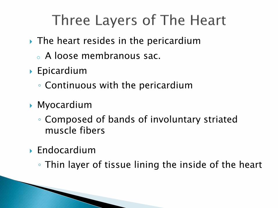
The heart resides in the pericardium
o A loose membranous sac.
Epicardium
◦ Continuous with the pericardium
Myocardium
◦ Composed of bands of involuntary striated muscle fibers
Endocardium
◦ Thin layer of tissue lining the inside of the heart
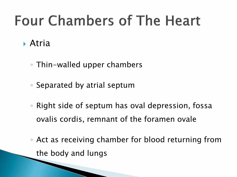
Atria
◦ Thin-walled upper chambers
◦ Separated by atrial septum
◦ Right side of septum has oval depression, fossa
ovalis cordis, remnant of the foramen ovale
◦ Act as receiving chamber for blood returning from
the body and lungs

Left atrium
Fossa ovalis cordis
Right atrium
Atrial septum
Epicardium
Myocardium
Endocardium

Ventricles
◦ Lower chambers which make up the bulk of the
muscle mass of the heart
◦ Left ventricle 2/3 larger than right ventricle
◦ Right ventricle is a thin-walled and oblong, like
pocket attached to left ventricle

Ventricles
◦ Contraction of left ventricle pulls in right
ventricle, aiding its contraction (termed left
ventricular aid)
◦ Separated by intraventricular septum
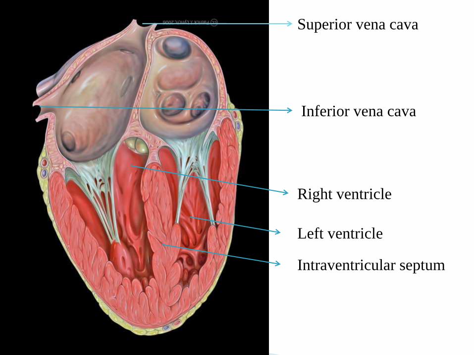
Right ventricle
Left ventricle
Intraventricular septum
Superior vena cava
Inferior vena cava
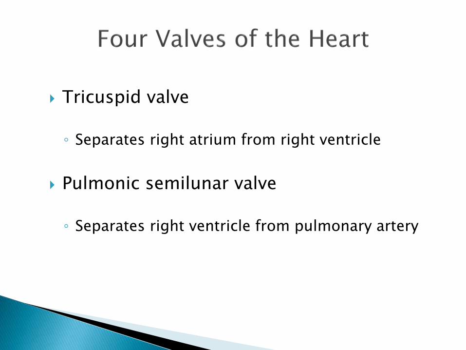
Tricuspid valve
◦ Separates right atrium from right ventricle
Pulmonic semilunar valve
◦ Separates right ventricle from pulmonary artery

Bicuspid (mitral) valve
◦ Separates left atrium from left ventricle
Aortic semilunar valve
◦ Separates left ventricle from aorta

Blood flow from right ventricle to lungs Blood flow from left ventricle to aorta
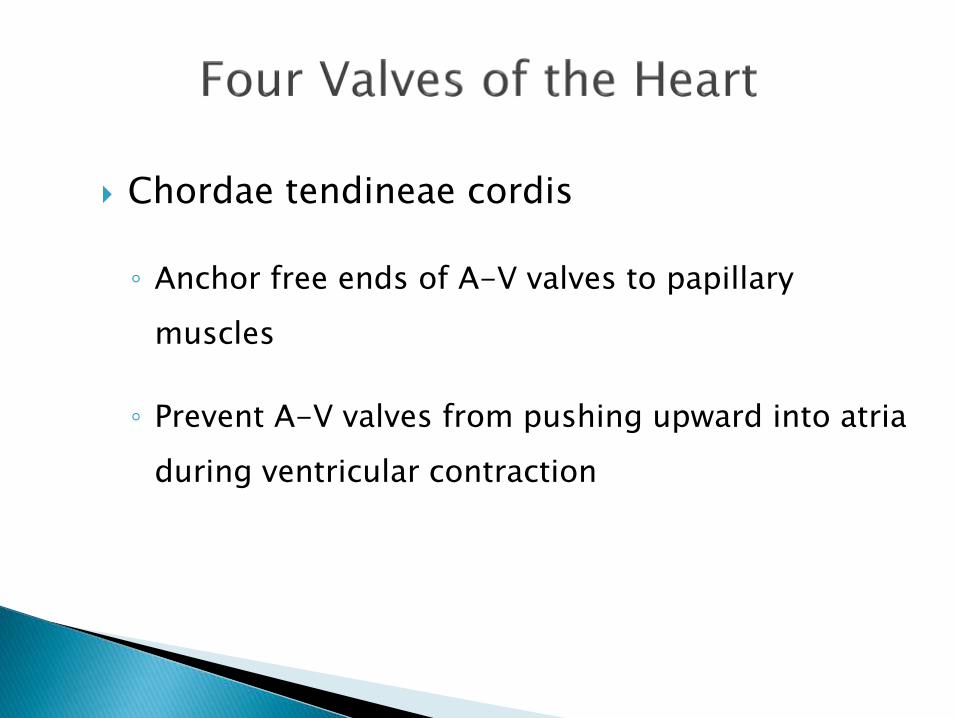
Chordae tendineae cordis
◦ Anchor free ends of A-V valves to papillary
muscles
◦ Prevent A-V valves from pushing upward into atria
during ventricular contraction
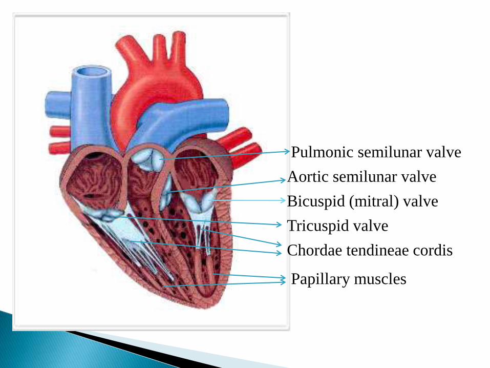
Aortic semilunar valve
Pulmonic semilunar valve
Bicuspid (mitral) valve
Tricuspid valve
Chordae tendineae cordis
Papillary muscles

Pulmonary artery to left lungPulmonary Artery to right
lung
Pulmonary veins from left
lung
Pulmonary veins from
right lung
Superior vena cava
Aorta
Brachiocephalic artery
Left common carotid artery
Left subclavian artery

Arises from root of the aorta
Left Coronary ArteryRight Coronary Artery
Anterior Descending Artery
Circumflex Artery
Posterior Descending Artery

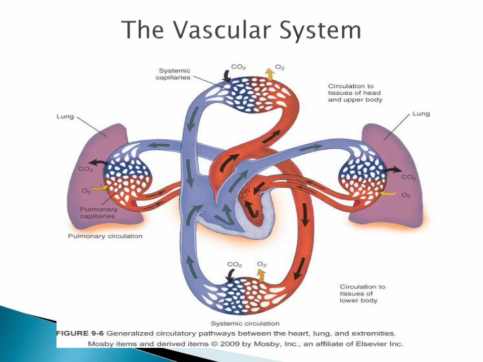
1) Blood enters the heart through the inferior and superior vena
cava, flowing into the right atrium.
2) The blood passes through the tricuspid valve into the right
ventricle.
3) It then passes through the pulmonic semilunar valve, entering
the pulmonary artery of the pulmonary circulation.
4) It flows through the pulmonary bed of the right and left lungs to
the pulmonary vein, reentering the heart at the left atrium.
5) It then flows through the bicuspid valve into the left ventricle.
6) Passing through the aortic semilunar valve, the blood enters the
aorta and systemic vascular system.

Anterior descending artery
◦ Supplies anterior sulcus and apex
◦ “Widow maker” heart attack
Circumflex artery
◦ Supplies posterior side of left ventricle

Together supply most of left ventricle, left
atrium, 2/3 of intra ventricular septum, half
of intra atrial septum, and part of right atrium


Posterior descending artery
◦ Supplies posterior intraventricular sulcus
Has numerous smaller branches

Supplies anterior and posterior portions of
right ventricular myocardium, right atrium,
sinus node, posterior 1/3 of intraventricular
septum, and portion of base of right ventricle

Closely parallel the arterial system
Some coronary venous blood enters the
heart through the Thebesian veins
◦ Thebesian veins empty directly into all chambers
thus creating some venous admixture lowering
Pa02

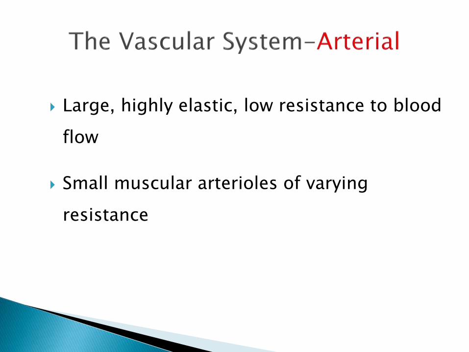
Large, highly elastic, low resistance to blood
flow
Small muscular arterioles of varying
resistance

Transport blood away from the heart
Generally contain oxygenated blood Exception: pulmonary artery
Composed of three layers◦ Tunica adventitia (external layer)◦ Tunica media (thickest layer)◦ Tunica intima (thinnest layer)

Tunica adventitia
◦ Consists of connective tissue surrounding collagenous and elastic fibers
◦ Supports and protects the vessel
◦ Contains lymphatic vessels and nerve fibers
◦ Has fine vessels that provide its blood supply

Tunica media
◦ Thickest layer
◦ Composed of concentrically arranged smooth muscle and elastic fibers
◦ Nerve fibers of tunica adventitia terminate in tunica media

Tunica intima
◦ Thinnest layer of the artery
◦ Consists of the epithelium – flat layer of simple squamous cells
◦ Common to all blood vessels including the endocardium

Large arteries are termed conductance or elastic arteries because the tunica media has less smooth muscle and more elastic fibers
Medium sized arteries are termed the nutrient arteries because they control the flow of blood to the various regions of the body
Arterioles have a thin tunica intima and adventitia, but a thick tunica media composed almost entirely of smooth muscle and control blood flow to the capillary bed
◦ Called resistance vessels because they control the rate that the blood leaves the arterial tree , control arterial blood volume and thereby blood pressure

Aorta
Brachial
Radial
Ulnar
Femoral
Anterior tibial
Peroneal artery
Posterior tibial
Aortic knob

Circle of Willis
Internal carotids
External carotids
Common carotids
Vertebral arteries

Microcirculation
Maintains constant environment for the cells and
tissues
Exchange of nutrients, gases, and wastes
The blood does not directly come in contact with
the parenchymal cells and tissues in the body,
but constituents of the blood first exit the micro
vascular exchange blood vessels to become
interstitial fluid, which comes into contact with
the parenchymal cells of the body. Lymph is
the fluid that is formed when interstitial fluid
enters the initial lymphatic vessels of the
lymphatic system

Pre-capillary sphincter valves
◦ Smooth muscle rings at the proximal end of the capillary
◦ Contraction decreases blood flow
◦ Relaxation increases blood flow
◦ Responsive to local changes in PaO2, PaCO2, pH, and temperature
◦ Called exchange vessels because they are the site of gas, fluid, nutrient, and waste exchange


Transport deoxygenated blood back to the heart – exception: pulmonary vein
Composed of the same layers as arteries, but are thinner
Called capacitance or reservoir vessels because 70% to 75% of the blood volume is contained in the venous system
Peripheral veins contain one-way valves.
◦ Valves are formed by duplication of endothelial lining
◦ Found in veins >2mm in diameter
◦ Are in areas subjected to muscular pressure, arms/legs
◦ Prevent retrograde flow of blood

Mechanisms aiding venous return to the heart:
◦ Sympathetic venous tone
◦ Skeletal muscle pumping or “milking” combined with the one-way valves
◦ Cardiac suction
◦ Thoracic pressure differences created by respiratory efforts (thoracic pump)


Consists of formed elements (cells) suspended & carried in plasma (fluid part)
Total blood volume: 60-80 mL/kg of body weight
Plasma is straw-colored liquid consisting of 90% H20 & dissolved solutes
◦ Includes ions, metabolites, hormones, antibodies, proteins
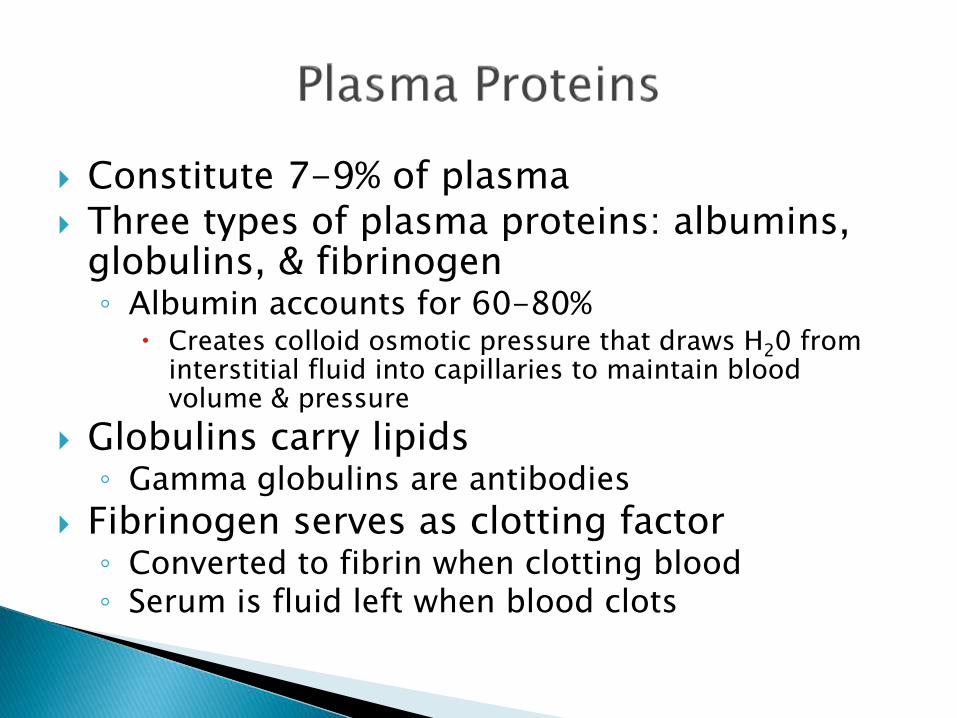
Constitute 7-9% of plasma Three types of plasma proteins: albumins,
globulins, & fibrinogen◦ Albumin accounts for 60-80%
Creates colloid osmotic pressure that draws H20 from interstitial fluid into capillaries to maintain blood volume & pressure
Globulins carry lipids◦ Gamma globulins are antibodies
Fibrinogen serves as clotting factor◦ Converted to fibrin when clotting blood ◦ Serum is fluid left when blood clots

Composed of erythrocytes (RBCs) & leukocytes (WBCs)
RBCs are flattened biconcave discs◦ Generated in the red bone marrow by the
process of erythropoiesis from the hemocytoblast, a common stem cell
◦ Shape provides increased surface area for diffusion
◦ Lack nuclei & mitochondria◦ Has semi-permeable membrane◦ Contains hemoglobin molecule that
transports oxygen◦ Approx. 30 trillion in the body

Is the formation of blood cells from stem cells in marrow (myeloid tissue) & lymphoid tissue◦ RBC’s increase in number above normal with
chronic hypoxia
Erythropoiesis is formation of RBCs◦ Stimulated by erythropoietin (EPO) from kidney
Leukopoiesis is formation of WBCs◦ Stimulated by variety of cytokines

2.5 million RBCs created daily
Lifespan of 120 days Old RBCs removed
from blood by phagocytic cells in liver, spleen, & bone marrow◦ Iron recycled back into
hemoglobin production

Have nucleus, mitochondria, & amoeboid ability
Formed in the myeloid tissue
Can squeeze through capillary walls (diapedesis)◦ Granular leukocytes help detoxify foreign
substances & release heparin
Include eosinophils, basophils, & neutrophils

Agranularleukocytes are phagocytic & produce antibodies
Include lymphocytes & monocytes

Specialized type of blood cell
Fragments into small irregular pieces of protoplasm called thrombocytes and platelets
Have no nucleus
Have a granular cytoplasm
Function in clot formation

Are smallest of formed elements, lack nucleus
Constitute most of mass of blood clots
Release serotonin to vasoconstrict & reduce blood flow to clot area
Secrete growth factors to maintain integrity of blood vessel wall
Survive 5-9 days

RBC’s – Males: 4.6 - 6.2 x 10 /mm
Females: 4.2 – 5.4 x 10 /mm
• Hb – Males: 13.5 – 16.5 g/dl
Females: 12 – 15 g/dl
• Hematocrit – Males: 42 – 54%
Females: 38 – 47%
• Leukocytes – 4500 – 11,500/mm

Neutrophils: 40 – 75%
Eosinophils: 0 – 6%
Monocytes: 2 – 10%
Basophils: 0 – 1%
Megakaryocyte: 150,000 – 400,000/mm

Systolic pressure
◦ Pressure during contraction phase of heart
◦ Normal value: 90 – 140 mmHg
Diastolic pressure
◦ Pressure during relaxation phase of heart
◦ Normal value: 60 – 90 mmHg

Mean arterial pressure (MAP)
◦ Average pressure in the arterial system over a
given time
◦ Normal value: 80 – 100 mmHg

Mean arterial pressure
MAP = (2 x diastolic pressure) + (systolic pressure)
3
A MAP of approximately 60 mmHg is necessary to perfuse coronary arteries, brain, kidneys.
Reflects right atrial pressure
Influenced by changes in right ventricular function
Measured with catheter placed in superior vena cava just above right atrium

Purpose◦ Assess blood volume status
◦ Administration of fluids
◦ Sampling of blood
◦ Measurement of SvO2
◦ Assessment of right ventricular pre-load
Normal valueoCVP: < 6 mmHg
oRight atrial pressure (RAP): 2-6 mmHg

Used to assess filling pressure of the left side of heart
Measured by flow-directed, balloon-tipped catheter
Measures◦ Pulmonary artery pressures – systolic, diastolic,
mean
◦ Right ventricular preload (via right atrial pressure)
◦ Right ventricular afterload (via PA systolic pressure)

Normal values
◦ Pulmonary artery pressure, systolic: 20-30 mmHg
◦ Pulmonary artery pressure, diastolic: 6-15 mmHg
◦ Pulmonary artery pressure, mean: 10-20 mmHg
◦ Pulmonary artery wedge pressure, mean: 4-12 mmHg

Total amount of blood pumped by the heart
per minute
Cardiac Output = Heart Rate x Stroke
Volume
Normal value – 5L/min

Cardiac Index
◦ Volume of blood pumped by the heart per
minute divided by body surface area
CI = CO
BSA
Normal range: 2.5 - 4.0 L/min per square meter
Low values can indicate cardiogenic shock

Amount of blood ejected from the ventricle
with each ventricular systole
End-systolic volume (ESV)
◦ Volume remaining after systole

End-diastolic volume (EDV)
◦ Volume to which the ventricles fill during
diastole
SV = EDV – ESV
Normal value: 60 – 130 ml/beat

Ejection fraction (EF)
◦ Proportion of EDV ejected on each stroke
EF = SV
EDV
◦ Normal value – 64%
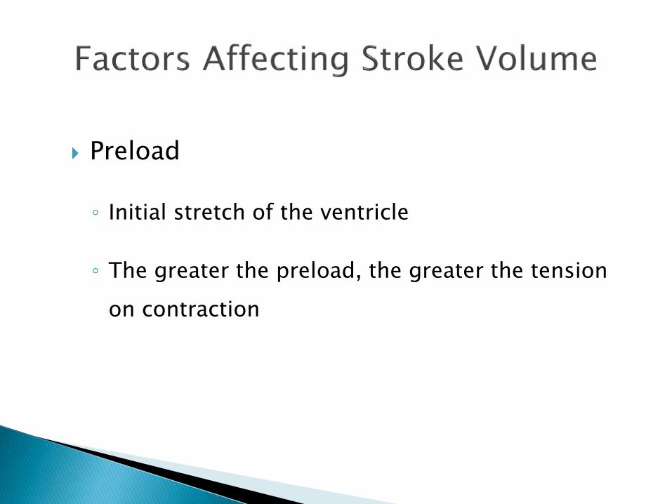
Preload
◦ Initial stretch of the ventricle
◦ The greater the preload, the greater the tension
on contraction

Afterload
◦ Force against which the heart must pump.
◦ In clinical practice, left ventricular afterload
equals systemic vascular resistance.

Contractility
◦ Amount of systolic force exerted by heart muscle at any
given preload.
◦ Increases in contractility leads to higher EF, lower end
systolic volume, and higher stroke volume
◦ Decreases in contractility lead to lower ejection fraction,
higher end systolic volume, and decreased stroke volume.

Contractility
Inotropism: any factor which affects the contractility of the heart
◦ Positive inotropism
Higher stroke volumes for a given preload: indicating an increase in contractility
◦ Negative inotropism
Decreased stroke volumes for a given preload; indicates a decrease in contractility

Heart rate
Autonomic nervous system
o Sympathetic: fight or flight: HR, RR, BP, pupil
dilation and bronchodilation
o Parasympathetic: rest and digest

Heart Rate
◦ Cardiac output directly proportional to heart rate
Relationship exists up to 160 to 180 beats/min
Filling time for ventricles insufficient at higher rates
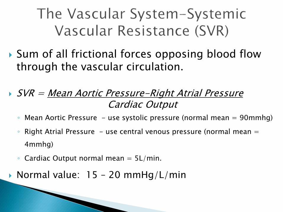
Sum of all frictional forces opposing blood flow through the vascular circulation.
SVR = Mean Aortic Pressure-Right Atrial PressureCardiac Output
◦ Mean Aortic Pressure - use systolic pressure (normal mean = 90mmhg)
◦ Right Atrial Pressure - use central venous pressure (normal mean =
4mmhg)
◦ Cardiac Output normal mean = 5L/min.
Normal value: 15 – 20 mmHg/L/min
Cardiac anatomy◦ Layers of the heart
◦ Chambers of the heart
◦ Valves
◦ Coronary arteries
Blood flow through the heart
Arterial system◦ Structure of artery
◦ Purpose
◦ Major arteries
Venous system◦ Structure of system
◦ Purpose
◦ Aids to venous flow
Capillary system◦ Structure of system
◦ Purpose

Composition of blood
Plasma proteins
Types of cells, functions, normal values, abnormalities◦ Erythrocytes
◦ Leukocytes
◦ Megakaryocytes
◦ Platelets
◦ Hemoglobin
◦ Hematocrit

Definition, normal values, and formula (if applicable)◦ Systemic vascular resistance
◦ Systolic pressure
◦ Diastolic pressure
◦ Mean arterial pressure
◦ Cardiac output and index
◦ Stroke volume, esv, edv, ef
Factors affecting stroke volume