Potential in vitro anti-allergic, anti-inflammatory and ...
12
RESEARCH ARTICLE Open Access Potential in vitro anti-allergic, anti-inflammatory and cytotoxic activities of ethanolic extract of Baliospermum montanum root, its major components and a validated HPLC method Weerachai Pipatrattanaseree 1,2 , Arunporn Itharat 1,2* , Nichamon Mukkasombut 1,2 and Ubonwan Saesiw 1,2 Abstract Background: The root of Baliospermum montanum has been used as an ingredient of traditional Thai medicines for the treatments of several diseases including itching eczema, muscle and joint inflammation, and cancer. Few studies have been done on phytochemical components of this root. In this study, we isolated major compounds of the crude ethanolic extract of B. montanum root and developed and validated a high performance liquid chromatographic (HPLC) method for the determination of its major components. We then investigated anti-allergic, anti-inflammatory and cytotoxic activities of the extract. Methods: The aims of this study were to investigate in vitro activities including inhibitory effect of β-hexosaminidase released from RBL-2H3 cells, inhibition of nitric oxide (NO) production from RAW 264.7 cells and cytotoxic activity against cancerous liver cell lines (HepG2 and KKU M156) by using sulforhodamine B (SRB) assay. Isolation of major components was conducted by using column chromatographic method. Isolated major compounds were analyzed by using high performance liquid chromatography (HPLC). Results: The crude extract exhibited the highest cytotoxic activity, with IC 50 less than 1 μg/mL, while its anti-allergy and anti-inflammation were also potent with IC 50 less than 6 μg/mL. Three propiophenones isolated from B. montanum root exhibited moderate cytotoxic activities (IC 50 > 20 μg/mL). Two of the propiophenones found were major components that can be detected by HPLC. The developed and validated HPLC method showed good accuracy, precision, and linearity. Conclusion: The results of this study suggested that ethanolic extract of of B.montanum root can be a potential source of anti-allergy, anti-inflammation, and anti-cancer compounds. The isolated compounds can serve as markers when B. montanum is used in herbal remedies but not as overall responsive markers. The HPLC method developed may be useful for quality control in the production of the extract and for further formulation developments. However, investigation of several associated biological activities is necessary before the development can proceed further. Minor active compounds should be isolated and a more sensitive analytical method should be developed to detail the key responsive components of the ethanolic extract of B. montanum root. Keywords: Anti-allergy, Anti-inflammation, Cytotoxicity, Baliospermum montanum, Propiophenones * Correspondence: [email protected] 1 Department of Applied Thai Traditional Medicine, Faculty of Medicine, Thammasat University, Klongluang, Pathumthani 12120, Thailand 2 Center of Excellence on Applied Thai Traditional Medicine Researches, Faculty of Medicine, Thammasat University, Klongluang, Pathumthani 12120, Thailand © The Author(s). 2019 Open Access This article is distributed under the terms of the Creative Commons Attribution 4.0 International License (http://creativecommons.org/licenses/by/4.0/), which permits unrestricted use, distribution, and reproduction in any medium, provided you give appropriate credit to the original author(s) and the source, provide a link to the Creative Commons license, and indicate if changes were made. The Creative Commons Public Domain Dedication waiver (http://creativecommons.org/publicdomain/zero/1.0/) applies to the data made available in this article, unless otherwise stated. Pipatrattanaseree et al. BMC Complementary and Alternative Medicine (2019) 19:45 https://doi.org/10.1186/s12906-019-2449-0
Transcript of Potential in vitro anti-allergic, anti-inflammatory and ...

RESEARCH ARTICLE Open Access
Potential in vitro anti-allergic, anti-inflammatoryand cytotoxic activities of ethanolic extractof Baliospermum montanum root, its majorcomponents and a validated HPLC methodWeerachai Pipatrattanaseree1,2, Arunporn Itharat1,2* , Nichamon Mukkasombut1,2 and Ubonwan Saesiw1,2
Abstract
Background: The root of Baliospermum montanum has been used as an ingredient of traditional Thai medicines forthe treatments of several diseases including itching eczema, muscle and joint inflammation, and cancer. Few studieshave been done on phytochemical components of this root. In this study, we isolated major compounds of the crudeethanolic extract of B. montanum root and developed and validated a high performance liquid chromatographic(HPLC) method for the determination of its major components. We then investigated anti-allergic, anti-inflammatoryand cytotoxic activities of the extract.
Methods: The aims of this study were to investigate in vitro activities including inhibitory effect of β-hexosaminidasereleased from RBL-2H3 cells, inhibition of nitric oxide (NO) production from RAW 264.7 cells and cytotoxic activityagainst cancerous liver cell lines (HepG2 and KKU M156) by using sulforhodamine B (SRB) assay. Isolation of majorcomponents was conducted by using column chromatographic method. Isolated major compounds were analyzed byusing high performance liquid chromatography (HPLC).
Results: The crude extract exhibited the highest cytotoxic activity, with IC50 less than 1 μg/mL, while its anti-allergy andanti-inflammation were also potent with IC50 less than 6 μg/mL. Three propiophenones isolated from B. montanum rootexhibited moderate cytotoxic activities (IC50 > 20 μg/mL). Two of the propiophenones found were major componentsthat can be detected by HPLC. The developed and validated HPLC method showed good accuracy, precision,and linearity.
Conclusion: The results of this study suggested that ethanolic extract of of B.montanum root can be a potentialsource of anti-allergy, anti-inflammation, and anti-cancer compounds. The isolated compounds can serve as markerswhen B. montanum is used in herbal remedies but not as overall responsive markers. The HPLC method developedmay be useful for quality control in the production of the extract and for further formulation developments. However,investigation of several associated biological activities is necessary before the development can proceed further. Minoractive compounds should be isolated and a more sensitive analytical method should be developed to detail the keyresponsive components of the ethanolic extract of B. montanum root.
Keywords: Anti-allergy, Anti-inflammation, Cytotoxicity, Baliospermum montanum, Propiophenones
* Correspondence: [email protected] of Applied Thai Traditional Medicine, Faculty of Medicine,Thammasat University, Klongluang, Pathumthani 12120, Thailand2Center of Excellence on Applied Thai Traditional Medicine Researches,Faculty of Medicine, Thammasat University, Klongluang, Pathumthani 12120,Thailand
© The Author(s). 2019 Open Access This article is distributed under the terms of the Creative Commons Attribution 4.0International License (http://creativecommons.org/licenses/by/4.0/), which permits unrestricted use, distribution, andreproduction in any medium, provided you give appropriate credit to the original author(s) and the source, provide a link tothe Creative Commons license, and indicate if changes were made. The Creative Commons Public Domain Dedication waiver(http://creativecommons.org/publicdomain/zero/1.0/) applies to the data made available in this article, unless otherwise stated.
Pipatrattanaseree et al. BMC Complementary and Alternative Medicine (2019) 19:45 https://doi.org/10.1186/s12906-019-2449-0

BackgroundBaliospermum montanum (Willd.) Muell-Arg is a leafystout monoecious undershrub plant, a member of Euphor-biaceae. It grows naturally in middle- and south-east Asiaand has been used as a traditional medicine in severalcountries. In Ayurvedic medicine, B. montanum root isused to treat skin diseases, worm infection, gastrointes-tinal tract diseases, and itching [1]. In traditional Thaimedicines (TTM), B. montanum root is an important in-gredient in several remedies such as ‘Sahastara’ for muscleand joint pain [2] and ‘Benja-Amarit’ for liver cancer [3].It is also used for the treatment of eczema itches [4].The biological activities of B. montanum root have
been investigated. Kakatum and co-workers reported invitro anti-inflammatory effect of B. montanum root, acomponent in a Thai polyherbal preparation called“Sahastara” used for the treatment of muscle and jointpain [5]. Ogura and co-workers reported in vitro cyto-toxicity of the isolated compounds from the root againstleukemia P-388 cells [6]. In an in vivo study, Venkateshand co-worker reported that leaves of B. montanuminhibited the degranulation of mast cells in systemicanaphylaxis model [7]. Other researchers reportedanti-oxidant [5], antimicrobial activities [8], immuno-modulatory activities [9] and hepatoprotective propertiesof B. montanum [10, 11]. Phytochemical screening re-vealed that chemical constituents in the root of B. mon-tanum were steroids, terpenoids, glycosides, saponins,alkaloids, flavonoids, phenolic compounds, tannins,sugars, and several other minor components [6–8].Based on the traditional uses and scientific evidences
above, we selected in vitro biological effects as preliminarymodels to investigate the associated biological propertiesof the ethanolic extract of B. montanum root. Exhaustivesearch reveals no reports on inhibitory effect ofβ-hexosaninidase released from RBL-2H3 cells and cyto-toxic activity against cancerous liver cells of B. montanumcrude extract. Neither were reports on its major compo-nents and their analytical methods found. This presentstudy aimed at investigating anti-allergic activity by deter-mining the inhibitory effect on β-hexosaminidase releasedfrom RBL-2H3 cells, anti-inflammatory activity by detect-ing the inhibition of nitric oxide (NO) production fromRAW 264.7 cells and cytotoxic activity against cancerousliver cell lines by using sulforhodamine B (SRB) assay. Theisolation of major components and evaluation of their ac-tivities were also conducted. Subsequently, a quantitativeHPLC method for the determination of its major compo-nents was developed and validated.
MethodsPlant materials and preparationsB.montanum roots were collected from Lampoon prov-ince, Thailand, in January 2016 and authenticated by
Department of Applied Thai traditional Medicines,Thammasat University. The vouchered specimen(SKP-071021301) was deposited at the herbarium ofSouthern Center of Thai Medicinal Plants, Faculty ofPharmaceutical Sciences, Prince of Songkla University,Songkhla, Thailand.Roots of B. montanum were washed, sliced thinly and
dried in a hot air oven at the temperature not more than45 °C, and ground to coarse powder. One kg of the driedroot was macerated with 95% ethanol (3 × 3 days). Theextracts were filtered, combined and concentrated with arotary evaporator under a reduced pressure at 45 °C.
Reagents and chemicalsAnalytical grade organic solvents: hexane, chloroform,ethyl acetate, methanol, acetonitrile and methanol (HPLCgrade) were purchased from RCI LabScan (Bangkok,Thailand). Purified water was prepared by Milli Q® systemfrom Millipore (Bedford, MA, USA).Chlorpheniramine, prednisolone, and vincristine were
purchased from Sigma (MO, USA). Chemical referencesubstances used for method validation, compounds 1 and2, were isolated from the crude ethanolic extract and iden-tified by the spectroscopic method including UV, MS, andNMR. The purities of isolated compounds were deter-mined to be more than 98% by using HPLC analysis.Rat basophilic leukemia cell line (RBL-2H3: ATCC
CRL-2256™), murine macrophage leukemia cell line(RAW 264.7: ATCC® TIB-71™) and hepatocellular carcinomacell line (HepG2: ATCC® HB-8065™) were purchased fromAmerican Type Culture Collection (ATCC®, VA, USA).Cholangiocarcinoma cell line (KKU-M156) was obtainedfrom Dr. Piti Aungareewithaya and Prof. Dr. VeerapholKukongviriyapan, Faculty of Medicine, Khon-Kaen Univer-sity. Normal human keratinocyte immortal cell line (HaCaT)was purchased from CLS cell line service (No. 300493-SF).Fetal bovine serum (FBS) and trypsin-EDTA were pur-
chased from Gibco® (OK, USA). Minimum essentialmedium (MEM), Dulbecco's Modified Eagle's Medium(DMEM), Roswell Park Memorial Institute medium 1640(RPMI-1640), penicillin-streptomycin (P/S), and phos-phate buffer saline (PBS) were purchased from Biochrom(MA, Germany). Nutrient Mixture F-12 Ham medium(HAM F-12), 3-(4, 5-dimethyl-2-thiazolyl)-2, 5-diphenyl-2H-tetrazolium bromide (MTT), Sulforhodamine B (SRB),4-Nitrophenyl N-acetyl-β-D-glucosaminide (PNAG), anti-dinitrophenylated bovine albumin (DNP-BSA) and anti-dinitrophenyl immunoglobulin E (Anti-DNP IgE) werepurchased from Sigma (MO, USA). Dimethyl sulfoxide(DMSO) was purchased from Fluka (Munich, Germany).
InstrumentsHPLC analysis was performed on an Agilent® 1200HPLC system (Agilent Technologies, USA) composing
Pipatrattanaseree et al. BMC Complementary and Alternative Medicine (2019) 19:45 Page 2 of 12

of a solvent degasser (G1322A), a quaternary solventpump (G1311A), an autosampler (G1329A), a columnoven (G1316A) and a photodiode array detector(G1315D). The chromatographic data were processed bythe Chemstation® software revision B.04.01 SP1. TheESI-HRMS were performed either on Shimadzu iontrap-time of flight spectrometer (IT-TOF) (ShimadzuCorporation, Japan) or Bruker microTOF (Bruker com-pany, USA). Nuclear magnetic resonance (NMR) spectrawere recorded either on Bruker Avance DRX-500 NMRspectrometer (Bruker Company, USA) or Varian UnityInova 500MHz (Variance, USA) using tetramethylsilaneas internal standard.
Anti-allergic activityInhibitory activity on β-hexosaminidase released fromrat basophilic leukemia cells (RBL-2H3) was evaluatedfollowing the report of Juckmetha and co-workers [12].RBL-2H3 cells were cultured in MEM supplementedwith 15% FBS, penicillin/streptomycin (each 100 μg/mL).Cells were seeded in 24-well plate with the concentra-tion of 5 × 10⁵ cells/well and incubated in 5% CO2 at 37°C for 1.5 h. RBL-2H3 were sensitized with 0.45 μg/mLof Anti-DNP IgE and incubated at 37 °C in 5% CO2 for24 h. Cells were then washed with 400 μL of Siraganianbuffer (Buffer A) [119 mM NaCl, 5 mM KCl, 5.6 mMglucose, 0.4 mM MgCl2, 1 mM CaCl2, 25 mM piperazi-ne-N,N′-bis (2-ethanesulfonic acid) (PIPES), 0.1% BSA,and 40 mM NaOH, pH 7.2]. Subsequently, 160 μL ofbuffer A was added into the wells and incubated at 37 °Cfor 10 min. The stock solution of the sample was dilutedin DMSO and the working samples were diluted withbuffer A. The concentrations of the working sampleswere 0.1 μg/mL, 1 μg/mL, 10 μg/mL and 50 μg/mL. Theserially diluted samples (20 μL) were added to each welland incubated for 10 min. The reaction ofantigen-antibody was obtained by adding 20 μL of anti-gen (DNP-BSA) to produce a final concentration of10 μg/mL and incubated at 37 °C for 20 min. Theamount of β-hexosaminidase released from the degranu-lation was determined by adding 50 μL of the substrate,PNAG (1 mM in 0.1M citrate buffer, pH 4.5). The en-zyme reaction was stopped by adding 200 μL of 0.1Mcarbonate buffer pH 10.0. The product of enzyme reac-tion, p-nitrophenol, was analyzed by measuring absorb-ance at wavelength 405 nm. Chlorpheniramine was usedas positive control. The percentage of inhibition was cal-culated by the following equation.
Inhibition %ð Þ ¼ 1−T−B−Nð ÞC−B−Nð Þ
� �� 100
Where
C =Optical density of control: [+] DNP-BSA, [−] Testsample;T = Optical density of test sample: [+] DNP-BSA, [+]Test sample;B = Optical density of blank: [+] DNP-BSA, [+] Testsample;N =Optical density of normal: [−] DNP-BSA, [−] Testsample.
Anti-inflammatory activityThe inhibitory activity of crude extracts and isolated com-pounds on NO production of LPS-induced RAW 264.7cells was investigated according to a previously reportedmethod [13]. RAW 264.7 cells were cultured in RPMImedium supplemented with 10% FBS, penicillin/strepto-mycin (each 100 units/mL) and incubated at 37 °C in 5%CO2 with 95% humidity. The cells were seeded into96-well plate (1 × 10⁵ cells per well) and incubated for 24h. Subsequently, the cells were induced by LPS at the finalconcentration of 2 ng/mL and the serially diluted samples(0.1 μg/mL, 1 μg/mL, 10 μg/mL and 50 μg/mL) wereadded. The plate was incubated for 24 h before beingtested for NO accumulation and cell viability. Prednisol-one was used as positive control. The accumulated NO re-leased from the LPS-induced RAW 264.7 cells wasdetermined by using Griess’s reagent (1% sulfanilamideand 0.1% N-(1- naphthyl) ethylenediamine dihydrochlor-ide in 2.5% phosphoric acid). The supernatant in each well(100 μL) was transferred to another 96-well plate and100 μL of Griess’s reagent was added. The optical densityof reaction was measured at 570 nm. The inhibitory activ-ity (%) of NO production was calculated by the followingequation and the IC50 values were calculated.
Inhibition %ð Þ ¼ ODcontrol‐ODsample
ODcontrol
� �� 100
The viability of the LPS treated RAW 264.7 cells werecolorimetrically determined by using 3-(4, 5-dimethyl-2-thiazolyl)-2, 5-diphenyl-2H-tetrazolium bromide (MTT)method. MTT solution (10 μL) was added into each welland the plate was incubated for 2 h. The remained for-mazan crystal in each well, after removing the medium,was dissolved by 0.04M HCl in isopropanol (100 μL).The optical density was measured by using microplatereader at wavelength 570 nm. The cell viability of morethan 70% (Table 2) indicated that the inhibitory effectwas not derived from cell death.
Cytotoxic activityThe crude extracts and isolated compounds were evalu-ated for cytotoxic activity by using sulforhodamine B(SRB) assay against two cancerous liver cells (HepG2and KKU-M156) and a normal cells (HaCaT). The SRB
Pipatrattanaseree et al. BMC Complementary and Alternative Medicine (2019) 19:45 Page 3 of 12

protocol has been described previously [14]. HepG2 wascultured in MEM; KKU-M156 was cultured in HAMF-12 media and HaCaT was cultured in DMEM. All cul-tured media were supplemented with 10% FBS, penicil-lin/streptomycin (each 100 units/mL). All cells wereincubated in 5% CO2 at 37 °C with 95% humidity. Basedon the growth curve, each cell was seeded into 96-wellplate (100 μL per well) with optimal density (HepG2:3 × 10³ cells per well, KKU-M156: 2 × 10³ cells per welland HaCaT: 8 × 10³ cells per well). Seeded cells were in-cubated at 37 °C in a 5% CO2 with 95% humidity for 24h. Cells were then treated with 100 μL of different con-centrations of sample solution (0.1 μg/mL, 1 μg/mL,10 μg/mL and 50 μg/mL). DMSO at the same concentra-tion as the sample (2% v/v) was used as the control solv-ent. Cell cultures were incubated for the exposure timeof 72 h. After the exposure time, the medium was re-moved and the plate was washed with 200 μL phosphatebuffer saline (PBS) and 200 μL fresh medium was addedinto each well. The plates were then incubated for 72 hfor the recovery period. The cells were then fixed byadding 100 μL of 40% trichloroacetic acid and the platesleft at 4 °C for 45 min. After the plates were washed,50 μL of SRB solution (0.4% w/v in acetic acid) wasadded into each well and the cells were allowed to bestrained for 30 min. The stained cells were dissolved byusing 100 μL of 10 mM Tris base pH 10.5. The %inhib-ition was calculated using the following equation.
Inhibition %ð Þ ¼ ODcontrol‐ODsample
ODcontrol
� �� 100
Isolation of major constituents from the root of B. montanumPure compounds were isolated by using silica columnchromatography (CC) and HPLC techniques. Ethanolicextract of B. montanum root (40 g) was subjected to a sil-ica gel column (49–63 μm, 200 g). The extract was elutedsequentially by using 100% hexane (700mL), hexane/chloroform 7:3 (700mL), 3:7 (700mL), 100% chloroform(700mL), chloroform/ethyl acetate 7:3 (700mL), 3:7 (700mL), 100% ethyl acetate (700mL), ethyl acetate/ methanol7:3 (700mL), 3:7 (700mL) and 100% methanol (700mL).Ten obtained fractions, namely FF01 to FF10, were ana-lyzed by using the HPLC method described below.Fractions FF03 and FF04 were combined (6.8 g) and
subjected to second silica gel column (90 g). Polarity gra-dient elutants were hexane, chloroform, ethyl acetate, andmethanol, consecutively. Fractions were collected accord-ing to the results from TLC monitoring. Eight fractionsobtained from the second CC were named SF01 to SF08.Fraction SF03 (536 mg) was subjected to third silica
gel column (24 g) and eluted by polarity gradient mobilephases from hexane to chloroform and ending with ethyl
acetate. Fractions were collected according to the resultsfrom TLC monitoring. Eight fractions obtained from thethird CC were named TF01 to TF08.Fraction TF02 (10.8mg) was further fractionated by
semi-preparative HPLC (Zorbax® 5 μm, 10mm× 250mm,5 μm; 40% H2O in methanol, flow rate 3.0ml/min) to yieldcompound 1 (4.0mg) and compound 3 (1.3mg). FractionTF03 (40.9mg) was subjected to semi-preparative HPLC(Zorbax® 5 μm, 250mm× 10mm; 50% H2O in acetonitrile,flow rate 3.0ml/min) to yield compound 1 (9.8mg), andcompound 2 (30.1mg). All isolated compounds were char-acterized by using 1H NMR, 13C NMR, 2D NMR, and MS.
High performance liquid chromatography analysisPreparation of samples and standard solutionsSample solutions of ethanolic extracts of B. montanumroot and their CC fractions were prepared at the con-centrations of 10 mg/mL and 1mg/mL, respectively.Samples were weighed accurately and dissolved with amixture of methanol:chloroform (9.5:0.5) with the aid ofsonication. Solutions of the isolated compounds wereprepared at the concentration of 1 mg/mL in methanolfor the determination of their purity. Different concen-trations of the isolated standard solutions were preparedin methanol according to the linear range. All preparedsolutions were filtered through 0.45 μm membrane be-fore injecting into the HPLC system.
Chromatographic systemMajor components of the crude extract were separatedalong the C18 reverse phase column (Zorbax® C18,4.6 × 150, 5 μm). Gradient mobile phase composing ofwater (A) and acetonitrile (B) was programmed as follows:0–10min, 20%B; 10–15min, 200%B - 65%B; 15.1–20min,20%B. The flow rate was set at 1.0 mL/min. Samples wereinjected into the HPLC system and detected with diodearray detector using the wavelength of 280 nm.
Method validationThe developed quantitative method for the determin-ation of compounds 1 and 2 in ethanolic extract of B.montanum root was validated for specificity, linearity,range, accuracy, precision, LOD and LOQ.Specificity of each compound was performed by com-
paring retention time and UV spectrum of the peak inthe chromatogram of crude extract with the respectivepeak in the chromatogram of standard solution. The lin-earity of each analyte was evaluated using the con-structed calibration curve of serial concentrations ofstandard solutions. LODs and LOQs were investigatedby using diluted standard solution. The signal to noiseratio (S/N) was calculated by dividing the peak height ofthe standard with the height of the background noise.
Pipatrattanaseree et al. BMC Complementary and Alternative Medicine (2019) 19:45 Page 4 of 12

The signal to noise (S/N) ratios calculated for LODs andLOQs were about 3 and 10, respectively.Accuracy and precision of the method were evaluated
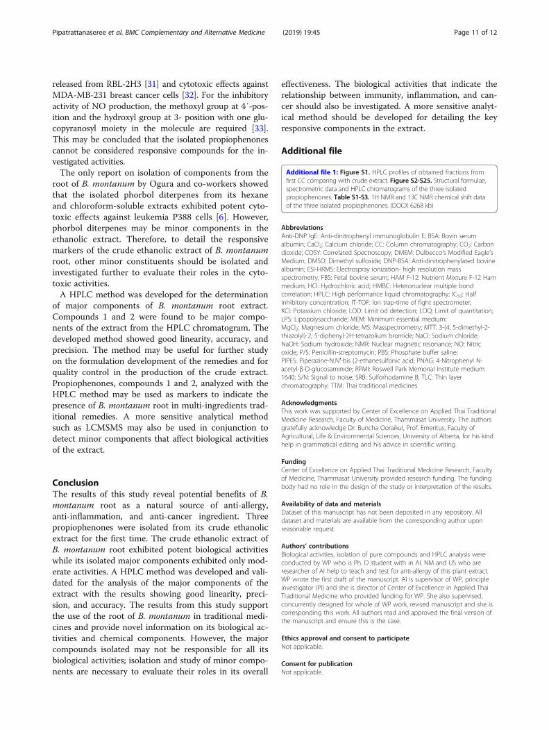
by using three levels of spiked samples containing threeconcentrations of the standards. Crude extract solutionswere spiked with the standard mixture solution of com-pound 1 (15, 25 and 40 μg/mL) and compound 2 (30, 50and 80 μg/mL). Accuracy was calculated as percentage re-covery. Intra-run and inter-run precision were calculatedas RSD% of the concentration found from the subtractedspiked samples. The accuracy and precision of the methodwere determined by three replicates within-run for threeconsecutive runs.
Statistical analysisThe results of the biological activities were reported asmean ± standard deviation (SD) from three replicated ex-periments. IC50 values were calculated by using a regres-sion analysis. Mean differences among groups wereanalyzed by Student’s t-test. Statistical analysis was con-ducted by using GraphPad Prism software (CA, USA).
ResultsIsolation and identification of pure compoundsIsolation of pure compounds from the ethanolic extractof B. montanum root produced three propiophenones(Fig. 1). The fractions obtained from the first CC weresubjected to HPLC analysis to guide the isolation ofmajor compounds (Additional file 1: Figure S1). FF03and FF04 were combined based on the similarity of theHPLC profiles which contained two major compoundssimilar to the crude extract. The combined fraction wassubjected to further silica gel CC. The fractions contain-ing the major compounds (from the third silica gel CC)were subjected to the semi-preparative HPLC. Identificationof the three isolated propiophenones was performed byspectrometric techniques (Additional file 1: Figure S2–S25and Table S1–S3) with the following results.Compound 1 was obtained as white solid (13.8mg,
0.035% crude extract). The UV spectrum showed λmax 229,274, 303 nm. Positive ESI-HRMS at m/z of 217.0835 [M+Na]+ suggested the molecular formula to be C11H14O3Na.
1H NMR (500MHz, CDCl3) δ 7.59 (H, dd, J = 8.4, 1.9Hz),7.52 (1H, d, J = 1.9 Hz), 6.88 (1H, d, J = 8.4 Hz), 3.93 (3H,s), 3.92 (3H, s), 2.95 (2H, q, J = 7.3Hz), 1.21 (3H, t, J = 7.3Hz). 13C NMR (125MHz, CDCl3) δ 199.4, 153.3, 149.2,130.4, 110.6, 110.3, 56.0, 31.3, 8.6. Based on 1H-1H COSYand HMBC correlations, compound 1 was suggested to bea known compound, 1-(3′,4′-dimethoxyphenyl) propan-1-one or propioveratrone. The structure was confirmed bycomparing 1H-NMR and 13C-NMR spectral data to thedata reported by Park and co-workers [15].Compound 2 was obtained as white solid (30.1mg,
0.075% w/w crude extract). The UV spectrum showedλmax 217, 278 nm. Positive ESI-HRMS at m/z of 274.0941[M+Na]+ suggested the molecular formula to beC12H16O4Na.1H NMR (500MHz, CDCl3) δ 7.22 (2H, s),3.93 (6H, s), 3.92 (3H, s), 2.97 (2H, J = 7.2 Hz), 1.22 (3H,J = 7.3 Hz). 13C NMR (125MHz, CDCl3) δ 199.5, 153.2,142.8, 132.3, 105.9, 60.9, 56.4, 31.5, 8.4. Based on 1H-1HCOSY and HMBC correlations, compound 2 was sug-gested to be a known compound, 1-(3′,4′,5′-trimethoxy-phenyl) propan-1-one. The structure was confirmed bycomparing 1H-NMR and 13C-NMR spectral data to thedata reported by Tanpure and co-workers [16].Compound 3 was obtained as white solid (1.3mg,
0.003% w/w crude extract). The UV spectrum showed λmax
236, 275, 340 nm. Positive ESI-HRMS at m/z of 233.0784[M+Na]+ suggested the molecular formula to beC11H14O4Na. 1H NMR (500MHz, CDCl3) δ 12.75 (OH, s),7.14 (1H, s), 6.45 (1H, s), 3.92 (3H, s), 3.84 (3H, s), 2.94(2H, q, J = 7.3Hz), 1.22 (3H, t, J = 7.3 Hz), 13C NMR (125MHz, CDCl3) δ 204.8, 160.0, 156.5, 141.8, 111.2, 110.8,100.7, 56.7, 56.2, 31.3, 8.6. Based on 1H-1H COSY andHMBC correlations, compound 3 was suggested to be aknown compound 1-(2′-hydroxy- 4′, 5′-dimethoxyphenyl)propan-1-one. The structure was confirmed by comparing1H-NMR and 13C-NMR spectral data to the data reportedby Mendieta and co-workers [17].
In vitro inhibitory effect on β-hexosaminidase release fromRBL-2H3 cell linesβ-hexosaminidase is a marker of IgE-mediated degranu-lation of mast cells relating to the symptoms of itching
Fig. 1 Chemical structures of propiophenons isolated from ethanolic extract of B. montanum root
Pipatrattanaseree et al. BMC Complementary and Alternative Medicine (2019) 19:45 Page 5 of 12

eczema [18]. Screening results of the anti-allergic activityagainst the releasing of β-hexosaminidase fromRBL-2H3 cells of the crude extract and the isolated purecompounds at concentration 50 μg/mL are shown inFig. 2. The crude extract exhibited significantly the high-est activity while other compounds showed comparable%inhibition with the positive control, chlorpheniramine.These results indicated the possible anti-allergic activityof all investigated compounds and the crude extract.The IC50 investigation showed that all samples hadconcentration-dependent inhibitory effect (Table 1). Thecrude extract exhibited the highest anti-allergic activity withIC50 value of 4.97 ± 0.43 μg/mL which was three times morepotent than the positive control (17.74 ± 1.61 μg/mL). Theisolated compounds 1 and 2 showed anti-allergic ac-tivity comparable to the positive control with IC50 of21.20 ± 2.01 μg/mL and 26.25 ± 6.06 μg/mL, respectively,while compound 3 showed moderate activity.
In vitro inhibitory activity of NO production from LPS-inducedRAW 264.7NO is a pro-inflammatory mediator released frommacrophage during inflammation. Excessive concentra-tion of NO production from inducible NO synthase(iNOS) in macrophage is linked to inflammatory reac-tions. NO has an important role along with several me-diators in inflammatory diseases including rheumatoidarthritis and osteoarthritis [19, 20]. The screening resultsof the inhibitory activity of NO production of the crude
extract and its isolated compounds at the concentrationof 50 μg/mL are presented in Fig. 3. The crude extractand all compounds exhibited %inhibition less than pred-nisolone (positive control). The crude extract, com-pounds 1 and 2 showed comparable %inhibition whilecompound 3 showed the least inhibitory effect. From thedetermination of IC50, the crude extract and isolatedcompounds inhibited the production of NO in aconcentration-dependent manner (Table 2). The crudeextract exhibited the highest inhibitory activity on NOproduction with IC50 of 6.06 ± 0.43 μg/mL while the iso-lated compounds showed moderate activity with IC50
more than 30 μg/mL. Among the three isolated propio-phenones, compounds 2 exhibited the highest inhibitoryactivity with IC50 of 33.61 ± 5.25 μg/mL. All compoundsand crude extract showed less inhibitory activity thanprednisolone which exhibited potent inhibitory activitywith IC50 value of 0.11 ± 0.02 μg/mL.
In vitro cytotoxic activityCytotoxic activity of the B.montanum root extract andisolated compounds were investigated against selectedtwo cancerous liver cells, HepG2 and KKU-M156, and anormal cell, HaCaT. All samples exhibited cytotoxic ac-tivity in direct proportion to their concentrations (datanot shown). Their IC50 are presented in Table 3. Thecrude extract exhibited potent cytotoxic activity againstHepG2 and KKU-M156 with IC50 less than 1 μg/mL.Interestingly, crude extract exhibited less cytotoxicity to
Fig. 2 Effect of ethanolic extract of B. montanum root and the isolated propiophenones at concentration of 50 μg/mL on β-hexosaminidase releasefrom RBL-2H3 cells. Results are present as mean ± SD from triplicate experiment. Mean differences between sample and positive control were analyzedby student’s t-test. *Significant difference comparing tested sample and positive control analyzed by Student’s t-test (p-value < 0.05)
Pipatrattanaseree et al. BMC Complementary and Alternative Medicine (2019) 19:45 Page 6 of 12

HaCaT with the IC50 more than 100 μg/mL. However,the isolated compounds exhibited moderate cytotoxicactivity to the cancerous cells with IC50 more than45 μg/mL. Though the crude extract and isolated com-pounds exhibited cytotoxic activity less than positivecontrol (vincristine), the cytotoxic effect of the extractand compounds against normal cells were lower com-pared to their effect against cancerous cells, showingtheir selective cytotoxic property.
HPLC method development and method validationIn the three isolated propiophenones, compound 1 andcompound 2 were the major peaks found in the HPLCchromatogram of the ethanolic extract of B. montanum
root, therefore, we used these two compounds asmarkers for quantitative HPLC analysis. Compound 3showed very small amount and its potency on the bio-logical activities were lower than those of the others.The selectivity of the method is shown in Fig. 4. Com-pounds 1 and 2 showed the retention times of about14.9 min and 16.6 min, respectively. No peaks werefound in the chromatogram of the blank (methanol) atthe same retention times as the standards. The UV spec-tra of the peaks of compounds 1 and 2 in the chromato-gram of the crude extract corresponded to therespective standards in the chromatogram of standardsolutions (Fig. 5a, b). Peak purity of each compound insample chromatogram was evaluated by comparing the
Table 1 Inhibitory effect of crude ethanolic extract and isolated compounds from Baliospermum montanum root on releasing ofβ-hexosaminidase from RBL-2H3 cell line
Sample %Inhibition at various concentraction (μg/ml) IC50 (μg/ml)
0.1 1 10 50 100
Crude extract – 21.59 ± 0.59 81.77 ± 5.94 92.28 ± 1.84 94.13 ± 2.49 4.97 ± 0.43*
Compound 1 −7.88 ± 3.01 4.84 ± 3.46 29.40 ± 2.01 77.74 ± 3.60 – 21.20 ± 2.01
Compound 2 −5.37 ± 2.30 15.05 ± 1.71 30.37 ± 6.70 75.18 ± 6.47 – 26.25 ± 6.06
Compound 3 −14.21 ± 5.67 8.52 ± 2.93 17.38 ± 5.72 60.84 ± 8.98 – 44.42 ± 7.33*
Chlorpheniramine (Positive control) – 14.89 ± 1.57 36.17 ± 2.84 71.43 ± 11.01 81.27 ± 9.51 17.74 ± 1.61
Data performed as mean ± SD from triplicate experiments. Mean difference of IC50 data of tested sample and positive control were compared by Student’s t-test.* Significant difference at p-value < 0.05
Fig. 3 Effect of ethanolic extract of B. montanum root and the isolated propiophenones at concentration of 50 μg/mL on NO production fromRAW 264.7 cells. Results are presented as mean±SD from triplicate experiment. Mean differences between sample and positive control wereanalyzed by student’s t-test. *Significant difference comparing tested sample and positive control analyzed by Student’s t-test (p-value < 0.05)
Pipatrattanaseree et al. BMC Complementary and Alternative Medicine (2019) 19:45 Page 7 of 12

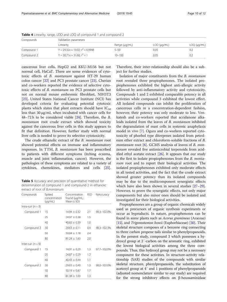
UV spectra at peak start, peak apex and peak end whichwere similar (Fig. 5c, d). The linearity, range, LOD, andLOQ of compounds 1 and 2 are shown in Table 4.LOD and LOQ of both compounds 1 and 2 were
0.05 μg/mL and 0.2 μg/mL, respectively. The methodshowed good linearity demonstrated by the coefficient ofdetermination (r2) being more than 0.999 within theranges of 5 μg/mL - 50 μg/mL, for compound 1 and10 μg/mL - 100 μg/mL for compound 2. The accuracy ofboth compounds are presented as %recovery as shownin Table 5. The recovery of both compounds were within97.7–103.0%. The precision shown as RSD% was evalu-ated both intra-run and between-run. The RSD% of eachcompound was less than 2.4% for both within-run andbetween-run showing good precision of the method.The developed HPLC method showed good linearity, ac-curacy, and precision and, hence, can be used to deter-mine the major components of the ethanolic extract ofB. montanum root.
DiscussionEvidential uses of traditional herbal medicines are estab-lished by ancient doctors in several societies around theworld. However, scientific information on these herbalmedicines is required to either support or dispute their
traditional uses. As the interest in alternative medicinesgrows, the knowledge accumulated over the years by thetraditional doctors can be used to guide modern scien-tists along the right path to discovery. In the presentstudy, we set out to study the root of B. montanum,which has been used in TTM as an ingredient in remed-ies for the treatments of itching eczema, muscle andjoint inflammation and cancer (especially liver cancer).Crude ethanolic extract of the root was fractionated,major components of each fraction were determined forwhich an analytical method was developed and its effi-cacy verified, followed by the analysis of biological activ-ities of the crude extract and its components against thetargeted diseases.The crude ethanolic extract exhibited potent inhibitory
effects in all biological activities investigated. It showedthree times higher anti-allergic activity than the positivecontrol, chlorpheniramine. Venkatesh and co-workers,using in vivo anaphylaxis model, have also shownanti-allergic effects of the leaves of B. montanum [21].For anti-inflammatory activity, the crude extract inhib-ited the inducible NO production from RAW 264.7 cellswith IC50 comparable to that reported by Kakatum andco-workers [5]. In cytotoxic study, the crude extract ex-hibited selective cytotoxic activity against the two
Table 2 Inhibitory effect of isolated compounds on LPS-induced nitric oxide release from RAW 264.7 cell lines
Sample %Inhibition at various concentration (μg/mL)(%Viability)
IC50(μg/mL)
0.01 0.1 1 10 50
Crude extract – 16.65 ± 6.18(92.51 ± 2.14)
32.42 ± 5.42(89.44 ± 3.74)
54.64 ± 2.23(83.49 ± 2.66)
68.38 ± 1.04(78.82 ± 2.83)
6.06 ± 0.43*
Compound 1 – 2.66 ± 0.23(84.66 ± 7.91)
3.91 ± 3.03(89.71 ± 12.08)
14.41 ± 1.71(84.71 ± 9.65)
69.15 ± 2.49(80.46 ± 3.10)
40.40 ± 1.32*
Compound 2 – 7.09 ± 6.86(94.52 ± 10.55)
11.12 ± 9.89(88.81 ± 8.07)
8.69 ± 6.93(80.85 ± 7.39)
58.83 ± 3.59(90.39 ± 9.92)
33.61 ± 5.25*
Compound 3 – −0.82 ± 3.26(102.40 ± 2.17)
2.64 ± 0.31(96.95 ± 0.98)
14.59 ± 3.43(97.27 ± 4.43)
34.44 ± 3.07(91.62 ± 3.87)
> 50*
Prednisolone(Positive control)
19.82 ± 3.43(100.40 ± 12.94)
47.72 ± 6.24(84.28 ± 4.56)
69.94 ± 3.76(89.46 ± 2.28)
77.94 ± 8.12(80.78 ± 2.71)
– 0.11 ± 0.02
Data performed as mean ± SD from triplicate experiments. Mean difference of IC50 data of tested sample and positive control were compared by Student’s t-test.*Significant difference at p-value < 0.05
Table 3 Cytotoxic activity of the crude ethanolic extract and the isolated compounds against cancerous liver cells (HepG2 and KKU-M156) and normal cells (HaCaT)
Sample IC50 (μg/mL) (Selective index)
HepG2 KKU-M156 HaCaT
Crude extract 0.06 ± 0.02* (> 100) 0.16 ± 0.02* (> 100) > 100*
Compound1 87.07 ± 1.36* (> 1.15) 58.32 ± 5.95* (> 1.71) > 100*
Compound2 47.61 ± 1.20* (> 2.10) 95.65 ± 0.48* (> 1.04) > 100*
Compound3 90.33 ± 5.65* (> 1.11) 83.38 ± 5.20* (> 1.20) > 100*
Vincristine 0.013 ± 0.0006 (< 0.008) 0.0030 ± 0.0015 (< 0.033) < 0.0001
Data performed as mean ± SD from triplicate experiments. Mean difference of IC50 data of tested sample and positive control were compared by Student’s t-test.*Significant difference at p-value < 0.05
Pipatrattanaseree et al. BMC Complementary and Alternative Medicine (2019) 19:45 Page 8 of 12

Fig. 4 HPLC chromatogram of blank (methanol)) (a), standard solution (10 μg/mL) (b) and crude extract solution in methanol (4 mg/mL) (c)
Fig. 5 Overlay UV spectra of compound 1 (a) and compound 2 (b) in crude extract solution (4mg/mL) compared with respective standards instandard solution (10 μg/mL); overlay UV spectrum of each compound at peak start, peak apex and peak end: compound 1 (c) and compound 2 (d)
Pipatrattanaseree et al. BMC Complementary and Alternative Medicine (2019) 19:45 Page 9 of 12
cancerous liver cells, HepG2 and KKU-M156 but notnormal cell, HaCaT. There are some evidences of cyto-toxic effects of B. montanum against HT-29 humancolon cancer [22] and PC3 prostate cancer [23]. Cherianand co-workers reported the evidence of selective cyto-toxic effects of B. montanum on PC3 prostate cells butnot on normal mouse embryonic fibroblast, NIH3T3[23]. United States National Cancer Institute (NCI) hasdeveloped criteria for evaluating potential cytotoxicplants which states that plant extracts should have IC50
less than 30 μg/mL when incubated with cancer cells for48–72 h to be considered viable [24]. Therefore, the B.montanum root crude extract which showed toxicityagainst the cancerous liver cells in this study appears tofit that definition. However, further study with normalliver cells is needed to prove its selective cytotoxicity.The crude ethanolic extract of the B. montanum root
showed potential effects on immune and inflammatoryresponses. In TTM, B. montanum has been prescribedto patients with different symptoms (itching eczema,muscle and joint inflammation, cancer). However, thepathologies of these symptoms are related to a variety ofcytokines, chemokines, mediators and cells [25].
Therefore, their inter-relationship should also be a sub-ject for further studies.Isolation of major constituents from the B. montanum
root revealed three propiophenones. The isolated pro-piophenones exhibited the highest anti-allergic activityfollowed by anti-inflammatory activity and cytotoxicity.Compounds 1 and 2 exhibited comparable potency in allactivities while compound 3 exhibited the lowest effect.All isolated compounds can inhibit the proliferation ofcancerous cells in a concentration-dependent fashion,however, their potency was only moderate to low. Ven-katesh and co-workers reported that acridanone alka-loids isolated from the leaves of B. montanum inhibitedthe degranulation of mast cells in systemic anaphylaxismodel in vivo [7]. Ogura and co-workers reported cyto-toxicity of phorbol type diterpenes isolated from petrol-eum ether extract and chloroform-soluble fractions of B.montanum root [6]. GCMS analysis of leaves of B. mon-tanum revealed five antimicrobial terpenoids from acid-ified ethyl acetate extract [26]. It appears that our studyis the first to isolate propiophenones from the B. monta-num root and to report their biological activities. Theisolated propiophenones exhibited only moderate effectsin all tested activities, and the fact that the crude extractshowed greater potency than its isolated compoundsmay be due to the multicomponent synergistic effectswhich have also been shown in several studies [27–29].However, to prove the synergistic effects, not only majorcomponents but also minor ones should be isolated andinvestigated for their biological activities.Propiophenones are a group of organic chemicals widely
used as precursors of organic synthesis experiments oroccur as byproducts. In nature, propiophenones can befound in some plants such as Acorus gramineus (Araceae)[15], and Trigonostemon howii (Euphorbiaceae) [30]. Theirskeletal structure composes of a benzene ring connectingto three carbon propene tails similar to phenylpropanoids.In the present study, compound 3 which possesses a hy-droxyl group at 2′-carbon on the aromatic ring, exhibitedthe lowest biological activities among the three com-pounds. Thus, this hydroxyl group may not be a necessarycomponent for these activities. In structure-activity rela-tionship (SAR) studies of the compounds with similarskeletal structure, phenylpropanoids, the substitution ofacetoxyl group at 4′ and 1 positions of phenylpropanoids(adjusted nomenclature similar to our study) are requiredfor the strong inhibitory effects on β-hexosaminidase
Table 4 Linearity, range, LOD and LOQ of compound 1 and compound 2
Compounds Validation parameters
Linearity Range (μg/mL) LOD (μg/mL) LOQ (μg/mL)
Compound 1 Y = 29.32x + 10.02; r2 = 0.9998 5–50 0.05 0.2
Compound 2 Y = 30.71x + 31.60; r2 = 1 10–100 0.05 0.2
Table 5 Accuracy and precision of quantitative method fordetermination of compound 1 and compound 2 in ethanolicextract of root of B.montanum
Compounds Spikedconcentration(μg/mL)
Concentrationfound (μg/mL;Mean ± SD)
RSD %Accuracy
Intra-run (n = 3)
Compound 1 15 14.94 ± 0.32 2.1 98.3–102.0%
25 24.97 ± 0.38 1.5
40 40.65 ± 0.39 1.0
Compound 2 30 29.63 ± 0.11 0.4 98.3–102.3%
50 50.04 ± 1.18 2.4
80 81.24 ± 1.63 2.0
Inter-run (n = 9)
Compound 1 15 14.91 ± 0.20 1.3 97.7–102.0%
25 24.87 ± 0.29 1.2
40 40.43 ± 0.44 1.1
Compound 2 30 29.83 ± 0.40 1.3 98.0–103.0%
50 50.19 ± 0.87 1.7
80 81.38 ± 1.03 1.3
Pipatrattanaseree et al. BMC Complementary and Alternative Medicine (2019) 19:45 Page 10 of 12
released from RBL-2H3 [31] and cytotoxic effects againstMDA-MB-231 breast cancer cells [32]. For the inhibitoryactivity of NO production, the methoxyl group at 4′-pos-ition and the hydroxyl group at 3- position with one glu-copyranosyl moiety in the molecule are required [33].This may be concluded that the isolated propiophenonescannot be considered responsive compounds for the in-vestigated activities.The only report on isolation of components from the
root of B. montanum by Ogura and co-workers showedthat the isolated phorbol diterpenes from its hexaneand chloroform-soluble extracts exhibited potent cyto-toxic effects against leukemia P388 cells [6]. However,phorbol diterpenes may be minor components in theethanolic extract. Therefore, to detail the responsivemarkers of the crude ethanolic extract of B. montanumroot, other minor constituents should be isolated andinvestigated further to evaluate their roles in the cyto-toxic activities.A HPLC method was developed for the determination
of major components of B. montanum root extract.Compounds 1 and 2 were found to be major compo-nents of the extract from the HPLC chromatogram. Thedeveloped method showed good linearity, accuracy, andprecision. The method may be useful for further studyon the formulation development of the remedies and forquality control in the production of the crude extract.Propiophenones, compounds 1 and 2, analyzed with theHPLC method may be used as markers to indicate thepresence of B. montanum root in multi-ingredients trad-itional remedies. A more sensitive analytical methodsuch as LCMSMS may also be used in conjunction todetect minor components that affect biological activitiesof the extract.
ConclusionThe results of this study reveal potential benefits of B.montanum root as a natural source of anti-allergy,anti-inflammation, and anti-cancer ingredient. Threepropiophenones were isolated from its crude ethanolicextract for the first time. The crude ethanolic extract ofB. montanum root exhibited potent biological activitieswhile its isolated major components exhibited only mod-erate activities. A HPLC method was developed and vali-dated for the analysis of the major components of theextract with the results showing good linearity, preci-sion, and accuracy. The results from this study supportthe use of the root of B. montanum in traditional medi-cines and provide novel information on its biological ac-tivities and chemical components. However, the majorcompounds isolated may not be responsible for all itsbiological activities; isolation and study of minor compo-nents are necessary to evaluate their roles in its overall
effectiveness. The biological activities that indicate therelationship between immunity, inflammation, and can-cer should also be investigated. A more sensitive analyt-ical method should be developed for detailing the keyresponsive components in the extract.
Additional file
Additional file 1: Figure S1. HPLC profiles of obtained fractions fromfirst CC comparing with crude extract. Figure S2-S25. Structural formulae,spectrometric data and HPLC chromatograms of the three isolatedpropiophenones. Table S1-S3. 1H NMR and 13C NMR chemical shift dataof the three isolated propiophenones. (DOCX 6268 kb)
AbbreviationsAnti-DNP IgE: Anti-dinitrophenyl immunoglobulin E; BSA: Bovin serumalbumin; CaCl2: Calcium chloride; CC: Column chromatography; CO2: Carbondioxide; COSY: Correlated Spectroscopy; DMEM: Dulbecco’s Modified Eagle’sMedium; DMSO: Dimethyl sulfoxide; DNP-BSA: Anti-dinitrophenylated bovinealbumin; ESI-HRMS: Electrospray ionization- high resolution massspectrometry; FBS: Fetal bovine serum; HAM F-12: Nutrient Mixture F-12 Hammedium; HCl: Hydrochloric acid; HMBC: Heteronuclear multiple bondcorrelation; HPLC: High performance liquid chromatography; IC50: Halfinhibitory concentration; IT-TOF: Ion trap-time of fight spectrometer;KCl: Potassium chloride; LOD: Limit od detection; LOQ: Limit of quantitation;LPS: Lipopolysaccharide; MEM: Minimum essential medium;MgCl2: Magnesium chloride; MS: Masspectrometry; MTT: 3-(4, 5-dimethyl-2-thiazolyl)-2, 5-diphenyl-2H-tetrazolium bromide; NaCl: Sodium chloride;NaOH: Sodium hydroxide; NMR: Nuclear magnetic resonance; NO: Nitricoxide; P/S: Penicillin-streptomycin; PBS: Phosphate buffer saline;PIPES: Piperazine-N,N′-bis (2-ethanesulfonic acid; PNAG: 4-Nitrophenyl N-acetyl-β-D-glucosaminide; RPMI: Roswell Park Memorial Institute medium1640; S/N: Signal to noise; SRB: Sulforhodamine B; TLC: Thin layerchromatography; TTM: Thai traditional medicines
AcknowledgmentsThis work was supported by Center of Excellence on Applied Thai TraditionalMedicine Research, Faculty of Medicine, Thammasat University. The authorsgratefully acknowledge Dr. Buncha Ooraikul, Prof. Emeritus, Faculty ofAgricultural, Life & Environmental Sciences, University of Alberta, for his kindhelp in grammatical editing and his advice in scientific writing.
FundingCenter of Excellence on Applied Thai Traditional Medicine Research, Facultyof Medicine, Thammasat University provided research funding. The fundingbody had no role in the design of the study or interpretation of the results.
Availability of data and materialsDataset of this manuscript has not been deposited in any repository. Alldataset and materials are available from the corresponding author uponreasonable request.
Authors’ contributionsBiological activities, isolation of pure compounds and HPLC analysis wereconducted by WP who is Ph. D student with in AI. NM and US who areresearcher of AI help to teach and test for anti-allergy of this plant extract.WP wrote the first draft of the manuscript. AI is supervisor of WP, principleinvestigator (PI) and she is director of Center of Excellence in Applied ThaiTraditional Medicine who provided funding for WP. She also supervised,concurrently designed for whole of WP work, revised manuscript and she iscorresponding this work. All authors read and approved the final version ofthe manuscript and ensure this is the case.
Ethics approval and consent to participateNot applicable.
Consent for publicationNot applicable.
Pipatrattanaseree et al. BMC Complementary and Alternative Medicine (2019) 19:45 Page 11 of 12

Competing interestsThe authors declare that they have no competing interests.
Publisher’s NoteSpringer Nature remains neutral with regard to jurisdictional claims in publishedmaps and institutional affiliations.
Received: 13 October 2018 Accepted: 24 January 2019
References1. Mali RG, Wadekar RR. Baliospermum montanum (Danti): ethnobotany,
phytochemistry and pharmacology- a review. Int J Green Pharm. 2008;2:194–9.
2. Pinsornsak P, Kanokkangsadal P, Itharat A. The clinical efficacy and safety ofthe sahastara remedy versus diclofenac in the treatment of osteoarthritis ofthe knee: a double-blind, randomized, and controlled trial. Evid BasedComplement Alternat Med. 2015. https://doi.org/10.1155/2015/103046.
3. Thai Clinical Trials Registry. http://www.clinicaltrials.in.th/index.php?tp=regtrials&menu=trialsearch&smenu=fulltext&task=search&task2=view1&id=2549. (2017) Accessed 20 Sept 2018.
4. Chuakul W, Saralamp P, Paonit W, Temsiririrkkul R, Clayton T. MedicinalPlants in Thailand, vol. II. Bangkok: Siambooks and Publication; 1997. p. 37.
5. Kakatum N, Jaiarree N, Makchucit S, Itharat A. Antioxidant and anti-inflammatory activities of Thai medicinal plants in Sahasthara remedy formuscle pain treatment. J Med Assoc Thail. 2012;95(Suppl 1):S120–6.
6. Ogura M, Koike K, Cordell GA, Farnsworth NR. Potential anticancer agentsVIII. Constituents of Baliospermum montanum (Euphorbiaceae). Planta Med.1978;33:128–43.
7. Venkatesh P, Mukherjee PK, Pal BC. Acridanone alkaloid in Baliospermummontanum--evaluation of its effect against anaphylaxis. Planta Med. 2011;77(17):1947–9.
8. Johnson M, Wesley EG, Hussain MIZ, Selvan N. In vivo and in vitrophytochemical and antibacterial efficacy of Baliospermum montanum (Willd.)Muell. Mg. Asian Pac J Trop Dis. 2010;3:894–7.
9. Patil KS, Jalalpure SS, Wadekar RR. Effect of Baliospermum montanum root extracton phagocytosis by human neutrophils. Indian J Pharm Sci. 2009;71:68–71.
10. Suresh Kumar SV, Mishra SH. Protective effect of extracts of Baliospermummontanum (Willd.) Muell.-Arg. Against paracetamol-induced hepatotoxicity-an in vivo and in vitro study. Anc Sci Life. 2014;33:216–21.
11. Suresh Kumar SV, Mishra SH. Hepatoprotective activity of Baliospermummontanum (willd) Muell.-Arg. In rats treated with carbon tetrachloride: Invivo and in vitro studies. Phcog Mag. 2009;5:196–202.
12. Juckmeta T, Thongdeeying P, Itharat A. Inhibitory effect on β-hexosaminidase release from RBL-2H3 cells of extracts and some pureconstituents of Benchalokawichian, a Thai herbal remedy used for allergicdisorders. Evid Based Complementary Altern Med 2014; ECAM: https://doi.org/10.1155/2014/828760
13. Makchuchit S, Rattarom R, Itharat A. The anti-allergic and anti-inflammatoryeffects of Benjakul extract (a Thai traditional medicine), its constituent plantsand its some pure constituents using in vitro experiments. BiomedPharmacother. 2017;89:1018–26.
14. Thongdeeying P, Itharat A, Umehara K, Ruangnoo S. A novel steroid andcytotoxic constituents from Dioscorea membranacea Pierre againsthepatocellular carcinoma and cholangiocarcinoma cells. J Ethnopharmacol.2016;194:91–7.
15. Park CH, Kim KH, Lee IK, Lee SY, Choi SU, Lee JH, Lee KR. Phenolicconstituents of Acorus gramineus. Arch Pharm Res. 2011;34:1289–96.
16. Tanpure RP, Harkrider AR, Strecker TE, Hamel E, Trawick ML, Pinney KG.Application of the McMurry coupling reaction in the synthesis of tri- andtetra-arylethylene analogues as potential cancer chemotherapeutic agents.Bioorganic Med Chem. 2009;17:6993–7001.
17. Mendieta A, Jiménez F, Garduño-Siciliano L, Mojica-Villegas A, Rosales-Acosta B, Villa-Tanaca L, Chamorro-Cevallos G, Medina-Franco JL, Meurice N,Gutiérrez RU, Montiel LE, Cruz Mdel C, Tamariz J. Synthesis and highlypotent hypolipidemic activity of alpha-asarone- and fibrate-based 2-acyland 2-alkyl phenols as HMG-CoA reductase inhibitors. Bioorg Med Chem.2014;22:5871–82.
18. Kawakami T, Ando T, Kimura M, Wilson BS, Kawakami Y. Mast cells in atopicdermatitis. Curr Opin Immunol. 2009;21:666–78.
19. Nagy G, Koncz A, Telarico T, Fernandez D, Érsek B, Buzás E, Perl A. Centralrole of nitric oxide in the pathogenesis of rheumatoid arthritis and sysemiclupus erythematosus. Arthritis Res Ther. 2010;12(3):210–6.
20. Vuolteenaho K, Moilanen T, Knowles RG, Moilanen E. The role of nitric oxidein osteoarthritis. Scand J Rheumatol. 2007;36:247–58.
21. Venkatesh P, Mukherjee PK, Mukherjee D, Bandyopadhyay A, Fukui H,Mizuguchi H. Potential of Baliospermum montanum against compound 48/80-induced systemic anaphylaxis. Pharm Biol. 2010;48(11):1213–7.
22. Nandagaon VS, Kulkarni AR. In vitro antioxidant and cytotoxicity activity ofBacopa monnieri and Baliospermum Montanum Muell Arg 2013;4:63–67.
23. Cherian AM, Snima KS, Kamath CR, Nair SV, Lakshmanan VK. Effect ofBaliospermum montanum nanomedicine apoptosis induction and anti-migration of prostate cancer cells. 2015;71:201–209.
24. Suffiness M, Pezzuto J. Assays related to cancer drug discovery. In:Hostettmann K, editor. Methods in plant biochemistry: assays for bioactivity.London: Academic Press; 1990. p. 71–133.
25. Grivennikov SI, Greten FR, Karin M. Immunity, inflammation, and cancer. Cell.2010;140:883–99.
26. Radhakrishna S., Kumari SP, GCMS Analysis of Total Terpenoids fromBaliospermum montanum and its Antimicrobial Activity. Journal foradvanced research in applied sciences. 5(3), 94–101. Accessed 20 Sept 2018.
27. Pathomwichaiwa T, Ochareon P, Soonthornchareonnon N, Ali Z, Khan IA.Alkaline phosphatase activity-guided isolation of active compounds andnew dammarane-type triterpenes from Cissus quadrangularis hexane extract.J Ethnopharmacol. 2015;160:52–60.
28. Ma XH, Zheng CJ, Han LY, Xie B, Jia J, Cao ZW, Li YX, Chen YZ. Synergistictherapeutic actions of herbal ingredients and their mechanisms frommolecular interaction and network perspectives. Drug Discov Today. 2009;14:579–88.
29. Stermitz FR, Lorenz P, Tawara JN, Zenewicz LA, Lewis K. Synergy in a medicinalplant: antimicrobial action of berberine potentiated by 50-methoxyhydnocarpin,a multidrug pump inhibitor. Proc Natl Acad Sci U S A. 2000;97:1433–7.
30. Ma J, Yang X, Wang P, Dong B, Su G, Tuerhong M, Jin DQ, Xu J, Lee D,Ohizumi Y, Lin J, Guo Y. Phytochemicals with NO inhibitory effects andinteractions with iNOS protein from Trigonostemon howii. Bioorg Chem.2017;75:71–7.
31. Matsuda H, Morikawa T, Managi H, Yoshikawa M. Antiallergic principles fromAlpinia galanga: structural requirements of phenylpropanoids for inhibitionof degranulation and release of TNF-α and IL-4 in RBL-2H3 cells. BioorganicMed Chem Lett. 2003;13:3197–202.
32. Liew SK, Azmi MN, In LL, Awang K, Nagoor NH. Anti-proliferative, apoptoticinduction, and anti-migration effects of hemi-synthetic 1’S-1′-acetoxychavicol acetate analogs on MDA-MB-231 breast cancer cells. DrugDes Devel Ther. 2017;11:2763–76.
33. Lee JW, Lee DY, Baek DR, Jeong RH, Lee DS, Kim YC, Kim GS, Baek NI, Lee YH.Phenylpropanoids from red kohlrabi sprouts inhibits nitric oxide production inRAW 264.7 macrophage cells. Food Sci Biotechnol. 2014;23(3):965–9.
Pipatrattanaseree et al. BMC Complementary and Alternative Medicine (2019) 19:45 Page 12 of 12