Posters (Abstracts 72 to 394) - Ruđer Bošković Institute · Posters (Abstracts 72 to 394) 072...
108
Posters (Abstracts 72 to 394) 072 Topic Implant Aesthetics The comparsion of osteointegration of SLActive, SLA, BL implants using DVT pictures, bone level profile device and ostell-mentor system: case report Szaniawska K, Wojtowicz A Medical University, Warsaw Introduction: SLActive, SLA and Bone Level, a members of Straumann implants family, are the products to the combina- tion of cylindrical-shape implant advantages, very good osteo- conductive properties of the surface and different concept of the level of installed implants. The aim: Of this work is to evaluate osteointegration of Straumann SLActive, SLA and Bone Level implants in two months after implantation in the patient with risk factors. Material and methods: Male aged 29, heavy smoker, perio- dontitis in family history, poor oral hygiene and partial defi- ciency of the teeth revealed in intraoral examination. DVT diagnosis showed horizontal atrophy of alveolar process in tooth-less region. On the base of DVT (Picasso), diagnostic models, patient was qualified (Straumann recommendation) for SLAactive (immediate installation) SLA and Bone Level Implant (esthetic area) with bone augmentation using BioOss and BioGuide membrane. Two months later the osteointegra- tion progress was evaluated in all installed implants using: 1. Mathematic analysys (Fourier transform, Fractal) of DVT, and dental X-rays, DVT of bone surrounding the implants. 2. Ostell Mentor – electro-magnetic camerton value (arbitrary units), recommended by ITI were used for that purpose. 3. Bone Profile Measurement device – direct evaluation augmen- ted bone profile around the implants. Results: In two months after implant installation some differ- ences of Fractal analysis of bone trabeculae direction/size and Ostell Mentor arbitrary units value were found. Conclusions: Presented case report of Straumann implants installation seems to confirm: 1. efficacy and excellent osteointegrational properties of SLActive implants. 2. high Ostell Mentor value of arbitrary units in all examined implants. Good parameters of osteointegration of Straumann implants family evaluated 2 month after installation i.e time of early loading are not long-term prognostic factor. Only after two months period of healing, high level of integration was observed, despite of the risk factors coexistence. It indicates for reduction of overall implant healing and makes early prosthetic treatment possible. 073 Topic Implant Aesthetics Immediate implant placement and bonymucosal papilla healing in aesthetic areas Di Alberti L 1 , Donnini F 2 , Camerino M 2 , Perfetti G 1 , Dolci M 1 , Trisi P 1 1 University of Chieti, Oral and Maxillo-Facial Unit, Chieti, 2 Private Practitioner, Chieti, Pescara The immediate restoration or loading of dental implants has been an intense area of clinical trial and research in the field of dental implantology over the last several years. The immediate placement of an implant, then, is not only possible but in some clinical situations advisable, with each case individually assessed and the time of placement determined by the clinician. In such cases, the immediate implant placement provides a considerable number of advantages over the traditionally established placement. Osseointe- grated implants have been increasingly also used for aesthetic, predictable restorative treatment. This study presents the 2-year postoperative results of patients treated with immediate, single, tapered implants (Seven, MIS, Israel) in the maxillary incisor region and the simultaneous placement of screwed provisional implant- supported crowns. Implant stability was assessed clinically and by means of resonance frequency analysis (RFA) at surgery and after 3 months. Wound healing was evaluated after 1, 2, 6 and 12 weeks post-operatively. Of the total of 32 implants placed, no implants were lost, resulting in a 100% survival and success rate. All 32 implants were reevaluated and judged to have no signs of mobility, peri-implant inflammation, or adverse reactions. This pilot study has demonstrated that tapered implants yielded clinically after immediate implant placement into the extraction socket and when used in selected cases, this technique facilitated maintenance of the gingival architecture adjacent to immediate transalveolar implants. 074 Topic Implant Aesthetics Screw-retained implant-supported zirconia crowns: 12 months study Camerino M 2 , Di Alberti L 1 , Rossi G 3 , Donnini F 2 , Perfetti G 1 , Dolci M 1 , Trisi P 1 1 University of Chieti, Oral and Maxillo Facial Unit, Chieti, 2 Private Practitioners, Chieti, Pescara, 3 Dental Technician, Alba Adriatica This study evaluated the clinical performance of screwed custo- mized zirconia abutments. Additionally, the marginal fit between the selected implant components was measured and the clinical gingival response was monitored. Twenty patients were consecu- tively selected for a prospective study of 30 implant-supported restorations. Customized zirconia abutment complexes were pre- pared, then ceramic was performed directly. The abutments were 860
Transcript of Posters (Abstracts 72 to 394) - Ruđer Bošković Institute · Posters (Abstracts 72 to 394) 072...

Posters(Abstracts 72 to 394)
072 Topic Implant Aesthetics
The comparsion of osteointegration of SLActive,SLA, BL implants using DVT pictures, bone levelprofile device and ostell-mentor system: case report
Szaniawska K, Wojtowicz AMedical University, Warsaw
Introduction: SLActive, SLA and Bone Level, a members of
Straumann implants family, are the products to the combina-
tion of cylindrical-shape implant advantages, very good osteo-
conductive properties of the surface and different concept of the
level of installed implants.
The aim: Of this work is to evaluate osteointegration of
Straumann SLActive, SLA and Bone Level implants in two
months after implantation in the patient with risk factors.
Material and methods: Male aged 29, heavy smoker, perio-
dontitis in family history, poor oral hygiene and partial defi-
ciency of the teeth revealed in intraoral examination. DVT
diagnosis showed horizontal atrophy of alveolar process in
tooth-less region. On the base of DVT (Picasso), diagnostic
models, patient was qualified (Straumann recommendation)
for SLAactive (immediate installation) SLA and Bone Level
Implant (esthetic area) with bone augmentation using BioOss
and BioGuide membrane. Two months later the osteointegra-
tion progress was evaluated in all installed implants using:
1. Mathematic analysys (Fourier transform, Fractal) of DVT, and
dental X-rays, DVT of bone surrounding the implants.
2. Ostell Mentor – electro-magnetic camerton value (arbitrary
units), recommended by ITI were used for that purpose.
3. Bone Profile Measurement device – direct evaluation augmen-
ted bone profile around the implants.
Results: In two months after implant installation some differ-
ences of Fractal analysis of bone trabeculae direction/size and
Ostell Mentor arbitrary units value were found.
Conclusions: Presented case report of Straumann implants
installation seems to confirm:
1. efficacy and excellent osteointegrational properties of SLActive
implants.
2. high Ostell Mentor value of arbitrary units in all examined
implants.
Good parameters of osteointegration of Straumann implants
family evaluated 2 month after installation i.e time of early loading
are not long-term prognostic factor. Only after two months period of
healing, high level of integration was observed, despite of the risk
factors coexistence. It indicates for reduction of overall implant
healing and makes early prosthetic treatment possible.
073 Topic Implant Aesthetics
Immediate implant placement and bonymucosalpapilla healing in aesthetic areas
Di Alberti L1, Donnini F2, Camerino M2, Perfetti G1,Dolci M1, Trisi P1
1University of Chieti, Oral and Maxillo-Facial Unit, Chieti, 2Private
Practitioner, Chieti, Pescara
The immediate restoration or loading of dental implants has been
an intense area of clinical trial and research in the field of dental
implantology over the last several years. The immediate placement
of an implant, then, is not only possible but in some clinical
situations advisable, with each case individually assessed and the
time of placement determined by the clinician. In such cases, the
immediate implant placement provides a considerable number of
advantages over the traditionally established placement. Osseointe-
grated implants have been increasingly also used for aesthetic,
predictable restorative treatment. This study presents the 2-year
postoperative results of patients treated with immediate, single,
tapered implants (Seven, MIS, Israel) in the maxillary incisor region
and the simultaneous placement of screwed provisional implant-
supported crowns. Implant stability was assessed clinically and by
means of resonance frequency analysis (RFA) at surgery and after
3 months. Wound healing was evaluated after 1, 2, 6 and 12 weeks
post-operatively. Of the total of 32 implants placed, no implants
were lost, resulting in a 100% survival and success rate. All 32
implants were reevaluated and judged to have no signs of mobility,
peri-implant inflammation, or adverse reactions. This pilot study
has demonstrated that tapered implants yielded clinically after
immediate implant placement into the extraction socket and
when used in selected cases, this technique facilitated maintenance
of the gingival architecture adjacent to immediate transalveolar
implants.
074 Topic Implant Aesthetics
Screw-retained implant-supported zirconia crowns:12 months study
Camerino M2, Di Alberti L1, Rossi G3, Donnini F2, PerfettiG1, Dolci M1, Trisi P1
1University of Chieti, Oral and Maxillo Facial Unit, Chieti, 2Private
Practitioners, Chieti, Pescara, 3Dental Technician, Alba Adriatica
This study evaluated the clinical performance of screwed custo-
mized zirconia abutments. Additionally, the marginal fit between
the selected implant components was measured and the clinical
gingival response was monitored. Twenty patients were consecu-
tively selected for a prospective study of 30 implant-supported
restorations. Customized zirconia abutment complexes were pre-
pared, then ceramic was performed directly. The abutments were
860

screwed onto the implants and restored with all-ceramic crowns.
Plaque and gingival indices were recorded monthly intervals over a
12- month period.
All ceramic zirconia abutments offered sufficient stability to
support implant-supported single-tooth reconstructions in anterior
and premolar regions. The soft and hard tissue reaction toward
zirconia was favorable.
075 Topic Implant Aesthetics
Extraction socket soft and hard tissue classification:reliability and validation
Juodzbalys G1, Wang HL2
1Department of Oral and Maxillofacial Surgery, Kaunas University of
Medicine, Kaunas, Lithuania, 2Department of Periodontics and Oral
Medicine, School of Dentistry, University of Michigan, Michigan, Ann
Arbor, USA
Objectives: The aim of this study was to present and validate a
new classification system for the maxillary anterior extraction
socket based upon soft and hard tissue parameters and to
determine indications, efficacy and advantages of the support
immersion endoscope (SIE) method for extraction socket assess-
ment.
Methods: Twenty-five maxillary anterior teeth from25 subjects
(15 men and 10 women; ages 18 to 51 years; mean¼32.4 years)
were used to validate the new proposed classification system.
Two independent surgeons recommended a treatment approach
based upon the classification proposed. These suggestions were
verified at the time of surgery. Weighted Cohen’s k was used to
calculate interobserver reliability. Statistical analysis was per-
formed using the paired t, Kolmagorov-Smirnov, and marginal
homogeneity tests. Extraction sockets were evaluated either
with conventional extraction site evaluation method (CESE)
alone or with CESE plus SIE. CESE includes visual evaluation,
periodontal probing, ridge-mapping with calipers, dental mirror,
orthopantomogram, and diagnostic wax-up.
Results: Interobserver agreement and weighted Cohen’s k were
96% and 0.94, respectively. This indicated a high reliability for
the proposed classification system. No peri-implant soft tissues
were classified as deficient when the newly developed classifi-
cation was used to recommend treatment. Overall, 80% of
sockets were graded as adequate based on soft tissue parameters
(P < 0.001). CESE plus SIE had significantly better accuracy in
examining extraction socket labial plate vertical position; labial
plate thickness; and bone quality when compared to CESE
alone.
Conclusion: The extraction socket classification proposed here
is an objective and helpful tool for socket assessment and for
promoting future implant esthetics. Support immersion endo-
scope can be used as an adjunct tool in assessing extraction
socket morphology and bone conditions without flap elevation.
076 Topic Implant Aesthetics
Simultaneous procedure including implantplacement, GBR and SECTG
Beitlitum IDepartment of Periodontology, The Maurice and Gabriela
Goldschleger School of Dental Medicine, Tel Aviv University,
Tel Aviv
The sub epithelial connective tissue graft (SECTG) predictably
results in root coverage and improves esthetics. GBR procedures
using resorbable membranes and deproteinized bovine bone miner-
aln (DBBM) are well documented. Applying both procedures simul-
taneously.may act synergistically.
Methods: Eight patients, missing upper first/second maxillary
premolars were evaluated for the purpose of implant placement
and correction of gingival recession in the adjacent tooth.
Delayed, tapered implant were placed to replace the missing
premolars. Bone deficiencies around the implant were augmented
using DBBM and covered with either cross-linkedw (CLM) or a non-
cross-linkedz (NCLM) collagen membranes. Gingival recessions
(Miller Cl I or Cl III) in the adjacent tooth were simultaneously
corrected using SECTG.
Results: Clinical outcomes presented a successful integration of
the implant, and harmonious gingival margin in both the
implant and the adjacent tooth. Almost complete coverage of
the Cl I recession (93%) and partial root coverage of the Cl III
recessions (61%) were shown.
Conclusion: This report presents a successful simultaneous
surgical approach for the correction of both bone and soft tissue
deficiencies. This simultaneous approach proved to be predict-
able, less time consuming and more pleasing to the patient in
comparison to a staged approach.nBio-Ossa Geistlich Sohne AG, Wolhusen, Switzerland.wOssixO ColBar Life Sciences Ltd, Herzliya, Israel.zBio-Gidea Geistlich Sohne AG, Wolhusen, Switzerland.
077 Topic Implant Aesthetics
Simultaneous GBR and immediate implant, leavingthe gingival collar intact
Beitlitum IDepartment of Periodontology, The Maurice and Gabriela
Goldschleger School of Dental Medicine, Tel Aviv University, Tel Aviv
Separation of the free marginal gingiva and the mucoperiosteum
from the alveolar bone triggers a cascade of cellular and molecular
events activating bone resorption. Flapless immediate implants
placement however limits the ability to correct localized fenestra-
tions or augment the peripheral bone.
A semilunar incision approch is presented, aimed at enabling
GBR procedures while maintaining the free gingival margin intact.
Methods: In eight patients requiring immediate implant place-
ment in the maxillary esthetic zone, an a traumatic extraction
was carried out, followed by insertion of a tapered implant
and raising a semilunar flap for correcting either an existing
861
fenestration or a peri-apical lesion using deproteinized bovine
bone mineraln and a native collagen membrane.
Result: Clinical outcomes presented successful integration of
the implants, and harmonious gingival margins around the
healing abutments, while treating bone defects around it.
Conclusion: Simultaneous GBR and Immediate Implant place-
ment, using a semilunar incision and leaving the gingival collar
intact, successfully retains esthetic gingival margin.nBio-Ossa Geistlich Sohne AG, Wolhusen, SwitzerlandwBio-Gidea Geistlich Sohne AG, Wolhusen, Switzerland
078 Topic Implant Aesthetics
Optimal function and esthetic in full-mouth implantreconstruction: case series
Artunc C1, Tekin U2, Comlekoglu ME3, Gungor MA4,Sagirkaya E5
1Ege University, School of Dentistry, Department of Prosthodontics,
Izmir, 2Ege University, School of Dentistry, Department of Oral
Surgery, Izmir, 3Ege University, School of Dentistry, Department of
Prosthodontics, Izmir, 4Ege University, School of Dentistry,
Department of Prosthodontics, Izmir, 5Ege University, School of
Dentistry, Department of Prosthodontics, Izmir
Implant therapy is one of the prosthetic treatment options con-
sidering biological and mechanical factors. Conventional complete
dentures as well as implant-supported fixed dentures are treatment
alternatives for edentulous patients. Rehabilitation of edentulous
patients with implant-supported fixed dentures provides significant
psychosocial achievements due to the reestablishment of dentate
state. Long-term clinical and esthetic success of implant-supported
dentures is related to the longevity of osseointegration and restora-
tion of periimplantary soft tissues.
Factors influencing the final esthetics of the implant-suppported
fixed dentures for edentulous patients have been described in this
clinical report. Treatment approach in esthetically compromised
situations are described with case series. Reconstruction of cases
with severe horizontal and vertical hard and soft tissue losses by
using implant-supported dentures is challenging. Advanced surgical
techniques for soft and hard tissue management might be required
to obtain optimal esthetics especially in premaxillary region where
treatment alternatives are limited due to anatomical and technical
difficulties. Implant localization, framework material, abutment
type, fixture-abutment-suprastructure assembly congruency, nat-
ural appearance and morphology of the prostheses are significant
factors related with the esthetic outcome of the implant-supported
restorations.
079 Topic Implant Aesthetics
Esthetic and biological evaluation of implant-supported restorations: case series
Gungor MA1, Tekin U2, Dundar M3, Artunc C4
1Ege University, School of Dentistry, Department of Prosthodontics,
Izmir, 2Ege University, School of Dentistry, Department of Oral
Surgery, Izmir, 3Ege University, School of Dentistry, Department of
Prosthodontics, Izmir, 4Ege University, School of Dentistry,
Department of Prosthodontics, Izmir
Implantology includes functional and esthetic rehabilitation of lost
tissues. Implant-supported restorations are fabricated in cases of
tooth loss due to periodontal diseases, caries and trauma and
resultant severe biomechanical deteoriations. Implant-supported
restorations are also important for patients’ psychological concerns.
This clinical report describes the surgical and prosthetic methods
used for the determination of factors influencing treatment plan-
ning and optimal esthetics. Advanced surgical techniques, guidance
of the emergence profile by postoperative temporary restoration
fabrication and achieving optimal esthetics by definitive restoration
application are discussed with case series at our clinics. The esthetic
and functional needs of the patients had been restored. Patients are
being recalled periodically. Clinical applications of different implant
systems (Straumann, Biolok, Astra, BioHorizons, Bego), abutment
types (metal, zirconium) and framework materials (metal-ceramic,
heat-pressed ceramic, zirconium oxide and aluminum oxide)
have been described. The clinicians’ success has increased by the
technological progress with scientific documentation. Non-trau-
matic surgical procedures, mechanical stability of the implant,
choice of ceramic material and technological progress
lead to highly esthetic and long-lasting restoration fabrication.
Multidisciplinary teatment approach is the key factor to successful
treatment.
080 Topic Implant Aesthetics
High-tech esthetics: The zirconia implant approach
Sarkis RPrivate Practice, Beirut
The Challenge of Esthetic Implant dentistry has always been
restoring what is lost to its original shape and color. Today, the
means, methods and opportunities for implant esthetic dentistry
seem to be at hand. The technological advances in biomaterials and
the better understanding of their applications in implant dentistry
allow us today the use of Zirconia white implants not only where
esthetics is compromised but also in classical situations where the
use of metal titanium is undesirable. Zirconia is very helpful in
such situations, and better soft tissue-preserving than the conven-
tional strategies. This presentation will give a closer look to the
clinical approach, the integration of zirconia in hard and soft tissues,
the indications and the results of the use of Zirconia white implants
at 24 months follow up.
862

081 Topic Implant Aesthetics
Significance of prosthetic components on peri-implant mucosa color changes
Rutkunas V1, Mizutani H2
1Institute of Odontology, Vilnius University, Vilnius, 2Department of
Masticatory Function Rehabilitation, Tokyo Medical and Dental
University, Tokyo
Objectives: To evaluate the influence of peri-implant mucosa
thickness on masking ability of different prosthetic compo-
nents. To estimate the effect of light transmission of zirconium
oxide based abutments and crowns on gingival esthetics.
Material and methods: Peri-implant mucosa color changes with
different prosthetic materials were investigated using 2 tita-
nium based (titanium only and titanium layered with ceramic)
and 5 zirconium oxide based specimens using in-vitro porcine
jaw model. Unstained, stained (A2, A3 and A4) and covered with
pink ceramic Zirconzahn (Zirconzahn GmbH, Germany) and
two types (white and yellow) of Cercon (DeguDent GmbH,
Gemany) ceramic specimens comprised the zirconia group.
DL, Da, Db and DE were calculated and compared for all speci-
mens with different thicknesses of mucosa. Special setting
was used to evaluate light transmission of different materials.
Specimens covered with mucosa were lightened by the daylight
or halogen light from one side and mucosal color changes
evaluated.
Results: Study revealed that overall color change of mucosa was
significantly influenced by the optic properties of the prosthetic
components (ANOVA, p < 0.05). With increased thickness of the
mucosa, color change was less expressed. Pink porcelain was
found to be an effective measure to prevent adverse optic effects
with thin peri-implant tissues. Significantly more light was
transmitted to the mucosa in the zirconia group.
Conclusions: Thin peri-implant tissues are associated with the
risk of mucosa color change due to the show-through of pros-
thetic components. Colored zirconia abutments ensure less
discoloration. Light transmitting feature of zirconia materials
might promote natural appearance of the marginal tissues.
082 Topic Implant Aesthetics
Immediate implantation in inflamated versus non-inflamed sockets: clinical, radiological outcomes
Kocar M1, Kansky A1, Gorjanc M2
1University Clinical Center Ljubljana, Department of Maxillofacial
and Oral Surgery, Ljubljana, 2Implant Institute, Ljubljana
Intruduction: Immediate implantation is becoming very used
method because of its predictable. Aim of our study was to
compare implant outcomes and stability of marginal bone after
immediate implantation between inflamed (ISO) and not
iflamed sockets (NISO).
Methods: Eight implants (seven patients) were placed and
prostethically restored by following immediate implantation
with non-functional loading protocol. In ISO-group (four pa-
tients) four implants were implanted where at least one sign of
inflammation was presented: fistula with push (3), evident
mucosal swelling with redness (1). Other implants were inserted
at NISO-group (three patients) after fracture of roots (4) were
established. Antibiotic was prescribed for 10 days. Clinical
controls and intraoral periapical x-rays were performed periodi-
cally at 1/6/12/24 week. Vertical changes of periimplant bone
level were observed on mesial and distal site of the implant
body. Bone apposition on implant shoulder was checked on last
control. X-rays (image size 3072 � 2048 pixels) were examined
with ImageJ program.
Results: All implants were osseointegrated and fulfilled other
requested criteria of success. Average rates of subcrestally
insertion were (ISO/NISO) mesially 2.540/2.805, distally
2.201/2.317 mm on x-rays at 1 week. After 24 weeks average
resorption rates were mesially 0.023/0.073, distally 0.028/
0.076 mm. With 5 implants, both shoulders were completely
overgrown by bone. No bone apposition was observed only on
one side (ISO).
Conclusions: Inflammation did not adversely affect osseointe-
gration; bony overgrowth is suggesting the opposite. Marginal
bone resorption with immediate implantation can not be
avoided. Antibiotics had important rule of overweighting the
threat of infectious non-integration. Small sample and minimal
changes of bone level require for continuing of study.
083 Topic Implant Aesthetics
Autogenous bone block with immediate implantplacement in anterior maxilla
Susic M1, Gabric Panduric D1, Pelivan I2, Katanec D1,Kobler P1
1Department of Oral Surgery, School of Dental Medicine, University of
Zagreb, Zagreb, 2Department of Prosthodontics, School of Dental
Medicine, University of Zagreb, Zagreb
Anterior teeth root fractures and chronical fistula usually results in
bone deficiency of anterior maxilla. Insufficient bone volume in the
anterior maxillofacial region often requires reconstructive surgery
for ridge augmentation to make placement of endoosseous implants
possible. In the majority of the cases the bone block grafts were
harvested from the mandible using specially designed instruments.
A patient was a 36-year-old male with clinical and x-ray signs of a
vertical fracture of the endodontically treated maxillary left central
and lateral incisor. After extensive diagnostic procedures, therapeu-
tical plan was made: two dental implants placement (XiVEs
Implant System) immediately after teeth extractions and bone
defect augmentation in the area 21 and 22. After immediately
implants placement, autologous bone graft was used for remaining
bone defect of the buccal cortical plate, combined with heteroge-
nous bone filling material (Bio-Osss
, Geistlich, Germany) and
finally covered with resorptive membrane (Bio-Gides
, Geistlich,
Germany). During the osseointegration period, onlay composite
resin bridge was made as the temporary prosthetic solution. Follow-
ing the osseointegration period of 6 months, all-ceramic single
tooth crowns on zirconium oxide abutments were used as final
prosthodontic restorations. The patient exhibited neither clinical
863
nor radiologic complications throughout the 6 months period of
clinical monitoring.
084 Topic Implant Aesthetics
Tooth replacement using an ankylos implant with acercon abutment and cercon crown
Solymosi PSolydent Dental Surgery, Gyor, Hungary
First case: The presented 32 year old patient of us, came in
March 2004. into our office. He told us he had an accident in his
childhood, because of his maxillary right central incisor, tooth
11 was extracted. Since then he had a removable denture.
Because of his young age and because of aesthetic reasons he
was interested in an implant solution. After taking a panoramic
x-ray, we have found that there is a possibility for an implanta-
tion. The amount and thickness of the bone was suitable for an
implantation. The operation was done on the 9. October 2004.
After raising the flap we have implanted an Ankylos implant
type B of 14 mm length an 4.5 mm diameter. We have not found
the amount of bone enough, this is why we have placed BIO-OSS
cancellous bone around the implant. Before closing the wound
the bone replacement material was fixed with a BIO-GIDE
membrane. We have made a recall examination after half a
year. Because of using a bone replacement material we have
waited a year for opening the implant site. We have taken the
impression September 2005. We used a CERCON abutment on
which a CERCON crown was fixed.
Second case: The 25 years old patient came in June 2005. into
our office for recementation of his post crown in his maxillary
right central incisor, tooth 11. After taking an x-ray we sug-
gested the extraction of the tooth. The tooth was extracted by
using a periotome to preserve the buccal and palatal wall of the
socket. A temporary removable denture was made. After we
have waited 3 months, an Ankylos implant, type B of 17 mm
length and 4.5 mm diameter was placed on site 11. The socket
was preserved, that is why an augmentation was not made. In
the middle of October the implant site was opened, after waiting
2 weeks the impression was taken. We used a CERCON
abutment and a CERCON crown was made.
085 Topic Implant Aesthetics
Gingivomorphometry—A new method for collectionand measurement of standardized & reproducible datain oral photography
Weinlaender M1, Lekovic V2
1Implantology & Periodontology Vienna, Austria, City Implant
Vienna, Vienna, 2Department of Periodontology, School of Dentistry,
Belgrade, Serbia
The Gingivomorphometry method is a two step concept for
evaluating certain intraoral soft tissue and crown parameters
through 1. standardized and reproducible data collection and 2.
standardized and reproducible parameter measurements. In order to
acquire standardized and reproducible data in oral photography
3 basic criteria have to be fulfilled: 1. Standardized and reproducible
patient positioning 2. Standardized and reproducible camera posi-
tioning 3. Standardized and reproducible mirror positioning for data
collection in the premolar and molar regions. A device to fulfill the
above mentioned criteria was developed and will be presented.
Standardized processing of the aquired data – the ‘‘Morphome-
trical’’ part of this concept – is based on the import of the acquired
data into a medical image processing software designed for naviga-
tion and visualization of multimodality and multidimensional
images. In our concept, the acquired data files are saved and
imported into an image processing software (e.g. Photoshops
).
With the help of this program a reference line connecting the
midfacial gingival levels (FGL) of the adjacent teeth next to the
implant restoration is added. The zeniths of the adjacent teeth can
then be marked as reference points and – together with the resulting
reference lines – they serve as standard orientation markers for the
necessary morphometrical measurements. A standardized protocol
of six different soft tissue measurements around implant supported
crowns include mesial/distal papilla height and area, as well as soft
tissue height of the implant supported crown and implant crown
soft tissue perimeter. Implant supported crown area and perimeter
can also be measured and compared to contralateral parameters.
086 Topic Implant Aesthetics
Dimensional comparison of interproximal soft-tissueon multiple-implants and contra-lateral natural teeth
Lee DW, Kim TH, Moon ISDepartment of Periodontology, Yong-dong Severance Dental Hospital,
Yonsei University, Seoul
Objectives: The purpose of the present study was to compare
and to correlate the dimension of soft tissue from the tip of
papilla to the crestal bone between 1) two adjacent implants 2)
two contra-lateral natural teeth. Thus, the dimensional relation-
ship and possible correlation would be investigated.
Methods: The present study involved 16 inter-implant papillae
and 16 contra-lateral interproximal papillae in 13 patients. All
the subjects had periodontal treatment prior to the implant
surgery. Patients who had inflamed mucosa around implants
with bleeding tendency and plaque accumulation were rejected.
Plaque index (PI) and gingival index (GI) was recorded. The
shortest distance from the tip of the papilla to the inter-implant
crestal bone (DI)/and contra-lateral inter-dental (DN) crestal
bone was measured with non-invasive method, developed and
published by our team, using radiograph. Wilcoxon-Signed-
Ranks-Test was performed in order to see the 1) difference of
PI/GI between implants and natural teeth and 2) difference
between DI and DN. Also, correlation between DI and DN
was calculated (Spearman’s correlation-coefficient).
Results: The mean PI/GI of implant (0.1� 0.08/0.1� 0.02) and
natural teeth (0.1� 0.05/0.1� 0.01) were not different. Statis-
tically significant difference was found between DI and DN
(4.08� 0.68 vs 2.88� 0.9, p¼0.001). Correlation between DI
and DN was not significant (p¼ 0.12, rho¼0.4).
864
Conclusion: The result of the present study showed that in
periodontally treated patients, the dimension of interproximal
soft tissue between multiple implants was significantly less
than that of contra-lateral natural teeth. However, the correla-
tion between two variables was not found.
087 Topic Implant Aesthetics
Extraoral fixtures for nasal epithese
Evrard L1, Baeyens W1, Poortmans P2, Glineur R1
1Service de Stomatologie et de Chirurgie Maxillo-faciale-ULB-Hopital
Erasme, Bruxelles, 2Laboratoire de prothese dentaire-ULB-Hopital
Erasme, Bruxelles
Maxillofacial defects caused by cancer treatment are a huge problem
affecting the quality of life of patients. Epithethic solutions are
indicated if plastic surgery reconstruction is not a valid option for an
extensive defect.
We present the case of a 42 years-old woman, who had underwent
a total nasal resection, for a well-differentiated epidermoid carci-
noma of the tip of the nose, resected in his totality. The anatomo-
pathologic analysis of the piece of resection did not reveal any
ganglionar metastase, then, the site of tumoral resection was not
irradiated.Three month later, four extraorals
Vistafix fixtures were
placed. After four months, each fixture as a bone anchorage was
connected with a retention device to be connected to the epithese.
After four weeks, cutaneous healing allowed the prosthetic steps to
be achieved.
The use of extraoral fixtures as bone anchorage for a nasal
epithese represents an alternative to more heavy and more time-
consuming surgical reconstruction techniques. Another advantage
is that the site can be monitored easily, as for the carcinologic
follow-up. It allows a good retention of the nasal epithese and an
esthetic result which is satisfying for the patient.
A multidisciplinary co-operation is essential if optimal results are
to be achieved.
088 Topic Implant Aesthetics
Multicentric prospective study of immediatelyprovisionalized postextractive implants
Clauser C1, Menini I2, Barone R1
1Accademia Toscana di Ricerca Odontostomatologica, Firenze,2Private Practice, Belluno
Background: Immediate restoration of post-extractive single
implants is gaining increasing popularity, but more information
is needed about their success rate, patient perception and risk
factors. The aim of this multicentric prospective study is
assessing the success rate and identifying possible risk factors
for aesthetic and functional failure.
Methods: Molar teeth were excluded from the study. One-year
follow-up was planned: the recruitment will be terminated after
the insertion of 250 implants in 15 centers.
Surgical protocol was designed to achieve optimum stability
while striving for light contact with the buccal cortical bone.
Cone-shaped implants (Nanotite Certain NT implants, Implant
Innovation Inc., Palm Beach, Fla) were selected to increase primary
stability and wound healing speed. Several morphologic variables
were recorded, including periodontal biotype, crestal bone levels,
marginal tissue level relative to adjacent natural teeth. A screwed
temporary crown had to be delivered in 48 hours after the end of
surgery. Any occlusal contact was eliminated.
Demographic and dental data were recorded in order to find
possible relationships with implant failure or complications, with
special emphasis on recession.
Results: The study was started in July 2007 and should be
completed in 2 years. The success rate was greater than 98% in
the first 88 cases. There have been too few complication to allow
for an analysis of the risk factors.
Conclusions: Immediate provisionalization with non-func-
tional loading is a safe option for postextractive implants in
the short run. Long term data is needed to recommend this
protocol for clinical practice.
089 Topic Implant Aesthetics
Outcome analysis of immediately-placed,immediately restored implants in the aesthetic area:the clinical relevance of different inter-implantdistances
Degidi M1, Novaes Jr AB2, Nardi D1, Piattelli A3
1Private Practice, Bologna, 2University of Sao Paulo, Sao Paulo,3University of Chieti-Pescara, Chieti
Background: The purpose of this study was to compare and
evaluate bone and soft tissue levels between immediately-
placed, immediately-restored implants positioned in the aes-
thetic anterior region with different inter-implant distances.
Methods: 49 patients requiring multiple implant restorations in
the anterior regions received 152 implants, which were imme-
diately restored. Periapical radiographs and digital images of 99
inter-implant sites were made in regular follow-up examina-
tions at 0, 6, 12 and 24 months after surgery. They were digitally
recorded and analyzed. The presence of the inter-proximal
papilla was assessed and compared to the distances between
the bone crest and the contact point between the natural teeth
and the restoration crowns.
Results: Implants with an inter-implant distance of less than
2 mm seems to lose less bone laterally. When the inter-implant
distance was less than 2 mm, vertical crestal bone loss was
significantly greater than in the other groups. Interproximal
papilla presence decreased in percentage when the distance
between the bone crest and the contact point between the two
restoration crowns was greater than 6 mm and when two
implants where placed at a distance of 4 or more millimetres.
Conclusions: To guarantee a better aesthetic result in immedi-
ately placed, immediately restored implants, the contact point
between the two prosthetic crowns should be placed at 3–4 mm
and never more than 6 mm from the bone peak. Two adjacent
implants should be placed at a distance greater than 2 mm and
lesser than 4 mm.
865

090 Topic Implant Aesthetics
Evaluation of implant-supported single crownesthetics: 2-year follow-up
Aladag A, Gungor MA, Comlekoglu ME, Dundar M,Tekin U, Artunc CEge University, School of Dentistry Department Prosthodontics, Izmir
Objectives: The aim of this study was to evaluate the esthetics
of implant-supported single crowns and adjacent soft tissues
with the use of an esthetic index.
Material and methods: 35 implant-supported single crowns and
their periodontal status were evaluated using Implant Crown
Esthetic Index (ICEI) including 9 items by three oral-maxillofa-
cial surgeons and three prosthodontists after 2 years. The rating
was carried out twice by each of the examiners. Weighted
Cohen’s k was calculated to express the intra- and interobserver
agreement.
Results: The agreements for prosthodontists, surgeons and
both groups (overall) are shown in the Table. Intraobserver
results indicated that the agreement among the ratings of oral
maxillofacial surgeons was fair (0.39) while the prosthodontists’
was moderate (0.45). The best interobserver agreement was
observed among the prosthodontists (0.51).
Item1
Item2
Item3
Item4
Item5
Item6
Item7
Item8
Item9
Prosthodontists 0.36 0.51 0.64 0.52 0.27 0.41 0.51 0.47 0.22Surgeons 0.59 0.06 0.49 0.01 0.51 0.24 0.55 0.18 0.47Overall 0.12 0.13 0.31 0.23 0.21 0 0.31 0.14 0.27
Conclusions: ICEI is an objective tool in evaluating esthetics of
implant-supported single crowns and surrounding soft tissues.
Prosthodontists exhibited the highest reliability in rating the
esthetics of restorations. Although ICEI is frequently used for
esthetic evaluation of implant restorations, it might be devel-
oped for further clinical data collection.
091 Topic Implant Aesthetics
Comparision of impression making procedures foresthetic implant restoration
Lee JH1, Yang JH2
1Department of Prosthodontics Dankook, University, Cheonan,2Department of Proshtodontics, Seoul National University, Seoul
Impression is the most important tool of communication between
dentist and dental technician. Impression should have to transfer as
much information as possible. To make more precise and conve-
nient there were several modified technique. Individual impression
coping, impression with temporary restoration as impression coping
and manual modification of stone cast were representatives. This
presentation will compare these technique with a newly developed
a simple and accurate technique to produce an implant definitive
cast with an analog block. An analog block is made from bis-GMA
temporary restoration material. This chairside procedure is designed
to transfer information regarding the contoured soft tissue and
provisional restoration, enhancing the aesthetics of the definitive
restoration. This method also reduces the number of parts used in
impression procedures and numbers of connecting/disconnecting.
So using this method will reduce the chance of misfit by repeated
screwing. With clinical case presentation, upper mentioned tech-
nique were compared.
092 Topic Implant Aesthetics
The influence of immediate implant loading on thegingival aesthetics
Vavrickova L1, Dufkova D1, Sobotka M1, Pilathadka S1,Simunek A1, Dostalova T2
1Department of Dentistry, medical faculty and university hospital in
Hradec Kralove, Charles University in Prague, Hradec Kralove,2Department of Paediatric Stomatology, 2nd Medical School, Charles
University Prague, Prague
Authors discuss the influence of immediate implantation with
immediate loading on the red aesthetics over the early implantation
with immediate loading (6 weeks after extraction).
Over 600 implats have been observed in years 2005–2008. The
situation has been photo documentated in the day of 1st stage
surgery, after 3 month and after 6 month.
The clinical experience shows, the best aesthetic results of
gingiva are possible to achieve usually in young patients, with
preoperative healthy gingiva and with sufficient bone support.
The implant width also acts the essential role in the red
aesthetics, the wider the implant, the better the outcome. Immedi-
ate implantation with immediate loading has better aesthetic result
than early implantation with immediate loading. Six month post
surgical gingival recession was more pronounced after early im-
plantation than after immediate implantation.
For good aesthetic result of gingiva, it is neccesary to have
preoperative healthy gingiva, the sufficient height of interdental
bone, wide implant platform and immediate implant loading.
Passive fit of the crown in the cervical part and perfect oral hygiene
are considered as the standard.
093 Topic Implant Aesthetics
Intraoral luting: new prosthetic design to achieveaesthetics and passivity
Longoni S1, Sartori M1, Tonelli P2, Brancato L2, Duvina M2,Maroni I3, Monguzzi R1, Baldoni M1
1University of Milano-Bicocca, Milano, 2University of Firenze,
Firenze, 3Private Practitioner, Varese
Intraoral luting between metallic framework and galvanic gold caps
(AGC) is a good procedure to achieve passivity of the prosthesis.
The limit of the conventional technique is the resultant thick
prosthesis margin made of the AGC, the cement for the luting
phase and the framework. A new intraoral luting technique is
presented. The peculiarity is the different prosthetic design with
866

the metallic framework that is 1.5 mm shorter than the margin of
the galvanic caps. As a consequence, only the thin prosthesis seal of
the AGC (0.3 mm) is placed just beyond the apical limit of the
aesthetic material with a good chromatic camouflage in the area
next to the free gingival margin.
The technique has the advantage that it can be performed with all
the implant systems which have milling abutments. The clinician
and the technician can choose the metal for the casting of the
framework and the aesthetic resin veneering material.
One disadvantage is the composite luting agent (Nimetic cem;
3 M ESPE, Seefeld, Germany), because it does not resist above
1201C. For this reason the aesthetic veneering is made with hybrid
ceramic (Estenia; Kuraray Medical Inc, Okayama, Japan). In the
future, the development of other luting materials which resist at the
temperature for low fusing porcelains should overcome this limit.
The procedure presented is efficient, standadized and assures a
prosthetic rehabilitation with a high aesthetic result.
094 Topic Implant Aesthetics
Ridge augmentation of the anterior maxilla usingcancellous freeze-dried block allografts
Nissan J, Mardinger O, Calderon S, Ghelfan O, Chaushu GTel Aviv University, Tel Aviv
Background: An esthetic final result depends on the presence of
an adequate bone volume, providing a predictable bony support
for the gingival margin and papillae. Therefore, pre-implant
augmentative surgery is a pre-requisite in many cases in the
anterior maxilla.
Aim: Evaluate the clinical, radiological and histological results
of endosseous implants placed in the anterior maxilla following
ridge augmentation with cancellous freeze-dried block bone
allografts.
Material and methods: Consecutive patients requiring ridge
augmentation of at least 4 mm in either vertical, horizontal or
both dimensions of the anterior maxilla were included in the
study. 55 implants were placed after a healing period of 4–6
months, based on age, defect size, radiographic and clinical
follow up. Bone biopsies were taken after 4–6 months during
implant placement. 19 implants were immediately loaded.
Results: 38 blocks were used in 26 patients (16 females and 10
males) aged 17–70 years (mean 34� 17 years). The mean follow-
up was 33� 13 months (range 8 to 53 months). Histological
evaluation demonstrated newly formed bone containing viable
osteocytes merged with residual grafted bone characterized by
empty lacunae devoid of osteocytes. Two blocks failed resulting
in 94.7% survival rate. One out of the immediately loaded
implants (98% survival rate) failed following trauma. After
3 months of waiting, the implant was reinserted and success-
fully osseointegrated. All patients received a fixed implant-
supported prosthesis. No further implants have been lost in
function.
Conclusion: Cancellous block-allografts appear to hold pro-
mises as a grafting material for the atrophic anterior maxilla.
095 Topic Implant Aesthetics
Marginal adaptation in provisional crowns ofimmediate single tooth implants
Borges T1, Carvalho A1, Carvalho C2, Carvalho V1
1Portuguese Catholic University, Viseu, 2CAEF, Centro Avancado de
Estetica Facial, Chaves
A body of literature describes good osseointegration outcomes
following provisionalization of single tooth immediate implants in
extraction sites. Good aesthetics results on the same day of the
surgical procedures and maintenance of the soft tissue contours are
the main advantages of this protocol. Cemented retained, screw
retained and friction retained crowns can be used for the provisional
restorations. The marginal adaptation of the provisional crowns
next to the soft tissues margin can be achieved by different ways,
always conditioned by the type of chosen provisional restoration.
This clinical report describes a method of improving marginal
adaptation of screw retained provisional crowns in immediate
implants placed in 8 patients in 10 extraction sites, with the use
of a temporary implant abutment, a polycarbonate crown and final
adaptation with flow composite by an indirect placement using a
silicon putty model. Different parameters like the papilla anatomy,
marginal bone loss and satisfaction of the patients were evaluated.
This technique offers a good soft tissue adaptation, a reduction of
treatment time and an improved degree of satisfaction for the
patients.
096 Topic Implant Aesthetics
Soft tissue and bone evaluation around custom madeabutments
Gultekin BA1, Bayraktar M1, Akilli E1, Abdel-Hak J1,Turkoglu P2, Siraliyev S1, Yalcin S1
1Istanbul University, Faculty of Dentistry, Department of Oral
Implantology, Istanbul, 2Istanbul University, Faculty of Dentistry,
Department of Prosthodontics, Istanbul
Introduction: Recent progress in the technology and research of
new dental materials has resulted in an increased number of
materials available for esthetic restorations. Increasing aware-
ness among patients of dental esthetics had led some to be
dissatisfied with the appearance of metal-ceramic crowns and
titanium abutments.
Material and methods: 32 implants were placed in 12 patients.
After implant surgery, impressions were taken and custom
made zirconia abutments were prepared by Proceras
(Gothen-
burg, Sweden). Implant supported zirconium restorations were
made after 3 months of healing period. Heavy smokers, patients
with systemic diseases were not included. The radiographic
examinations were made with panoromic radiographies after
0, 3,6,12 months.
Results: Satisfactory functional and mechanical results were
achieved in much of the restorations. 2 procera abutments with
diameter of 3.5 Nobel Biocare Replace Select system have
broken. The mean radiographic bone loss was 1.3� 1.2 mm
867
(range: 0.3–1.3). Periodontal health and aesthetic of the zirco-
nium restorations were favorable.
Conclusion: Patient mediated zirconium made abutments
would facilitate and improve the clinical prosthetic success
and also aesthetic appearance. Clinical studies are required to
confirm the long-term performance of this type of restoration.
097 Topic Implant Aesthetics
Congenital missing teeth: Prosthetic rehabilitationfollowing orthodontic treatment
Abdel-Hak J1, Akilli E1, Arici V1, Gultekin BA1, KarabudaC1, Qasrawi O2
1Istanbul University, Faculty of Dentistry, Department of Oral
Implantology, Istanbul, 2Istanbul University, Faculty of Dentistry,
Department of Orthodontics, Istanbul
Missing teeth in anterior part of the maxilla cause functional,
aesthetical and psychological problems. Especially in adolescents,
apart from conventional methods (removable partial dentures,
maryland bridges, fixed dentures), osseointegrated implants devel-
oped by Branemark et al. may be used for treatment of missing
teeth. On account of function and aesthetics, implant application is
reliable and predictable method. It is significant to apply implants
after active bone growth period in adolescents.
In this study, single patient undergones prosthetic rehabilitation
following orthodontic treatment. In the case, implants are applied
after the preparation of necessary room for congenitally missing
upper bilateral lateral incisors. After the distalization of canines,and
mesialization of the central incisors to their proper anatomical
location,two implants are applied in the gained space.
As a result, osseointegrated implant application in anterior part of
maxilla is fairly conservative and aesthetic method.
098 Topic Implant Aesthetics
Immediate implantation with zircon implants –a preliminary report
Steczko W, Dubis PSteczko Dental Practice, Krakow
The report describes a case involving the use of zirconium dioxide
implants in the aesthetic segment of a 30-year old female patient.
Teeth numbers 11 and 12 of the female patient were extracted
atraumatically following complications that arose during the course
of endodontic treatment. Using piezosurgery and osteotomes im-
plant beds were prepared for one-piece zirconium dioxide implants.
Implants 4 mm in diameter and 14 mm in length were then
inserted. The free space between the vestibular bone plate and the
implant was filled with Bio-Oss mixed with the patient’s venous
blood.
Due to the indication for the restoration of teeth 12 and 21 with
veneers, an adhesive resin bridge was inserted as a long-term
provisional. The WhiteSky system (Bredent) was used to make
the preparation of coronal part of the implants after insertion
possible.
The final prosthetic restoration using individual Procera Zirconia
crowns was completed 4 months later. Full osseointegration of the
implants and a very good aesthetic effect were achieved.
099 Topic Implant Aesthetics
Multidisciplinary treatment for missing upper centralincisor in an adolescent
Pelivan I1, Mestrovic S2, Susic M3
1School of Dental Medicine, University of Zagreb, Department of
Prosthodontics, Zagreb, 2School of Dental Medicine, University of
Zagreb, Department of Orthodontics, Zagreb, 3School of Dental
Medicine, University of Zagreb, Department of Oral Surgery, Zagreb
Implant restorations are commonly used to replace missing inci-
sors. However their use in adolescents is not very common. This
case report represents a multidisciplinary treatment approach for
missing upper central incisor in young patient. Fifteen-year-old girl
was referred with missing upper right central incisor which was
surgically removed at the age of 11. The tooth was impacted due to
traumatic injury of deciduous upper right central incisor at the age
of two which led to severe trauma of permanent tooth germ. Space
between tooth 12 and 21 was completely closed with teeth crowd-
ing in both maxilla and mandible. Dental radiographs and diagnos-
tic casts were made. Missing space was achieved by means of
orthodontic space opening procedure which lasted till girl was 17
years old. It was decided to make an implant restoration after the
orthodontist stated that the patient has completed her facial growth.
Friadent XiVE implant (3.0 mm diameter) was placed and period of
three months was recommended as healing time. During that time
patient had teeth-retained removable temporary tooth designed as
ovate pontic. It was used for aesthetic and soft tissue management
with non-invasive papilla reconstruction, respectively. Satisfactory
soft tissue emergence profile was achieved during healing period.
After impression taking, PFM crown was made on 15degrees angled
aesthetic abutment. Regular 6 months follow up showed excellent
functional and esthetical results after one year.
100 Topic Implant Aesthetics
Implant treatment of pediatric oligodontia
Kim NH, Yun KI, Choi YJDental Center, Chung-Ang University Hospital, Seoul
The oligodontia patients have not only positional, dimensional and
morphological abnormalities in tooth but also abnormalities in
maxillo-facial skeleton. So it is important to cooperate with
pedodontist, orthodontist, prosthodontist, and oral and maxiilofa-
cial surgeon.
We report a case of pediatric oligodontia treated by multidisci-
plinary procedures including dental implants.
868
101 Topic Implant Aesthetics
Implant replacement of congenitally missing lateralincisor in maxilla
Eccellente T, Piombino M, Rossi A, D’errico M, Festa PClinic for Periodontal and Implant Surgery, Grumo Nevano (NA)
Purpose: Implant restorations are used to replace congenitally
missing lateral incisors in orthodontic patients. The present
study reports on the clinical and aesthetic results of implant-
prosthetic treatment of this anomaly.
Material and methods: In 9 patients affected by monolateral
agenesis and 17 patients with bilateral agenesis 43 implants (5
Branemark, 14 Straumann and 24 Ankylos implants) were
inserted. The minimum length of implants was 8 mm. In
different time intervals PlI, BOP, standardized periapical radio-
graphs, technical complications and patients’ satisfaction were
recorded. All patients were submitted to maintenance therapy.
Results: Six months after surgery all implants were osseointe-
grated from clinical and radiographic point of view and loaded
with temporary resin crowns. 31 implants received final restora-
tion using Auro Galvan Crowns veneered with ceramic, the
remaining 12 implants with metal-ceramic crowns. After 40.8
months of function (range 32–57 months) no implant failed. The
majority part of implants presented healthy peri-implant soft
tissue conditions (PlI < 1, BOP < 1). Marginal bone resorption
ranging from 0.38 to 1.52 mm. During observation period, two
cases of abutment screw loosening were recorded in a Brane-
mark system. Two Straumann implants were considered as not
being satisfactory for aesthetics.
Conclusion: The implant-prosthetic replecement of congeni-
tally missing lateral incisor has proved to be a predictable
treatment for both re-establishment of function and aesthetics.
The long-term success of the rehabilitation depends on inter-
disciplinary treatment planning. The absence of microgap and
micro-movement of the internal tapered implant-abutment
connection could have a positive influence on the long term
stability of peri-implant soft and hard tissue.
102 Topic Implant Aesthetics
Implant treatment protocol for the patients withParkinson’s disease
Kong KA, Choi YJDental Center, Chung-Ang University Hospital, Seoul
It is essential to consider systemic or localized diseases in establish-
ing implant treatment and maintenance plan. In addition, for
successful implant treatment, it is necessary not only for patients
to keep their oral hygiene care, but also for implant dentists to
supply continuous maintenance care to patients. In case of medi-
cally compromised patients, the followings must be reflected on
treatment plan; the effect of systemic disease on implant surgery,
the way to overcome the influence of disease, and maintenance
strategy of implant prosthesis.
Parkinson’s disease, among any other systemic diseases, a
degenerative disorder of the central nervous system that often
impairs the sufferer’s and speech. And this disease belongs to a
group of conditions called. It is characterized by muscle rigidity,
tremor, a slowing of physical movement (bradykinesia) and, in
extreme cases, a loss of physical movement (akinesia).
In this presentation, I’d like to suggest a protocol regarding
implant treatment and maintenance for the patients with Parkin-
son’s disease.
103 Topic Implant Aesthetics
Ridge preservation using Bio-Osss
and collagenmembrane without flap primary closure
Huang KC, Chen CJ, Tseng CCChimei Medical Center, Liouying Campus, Tainan
The development of dental implant has mature gradually until now.
The result of dental implant therapy is predictable. The good bone
support around the dental implant is necessary. After tooth re-
moved, the extracted wound heals and the bone be resorpt after
ridge remodeling. Hence there are many kinds of socket preserva-
tion techniques to develop to prevent the bone collapse after tooth
removed, and keep the ridge width for the implant placement.
The case report is that a 23 year-old female patient, upper left
central incisor had fractured due to traffic accident. After discussion
with the patient, the implant placement was decided. Therefore,
when the central incisor was removed, the ridge preservation with
Bio-Oss and collagen membrane was applied simultaneously, and
the flaps was suture without primary closure.
After 6 months, the implant was placed the gap, and the
restoration had been finished after osseointegration. After one
year follow-up, the soft tissue and bone condition is stable. So,
the case is reported and the ridge preservation technique will be
compared.
104 Topic Implant Aesthetics
The influence of management of periodontal biotypesin sustaining soft tissue aesthetics around anteriormaxillary implants: strategies for success
Surathu NMaulana Azad Institute of Dental Sciences, New Delhi
The anterior maxillary implant site is most sensitive to aesthetic
excellence. Success in this all important zone is defined by several
factors, but aesthetic success does take precedence. Several myriad
components contribute to the clinician’s understanding and suc-
cessful management of this zone. Of these, a biologic understanding
of soft tissue biotypes and their influence on aesthetic success, is
possibly paramount.
The anterior maxilla presents in a variety of tissue biotypes.
Often the biotype is also a complex interplay between several
components including tissue thickness, bone sufficiency, histolo-
gical constitution and shape. While some biotypes lend themselves
to more successful and easier management, others require careful
diagnosis and handling to achieve similar success. Recognition of a
869

difficult biotype is all important and can sometimes make the
difference even between success and failure of the implant.
This paper will seek to identify and classify potential biotypes
that may be encountered. It will also endeavour to offer clinical
solutions for various biotypes and consequent situations. Emphasis
will be laid on seeking to identify the right clinical technique; in
choosing between immediate implant placement, site development
and interdisciplinary intervention or perhaps even combining all of
these techniques. Cases will be used to illustrate various biotypes
providing illustrative evidence of the presenter’s understanding. An
objective assessment will also be made of techniques that provide
the recipe for long term clinical success.
105 Topic Implant Aesthetics
Bone response between platform switched implantsin minipigs
Elian N, Bloom M, Cho S, Cardaropli G, Tarnow DNew York University, College of Dentistry, New York
The aim of the study is to investigate the bone tissue alterations at
two adjacent implants with an inter-implant distance of 3 mm and
4 mm. The mandibular premolars and the first molar from 12
minigigs were extracted. After 3 months of healing 72 implants
were placed using a template guide. Three experimental platform
switching implants with SLActive surfaces (Institute Straumann,
Switzerland) were placed on one side of the mandible with an inter-
implant distance of 3 mm while on the contralateral side the
distance was 4 mm. One stage procedure was used with abutment
placement at time of surgery utilizing a transmucosal abutment
healing cap.The minipigs were sacrificed 8 weeks after implant
placement. Histomorphometric analyses including first bone to
implant contact (fBIC) and bone to the implant contact in a defined
region of interest (ROI) were performed. The results indicated a BIC
of the interproximal sides of 88.8� 9.7% for the 4 mm group and
85.1� 12.2% for the 3 mm group. There was no significant
differences between the groups. A mean bone gain of 0.5 mm
� 0.8 mm adjacent to the implants was recorded in the 3 mm
inter-proximal distance group while a bone gain of
0.6 mm� 0.5 mm in the 4 mm inter-proximal distance group
was measured. The results were not significant. The results of
the present study revealed no bone loss and no statistically
significant differences in the bone maintenance in the proximal
areas between the implants with inter-implant distances of 3 mm
and 4 mm. The study was sponsored by Straumann.
106 Topic Implant Aesthetics
Implant restoration with congenital absence ofmaxillary lateral incisors
Siormpas K1, Siormpa-Kontsiotou E1, Mitsias M2
1Private Dental Clinic, Larissa, 2Dental-Center, Athens
Introduction: The use of osseointegrated implants for the con-
genital absence of maxillary lateral incisors in adults is an ideal
treatment plan with a very high success rate. In this project,
three clinical cases are presented. Intention of this study is also
to point out the advantages and disadvantages of the method as
well as to discuss the potential problems and requirements, so
that a successful aesthetical and functional result can be
accomplished. The purpose of this study is to assess the alter-
native treatment of implant restoration on young patients with
congenital absence of maxillary lateral incisors, comparing it to
the conservative bridge restorations. The major advantage this
treatment plan has to offer in comparison to the standard fixed
prosthodontic choice is the conservation of the natural teeth
which lie adjacently to the congenitally missing tooth, when the
preparation of the former is required.
Material and methods: Thirty three female patients were in-
cluded in this study. Their age ranged between 17–22 years old.
After the completion of the orthodontic treatment, Xive im-
plants (DENTSPLY-Friadent) with immediate provisional
crowns were inserted. Aesthetic abutments were used to sup-
port permanent zirconia crowns.
Results: After a 5-year observation period none of the implants
failed. After evaluation peri-implant bone loss and soft tissue
recession were not detected. The initial satisfactory aesthetic
result was not altered throughout this period.
Conclusion: This study showed that implant restoration of
congenitally missing maxillary lateral incisors is a trustworthy
and well documented treatment plan with a high rate of success.
References: 1. Salama H., Salama M., Garber DA.: Tecniques
for development optimal periimplant papillae within the es-
thetic zone. Guided soft issue augmentation: the three stage
approach. J Esthet. Dent 1995; 7:3–9.
2. Smucler H., Castellucci F., Capri D.: The role of the
implant housing in obtaining aesthetics: Part 1 Generation of
periimplant gingivae and papillae. Pract Perio Aesthet Dent 2003;
15:141–149.
107 Topic Implant Aesthetics
Osteogenic distraction for implant placementcorrection
Beltrao R1, Beltrao G1, Finco N2, Abreu A2, Fritscher G1,Chaves Jr A1
1Pontificia Universidade Catolica RS, Porto Alegre, 2SOBRACID,
Porto Alegre
The aim of this study is to demonstrate a surgical technique of
implant placement correction using the osteogenic distraction
principles. One of the aesthetics factors in implantology is related
to the place of the implants after surgery. We develop a surgical
protocol for replace implants already osteointegrated using osteo-
genic distraction. This procedure is simple, well accepted by the
patients and do not require edentulous period. After surgery the
distractor is placed and it contains the provisory tooth. After seven
days the movement starts and in 20 days the implants is in the new
position. The patients didn’t related pain or any kind of complica-
tions as excessive bleeding or edema. Five cases will be presented
and in all of those success was achieved. The study allows to
870
concludes that this method is safe and gives a very important
aesthetic result to our patients.
108 Topic Implant Aesthetics
Preservation of buccal bone wall after immediateimplant placement followed by immediate function.A case report
Muglia V1, Novaes Jr A2, Oliveira R2
1Department of Dental Material and Prostheses, School of Dentistry of
Ribeirao Preto, University of Sao Paulo, Ribeirao Preto, SP, Brazil,2Department of Bucco-Maxillo-Facial Surgery and Traumatology and
Periodontology, School of Dentistry of Ribeirao Preto, University of
Sao Paulo, Ribeirao Preto, SP, Brazil
Immediate placement of an implant into an extraction socket
provides both patient and the clinician with the advantages of
significantly decreasing treatment time by minimizing the surgical
stages and helping to maximize the esthetic outcome by preventing
alveolar ridge and gingival resorption. The aim of this case report is
demonstrate through CT scan analysis the possibility of mainte-
nance of buccal bone wall after immediate implant placement
followed by immediate function protocol.
109 Topic Implant Aesthetics
Immediate implant combined with tissue graft, a newprotocol for predictable esthetic results: retrospectivestudy
Bonnet F, Becker GDDS, Cannes-Le Cannet
Purpose: Evaluation of a surgical procedure of maxillary incisor
immediate implant with anorganic bovine bone graft with or
without a connective tissue graft.
Material and method: We used a NobelBiocares
implant system
(Branemark Systems
or NobelSpeedys
) with Proceras
Zirconia
Abtument. 26 implants had been placed, 4 to 1 years of follow-
up. In all cases, an anorganic bovine bone graft had been done at
implant placement time and in 19 cases a connective tissue graft
had been added. Temporary acrylic resin restorations, which
were fabricated with a temporary abutment at implant level,
were connected at implant placement time.
Results: After three to four months, the provisional crowns
were replaced by definitive ceramic crowns. Regular follow-ups
were performed during the investigation period. No implants
were lost. The Pink Esthetic Score andarchitecture of peri-
implant tissuesare maintained or increased, thanks to the
anorganic bovine bone and connective tissue grafts and prosthe-
tic emergence profile. All patients were very satisfied with the
esthetic outcome.
Discussion: Different parameters are important to achieve a
predictable result.
Conclusion: This study suggests that this procedure is predict-
able for every kind of pathology, with a two pieces implant
system in aesthetic region.
110 Topic Implant Aesthetics
The influence of the teeth on the midbuccal tissueand the papilla
Ascenzi F, Lee JA, Kim S, Cho SC, Froum S, Elian N, Tarnow DNew York University, New York
The challenge of modern implant dentistry is achieving an esthetic
as well as a functional implant restoration. An esthetic implant
supported restoration requires ideal hard and soft tissue topography.
This in large part depends on the interproximal papilla and the
height of the midbuccal tissue. The purpose of this study was to
determine whether changes in the bucco-lingual position of the
maxillary anterior teeth were accompanied by a change in the
height of the midbuccal tissue and interproximal papilla after
orthodontic movement, and to discuss the relevancy of this finding
to implant position and soft tissue height.
Material and methods: A total of 10 patients (2 males, 8 females)
between 26 to 34 years of age (average 30.8 years old) were
selected for this study. All patients were treated to improve the
alignment of the upper anterior teeth. Ten pre-treatment and ten
post-treatment casts of same patients were used for measure-
ments. Only, maxillary anterior teeth (canines and incisors)
were used for measurements in this study.
Results: Buccal and lingual movement following orthodontic
treatment showed different effects on the gingival margin height
as measured midbuccally on the canine teeth. Buccal movement
of an average of 1 mm showed an average loss of 1.1 mm in
bucco-gingival height, while lingual movement showed rela-
tively no change (� 0.1 mm) in gingival height.
Conclusion: When canines were moved an average of 1.0 mm
buccally, the buccal free gingival margin receded of an average of
1.1 mm. When the central incisors were moved an average of
1 mm buccally, the papilla height decreased of 0.5 mm.
111 Topic Implant Aesthetics
Edentuous, mandible, atrophicImplant supported prosthesis for patient withatrophic edentulous mandible
Shim HDepartment of Prosthodontics, Dental Center, Hallym University
Sacred Heart Hospital, Anyang
Most patient who have conventional mandibular full denture often
complain about the reduced retention and stability, difficulties with
eating and speech, pain and so on. Insufficient bone support and
lack of attached gingiva may bring about the discomforts such as
sore spots due to the impingement of thin gingival. And changes in
neuromuscular system may results in impaired function and
compromised esthetics and phonetics. Implant can be available to
these patients. And implant has resulted in a drastic change in the
treatment concepts for management of the atrophic edentulous
mandible. Fixed type implant-Supported Denture is stable and
useful to masticate rigid food, but it is not popular to all of the
elderly patients with edentulism, because of its cost and needs of
871
additional surgical treatment. 2–4 implants without any additional
surgery, the patient with atrophic edentouls mandible may get
satisfaction.
This presentation give the option to treat the atrophic edentulous
mandible with various prosthodontic design.
112 Topic Implant Aesthetics
Nobel active implants in esthetic zone, in the absenceof leteral incisiors
Witkowski R1, Oksinski J2
1Private Implant Clinic Nobel Biocare, Opole, 2Dental Laboratory
Techdent, Warszawa
Purpose: Nobel Active is an implant of the III generation which
allows us to solve many esthetic clinical problems. It involves:
- self drilling,
- self cutting,
- self condensing abilities,
- minimal trauma during implantation
- possibility to redirect implant position during implant place-
ment.
All these features make Nobel Active Internal implants of choice
in cases of lack of lateral incisiors.
Material and methods: 9 patients were treated because of the
absence of lateral incisors. In 5 of them we used Nobel Active
Internal implants. 4 patients needed tissue reconstruction
around implants, which was performed by the method of GBR
technique and (or) soft tissue management. All of them received
individual abutments and Procera crowns.
Results: All patients were ortodonticly treated before implanta-
tion. In one case a small inflammation around one implant
developed, which reduced after antibiotics and healing abut-
ment placement. Good esthetic results were obtained in all
cases of tissue reconstruction after implant osteointegration and
placing esthetic Procera crowns on individual abutments.
Conclusions: By useing Nobel Active implants we improve:
- primary stability of the implants by condensing a bone
shape of the alveolar process thanks to osteotom abilities of these
implants
- time of the esthetic restoration.
Shape of the gingiva profile was improved after submucosus
transplant placement and we also obtain good esthetic results
with Procera individual crowns.
113 Topic Implant and Guided Surgery
Retrospective study on RFA measurements of 385ITI dental implants
Karl M1, Graef F2, Heckmann S1, Krafft T3
1Department of Prosthodontics, University of Erlangen-Nuremberg,
Erlangen, 2Institute of Applied Mathematics, University of Erlangen-
Nuremberg, Erlangen, 3Private Practice, Weiden
Objectives: There is no proven clinical tool to evaluate the
amount of osseointegration and stability around dental
implants. Therefore, the aim of this retrospective clinical study
was to evaluate resonance frequency analysis (RFA) values of
385 ITI solid screw implants.
Experimental methods: Both at implant placement and after
healing, implant stability quotients (ISQ) were determined. For
statistical analysis, Pearson correlation coefficients, indepen-
dent and paired samples t-tests were computed at a level of
significance of alpha¼ 0.05.
Results: ISQ values ranged from 39 to 86 at implant placement
and from 35 to 89 after healing showing a significant increase.
The highest ISQ values at both stages were obtained in the
posterior mandible (p¼ 0.000). After healing, ISQ values in
the anterior mandible were also significantly higher than
in the anterior (p¼0.001) and posterior maxilla (p¼ 0.013).
Implant length had a significant influence upon ISQ in the
anterior mandible (p¼ 0.000) at insertion and in the anterior
(p¼0.005) and posterior mandible (p¼ 0.036) after healing. Im-
plant diameter and ISQ at insertion correlated in the anterior
mandible (p¼ 0.037). After healing, a significant influence was
found for all regions except the posterior maxilla (p¼ 0.795).
With the exception of the anterior maxilla (p¼ 0.542), ISQ at
placement had a significant influence upon ISQ after healing.
In the anterior maxilla (p¼0.002) and in the posterior mandible
(p¼0.007) healing time significantly influenced ISQ after
healing.
Conclusions: It appears that only repeated ISQ measurements of
a specific implant bear some diagnostic benefit although the
parameters influencing the absolute values still remain unclear.
114 Topic Implant and Guided Surgery
Extra-oral implants: new expanded prosthetic options
Sullivan M, Casey DRoswell Park Cancer Institute, Buffalo, New York
The titanium extraoral implant has become state-of-art treatment
for retention of facial prostheses since first introduced by Tjellstrom
et al. in 1981. The extraoral implant has also become the forgotten
child of the endosseous implant market. Due to sales of small
numbers of these implants, there has been little or no attempt by
their manufacturers to improve and upgrade their original systems.
Surface modifications have not kept pace with those of intraoral
implants, and prosthetic systems have not come forward with new
or improved systems.
Each of the systems has their own advantages, as well as short-
comings. The number of systems world-wide is very limited, with
only two systems currently available in North America. These will
be discussed along with the description of the treatment of a patient
where current restorative options were expanded by combining the
restorative parts from one manufacturer’s extraoral system, with
another manufacturer’s intraoral system. This is the first time this
approach has been reported. Advantages and cost savings will be
among topics discussed in this approach of combining compatable
parts from different systems.
872

115 Topic Implant and Guided Surgery
Immediate full arch occlusal loading with Seven andMistral Implants
Di Alberti L1, Donnini F2, Camerino M2, Calderini M1,Rossi G3, Perfetti G1, Dolci M1, Trisi P1
1University of Chieti, Chieti, 2Private Practitioner, Chieti, Pescara,3Dental Technician, Alba Adriatica
Prosthetic rehabilitation with implant-supported prostheses in the
atrophic edentulous maxilla often requires a bone augmentation
procedure to enable implant placement and integration. This paper
reports the preliminary data from a clinical study of immediately
loaded, full-arch, screw-retained prosthesis with distal extensions
(hybrid prosthesis) supported by Mistral and Seven implants placed
in the edentulous maxilla. Five patients who received 60 implants
were enrolled in this study. All patients received all implants
immediately loaded and the metal framework temporary prosthesis
within 4 hours of surgery, and the hybrid prosthesis, made of a
titanium framework and acrylic resin teeth, was placed after
3 months with no additional surgery. Marginal bone loss was
monitored via periapical radiographs by a computerized technique.
Results: One failure (out of the 60 immediately loaded im-
plants) occurred after 3 weeks of function because of infection.
A cumulative success rate of 98.9% was achieved for up to 18
months of follow-up, while the prosthetic cumulative success
rate for the same period was 100%. RFA measurements of Seven
and Mistral implants showed an increased RFA value with time,
indicating an increased stability. Marginal bone loss at the
immediately loaded implants was within the generally accepted
conventional limits for standard delayed loading protocols.
Discussion: This technique can reduce surgical treatment time
but should be applied with caution.
Conclusion: The preliminary results of this study suggest that
rehabilitation of the edentulous maxilla by an immediately
loaded prosthesis supported by 6 implants may represent a
viable alternative treatment to the classical delayed loading
protocols.
116 Topic Implant and Guided Surgery
Ridge widening and immediate implant placement:a simplified technique
Di Alberti L1, Camerino M2, Donnini F2, Perfetti G1,Dolci M1, Trisi P1
1University of Chieti, Oral and Maxillo – Facial Unit, Chieti, 2Private
Practitioner, Chieti, Pescara
Alveolar atrophy may present an anatomical limitation to the
placement of endosseous implants. So narrow alveolar ridges
remain a serious challenge for the successful placement of implants.
This article reports a technique for widening the atrophic ridge by
splitting the alveolar bone longitudinally using a novel bone
expansion screw kit; treatment of ridges as thin as 2.5 mm at the
alveolar crest and simultaneous placement of dental implants.
A novel approach for ridge expansion without the use of osteo-
tomes and surgical hammer has been compared with classical
techniques. The new compression and expansor kit developed by
MIS improved the treatment in split crest and soft bone compres-
sion. A simple bone expansion procedure had enable a better
implantation and better implant primary stability. The expansion
and compression kit has prevented traumatic osteotomy, increased
bone density, increased implant primary stability and gave a perfect
gradual control of bone expansion. Ten patients have been divided in
two groups of five. The study group has been treated with the novel
expansor screws and the control group have been treated with
classical technique.
Results showed that the expansor screws gave better stability of
the implants and better control of the expansion procedure for a
more secure and atraumatic surgery.
The advantages of this technique for patients include less surgical
trauma and reduced treatment time.
Based on these findings, split bone with this widener screws are a
promising alternative for alveolar ridge reconstruction in dental
implantology.
117 Topic Implant and Guided Surgery
Preferable interradicular implant sites for orthodonticminiscrews
Mayer G1, Pichelmayer M2, Jakse N1
1Department of Oral Surgery and Radiology, Graz, 2Department of
Orthodontics, Graz
Introduction: The interradicular space is the most common
location for the placement of orthodontic miniscrews. However,
as far as bone support is concerned, only little investigation has
been performed on this topic so far. Therefore, the aim of this
study was to evaluate the interradicular bone dimensions and as
a consequence to find favourable interradicular implant sites for
orthodontic miniscrews.
Material and methods: Randomized selected axial CT images of
24 maxillae and 21 mandibles were examined. Every interradi-
cular space was measured horizontally at a depth of 3, 4, 5 and
6 mm apical from the cemento enamel junction by an accuracy
of measurement of 0.1 mm.
Results: The highest amount of bone was found between the
first molar and the second premolar in the upper jaw (average
2.65 mm� 0.71), as well as between the first and second molar
in the lower jaw (average 3.19 mm� 0.72). In general, the
interradicular distance increased significantly from coronal to-
wards apical and the posterior teeth showed more interradicular
distance than the anterior ones. Gender dependency has exclu-
sively been detected in the area between the first molar and the
second premolar in the upper jaw.
Conclusions: Although there are individual variations in the
interradicular distance, a number of preferable locations for the
placement of miniscrews were detected. In combination with a
correct surgical technique, the risk of root damage can be
minimized and the survival rate can be increased. The reflection
of the physiological relations between dental roots might also be
used for further implantological research.
873

118 Topic Implant and Guided Surgery
One-piece implant-retained overdentures – clinicalprocedures
Naoko NO, Mariko MK, Ohkubo OC, Junichi JS, TakashiTA, Kanichi KS, Toshio THTsurumi University, Yokoham
Ojectives: Although Nobel Direct (Nobel Direct: ND; Nobel
Biocare, Sweden) has been generally used for regular crown and
bridge rehabilitations, ND might be used for removable pros-
theses. The purpose of this paper is to present clinical proce-
dures of one-piece implant-retained overdentures.
Material and methods: If existing mandibular denture has poor
retention and instability, patient requires prompt improvement.
For this situation, 2 to 4 NDs have been placed so that each
implant is strictly parallel using a surgical guide. Ideally, NDs
should be placed with minimally invasive flapless surgery using
Nobel Guide. The intaglio surface of the denture can be adjusted
with autopolymerized PMMA resin to fit the abutment head.
The existing complete denture can be immediately modified to a
ND-retained overdenture.
Results: Rubber O-rings are fitted on the abutment head of the
NDs to obtain sufficient retentive force and flexible connection
between the denture base and implant. Alternatively, the abut-
ment head of the ND might be used as an inner crown for a
Konus telescope system since it has a conical shape with six
degrees of taper. Based on ‘‘one metal rehabilitation’’ concept,
outer crown can be made with commercially pure titanium.
Conclusions: One-piece implant (ND)-retained overdentures
have been tried to apply with immediate loading to elderly
edentulous patients. Using commercially pure titanium for the
outer telescope copings, the one-piece implant (ND)-retained
Konus telescopic denture can provide appropriate retention and
stability. Partially supported by a research grant from Nobel
Biocare Research Foundation (Project No. 2007-556).
119 Topic Implant and Guided Surgery
Aplications of 3D software in dental implantology
Caubet Biayna J, Iriarte Ortabe J, Morey Mas M, SanchezMayoral J, Saez-Torres C, Caballer Casas IUnit of Maxillofacial Surgery and Bone Regeneration, Gbcom, Palma
Purpose: We report our experience with the use of software for
dental implant planning and implant placement.
Material and methods: During the period 2005–2008 523 dental
implant were planned by using Simplant platform (Simplant
Planner, Materialise, Belgium). Basic radiographic evaluation
consisted of a conventional ortopantomogram. In all the cases
CT scan was performed when clinical examination or ortopan-
tomogram revealed the possibility of complications in dental
implant placement. CT scan were processed to allow 3D me-
saurement with simplant software. In some cases the planned
case was sent to a manufacturing facility for splint and prosthe-
sis construction.
Results: 512 (98%) of the 523 planned implants were placed.
Only 8 implants could not be placed due to the lack of primary
stability. Indications of the placed implants were: a) Fixed
rehabilitation, b) Posterior mandible rehabilitation, c) Posterior
maxillary rehabilitation, d) Aesthetic zone.
Surgical splints and cad-cam prosthesis were performed In se-
lected patients.
Conclusions: The use of implant software may allow a more
precise preoperative planning for implant placement. In some
cases the planned case can be sent for splint and prosthesis
construction.
We report our experience in guided surgery with bone level
Straumann implant.
120 Topic Implant and Guided Surgery
Immediate loading of implants in edentulous maxilla:case series
Comlekoglu ME, Dundar M, Gungor MA, Gunbay T,Artunc CEge University, School of Dentistry, Department of Prosthodontics,
Izmir
Objective: To evaluate the service period of immediately loaded
Straumann-ITI sand-blasted, large-grit, acid-etched (SLActive)
solid-screw dental implants in the edentulous maxilla after 12
months of loading.
Material and methods: Five patients (mean age 58 years) with
edentulous maxillae each received 6/8 implants and 1 implant-
supported fixed provisional prosthesis within 48 hours after
surgery. After a mean healing time of 16 weeks, the patients
received a definitive, solid/angled-screw retained, implant-sup-
ported, metal-ceramic fixed prosthesis. A total of 34 implants
were placed. Clinical and resonance fequency analysis (RFA)
evaluations were recorded after 1 month of loading with the
implant-supported fixed prostheses as well as 12 months after
implant placement. Radiological examinations were made at
implant placement and after 12 months.
Results: The mean RFA values at 1st month were 67� 1.8
while the mean values at 12-month follow-up were 81� 2.1.
Two implants failed during the healing period.
Conclusion: The increase in RFA values might be attributed to
splinting the implants immediately after placement. Immedi-
ately loaded ITI SLActive solid-screw implants bearing fixed
prostheses exhibited successful survival rates after 12 months.
121 Topic Implant and Guided Surgery
A study of 24 zygomatic implants submitted toimmediate loading with a follow-up of 12 to 25months
Moraes E1, Moraes N2
1UNIFESO, Teresopolis, 2UFF, Niteroi
Objective: The authors present a clinical retrospective study of
total maxilla rehabilitations using zygomatic implants sub-
mitted to immediate loading in patients of their private center
and the University Clinic. The purpose of this study is to
present treatment options to atrophic maxilla, sequelae of
implants complications and postmaxillectomy deficiency.
874

Material and methods: Between July 2005 to September 2007 a
total of 8 patients with severely atrophic maxillae were sub-
mitted a surgical protocol to placement of zygomatic implants
The zygomatic implants were placed in zygoma area, conven-
tional implants in anterior region using palatine approach and in
the pterigopalatine region. The zygomatic implants and conven-
tional implants were placed with a anchorage torque of 45 N.cm
and submitted to immediate loading. The patients were re-
viewed a time for month and the prostheses were removed after
a period of 6 months. The osseointegration was evaluated, using
reverse torque test and percussion.
Results: Eight patients (2 women and 6 men with mean age of
58 years) were rehabilitated with 24 zygomatic implants and 15
standard implants. Two patients with 2 zygomatic implants and
four conventional implants in maxilla anterior region, one
patient with 2 zygomatic implants and three conventional
implants in maxilla anterior region, one patient with 2 zygo-
matic implants, two anterior implants and one remanescent
implant, one patient with 4 zygomatic implants and two con-
ventional implants in pterigopalatine region and three patients
with 4 zygomatic implants. The patients with maxillae seque-
lae, were rehabilitated with removable prostheses presented a
framework design of maxillary obturator prostheses, with im-
plants splinted by a metallic bar. The follow-up examinations
for the sample occurred between 12 to 25 months after pros-
theses installation. None implant presented mobility or failure.
One patient presented soft tissues inflammation in two zygo-
matic implants region.
Conclusion: Zygomatic implants submitted to immediate load-
ing presented in this study a good result to rehabilitation of
atrophic maxillae, using 2 zygomatic implants and standard
implants placed in the anterior area, and 4 zygomatic implants
in the treatment of complex cases.
122 Topic Implant and Guided Surgery
In-loco cad/cam abutment and crown fabrication atthe surgical procedure
De Luca S1, De Luca J1, Souza Pinto V3, De La Fontaine J2,Spinola A2, Souza E2
1Clinica De Luca, Rio de Janeiro, 2Pontificia Universidade Catolica do
Rio de Janeiro- PUC, Rio de Janeiro, 3Clinica Souza Pinto, Sao Paulo
The association of two cad/cam machines – the BioGenie and
CEREC systems, capable of milling individualized titanium abut-
ments and temporary resin crowns respectively – created a new
approach for immediate loading in osseointegrated implantology.
For the first time both abutment and crown where machined and
installed in a short period during the surgical procedure. Placing the
final abutment and temporary crown, at the moment of implant
installation, had the added benefits of better preserving the mucosal
barrier without the need for abutment inventory, reducing treat-
ment time, stress, and work for both patient and dentist. This new
friendly approach can be a helpful aid or an alternative to promote
the rehabilitation with guided surgery.
A clinical case is presented.
123 Topic Implant and Guided Surgery
Use of OPRA technique for the rehabilitation ofabove knee amputees suffering from vascular disease– experiences in Hungary
Ferencz S1, Mangold V4, Moricz O5, Bertok S1, Horvath S1,Derczy K2, Branemark R3, Roth E1, Weber G1
1Department of Surgical Research and Techniques, Medical Faculty,
University of Pecs, Pecs, 2Diagnostic Centre of Pecs, Pecs,3Sahlgrenska University Hospital, Gothenburg, 4Department of
Surgery, Medical Faculty, University of Pecs, Pecs, 5Department of
Traumatology, Medical Faculty, University of Pecs, Pecs
Introduction: Main problems with lower limb amputation are
high mortality and difficulties with prostheses. One of the
cheering techniques is intramedullary osseointegration called
OPRA technique (introduced by Dr. Branemark).
Aim was to adapt this technique for rehabilitation of vascular ill
lower limb amputees, as collaborative program of work between
University of Pecs and Branemark team, Gothenburg.
Methods: The rehabilitation program for osseointegration can-
didate is a long and intensive program following two surgical
operations (at first a titanium fixture is built into the femoral
bone; after a half year an abutment is inserted into previously
implanted titanium fixture).
Till now in six patients titanium screw was built in, three of
them received abutment also.
Results: Control X-rays after operation showed excellent in-
corporation of implanted titanium fixture in every case. Eight
months after first operation one patient died because of myo-
cardial infarction. During early phase of rehabilitation patients
had no complains. One female patient had pain while loading
during loading with full-length prosthesis. New X-ray and CT
scan showed expressed signs of osteoporosis. Rehabilitation was
stopped immediately. After short time moderate loading was
started again with success. All patients reported much improved
proprioception of the prosthetic limb, to a degree where the
roughness of the walking surface could be felt through the
prosthetic foot. Hereupon they feel their walking safer.
Conclusion: With careful patient selection OPRA technique can
be used also for vascular ill above knee amputees, however,
some aspects need to be critically evaluated before it becomes a
routine clinical procedure.
875

124 Topic Implant and Guided Surgery
The stability assessment of the non-submerged,internal connection type implants simultaneouslyplaced in maxillary sinus with deproteinized bovinebone by resonance frequency analyzer
Lee J1, Pang EK2, Kang N3
1Ewha Womans University, Graduate School of Clinical Dentistry,
Department of Implant Dentistry, Seoul, 2Ewha Womans University,
School of Medicine, Department of Periodontology, Seoul, 3Ewha
Womans University, School of Medicine, Department of Oral and
Maxillofacial Surgery, Seoul
The aim of this study was the stability assessment of the non-
submerged, internal connection type implants placed in maxillary
sinus with deproteinized bovine bone mineral (BioOsss
) by reso-
nance frequency analyzer, when residual alveolar bone height is
below 8 mm.
Material and methods: A total of 20 implants was placed into 5
grafted maxillary sinuses in 5 patients. Deproteinized bovine
bone mineral (BioOsss
) used as graft material. Osstem SSII
implants (diameter 4.1 mm, and length 11.5 mm, RBM surface)
were placed. All of the patients received maxillary sinus graft
procedure by 1-step technique. Residual bone height was
1.3 � 7.8 mm (mean 4.2 mm) measured by panoramic radio-
graphy. After implant placement, RFA was measured at the time
of 4, 8, 12, 20 weeks. Measured RFA was divided into two group,
less than 4 mm and more than 5 mm of bone height. It was
statistically analyzed.
Results: 1. The stability of all implants placed in the posterior
maxilla with sinus graft was increased with time.
2. The RFA of the implants placed in the group of ‘‘alveolar bone
height over 5 mm’’ presented 65 ISQ at 20 weeks. (P < 0.01).
3. The RFA of the implants placed in the group of ‘‘alveolar bone
height under 4 mm’’ presented also 65 ISQ at 20 weeks. (P < 0.01).
125 Topic Implant and Guided Surgery
Gel-pressure technique for flapless transcrestalmaxillary sinus floor elevation
Pommer B, Watzek GDepartment of Oral Surgery, Bernhard Gottlieb School of Dentistry,
Medical University Vienna, Vienna
Purpose: A minimally invasive surgical technique for flapless
transcrestal maxillary sinus floor elevation via surgical tem-
plates is presented.
Material and methods: The gel-pressure technique was carried
out in 10 patients (mean age: 54 years) with deficient posterior
maxillary bone. CT scan-designed surgical templates were
manufactured preoperatively using 3D implant planning soft-
ware. Flapless elevation of the sinus membrane was accom-
plished by navigated drills to the floor of the sinus followed by
injection of a radiopaque gel. The pressure of the gel was
controlled to elevate the sinus membrane without perforation.
After interoperative radiographic verification of membrane
integrity, augmentation material was inserted thru the drill
holes and implants were placed simultaneously.
Results: Membrane perforation could be avoided successfully in
all cases. One patient had to be reoperated due to a planning
error. All planned implants could be inserted with adaequate
primary stability.
Conclusions: The gel-pressure technique for flapless transcres-
tal maxillary sinus floor elevation represents a minimally
invasive method to increase bone volume in the resorbed
posterior maxilla.
126 Topic Implant and Guided Surgery
Densitometric comparison of implant placementbetween flapless and two-stage technique
Gabric Panduric D1, Katanec D1, Susic M1, KomljenovicBlitva D2
1Department of Oral Surgery, School of Dental Medicine, University of
Zagreb, Zagreb, 2Private Dental Practice, Rovinj
Flapless technique is a surgical approach of implant placement
without raising a mucoperiosteal flap. Such approach has many
advantages: shorter surgical treatment, minimal bleeding, post-
operative discomfort for the patient is reduced; possibility of
immediate loading of the inserted implant, faster procedure of
implant placement and by that less time is needed for the complete
implant-prosthetic restoration. Purpose of this study was radio-
graphic assessment of flapless technique and determination of its
clinical values in comparison with two-stage dental implant tech-
nique through computerized densitometric analysis.
The sample consisted of 10 patients with missing teeth in the
premolar region in the upper jaw, where dental implants (Nobel
Replaces
Tapered) were inserted. In the first group of 5 patients the
implants were placed using flapless technique, and with two-stage
technique implants were placed in the other group of 5 patients.
All inserted implants were loaded with metal-ceramic crowns
3 months after placement. The patients were followed for 18
months through clinical follow-ups and radiovisiographical (RVG)
images made after 3, 12 and 18 months. After comparing the
average densities, the results showed similar decrease of density in
both groups, conventional two-stage technique showed 3.24 (des-
rease of average densities around inserted implant) and flapless
technique 1.23 (desrease of average densities around inserted
implant).
It can be concluded that flapless technique in everyday clinical
usage has the same result as the two-stage dental implant technique.
127 Topic Implant and Guided Surgery
Prospective study of SLActive and SLA implants inirradiated patients
Heberer S, Nelson KCharite/CMF-Surgery – Clinical Navigation and Robotics, Berlin
In patients having undergone radiotherapy after treatment of oral
cancer, osseointegration can be impaired because irradiation reduces
876
bone vitality due to vascular changes. The aim of this study was to
evaluate the success rate of chemically modified and conventional
SLA titanium implants loaded after a reduced healing period in
irradiated oral squamous cell carcinoma (OSCC) patients.
Material and methods: 10 patients (10 males) with a mean age of
57.6 years were treated with dental implants after ablative
surgery and radio-chemotherapy of oral cancer. The placement
of SLActive and SLA implants was performed bilaterally accord-
ing to a split-mouth design. All 53 implants placed showed an
unloaded healing time of 6 weeks in the mandible and 12 weeks
in the maxilla. At time of implant and abutment placement
different variables and the success of the implants over time was
determined by the criteria of Buser.
All patients received spaced standardized radiological examina-
tion for evaluation of periimplant crestal bone loss. As a baseline for
the evaluation of bone loss orthopantomographic x-rays were
obtained immediately after implant placement.
Results: Of 53 implants 27 SLActive implants and 26 SLA
implants were placed.
21 implants (10 SLA implants, 11 SLActive) were located in
maxilla and 32 (16 SLA, 16 SLActive). The average observation
period was 9.1 months (7–12 months). The amount of bone loss at
the implant shoulder at SLA active implants was 0.19 mm mesial
(0.12–0.37 mm) and 0.21 mm distal (0.17–0.42 mm). The SLA
implants displayed a bone loss of mesial 0.24 mm (0.21–0.46) and
distal 0.22 mm (0.19–0.43 mm). No implant was lost resulting in a
success rate of 100%.
Conclusion: Regarding the data found in this investigation we
can conclude that implants with chemically modified (SLAc-
tive) and conventional SLA titanium surface show equivalent
success rates in irradiated patients; even when the time of
unloaded osseointegration was shortened. Sandblasted, acid-
etched implants with or without a chemically modified surface
placed in the maxilla regardless of the location can be restored
after a 12-week healing period with a high predictability of
success.
128 Topic Implant and Guided Surgery
Bone bed preparation through laser for implantinstallation
Kim SK1, Heo SJ1, Koak JY1, Yang JH1, Lee JB1, Han JS1,Lim YJ1, Kim SH1, Kim MJ1, Lee JH2
1Department of Prosthodontics and Dental Research Institute, School
of Dentistry, Seoul National University, Seoul, 2Department of
Prosthodontics, Asan Medical Center, College of Medicine, University
of Ulsan, Seoul
Preparation of implant beds using with lasers is being tryed as an
adjunctive method, but the accuracy of this technique has not been
examined. The aim of the study was to evaluate the accuracy and
effectiveness of implant bed preparation using an Er, Cr:YSGG laser
with various laser tips. The laser was applied to pig rib bone. The
laser was employed at a 5.75 W power setting, 30 Hz/sec pulse
repetition, with 50% water and 60% air spray. According to laser
tips the groups were divided as follows; Group 1: paralleled – shaped
sapphire tip (0.6 mmF), Group 2: paralleled – shaped zirconia tip
(0.6 mmF), Group 3: tapered sapphire tip (0.4 mmF). The laser tip
was separated by 1 mm from the bone and applied for 15 seconds in
a non-contact mode. After the application, the bone was sectioned
for specimens, and histologic measurements were determined by
computerized morphometry. The length of the prepared bone sur-
face was measured and the width of the entrance was measured.
The prepared length of Group 3 was longer than that of Group 2.
The prepared bone width was larger than the width of the laser tip in
every group. Additional bone removal was observed adjacent to the
prepared area and displayed an irregular surface. Different cutting
effects were observed according to the laser tip, emphasizing the
importance of proper tip selection in the clinical setting.
129 Topic Implant and Guided Surgery
Histomorphometric analysis of delayed implantationafter distraction osteogenesis in dogs
Jung MK, Kim SG, Kim HK, Moon SYChosun University Dental Hospital, Gwangju
The purpose of this study was to evaluate the osseointegration at
implantation after consolidation in distracted narrow alveolar bone.
Three adult mongrel dogs weighing 9–10 kg were studied. The
lower premolars were extracted and horizontal distraction was
performed using a distraction device for 8 weeks. Eight weeks after
distraction, screw-type implants were installed. The dogs were
sacrificed after 4 weeks implantation.
Direct bone contact was achieved, with no significant difference
new bone formation observed between implants placed in the
distracted and undistracted bone after 4 weeks.
It was concluded from this study that horizontal distraction is a
useful technique in augmenting a narrow alveolar ridge necessary
for implant placement.
130 Topic Implant and Guided Surgery
Effects of final drill diameter size on bone formationin the mandible of micropig
Eom TG1, Cho IH1, Kim MD1, Cho YS2, Choi KO1
1Osstem Implant Co., Ltd, R&D Center, Busan, 2Apsun Dental
Clinic, Seoul
Purpose: Final drill diameter (FDD) was controlled to insure a
good primary stability on the clinical level. FDD have influ-
enced on the bone compaction and wound chamber size. The
aim of this study was to evaluate the effect of FDD on the
primary stability and bone formation.
Material and methods: A total of 60 internal submerged type
implants with 3.5 mm diameter and 8.5 mm length (GS II,
Osstem implant Co., Ltd, Korea) were inserted in the mandible
of 10 micropigs. The implantation sites were prepared as follow;
1) G1 (+3.0 mm drill): bone compaction existence in the most of
surrounding bone
2) G2 (+3.3 mm drill): bone compaction and wound chamber
together existence
877

3) G3 (+3.5 mm drill): only wound chamber existence in 5 mm
upper area of implant
Primary stability was measured with RFA. The polyfluoro-
chrome sequential labeling was performed at 3 days and 2 and 4
weeks. The animals were sacrificed at 0, 1, 3 and 5 weeks. Bone
formation was evaluated by histomorphometric and fluorescence
microscopy analysis.
Results: The primary stability of G1, G2 and G3 were
82.6� 4.79, 78.3� 6.24, 60.5� 14.69, respectively. BIC value
of G1 was the highest at 1 week and 3 weeksand decreased
gradually from 3 weeks. BIC values of G1, G2 and G3 were
71.60� 12.6%, 79.37� 15.8%, 59.16� 16.7% at 5 weeks. For
the evaluation of bone mineral apposition, G2 and G3 were
higher than G1. In the fluorescent microscopic analysis.
Conclusions: In conclusion, the selection of optimal drilling
diameter is very important for the primary stability and bone
formation.
131 Topic Implant and Guided Surgery
Bone healing around implants following flapless andmini-flap surgeries in the canine mandible
Choi BH1, Li J2, Jeong SM3, Kim HS4, Ko CY5, Xuan F2
1Professor, Department of Oral & Maxillofacial Surgery, College of
Dentistry, Yonsei University, Seoul, 2Research Assistant, Department
of Dentistry, Yonsei University, Wonju College of Medicine, Wonju,3Assistant Professor, Department of Dentistry, Yonsei University,
Wonju College of Medicine, Wonju, 4Associate Professor, Department
of Biomedical Engineering, College of Health Science, Institute of
Medical Engineering, Yonsei University, Wonju, 5Graduate Student,
Department of Biomedical Engineering, College of Health Science,
Institute of Medical Engineering, Yonsei University, Wonju
Objective: The aim of this study was to compare the effects of
flapless and mini-flap surgeries on crestal bone changes around
implants using a canine mandible model.
Study design: Bilateral, edentulated, flat alveolar ridges were
created in the mandible of 12 mongrel dogs. After three months
of healing, two implants were placed on each side of the
mandible by flapless or mini-flap procedures. After a healing
period of three months, six dogs were sacrificed to evaluate
crestal bone changes after stage I surgery. In the other six dogs, a
second stage surgery and transmucosal abutment attachment
was performed for mini-flap implants. After a healing period of
two months, the remaining dogs were sacrificed to evaluate
crestal bone changes after stage II surgery.
Results: When the mean peri-implant bone levels for flapless
and mini-flap implants from stage I to stage II surgeries were
compared, the mini-flap implants had a significantly greater
height than the flapless implants (p < 0.05); however, after stage
II surgery, both flapless and mini-flap implants had comparable
bone levels (p > 0.05).
Conclusion: The study demonstrated that, although there were
no differences between flapless and mini-flap surgeries with
respect to the overall amount of peri-implant bone loss,
mini-flap surgery was more effective than flapless surgery in
improving implant anchorage in the early phase after implant
placement.
132 Topic Implant and Guided Surgery
Clinical and radiographic evaluation of low drillingspeed without irrigation protocol
Shahzad Dowlatshahi M1, Ahmadzade A2, Koosha S3
1Private Practice, Ahwaz, 2Jondishapor University, Ahwaz,3Jondishapor University, Ahwaz
Obtaining autologous bone with no risk to the patient and better
control over the drilling depth which minimizes the potential risk of
invading the adjacent tissues without increasing temperature are
very important issues which presented in low drilling speed
(50 rpm) without irrigation protocol.
The goal of this study is to evaluate this protocol in clinical
situation.
Material and methods: Seventy implants (bti implants, different
sizes and lengths) were placed for 23 patients (14 female, 9
male). Every type and part of bone was considered; every drilling
step was performed with low speed (50 rpm) without irrigation,
except initial drill with active point (800 rpm with irrigation).
Standard X-rays were performed after placement of the implants,
and then after 6 months and one year in follow up appoint-
ments.
Results: After an average time of one year of loading, osseous
integration based on Albreksson success criterion’s was re-
corded for all implants and none of implants were failed or
failing after one year. X-rays evaluations did not show either
craterisation or abnormal bone loss around implants.
Conclusion: Implant socket can be prepared with success at low
drilling speed (50 rpm) without irrigation. This technical
approach can provide the above mentioned advantages.
133 Topic Implant and Guided Surgery
The condition of soft tissues after 1-year ofimmediate loading
Komiyama A, Klinge B, Hultin MKarolinska Institutet, Institute of Odontology, Huddinge
Objective: The objective of this study was to evaluate the
condition of the soft tissues following 1 or more years of
immediate loading in patients that were treated with compu-
ter-assisted implant installation combined with the flapless
surgical procedure.
Material and methods: Since 2003, 34 edentulous jaws have been
treated using the Teeth-in-an-HourTM
protocol. During this per-
iod, suprastructures were removed in 22 cases (15 maxillary, 7
mandibular) after 1 or more years of functional loading. The clinical
conditions were evaluated surrounding 126 implants and 79 residual
teeth in the opposite jaw. Supragingival plaque around the implants
and residual teeth was recorded before the removal of the implant-
supported prosthetic suprastructure. Upon removal, probing pocket
878
depth was measured and clinical inflammation was assessed by
bleeding on probing.
Results: Plaque index showed the presence of plaque on 46% of
implants surfaces and on 63% of teeth. A significant difference
(p¼0.03) in visible plaque around the implants and teeth was
observed. Bleeding on probing registered at 87% of the implants,
which was significantly higher (p¼0.005) than around the teeth
(45%). Although, deeper pockets were found around the im-
plants (2.7 mm) than around the teeth (1.9 mm) (p¼ 0.02) this
did not exceed 5 mm.
Conclusion: In this study, clinical inflammation was more
excessive and deeper periodontal pockets were found around
implants than around residual teeth. The design of abutments
and the suprastructure may have influenced this clinical
finding.
134 Topic Implant and Guided Surgery
The influence of thin gingival tissues on crestal bonestability around implants with platform switching
Linkevicius T1, Apse P2, Grybauskas S1, Puisys A1
1Vilnius Implantology Center, Vilnius, 2Riga Stradins University, Riga
Evidence from clinical and animal studies suggests that platform
switching concept may reduce early crestal bone loss around
implants, as implant-abutment interface is shifted away from
bone. However, some studies indicate that thin gingival tissues
may cause marginal bone loss. It is not clear if the bone will be
stable around implants with modified platform, placed in thin
gingival tissues.
Objective: To evaluate the influence of thin gingival tissue on
crestal bone stability around implants with platform switching.
Material and methods: Ten 3i implants with platform switching
and 10 Biohorizons implants with traditional implant-abutment
connection were placed in 6 patients, using non-submerged
approach. Gingival tissue thickness was measured with perio-
dontal probe after partial flap elevation. 3i implants were posi-
tioned equally with bone crest, while Biohorizons implants
were inserted 2 mm supracrestally. After appropriate healing
time and prosthetic manipulations, implants were restored with
metal-ceramic fixed partial dentures, connecting both types of
implants. Radiographic images were taken after implant place-
ment, after prosthetic treatment and 1-year follow-up.
Results: All implants at a time of evaluation were successfully
integrated. Mean bone loss around 3i implants was
1.58 mm� 0.16, while Biohorizons implants underwent
1.61 mm� 0.24 of bone loss. T-test revealed no statistical
difference between tested implants.
Conclusion: Within the limitations of this pilot study it can be
concluded that implants with platform switching do experience
crestal bone loss, if at a time of implant placement thin gingival
tissues were present.
135 Topic Implant and Guided Surgery
Maxillary sinus augmentation for implant placement:1–12 years follow-up study
Zabaras D, Gisakis IG, Bouboulis S, Spanos A, Petsinis VDepartment of Dental Implants & Bone Regeneration, HYGEIA
Hospital, Athens
Objective: Aim of this retrospective study was to evaluate the
results after 12 years of application of the sinus augmentation
procedure and the complications arised.
Material and methods: The material for the study came from the
records of 275 adult patients (148 male and 127 female) aged of
30–72 years old, which underwent in 351 interventions of sinus
membrane elevation. Two surgical techniques were used: lateral
osteotomy window (282 cases), and osteotomes (69 cases). As
graft materials were used: a) only autologous bone (187 cases), b)
autologous bone in combination with platelet-rich plasma (38
cases), and c) autologous bone in combination with allograft
(126 cases). Regarding the barrier membranes used, in 192 cases
were resorbable and in 159 cases non-resorbable. In total, 676
Straumann implants (Institute Straumann AG, Basel, Switzer-
land) were placed in the sites of sinus augmentation. All
patients had a detailed clinical and radiographic examination
every 6–12 months. CT scans were taken in all cases where
delayed implant placement was followed.
Results: Sinus augmentation was achieved in all cases and no
implant failure occurred during the osseointegration period. In
cases where combination of autograft with non-resorbable
membrane was used, bone maturation and augmentation was
faster according to clinical and radiographic evaluation. Accord-
ing to radiographic examination bone grat resorption was 0–
0.5 mm in 188 cases (53.56%), 0.6–1 mm in 129 cases (36.75%)
and > 1 mm in 34 cases (9.69%) (p < 0.05). In 252 cases (71.79%)
were not observed complications. Complications existed in 99
cases (28.21%) (p¼0.083). Out of these: 68 (19.37%) concerned
perforation of sinus membrane, 15 (4.27%) exposure of barrier
membrane, 4 (1.14%) infection of barrier membrane and 12
(3.42%) loss of implants (p¼ 0.003). In total, 25 implants were
lost (3.69%), mainly due to poor oral hygiene. In 63 cases of
sinus membrane perforation, the size of damage was small. In
these cases the re-establishment was done with the use of a fast
resorbing collagen membrane. In 5 cases the membrane almost
destroyed. 4 of these cases were faced with the use of autograft
in combination with platelet-rich plasma, while in 1 case the
intervention was stopped. There was no statistical significant
correlation between the occurrence of complications and the
gender or age of the patients (p¼0.067).
Conclusions: Autologous bone in combination with non-resorb-
able membrane appears to have more predictable results in sinus
augmentation. In cases where PRP was used, clinical and radio-
graphic evidences of bone maturation were earlier. In the
majority of cases were not observed complications, while the
more serious complication of intervention was the perforation
of sinus membrane. The total rate of implant success was
96.31%.
879

136 Topic Implant and Guided Surgery
Infected tooth/root extraction and immediate implantplacement with ultra-sonic surgery
Blus C1, Szmukler-Moncler S2, Rispoli L3, Savoldi E1
1Department of Periodontology, Uni Brescia, Brescia, 2Department of
Stomatology & Maxillofac Surgery, Paris, 3PG Department of Oral
Surgery, Uni la Sapienza, Roma
Reliability of ultra-sonic bone surgery (USBS) preparing implant
osteotomies was investigated. Procedures included atraumatic
tooth extraction and immediate implant placement in, infected or
not, extraction sockets.
Inclusion criteria were: 1. Teeth/roots with acute, chronic or no
inflammation; 2. Buccal table preserved; 3. Sites accommodating at
least 10 mm long implants, 4. loading > 3 months. During
01.2006–07.2007, 41 patients were treated with 74 immediate
implants following immediate or early loading protocols. Sites in
the anterior maxilla were 25, 26 in the posterior, 23 in the
mandible. Acute, chronic and no infected sites were 30, 29 and
15, respectively. Atraumatic extraction and implant osteotomy was
performed with UBSr. Osteotomy was performed with full conical
tips of increasing diameter; working surface was located at the
apical portion. Placed implants were +3.75 � 15 and +4.5 � 59,
length was 8 � 10 mm, 24 � 11.5 and 42 � 13.
Teeth and roots were atraumatically extracted after cutting the
PDL fibres over up to 10 mm in depth. Drilling at the apical third of
the palatal table of anterior extraction sockets with the pilot tip and
shifting palatally from socket axis was uncomplicated, without
skidding; the same for the thin interradicular crest of the 14
biradicular teeth. The tips couldn’t damage the fragile buccal table
because the working surface was apical. Mean healing period was
1.8 months. All implants osseointegrated, whatever the sites were
infected or not; 87.8% have been now loaded for 6–24 months.
Conclusion is that atraumatic tooth/root extraction, proper im-
plant orientation and placement without skidding at extraction site
can be performed by UBS with same high predictability, in infected
and non infected sites.
137 Topic Implant and Guided Surgery
Simultaneous flapless implant placement and peri-implant defect correction: an experimental study indogs
Jeong SM1, Choi BH2, Li JX1, Lee DH1, Xuan F1, Lee SH3,Chung CH4
1Department of Dentistry, Yonsei University, Wonju College of
Medicine, Wonju, 2Department of Oral & Maxillofacial Surgery,
College of Dentistry, Yonsei University, Seoul, 3Department of
Dentistry, Ilsan Hospital, Ilsan, 4Department of Prosthodontics,
College of Dentistry, Chosun University, Kwangju, Kwangju
Background: Minimally invasive implant surgery allows clin-
icians to place implants in less time, without extensive flaps,
and with less bleeding and postoperative discomfort. The pur-
pose of this study was to evaluate a new surgical technique by
which implants are inserted in a deficient alveolar ridge using a
flapless technique simultaneously with a peri-implant defect
correction that is performed using a subperiosteal tunneling
procedure.
Methods: Bilateral, horizontal defects of the alveolar ridge were
created in the mandibles of five mongrel dogs. After three
months of healing, one implant was placed on each side of the
mandible by a flapless procedure. The exposed threads of the
implant on one side of the mandible were covered with a 1:1
autogenous bone/xenograft mixture using a subperiosteal tun-
neling technique. Four months later, biopsies of the implant
sites were taken and prepared for ground sectioning and analysis.
Results: All implants were well osseointegrated with the host
bone. All of the peri-implant defects at the test sites were
covered with tissue that resembled bone. In all specimens, a
mixture of bone, connective tissue and residual bone particles
was observed in the graft area. In the control sites where no graft
was used, none of the exposed threads on any implants were not
covered with new bone.
Conclusion: This preliminary report indicates the potential
use of a minimally-invasive flapless technique as a substitute
for a more invasive implant placement and ridge augmentation
procedure.
138 Topic Implant and Guided Surgery
Radiological evaluation of template (NobelGuide)inserted implant positions. A prospective study
Vasak C, Watzak G, Strbac G, Gahleitner A, Tepper G,Watzek G, Zechner WDepartment of Oral Surgery, Gottlieb Bernhart University Clinic of
Dentistry, Medical University Vienna, Vienna
The challenge of every complex implant supported prosthetic
restoration is the connection of prosthetical and surgical planning.
Treatment concepts for restorative-driven implant surgery were
developed to enable sophisticated guided implant placement with-
out any complications. The purpose of this study was the evalua-
tion of the overall deviation in a clinical t reatment situation to
assess the possible impact on the treatment safety of computer
assisted, template guided implantology.
After computer aided planning (Proceras
Software, Nobel Bio-
care, Sweden) 86 implants were placed in 18 partially or fully
edentulous patients with the NobelGuides
treatment concept
(NobelBiocare, Sweden). On the basis of the merged preoperative
and postoperative CT-scans (fusion) the deviations between the
virtually planned and the actually placed implants were measured
to assess the treatment safety.
All patients underwent an uneventful one-stage implant surgery
and were provided with healing abutments during conventional
healing times of two months (lower jaw) and three months (upper
jaw). The average linear deviation measured on implant shoulder
and implant apex was below 0.9 mm for the mesio-distal direction
and less than 0.6 mm for the bucco-lingual direction. The angular
deviation between the proposed direction and the actual direction
was less than 5 degrees.
880

The computer aided NobelGuides
template enables a guided
flapless implant surgery. The study outcome of the NobelGuides
concept demonstrates those high accuracy required for transferring
complex preoperative planning into surgical reality.
139 Topic Implant and Guided Surgery
A surgical guide for dental implant placement inedentulous anterior regions
Tekin U1, Gungor MA2, Aladag A2, Artunc C2
1Department of Oral and Maxillofacial Surgery, Ege University Faculty
of Dentistry, Izmir, 2Department of Prosthodontics, Ege University
Faculty of Dentistry, Izmir
Tools for surgical guidance during implant insertion aim at trans-
ferring pre-operative planning to the intra-operative site. After
computed tomography (CT)-based selection of the implant site,
transfer of planning and insertion of implants can be accomplished
via template or computerassisted navigation or a combination of
both methods.
Implant placement after virtual planning of implant positions
using CT data and surgical templates can be reliable for preoperative
assessment of implant size, position, and anatomical complica-
tions. It is also indicative of cases amenable to flapless surgery.
As a result, we were able to perform transmucosal drilling and
implant placement in all patients without disruption of important
anatomical structures; none of the patients had signs of post-
operative sensory changes in the lip or chin region.
The combination of computer and surgical guidance of dental
implants in the interforaminal region provides excellent results in
mandibular edentulism.
In this presentation, oral implants were planned on CT. A special
surgical template was fabricated and the surgical bur tubes were
directly positioned in this template. Bur tube positioning may
represent a precise means for CT-guided template production.
140 Topic Implant and Guided Surgery
The role of matrix protein in the development ofimplantation by fresh socket
Paknejad M1, Soleimani Shayesteh Y1, Talaieepour M2
1Center of Dental Research- Dental School of Tehran University,
Tehran, 2Dental School of Tehran University, Tehran
Aim: One of the main problems of immediate implantation is
crestal bone resorption. It seems that application of Emdogain
(EMD) following implantation in fresh socket could be able to
prevent it. In this randomized clinical trial study we decided to
compare crestal bone resrption up to 1 year after implantation in
fresh socket with and without Emdogain.
Material and methods: 28 patients volunteer immediate implan-
tation in upper anterior site regarding to inclusion and exclusion
criteria were selected. Min thickness of remaining buccal bone
was 1 mm, therefore GBR technicque was not indicated. Also
the distance between the shoulder of implant and crestal bone
was not more than 2 mm. In test group (EMD) was used. All the
fixtures submerged. 3 months later prosthesis was loaded.
Digital radiographies with RVG system were taken before,
immediately, 3, 6, 9 and 12 months after surgery. The statistical
analysis was t test.
Result: Although in all images no significant difference was
observed, but after 6, 9 and 12 months later the amount
of crestal bone resorption slightly was less in test than
control group. After 3 months it was 0.18 in test versus 0.17
in control group, after 6 months 0.23 versus 0.24, After 9 months
0.27 versus 0.38 and finally after 12 months 0.31 mm in test
versus 0.59 mm in control group. All the implants were
successful.
Conclusion: Regarding to mentioned results it is concluded that
immediate implantation is an acceptable procedure. Although
using of (EMD) was not statistically significant, but it could
prevent crestal bone resorption slightly.
Keywords: Enamel matrix proteins, Immediate implantation crestal
bone resorption, Radio Visio Graphy (RVG).
141 Topic Implant and Guided Surgery
Immediate loading of 2-implants mandibularoverdentures - Active-in-one-day
Stoker G2, Wismeyer D1
1Free University ACTA Dental School, Amsterdam, 2Private Practice
for Implantology and Prosthetic Dentistry, Rotterdam-Spijkenisse
Objectives: Osseointegration-time of implants demands a
downtime for the complete denture patient with discomfort
and demands of aftercare. With the introduction of implants
with the SLActive-surface, immediate loading of two implants
interconnected by a bar with an overdenture has become a
possible treatment option. In this study this treatment is
evaluated.
Material and methods: One hundred and twenty four edentulous
patients with atrophic mandibles were recruited and treated in a
private Implant and Prosthetic practice. The procedure started
with the manufacturing of a conventional denture. For implant
placement this new denture was used as surgical guide and
directly after surgery as impression tray. This impression was
used to manufacture the bar and to mount the activated
retention-clip in the denture. Overdenture and bar were placed
at the same day as surgery. Instructions for the patients included
almost no limitations in use of the overdenture.
Results: Only 3 implants were lost in 3 patients. Patients
reported hardly complaints about pain, swelling, sore spots.
The rate of acceptance of the overdenture was high. Compared
with the traditional implant-procedure less appointments were
necessary for aftercare. ISQ measurements (Osstell Mentor)
proved to be of additional value for the prosthodontist and the
patients.
Conclusions: The results of this clinical trial with immediate
loading by a mandibular overdenture with 2 implants
interconnected with a bar show that this new procedure is
predictable and less time consuming for patients and
professional dental workers. This Active-in-one-Day concept
demands no special skills of the surgeon, prosthodontist and
dental technician.
881

142 Topic Implant and Guided Surgery
Immediate loading of short implants with chemicalmodified SLA surface
Kokovic VClinic of Oral Surgery, University of Belgrade, Belgrade
According to experiences and results of many studies about using of
short implants, the goal of the this study was to examine changes of
stability for immediate loaded short implants with chemical mod-
ified SLA surface and to compare their out – come to those of
control implants. The implant stability was assessed according two
different implant surfaces and functional loading. Study was per-
formed on 12 patients with bilateral partial edentulous mandible.
Three implants for both sides were placed in the positions of second
premolar, first and second molar. Implants on one side were
immediate loaded (IL group) and on the other side implants were
early loaded (EL group). In the study 36 tapered implants with
chemically modified SAL surface (SLActive) and 36 with standard
SLA surface, 8 mm length were analyzed. Each implant was tested
for primary stability with resonance frequency analysis (RFA –
Osstel Mentor) and RFA was performed at examinations in the
period 6 weeks following surgery. Mean of primary implant stability
for SLActive implants was 80.33� 5.17 ISQ (IL – 81.83� 3.13;
EL – 78.33� 6.06) and for SLA implants was 74.15� 7.26 ISQ (IL
– 73.88� 7.97; EL – 74.41� 6.70). The primary stability of
SLActive implants was significantly higher then in the group of
SLA implants (p¼0.01). The decrease of implant stability wasn’t
presented in the both groups of SLActive implants. In the IL group
of SLActive implants, increase of ISQ values has been noted 4
weeks after insertion and in EL group increase was detected in the
2nd week of study. In both functional loading groups of SLActive
and SLA implants statistically significant increase of implant
stability has been noted between value of primary stability and
value of implant stability in 6th week. Based on these results,
special anatomical shape with tapered effect of implant body
provide higher value of primary implant stability as a main factor
of immediate loaded procedure. Also, chemically modification of
SLA surface influenced in the increasing of that value. Concerning
that, short Straumann TE implants with SLActive surface is mainly
indicated in immediate loading procedure.
143 Topic Implant and Guided Surgery
Maxillofacial rehabilitation of patient withectodermic dysplasia; case report
Frank SZ1, Wojtowicz A1, Krzywicki D1, Szmanski S2,Roszkowski W1, Jasek A3
1Medical University of Warsaw, Department of Oral Surgery, Warsaw,2Private Practice, Olsztyn, 3Union Dental Laboratory, Warsaw
Introduction: Ectodermal dysplasia is a group of conditions in
which there is abnormal development of the skin, hair, nails,
teeth, and sweat glands. While females are carriers of the
disorder, the disorder occurs mostly in males. The disorder
occurs when abnormal skin tissue differentials, such as denti-
tion, hairiness and sweat glands. Disorder can be observed in
enlarged forehead, hollow nose and a thicker lips. The disorder
also leads to a complete or partial missing teeth. Persons with
ectodermal dysplasia may not sweat or have decreased sweating
because of a lack of sweat glands. Children with the disease may
have difficulty controlling fevers. Mild illness can produce
extremely high fevers, because the skin can’t sweat and control
temperature properly. Affected adults are unable to tolerate a
warm environment and need special measures to keep a normal
body temperature. Other symptoms include: abnormal nails,
abnormal and missing teeth, absent or very thin hair, absent
tears (occasional), decreased skin color (pigment), foul-smelling
nasal discharge, inability to sweat, large forehead, light coloring,
lower-than-normal number of teeth.
Aim of the work: The aim of this work was to analyzed stages of
maxillofacial rehabilitation of patient with ecodermal dysplasia.
Patient: A seventeen year old female patient was admitted to
Department of Oral Surgery Medial University of Warsaw.
Patient was diagnosed with ecodermal dysplasia. Symptoms
included oligodontia, inherited lack of teeth: 15, 14, 13, 12, 22,
23, 24, 25, 31, 32, 41, 42. The patient’s treatment involved
surgical bone grafting from the hip and implantation of Nobel
Replace implants. The surgery was conducted under general
anesthesia. The autogenic material was used in conjunction
with synthetic R.T.R. Septodont Cone.
Results: The two year patient observation showed full osteoin-
tegration of implants. After prosthetic reconstruction, full
chewing ability was achieved, as esthetic appearance.
Conclusion: The implant – prosthetic treatment of patients
with ecodermal dysplasia in whom ogligodontia occurs is the
correct oral treatment which enables complete rehabilitation of
chewing function. The use of prosthetic rehabilitation based on
dental implants results to improvement of facial esthetical
appearance. The use of autogenic material from hip bone
combined with Calcium Triphosphoran results in adequate
solution in patients with significant bone loss in maxilla.
144 Topic Implant and Guided Surgery
Marginal bone resorption: comparison betweentraditional or computer guided technique
Nowakowska J, Basso M, Del Fabbro M, Taschieri S,Corbella S, Francetti LUniversity of Milan, Galeazzi Institute, Section of Odontology, Milan
Aim: This prospective study evaluated marginal bone resorp-
tion between a computer guided flapless technique (Nobel
Guides
, Nobel Biocare) and a traditional protocol for implant
positioning.
Material and methods: A total of 132 implants (Nobel Speedys
and MK-IVs
, Nobel Biocare) were placed in 33 fully edentulous
patients. They were rehabilitated by immediately loaded full arch
fixed prostheses, anchored to 4 implants, both axial and tilted. 21
patients (84 implants) were rehabilitated by traditional protocol,
and 12 patients (48 implants) were treated with flapless computer
guided technique. Bone level evaluation was assessed by image
software (Image Tools
3.0, UTHSCSA), analyzing intraoral radio-
graphs obtained through individual positioning device.
882
Results: Cumulative implant survival rate for traditional and
computer guided protocol was 100% for both treatments after
two years of follow-up. Marginal bone loss averaged
1.62� 0.4 mm at 24 month evaluation for traditional protocol,
and 1.1� 0.33 mm for computer guided technique. No signifi-
cant difference in marginal bone loss was recorded between
upright and tilted implants in the same group. A satisfaction
questionnaire filled by patients revealed better results for com-
puter guided technique, concerning pain, stress, oedema, and
provisional prosthesis aesthetic and function.
Conclusions: This study indicated that computer guided techni-
que might result in lower bone resorption around dental im-
plants in respect to traditional protocols, executed by surgical
flap elevation. A few contrasting data are actually available in
Literature regarding to computer guided implantology. This
might be depending on diagnosis, pre-surgical preparation, and
careful protocols application in computer planning and surgical
procedures.
145 Topic Implant and Guided Surgery
Bone-to-implant-contact of zirconia versus titanium:an experimental animal study
Koch FP1, Weng D2, Kramer S3, Wagner W4
1Klinik fur Mund-, Kiefer- und Gesichtschirurgie der Universitat
Mainz, Mainz, 2Poliklinik fur zahnarztliche Prothetik der Universitat
Wurzburg und Privatpraxis Starnberg, Starnberg, 3Klinik fur Mund-,
Kiefer- und Gesichtschirurgie der Universitat Mainz, Mainz, 4Klinik
fur Mund-, Kiefer- und Gesichtschirurgie der Universitat Mainz,
Mainz
Introduction: Zirconia (ZrO2) seems to be a favourable material
for dental implants. The aim of this study was to evaluate the
bone-level of the uppermost part of zirconia versus titanium
implants in dependence on the insertion level.
Material and methods: Two month after teeth extraction in the
lower jaw in six dogs, four different implant types were inserted
in each side of the mandibula: a zirconia implant, a coated
(TiO2, calcium liberating) zirconia implant, a titanium implant
and an experimental synthetic implant. All implant types had
the same macro- and microstructure (sand-blasted). In a split
mouth manner they were inserted in a submerged and non-
submerged gingival healing mode. During the healing period the
epicrestal implant parts were immediately stressed by mastica-
tory function. Histometrically the bone contact at the screw
part and the bone level of the upper implant part were measured
after a healing period of four month. (The synthetic implant was
not evaluated.)
Results: All implant were clinically stable osseointegrated. No
implant failed histometrically the bone-levels of the submerged
test implants and the control implants of titanium were similar:
zirconia 2.2 mm, coated zirconia 1.8 mm and titanium 1.9 mm.
The non submerged implants showed a longer distance from the
implant shoulder to the uppermost bone-to-implant contact:
zirconia 3 mm, coated zirconia 2.5 mm, titanium 2.6 mm.
Summary: Zirconia- and titanium-implants didn’t show signif-
icant differences of bone-to-implant contact in the screw-parts.
The bone-level of the upper implant part seems to be dependent
on the insertion depth and healing mode, but not on the implant
material.
146 Topic Implant and Guided Surgery
Implant position indicator: a new device
Fritscher G1, Finco N2, Finco E2, Beltrao G1, Finco C2,Abreu A2
1Pucrs, Porto Alegre, 2Sobracid, Porto Alegre
Although oral rehabilitation with dental implants is a well known
technique worldwide, the correct position to place the implant is an
important step for the success of the case. Different surgical
techniques have been developed to optimize the prognosis, such
as the use of a positioning indicator and guided surgery. Especially
for beginners, but also to all implants surgeons we developed a new
positioning indicator, different from those used in the surgical kits
from the implants companies. In this new instrument we create a
tooth crown over the position indicator, one for each kind of tooth.
By using this model the surgeon has the possibility to view how the
prosthesis will be during the surgery. With this work we intend to
demonstrate the new device and to show how to use in the surgical
procedure. We have observed that our students are having more
facilities to understand the need of placing the implant in the correct
position. Even small angulations mistakes are easier to be diagnosed
and corrected. We do believe that this new device for positioning the
implants should be used by all implants surgeons.
147 Topic Implant and Guided Surgery
The clinical study on the change of osseointegrationand stability of dental implants; A clinical follow-upof ISQ values for two months
Yoo CK, Seo MH, Lee EK, Suh JDDepartment of OMFS SNU Boramae Hospital, Seoul
Purpose: The purpose of this study is to evaluate the predictive
success rate of dental implant indirectly by measurement of ISQ
values.
Patients and methods: We selected a one-stage rough surface
implant of which was used for implant installations at the
department of OMFS, SNU Boramae hospital from May 2004
to January 2007. In all, 73 implants were selected in 34 patients,
of which 17 were males (mean age 53.2) and 17 were females
(mean age 49). Of the 73 implants, 29 were installed in the upper
jaw, and 44 in the lower jaw. The ISQ values were taken at the
time of installation, 1 month after installation, and and 2
months after installation.
Results: The mean ISQ value of fixtures installed immediately
in the mandible and maxilla is 76.4� 4.19 and 72.5� 6.37
respectively. The 1-month postoperative ISQ value in the mand-
ible was measured as 78.4� 3.00, whereas the maxilla produced
an ISQ value of 74.7� 4.87 at 2 months after installation.
Conclusion: Although some authors say that one-stage rough
surface type implants show decreasing one-month postoperative
883

ISQ values when tested expeimentally, our clinical results
showed otherwise. We thus, conclude that using one-stage rough
surface type implants show increasing ISQ values and increasing
osseointegration tendency, making early loading possible.
148 Topic Implant and Guided Surgery
Effect of bone quality on insertion torque duringimplant placement
Bae BR, Jeong JD, Cho IH, Lee JH, Lee YI, Keum EC,Zhao CRDepartment of Prosthodontics, Dental School, Dankook University,
Cheonan, Choongnam
Purpose: The aim of the study was to assess the influence of
insertion torque of bone quality and to compare axial force,
moment and von Mises stress using finite element analysis of
plastoelastic property for bone stress and strain.
Material and methods: The Branemark MKIII. RP implant and
cylindrical bone finite model were designed as cortical bone at
upper border and trabecular bone below the cortical bone. 7
models were made according to thickness of cortical bone,
density of trabecular bone and bicortical anchorage and von
Mises stress, axial force and moment were compared by running
time.
Results: The axial force was measured highest when Branemark
MKIII implant flange inserts the cortical bone. And maximal
moment was measured highest after axial force suddenly de-
creased when the flange impinged at upper border and the
concentration of von Mises stress distribution was at the same
site. The influence of density in trabecular bone to axial force
was less when cortical bone was 1.5 mm thick but it might be
more affected when the thickness was 0.5 mm. The total axial
force with bicortical anchorage, was similar when upper border
thickness was the same. But at the lower border the axial force
of bicortical model was higher than that of monocortical model.
Conclusion: Within the limitation of this FEA study, the
insertion torque was most affected by the thickness of cortical
bone when it was placed the Branemark MKIII implant in
premolar region of mandible.
149 Topic Implant and Guided Surgery
A new lateral approach for sinus elevation
Anitua E1, Orive G2
1Clinical Private Practice in Implantology, Vitoria, 2Clinical Private
Practice in implantology, Vitoria
In the present study a new lateral approach for sinus elevation and
the installation of dental implants following a 2-stage approach is
described. This approach includes the use of an ultrasonic generator
to open the window which enables an increased tactile control and
avoids soft tissue damage, the application of a mixture of bovine
anorganic bone and preparation rich in growth factors (PRGF) as
graft material and the use of autologous fibrin as sealing biomater-
ial. Initially, a bilateral sinus pilot study (n¼ 5) was carried out to
evaluate the effect of using PRGF technology in the new sinus
elevation approach. Results showed that the use of PRGF improved
graft volume and facilitating its posterior manipulation. Addition-
ally, the histomorphometrical data demonstrated that PRGF pro-
vided a 3-fold increase in the percentage of living bone comparing
with the control side 5 months post-treatment. These results
supported the 2-stage implant installation in a larger group receiving
the new lateral approach. 18 patients received 43 dental implants
after this new sinus floor elevation approach. The mean follow-up
period for all implants was 33� 7 months. The overall survival
rate of dental implants installed after sinus floor elevation was
100%. In addition, the histomorphometrical evaluation of 8 sam-
ples from PRGF grafted sinus involved in the study evidenced a
25.24% � 4.62% of vital newly formed bone, 50.31%� 15.56%
of soft connective tissue and the remaining 24.46� 12.79 of
bovine anorganic bone. This new approach for sinus elevation and
implant installation is safe, effective and predictable.
150 Topic Implant and Guided Surgery
Treatment outcome following guided surgery:a 3-year preliminary report
Dada K1, Daas M2, Vicaud F2, Postaire M2
1Private Practice, Paris, 2Department of Prosthodontics, University
Paris 5, Paris
Purpose: The goal of this study was to investigate the outcome
of NobelGuideTM
in the treatment of totally or partially edentulous
patients.
Material and methods: Over a three year period, a total of 39
consecutively treated patients have undergone implant treat-
ment using NobelGuideTM
system: 19 women and 20 men
-8 patients were totally edentulous for both mandible and
maxilla;
-18 patients were totally edentulous in the mandible or in the
maxilla;
-15 patients were partially edentulous.
A total of 49 totally or partially edentulous arches were treated.
A total of 258 implants have been inserted: 172 NobelSpeedy-
TiUniteGroovyTM
, 74 MKIIITiUniteGroovyTM
and 12 NobelRe-
placeTiUnite GroovyTM
.
212 implants were immediately loaded.
In all partially cases with immediate function, temporary pros-
thesis were used.
In cases of full edentulism with immediate function: 12 rehabi-
litations were definitive prosthesis supported by Procera Implant
BridgeTM
and 17 rehabilitations were temporary prosthesis (because
of a reduced occlusal height or inappropriate angulation):
Outcome measures were prosthesis and implant success, biologic
and prosthetic complications, pain and oedema evaluation.
Results: All the restorations immediately loaded were delivered
the same day of the surgery.
3 implants failed in two patients but were successfully
replaced.
7 implants showed marginal bone loss higher than 2.5 mm
All implants with immediate loading were successful.
No major complication occurred.
884

3 patients with temporary prosthesis immediately loaded experi-
enced prosthetic complication (resin break), no definitive prosthesis
failed.
Conclusion: The present preliminary prospective study indi-
cates that implant therapy with NobelGuideTM
is a reliable
treatment option.
151 Topic Implant and Guided Surgery
Oral rehabilitation of a totally edentulous patientwith surgically guided implant supported fixedprosthesis
Sencimen M1, Durmaz CE1, Altug HA2, Dogan N1, SahinS3
1Gulhane Military Medical Academy, Department of Maxillofacial
Surgery, Ankara, 2Diyarbakir Military Hospital, Clinic of Maxillofacial
Surgery, Diyarbakir, 3Gulhane Military Medical Academy,
Department of Periodontology, Ankara
Treatment of the edentulous jaws using a conventional complete
removable denture is a common clinical undertaking, yet at times it
can be a difficult and challenging intervention. Implant therapies
have increased the range of prosthodontic options for the treatment
of edentulism. Osseointegrated implant treatment can be originally
designed for the edentulous patient to support a fixed prosthesis.
Proper implant placement is crucial for successful implant-sup-
ported restorations. A surgical guide can also be used either for
radiographic evaluation during treatment planning or during surgi-
cal procedures.
In this case report, we presented a patient who rehabilited with
implant supported fixed dental prosthesis, and a bone supported
surgical guide was used for proper position.
Key words: Edentulism, osseoinegration, implant supported fixed
prosthesis
152 Topic Implant and Guided Surgery
Clinical experience in CAI: preliminary results
Gro�selj D1, Gro�selj H2
1Medical Faculty, Department of Dental Medicine, Ljubljana, 2Megfid
d.o.o., Ljubljana
Objectives: The advantages of computer aided implantology
(CAI) are the better prepared surgery with visualisation of
critical anatomic structures, assessment of available bone and
data about bone quality, increased confidence for the surgeon,
decreased operative time, less frequent use of bone grafts, higher
quality of collaboration between dentists and prosthetic lab and
better communication with patients. A thorough aesthetic
evaluation should be performed especially in planning and
replacement of anterior teeth.
Patients and methods: The study describes usefulness of
computer assisted fabricated surgical templates as the static
method for transfer of three-dimensional pre-operative planning
to the patients. For the static method, the system SimPlant was
used for placing in total 33 dental implants (P1H) in 6 females
and 10 males, mean age 62� 7.5 years.
Results and conclusions: With the support of computer guided
implantology, the precision achieved in the planning phase can
be transferred to the patient so that the accuracy of the dental
implant surgery and aesthetic are improved. Complications
related to an incorrect position of the implants have not been
observed so far; the preoperative planning could be exactly
carried out. There was one failure within the first 6 months in
the examined insertion group. A computer-aided method
included in this study were successfully applied in a clinical
treatment after a start-up period. Due to uncomplicated
handling the static template technique can be recommended
as the method of choice for the majority of cases.
153 Topic Implant and Guided Surgery
Guided surgery – clinical and model analysis
Abboud M, Wahl G, Kessler B, Pohl YUniversity of Bonn, Bonn
Objectives: The aim of this study is to analyse the benfits and
precision of CT based implant planning and guided implant
placement.
Methods: After CT based implant planning with the SimPlant
software (Materialise, Leuven/Belgium) Ankyloss
(Friadent
GmbH, Mannheim/Germany) implants are placed with a drill
guide to the planned location with immediate provisionaliza-
tion. The drill guides are custom manufactured by a stereolitho-
graphy process for each patient. During the operation, the drill
guide is tooth or bone supported. Furthermore on a model the
precision of the CT scan and the rapid prototyping production is
evaluated. Angulations, entrance points of the implants placed
in the model with surgical template were evaluated.
Results: 23 implants have been placed and immediately loaded.
Knowledge of the exact location of important anatomy, such as
the mandible nerve and the maxillary sinus cavities, in combi-
nation with the drill guide helps to insure that all implants are
properly placed. The model experiment showed a maximum
deviation of 9.71 of the actual implant position. 87% of the
implants were within less than 1 mm from the intended posi-
tion.
Conclusions: The probability for a successful operation with a
CT based implant planning and guided surgery seems to be
increased due to a thorough knowledge of anatomy. The func-
tion of the provisional restoration can be significantly enhanced
by CT guided implant insertion. Still the results from the model
experiment indicate that there is room for improvement.
154 Topic Implant and Guided Surgery
A protocol for using CAD-CAM technique inextraction sites
Cantoni T, Polizzi GPrivate Practice BSC, Verona
Purpose: To immediately restore compromised dentition with
flapless implant surgery in fresh extraction sites, using a
885
specially designed radiographic stent in conjunction with No-
belGuideTM
system.
Method: The analysis was done with a two pieces radiographic
guide. This guide is not affected by the presence of undercuts
and it is well adherent to the gingiva allowing for a more precise
planning. Also it will carry the expected final teeth position,
independently from the position of the teeth to be extracted.
Implants in extraction sites were virtually planned with teeth/
roots still in their alveolus. Extractions were performed just before
fixation of the surgical guide.
A provisional screw-retained fixed bridge with titanium cylinders
connection was delivered 2–3 hours after the surgery.
Results: 13 patients (8 female, 5male) mean age 56 (range 34–72)
were treated following this method. A total of 76 implants (35 in
extraction sites) were flapless inserted.
9 patients received total cross-arch screw-retained bridges,
4 received 6 partial bridges. During the follow up period (range
6–18 months) all implants, individually checked at 6 months, were
stable. The patients showed aesthetic and functional satisfaction.
Conclusions: This method provides an easy and precise virtual
planning and flapless surgery in post-extraction cases. The
patient’s desire, to have a smooth and immediate transition
from a residual hopeless dentition to a new stable dentition on
implants, is possible in few hours with little discomfort.
155 Topic Implant and Guided Surgery
Inmediate loading on zygomatic implants
Mate Sanchez de val JE1, Ramos Medina B2,Cerezuela Furntes E3
1Universidad de Murcia, Murcia, 2Hospital Del Rosell, Cartagena,3Public Buco-Dental Health System, Cartagena
Introduction: Zygomatic implants (Nobel Biocare, Goteborg,
Sweden), designed for implant supported rehabilitation of pa-
tients with severe atrophy in the posterior maxilla, have shown
a success rate of more than 97%. From these data, new techni-
ques have been developed to facilitate surgical insertion and for
use in immediate loading protocols.
Objective: To assess the success rate and complications in the
use of zygomatic implants protocol with immediate loading.
Method: Ten patients with full maxilla edentulism were in-
cluded in the study. Two zygomatic implants (n¼20) were
placed, along with a number from 4 to 6 conventional implants
in the anterior maxillary area, which have been immediately
loaded within the first 24 hours; The insertion of the zygomatic
implants has been made through a modified protocol called
‘‘sinus slot’’, which facilitates their insertion while allowing the
implant – crown connection in the alveolar ridge in the upper
first molar area, instead of the usual palatal location between
the upper premolars; The temporary prosthesis used in the
immediate loading protocol is replaced by the final ceramo-
metal prosthesis at 6 months. Success rate (asymptomatic
implant, with no mobility or clinical signs of infection) in an
average time of 3 years is assessed.
Discussion: Zygomatic implants are designed for those situa-
tions in that upper jaw bone atrophy would otherwise require
the use of onlay and/or inlay (‘‘sinus lift’’) bone grafts, showing a
success rate of more than 97%, overcoming the success rates of
conventional bone graft/implant protocols used for the rehabi-
litation of people with severe reabsorbed jaws. The technique of
‘‘sinus slot’’ allows for a more predictable and easy insertion of
these implants, which also will be located in an ideal prosthetic
position (alveolar ridge of the theoretical upper first molar),
preserving more bone and ensuring greater bone/implant surface
contact than the standard zygomatic implants protocol; In this
series a total of 20 zygomatic implants have been placed using
this technique, in combination with 4 or 6 conventional im-
plants in the anterior maxilla, which are functionally loaded in
the first 24 hours; A success rate of 100% has been achieved,
with a mean follow-up of 3 years (2 to 4), with good functional
and aesthetic results. Therefore we conclude that this protocol
has high reliability and validity and should be considered in the
rehabilitation of complex cases of severe maxillary atrophy,
allowing a very satisfactory rehabilitations.
156 Topic Implant and Guided Surgery
Immediate loading of implants immediately placedafter teeth extractions in total rehabilitation
Gebran G1, Yahfoufi Z2
1Private Pracice, ITI fellow member, Beirut, 2Private Practice, Beirut
As favourable loading conditions are obtained through rigid fixed
supra construction (splinting) during the initial healing period; in
order to reduce stresses on implants and to keep micro-motion
below the critical threshold, initial and direct loading of the
implants could be a successful treatment modality.
Between January 2000 and December 2005, seven patients,
5 women and 2 men, were selected for total immediate rehabilita-
tion of implants immediately placed after teeth or roots extractions.
The selection of these patients was based on their refusal to wear
full temporary removable dentures during the 2–3 months healing
period before the final fixed rehabilitation on the implants. They
were treated using a one-day visit approach for extraction, implant
placement, and total rehabilitation of the mandible and/or maxilla
by means of full implant-supported fixed restoration. A total of 68
Straumans
SLA implants, with a maximum length of 12 mm, were
placed directly after extraction and immediately loaded the same
day of the surgery.
Among the 68 immediately loaded implants, one early failure
was observed and no late failure was recorded over a period of 2–7
years. The success rate (SR) for implants was 98.5%. There were no
surgical complications. Fracture and debonding of the acrylic resin
provisional bridge occurred for 1 patient after the first 8 weeks of
treatment.
This approach has the advantage of giving the patient a total fixed
rehabilitation after teeth extraction, maintaining vertical dimension
and skipping the uncomfortable interim period of patient adaptation
with the removable complete denture.
886
157 Topic Implant and Guided Surgery
One stage operation of large oroantral fistula closureand sinus lifting for dental implant installation
Lee BKDepartment of Oral and Maxillofacial Surgery, Asan Medical Center,
University of Ulsan, Seoul
Bone grafts at the maxillary sinus are often required after closure of
an oroantral fistula (OAF) to allow for subsequent implant installa-
tion. The present report describes a single procedure that provides
large OAF closure and bone grafting at the involved sinus. This
technique involves sinus mucosal lifting via elevating the sinus
membrane, which is recovered as a continuous layer by combining
the residual sinus membranes with a flipped part of the oral mucosa
around the OAF. Autogenous bone from the Ileum was grafted
into the prepared sinus space, and the oral side of the graft was
covered by a rotated palatal flap. This technique was used to treat 3
patients who had large OAFs in the atrophied posterior maxillay
region due to previous multiple implant failures after sinus lifting.
The treatment was successful in all cases. This technique appears
to be suitable for large OAFs where implants are subsequently
required.
158 Topic Implant and Guided Surgery
Primary stability increase of dental implants usingosteotome technique: A human cadaver study
Cheol-Won LEE1, Ki-Hong PAR2, Yeon-Ho SEO1
1The Graduate School of Clinical Dental Science, Seoul, 2St. Mary’s
Hospital, the Catholic University, Korea, Seoul
The objective of the present study was to determine the primary
stability after using the osteotome technique in the maxilla of six
human cadavers. All of the 28 tapered design implants (Warantec,
Seoul, Korea) were inserted using osteotome and conventional
drilling techniques. The implant stability data was recorded for
each fixture site after placement. Subsequently, histologic analysis
was performed using undecalcified ground sections. The results
were as follows:
1. There was no significant difference in the peak insertion torque
& periotest values between type 4 bone. In the type 3 bone, the
peak insertion torque & periotests values of osteotome technique
were significantly higher than that of the drilling technique
(p < 0.05).
2. There was no significant difference in the RFA values among
the groups (p > 0.05).
3. Histologic evaluations in the osteotome group showed that
fractured bone chips were densely compacted inside the threads and
the contact area with cortical bone in the neck threads were
more wide than that of the drilling group. In the conventional
drilling group, there were fewer bone chips in the threads and less
cortical bone implant contact due to the countersinking and tap
drilling.
159 Topic Implant and Guided Surgery
Immediate load for edentulous mandibles andmaxillae
Guimaraes M, Yao C, Faga A, Miyazaki M, Martinez T,Sarzedo RProfessional Improvement School – Paulista Dental Association (EAP/
APCD), Department of Dental Implant, Sao Paulo, Brazil, Sao Paulo
Objectives: The accepted protocol for forecasting the success
and predictability of osseointegrated implants included a stress-
free healing period; however, the currently knowledge has been
showed an equivalence to the functional concepts about delay
and immediate load. The aim of this study was to clinically
evaluate the viability and the survival rate after restore mand-
ible and maxillae using four implants to support complete fixed
dental prostheses made with a metal substructure and acrylic,
24 hours after surgery.
Material and methods: For this clinical study, 60 patients
received 240 immediately loaded implants (NobelBiocares
,
Sweden). The implants were placed in the anterior region of
the mandibles and maxillae and the bilateral posterior implants
were tilted about 301 relative to the occlusal plane. Multifunc-
tional trays were used to guide the localization of the four
implants during the surgery and after that in the prosthetic
phase it was used as an individual tray assisting the casting and
the determination of the occluding intermaxillary relationship.
Results: Clinical implant evaluations were performed in the
periimplant areas. The overall implant success based on the
sampling of the total number of the implants were 99.58%, and
the prostheses survival rates were 100%, suggesting the viability
of the proposed treatment.
Conclusions: The immediate loading fixed dental prostheses,
made with a metal substructure and acrylic to support four
implants showed to be a safe and predictable technique to settle
the difficulties in the treatment of the atrophic mandibles and
maxillae moreover with the complexity of the bone grafts
procedures.
160 Topic Technical and Biological Complications
Implant wound and periodontal pocket repair withListerine
s
formulation
Di Alberti L1, Donnini F2, Camerino M2, Di Alberti C2,Perfetti G1, Dolci M1, Trisi P1
1University of Chieti, Oral and Maxillo – Facial Unit, Chieti, 2Private
Practitioners, Chieti, Pescara
The aim of this study was to evaluate the clinical results of local
delivery of adjunctive antimicrobials in the treatment and manage-
ment of patients with periodontitis and perinplantitis. The study
population included 20 patients, with chronic adult periodontitis
or perimplantitits. Ten out of 20 received local treatment with
listerine in gel formulation injected directly into the periodontal
pocket; 10 received local treatment with a chlorhexidine-based gel
(1%) in situ.
887

In the 10 patients treated with Listerine we observed a reduction
in attachment loss and a reduction in probing depth of 1.4 mm; in
the 10 chlorhexidine-treated patients we observed a reduction in
attachment loss and a reduction in probing depth of 0.9 mm.
Our results suggest that direct delivery of antimicrobial agents to
the infection site may be a useful adjunctive to conventional
periodontal treatment. Intra-pocket chlorhexidine-based gel (1%)
is less effective than injected Listerine in controlling infection.
161 Topic Technical and Biological Complications
Periimplant parameters in head & neckreconstruction—influence of extraoral or intraoraltissue
Kwon YD1, Karbach J2, Wagner W3, Al Nawas B3, Yu1
1Department of Oral & Maxillofacial Surgery, Kyung Hee University
Dental School, Seoul, 2Department of Oral Surgery, Johannes
Gutenberg University, Mainz, 3Department of Oral & Maxillofacial
Surgery, Johannes Gutenberg University, Mainz
Objective: This study is designed to assess soft tissue para-
meters of dental implants supporting overdentures in edentu-
lous patients with head and neck malignancies. We will look for
possible implication of extraoral tissue transfer on peri-implant
parameters including microbiology.
Material and methods: A total of 33 implants supporting over-
dentures in 33 oral cancer patients were examined. Clinical
parameters (mPI, probing depth, BOP, origin of peri-implant soft
tissue and amount of radiation) were taken, and microbiological
identification was carried out by DNA-DNA hybridization. To
identify yeast species, the samples were cultivated on after
Sabouraud agar plates then identified by API 20C AUX plates.
The implants observed were in place for at least 6 months after
the exposure of the transgingival portions.
Result: Transferred extraoral soft tissue was observed in eight
cases, and oral mucosa was observed in the rest of the peri-
implant area. Colonization of periopathogen was found on 14
implants. Ten of these were from peri-implant pocket of oral
mucosa and 4 were from the transferred extraoral counterpart.
There was no significant difference in the prevalence of perio-
pathogen between the transferred extraoral tissue group and the
oral mucosa group had no statistical significance. Yeast species
were found in 14 cases consisting of 4 cases of C. albicans,
9 cases of C. glabrata and one case of C. tropicalis. Regarding the
difference in the prevalence of Candida, the result was not
statistically significant (Fischer’s exact p > 0.05) according to the
origin of peri-implant soft tissue although a low incidence of
yeast was observed in the extraoral soft tissue group. However,
considering irradiation as the confounding factor, Candida sp. is
less frequently observed in the transferred extraoral soft tissue
group with statistical significance (p¼ 0.03, Mantel-Haenzel
Common Odd Estimate).
Conclusion: Peri-implant soft tissue originating from trans-
ferred skin may be less sensitive to irradiation in the
context of prevalence of Candida sp. though further studies
with greater number of subjects would be required to support
this finding.
162 Topic Technical and Biological Complications
Atraumatic removal of very well integrated butmisplaced or misangulated implants
Jafari SM1, Jafari SM2
1Tehran University of Medical Sciences, Tehran, 2Azad University
Dental School, Tehran
Sometimes an implant is very well integrated but its direction or
angulation is not quite satisfactory or even it is a sleeping implant
that its space is needed or due to some changes in prosthetic
designing, the place of a fixture in the jawbone needs to be changed.
The problem is that in many cases, only one or one and half a
millimeter of bone is left at buccal and/or lingual side of the implant
which is to be removed. Creating a through and through bone defect
is what really happens when a trephine is used to bring out the
implant and its surrounding bone from the jaw. Then, the practi-
tioner has to go through the time and money consuming procedures
of repairing or reconstructing the alveolus using bone or bone
substitutes. To avoid the above mentioned problem(s), I have
been using the follwing techninque along the years and in several
cases that the implants had to be removed because the restoring
prosthodontist was not quite satisfied with the position or angula-
tion of the implants or had changed his mind about the restoration
planning.
First, the overlying mucosa or surrounding gingiva is completely
detached from the implant neck and kept away. Then, one pole of
the electrocautery is attached to the platform of the implant while
the other pole is attached to one of the patient’s limbs. The
electrocautery is regulated on coagulation mode and kept working
for five seconds. This procedure will cauterize and electro-necrotize
the layer of bone which is in close proximity, or integrated to the
implant body. The mucosa is then sutured, some analgesics are
prescribed and the patient is dimissed. After seven to ten days the
patient comes back to the office and the implant is unscrewed out of
the bone very easily, even without the need for anesthetic injection.
In my cases, there has never felt any necessity for using antibiotics
and no complication in natural healing of the implant socket has
ever been met.
163 Topic Technical and Biological Complications
Meta-analysis of peri-implant crestal bone loss ofdifferent implant surfaces
Zechner W, Kaml C, Vasak C, Georg WDepartment of Oral Surgery, Bernhard-Gottlieb-Dental School of the
Medical University of Vienna (MUW), Vienna
Introduction: The implant surface and gross structure are major
determinants of implant success and the stability of the bone-to-
implant interface. Comparative studies are time-consuming and
challenging, because study designs are often not standardized.
In the present study the effects of the implant surface structure
on crestal bone loss and clinical success rates are evaluated.
Material and methods: 550 reports were collected in a manual
and Medline-supported literature search using stringent
888
inclusion and exclusion criteria. Of these, 12 studies meeting
the stringent inclusion criteria were included in the meta-
analysis. 2,281 machined and 1,693 surface-roughened implants
were compared. The Cochrane Collaboration Review Manager
was used for statistical evaluations. Losses were quantified with
odds ratios for each study and pooled with a random-factor
model.
Results: There was no significant difference in peri-crestal bone
loss (379 machined versus 384 surface-roughened implants;
p¼ .65). Overall, 179 implants were lost. Of these, 119 were
machined and 59 surface-roughened (p¼ .01).
Conclusions: The survival rate of machined implants is signifi-
cantly lower than that of surface-roughened implants
(2.38 � higher loss rate for machined implants at a 95% con-
fidence intervals). There was no significant relation between the
implant sites and the crestal bone loss.
164 Topic Technical and Biological Complications
Immediate implantation and buccal bone remodelingfollowing flap/flapless approach
Novaes Jr A1, Barros R1, Papalexiou V2
1School of Dentistry of Ribeirao Preto, University of Sao Paulo,
Ribeirao Preto, 2Pontifical Catholic University of Parana, Curitiba
Background: Resorption of the buccal bone following immedi-
ate implantation with the flap approach has a significant impact
on the aesthetic result if performed in the anterior region.
Purpose: The aim of the study is to evaluate in dogs the loss of
buccal bone height after immediate implantation using the
flapless or flap approach.
Method: The lower premolars were extracted from 3 dogs, in the
test group (TG) the teeth were extracted with the flapless
approach and in the control group (CG) using flaps. In sequence
18 Ankylos implants were placed, three on each side, they were
placed at the level of the bone crest and 1 mm away from the
buccal bone, no grafting material was used in the void between
the implant and buccal bone. Gingiva formers were placed and
the nonsubmerged healing went on for 8 weeks. At that time the
animals were sacrificed and the bone blocks were prepared for
undecalcified sectioning. On the histologic specimens, sec-
tioned in a buccal-lingual direction, the loss of buccal bone
height was measured in relation to the top of the lingual bone.
Results: For the CG the buccal bone had a mean resorption in
height of 2.14� 0.34 mm and for the TC the loss was
0.98� 0.45 mm.
Conclusion: Immediate implantation with the flapless approach
had less than half of buccal bone resorption when compared to
the flap approach, even in the absence of grafting materials.
This protocol was approved by the Institution’s Research Com-
mittee for Animal Experimentation.
This study was in part sponsored by Dentsply Friadent and the
Coordination for the Development of Personnel in Higher Educa-
tion (CAPES).
165 Topic Technical and Biological Complications
The use of laser in dental implants decontamination
GoncAlves F1, Granjeiro JM2, Zanetti RV1, Zanetti AL1,Martelli FS3
1Universidade Cidade de Sao Paulo (CEIO), Sao Paulo, 2Universidade
Federal Fluminense, Rio de Janeiro, 3Microdentistry, Firenze
This research aimed to evaluate the diode laser 980 nm and the
Nd:YAG laser 1064 nm extra long pulse potentials on reducing the
bacteria after irradiation of 3 different titanium dental implant
surfaces (machined; acid etched; sandblasted-TiO2) contaminated
with E. faecalis and P. gingivalis, as well as the possible changes on
the irradiated implant surfaces, considering their clinical use for the
treatment of periimplantitis. The power settings studied were 2.5
and 3.0 W. When using the diode laser the results showed 100% of
the bacteria reduction on the implants irradiated with 3.0 W. It was
also achieved 100% of bacteria reduction on the implant surfaces
contaminated with P.gingivalis when irradiated with 2.5 W and
3.0 W. The bacteria reduction was not complete for the implants
contaminated with E. faecalis, irradiated with 2.5 W and sand-
blasted-TiO2 surface (78.6%) and acid etched surface (49.4%).
The Nd:YAG laser extra long pulse provided 100% of the bacteria
reduction in all samples, except on implants contaminated with
E. faecalis irradiated with 2.5 W and acid etched surface (97.2%).
The SEM analysis showed that, under the power settings used, no
implant surface changes were found.
Conclusion: The diode laser 980 nm and Nd:YAG 1064 nm extra
long pulse achieved, in vitro, bacteria reduction in three differ-
ent implant surfaces, under the power settings applied, and may
be indicated for the clinical treatment of periimplantitis.
166 Topic Technical and Biological Complications
Anaerobic bacterial contamination of bone piecescollected during implant surgery
Koga T, Ishihara K, Okuda KTokyo Dental Collage Department of Microbiology, Chiba
Objectives: During dental implant surgery, drilling procedures
produce some amounts of bone pieces. Such pieces can be used
as minor ridge augmentation materials. To clarify the contam-
ination risk of bone pieces by oral microorganisms, correlation
between contamination degree and clinical variables were
examined.
Methods: 51 partially edentulous patients were enrolled in this
study. Bone pieces were collected by a bone trap inserted
between the suction tip and tube in the implant surgery. They
were immediately crushed aseptically, dispersed in sterilized
PBS (pH7.2). The suspension was inoculated on blood agar plate
and cultured anaerobically for 7 days to evaluate contamination
by oral bacteria. To examine the effect of irrigation of bone
pieces, 1 liter of sterile saline was suctioned through the bone
filter in a part of patients.
Results: Average CFUs in upper and lower jaw samples were
20,174 and 37,337, respectively. The average CFUs per implant
889

number in a subject was higher in the lower than in the upper
jaw (p < 0.05). The number of contaminating anaerobic bacteria
correlated well with the number of implants in the upper jaw
(r¼0.673, p < 0.05) not with that in the lower jaw. Irrigation of
bone pieces by 1 liter of sterile saline has slightly reduced CFUs.
Conclusion: Lower jaw has relatively more contamination risk
of bone pieces probably because of saliva. Such anaerobic
bacterial contamination indicates that antibacterial treatment
of bone before and after minor ridge argumentation is essential
for dental implant patients.
167 Topic Technical and Biological Complications
Computer-aided operation planning and3-dimensional osteotomy guidance for alveolardistraction
Yanai C1, Iizuka T2, Takamori H1, Shinya A3, Shinya Y3,Ono H4, Sugiyama H4
1Oral and Maxillofacial Surgery, Oral Implant Center, The Nippon
Dental University Hospital, Tokyo, 2Department of Cranio-
Maxillofacial Surgery, University of Berne, Berne, 3Department of
Crown & Bridge, The Nippon Dental University School Life Dentistry
at Tokyo, Tokyo, 4Ono & Co. Ltd, Tokyo
Objectives: In alveolar distraction, in cases of severe atrophy in
particular, it is often difficult to perform osteotomies in order to
make a transport segment in optimal size and shape. Moreover
care must be taken, not to damage the closely locating anato-
mical structures such as the maxillary sinus, the inferior
alveolar nerve, and the roots of the neighboring teeth. For setting
ideal osteotomy lines exactly, we have developed a CT-based
preoperative planning tool.
Methods: 3-dimensional visual reconstruction of the jaw is
created from the preoperative CT scans (1.0-mm slice thick-
ness). Using the image-processing software Mimics (Materialise,
Yokohama, Japan), various procedures of virtual cutting are
simulated first to determine optimal osteotomy lines and to
design an ideal transport segment. After the computer planning,
data from the virtual solid model are transferred to a rapid
prototype model, and a guiding splint is made to transfer the
planned surgical simulation to the actual surgery.
Results: The method was used in a case of severe atrophy of the
anterior maxilla. The patient had a large maxillary sinus requir-
ing a precise osteotomy in this critical area. Using the splint
allowing a 3-dimensional guidance, alveolar osteotomies were
easily done to achieve a transport segment in sufficient dimen-
sion as planned, and any perforation of the maxillary sinus could
be avoided. Finally the alveolar distraction of 10 mm has suc-
cessfully been performed.
Conclusion: The preoperative planning method and the guiding
splint described here are useful in problematic cases requiring
an extremely precise osteotomy due to lack of bony space.
168 Topic Technical and Biological Complications
Applying vestibuloplasty with free gingival graft tothe lip mucosa around endosteal implants. A casereport
Sahin S1, Kaya Y2, Saygun I1, Sencimen M3, Okcu KM3,Altug HA4
1Gulhane Military Medical Academy, Dental Sciences Center,
Department of Periodontology, Ankara, 2Periodontist, Private Practice,
Istanbul, 3Gulhane Military Medical Academy, Dental Sciences
Center, Department of Oral and Maxillofacial Surgery, Ankara,4Specialist, Military Hospital, Dental Service, Diyarbakir
Objective: The aim of this present case report was the treatment
of soft tissue complication adjacent to endosteal implants, due
to inadequate vestibular depth and insufficient keratinized
tissue, by applying vestibuloplasty with free gingival graft to
the lip mucosa.
Case Report: Mandibular anterior part of a 23-year-old male was
reconstructed with a microsurgical fibula osteoseptocutaneous
flap after a gun shot, resulting with a trauma between the right
second molar to the left second molar. Four implants were
placed in the anterior mandible region for prosthetic rehabilita-
tion, 1 year after the surgery. After the implant surgery, severe
gingival hyperplasia with labile mucosa from the lower lip was
observed, owing to shallow vestibular groove with insufficient
keratinized tissue around dental implants. Placement of the free
gingival graft with vestibuloplasty was performed for solving
this problem 6 months after the implant placement as a
secondary procedure. After the vestibuloplasty, free gingival
graft was placed on the lower lip mucosa, providing keratinized
tissue for to prevent the migration of labile mucosa with
increasing vestibular depth.
Results: Totally healthy gingival tissue was created around
dental implants by applying vestibuloplasty with free gingival
graft on the lower lip mucosa.
Conclusion: Vestibuloplasty with free gingival graft placing on
lip mucosa has an excellent overcome for labile mucosa in
shallow vestibular groove.
169 Topic Technical and Biological Complications
Titanium granules for treating peri-implantitis –clinical study with human histology
Wohlfahrt JC1, Aass AM2, R�nold HJ3, Lyngstadaas SP1
1Department of Biomaterials, Dental Faculty, Institute of Clinical
Dentistry, University of Oslo, Oslo, 2Department of Periodontology,
Dental Faculty, Institute of Clinical Dentistry, University of Oslo,
Oslo, 3Department of Prosthodontics, Dental Faculty, Institute of
Clinical Dentistry, University of Oslo, Oslo
Objective: Evaluation of porous titanium granules (PTG) for
treatment of peri-implantitis defects.
Material and methods: Ten patients with peri-implantitis were
included in this case control clinical study. The patients were
randomized into either solely open flap debridement with
surface decontamination, or additional treatment with PTG.
890
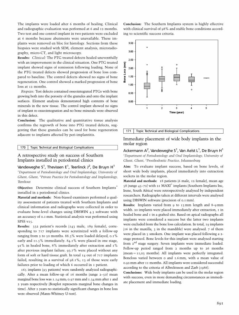
The implants were loaded after 6 months of healing. Clinical
and radiographic evaluation was performed at 6 and 12 months.
Two test and one control implant in two patients were excluded
at 6 months because abutments were unavailable. These im-
plants were removed en bloc for histology. Sections from these
biopsies were studied with SEM, element analysis, microradio-
graphy, micro-CT, and light microscopy.
Results: Clinical: The PTG treated defects healed uneventfully
with an improvement in the clinical situation. One PTG treated
implant showed signs of remission following loading. None of
the PTG treated defects showed progression of bone loss com-
pared to baseline. The control defects showed no signs of bone
regeneration. One control showed a marked progression of bone
loss at 12 months.
Biopsies: Test defects contained osseointegrated PTGs with bone
growing both into the porosity of the granules and onto the implant
surfaces. Element analysis demonstrated high contents of bone
minerals in the new tissue. The control implant showed no signs
of implant re-osseointegration and no bone minerals were observed
in this defect.
Conclusion: The qualitative and quantitative tissue analysis
confirms the regrowth of bone into PTG treated defects, sug-
gesting that these granules can be used for bone regeneration
adjacent to implants affected by peri-implantitis.
170 Topic Technical and Biological Complications
A retrospective study on success of SouthernImplants installed in periodontal clinics
Vandeweghe S1, Thevissen E1, Teerlinck J2, De Bruyn H1
1Department of Periodontology and Oral Implantology, University of
Ghent, Ghent, 2Private Practice for Periodontology and Implantology,
Turnhout
Objective: Determine clinical success of Southern Implantss
installed in 2 periodontal clinics.
Material and methods: Non-biased examiners performed a qual-
ity assessment of patients treated with Southern Implants and
clinical information and radiographs were collected in order to
evaluate bone-level changes using DBSWIN 4.3 software with
an accuracy of 0.1 mm. Statistical analysis was performed using
SPSS v15.
Results: 332 patient’s records (143 male, 189 female), corre-
sponding to 717 implants were scrutinized with a follow-up
ranging from 2 to 50 months. 88.3% were loaded delayed, 0.1%
early and 11.5% immediately. 84.1% were placed in one stage,
91% in healed bone, 8% immediately after extraction and 1%
after previous implant failure. 93.7% were placed without any
form of soft or hard tissue graft. In total 14 out of 717 implants
failed, resulting in a survival of 98.1%; 13 of those were early
failures prior to loading of which 6 occurred in 1 patient.
165 implants (93 patients) were randomly analyzed radiographi-
cally. After a mean follow-up of 16 months (range 2–50) mean
marginal bone loss was 1.14 mm; 0.97 mm and 1.34 mm after 1 and
2 years respectively (Boxplot represents marginal bone changes in
time). After 2 years no statistically significant changes in bone loss
were observed (Mann-Whitney U-test).
Conclusion: The Southern Implants system is highly effective
with clinical survival of 98% and stable bone conditions accord-
ing to scientific success criteria.
171 Topic Technical and Biological Complications
Immediate placement of wide body implants in themolar region
Ackermann A2, Vandeweghe S1, Van Aelst L1, De Bruyn H1
1Department of Periodontology and Oral Implantology, University of
Ghent, Ghent, 2Prosthodontic Practice, Johannesburg
Aim: To evaluate implant success, based on bone levels, of
short wide body implants, placed immediately into extraction
sockets in the molar region.
Material and methods: 18 patients (6 male, 12 female), mean age
58 (range 45–72) with 21 MAXIs
implants (Southern Implants Inc,
Irene, South Africa) were retrospectively analysed by independent
researchers. Radiographs taken at different intervals were analysed
using DBSWIN software (precision of 0.1 mm).
Results: Implants varied from 9 to 13 mm length and 8–9 mm
width. 20 implants were placed immediately after extraction, 1 in
healed bone and 1 in a grafted site. Based on apical radiographs all
implants were considered a success but the latter two implants
were excluded from the bone loss calculation. In total, 19 implants
(16 in the maxilla, 3 in the mandible) were analysed. 7 of them
were placed in 5 smokers. One implant was placed following a 2-
stage protocol. Bone levels for this implant were analysed starting
from 2nd stage surgery. Seven implants were immediate loaded.
Follow-up period ranged from 2 months up to 26 months
(mean¼ 11,03 months). All implants were perfectly integrated.
Boneloss varied between 0 and 1.6 mm, with a mean value of
0.92 mm after 11 months. All implants were considered successful
according to the criteria of Albrektsson and Zarb (1986).
Conclusions: Wide body implants can be used in the molar region
with success, even in more demanding circumstances as immedi-
ate placement and immediate loading.
891
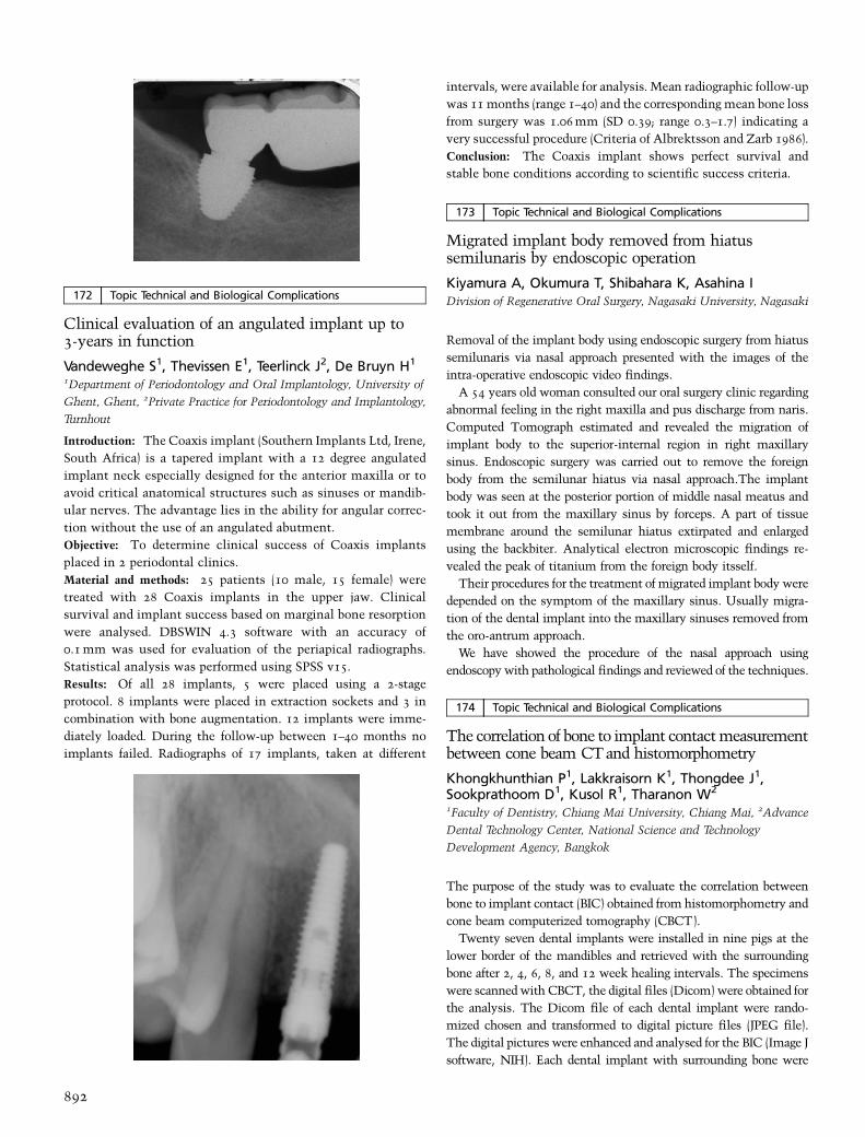
172 Topic Technical and Biological Complications
Clinical evaluation of an angulated implant up to3-years in function
Vandeweghe S1, Thevissen E1, Teerlinck J2, De Bruyn H1
1Department of Periodontology and Oral Implantology, University of
Ghent, Ghent, 2Private Practice for Periodontology and Implantology,
Turnhout
Introduction: The Coaxis implant (Southern Implants Ltd, Irene,
South Africa) is a tapered implant with a 12 degree angulated
implant neck especially designed for the anterior maxilla or to
avoid critical anatomical structures such as sinuses or mandib-
ular nerves. The advantage lies in the ability for angular correc-
tion without the use of an angulated abutment.
Objective: To determine clinical success of Coaxis implants
placed in 2 periodontal clinics.
Material and methods: 25 patients (10 male, 15 female) were
treated with 28 Coaxis implants in the upper jaw. Clinical
survival and implant success based on marginal bone resorption
were analysed. DBSWIN 4.3 software with an accuracy of
0.1 mm was used for evaluation of the periapical radiographs.
Statistical analysis was performed using SPSS v15.
Results: Of all 28 implants, 5 were placed using a 2-stage
protocol. 8 implants were placed in extraction sockets and 3 in
combination with bone augmentation. 12 implants were imme-
diately loaded. During the follow-up between 1–40 months no
implants failed. Radiographs of 17 implants, taken at different
intervals, were available for analysis. Mean radiographic follow-up
was 11 months (range 1–40) and the corresponding mean bone loss
from surgery was 1.06 mm (SD 0.39; range 0.3–1.7) indicating a
very successful procedure (Criteria of Albrektsson and Zarb 1986).
Conclusion: The Coaxis implant shows perfect survival and
stable bone conditions according to scientific success criteria.
173 Topic Technical and Biological Complications
Migrated implant body removed from hiatussemilunaris by endoscopic operation
Kiyamura A, Okumura T, Shibahara K, Asahina IDivision of Regenerative Oral Surgery, Nagasaki University, Nagasaki
Removal of the implant body using endoscopic surgery from hiatus
semilunaris via nasal approach presented with the images of the
intra-operative endoscopic video findings.
A 54 years old woman consulted our oral surgery clinic regarding
abnormal feeling in the right maxilla and pus discharge from naris.
Computed Tomograph estimated and revealed the migration of
implant body to the superior-internal region in right maxillary
sinus. Endoscopic surgery was carried out to remove the foreign
body from the semilunar hiatus via nasal approach.The implant
body was seen at the posterior portion of middle nasal meatus and
took it out from the maxillary sinus by forceps. A part of tissue
membrane around the semilunar hiatus extirpated and enlarged
using the backbiter. Analytical electron microscopic findings re-
vealed the peak of titanium from the foreign body itsself.
Their procedures for the treatment of migrated implant body were
depended on the symptom of the maxillary sinus. Usually migra-
tion of the dental implant into the maxillary sinuses removed from
the oro-antrum approach.
We have showed the procedure of the nasal approach using
endoscopy with pathological findings and reviewed of the techniques.
174 Topic Technical and Biological Complications
The correlation of bone to implant contact measurementbetween cone beam CT and histomorphometry
Khongkhunthian P1, Lakkraisorn K1, Thongdee J1,Sookprathoom D1, Kusol R1, Tharanon W2
1Faculty of Dentistry, Chiang Mai University, Chiang Mai, 2Advance
Dental Technology Center, National Science and Technology
Development Agency, Bangkok
The purpose of the study was to evaluate the correlation between
bone to implant contact (BIC) obtained from histomorphometry and
cone beam computerized tomography (CBCT).
Twenty seven dental implants were installed in nine pigs at the
lower border of the mandibles and retrieved with the surrounding
bone after 2, 4, 6, 8, and 12 week healing intervals. The specimens
were scanned with CBCT, the digital files (Dicom) were obtained for
the analysis. The Dicom file of each dental implant were rando-
mized chosen and transformed to digital picture files (JPEG file).
The digital pictures were enhanced and analysed for the BIC (Image J
software, NIH). Each dental implant with surrounding bone were
892

prepared for the undecalcified ground section technique. The digital
photographs of each section were used for the BIC histomorphome-
try analysis (Image J soft ware, NIH). The BIC obtained from both
method were statistically analysed for the correlation.
Pearson’s correlation showed that BIC obtained from CBCT and
histomorphometry were significantly positive correlated (r¼ 0.716,
p < 0.01).
The measurement of BIC using CBCT could be clinically used.
However, the development of the computer software based on the
proposed method is required.
175 Topic Technical and Biological Complications
Immediate NanoTite implants plus platformswitching technique preserving crestal bone
Delgado Ruiz RA1, Mate Sanchez JE1, Calvo Guirado JL2,Ortiz Ruiz A3
1Associate Professor of Restorative Dentistry, School of Medicine and
Dentistry, Murcia University, Murcia, 2Senior Lecturer of General
Dentistry, School of Medicine and Dentistry, Murcia University,
Murcia, 3Senior Lecturer of Pediatric Dentistry, School of Medicine and
Dentistry, Murcia University, Murcia
Purpose: The aim of this study was to evaluate the crestal
bone loss levels when use a mixed technique with immediate
implants with Nano-TiteTM
surface and platform switching
technique (case), and compare this with the crestal bone loss
levels when use immediate implants with Osseotites
surface and
traditional corresponding abutment (controls) in a 12 month
follow-up.
Material and methods: Patients that have bilateral failed upper
teeth without active infectious condition, that need extraction
was included in the study. A bite-block will be prepared to
ensure proper radiographic alignment for subsequent periapical
radiographs. Two to four case implants or control implants will
be placed adjacent to support a fixed prosthesis in both sides in
the same patient. Cantilevers will not be allowed, a interim-
plant distance of 3 mm, and implant-tooth distance of 1.5 mm.
A immediate post extraction implant placement protocol will be
used, dont load apply, and the prosthesis attached at 3 months.
Periapical radiographs will be obtained immediately after im-
plant placement, at 2, 3, 6 and 12 months and analyzed with a
computer image program.
Results: Fifteen case implants and Fifteen control implants was
inserted in six patient three men and three women. A prelimin-
ary report at 6 months shows a mean bone-loss of � 0.9 mm
on the mesial aspect, and � 1.1 mm on the distal aspect
around the control implant. And a mean bone loss of
� 0.09 mm on the mesial aspect, and � 0.12 mm at the distal
aspect around the case implant. None implant loss at this
evaluation point.
Conclusions: Immediate implantation of Nano TiteTM
Prevail
implants combined with the Platform Switching technique in a two
phase implants protocol, shows a positive effect on the crestal bone
preservation when compared with the immediate implantation of
Osseotites
Certain implants without platform switching technique
in a preliminary 6 month follow-up results.
The Nano-Tite surface applied to the traditional prevail implant
body and the platform switching technique combined will translate
in to more consistent bone preservation outcomes.
176 Topic Technical and Biological Complications
Evaluation of biomechanical weak point onosseointegrated implant with vertical boneresorption: finite element study
Eom TG1, Suh SW1, Kim MD1, Kim SG2
1Osstem Implant Co., Ltd, R&D Center, Busan, 2Department of Oral
& Maxillofacial Surgery, College of Dentistry, Chosun University,
Gwangju
Purpose: This study evaluated the structurally weak points of
fixtures depending on the extent of exposure of implants owing
to the marginal bone resoption around the internal non-sub-
merged type implants.
Material and methods: For this study, single implant fixture
(+4.0 � L10.0 mm, HS II implant, Hiosssen, Philadelphia, USA)
was anchored in the cylindrical alveolar bone (+28 � L30 mm).
It was modeled including 1-piece type Solid Abutment and
simplified crown. With varying bone levels, decreases of 0, � 1,
� 2, and � 3 mm were applied to four bone-implant models.
Then, applying the inclined (301) occlusal load to the center of
the crown parallel with the axis of implants. They are evaluated
on the maximum stress of the fixtures and its position.
Result: There was no difference in stress when bone level
decreased by � 1 mm In contrast, stress increased when bone
level decreased by � 2 mm or more (above 14%). In terms of
stress concentrated position, a decrease in bone level caused be
moved the position on bone level circumferences with the
exposed fixture thread.
Conclusion: It was expected to be weak point on the implant-
abutment connection regions in case of internal non-submerged
type implant. It follows in result, in Bone level decreases,
maximum stress increased and the position moves on bone level
circumferences with the exposed fixture thread. Thus, clinical
caution is required in marginal bone resorption causes increasing
the possibility of implant fracture.
177 Topic Technical and Biological Complications
Influence of early exposure on the crestal bone lossaround implants
Moon IS, Kim TH, Lee DWDepartment of Periodontology, Yong-dong Severance Dental Hospital,
Yonsei University, Seoul
Objectives: Plaque accumulation, bacterial colonization and
epithelial invagination can occur during osseointegration period
when a direct communication between the implant surface and
the oral environment is established, and it can be a harmful
factor resulting in early crestal bone loss.
Methods: The present study consisted of 371 patients who were
treated with dental implants. 865 threaded internal implants
were placed following a 2-stage surgical protocol (443 in maxilla,
893

422 in mandible). Among the study population, there were 22
subjects who had both early exposed and non-exposed implants
(26 early exposed and non-exposed implants in each). Total early
exposure rate was examined, and the crestal bone change of
exposed and non-exposed implants in identical subjects was
compared with Wilcoxon Signed Ranks Test.
Results: 42 implants in 33 patients were exposed to the oral
cavity through the mucosa before uncovering surgery (4.9%), 16
implants in the maxilla presented spontaneous early exposure
(3.6%) and 24 implants in the mandible (5.7%).
The crestal bone change was examined when the final restora-
tions were inserted. The mean crestal bone change of exposed
implants was � 0.43 mm� 0.51, ranged from a loss of 2.15 mm
to 0.00 mm. The mean crestal bone change of non-exposed im-
plants was �0.23 mm� 0.35, ranged from loss of 1.20 mm to
0.00 mm. Wilcoxon Signed Ranks Test revealed that there was
statistically significant difference between the crestal bone loss of
exposed and non-expoesd implants (p¼ 0.006).
Conclusion: The early exposure of the implant seems to facil-
itate periimplant crestal bone loss. Initial healing phase follow-
up may be critical for implant success.
178 Topic Technical and Biological Complications
Safety of preoperative panoramic radiographs foranterior mandibular implants
Vazquez L1, Belser U2, Samson J1, Bernard JP1
1Department of Oral Surgery, Geneva, 2Department of Prosthodontics,
Geneva
Objectives: Panoramic radiographis the standard examination
tool for implant treatment planning. Haemorrhage in the floor of
the mouth following implant placement in the anterior mand-
ible has led to recommend CT examination before surgery in the
interforaminal region.The aim of this study is to evaluate the
incidence of bleeding complications after implant placement in
the anterior region of the mandible in cases when a panoramic
radiograph was the only preoperative imaging technique used.
Methods: The study included 1200 Straumanns
implants (Strau-
mann AG, Basel, Switzerland), inserted in the anterior region of
the mandible of 735 patients (mean age 61� 13 years). The bone
morphology was assessed by clinical examination and palpation
of the bone ridge at the implant site during the preoperative
planning and before local anaesthesia. The bone height was
evaluated from the alveolar crest to the inferior border of the
mandible on a standard panoramic radiograph. No implants
longer than 12 mm were used. Implants were placed following
a one-stage surgical procedure with an incision along the crest of
the alveolar ridge and careful elevation of mucoperiosteal flaps.
112 implants were inserted in the incisor region, 702 implants in
the canine region and 386 in the region of the first premolar.
Results: No haemorrhage or haematoma in the floor of the
mouth following implant placement were reported.
Conclusions: Careful surgical technique and clinical examina-
tion combined with panoramic radiography, can be considered a
safe procedure. CT examination, implying a supplementary
radiation burden for the patient, is not justified.
179 Topic Technical and Biological Complications
Surgical repair of sinus membrane perforations usingPRF (Platelet-rich Fibrin) technique
Bolukbasi N, Ersanli S, Ozdemir TIstanbul University, Faculty of Dentistry, Department of Oral
Implantology, Istanbul
The most common complication during sinus graft surgery is
perforation of sinus membrane. Opening of Schneiderian mem-
brane may be caused by tearing during scoring of the lateral window
or elevating the membrane from the bony walls. Sinus perforation
may cause bacterial penetration into graft material through the torn
membrane or graft material may migrate and obstruct ostium. Both
these conditions increase the risk of infection and implant failure. If
perforation occurs the opening can be sealed with resorbable
membrane or sutured. PRF is a new technique to treat the perfora-
tion of sinus membrane. It is a new regeneration platelet concen-
trate and contains many growth factors. PRF technique is very
simple. The blood sample, taken without anticoagulant, is centri-
fuged for 12 minutes. The resultant product consists of three layers.
Acellular plasma (PPP) is concentrated at the top layer and red
corpuscles are concentrated at the bottom layer. Fibrin clot (PRF) is
obtained in the middle of the two layers. When fibrin clot is packed
in two sterile compresses, fibrin membrane transferable to the
Schneiderian membrane can be obtained. In this presentation PRF
technique is described on cases for the management of sinus
membrane perforation.
180 Topic Technical and Biological Complications
Clinical effects and literature analysis of 980 nm-diode laser in implantology
Shenouda A, Romanos GEastman Dental Center, Division of Periodontology, Rochester, New York
Background: Different wavelengths have been used in order to
excise, ablate or decontaminate peri-implant soft and hard
tissues.
Objectives: The aim of this presentation was to evaluate the
intra-operative efficacy and post-operative clinical outcomes of
the 980 nm-diode laser in peri-implant soft tissue surgery and
treatment of peri-implantitis. An additional aim was to review
the current literature of the laser use in Implantology.
Material and methods: 30 patients were selected for application
of the 980 nm-diode laser (Kavo Gentleray 980, Biberach, Ger-
many). The laser was used in the excision of peri-implant
hyperplasias, in second-stage and in soft tissue pre-prosthetic
surgery (focused beam). It was also used in the decontamination
of implant surfaces before augmentations (defocused, ablative
mode). Low power settings were applied (2–4 W, CW and pulse
mode) in order to achieve sufficient coagulation, bacterial
reduction and tissue excision, according to laser-tissue interac-
tions and safety parameters. 30 papers selected from Pubmed/
Medline search databases using the keywords ‘‘dental implant
and laser’’ from the period of 1990–2007 were evaluated.
894

Results: We were able to show a precise incision quality with
sufficient haemostasis and without scar tissue formation in the
peri-implant mucosa. All of the peri-implantitis patients pre-
sented clinically healthy soft tissues with significant bone fill.
The literature presented limited data in the use of the 980 nm-
diode laser compared to different laser wavelengths.
Conclusions: The 980 nm-diode laser may be a new modality
with advantages in Implantology offering promising results.
More studies are necessary to support our observations and to
gain clinical evidence.
181 Topic Technical and Biological Complications
Determination the primary stability and the changesin early healing for ITI and Replace Select taperedimplants
Rokn A, Rasooli Ghahroodi A, Miremadi A, Mesgarzade ATehran University of Medical Sciences, Tehran
Purpose: To determine the primary stability and the changes in
stability as a reflection of early healing around single staged
implants in human utilizing resonance frequency analysis (RFA).
RFA makes use a smart peg, attached to an implant which is
excited with a probe measurement of osstell mentor device over
a range of sound frequencies with subsequent response analysis.
Material and methods: 125 patients had 1 to 8 implants placed in
the canine premolar and molar regions of the maxilla and
mandible. Bone type was classified into 1 to 4 groups according
to the lekholm & zarb index (1985). RFA was used for direct
measurement of implant stability on the day of implant place-
ment and consecutively at 14, 30, and 60 days after placement.
Results: 306 implants (153 ITI SLA and 153 Nobelbiocare Re-
place select tapered) placed. No early failure occurred during
healing phase. All 306 implants distributed as follows: 3.36% ITI
and 8.90% Replace in type 1 bone, 72.84%ITI and 48.63%
Replace in type 2 bone, 17.88% ITI and 38.38% Replace in
type 3 bone and 5.98% ITI and 4.10% Replace in type 4 bone.
In Replace implants the lowest mean stability measurement was
at 30 days for all bone types. In ITI SLA implants the lowest
mean stability was at 60 days for type 1 and at 30 days for type 2
and at the base line for type 3&4. A boneferroni adjust student t
test comparison of bone groups.
At each time point revealed highly significant differences be-
tween implant stability in type 1 and 4 bone (p < 0.001) and a
moderately significant difference between type 2 and 3, type 3 and
4 at all times in ITI SLA implants (p¼0.07). In Replace implants
stability did not change significantly in all bone types (P > 0.05).
Conclusion: The geometry of implant and bone type are the
main factors for implant stability in first 2 months and Replace
implant is better for Immediate loading.
182 Topic Technical and Biological Complications
Clinical evaluation of fixed zirconium-basedrestorations on implants
Linkevicius T1, Vladimirovas E2, Grybauskas S2, Puisys A2
1Vilnius University, Vilnius, 2Vilnius Implantology Center, Vilnius
Lack of elasticity and proprioreception makes implant-borne re-
storations more prone to technical complications. Material research
did not prove the presence of chemical bond between zirconium and
overlaying ceramic. Nonetheless, the use of zirconium-based re-
storations on implants is increasing in clinical practice. However,
clinical evidence of success rates of zirconium-based crowns on
implants is very sparse in the literature.
Objective: To evaluate the short-term clinical performance of
zirconium-based restorations on implants.
Material and methods: Thirty-four patients who received 76
titanium implants, subsequently restored with 63 zirconium-
based prostheses were recalled for retrospective evaluation.
Fifty-one single crowns and 16 fixed partial dentures with
regular occlusion scheme were delivered. Twenty-four restora-
tions were inserted in anterior area, while lateral segments
received 39 prostheses. All restorations were examined for
presence or absence of contact during eccentric mandibular
movements – 3 protrusion, 3 lateral guidance, 57 were guidance
free. Three different zirconium systems were used for restora-
tion of implants – VITA In-Ceram (43 cases), Ceramill Zi (13
cases) and Zirconzahn (7 cases). Mean follow-up period was 13.4
months, ranging from 7 to 29 months.
Results: All implants at a time of evaluation were successful.
No fracture of framework was recorded. Fractures of ceramic
occurred in 3 patients (8.82%) and in 3 restorations (4.83%). In
anterior segment 1 fracture occurred (4.16%) and in lateral – 2
(7.69%). There was 1 failure in group of single crowns (1.96%)
and 2 in fixed partial denture group (16.6%). In-Ceram encoun-
tered 2 fractures (4.65%), Zirconzahn – 1 fracture (14.2%). Two
ceramic fractures occurred in lateral guidance (66%), 1 failure in
guidance free group (1.75%).
Conclusions: Most failures occurred in fixed partial dentures
with contact on lateral movement Zirconium-based restorations
on implants do need more long-term research.
183 Topic Technical and Biological Complications
Clinical and radiographical evaluation of narrowdiameter implants
Gultekin BA, Sirali S, Bayraktar M, Ozgen M, Akilli E,Abdel-Hak J, Yalcin SIstanbul University, Faculty of Dentistry Department Of Oral
Implantology, Istanbul
The purpose of this study was to evaluate the clinical performance
of narrow diameter Nobel Biocare (Replace) implants in limited
available bone.
Totally 84 dental implants (3.5 mm narrow diameter Nobel
Biocare/Replace) were inserted in 37 patients. All implants were
895

loaded after two or four months healing period in mandible and
maxilla respectively. 22 single tooth and 62 partial fixed prosthesis
were delivered. Panoramic and periapical radiographs were taken
right after the operation, prosthesis delivery and 12, 24, 36 months.
Peri-implant index scores (plaque and gingival indexes) were also
recorded at 12, 24, 36 months after the operation.
The cumulative survival rate was % 96.4. All lost implants
(three/10 mm length) were placed in posterior maxilla. None of
splinted narrow diameter implant supported restorations were lost.
Two single tooth restoration abutment screws were loosened that
were placed most distally in posterior maxilla 1 year after delivery.
Total mean bone loss was 0.9� 0.004 mm for all implants.
Implants placed in maxilla were slightly showed more bone loss
than mandibular implants but not significantly. There were no
significant differences in peri-implant health (Modified Plaque
Index, Gingival Index, Bleeding Index) between maxillar and
mandibular implants.
Fixed prosthetic treatment choice supported with narrow dia-
meter implants is a reliable and predictable method. However in
low quality bone, splinting implants with fixed prosthesis may
increase the success and also inhibit screw loosening of abutments.
184 Topic Technical and Biological Complications
Clinical performance of tooth-implant fixedprosthesis
Gultekin BA, Ozgen M, Bayraktar M, Abdel-Hak J,Siraliyev S, Yalcin S, Ozdemir TIstanbul University, Faculty of Dentistry, Department of Oral
Implantology, Istanbul
The aim of this study was to investigate the success rate and
marginal bone loss around implant and tooth-supported prostheses.
Thirty five partially edentulous patients consecutively who
received 15 three-occlusal unit with one pontic between implant
and natural tooth and 20 two occlusal unit without pontic with
terminal implant and tooth support were subjected to quantifica-
tion. The radiographs of the implants were digitized, and the areas
of bone atrophy mesial and distal to the implants were determined.
Radiographs were taken right after implant surgery, 6–18–30
months after prosthesis delivery. The distance between the tooth
and implant was ranged 2–5 mm in prosthesis without pontic and
ranged 7–12 mm in prosthesis with pontic between implant and
natural tooth.
After 18 months 0.84 mm at mesial side 0.76 mm at distal side;
after 30 months 1.25 mm at mesial side 1.12 mm at distal side
mean bone loss observed around the implant splinted with natural
tooth without pontic. After 18 months 1.16 mm at mesial side
0.86 mm at distal side; after 30 months 1.45 mm at mesial side
1.22 mm at distal side mean bone loss observed around the implant
splinted with natural tooth with a pontic between a natural tooth
and implant.
This study showed that the distance between implant and natural
tooth effect the bone loss around the implant neck.especially bone
loss at the mesial side of implants seemed more effected compared
to distal side of the implant.
185 Topic Technical and Biological Complications
Clinical and radiographical evaluation of Biohorizonimplants
Karabuda BA, Gultekin E, Akilli J, Abdel-Hak C, Ersanli S,Yalcin S, Ozdemir TIstanbul University, Faculty of Dentistry, Department of Oral
Implantology, Istanbul
Background: Dental implant rehabilitation is one of the most
reliable and also predictable treatment method in edentulous
cases. The purpose of this study was to evaluate the clinical and
radiographical success of Biohorizon dental implants.
Material and methods: Totally 112 dental implants were in-
serted in 34 patients. Grafted sites, medically compromised
patients and smokers were excluded from the study. 6 implants
in lower, 8 implants in upper jaw were immediately loaded. 98
implans were loaded in conventional manner (3 and 6 months
healing for lower and upper jaw respectively). Panoramic radio-
graphs were taken right after the operation, at prosthesis deliv-
ery and 6, 18, 30 months after prosthesis delivery. Peri-implant
index scores (Modified Plaque Index, Gingival Index) were also
recorded at 6, 18, 30 months after prosthesis delivery.
Results: The cumulative survival rate was % 94.7. Six implants
were lost. Five of lost implants were immediately loaded. Total
mean bone loss was 0.06� 0.8 mm for all implants. No sig-
nificant differences were found between immediate and con-
ventional loading respective with peri-implant bone resorption.
Implants placed in maxilla were slightly showed more bone loss
than mandibular implants. Satisfactory peri-implant index
scores were gained (less than score 1) in this study.
Conclusion: Conventional treatment protocol survival results
are satisfactory and also predictable, however more attention is
needed for immediate loading. More clinical studies are required
to confirm the long term performance.
186 Topic Technical and Biological Complications
Vascular endothelial growth factor in gingivalcrevicular fluid as a prognostic factor in implanttreatment
Lomzynski L1, Mierzwinska-Nastalska E1, Gladkowski J1,Jaworska-Zaremba M1, Demkow U2
1Department of Prosthodontics, Medical University of Warsaw,
Warsaw, 2Department of Laboratory Diagnostics and Immunology,
National Institute of T.L.D., Warsaw
Angiogenesis is a process regulated by the cytokines and growth
factors. It plays a crucial role in physiology and pathology. One of
the most important angiogenic cytokine in inflammatory and
immune responses of the tissues surrounding dental implants is
the vascular endothelial growth factor (VEGF).
Objective: The aim of the present study is to investigate the
levels of VEGF in GCF of healthy and inflamed sites around
dental implants.
Material: Group of 32 edentulous patients (15M, 17F; mean age
64.5 (45–84)) after implant treatment with the use of OVD.
896
59 samples of GCF were collected with the use of Periopapers
(Ora Flow Inc., NY, USA), 39 from sites with clinical symptoms
of inflammation and 20 from clinically healthy sites (control
group).
Methods: Periodontal status was assessed with the use of
Florida Probe, marginal bone loss was evaluated on dental
radiographs. Immunoabsorbent assay – ELISA (R&D
Systems, Minneapolis, USA) evaluated the concentrations of
VEGF.
Results: Statistically significant (p < 0.05) higher levels of VEGF
were detected in the GCF of patients with clinical symptoms of
periimplantitis (15.17� 0.38 pg/ml) compared to the clinically
healthy sites (11.40� 0.20 pg/ml), and were correlated to the
pocket depth.
Conclusions: VEGF is a probable factor of importance in the
exacerbation of periimplantitis, possibly by promoting angio-
genic processes in the tissues surrounding implants thus
enhancing the distribution of inflammatory mediators and
proinflammatory cells to the inflamed implant site.
187 Topic Technical and Biological Complications
Bone qualitiy using the fractal dimension and theimplant stability quotient
Kim MH1, Ku Y1, Rhyu IC1, Heo MS2, Huh KH2
1Department of Periodontology and Dental Research Institute, School
of Dentistry, Seoul National University, Seoul, 2Department of Oral
and Maxillofacial Radiology and Dental Research Institute, School of
Dentistry, Seoul National University, Seoul
Bone qualitiy using the fractal dimension and the implant stability
quotient.
The purpose of this study is to investigate whether the fractal
dimension of the panoramic radiograph is related to the primary
stability of the implant as represented by RFA. This study included
22 patients who underwent dental implant installation at the
Department of Periodontology of Seoul National University Dental
Hospital. Morphometric analysis and fractal analysis of the bone
trabecular pattern were performed using panoramic radiographs, and
the implant stability quotient (ISQ) values were measured through
RFA after the implant installation. The radiographs of 52 implant
sites were analyzed, and the ISQ values were compared with the
results from the morphometric analysis and fractal dimension.
The Pearson correlation showed a linear correlation between the
ISQ values of RFA and the parameters of morphometric analysis
but there was no statistical significance. The fractal dimension had
a linear correlation with a statistical significance. The correlation
was more prominent in the mandible. In conclusion, we suggest
that the fractal dimension acquired from the panoramic radiograph
may be a useful method to predict the initial stability of dental
implants.
188 Topic Technical and Biological Complications
‘‘GOOD’’ biphosfonates in oral implantrehabilitation
Borgioli A1, Brancato L1, Viviani C1, Mascherini C1, SartoriG2, Longoni G2, Duvina M1, Duvina P1, Tonelli 1
1University of Florence, Oral Surgery Department, Postgraduate
School of Surgery, Florence, 2University of Milano-Bicocca,
Prosthodontic Department, Milan
Intoduction: An increasing number of patients affected by
oncologic and osteoporotic pathologies in treatment with Bipho-
sphonates develops osteonecrotic lesions localized in the max-
illary bone. In these patients the oral surgery should be avoided
because of the poor implant prognosis during healing period.
According to several Authors this kind of failure is related to all
biphosphonate drugs. However, first generation biphosphonates
(‘‘good biphosphonates’’ e.g. Clodronate) are able to improve
implant osteointegration.
Methods: The Authors analyzed a series of 3 patients. Implant
dental surgery was performed in one patient with zoledronate
treatment. During the healing period the premature loss of the
screws was noted and the implant situs showed extended bone
necrosis.
In the other two cases, implant rehabilitation was preceded by
intravenous clodronate administration for severe osteoporosis. A
condition of mandibular bone loss indicated removable dental
implants rehabilitation (overdenture with interforaminal located
implants). We used Ankiloss
plus Dentsply implants.
Results: In the patients with clodronate treatment was noted
good bone vascularization and excellent primary stability. The
load of the systems was carried out three months from their
insertion. The healing period was uneventful and 12 months
later, the radiological examination showed an optimal implant
osteointegration.
Conclusion: The chemical structure of BPs ‘‘good biphospho-
nate’’ and strict selecting patient criteria can enhance the
results.
189 Topic Technical and Biological Complications
Connective tissue graft for anterior maxilla defect
Finco N1, Fritscher G2, Romanelli V1, Finco C1, Abreu A1,Beltrao R2
1SOBRACID, Porto Alegre, 2PUCRS, Porto Alegre
The reconstructive therapies with dental implants intend to recup-
erate function, good hygiene and aesthetic. Many factors are
involved in this treatment, such as bad positioned implants,
periimplant infections or even bone defects. Furthermore, some of
these implants are on anterior region of maxilla, turning into poor
prognosis. The solution for these cases may be very complex, and
then simple and safe actions must be tried. The aim of this work is
to report a variation technique of connective tissue graft. First of all
a connective graft is removed from posterior palatal area next to the
first molar, without epithelium. Then two oblique incisions adja-
cent the defect area are made and a divided flap detachment is also
897
done. A local antibiotic therapy over the implant surface is done for
five minutes with tetracycline. Over the periimplant defect area the
connective tissue is placed. In the end, the flap is positioned over
the graft and sutured. After three to four months a new surgery is
taken to expose the platform and an impression copy is performed to
build the provisory acrylic crown immediately. This technique has
been showing good results, leading to better aesthetic and also
simplifying hygiene practices.
190 Topic Technical and Biological Complications
Complications after implantation may have allergicbackground
Popinski J1, Buczylko K2
1Specjalistyczna Praktyka Stomatologiczna, Lodz, 2Uniwersytet
Medyczny, Lodz
Aim: A study of correlation between allergy to metal ions (A)
and implant integration (I) presented on the basis of to 4 options
Aþ Iþ (successful implantation despite of allergy), Aþ I� (un-
successful implantation probably caused by allergy),
A� Iþ (successful implantation without allergy at all), A� I�(unsuccessful implantation without allergy).
Methods: Implanto-prosthetic procedures according totwo faze
procedure, CT and panoramic radiological examination, allergic
patch tests with special set of components used in implanto –
prosthetic treatment.
Material: 60 subjects with implants and different prosthetic
restorations, 41 to 65 year olds, 37 women and 23 men.
Results: In case of 1/4 of patients allergic to metal ions, a proper
integration of titanium implants was observed. In case of 3/60
subjects, allergic to Ni, Cr, Co, Au metal ions, improper
integration of implants occurred. There was 7 times more
complicationin case of patients being allergic. A proper implant
integration without allergy concerned 3/4 subjects. An improper
implant integration among non-allergic patients occurred once.
Conclusions: Among own 60 cases, 3/4 complications after
implantation had some allergic background.
Conclusion: The resultsof the research suggest allergic tests to
be conducted as a routine before implant – prosthetic treatment.
191 Topic Technical and Biological Complications
Comparison of Panoramic radiography andComputed Tomography (CT) for diagnosis of inferioralveolar nerve injury after implant surgery
Kim ST1, Eun SA2
1TMJ & Orofacial Pain Clinic, Yonsei University, Dental College, Seoul,2Department of Oral and Maxillofacial Radiology, Yonsei University,
Dental College, Seoul
The aim of this study was to compare the relationship of implants
and mandibular canal in panoramic radiography and Computed
Tomography (CT) in inferior alveolar nerve injury patients after
implant surgery.
56 patients, who complained of inferior alveolar nerve injury after
implant surgery, were enrolled in this study. All the patients had
been taken panoramic radiography and CT (DentaScans
) and
examined the symptom area and previous dental history. In pa-
noramic radiography, we classified the relationship of implants
with the mandibular canal to three types (distant, contact, perfora-
tion type). And also four types (distant, contact with continuous
cortical layer, contact with discontinuous cortical layer, perforation
type) were classified in Computed Tomography.
In panoramic radiography, there were 18 cases (32%) of distant
type, 28 cases (50%) of contact type and 7 cases (12%) of perfora-
tion type. In CT, there were 9 cases (16%) of distant type, 9 cases
(16%) of contact with continuous cortical layer, 19 cases (33%) of
contact with discontinuous cortical layer and 13 cases (23%) of
perforation type. The most common type of injury was contact
type. (50% in panorama and 49% in CT). 9 cases (16%) of distant
type in panorama were investigated as contact type in CT and 6
cases (11%) of contact type in panorama were evaluted as perfora-
tion type in CT.
In this limited study, there were significant differeces between
the results of panorama and CT in inferior alveolar nerve injury
patients. We concluded that CT may be useful to diagnose the
inferior alveolar nerve injury after implant surgery, when panorama
does not show any evidence of nerve injury symptoms.
192 Topic Technical and Biological Complications
Mean implant axis and abutment inclination
Risciotti E1, Squadrito N2, Ortenzi G3
1University of Genova, Genova, 2University of Genova, Genova,3University of Pavia, Pavia
Purpose: The aim of the present study was to determine the
effectiveness of the Mean Implant Axis (MIA) methodology in
controlling the angular preparation of multiple implant abut-
ments compared to conventional abutment preparation.
Material and methods: Out from an actual clinical case 6 stone
models were made and 5 were sent to 5 different dental
laboratories not familiar with the proposed procedure. We
requested them to define and mark on the model their milling
arbitrary implant axis (AIA) and to prepare the implant abut-
ments with a 61milling taper. On one model we defined the
implant milling axis by recording the implant angulations and
by calculating the mean implant axis (MIA) and by preparing
each abutments within the possible angular ranges offered by
the 61milling taper.
Among each set of prepared abutments we compared the milling
angle used for the most angulated one.
Results: On the sagital plane the AIA preparation angle of the of
the most angulated abutment average 261.120 with a range from
231.46 and 281.37 with a SD of � 21.250. The MIA most
angulated abutment was 131.500 (p < 0.001). No statistically
significant differences were found among preparation angles
on the frontal plane.
Conclusion: The difference between the AIA mean preparation
angle and the MIA one is more then 5 times the SD. and it is
898

strongly correlated to the effectiveness of the proposed method
in controlling the possible angular compensations when multi-
ple implants need to be prepared with a common milling axis.
193 Topic Technical and Biological Complications
Early bone healing around endosseous implants ofdifferent bulk designs
Suzuki M1, Coelho PG2
1Tufts University School of Dental Medicine, Boston, 2New York
University, New York
Although great advancements and information regarding novel
surface treatments are currently available, little attention and
research has been devoted to the implant’s bulk design and its
effect in endosseous implants early healing. The purpose of this
investigation was to analyze the bone healing around three different
designs on the bone healing on a dog model.
Methods: Plateau (P) shaped, screw (S) shaped, and a mixed
plateau and screw (PS) shaped implants were placed in the tibia
of six adult beagle dogs and left in vivo for 2 and 4 weeks.
Following euthanization, the implants separated in individual
blocks. The samples were non- processed to nondecalcified
sections, reduced to �30mm thickness plates, and bone-to-
implant (BIC) was determined through computer software.
Results: Transmitted and polarized light microscopy showed a
�400 mm region of newly deposited bone along the perimeter of
implant regions which remained in close proximity to cortical
bone after surgery. This appositional bone healing was observed
along the whole perimeter of S implants and plateau tips for
P and SP implants. In contrast, substantial amounts of woven
bone formation was observed in regions where surgery and
implant design resulted in larger void spaces for the P and SP
implants. Blood vessels were observed parallel to the space
between fins of P and SP implants. No significant differences
in BIC were observed for the different groups.
Conclusion: Different implant designs and surgical protocols
resulted in different osseointegration pathways with comparable
rates.
194 Topic Technical and Biological Complications
Osteonecrosis related to bisphosphonate treatmentand dental implants
Abi Najm S1, Bernard JP2, Lesclous P3, Zagury T2, CarrelJP2, Lombardi T2, Samson J2
1Ardentis, Clinique Dentaire, Swiss Dental Clinics Group, Lausanne,2School of Medecine, Geneva, 3Faculty of Dentistry, University of
Paris Descartes, Paris
Bisphosphonates are therapeutic agents used to inhibit bone resorp-
tion and therefore bone renewal. They are established as effective
treatment in clinical disorders such as multiple myeloma, bone
metastasis, osteoporosis and Paget’s disease of bone. Osteonecrosis
of the jaws has been recently described as an adverse side effect of
bisphosphonate treatment.
Since December 2003, 35 cases of osteonecrosis related to
bisphosphonate treatment were treated in the School of Dental
Medicine, Geneva, Switzerland. Two cases were related to implant
placement. In our first case the patient was taking alendronate for
44 months at a dosage of 70 mg/week for her osteoporosis. She lost
her two implants after osteointegration for two years and despite
implant removal no healing occurred and a wide infected area of
bone necrosis appeared after implant removal. Complete healing
was achieved within 18 months for the soft tissue and 2 years for
the bone. Our second case was treated by pamidronate and later
zoledronic acid for bone metastasis from a breast cancer, no
osteointegration was obtained and exposed bone surrounding the
implant was seen. Complete healing is not achieved within 3 years.
At present implant placement in patients undergoing intravenous
bisphosphonates therapy is contraindicated.
Osteonecrosis and implant failure in patients treated by oral
bisphosphonates seems to be rare, but osteointegration can be lost
and the risks of osteonecrosis increase especially after long term
treatment with oral bisphosphonate ( > 3 years). Informed consent
should be provided related to possible future implant failure and
possible osteonecrosis of the jaws.
195 Topic Technical and Biological Complications
Ultrasound bone surgery vs. traditional rotativeinstruments for third molar surgery: a randomizedclinical trial
Barone A, Marconcini S, Covani UDepartment of Oral Pathology and Medicine, University of Genova,
Genova
Background: Osteotomy is one of the major parts of third molar
surgery. Edema, ecchymosis and trismus most commonly ap-
pear as a result of this surgical procedure. A double-blind,
randomized study was performed to determine the efficacy of
ultrasound bone surgery in decreasing edema, trismus and pain
after third mandibular molar extraction.
Methods: Twenty patients undergoing third molar extraction
were included in the study. Each patient had 2 symmetrical
impacted third molar which were randomly allocated in 2
groups: control group (n¼10), the mucoperiosteal flap elevation
and osteotomies were performed with traditional instruments;
test group (n¼10), the mucoperiosteal flap elevation and osteo-
tomies were performed with the ultrasound bone surgery in-
strument. Patients were evaluated during surgery for bleeding.
All the patients were scored at 2, 5, 7, and 10 days after surgery
for the following parameters: edema was evaluated according to
a visual scoring system that describes the site, extension and
degree of swelling; maximum interincisal distance was mea-
sured using calliper to evaluate the trismus; pain was objec-
tively measured by counting the number of analgesic tablets
required; the patients’ perception of the severity of symptoms
was assessed with a follow-up questionnaire (Visual scale).
Results: The bleeding during surgery showed lower level in the
test group when compared with the control group. Edema and
899

trismus did not show significant difference between the control
and test group at 2 days after surgery. Moreover, the scores for
edema, trismus and pain showed higher values in the control
group when compared to the test group at 5, 7 and 10 days after
surgery. The evaluation of pain showed higher scores in the
control group when compared with test group.
Conclusion: Ultrasound bone surgery used for flap elevation and
osteotomies during third molar surgery showed better results in
the prevention of edema, trismus and pain in the postoperative
healing period.
196 Topic Technical and Biological Complications
Mandibular fractures following implant placement:rarity or relevant risk?
Nkenke E, Schlegel A, Neukam FDepartment of Oral & Maxillofacial Surgery, University of Erlangen-
Nuremberg, Erlangen
The aim of a retrospective study was to assess the number of
factures of atrophic mandibles following implant placement, to
describe the treatment concept of these fractures and to assess the
different complications and the outcome of this treatment.
2239 patients received implants between 1996 and 2006. By
searching the electronic patient database persons were identified,
who were treated for mandibular fractures after implant placement.
The aetiology of the fracture was analysed. Kind of fracture treat-
ment, sensitivity impairment and other complications were com-
piled and the kind of the final prosthodontic restoration was
documented.
Four patients with mandibular fractures following implant place-
ment could be identified (2 female, 2 male, age range 65–77 years).
Two mandibular fractures occurred after removal of implants. In the
remaining two patients mandibular fractures occurred at the
implant-bone interface after long lasting peri-implant disease.
The fractures were treated by interpositional bone grafts from the
iliac crest and osteosynthesis plates. In one patient two additional
fractures of the mandible occurred during the treatment period. In
three patients sensitivity disorders in the innervation area of the
inferior alveolar nerve were still present after 12 postoperative
months. In three patients an implant borne removable denture
could be installed, while one patient remained without denture at
the end of the treatment period.
This retrospective study reveals that fractures of the edentulous
atrophic mandible following implant placement are rare events.
However, if these fractures occur they are difficult to treat and the
re-occurrence of a fracture cannot be excluded. Moreover, there is
no guarantee that patients who suffered from a mandibular fracture
as a consequence of implant placement can ever be treated with an
implant-borne prosthesis or even a conventional denture again.
197 Topic Technical and Biological Complications
Conventional vs. implant-supported prosthetictreatment in lateral area – case report
Dulcic N1, Pelivan I1, Dulcic D2
1University of Zagreb, School of Dental Medicine, Department of
Prosthodontics, Zagreb, 2Private Dental Practice, Schrobenhausen
Conventional prosthetic treatment is often the dentist’s first choice
in reconstruction of the lateral area of the dental arch. However,
sometimes an implant-supported prosthetic solution is narrowly
indicated, as it is shown in the following case report.
Clinical examination of a 43-year-old patient found missing tooth
16 and 14 with very short clinical tooth crowns and habitual
occlusion without interferences. An implant-supported prosthetic
treatment was recommended for the area 16 and 14, which the
patient refused and visited another dentist. He made a bridge with
tooth abutments at 17 and 15, pontic at 16 and mesial cantilever at
14. This bridge loosed 4 times in a year, the patient was not
satisfied with it, and she returned to the first dentist and agreed to
the implant-supported prosthetic treatment.
XIVE (Dentsply) implants were placed. One implant (3.8/13) was
placed in the area 14 with buccal soft tissue augmentation and
second (4.5/11) in the area 16 with internal maxillary sinus floor
elevation. During healing time of 3 months the patient wore a
temporary acrylic bridge on the abutment teeth of the old bridge and
satisfactory soft tissue emergence profile was achieved with pontic
and cantilever. Impression was taken 14 days after the implant was
opened and a healing abutment was placed. Standard abutments
were used as implant build up, and 4 metal-ceramic crowns with
functionally shaped occlusal surfaces were fabricated. Regular
check-ups of the patient every 3 months and after 3 years showed
excellent aesthetic and functional results.
198 Topic Technical and Biological Complications
Oral surgery and compromised coagulation –prospective evaluation of the graz guidelines
Acham S, Klampfl A, Jakse NDepartment for Oral Surgery and Radiology, Dental School Graz,
Medical University Graz, Graz
Introduction: Oral surgery including implantation and bone
grafting is, dependent on extension of intervention, often even
possible in haemostaseological deficient patients. The Graz
Guidelines of compromised Coagulation (GGcC) provide a
standardized concept for the individual management of different
surgical extensions. To evaluate the intra- and postoperative
haemorrhagic risk when GGcC is considered a prospective non-
randomised clinical trial was performed.
Methods: In a time period from 03/2005 to 11/2006 a total of 491
surgical interventions in haemostaseologically compromised
patients was performed. Three different samples of patients
were defined:
1) maintenance of anticoagulant resp. antiplatelet drugs
2) discontinued anticoagulant resp. antiplatelet therapy without
substitution
900
3) discontinued oral anticoagulation with substitution by LMH
(low molecular heparine).
Frequency and intensity (mild/moderate/severe) of intra- or post-
operative bleeding and other bleeding-associated complications
were investigated.
Results: Oral Anticoagulants
Regarding intra- or postoperative haemorrhagic complications
there was no significant difference between the samples of main-
tenance and discontinuation of the anticoagulant medication with-
out replacement. Merely the LMH-substituted collective exposed
higher quantitative and qualitative bleeding-data. A clinically
relevant bleeding-course did not occur in a single case.
Antiplatelet Drugs
We observed a moderate increase of the intra-operative intensity
of bleeding but no higher risk of postoperative bleeding complica-
tions. The frequency and intensity of postoperative bleeding epi-
sodes did not differ within the collectives. A clinical relevance of
bleeding was not observed.
Conclusion: Oral-surgical including implantological procedures
in haemostaseologic deficient patients can be performed safely,
assuming the consequent adherence of the Graz Guidelines of
compromised Coagulation (GGcC). In common of oral surgery
cases there is mostly no indication for discontinuation of the
anticoagulant resp. antiplatelet medication.
199 Topic Technical and Biological Complications
Assessing blood supply to the mental region usingCAT scans
Koa A, Garcia G, Hanawa Y, Cho SC, Froum S, Elian N,Tarnow DNew York University, New York
During implant placement, caution must be exercised when releas-
ing a flap as well as when placing an implant in the mandibular
anterior area, because the submental and sublingual arteries, may
enter accessory foramina through the lingual plate and be present in
this area (1). The purpose of this study is to assess the proximity of
the mandibular lingual vascular canal to implants placed in the
interforaminal area between the mandibular right and left central
regions.
Material and methods: Consecutively stored 100 computerized
axial tomography(CAT)-scans of the mandible were examined.
Results: The lingual vascular canal was found in 96% of the
images. The number of canals ranged from 1 to 2, and they were
positioned as follows:
1-ingual canal found at the midline (48%)
2-ingual canal found in the area of tooth 24 (22%)
3-ingual canal found in the area of tooth 25 (26%)
4-canals were vertically aligned on the midline (6%)
Conclusions: The median lingual vascular canal was found to be
present in 96% of the edentulous and partially edentulous group
and in 94% of dentulous patients.
200 Topic Technical and Biological Complications
Allergy to titanium dental implants – clinicalprotocol of diagnosis
Witkowski R1, Kwapinska A2, Obtulowicz K2
1Private Implant Clinic Nobel Biocare, Opole, 2Department of Clinical
and Environmental Allergology CMUJ, Krakow
Purpose: Metals are among the main reasons for contact aller-
gies occurrences.
Most allergenic metals are: nickel, chromium, cobalt; rarely
molybdenum, aluminum, palladium, copper and gold. Titanium
has some physical – chemical properties; it is biocompatible,
bacteriostatic, easily enters the process of osteointegration and has
no influence on enzymes responsible of toxic reactions. All these
means that it very seldom causes hypersensitivity reactions.
There are, though, recorded cases in scientific works which give
record of allergic contact dermatitis, disorders in wound healing
with granulation and sterile discharge after inserting a pacemaker or
orthopedic implant with a titanium component.
Material and methods: 7 patients suspected of hypersensitivity
to titanium were examined in Department of Allergology in
Jagiellonian University Medical College, Krakow, in 2004.
4 of them, who underwent an insertion of titanium dental
implants, were referred to Department of Allergology because of
osteointegration disorders. Patch test has been a method of choice
in diagnostic of contact allergies to metals for many years.
Results: A performed diagnosis in a form of a patch test did not
show hypersensitivity to any contact allergen including titanium.
1 patient, with a medical history of contact allergy to such metals
as: nickel, chromium and cobalt, was diagnosed for allergy to titanium
before a planned procedure of titanium tooth implant insertion.
Conclusions: During the diagnostic process, a doubtful result in
a patch test was obtained for filings of pure titanium. Allergy to
titanium seems to be very rare. The mechanism of developing
allergies to metal implants has not been fully examined. We
lack fully standardized tests in allergy diagnostics for titanium.
201 Topic Technical and Biological Complications
Is the implantological treatment using Nobel Activepossible in patients with renal osteodysthropy?
Kukula K1, Frank SZ1, Chmura A2, Wojtowicz A1
1Department of Oral Surgery, Medical University of Warsaw, Warsaw,2Department of General and Transplant Surgery, Medical University of
Warsaw, Warsaw
Introduction: Patients suffering from chronic renal failure are
treated using haemodialysis. This leads to endocrinal and
secretion dysfunction, reconstruction in osseous tissue, archi-
tectonics and quantities changes. Decreased amount of osseous
tissue in jaws, besides indication to extractions, is the cause of
premature teeth loss. Progress of implantology and replacement
therapy of renal diseases allow to consider the prosthetic treat-
ment based on implants in stabilized systemic diseases.
Aim: The aim of work was to estimate capabilities of implan-
tological treatment of patients with renal osteodysthropy.
901
Material and methods: Supervisory group determined 50 gener-
ally healthy patients (average age 44 years). Investigated group
consisted of 48 patients treated using haemodialysis (average age
45 years). X-rays were made using Planmeca 2002 PM.
Results: The radiodensynometric and radiometric analysis re-
viled decline of bone tissue in maxilla and mandible. 4 patients
were excluded because of bad condition of bone tissue,
3 patients because of presence of active viral infection of HPV.
Conclusions: In investigated group decline of maxilla and
mandible was observed. Although, this is not a contradiction
for implantological treatment.
Decrease of optical bone density in the investigation group seems
not to be objection for implantological treatment, because of
possibility of using implants with osseous condensation surface
around the implant.
Referring to the duration of haemodialysis, the percentage of
patients, which are not qualified to implantological treatment, is
higher. The length of implants are also different.
1-Department of Oral Surgery Medical University of Warsaw.
2-Department of General and Transplant Surgery Medical Uni-
versity of Warsaw.
202 Topic Technical and Biological Complications
The effect of Chlorohexidine on microgap invasion ofperiodontal pathogens
Ju JE1, Lee SH2, Yoon HJ1, Kim SG1, Park JU1
1Catholic Medical Center, Seoul, 2Seoul National University, Seoul
The abutment-implant interface at the alveolar bone crest is
related to the peri-implantitis and considered to be the potential
contributing-factor and source of pathogen that induces the loss of
alveolar bone regarding to the implant. Although the study on the
invasion and colonization of the bacteria into the abutment-implant
interface has frequently been carried out, the consideration on
the clinical management or improvement plan should further be
pursued.
In this study, the nonmotile Porphylomonas gingivalis and
motile Treponema denticola that are thought to be the major
bacteria responsible for the peri-implantitis and belong to the red
complex were used. In the first group, the microgap existing
between the GS II (Internal Hexatype, Korea) implant fixture and
healing abutment was not treated with the irrigants, while in the
second group, the microgap was treated with normal saline and
preserved in wetness, in the third group it was treated with
Chlorohexidine and preserved in wetness, and in the fourth group,
it was treated with Chlorohexidne and dried by sterile paper points.
P. gingivalis and T. denticola were cultivated under anaerobic
conditions during 1 week, and they that invaded the microgaps of
the four groups were dyed with fluorescent pigments and the
number was compared among 4 groups. On the basis of the result
of this study, the aspect and degree of the invasion into the
microgaps according to the type of irrigants (Chlorohexidine, or
normal saline) and whether cleansed or not are investigated and the
clinical significance was searched.
203 Topic Tissue Augmentation and Tissue Engineering
Early implantation after crest widening by distractionosteogenesis
Laster Z1, Jensen O2
1Poriya Hospital, Tiberias, 2Private Practice, Debver
Aim: To introduce a technique of early implantation after
widening a narrow alveolar crest by Distraction Osteogenesis.
Material and methods: New distractor was used to widen a
narrow crest. Under local anesthesia, three mucoperiosteal
incisions were performed. Through these incisions, bone cuts
were made without stripping the mucoperiosteum. The distrac-
tor was inserted into the crestal cut after fracturing with an
osteotome. Distraction begun 7 days post-op by rotating the
activating screw 1/4 of a turn 3 times a day. The distraction was
performed by the patients. Consolidation period was 1–2 weeks.
Implants were inserted at the same session of device removal or
week later (5–6 weeks post-op).
Results: The study group consisted of 42 patients over period of
3 years. The amount of widening ranged from 4–8 mm. Distrac-
tion periods ranged from 10–18 days. Latency period was 7 days.
Unfavorable fracture appeared in 2 cases and were redone
successfully 2–3 months later. Total of 93 implants were
inserted. 43 implants were inserted 1–2 weeks after removal of
the distractor. 50 implants were inserted on the day of distractor
removal. 5 implants failed and were replaced successfully
8 weeks later. Prosthetic treatment of the implants was com-
pleted 3–4 months after implantation. No complication was
observed 18–36 months after prosthetic treatment.
Conclusions: Crest widening by distraction may be a better and
faster alternative to bone augmentation. It provides distraction
of the attached gingiva and enables very early implantation (5–6
weeks post-op).
204 Topic Tissue Augmentation and Tissue Engineering
Mini-implants used to correct a postoperative openbite in a patient with TMJ anchylosis. A case report
Kantola R1, Panula K2, Ehrnrooth M3, Keski-Nisula K3
1Department of Clinical Dentistry, Vaasa Central Hospital, Vaasa,2Department of Oral and Maxillofacial Surgery, Vaasa Central
Hospital, Vaasa, 3Department of Orthodontics, Vaasa Central
Hospital, Vaasa
Mini-screws, microscrews and miniplates have been used in ortho-
dontic treatment, for example in treating open bite, and as anchors
for intrusion of the teeth.
The case report describes the treatment of a 37 year old, HLAb27
positive female, who, due to surgical treatment of TMJ anchylosis,
developed an open bite.
The patient was referred to the Department of Clinical Dentistry,
Vaasa Central Hospital, because of restricted mouth opening
(24 mm) and pain during function of the right TMJ. She had earlier
received orthodontic treatment. In spite of conventional TMD
treatment; occlusal splint, jaw excersises, physical therapy, and
902

surgical treatment; artroscopy/lavage and discectomy of the right
TMJ, the maximum mouth opening was further restricted to 12 mm.
An artroplasty with interpositional temporal muscle flap was
done, and three months postoperatively the maximum mouth
opening was 35 mm. Due to the structural changes of the right
TMJ, the patient developed an open bite, with occlusal contacts
only at the molars of the right side.
The open bite was treated orthodontically, intruding the upper
molars by using two buccally placed mini-plates (2 mm L-shaped
AO miniplates, Synthes, Switzerland) on each side. The mini-
plates were serving as anchors while intruding the molars using
elastic power chain (OM65, Ormco). A palatal arch served as
anchor to prevent buccal tilting of the molars.
To prevent further TMD-problems, the patient used a soft TMD-
appliance (LBB Appliance, Masel, UK) during the orthodontic
treatment.
205 Topic Tissue Augmentation and Tissue Engineering
3D analysis of intraoral MSC on different bonesubstitutes
Payer M1, Lohberger B2, Kirmeier R1, Bartmann C3,Strunk D3, Windhager R2, Jakse N1
1Department of Oral Surgery and Radiology, School of Dental
Medicine, Medical University, Graz, 2Department of Orthopaedic
Surgery, Medical University, Graz, 3Department of Internal Medicine,
Division of Hematology and Stem Cell Transplantation, Medical
University, Graz
Objectives: Aim of the study was to investigate 3D migration
and differentiation of mesenchymal stem cells (MSC) isolated
from intraoral bone on 2 different bone substitutes.
Material and methods: MSC isolated from maxillary bone from
five donors at the age of 14 to 20 years, were cultivated and
differentiated both towards the osteogenic and the adipogenic
lineage to proof multipotency. Alizarin Red S, Alkaline phos-
phatase and Osteocalcin were used as preliminary biomarkers of
osteogenic differentiation. Oil Red O was used to verify adipo-
genic differentiation.
The osteogenic lineage and an undifferentiated control group of
MSC were further cultured on natural bone mineral of bovine origin
(BioOsss
) and b-tricalcium phosphate (Vitosss
) scaffolds. A three
dimensional observation of cell migration and growth behavior was
performed by means of Laser Scanning Microscopy. Further a
method was established to constitute seeding efficiency, osteogenic
differentiation, cell distribution and proliferation behaviour.
Results: Cells isolated from maxillary bone could be differen-
tiated towards an osteogenic and adipogenic lineage and there-
fore proofed multipotency. Cell migration and similar 3D
growth behavior could be observed on both scaffolds. Both
osteogenically differentiated MSC and undifferentiated MSC of
the control group on the scaffolds could be stained for ALP
activity on the scaffolds. No ALP secretion was observed in the
undifferentiated control group without scaffolds.
Conclusion: Multipotency could be proofed for MSC isolated
from maxillary bone. Similar 3D growth behavior could be
studied on both bone substitutes. Both grafting materials seem
to induce ALP secretion and osteogenic differentiation of MSC
in vitro.
206 Topic Tissue Augmentation and Tissue Engineering
Quantitative histologic outcomes of maxillary sinusfloor augmentation with allomatrix
Dellavia C1, Tartaglia GM1, Orlando F1, Malinverni A2,Sforza C1
1Department of Human Morphology, Milano, 2DMCO Clinica
Odontoiatrica Polo San Paolo, Milano
Objectives: The present histologic study aimed to quantify bone
regeneration in maxillary sinus lift procedures three months
after grafting with a new bone osteoinductive allograft.
Methods: Twelve adult patients of both sexes underwent a
unilateral or bilateral maxillary sinus floor augmentation using
Allomatrix Injectable Putty (Wright Medical Technology,
Tennessee, USA) and simultaneous placement of machined
titanium implants. During the uncovering stage performed at
3 months of healing, bone biopsies were taken for histologic
processing. A histomorphometric analysis was performed on the
undecalcified sections using a standard point-counting techni-
que. The percentage volume occupied by new bone, residual
graft and bone marrow spaces was recorded.
Results: At 3 months of healing, no fibrous tissue, inflamma-
tory cells or foreign body reactions were observed within the
histologic specimens. In all the 15 augmented sites, newly
formed lamellar bone with large trabeculae and osteons incor-
porated the putty, mostly in proximity of the reforming buccal
cortex. Only few areas still showed a woven bone structure. Four
specimens contained a few calcium-sulfate carrier remnants,
surrounded by areas of bone remodelling with intense cellular
activity and Howship’s lacunae. Regenerated bone (mean den-
sity, 33.82� 8.63%), marrow spaces (mean density, 32.25�10.33%), and residual graft (mean density, 33.88� 9.03%) occu-
pied each about one third of the bioptical volume. No effects of
patient sex or age on the percentage composition of the bone
biopsies were observed.
Conclusions: Allomatrix resulted an effective bone graft for
maxillary sinus floor augmentation, by accelerating bone regen-
eration and thus reducing the healing time in comparison with
other bone substitutes.
207 Topic Tissue Augmentation and Tissue Engineering
Peri-implant surgery: evaluation of keratinizedmucosa
Speroni S, Maridati P, Briguglio F, Beretta M, Maiorana CReparto di Implantologia Clinica Odontoiatrica I.C.P., Milan
Objective: Peri-implant plastic surgery (PPS) techniques can be
used during implant treatment in order to improve aesthetics
architecture around implants. The object of this study was to
investigate the modification of connective tissue grafts using
different PPS’s techniques. Different influence variables were
903

investigated in order to evaluate a different type of healing.
Methods: This protocol considered a group of 14 patients, who
presented an insufficient amount of keratinized mucosa in the
second surgery phase, needing a peri-implant plastic surgery
(PPS). Patients were treated with free gingival grafts or grafts
subpedicled connective tissue grafts.
Results: Although the patients’ sample was limited, a stability
of the graft was obtained after almost 8 months from the
surgery. A 36 months follow-up showed that the size of the
graft was maintained. Statistically significant differences did
not exist in patients with different type of graft, type of implant,
recipient site, age and sex, but statistically significant differ-
ences were noticed in patients with different morphotype and in
grafts positioned in maxilla or mandible.
208 Topic Tissue Augmentation and Tissue Engineering
Short implants in mandibular reconstruction usingfree fibular graft
Macan D1, Zabarovic D2, Brajdic D1, Uglesic V1, KnezevicP1, Manojlovic S3, Gasparovic S4, Zajc I1
1Department Maxillofacial & Oral Surgery, University Hospital Dubrava,
School of Dental Medicine, Zagreb, 2Department Prosthetic Dentistry,
University Hospital Dubrava, School of Dental Medicine, Zagreb,3Department of Pathology, University Hospital Dubrava, Medical School,
Zagreb, 4Department of Anesthesiology, Resuscitation and Intensive
Care, University Hospital Dubrava, School of Dental Medicine, Zagreb
Chondromyxoid fibroma is a rare slow-growing, benign neoplasm.
Only about 2% of all reported cases involved mandible. Free fibular
graft’s low vertical height and tendency to resorb over time have
been considered potential drawbacks. Ideal dental implant treat-
ment is not achieved easily in patients who have undergone
mandibular reconstruction with a free vascularized fibula flap,
because of limited length, height, and width of the bone graft.
Short-length implants are considered disadvantage because of more
limited surface area to resist occlusal forces. Clinicians have
avoided the use of short-length implants in areas of compromised
bone. Nevertheless, anecdotal observations find many short im-
plants performing well in different restorative conditions and recent
clinical studies indicate that short implants may support most
prosthetic restorations quite adequately, but still clinical documen-
tation is sparse. This case report describes the use of short implants-
supported partial denture in a vascularized fibular bone graft to
reconstruct segmental resected mandible because of recurrent
chondromyxoid fibroma, seven years after initial operation. A 28-
year-old man presented with bilateral parasymphyseal composite
mandibular defect. The residual ridge of the remaining mandible
was at a higher level than the grafted bone. Six 3.5-mm-wide,
8 mm-long implants (Ankylos, Friadent, Germany) were placed
into grafted bone. Although one implant did not achieve primary
stability, eight months later, all implants were integrated. An arch-
bar supported partial denture was processed. No complications
occurred, graft showed stability and the result is optimal as is the
quality of life of young patient 1.5 year after implant loading.
Supported by Grant 065-1080057-0429
209 Topic Tissue Augmentation and Tissue Engineering
New tunnel with acellular dermal matrix prior toblock graft
Alghamdi AKing Abdulaziz University, Jeddah
Background: The purpose of the study was to evaluate if a newly
designed tunnel technique (NTT) using acellular dermal matrix
(ADM) allograft for soft tissue augmentation prior to block
grafting will minimize post-block grafting soft tissue complica-
tions.
Methods: Thirteen patients who required block grafting prior to
implants participated in the study. They were divided into two
groups: Group A: eight patients received block graft from
symphysis, & Group B: five patients received ADM graft with
a NTT for soft tissue preparation, followed, after 8 weeks by
block graft from symphysis. Healing checked at 2, 4, 12, and 24
weeks post-surgery.
Results: Group A: Three patients had wound dehiscence and
graft exposure. The first patient had 1 � 3 mm exposure at the
fourth week post-surgery & 25% resorption of the graft. The
second patient had 2 � 4 mm exposure at the fourth week &
45% resorption. The third patient had 4 � 5 mm exposure at the
fourth week, infection at the seventh week, & 75% resorption.
In the other five patients, recipient sites healed with no compli-
cations and minimal graft resorption (10–20%).
Group B: In all cases, recipient site healed with no complications
or infection, and minimal graft resorption (10–20%).
Conclusion: The New Tunnel Technique for soft tissue aug-
mentation using ADM allograft prior to mono-cortical block
grafting seems to be a valid approach in minimizing post-block
grafting soft tissue complications. Further researches with larger
sample size are needed to affirm this conclusion.
210 Topic Tissue Augmentation and Tissue Engineering
The schneiderian membrane containsosteoprogenitor cells: in vivo and in vitro study
Srouji SDepartment of Oral & Maxillofacial Surgery, Carmel Medical Center-
Department of Anatomy and Cell Biology, Faculty of Medicine,
Technion-I.I.T, Haifa
Recent studies succeeded to demonstrate a new bone formation in
the maxillary sinus, obtained by mucosal membrane lifting without
following use of any graft material. The aim of this work was to
prove the osteogenic potential of human maxillary sinus schneider-
ian membrane (MSSM) using both in vitro and in vivo assays.
Human MSSM was extensively rinsed with PBS – antibiotics,
and processed for histology or extensively cut to obtain MSSM cell
culture. Flow cytometry analysis was performed on P0, P1, P2
cultures using classic mesenchymal progenitor cells (MPCs) mar-
kers (CD 105, CD 146, CD 73, CD 166). At the end of the in vitro
experiment (P1), cells were either fixed and studied by morphology,
histochemistry, immunohistochemistry, or harvested for RT-PCR
904
analysis and flow cytometry. In vivo tests were performed using
ectopic subcutaneous model in immunodeficient mice. hMSSM
cells (P2; 1 � 106 cells) complexed with HA/b-TCP scaffold were
implanted in the back area of the animals for 8 weeks.
FACS analysis demonstrated the presence of CD 105, CD 146,
CD 73, CD 166. Osteogenic potential of MSSM culture was shown
by expression of classic osteogenic genes (ALKP, BMP-2, Osteo-
pontin, Osteonectin, osteocalcin), positive Osteocalcin immunos-
taining and extracellular matrix deposition. Pronounced new bone
formation from human origin was observed in the subcutaneous
transplants after 8 weeks.
This study provides the biological background for understanding
of the observed clinical phenomena in sinus lifting. Our results
prove the osteogenic potential of the hMSSM and contribute to
development of successful sinus augmentation techniques.
211 Topic Tissue Augmentation and Tissue Engineering
Immunohistochemical observations after sinus floorelevations with a reduced healing period
Mertens C1, Kosmehl H2, Frankmann-Pricoli J1,Steveling H1
1Department for Oral and Maxillofacial Surgery, University
Heidelberg, Heidelberg, 2Department of Pathology, Helios Clinics
Erfurt, Erfurt
Objective: Tenascin, an extracellular matrix glycoprotein, is
present in the matrix surrounding osteoblast precursors and
osteoblasts during bone development, but is absent from miner-
alized bone matrix and connective tissues adjactent to bone. In
this study, biopsies which were harvested 4 months after sinus
floor elevations were stained immunohistochemically with
Tenascin specific antibodies and evaluated. Additionally histo-
morphemetric analysis was performed to quantify the amount of
new bone.
Material and methods: In this clinical study, 34 two-stage sinus
floor elevations were performed in 24 patients in accordance
with the method described by Tatum. The chosen augmentation
material was a synthetic bone graft material based on Calcium
phosphate (BONITmatrixs
, DOT GmbH, Germany), consisting
of nano HA- and nano �-TCP-crystals (60:40) embedded in a
bioactive Silicon dioxide matrix. After 4 months of healing the
patients recieved implants. During surgery 10 cylindric bone
biopsy specimes were taken from the grafted posterior maxilla
using a trephine bur. The bone cores were analysed immuno-
histolochemically and histomorphometrically.
Results: All bone core cylinders showed high tenascin activity,
indicating bone growth after 4 months of healing. Histomor-
phometry revealed that 24.3% of the tissue volume was bone
tissue, 32% residual graft and 43.7% connective tissue. All
implants clinically proved high primary stability on insertion.
Discussion: Immunohistochemical analysis, using the highly
specific glycoprotein tenascin, proved that the newly formed
bone was actively growing. Through additional histomorphe-
metric analysis the exact amount of newly formed bone and the
rate of resorption of graft material could be quantified.
212 Topic Tissue Augmentation and Tissue Engineering
Clinical outcomes of dental implants placed inpatients with atrophic ridges using variousaugmentation methods
Hepsenol Y, Koca H, Kazanc A, Seckin TEge University School of Dentistry, Izmir
Contemporary approach to the rehabilitation of edentuluous pa-
tients involves utilisation of dental implants. Atrophied ridges and
the proximity to vital anatomical structures could be an obstacle in
treatment planning. Such obstacles will neccesiate application of
various ridge augmentation techniques.
Our study included 137 patients with 352 dental implants.
Patients were included in the study after detailed clinical and
radiographic examinations. Dental implants were allocated into
two groups, 1st group included 188 implants that were placed with
no need for bone augmentation and the 2nd group included 164
implants placed in atrophic ridges after bone augmentation. Bone
augmentation techniques used were guided tissue regeneration,
ridge split, sinus lifting, and autologous onlay bone grafting. The
success of these two treatment modalities were compared. Addi-
tionally different bone augmentation techniques were compared.
This study shows that the indications and the techniques for
ridge augmentation are critical. The clinical success rate for dental
implants placed after ridge augmentation is lower and that sinus
lifting and ridge split are the most successful while autologous onlay
bone grafting is the least successful bone augmentation technique.
213 Topic Tissue Augmentation and Tissue Engineering
Comparison of space maintenance method in delayedimplantation after ridge split surgery. A 2 year casereport
Park KD, Oh HK, Park HJ, Kook MSDepartment of OMFS, Chonnam National University, Gwang-Ju
Objective: Ridge split technique is recently considered as a safe
procedure in narrow ridge. Delayed implant placement is pre-
dictable and more aesthetic than simultaneous implant place-
ment in ridge split surgery. However, precise guidelines for
suitable timing of implant have not been established. The aim
of the presentation is to evaluate the ridge widening in 5 kinds of
ridge split surgery, radiographic and clinical result of the delayed
implant placement. The partial thickness flap was applied to
reduce the risk of buccal plate fracture during ridge split
procedure and to prevent crestal bone loss.
Methods: Split spaces were maintained by five methods as
follows. (1) fixed with screw, (2) autogenous bone chips, (3) rigid
strength by surrounding bone, (4) one implant placement at the
end-side of ridge split, (5) resorbable graft material (B-TCP).
From February 2006 to December 2007, fifty five ridge split
surgery were done in forty three patients and 127 delayed
implants were placed. The diameter and the length of implant
ranged from 4 mm and from 8.5 to 13 mm, respectively.
Implants were placed after 4 to 6 (mean 4.7) months.
905

Results: The healing was uneventful after ridge split and the
survival rate of implant was 100% for all groups. Space gaining
was 1–5 mm. Space gaining by (1) fixed with screw method had
the best result. Method (2) and (3) showed space relapse. Healing
period of 4 months was enough in method (1–4). In this protocol,
(5) B-TCP did not resorbed until 6 months after procedure, so, it
needs more healing period. Although, we applied the partial
thickness flap to reduce the buccal plate resorption, surface
resorption seems to occur.
Conclusion: Delayed implant placement is a predictable and
aesthetic approach in ridge split surgery.
214 Topic Tissue Augmentation and Tissue Engineering
Natural bone chips harvested with piezo-surgery forgbr procedures
Baldini N, Abati A, Viviani C, Tonelli PUniversity of Florence, Florence
The objective of the study is to evaluate the advantages of bone
chips harvested with piezosurgery in adjunction to bioamaterials in
GBR procedures for narrow edentulous ridges.10 patients with
conditions of narrow edentulous ridges (3–5 mm wide) have been
selected for GBR procedures and contemporary implant placement.
Patients have been randomly divided in two treatment groups (A-B).
Group A patients received a GBR procedure with only biomaterial
insertion (Bio-Oss) and membrane (Bio Gide) at the same time of
implant surgery. Group B patients received a GBR procedure with
biomaterial (Bio-Oss) and about 50% of natural bone chips har-
vested with a piezo-surgery instrument (Mectron) in sites adjacent
to the edentulous ridge or from tuber maxillae. Also in group B a
collagen membrane(Bio Gide) was placed. All implants were 3.8
diameter (Xive Friadent). Measures of bone width at implant site
have been taken with a specific instrument at flap opening, at the
end of the surgery before flap closure and at flap reopening for
second stage surgery.
At this time no implant have been lost. The mean value of bone
width before surgery was 4.7 mm in Group A and 4.4 in Group B
and 7.5 group A and 7.8 group B after biomaterial and membrane
placement. No implant have been lost. At reopening Group A
mean value was 5.6 mm and group B 6.2 mm.
215 Topic Tissue Augmentation and Tissue Engineering
Clinical histology of SLA microimplants placed intwo different biomaterials
Hallman MH1, Lindgren C2, Mordenfeld A1,Strandkvist T1, Sennerby L3
1Oral and Maxillofacial Surgery, Gavle, 2Oral and Maxillofacial
Surgery, Linkoping, 3Handicap and Biomaterial Res, Goteborg
This randomized, controlled clinical study was designed to compare
bone formation around micro-implants (MIs) with an SLA surface
placed after maxillary sinus floor augmentation (MSFA) with a
synthetic biphasic calcium phosphate (BCP) or deproteinized bovine
bone (DBB), in a split mouth design. Ten completely edentulous
patients and one partially edentulous patient with a bilateral need of
MSFA were included in the study. Patients were randomized for
augmentation with BCP in one side and DBB at the contralateral
side. Simultaneously with the augmentation, a customized MI was
placed vertically from the top of the alveolar crest, penetrating the
residual bone and into the grafting material. After 8 months of
healing, at the time of ordinary implant placement, all 22 MIs were
retrieved with a surrounding bone core for histological analyses. All
22 MIs were surrounded by varying amounts of new bone, bone
graft material and soft tissue. The percentage bone contact with the
SLA surface (mean value) was 55% for DBB and 65% for BCP. The
difference was not statistically significant. The total area of miner-
alized bone and bone graft material was similar for both materials,
but the percentage of bone graft particles in contact with miner-
alized bone was in favour of DBB (P¼ .007). In conclusion, bone
formation around micro-implants placed after MSFA using BCP or
DBB was found to be equal.
Study supported by Straumann.
216 Topic Tissue Augmentation and Tissue Engineering
Osteogenic diferentiation of adult mesenchymal stemcells derived from buccal fat pad by BMP-2
Pyo S, Lee W, Kim C, Park JDepartment of Oral and Maxillofacial Surgery, College of Medicine,
The Catholic University of Korea, Seoul
Autologous adult mesenchymal stem cells provide new and inno-
vative tools in tissue engineering. Recently adipose tissue has been
described as an alternative source for adult mesenchymal stem cells
(MSC). BMP-2 has been shown to induce osteogenic differentiation
in mesenchymal stem cells. Therefore, we investigated the effects
of BMP-2 on osteogenic differentiation of MSCs.
Buccal fat-derived cells (BFDC) were obtained from human
buccal fat pad and cultured. BFDC were analyzed for presence of
stem cell and osteogenic differentiation was induced by BMP-2.
At that time, the different concentration of BMP-2 was applied to
BFDC by 0, 10, 50, 100 and 200 ng, respectively. The cells were
harvested at day 7, 14, 21 day and extent of osteogenic differentia-
tion was checked by alkaline phosphatase (ALP) staining, Alizarin
red staining and RT-PCR for osteocalcin (OC) gene expression.
Incubation of BFDC with BMP-2 significantly up-regulated ALP
activity and showed increased intensity of Alizarin Red with
concentration of 100 ng all three group. And also it demonstrated
significantly increased relative OC gene expression at 100ng con-
centration.
These results suggest that the BFDC was likely to be differen-
tiated into osteoblastic phenotype with application of 100ng con-
centration of BMP-2 and short treatment with growth factors could
be of clinical importance.
906
217 Topic Tissue Augmentation and Tissue Engineering
Nasal prosthesis supported with bioactiveself-tapping implants surface
Holakovsky J1, Dostalova T2, Polinsky D3, Strnad J3
1Charles University – 1st Medical Faculty, Department of
Stomatology, Prague, 2Charles University – 2nd Medical Faculty,
Department of Paediatric Stomatology, Prague, 3Lasak, Prague
Facial defects secondary to the treatment of neoplasms, congenital
malformations, and trauma result in multiple functional and
psychosocial difficulties. Prosthetic rehabilitation with implants
support attempts to restore these facial disfigurements and may
improve the level of function and self-esteem for these patients.
However, difficulties with facial prostheses arise due to movable
tissue beds, quality of prosthesis retention, and associated skin
reactions to adhesives. The use of osseointegrated implants in the
craniofacial region reduces prosthesis limitations associated with
medical-grade adhesives and has been proven to be a reliable
treatment option with high long-term success rates for facial
prostheses. Patient acceptance of facial prostheses may be signifi-
cantly enhanced thanks to the quality of prosthesis retention and
stability afforded by craniofacial implants. Case report evaluated
treatment option where were used 3 implants with magnet-sup-
ported nasal prosthesis. 74-years old women used nasal prosthesis
with glass. Due to stability of oncological process the 3 implants
have to be inserted (STI-BIO-C Impladent, Lasak). As soon as
implants were integrated (6 months), a surgeon performed the X-
ray examination and checked the grade of osseointegration. After
the healing process the individual abutments with magnets were
prepared and nasal prosthesis (Multisil epithetic set, Bredent) was
prepared. Newly prepared self-tapping implant design with bioac-
tive surface help us to insert the implants to fragile and porous bone.
The case report reviews the rationale and principles of treatment,
which needs individual approach to the patient rehabilitation.
Supported by the project MZO 00064203.
218 Topic Tissue Augmentation and Tissue Engineering
The effects of alloplasts on peri-implant defects indogs
Yang JH1, Jung JY1, Chae GJ1, Jung UW1, Jung SM2,Lee IS3, Cho KS1, Choi SH1
1Department of Periodontology, Oral Science Research Center, College
of Dentistry, Yonsei University, Seoul, 2Dentiums
, Seoul, 3Institute of
Physics & Applied Physics, and Atomic-Scale Surface Science
Research Center, Yonsei University, Seoul
Purpose: The objectives of this study were to evaluate the
combined effect of b-tricalcium phosphate hydroxyapatite allo-
plasts(Osteons
) and SLA surface dental implants(Implantiums
)
placed in the surgically created two defects: circumferential and
dehiscence gap.
Methods: In six mongrel dogs, all mandibular premolars and the
first molar were extracted. After 8-weeks of healing period,
submerged type implants were placed with circumferential
cylinderical 2 mm coronal defects and 3 � 5 mm dehiscence
defects surgically created around the implants by using custo-
mized step drills. The defects were treated with 1 of the
following 4 modalities: Control 1) No treatment on peri-implant
circumferential defects, Control 2) No treatment on peri-im-
plant dehiscence defects, Experimental group 1) Bone graft on
peri-implant circumferential defects, Experimental group 2)
Bone graft and resorbable membrane(Gore Resolut XTs
) applied
on peri-implant dehiscence defects. The dogs were sacrificed
following an 8-weeks healing period, specimens were analyzed
histologically and histomorphometrically.
Results: Healing was uneventful and implants were all well
maintained. The implant surface was covered with a bone layer
as a base for intensive bone formation and remodeling. New
bone formation was observed on peri-implant circumferential
defects due to self contained gap. New bone formation was
observed along the implant surface in the experimental group 2.
The bone-to-implant contact (BIC) for experimental group 1
showed higher value than the control 1 and the value for
experimental group 2 was higher than that for control 2.
Conclusion: The results suggests that b-tricalcium phosphate
hydroxyapatite alloplasts are effective on peri-implant defects of
circumferential and dehiscence.
219 Topic Tissue Augmentation and Tissue Engineering
A novel sinus augmentation using tissue-engineeredbone for immediate function
Nakamura S1, Yamada Y2, Ito K3, Hibi H2, Ueda M1
1Department of Oral and Maxillofacial Surgery, Nagoya University
Graduate School of Medicine, Nagoya, 2Center for Genetic and
Regenerative Medicine, Nagoya University School of Medicine,
Nagoya, 3Department of Clinical Cell Therapy and Tissue
Engineering, Nagoya University School of Medicine, Nagoya
The objectives of the investigation: A purpose of this clinical
study is to evaluate the effects of injectable tissue-engineered
bone (T.E.B.) on maxillary sinus floor elevation for the severe
bone resorption of the alveolar arrest with simultaneous im-
plant placement for earlier healing time, bone regeneration and
immediate function.
Experimental methods used: This new technology, T.E.B. bone
regeneration, used mesenchymal stem cells (MSCs) as cells of
three elements in tissue engineering and platelet-rich plasma
(PRP), which provides signal molecules, as an autologous scaf-
fold. We applied T.E.B. to 16 sinus augmentations with 41
dental implants in 12 patients. The new regenerated miner-
alized tissue was evaluated by radiographic examination, com-
puted tomography (CT) and orthopantomograph.
Results: The residual alveolar crestal bone height between the
sinus membrane and nature bone was 5.5� 1.6 mm on average
(range: 3–10 mm) at preoperation. The height of mineralized
tissue showed the mean increases of 8.8� 1.6 mm compared to
preoperative values. The survival rate of the fixtures placed with
T.E.B. was 100%, and no adverse effects and remarkable bone
absorption were seen for 6 years follow up time.
907

Conclusions: Although these results are preliminary, injectable
tissue-engineered bone would stably predict the earlier success
of bone formation and dental implants, reduce patient burden,
and provide minimally invasive cell therapy for immediate
loading and function.
220 Topic Tissue Augmentation and Tissue Engineering
RFA values of implants placed in sinus grafted andnongrafted sites after 6 and 12 months
Degidi M1, Daprile G1, Piattelli A2
1Private Practice, Bologna, 2University of Chieti-Pescara, Chieti
Background: Maxillary sinus floor elevation surgery is widely
used as a preimplantology method to permit implant insertion.
Nevertheless, very few data are available about long-term
stability of dental implants inserted in grafted sites.
Purpose: The aims of the present study are to evaluate the
evolution of RFA values at 6 and 12 months from the implant
insertion in sinus grafted sites and non-grafted sites.
Material and methods: In 14 patients 80 Xive implants were
inserted. 63 implants were inserted in a site previously treated
with a sinus lift, 17 implants were inserted in healed or post-
extraction sites. For each implant diameter, length, bone den-
sity, insertion torque and percentage of implant fixed to a non-
grafted bone were recorded. RFA values at implant insertion,
after 6 and 12 months were recorded.
Results: After 6 and 12 months, grafted sites showed higher
RFA values than control sites; after 12 months the difference
was statistically significant (0.007). A statistically significant
positive correlation was found between resonance frequency
values and bone quality after 12 months (0.05). No statistically
significant correlation between RFA values and all the other
variables considered was found.
Conclusions: Sites treated with sinus lift can offer good long
term stability. After 6 and 12 months, the geometrical char-
acteristics of the implant are no longer important to obtain high
RFA values and the bone-implant interface seems to be deter-
minant.
221 Topic Tissue Augmentation and Tissue Engineering
Healing of augmented extraction sockets after 6 and12 weeks
Heberer S, Al-Chawaf B, Nelson KCharite/CMF-Surgery – Clinical Navigation and Robotics, Berlin
In this present study new bone formation of fresh extraction socket
after augmentation with Bio-Oss Collagen was analyzed after a
healing period of 6 and 12 weeks using histomorphometry.
Material and methods: 31 patients with an average age of 52
years and representing 34 extraction sockets were included in
this evaluation. The extraction sockets were instrumented to
eliminate all remnants of periodontal ligament tissue. In all
patients the walls of the extraction sockets were intact and were
augmented with Bio-Oss Collagen (Geistlich, Switzerland) with-
out flap management. After a 6 weeks- (16 patients-17 extrac-
tion sites) and 12 weeks- (15 patients-17 extraction sites) healing
period, at implant placement, bone biopsy samples were ob-
tained with a trephine bur and evaluated histomorphometri-
cally. Descriptive analysis of the amount of new bone
formation, Bio-Oss remnants and connective tissue was per-
formed using a digital imaging system (AxioVision; Zeiss).
Statistic analyses was performed using the Wilcoxon-Mann-
Whitney test.
Result and discussion: Histomorphometric measurements after
a 6-week healing period showed an increase of median new bone
formation of 28%, 12% for remaining BioOss-collagen particles
and 55% for the connective tissue. After a 12-week healing
period, the median percentage part of new bone formation was
25%, and the percentage of remaining BioOss-collagen particles
and the connective tissue 15% and 60% respectively.
These results encourage an early onset of implant placement after
a healing period of 6 weeks.
222 Topic Tissue Augmentation and Tissue Engineering
The vital bone formation after augmentationprocedures – How far is it possible
Pituch D1, Pospiech J2
1ADP Clinic, Lublin, 2Implantpoint, Poznan
Purpose: Evaluation of the influence of stem cells on bone
formation after augmentation procedures using allogenic and
synthetic bone grafts.
Material and methods: Bone marrow containing adult stem cells
was added to allogenic bone (group A) and to synthetic material
based on beta-TCP (group B). Control groups were with allogenic
bone (group C) and beta-TCP (group D) only without the
addition of stem cells. After four months healing time core
specimens of augmented sites in groups A and B were taken.
According to contemporary protocol, core specimens in group C
and D were taken between 6–12 months after surgery. Histolo-
gical and histomorphogenic analyses were performed. The per-
centage of vital bone and residual graft material was measured.
If the graft was matured for implantation-tapered screw im-
plants were inserted. The primary stability of implants was
measured using Resonance Frequency Analysis device immedi-
ately after placement.
Results: The comparison between patients reciving bone
augmentation together with stem cells to those without
this support show significantly better and earlier new vital
bone development in groups which were enhanced with bone
marrow.
The technique of taking out bone marrow is simple and much
less invasive than autogenous bone harvesting.
This method of bone augmentation reduces the time necessary
from initial surgery procedures to final prosthetic reconstruction on
dental implants.
908

223 Topic Tissue Augmentation and Tissue Engineering
Boneceramic as alternative for biooss to treatdehiscencies along implants
Van Assche N1, Jacobs R1, Coucke W2, Naert I1,Quirynen M1
1Catholic University Leuven, Leuven, 2Scientific Institute for Public
Health, Brussel
Background: The unpreventable resorption of the alveolar crest
after tooth extraction implies the use of a bone substitute to
cover bone dehiscences after implant insertion. In this study we
compared a synthetic bone substitute to a bovine bone mineral.
Material and methods: This split-mouth RCT compared the
healing capacity of two bone substitutes: BioOsss
(BO, Geistlich
Biomaterials) and Straumann BoneCeramics
(BC) to treat bony
dehiscences along implants. Thirteen patients, with a very
narrow maxillary ridge, received 4 to 6 implants (SLActives
,
Straumann) to support an overdenture. Two comparable dehis-
cences (at least 4 mm in height) were first covered with a layer of
autogenous bone, followed by a layer of one of the bone
substitutes, respectively. Finally a resorbable membrane (Bio-
Gides
, Geistlich Biomaterials) sealed the augmented area. A
submerged healing of 6 months was respected. A Cone Beam CT
(Accuitomos
) was taken one week after implant placement and
before second stage surgery. At abutment connection a vestib-
ular flap was reflected to inspect the healing of both sites.
Clinical and radiological parameters were compared at both
time points.
Results: All implants integrated. Both substitutes led to com-
parable defect fill (initial defect height from implant shoulder:
BO: 6.4 mm, range 4.0–9.0 mm; BC: 6.5 mm, range 4.0–12.0 mm;
at reentry: BO: 1.5 mm, range: 0–5.0 mm; BC: 1.7 mm, range:
0–4.0 mm). Intra-product differences in clinical parameters were
never observed (p > 0.05).
Conclusion: Both bone substitute materials are equally effective
in the treatment of dehiscences along implants.
Institut Straumann AG sponsored this research.
224 Topic Tissue Augmentation and Tissue Engineering
Guided tissue regeneration in periapical surgery:a randomized clinical study
Taschieri S, Del Fabbro M, Nowakowska J, Guastalla E,Weinstein RUniversity of Milan, Galeazzi Institute, Section of Odontology, Milan
Aim: This prospective clinical study evaluated success rate of
endodontic surgery in large periapical lesions by using or not
deproteinized bovine bone associated with a resorbable collagen
membranes.
Material and methods: A total of 73 teeth in 55 patients were
included in the study. Teeth treated surgically showed a perira-
dicular lesion at least 10 mm wide. Full mucoperiosteal tissue
flap were used. After the root end shaving away, root end cavities
were prepared with ultrasonic tips. Root-ends were filled using
Super Seal (Ogna Pharmaceuticals). The choice of using or not
GTR associated with a xenograft (Bio-Oss, GeistlichPharma) for
each patient was made by a computer-generated randomized
table. In all GTR cases were used a resorbable collagen mem-
brane (Bio-Gide, GeistlichPharma). The outcome was assessed
by clinical and radiographical evaluation at 1 year follow up.
Results: Sixty-nine teeth were investigated up to one year.
Thirty- one cases were through-and-through lesions. Fifty five
teeth (79.7%) successfully healed, 11 teeth had doubtful healing
and three were classified as failure. The total outcome of cases
treated using GTR was significantly better as compared to the
group in which GTR was not used (P¼0.03). A significant
advantage of the use of GTR associated to periradicular surgery
was found for the through-and-through lesions (P¼0.02) but not
for the four walls defects (P¼ 0.21).
Conclusions: This study showed that the combined use of
membranes and deproteinized bovine bone in periapical surgery
can improve the outcome of the treatment for through and
through periradicular lesions.
225 Topic Tissue Augmentation and Tissue Engineering
Retrospective study of implant site augmentationwith synthetic bone graft
Levin BUniversity of Pennsylvania, Philadelphia
The primary objective of the study was to evaluate implant survival
when implants are placed into augmented extraction sockets or
when augmentation is performed in conjunction with implant
placement for sinus elevation, extraction socket, dehiscence, or
fenestration defects.
This study is an IRB approved retrospective, consecutive patient,
single-center study of approximately 150 patients. All surgical
procedures were performed by one surgeon. Augmentation proce-
dures were performed using Straumann BoneCeramic (SBC) in
combination with a resorbable collagen membrane (GBR). SBC is
a synthetic bone graft substitute of medical grade purity in particu-
late form, composed of biphasic calcium phosphate. In representative
cases, histologic analysis was performed to confirm the presence of
vital bone in implant receptor sites. All implants placed were SLA
or SLActive. Radiographs were taken at time of implant placement
and at time of restoration for analysis of proximal bone levels.
Implant survival was 100% in this study. All augmentation
procedures facilitated implant placement in prosthetically-driven
positions. All simultaneous grafting procedures resulted in osseoin-
tegrated implants with healthy peri-implant tissues which were
ready for restoration within 3 months post implant placement. No
serious adverse events or graft failures were present. Histologic
analysis revealed significant substitution of SBC by vital woven and
lamellar bone at time of implant placement. Residual graft particles
were always present, and well-incorporated with newly-formed
bone without foreign body reaction or inflammation.
The results of this study demonstrate that SBC is a satisfactory
bone graft material for socket preservation and/or simultaneous
bone augmentation with implant placement.
909
226 Topic Tissue Augmentation and Tissue Engineering
Different techniques of implant site preparation.Comparison of primary stabilization
Stupka MOSIS-EDI, Krakow
Problem: The technique of implant socket forming contributes
to initial implant stability.
The aim of this study was to compare primary stability of various
types of Camlog implants placed in sockets formed with burs and
osteotome technique.
Material and methods: In animal bone (pig rib), implant sockets
were formed using burs and osteotome technique. Then the
implants of the same parameter were placed, using the
minimum torque, which is indispensable for inserting the
implant into the bone. Thirty trials were performed for each
type of implants (screw, root, screw-cylindrical) and for both
implant socket forming techniques. The results underwent
statistical analysis with t-Student test. The X-rays of the im-
plants were evaluated with Aphelion 3.1 computer programme.
Results: The highest value of minimum torque for implant
placement in sockets formed with osteotomes was for screw
implants; the lowest – for screw-cylindrical ones.
In percentage terms, the greatest increase was observed for screw
implants.
The analysis of the results showed high statistical significance
(p < 0.001).
The analysis of the X-ray picture using Aphelion 3.1 programme
showed the extent of the increase in bone density during preparation
of the implant socket with osteotome technique.
Conclusions: 1. In vitro studies demonstrated that primary
implant stability for each type of implants was better for the
implant sockets formed with osteotome technique.
2. To increase primary implant stability in implant sockets
formed with osteotome technique, special osteotomes in the shape
matched to each type of implants should be used. The universal
shape is not optimal.
Acknowledgements: I am especially indebted to Prof. Leszek
Wojnar and Dr. Zbigniew Latała from the Technical University
in Krakow for help in analyzing the pictures with computer
programme Aphelion 3.1.
227 Topic Tissue Augmentation and Tissue Engineering
Soft tissue response to platelet-rich fibrin (PRF) andresorbable collagene membrane in augmentationsites: preliminary results
Bolukbasi N, Ersanli S, Ozdemir TIstanbul University, Faculty of Dentistry Department of Oral
Implantology, Istanbul
Purpose: Platelet rich fibrin (PRF) is a new generation platelet
concentrate. Unlike commercially available fibrin adhesives or
platelet rich plasma, it results from progressive polymerization
occurring during centrifugation. PRF technique is very simple.
The blood sample, taken without anticoagulant, is centrifuged
for 12 minutes. The resultant product consists of three layers.
Acellular plasma (PPP) is concentrated at the top layer and red
corpuscles are concentrated at the bottom layer. Fibrin clot
(PRF) is obtained in the middle of the two layers.
Material and methods: Bimaxillary edentulous 20 patients went
under sinus lifting surgery. All patients were treated with Bio-
oss with Bio-gide one side and Bio-oss with PRF in the other
side. Following the surgery all patients were screened for soft
tissue response and consequent complications.
Results: Two sites treated with Bio-Oss with Bio-Gide showed
membrane exposure following the 15th day after surgery with
nasal bleeding including graft particles. No complications were
observed in PRF treated sites. Soft tissue recovery was observed
to be faster in sites treated with PRF.
Conclusion: Sites treated with PRF were observed to be com-
plication free. Growth factors inherited by PRF may be a
supporting factor for faster soft tissue healing. Further investiga-
tion may render the clinical effectiveness of PRF.
228 Topic Tissue Augmentation and Tissue Engineering
Effect of MBCP block carrier for RHBMP-2 withEPTFE membrane
Yun JH1, Shin CW2, Jung SW2, Kim CS2, Choi SH2, Cho KS2
1Department of Dentistry, College of Medicine, Kwandong University,
Myongji Hospital, Goyang, Gyeonggi, 2Department of Periodontology,
Research Institute for Periodontal Regeneration, College of Dentistry,
Yonsei University, Seoul
Objectives: The carrier used as delivery agent for bone morpho-
genetic proteins(BMPs) should also act as a scaffold for new bone
formation. Moreover, bone formation should be predictable in
terms of the volume and shape.
This study examined the osteogenic effect of macroporous
biphasic calcium phosphate (MBCP) block combined with ePTFE
membrane as a carrier for recombinant human bone morphogenetic
proteins (rhBMP-2). In addition, the additive effect of ePTFE
membrane on bone formation was evaluated.
Methods: Eight-millimeter critical sized calvarial defects were
created surgically in 28 male Sprague-Dawley rats. The animals
were divided into 2 groups containing 14 animals each. The
defects were treated with either rhBMP-2/MBCP block (rhBMP-
2/MBCP group) or rhBMP-2/MBCP block/ePTFE membrane
(rhBMP-2/MBCP/ePTFE group). A disc-shaped MBCP block
(3 mm height and 8 mm diameter) was used as the carrier for
the rhBMP-2 and ePTFE membrane was used to cover the
rhBMP-2/MBCP block. The histological and histometric para-
meters were used to evaluate the defects after 2- or 8-week
healing period (8 animals/group/healing interval).
Results: The level of bone formation in the defects of both
groups was significantly higher at 8 weeks than at 2 weeks
(P < 0.05). The ePTFE membrane has no additional effect com-
pared with the rhBMP-2/MBCP block only. However, at
8 weeks, rhBMP-2/MBCP/ePTFE group showed more even
bone formation on the top of the MBCP block than the
rhBMP-2/MBCP group.
910
Conclusions: These results suggest that the ePTFE membrane
has no synergistic effect on bone formation when a MBCP block
is used as a carrier for rhBMP-2.
229 Topic Tissue Augmentation and Tissue Engineering
Successful restoration after sinus lift using eitherboneceramic or bio-oss
Caruso GCagliari, Italy
Objectives: The goal of this clinical investigation was to
evaluate implant-based restoration in the posterior maxilla
previously augmented with a synthetic biphasic calcium
phosphate on one side and an anorganic bovine bone mineral
on the other side.
Method: Four patients with inadequate alveolar bone height in
the posterior maxilla were enrolled. Bilateral sinus lift was
performed exclusively using a synthetic biphasic calcium phos-
phate (BCP, Straumanns
BoneCeramic) on one side and an
anorganic bovine bone substitute (ABM, Bio-Osss
) on the other
side. After eight to 16 months, dental implants were placed and
loaded between three and seven months thereafter. Cores
retrieved during implant bed preparation were histologically
evaluated. The clinical outcome of the augmentation and the
dental implant success was evaluated.
Results: All patients healed uneventfully. Histological evalua-
tion showed similar new bone formation for BCP and ABM.
Good primary stability was achieved for all dental implants
except for one placed at a site augmented with BCP. The dental
implant could still be placed manually achieving limited
primary stability.
All dental implants, including the one with limited primary
stability were loaded as scheduled and no loss of dental implant
was observed up to now.
Conclusions: Within the limits of this investigation, it was
observed that the exclusive use of BCP showed similar new bone
formation to ABM and that successful implant based restoration
was achieved. It could also be observed that the primary stability
of dental implants placed at augmented sites was not necessarily
correlated to the amount of newly formed bone.
230 Topic Tissue Augmentation and Tissue Engineering
Stem cell in vitro proliferation over collagenmembrane and Plga scaffold
Fritscher G1, Machado D1, Viezzer C1, Loro R1, LubiancaJ1, Beltrao R1, Townsend R2
1PUCRS, Porto Alegre, 2ULBRA, Canoas
The bone marrow besides having a high number of heterogenic
cells, like connective, hematopoietics, and neuronal cells, also
presents a great number of stem cells. These cells are pluripotent,
so can differentiate in any connective cell when a stimulus is given.
The aim of this study was to evaluate bone marrow stem cells
adhesion, proliferation, and differentiation to osteoblast in vitro,
over a collagen membrane and a poli-lactic (glycolic) acid scaffold
(PLGA), using either recombinant human bone morphogenetic
protein (rhBMP-4) and platelet rich plasma (PRP). Bone marrow
cells were aspirated from iliac crest and purify using specifics
techniques. These cells were separated in main big groups cultured
over collagen membrane and PLGA scaffold. Each culture had 4
distinct treatments: (1) with rhBMP-4; (2) with rhBMP-4 and PRP;
(3) with PRP; and (4) control (only standard medium). The groups
were evaluated after 6, 9 14 and 21 days. Microscopic analysis of
collagen membrane and PLGA scaffold were performed using either
Propidium Iodide, and Eosin and Hematoxilin. Real time PCR was
performed to identify the expression level of osteopontin and
osteocalcin, and standard PCR to osteopontin. The results showed
cellular adhesion in both biomaterials, and suggest a higher cell
proliferation and differentiation in rhBMP-4 and PRP, and PRP
groups. The groups with rhBMP-4 had more differentiate cells that
control group. The data obtained here indicate that PRP and/or
rhBMP-4 induce adhesion, proliferation and differentiation of bone
marrow stem cells into osteogenic lineage.
231 Topic Tissue Augmentation and Tissue Engineering
Bone formation rate in various size defects inminipigs
Ruehe B1, Niehues S.M2, Nelson K1
1Department of Oral and Maxillofacial Surgery, Charite Campus
Virchow, Augustenburger Platz 1, 13353 Berlin, 2Department of
Radiology, Charite Campus Virchow, Augustenburger Platz 1, 13353
Berlin
Introduction: It is of importance to investigate new bone
formation in created defects to obtain information to determine
the critical size defect in minpigs. To our knowledge, this was
the first study investigating the bone formation rate in marginal
resections of the alveolar crest to obtain information about the
influence of the size of the defect in minipigs.
Material and methods: Marginal resection of the alveolar crest of
the lower jaw were performed in three female miniature pigs.
The animals used in the study were three years of age and
weighing approximately 55 kg. In surgery the implants inserted
for the use of a study with the surrouding peri-implant tissue
were harvested 10 weeks after implant placement. For this, bone
segments including the implants were removed from each side
of the mandible. The sizes of resected bone blocks varied
showing a diameter of 4.5 length � 1.6 cm width, and 1.7
length � 1.4 cm width. Periosteal coverage of the defects was
performed. A computed tomography (CT) of the skull the
miniature pigs was performed directly after the samplling
procedure as well as seven weeks postoperative using a
64-channel multislice scanner.
Results: The CT has shown, that seven weeks after obtaining
the biopsies, the filling of the defects with new bone varied.
Whereas the smaller defect sized 1.9 cm3 showed a new bone
formation rate of nearly 63%, the defect with an extent of
10.1 cm3 offered 47.7% neoossification.
911

Conclusion: The rate of new bone formation is dependent on
the size of the created defect, whereas the critical size defect of
the mandible in miniature pigs needs further investigations.
232 Topic Tissue Augmentation and Tissue Engineering
Stem cell from dental pulp
Yoon HJ1, Lee SH1, Sohn YS2, Choi YW2, Park NH2
1Department of Oral and Maxillofacial Surgery, St. Mary’s Hospital,
The Catholic University of Korea, Seoul, 2Catholic Institute of Cell
Therapy, Seoul
Introduction: Studies on adult stem cells for alveolar bone
reconstruction have been increased through years. Recently,
dental pulp from third molar is considered as less invasive
source of multipotent mesenchymal stromal cells (MSC). Pre-
viously, we reported the possibility of MSC population in adult
dental pulp after analyzing their phenotypes.
Objectives: The present study was performed to evaluate the
differential potential of MSC from the adult human dental pulp.
Material and methods: Dental pulps were obtained from 11
human third molars. Teeth with dental pathology or from
generally compromised patients are excluded. The tissues
were digested and cells were cultured in low glucose DMEM
supplemented with 10% fetal bovine serum and 1% antibiotics
and antimycotics.
After expansion, the phenotypes were analyzed using the follow-
ing antibodies: CD34, CD45, CD73, CD90, CD166, HLA-DR and
CD105. Expanded cells were induced osteogenic, adipogenic, chon-
drogenic differentiation. We performed alizarin red, Oil red O stain
and Type II collagen immunostain to evaluate their differential
potential. We also performed RT-PCR by FABP, OCN, PPARr,
RUNX2 to evaluate osteogenesis.
Results: They also expressed CD105, CD73 and CD90, but
showed lack expression of CD45, CD34 and HLA-DR surface
molecule. Expanded cell samples showed positive alizarin red
stain, Oil red O stain and Type II collagen immunostain. FABP,
OCN, PPARr, RUNX2 were increased in osteogenesis differen-
tiation.
Conclusion: These data revealed the presence of MSC popula-
tion in adult human dental pulp and suggest that these cells can
be used to reconstruct the defect of alveolar bone.
233 Topic Tissue Augmentation and Tissue Engineering
Bone formation in extraction sockets augmented withBio-Oss collagen after a healing period of 6 weeks
Al-Chawaf B1, Heberer S1, Hildebrand D2, Nelson K1
1Department of Oral and Maxillfacial Surgery, Charite’-
Universitatsmedizin Berlin,Campus Virchow-Klinikum, Berlin,2Private Practice, Berlin
Objectives: In this prospective study, bone formation was
quantified of extraction sockets augmented with Bio-Oss Col-
lagen after a 6-week healing period using light microscopy.
Material and methods: Sixteen patients (10 females and 6 males),
with a mean age of 50.5 years (range 28–69) underwent augmen-
tation of the extraction socket was performed using Bio-Oss
Collagen without raising a mucoperiosteal flap. After a six-week
healing period at the time of implant placement, histologic
specimens were taken from the grafted socket with a trephine
bur (+ 2 mm) and analyzed by light microscopy using a digital
imaging system (AxioVision). Descriptive analysis of the
amount of new bone, Bio-Oss remnants, connective tissue,
and statistical comparison between the apical and coronal
region of the specimens was performed using the Wilcoxon
signed-rank test.
Results: No signs of infection were evident clinically or histo-
logically. The histologic appearance of the augmented region
varied. A total of 17 sites in 16 patients were analyzed histo-
morphometricaly and showed a mean of 28% (range 9–57%) of
newly formed bone and 11% (range 3–31%) of remaining Bio-
Oss particles were found. Of the biopsies, 15% presented less
than 10% of new bone formation and 75% had mature provi-
sional matrix. There was a significant difference of the amount
of newly formed bone in the apical and coronal region (P¼0.02).
Conclusion: This study provides data showing that the bone
formation in human extraction sockets shows variation after a
healing period of 6 weeks; with sockets presenting bone forma-
tion rates similar to those found after a three-month healing
period. Bone formation in extraction sockets with no primary
closure is initiated from the apical region.
234 Topic Tissue Augmentation and Tissue Engineering
Bone regeneration with DBP/PLGA hybrid scaffold
Park J1, Jang H1, Lee E1, Rim J1, Khang G2
1Department of Oral & Maxillofacial Surgery, Korea University, Seoul,2Department of Biomedical Engineering, Chonbuk National
University, Chonju
Objective: This study was designed to investigate the influence
of demineralized bone particles (DBP)/PLGA hybrid scaffold on
osteogenesis in a calvarial defect model.
Methods: DBP/PLGA scaffolds were manufactured by solvent
casting/salt leaching method, and each scaffold contained 100
wt% DBP of PLGA, respectively. Cranial CSD(8 mm diameter)
was created by placing bicortical holes on rat calvaria, and the
defects were filled with DBP/PLGA scaffolds with or without
osteoblasts which were osteogenically differentiated from bone
marrow mesenchymal stem cells. After 1, 2, 4 and 8 weeks,
specimens were taken and, histologic, radiographic analyses
were carried out concerning density of regenerated bone.
Results: On week 1, in all experimental groups, bone formation
occurred in a direction from defected margin of calvarium to
center of implanted scaffold and new vessel formation took
place in front of the osteogenic regeneration front. From week 4,
we could see various stages of differentiation and maturation of
bone, starting from cortex to center of the implant. The area of
new bone inside of scaffold was found higher in DBP/PLGA
groups than PLGA control group.
Conclusion: DBP inside of PLGA scaffold is attracting
912

osteoblasts into the bony defect site and stimulating their
differentiation, which plays an important role in bone forma-
tion and maintenance, and bone tissue differentiation. We
found that the DBP/PLGA scaffold had ability to regenerate
new bone.
235 Topic Tissue Augmentation and Tissue Engineering
Particle size effect on augmented bone volume inautogenous graft
Kon K, Shiota M, Ozeki M, Kimura J, Kasugai SOral Implantology and Regenerative Dental Medicine, Tokyo Medical
and Dental University, Tokyo
Introduction: Autogenous bone has been recognized as the gold
standard for bone augmentation. However, grafted bone has
suffered resorption and induced problems in implant treatment.
The particle size of bone graft could be one of the factors of
decreasing augmented volume.
The purpose of this study was to evaluate the remodeling process
and volume of two different size autogenous bone graft.
Material and methods: 24 Japanese white rabbits were divided
into four groups by experimental periods: 1, 2, 4 and 8 week.
Small and large (42–140 and 8.6–16 mesh respectively) bone graft
was fabricated from tibia. The same weight of small particle bone
(SB) and large particle bone (LB) were filled into Polytetrafluor-
oethylene chambers fixed on temporal bone.
After experimental periods, specimens processed for micro-CT
and histological analysis.
Results: Histology
SB and LB occupied most of the chamber at 1 week. However, in
SB group, bone height was decreasing by time-dependent manner.
Meanwhile, in LB group, bone kept its height at all experimental
periods. SB showed rapid resorption and favorable connection with
newly formed bone. LB showed slower resorption than SB in all
experimental periods.
Micro-CT analysis
SB group lost augmented volume by time-dependent manner. In
LB group, stable bone volume was exhibited in all experimental
periods. Statistical significance was observed between SB and LB at
4 and 8 week.
Conclusion: LB showed moderate remodeling process, and
greater bone volume than SB. SB lost height and volume by
time-dependent manner; however it showed satisfactory inte-
gration with newly formed bone connection.
236 Topic Tissue Augmentation and Tissue Engineering
The comparison of the effect of emdogain onepithelial and fibroblast-like stem cells in vitro
Wojtowicz A1, Wlodarski K2, Szaniawska K3
1Department of Oral Surgery, Medical University of Warsaw, Warsaw,2Department of Histology, Medical University of Warsaw, Warsaw,3Department of Oral Surgery, Medical University of Warsaw, Warsaw
Introduction: Emdogain is a enamel-dentin growth proteins and
clinically recommended as a periodontal regenerative factor.
From theoretical point of view emdogain act on periodontal
tissue i.e stem cells: epithelial as well mesenchymal. Trans-
formed human epithelial HeLa cells c stimulate heterotpic bone
induction via BMP mechanisms.
The Aim: of the study was evaluate and compare the effect of
Emdogain on proliferative properties on mice C57B1-fibroblas-
toid stem cells, and human epithelial transformed HeLa cells in
vitro study.
Material and methods: The primary cells culture (TPR plates) are
incubated in 0,1 ml stock solution (empirically 0.35 ml E-Emdo-
gainþ 0.65 ml PBS¼ 10 mg/ml) for 2 ml final media solution. In
control culture only PBS was used for final media solution
(without emdogain). There are 2 cell culture: epithelial trans-
formed HeLa cells, ibroblastoid C57B1 cells (22 passage of stem/
fibroblastoid cells). The number of cells was counted directly
using Burcker, 48 after sub-confluent cell culture (cells covered
80% of area of culture plates).
Results: The number of HeLa cells in control: 0.48 � 106,
0.50 � 106, and 0.64 � 106¼ 0.54 � 106 cells. The number of
HeLa cells in Emdogain culture: 0.84 � 106, 0.98 � 106,
0.94 � 106¼ 0.92 � 106. The number of C57B1 cells in control
culture: 70, 60 and 70 thousand of cells/plate, Emdogain: 83, 67,
and 50 thousand of cells/plate.
Summary: Emdogain is a potent factor, which stimulate the cell
culture of HeLa epithelial cells (their proliferation). The pro-
longed time of exposition of C57B1 f-stem cells to Emdogain
stimulate their proliferation. It seems, that Emdogain can be use
also in bone augmentation tissue engineering methodology
around the implants.
237 Topic Tissue Augmentation and Tissue Engineering
Cancellous block-allograft for sinus flooraugmentation with simultaneous implant placement
Chaushu G, Mardinger O, Calderon S, Nissan JTel Aviv University, Tel Aviv
Background: Placement of dental implants in the posterior
atrophic maxilla presents a challenge in many cases. Today,
there is no specific bone height limit, and the surgical require-
ment is to stabilize the implant to enable osseointegration. The
present study examined the use of cancellous block-allograft for
sinus floor augmentation to allow stabilization of simulta-
neously placed rough-surface screw-type implants.
Patients and Methods: 28 patients (13 females and 15 males) aged
25–65 years (mean 54 � 9 years) were referred for implant-
supported reconstruction of posterior atrophic maxillae. Resi-
dual alveolar ridge height of less or equal to 4 mm was a
mandatory inclusion criteria. Bone biopsies were taken after 9
months during implant exposure.
Results: All the procedures were performed successfully. 2–4
implants were placed in each patient (total 66). 62 implants were
clinically osseointegrated yielding 93.9% success. Four im-
plants failed at implant exposure. After 3 months of waiting,
the implants were reinserted and successfully osseointegrated.
All patients received a fixed implant-supported prosthesis.
913

The mean follow-up was 24 months (range 6 to 43 months).
Histological evaluation demonstrated newly formed bone con-
taining viable osteocytes merged with residual grafted bone
characterized by empty lacunae devoid of osteocytes.
Conclusion: Cancellous block-allograft appears to hold pro-
mises as a grafting material for sinus floor augmentation with
simultaneous implant placement.
238 Topic Tissue Augmentation and Tissue Engineering
Genome-wide pathway analysis of osseous healingafter guided bone regeneration
Retzepi M, Donos NUCL Eastman Dental Institute, London
Aim: To characterise the gene profiles expressed at different
stages of intramembranous osseous healing following Guided
Bone Regeneration.
Material and methods: Critical size calvarial defects were created
in each parietal bone of sixteen Wistar rats. One defect was
treated with intracranial and extracranial placement of ePTFE
membranes (test), whereas the contralateral served as sham
operated control. The animals were sacrificed after 7 and 15
days of healing and specimens were prepared for histological
analysis (n¼ 20). RNA isolated from tissue samples was hybri-
dised to Affymetrix 230 GeneChips (n¼6). Differential gene
expression between experimental groups was statistically tested,
using the Linear Models Bioconductor test. Pathway and Gene
Ontology analysis were conducted using GenMAPP for identifi-
cation of global biological trends in gene expression data.
Results: At 7 days of healing, granulation tissue formation was
observed in the test and control defects, whereas, at 15 days,
significant intramembranous bone formation occurred in the
GBR treated defects. The inflammatory response pathway was
over-expressed at 7 days (z-score > 2), with up-regulation of genes
encoding for IL-1b, IL-6, PGE receptor, chemokine ligands, TNF
superfamily and chondroitin sulfate proteoglycan (p < 0.05). At 15
days, the cytoplasmic ribosomal proteins pathway was signifi-
cantly enriched (z-score > 2) with up-regulation of genes involved
in osteoblast differentiation and bone mineralisation, such as
BMP4, BMP receptor 1A, ras gene family, integrin b5, IGF,
VCAM, FGF and growth hormone receptor.
Conclusion: Genome-wide expression profiling revealed tran-
scriptomic variation during intramembranous bone formation.
The data provide suggestions on the molecular pathways in-
volved in osseous healing following Guided Bone Regeneration.
239 Topic Tissue Augmentation and Tissue Engineering
Hard tissue alterations after overaugmentation of theextraction socket
Fickl S1, Hinze M2, Zuhr O2, Wachtel H2, Bolz W2, HurzelerM2
1Department of Periodontology and Implant Dentistry, New York
University, New York, 2Institute for Periodontology and Implantology,
Munich
Objectives: The aim of the following experimental study was to
assess bone changes in the horizontal and vertical dimension
when the buccal aspect of the extraction socket was overaug-
mented at time of tooth extraction.
Material and methods: Four extraction sites in the beagle dog
were randomly assigned to the following treatments: Tx1:
Augmentation of the buccal plate with BioOss Collagens
and
BioGuides
Tx2: The buccal plate was forced into the buccal
direction with a specially designed instrument Tx3: The buccal
soft tissue was augmented with a modified soft tissue graft Tx4:
No treatment of the buccal plate. In each experimental site
socket-preservation was conducted with BioOss Collagens
and a
free gingival graft.
Results: After four month the vertical height of the buccal
bone plate was: � 3.6� 3.2 mm for Tx1; �4.7� 5.0 mm for
Tx2, � 3.9� 4.3 mm for Tx3 and � 1.3� 1.5 mm for Tx4.
Horizontal dimension of the alveolar process: 2.7� 0.9 mm for
Tx1, 2.9� 1.1 mm for Tx2, 3.5� 0.9 mm for Tx3 and 2.9� 0.8
for Tx4. When tested with the analysis of variance, no statistical
significance could be found between the different treatments.
Conclusion: Socket-preservation and overaugmentation of the
buccal plate seems to have no influence on the resorption
process of the buccal bone plate. The additional trauma on the
buccal bone plate by overaugmentation procedures seems to be
of determining importance.
240 Topic Tissue Augmentation and Tissue Engineering
Combination with alpha-tricalcium phosphate andsimvastatin stimulates bone regeneration
Nyan M, Kihara H, Machida T, Ueno H, Kasugai STokyo Medical and Dental University, Tokyo
Simvastatin, a cholesterol-lowering drug, stimulates BMP2 expres-
sion in osteoblasts and alpha-tricalcium phosphate (a-TCP) is osteo-
conductive and biodegradable bone substitute. Thus, the combination
with simvastatin and alpha-TCP would be a biodegradable bone
substitute stimulating bone formation. The purpose of the present
study was to test this hypothesis and to examine the optimal dose of
simvastatin in the combination with alpha-TCP particles in rat
parietal bone defect model. Bilateral 5-mm diameter calvarial defects
were created in adult Wistar rats and filled with preparations of
different doses of simvastatin (0, 0.01, 0.1, 0.25 and 0.5 mg) combined
with alpha-TCP particles or left empty. The animals were sacrificed
at 2, 4 and 8 weeks and analyzed radiologically and histologically.
Half of the animals of 4 and 8 weeks were labeled with fluorescence
914
dyes and histomorphometrically analyzed. Simvastatin doses of 0.25
and 0.5 mg caused inflammation of the soft tissue at the graft site
whereas control and other doses did not. New bone formation was
evident in the defects grafted with 0.1 mg simvastatin and a-TCP
combination (TCP-0.1 group) at 2 weeks. Radiological and histolo-
gical analyses revealed that in TCP-0.1 group bone volume in the
defect was the highest at all three-time points. In TCP-0.1 group the
highest mineral apposition rates were also observed at 4 and 8 weeks
and the defect was almost completely closed by 8 weeks. Conclu-
sively, when combined with a-TCP particles, 0.1 mg simvastatin is
the optimal dose for stimulation of the maximum bone regeneration
in rat calvaria defects without inducing inflammation and it could be
applied as an effective bone graft material.
241 Topic Tissue Augmentation and Tissue Engineering
Clinical applications of autologous osteoblasts in oral& maxillofacial lesions
Lee J1, Kim EC2, Min SK3, Jang JD4, Sung HM5
1Oral & Maxillofacial Surgery, School of Dentistry, Wonkwang
University, Dae Jeon, 2Oral & Maxillofacial Pathology, School of
Dentistry, Wonkwang University, Iksan, 3Oral & Maxillofacial
Surgery, School of Dentistry, Wonkwang University, Iksan, 4Sewon
Cellontech, RMS, Seoul, 5Green Cross Service, Seoul
Tissue engineering is a new approach for regenerative part of the
bone tissue. According to Stem cell concept, human bone can be
regenerated by using of stem cell, scaffolds and related cytokines.
It is useful fields in the oral and maxillofacial surgery for recon-
structive use of the bone.
Technically we aspirated the iliac crest bone marrow with 13 G
aspiration needle and cultured for 3 weeks in lab. We have applied
prepared autologous osteoblasts (1n107 cells/ml) which incorporated
with fibrin scaffolds into bone defects.
Overall length of regenerated bone was over 2 cm3 volumetri-
cally. Actually we reconstructed a half of mandible defects success-
fully in hemangioma case. Totally 20 cases of defect wounds were
filled with regenerative new bone.
242 Topic Tissue Augmentation and Tissue Engineering
Management of soft tissues for dental implants(systematic review)
Esposito M, Grusovin G, Maghaireh H, Coulthard P,Worthington HManchester University, Manchester
Objective: Dental implants are usually placed by elevating a
soft tissue flap, but in some instances, they can also be placed
flapless reducing patient discomfort. Several flap and suturing
techniques have been proposed. Soft tissues are often manipu-
lated and augmented for aesthetic reasons. It is often recom-
mended that implants are surrounded by a sufficient width of
attached/keratinized mucosa to improve their long-term prog-
nosis. This review aims to look into the random controlled trials
(RCT) of root-form osseointegrated dental implants in relation
to these aspects affecting the soft tissue around dental implants.
Methods: Screening of eligible studies, assessment of the meth-
odological quality of the trials and data extraction were con-
ducted in duplicate and independently by two review authors.
Authors were contacted for missing information. Results were
expressed as random-effects models using mean differences for
continuous outcomes and risk ratios for dichotomous outcomes
with 95% confidence intervals (CI). Heterogeneity was to be
investigated including both clinical and methodological factors.
Results: Eight potentially eligible RCTs were identified and five
trials including 140 patients in total were included. On a
patient, rather than per implant basis, implants placed with a
flapless technique and implant exposures performed with laser
induced statistically significant less postoperative pain than flap
elevation. There were no other statistically significant differ-
ences for any of the remaining analyses.
Conclusions: Flapless implant placement is feasible and has
been shown to reduce patient postoperative discomfort in
adequately selected patients. There is insufficient reliable evi-
dence to provide recommendations on the other issues men-
tioned in this review.
243 Topic Tissue Augmentation and Tissue Engineering
Comparoson of synthetic HA/SIO2 matrix andbovine derived HA
Weber F1, Kruse A1, Jung R2, Graetz K1
1Department, Cranio-Maxillofacial Surgery University Hospital,
University Zurich, CH-8091 Zurich, Zurich, Switzerland,2Department of Fixed and Removable Prothodontics and Dental
Material Science, University of Zurich, Zurich, Switzerland
The substitution of autologous bone with synthetic materials for
the treatment of bone defects is still a challenge, especially since
autologous bone is composed of nano-cristalline HA, which can not
be processed easily. Sol gel technology can be applied to embed
nanocristalline hydroxyapatite (HA) in a porous silica gel. The
substitute material NanoBone formed via the sol-gel process is
characterized by small pores of 5–100 m in diameter and nano-pores
of 10–20 nm in diameter. This interconnective porosity is assumed
to be responsible for the induction of new bone. In this study we
used NanoBone to treat non critical size cranial defects in rabbits
and compared it to BioOss, a bovine derived HA based bone
substitute material. The study was performed in 6 rabbits with
one defect being untreated and the other 2 defects being treated
either with NanoBone or BioOss. 4 weeks after operation the
animals were sacrificed and the samples embedded without dec-
alcification. Histomorphometric analysis was performed using the
middle section. The results showed that all in categories deter-
mined including bone to bone substitute contact, bone formation in
defect, and bone bridging NanoBone performed better but not
significantly better than BioOss.
915

244 Topic Tissue Augmentation and Tissue Engineering
The effect of iliac block bone graft with or withoutperiosteum on calvarium of rabbits
Kook MS, Choi GH, Hong HS, Park HJ, Oh HK, ParkChonnam National University, Gwang-Ju
This research was performed to evaluate the effect of periosteum
attached to iliac block bone graft on resorption of grafted bone.
Twenty one rabbit was used. Iliac bone was harvested with a size of
8 mm � 8 mm � 4 mm with periosteum (experimental group) or
without periosteum (control group). The iliac bone was grafted on
rabbit calvarium and fixed with miniscrews. Experimental animals
were sacrificed at 1, 4, and 8 week after surgery. Radiographic and
histological evaluations were done.
Results were as follows:
1. In radiographic evaluation, the area of grafted bone was reduced
with as time passed in experimental group than in control group
(p < 0.01).
2. The distance from the top of miniscrew to the top of grafted
bone was lesser in the experimental group than in the control group
(p < 0.001).
3. In the control group, all of the grafted bone were taken to
recipient site without inflammation or rejection. Bone resorption
was observed adjacent to periosteum of recipient site in all speci-
mens. At 8 week, the grafted bone was resorbed severely, and
overall shape was changed with remodeling.
4. In experimental group, the periosteum attached to the grafted
bone can be observed, and periosteum of recipient site was not
distinguished from that of grafted bone. At 8 weeks, overall shape of
grafted bone was maintained with reduction in size.
These results suggested that periosteum attached to the grafted
bone may help to establish early revascularization and prevent
resorption of grafted bone.
245 Topic Tissue Augmentation and Tissue Engineering
Use of collagenized porcine bone in sinus liftprocedures
Calvo-Guirado J, Ortiz-Ruiz A, Lopez-Marı LFaculty of Medicine and Dentistry, University of Murcia, Murcia
Objective: The aim of this randomized clinical study was to
evaluate with a clinical and histological analysis whether
collagen and corticocancellous porcine bone is suitablefor sinus
lift after 2 years follow up period.
Methods: Twenty-four patients with bilateral sinus lift, 12
female and 12 men ranging in age between 25 to 68 years,
were included in the study divided in two groups. Group 1
received sinus lift with bovine bone (control group) group 2
corticocancellous porcine bone MP3 (test group). Seven months
after surgery a bone biopsy were obtained with a trephine from
all sites before implant placement. Fourty eight expanded plat-
form nanotite implants were placed.
Results: The mean ridge heigh was 13 mm the test group, while
the mean heigh was 11.5 mm in the control group. The sites
which received porcine bone showed presence of vital bone and
les residual biomaterial than the control group. The particle
were surrouded by newly formed bone. No inflammatory infil-
trate and no tissue reactions were observed. No implants were
lost during 2 years follow up.
Conclusion: The sinus lift procedures using a mixture of
collagen and corticocancellous porcine bone were efficacy,
when compared to sinus lift made with bovine bone. This
technique contributed to maintain stable the alveolar bone
dimension, after 2 years. The histologic analysis showed that
the porcine bone is biocompatible and can be used for sinus lift
procedures without interfering with the physiological reparative
bone processes.
246 Topic Tissue Augmentation and Tissue Engineering
Clinical, radiographic and resonance frequencyevaluation of implants in regenerated andnonregenerated bone
Pieri F, Corinaldesi G, Sapigni L, Sozzi M, Marchetti CDepartment of Odontostomatological Sciences, Bologna
Objective: The aim of this study was to compare the clinical
and radiographic outcome of implants placed in alveolar ridges
vertically augmented with a mixture of anorganic bovine bone
and autogenous bone in combination with a titanium mesh
with that of implants placed in non-augmented edentulous
ridges in the same group of patients.
Material and methods: Eighteen partially edentulous patients
were subjected to bone augmentation procedures 8–9 months
prior to placement of 31 test implants. All patients received at
least 1 implant, that was placed into an adequate volume of
native bone in the same jaw, for a total of 27 control implants.
Implant stability quotient (ISQ) was evaluated at implant place-
ment (baseline), 3–4 months postplacement (at fixed prosthesis
delivery) and 1 year postloading. Peri-implant bone level (PBL)
changes were assesses using standardized radiographs at base-
line, after 6 and 12 months postloading.
Results: ISQ values demonstrated a similar pattern in both
groups: a significant increase, approximately 10%, was observed
passing from baseline to 1 year postloading (Student t test,
P < 0.01). The mean MBL was 1.27� 0.32 mm for test group
and 0.89� 0.22 mm for control group after 1 year. The MBL
values were found to increase significantly with time (Student t
test, P < 0.01) and differed significantly among the treated groups
(Mann-Whitney U test, P < 0.01).
Conclusions: This study demonstrated that implants placed in
regenerated or nonregenerated bone yielded clinically equiva-
lent short-term outcomes in terms of survival rate and ISQ
measurements, but that peri-implant bone resorption was more
pronounced in augmented sites.
916

247 Topic Tissue Augmentation and Tissue Engineering
Tooth autotransplantation for bone preservation andimplant placement-case report
Carvalho V1, Carvalho C2, Carvalho A3, Borges T4,Afonso A5, Carvalho V6, Rodrigues N7
1Portuguese Catholic University, Viseu, 2CAEF, Centro Avancado de
Estetica Facial, Chaves, 3Portuguese Catholic University, Viseu,4Portuguese Catholic University, Viseu, 5Faculty of Dentistry –
University of Porto, Porto, 6Faculty of Medicine – University of Porto,
Porto, 7Faculty of Medicine – University of Porto, Porto
Pediatric and adolescent patients could benefit from the auto
transplantation to replace early missing teeth. Recent studies
demonstrate that auto transplantation of teeth is a viable option
for teeth replacement in carefully selected patients. This case report
describes a 15 year old adolescent female patient with an impacted
canine with the root completely formed. The surgical procedure
consisted in the extraction of the impacted canine and the place-
ment of the tooth in a prepared recipient socket. At the age of 23 the
patient came to our appointment to extract the transplanted tooth
and place a dental implant. The radiographic images showed
a replacement resorption (ankylososis) of the tooth. Before the
implant placement a bone trephine collected a bone sample of the
transplant site for a histological evaluation. Crestal bone and soft
tissues around the placed implant were evaluated at 1, 3, 6 and 12
months after surgery. The transplant can replace missing teeth to
ensure preservation of bone until growth as ceased, so the patient
would be able to become a candidate to dental implants.
248 Topic Tissue Augmentation and Tissue Engineering
Analysis of cell differentiation and bone remodelingpatterns after sinus grafting
Galindo-Moreno P1, Avila G2, Moreno-Riestra I2, Rios H2,Rudek I2, Wang HL2, O’Valle-Ravassa F1
1School of Dentistry, University of Granada, Granada, 2Graduate
Periodontics, School of Dentistry, University of Michigan, Ann Arbor
Introduction: Since sinus grafting procedures were described,
several modifications of the original technique have been intro-
duced. Advances in biomaterials have enabled clinicians to apply
a wide variety of grafting materials with different biologic
properties. Currently, there is clinical evidence showing that
regardless the material used, clinical outcomes after sinus graft-
ing are commonly satisfactory. However, bone maturation stages
could be determined by particular material characteristics.
Aims: Evaluate clinical success of a sinus grafting technique and
bone remodeling patterns using a composite bone grafting material.
Material and methods: Thirteen patients were recruited for this
study. Twenty-six bilateral sinus grafting procedures were per-
formed, using a composite material consisting of cortical autoge-
nous bone and anorganic bovine bone. Implant placement was
delayed in every case. Bone core biopsy samples were taken 6
months later, at the time of implant placement, for a total of 59
implants. Samples were processed for TEM, light microscopy,
histomorphometric, immunohystochemical and stereological ana-
lysis to determine cell behavior in terms of differentiation and
distribution, as well as osteogenesis and bone resorption patterns.
Results: All sinus grafting procedures were successful. Mean value
for vital bone present was around 40%. Osteocytic distribution and
osteoblastic activity (positive expresion of osteopontin and BSP)
were compatible with normal bone remodeling, in absence of signs
of chronic inflammation, supported by the downregulation of
COX-2. Microvessels density shows adequate tissue maturation
after bone remodeling.
Conclusion: Bone remodeling phenomena suggest that the techni-
que evaluated in the present study is valid to obtain adequate
clinical outcomes.
249 Topic Tissue Augmentation and Tissue Engineering
Purification and biological activity of RHBMP-2produced by E.Coli expression system
Yun JH1, Choi KH2, Moon KO2, Kim SH2, Jang KL3,Cho KS4
1Department of Dentistry, College of Medicine, Kwandong University,
Myongji Hospital, Goyang, Gyeonggi, 2Research Development
Institute, Cowellmedi Co. LTD., Pusan, 3Depatment of Biological
Science, College of Natural Science, Pusan National University, Pusan,4Department of Periodontology, Research Institute for Periodontal
Regeneration, College of Dentistry, Yonsei University, Seoul
Objectives: Bone morphogenetic protein-2 (BMP-2) has been
shown to possess significant osteoinducitve potential. There
have been attempts to overcome a limitation of mass produc-
tion, and economical efficiency of BMP. The aim of this study
was to produce recombinant human BMP-2 (rhBMP-2) from E.
coli in a large scale and evaluate its biological activity.
Material and methods: The E.coli strain BL21(DE3) was used as a
host for rhBMP-2 production. Dimerized rhBMP-2 was purified
by affinity chromatography using Heparin column. To deter-
mine the physicochemical properties of the rhBMP-2 expressed
in E. coli, we examined the HPLC profile and performed
Western blot analysis. The effect of the purified rhBMP-2 dimer
on osteoblast differentiation was examined by alkaline phos-
phatase (ALP) activity and representing morphological change
using C2C12 cell.
Results: E. coli was genetically engineered to produce rhBMP-2 in
a non-active aggregated form. We have established a method
which involves refolding and purifying a folded rhBMP-2 dimer
from non-active aggregates. The purified rhBMP-2 homodimer was
characterized by SDS-PAGE as molecular weight of about 28 kDa
and eluted at 34% acetonitrile, 13.27 min(retention time) in the
HPLC profile and detected by Western blot. The purified rhBMP-2
dimer stimulated ALP activity and induced the transformation
from myogenic differentiation to osteogenic differentiation.
Conclusions: rhBMP-2 was produced in E. coli using genetic
engineering. The purified rhBMP-2 dimer stimulated ALP activity
and induced the osteogenic differentiation of C2C12 cells.
917
250 Topic Tissue Augmentation and Tissue Engineering
Hereditary gingival fibromatosis by photometricevaluation: a case report
Sahin S1, Saygun I1, Canakci CF2, Gozlu M3, Ucok O4
1Department of Periodontology, Gulhane Military Medical Academy,
Ankara, 2Department of Periodontology, School of Dentistry, Ataturk
University, Erzurum, 3Dentestetik Dental Center, Konya,4Department of Oral Diagnosis and Radiology, Gulhane Military
Medical Academy, Ankara
Hereditary gingival fibromatosis (HGF) is a rare gingival lesion
that presents as localized or generalized enlargement of the attached
gingiva. The gingiva is characterized as pink, firm, and very fibrous,
painless, enlargement with normal gingival color and little ten-
dency to bleed. Gingival fibromatosis usually occurs as an isolated
disorder or can be associated with a variety of other syndromes
such as cherubism, hypertrichosis, sensorineural hearing loss,
Laband syndrome, and Ramon syndrome. Treatment of HGF
depends on its enlargement and basic concept has been suggested
that the enlargement is minimal, thorough scaling of teeth and
home care may be all that are required to maintain good appearance.
On the other hand, overgrowth tissues should be surgically re-
moved, including external or internal bevel gingivectomy in asso-
ciation with gingivoplasty. Despite the there is no consensus
about evaluation of successfully treatment, the digitalized
methods such as photometric evaluation may be useful in cases
of HGF. The aim of this case was to assess the surgical HGF
treatment by using photometric evaluation analysis. A systemically
healthy 20-year-old girl referred to the Department of Perio-
dontology of the Gulhane Military Medical Academy with a
compliant of excessive gum enlargement. In our case, treatment
consistent of gingivectomy. Dental casts collected before treatment
and after 1 month periodontal surgery. Operation area were mea-
sured by photogrametric method. The volume of removed tissue
that observed in clinically was detected easily by this analysis
technique. Photogrametric method may be useful for the evaluation
of operation sites.
Key words: Gingival, Fibromatosis, Photometric evaluation
251 Topic Tissue Augmentation and Tissue Engineering
Reliability assessment on volumetric measurementsof maxillary sinuses
Kirmeier R1, Mertz S1, Gluhak C1, Michael P1, Robl T2,Lorenzoni M3, Norbert J1
1Department of Oral Surgery and Radiology, School of Dentistry,
Medical University of Graz, Graz, 2Department of Radiology, Medical
University of Graz, Graz, 3Department of Prosthodontics, School of
Dentistry, Medical University of Graz, Graz
Objective: The intra- and inter-examiner reliability of maxillary
sinus quantification by means of commercially available soft-
ware based on axial CT data sets is of particular interest to
clinicians and researchers in determining the volume change
according to internal sinus augmentation.
Material and methods: The validity for the applied method was
assessed by comparing the true volumes of two testing objects of
amorphous form, filled with contrast liquid of defined quantity,
and measured volumes. Additionally, the required number of
objects and testers was calculated based on a preliminary series
of 10 maxillary sinuses utilizing the same setting for volumetric
quantification reported as follows. All measurements were
carried out independently three times with an interval of
minimum one week by each examiner tracing the maxillary
sinus boundary manually on each single slice with a cursor.
Thus the 3-dimensional template of the maxillary sinuses has
been reconstructed and volume was calculated automatically.
Results: The measured values of the volumes of the testing
objects deviate from the real values about 4.6%. Thirty-four
healthy maxillary sinuses in 17 patients were independently
investigated by three examiners. When dealing with the max-
illary sinuses the intra-examiner reliability expressed by the
intra-class correlation coefficient (ICC; model 3, k) was 0.997 for
tester A, 0.999 for tester B and 0.998 for tester C. The ICC for
inter-examiner reliability was 0.997.
Conclusion: Our results propose that the applied method is
suitable for volumetric analysis of maxillary sinuses.
252 Topic Tissue Augmentation and Tissue Engineering
Enhancement of experimental implantosseointegration by systemic administration ofhuman parathyroid hormone (PTH 1–34)
Alkhiary YKing Abdulaziz University, Faculty of Dentistry, Jeddah
Substantial progress has been made in the morphology and materi-
als of dental implants to enhance osseointegration. However, a
systemically administered therapy that could enhance implant
osseointegration would be desirable. We therefore tested the hy-
pothesis that a once-daily subcutaneous injection of Parathyroid
hormone 1–34 (PTH) could enhance implant osseointegration. 500
grams Male Sprague Dawley rats were used for this study. PTH or
aqueous vehicle were administered to the animals by daily sub-
cutaneous injection. 2 experimental groups containing N¼ 9 each
were used. Groups included: 1) Control Implant, aqueous injection;
2) Implant, 30 mg/Kg PTH injection. Each group contained 3
subgroups: a) Euthanasia day 21; b) Euthanasia day 35; c) Dosing
ending day 35, and euthanasia day 84. Implants were placed in the
rat femur according to implant placement protocol. At the time of
euthanasia, the femurs were harvested and then prepared for
histomorphometric analysis. The effects of PTH treatment was
evaluated by analysis of variance (ANOVA) at p < 0.05. There was
no significant effect of PTH on cartilage percentage, but showed
higher cartilage percentage at day 21. Bone percentage showed
significant increase PTH at day 35. No statistical difference was
found between the experimental groups at day 84 either on cartilage
percentage, bone percentage or voids percentage. Our results clearly
added conformation to the anabolic ability of PTH around the
Titanium implant.
918
253 Topic Tissue Augmentation and Tissue Engineering
Enhanced bone formation using syntheticoligopeptide-coated bovine bone in vivo
Park JB1, Lee JY2, Park YJ3, Kim TI1, Seol YJ1, Lee YM1,Ku Y1, Rhyu IC1, Han SB1, Chung CP1
1Department of Periodontology, School of Dentistry, Seoul National
University, Seoul, 2Nano-Intelligent Bioengineering
Corporation(NIBEC), Seoul, 3Craniomaxillofacial Reconstructive
Sciences Major, School of Dentistry, Seoul National University, Seoul
Introduction: Growth factors are difficult to use because of high
molecular weights, immunological responses and difficulty in
coupling to scaffolds. The objective of this study is to evaluate
the ability of bovine bone mineral coated with synthetic oligo-
peptide to enhance bone regeneration in vitro and in vivo.
Methods: (1) A peptide sequence containing the binding do-
mains for BMP receptors was applied onto the deproteinized
bovine bone.
(2) The cell attachment was evaluated using confocal microscopy
and cell proliferation was measured. Alkaline phosphatase (ALPase)
activity and osteocalcin level was measured and amount of miner-
alization was determined by using Alizarin Red S staining.
(3) Two symmetrical defects were made in each rabbit. The
peptide-coated bone mineral and uncoated one were placed. Com-
puter-assisted histomorphometric measurements were obtained
using an automated image analysis system at 1,2 and 4 weeks.
(4) L-shaped defects were prepared at the central part of the
extraction site in beagles. The newly formed bone was evaluated
at 4 and 6 weeks.
Results: The cell attachment and cell proliferation of the pep-
tide-coated group were significant higher than the peptide-un-
coated control. The ALPase activity and osteocalcin level in the
test group was significantly higher than those of control group.
The ratio of new bone formation was significantly greater in the
test group compared to that of control in 2- and 4-week rabbit model
and 4-week beagle model.
Conclusion: The use of these peptide revealed to be effective
and application as peptide-coated bone is a promising material
for bone regeneration.
254 Topic Tissue Augmentation and Tissue Engineering
The bone regeneration effect as membranemaintenance period in rabbit
Na IC, Lee KH, Jung MG, Kim BODepartment of Periodontology, College of Dentistry, Chosun
University, Gwangju
Aim of study: To compare bone regeneration effect relative to
maintenance period of PTFE membrane in rabbit calvarial defects.
Material and methods: Eight rabbits were used. Four defects were
made in calvaria and classified into 3 groups; control group(no
graft), experimental group 1 (autogenous bone) and group 2
(deproteinized bovine bone). Defects were covered with PTFE
membrane. Membranes were removed after 1, 2, 4 and 8 weeks
and observed histologically.
Results: Control group. After 1 week no new bone formation was
evident. After 2 weeks minimum of bridge-shaped new bone are
formed. After 4 weeks defect was filled with new bone, thickness
was less than adjacent bone. After 8 weeks new bone formation
appears incomplete in bony cavity, with superficial connective
tissue invasion.
Experimental group 1. At 1, 2 weeks new bone filled defects at
adjacent bone with superficial connective tissue invasion. At 4, 8
weeks new bone formation appears complete in cavity without con-
nective tissue and autogenous bone particle is blended with new bone.
Experimental group 2. At 1, 2 and 4 weeks defects were filled
with new bone at original bone thickness. At 8 weeks complete
new bone formation around deproteinized bovine bone particle was
appeared in cavity without connective tissue.
Conclusion: Graft materials, membranes were necessary in
GBR procedure. When PTFE membranes were removed early,
the most favorable bone regeneration was revealed in experi-
mental group I > group II > control group. Membrane should
maintain for 4 weeks with autogenous graft. Use of xenograft
need longer maintenance period than autogenous bone.nThis study is was supported(in part) by research funds from the
Second stage of BK21.
255 Topic Tissue Augmentation and Tissue Engineering
Pre-implant reconstruction of the mandible:possibility and limits
Abati A1, Baldini N1, Spinelli G2, Tonelli P1
1Department of Oral Surgery, Azienda Ospedaliero-Universitaria
Careggi, Florence, 2Department of Maxillo-Facial Surgery, Azienda
Ospedaliero-Universitaria Careggi, Florence
Material and methods: 27 patients operated for preprosthetic
posterior mandible reconstruction. 16 patients were treated
with mandibular block bone grafts, 8 with multiple calvarial
block grafts, 3 with fibular free flap. After a 4–6 months healing
period 76 implants were positioned in the grafted bone.
Cases presented were treated with bone grafts harvested form
mandibular ramus, from calvaria or with fibular free flaps, accord-
ing to the severity of atrophy and to the quantity of bone needed for
the reconstruction.
For the treatment of Cawood and Howell class III and IVatrophy
and for monolateral of localized class V we used bone grafts
harvested form mandibular ramus.
For bilateral class V atrophy we used clavarial bone grafts and for
total edentulous patients with class V or VI we used fibular free
flaps.
The clinical parameters valued were: survival of bone graft,
complications of the grafting procedure, complications on the donor
site, graft resorption, and success of the implants.
Results: No complications were observed during the surgical
procedures and during the healing period. Checkups showed a
minimal degree of bone resorption and an excellent osteo-
integrative process.
Conclusion: The Authors report case series successfully treated
with inlay, onlay, veneer or combined surgical technique before
implant placement using mandibular ramus or calvarial bone
919

block grafts, solving many clinical situations (single tooth
restoration, localized ridge augmentation, full-arch restoration)
showing the great versatility of these harvesting sites.
Alveolar ridge augmentation using autogenous block graft is
provided as a reliable and predictable possibility of restoring the
adequate bone volume for implant insertion and to reestablish a
correct maxillo-mandibular relationship in both sagittal and vertical
dimension enhancing a better prosthetic results.
256 Topic Tissue Augmentation and Tissue Engineering
Effect of emdogain in new bone formation inextraction sockets following socket preservationwith punch technique
Ayhan E1, Parlar A2, Tokman B2, Senguven B2
1Primadent Private Clinic, Ankara, 2University of Gazi, Ankara
Objective: The purpose of this study was to evaluate the healing
and the formation of new bone in extraction sockets filled with
Emdogains
and Bio-Osss
Collagen and covered by using the
punch graft technique.
Methods: Ten (5 male and 5 female) patients having symme-
trical teeth in contralateral quadrants of the same jaw con-
demned for extraction were participated in our study. After the
initial periodontal therapy, the surgical operations were started.
In the first operation, following tooth extraction, one site was
received Emdogains
while the other socket was filled with Bio-
Osss
Collagen. Primary coverage of the socket was achieved by
using a mucosal punch graft harvested from the palate. After 3
months of healing period, in the second operation, the augmen-
ted extraction sites were re-entered. Bone biopsies were obtained
and the histological examinations were started. Dental implants
were placed. The numeric stability values of implants were
evaluated by resonance frequency analysis (RFA). Implant stabi-
lity measurements were re-evaluated at first and third months.
Results: Histologic examination revealed new bone in all speci-
mens. When we compare the new bone between Emdogains
and
Bio-Osss
Collagen groups, there was no statistically significant
differences respectively in apical and coronal sections of biopsies
between two groups. RFA values were statistically higher for
implants placed in Emdogains
sites at first and third months
while no significant differences were seen in RFA values for
implants placed in Bio-Osss
Collagen sites. There was no
correlation between the amount of new bone in sockets and
baseline RFA values for implants in Emdogains
and Bio-Osss
Collagen groups.
257 Topic Tissue Augmentation and Tissue Engineering
Split crest with simultaneous implants placement.Surgical protocol and clinical study
Abati A, Baldini N, Tonelli PDepartment of Oral Surgery, Azienda Ospedaliero-Universitaria
Careggi, Florence
Introduction: The present work shows our surgical protocol for
the treatment of horizontal maxillary bone defects with split
crest technique and simultaneous placement of osteointegrated
implants.
Material and methods: For this study were selected patients with
maxillary horizontal bone defects (residual crest width between
2.5 and 4 mm, measured 2 mm apically from the top of bone
crest) and sufficient bone height (at least 10 mm). Split crest was
performed with ultra-sonic instruments and screwed bone ex-
panders, in the same time were positioned implants with
covered healing technique.
During the first surgery bone width was measured before and
after split crest. During second stage surgery, for the exposure of
implants, were analyzed bone crest width, implant integraton and
were taken peri-implant bone measurement on the vestibular,
lingual mesial and distal surfaces.
8 patients were operated with split crest and selected for this
study. 17 implants were positioned in the expanded ridge. One of
the implants failed and was removed during the exposition proce-
dure, all the other were correctly integrated. The mean bone with
gain was estimated in 2.3 mm.
Conclusions: Split crest isn’t a new technique, first studies were
publicated in 1992, but there aren’t many works on it yet.
From literature review and from our clinical experience we can
conclude that split crest is a predictable technique for the treatment
of horizontal bone defect, but rigid indication must be respected:
sufficient bone height, minimum width of the bone crest 2.5 mm,
presence of cancellous bone between the cortical plates. Maxillary
ridge is more indicated than mandibula, and the better results were
obtained for the placement of 2–3 implants in the latero-posterior
zone of the upper jaw.
Main advantage of this technique are possibility to set implants in
the same time of the regenerative surgery, without additional
surgical procedures and time and the reduced necessity of bio-
materials or bone harvesting procedure.
258 Topic Tissue Augmentation and Tissue Engineering
Preliminary in vivo investigation of the biodegradabilityof a novel elastomer material for bone regeneration
Kapos T1, Weber HP1, Yelick P2, Karp JM3, Gallucci G1
1Department of Restorative Dentistry and Biomaterials Sciences,
Harvard School of Dental Medicine, Boston, 2Division of Craniofacial
and Molecular Genetics, Tufts University School of Medicine, Boston,3Harvard-MIT Division of Health Science and Technology,
Massachusetts Institute of Technology, Boston
Objectives of the investigation: Assess in vivo the biodegradabil-
ity and the bone formation of a novel elastomer material
920
(poly-glycerol sebate, PGS) in a bony environment of a rat calvarial
model. Microcomputed tomography (micro-CT) imaging analysis,
histological analysis and histomorphometric analysis will be used
to determine the behavior of PGS. The specific hypothesis of this
investigation was that PGS when placed in a bony defect will
possess biodegradable properties that will lead to bone formation.
Experimental methods: Two 3 mm in diameter, circular full
thickness non-critical size (Non-CSD) bone defects were surgi-
cally created in the parietal bones of 12 female Sprague-Dawley
rats. The left side defect was filled with PGS. The right side
defect was left empty and was used as a control. PGS supplied in
2 different porosities (50% and 60%) creating thus 2 test groups.
Three rats from each test group were sacrificed at 2 (n¼ 6), and 4
(n¼ 6) weeks. Cranial bones were harvested to assess the
biodegradability of the PGS.
Essential results: The micro-CT scan analysis provided the
following average Hounsfield values for each group:
WEEK-2 Hounsfield Units SDGroup 1 (50/50) � 338.333 20.66667Group 2 (60/40) � 253 18.66667WEEK-4Group 1 (50/50) � 252 15.66667Group 2 (60/40) 244 18.66667
In both groups 1 and 2 there was an increase of Hounsfield Units
from week 2 to week 4.
Conclusions: Based on the Hounsfield Units value the biode-
gradability of the material was confirmed, however less bone
formation was observed when compared to the control site
Project supported by:
1. ITI Research Committee Grant
2. American College of Prosthodontists Education Foundation
(ACPEF) Research Fellowship Award
259 Topic Tissue Augmentation and Tissue Engineering
Growth of pulp derived stem cells on differentimplant surfaces in vitro
Scheer M1, Neugebauer J1, Arnhold S2, Lichte K1,Salimi-Amin N1, Zoeller JE1
1University of Cologne, OMFS Deapartment, Cologne, 2University of
Giessen, Veterinary Anatomy, Giessen
Osteointegration of dental implants on the cellular level depends
on surface characteristics as well as attachment of surrounding
osteoblasts. In recent years tissue engineering techniques for aug-
mentation purposes have gained much interest. For bone repair
mesenchymal progenitor cells as source for viable osteoblasts have
been propagated.
The aim of our study was to compare the influence of different
implant surfaces on morphology and osteogenic differentiation of
mesenchymal progenitor cells (MPCs) derived from human pulp
tissue. Additionally the effects of incorporated BMP-2 on MPCs
formation on implant surfaces in vitro were evaluated.
After informed consent was given MPCs from human pulp tissue
were cultivated in DMEM, supplemented with 10%FCS, penicil-
lin/streptomycin and 50 mg/ml ascorbic acid under standard condi-
tions. MPCs from were cultivated on implant surface specimens
with 10 mm diameter. Titanium and ZrO-ceramic discs with
machined and grid blasted surfaces. The numbers of viable and
proliferating cells on surfaces were assessed by WST-1 and BrDu
assay respectively. The osteogenic phenotype of adherent cells was
proved by alkaline phosphatase assay. Our results revealed differ-
ences between the ZrO and titanium surfaces. Incorporation of
BMP-2 in a calcium phosphate coating enhanced osteogenic differ-
entiation.
The contact area between implant surface and alveolar bone
especially in areas lacking sufficient bony support needs special
consideration. Stimulation and recruitment of MPSc for osteointe-
gration is mandatory. BMPs can act as response modifier for
accelerated osteointegration in augmented areas.
260 Topic Tissue Augmentation and Tissue Engineering
Mucogingival surgery by laser technology
Ruiz Magaz V, Santos Alemany AUniversitat Internacional de Catalunya, Barcelona
In the last years laser technology has been introduced in the surgical
periodontal treatment. A less post-surgical morbidity has been
found as one of the advantages of its use.
When soft tissue are treated by laser, they follow a similar wound
healing process as the one found in conventional surgery but
without scare formation and without wound contraction.
Experimental studies in animal models compare the wound
healing in oral mucosa, done with either conventional scalpel and
with CO2 or Nd:YAG laser. A delay in wound healing is observed
due to the thermal necrosis and foreign body reaction. Wounds
treated with this type of laser have a delayed healing in comparison
with conventional scalpel because of the slower connective tissue
regeneration as well as slower epithelialisation. Nevertheless, soft
tissue healing after use of Er-Cr:YSGG laser is similar to the
obtained with conventional scalpel because this type of laser has
less thermal effects than the previously cited.
This poster shows the use of Er-Cr:YSGG laser as an alternative
use of conventional scalpel within free gingival tissue grafts.
Clinically is been observed that the use of this device in mucogin-
gival surgery, can give some benefits in comparison with the
traditional technique. These are: hemostasia during and after
surgery, diminished post surgical edema, absence of sutures and
scares in the donor site, reduced surgical time, diminished mechan-
ical trauma, faster healing in the donor site, use of less quantity of
local anesthesia, less vasoconstrictor concentration and avoid the
use and confection of a palatal stent.
921

261 Topic Tissue Augmentation and Tissue Engineering
Homologous-bone-grafting for atrophic maxilla:is it an evidence-based decision?
Clementini M1, Boniello R2, Gasparini G2, Moro A2,Macrı LA2, Agrestini C1, Campanella V1, Arcuri C1,Pelo S2, Barlattani A1
1Universita Tor Vergata, Roma, 2Universita Cattolica Sacro Cuore,
Roma
Objective: Homologous bone is one of the materials used in the
surgery of severe atrophic maxilla for an implant-supported
rehabilitation. The aim of this study was to evaluate if homo-
logous bone graftins is an evidence-based treatment to create
adequte bone volume in the maxilla for the placement of
endosseous implants.
Method: A systematic review of the literature on homologous
bone grafting was undertaken, starting from the observation of a
55 year old patient with a Cawood and Howell’s Class V atrhopy
of the maxilla, an augmtented vertical jaws relationship and no
local and general controindication to an implant supported
prosthesis rehabilitation, to evaluate the effects of homologous
bone grafting to solve both atrhopic maxilla and vertical jaws
relationship in term of long-term stability or resorption, histo-
logical and histomorphometrical analysis and implant survival
and succes rate. The search was undertaking using the PubMed
database and manually. The same criteria were used to assess
each article using the EBM evaluation parameters.
Results: Thirty-eight articles were found in the literature of
which eleven were eligible. Outcome measures weren’t the
same in all articles: five articles investigated histology and
histomorphometry frome bone biopsy core samples, one showed
a quantitative radiographic analysis, five evaluated implant
survival and succes rate. There were no articles concerning
long-term stability or resorption. None of these articles consid-
ered all the searched parameters together.
Conclusion: The query of the research was not satisfied. More
well-done studies are required.
262 Topic Tissue Augmentation and Tissue Engineering
Perforations of the schneiderian membrane duringsinus floor elevation
Becker ST1, Terheyden H2, Steinriede A1, Behrens E1,Springer I1, Wiltfang J1
1Department of Oral and Maxillofacial Surgery, University of
Schleswig-Holstein, Kiel, 2Red Cross Hospital, Kassel
Objectives: The aim of this study was to follow 41 intraopera-
tive perforations of the Schneiderian membrane during sinus
floor elevation and to identify potential differences to patients
without perforations.
Material and methods: 201 sinus floor elevations were done
in the department of oral and maxillofacial surgery of the
University Hospital of Schleswig-Holstein in the years 2005
and 2006. 41 intraoperative perforations (20.4%) were documen-
ted and treated according to the following scheme: Defects
smaller than 5 mm were covered with a collagen membrane.
Larger Defects were additionally sutured. Particulated jaw bone
mixed 50:50 with bone substitute (25 cases) and 50:50 mix of
particulated iliac crest bone and BioOsss
(6 cases) mainly served
as graft material in the perforation group. In 12 cases implants
were installed at the time of sinus grafting and in 27 cases a
second operation was performed.
Results: Four sinus lift procedures had to be discontinued
intraoperatively. Over a mean control interval of 162 days, one
implant of the 93 inserted had to be replaced in the perforation
group. After one year, implant survival rate was 14 out of 14 in
the perforation group versus 81/92 in the control group.
Conclusions: With appropriate treatment, intraoperative sinus
membrane perforations did not represent an elevated risk for
implant loss, infectious complications or displacement of graft
material in the investigated population.
263 Topic Tissue Augmentation and Tissue Engineering
Peri-implantitis: regeneration of bone defects treatedwith a osteoinductive demineralized xenogenic bonegraft (colloss
s
)
Zernial O, Behrens E, Springer IN, Wiltfang JDepartment of Oral and Maxillofacial Plastic Surgery UKSH, Kiel
Therapy of periimplantitis and the associated bone loss are still a
challenge. Currently treatment is limited to decontamination,
guided tissue regeneration and autologous bone graft augmentation.
The aim of this study was to examine the clinical success of
regenerative therapy of periimplant bone lesions with a deminer-
alized xenogeneic bone graft material (Collosss
) which contains
native BMPs.
36 implants of 22 patients with active infection and periimplant
bone lesions were treated. After supracrestal smoothing and sub-
crestal cleansing implant surface was decontaminated with chlor-
hexidine. A mixture of autologous bone and Collosss
was
augmentated afterwards. Periimplant pockets and bone lesions
were measured and compared before and one year after surgery.
The regenerative therapy with Collosss
was at 35 out of 36
implants successful. Periimplant soft tissue pockets were reduced
on average by 4 mm of 7.5 mm to 3.5 mm (p < 0.001). Periimplant
bone showed after regenerative therapy with Collosss
, on average, a
decrease of 3.5 mm by 5.1 mm to 1.6 mm (p < 0.001).
The clinical application of osteoinductive bone graft material
Collosss
in combination with autologous bone is on the basis of this
collective a safe and successful regenerative treatment after periim-
plantitis. In comparison to previous therapy approaches this treat-
ment is able to improve periimplant bone regeneration significantly.
922
264 Topic Tissue Augmentation and Tissue Engineering
Xenograft versus autogenous bone for maxillary sinusaugmentation. A pilot clinical trial
Covani U1, Calvo J2, Barone A1
1Section of Oral Pathology and Medicine, Nanoworld Institute,
University of Genova, Genova, 2Section of Implantology, School of
Dental Medicine, University of Murcia, Murcia, Spain
Background: Implant placement in the posterior maxilla may
often be contraindicated because of insufficient bone volume
and presence of the maxillary sinus. In these situations the sinus
floor lifting and grafting have been frequently proposed as the
best treatment. The aim of this study was to histologically
compare the use of autogenous bone (100%) versus with a
corticocancellous porcine bone.
Methods: Seven patients requiring a bilateral maxillary sinus
augmentation were selected for this study. Each patient received
in one sinus, randomly selected, autogenous bone 100% (control
side), and in the contra lateral sinus a corticocancellous porcine
bone (test side). Seven months after augmentation procedure
bone biopsies were taken at the time of implant placement from
the lateral window.
Results: No complications were observed during the surgical
procedures. All the patients had an uneventful healing, no signs
or symptoms of maxillary sinus disease were observed during
the 7 months after surgery. No significant differences of trabe-
cular bone percentages were observed in bone biopsies from test
and control sites. Moreover, higher fraction of mineralized
tissue were observed in the test sites when compared with the
control sites.
Discussion: It could be concluded from this study that the
corticocancellous porcine can be successfully used in maxillary
sinus augmentation. More wide and long-term studies are
required to evaluate the success of implants placed in both
test and control sites.
265 Topic Tissue Augmentation and Tissue Engineering
Combined graft technique of severe atrophic maxillaewith ultrasonic surgery
Pelo S1, Rispoli L1, Blus C2, Szmukler-Moncler S3, Moro A1,Gasparini G1
1Catholic University ‘‘A. Gemelli’’ Hospital, Department of
Maxillofacial Surgery, Rome, 2University of Brescia, Brescia,3University of Paris VI, Department of Stomatology and Maxillofacial
Surgery, Paris
Severe atrophy of the maxilla (Cawood and Howell class V to VI)
require bone augmentation procedures to increase vertical and
horizontal bone volume to allow a 3D restorative-driven approach.
Implant position and angulation compromises, instead, can lead
to aesthetic dissatisfaction, mechanic overload and eventual
implants loss. A simultaneous technique to correct class V and
VI 3D maxillary defects with a combination of segmental
maxillary osteotomies, interposition of endochondral bone grafts,
membranous onlay grafts and sinus lift procedures using ultrasonic
bone scalpels is presented. Prior to the procedure presurgical
planning was made with a surgical guide made on casts models.
The osteotomies, at donor and recipent sites, were carried out using
the UBSs
ultrasonic surgery device.
Major complications like surrounding soft tissue damage, exces-
sive bleeding, inaccurate bone splitting, adverse effects or delayed
healing were recorded over a 12-month follow-up period.
6 patients have been treated. The ultrasonic bone chisels allowed
for a precise procedure in bone cutting. and avoided any hard or soft
tissue complication. 18 implants were inserted in an anatomically
favourable position after 6 month of bone healing. After 3 months,
implants supported 2 cross-arch bridges and 6 partial dentures.
As surgical, aesthetic and functional results were satisfying, we
can conclude, therefore, from these preliminary data that the
surgical approach with UBS is efficient, comfortable and results
in minimal morbidity.
266 Topic Tissue Augmentation and Tissue Engineering
Straight long ultrasonic tips for orthognathic surgery:A technical innovationstraight long ultrasonic tips fororthognathic surgery: A technical innovation
Pelo S1, Blus C2, Rispoli L1, Szmukler-Moncler S3, GaspariniG1, Moro A1
1Catholic University ‘‘A. Gemelli’’ Hospital, Department of
Maxillofacial Surgery, Rome, 2School of Dental Medicine, Periodontics
Department University of Brescia, Brescia, 3University of Paris VI,
Department of Stomatology and Maxillofacial Surgery, Paris
Ultra-sonic bone surgery (USBS) has been recently introduced and
expansion of its application fields is still open and unknown. New
shapes of vibrating tips can lead to new applications of USBS.
Objective of the present clinical study was to assess, on the
UBSs
, the safety and efficacy of innovative vibrating tip designs.
The aim was to perform LeFort I osteotomies and bilateral sagittal
splits in orthognathic surgery with USBS. The designed tips were
made of titanium alloy, straight in shape, 55–65 mm long. The
purpose was to ease access to the pterygoid process, increase
the visibility during surgery and minimize the risk of injuring
the surrounding neuro-vascular elements.
Ten patients with class II and III malocclusion were treated by
bilateral sagittal split and LeFort I osteotomies. The following
parameters were recorded: 1. Cutting efficacy, 2. Ease of access to
the surgery site, 3. Intra-operative visibility, 4. Surgical morbidity,
5. Post-operative events. The tips were efficient in bone cutting,
comparable to oscillating and reciprocal saws classically used for
this application. Access to the remote posterior area was easy
because the tips were long, straight and not bulky. Visibility was
enhanced because of decreased bleeding and permitted precise
osteotomy cuts, especially in remote areas like the pterygoyd
process and posterior border of mandibular ramus. No soft tissue
injury was recorded and healing was uneventful.
We conclude that USBS with these straight, long ultrasonic tips
made of titanium alloy can be considered as a safe and comfortable
alternative method for maxillofacial complex surgeries.
923
267 Topic Tissue Augmentation and Tissue Engineering
Bio-inspired composite nanofibers for controlling invitro mineralization of human mesenchymal stemcells and in vivo bone formation
Lee BK1, Jeong SI1, Ko EK2, Rim NG2, Shin HS2
1Department of OMFS, Asan Medical Center, University of Ulsan,
Seoul, 2Department of Bioengineering, Hanyang University, Seoul
Tissue engineering has become an alternative method to traditional
surgical treatments for the repair of bone defects, and an appropriate
scaffold supporting osteoconductivity is a key element in this
approach. In the present study, nanofibrous organic/inorganic
composite scaffolds containing nano-sized demineralized bone
powders (DBP) with biodegradable poly(L-lactide) (PLA) were devel-
oped by an electrospinning process for engineering bone. In order to
assess their osteoconductivity, in vitro osteogenic differentiation of
human mandible-derived mesenchymal stem cells (hMSCs) cul-
tured on PLA or PLA/DBP composite nanofiber scaffolds were
examined. During 21 days of culture, the PLA/DBP nanofiber
scaffolds remarkably enhanced calcium deposition of hMSCs in
comparison with the PLA nanofiber scaffold. The in vivo osteo-
conductive effect of PLA/DBP nanofibrous scaffolds was further
investigated using rats with critical-sized skull defects. Micro-
computerized tomography revealed that a greater amount of newly
formed bone extended across the defect area in PLA/DBP scaffolds
in comparison to the non-implant and PLA scaffolds 12 weeks after
implantation, and that the defect size was reduced by almost 90%.
Therefore, PLA/DBP composite nanofiber scaffolds may serve as a
favorable matrix for the regeneration of bone tissue.
268 Topic Tissue Augmentation and Tissue Engineering
Treating buccal dehiscence defects in implants with acollagen lyophilisate
Stavropoulos A, Karring TDepartment of Periodontology, School of Dentistry, University of
Aarhus, Aarhus
Objectives: To explore the potential of a collagen lyophilisate
for the treatment of dehiscence defects on the buccal surface of
implants.
Material and methods: 3 dogs were used in the study. 6 months
after extraction of all mandibular premolars, 2 implant sites
(2.5 mm � 10 mm long) were prepared in both sides of the
mandible in each dog. Then, the coronal 5 mm of each implant
site was enlarged to 5 mm in diameter and had additionally
its buccal plate removed. Thus, after installation of
3.25 mm � 10 mm long implants with their shoulder flush
with the alveolar crest mesio-distally, a 5 mm deep buccal
dehiscence defect and a ca. 0.8 mm circular defect were present
at each implant site. The defects in each site were treated with
application of a) the collagen lyophilisate (COL) alone, or b)
COLþ b-TCP, or c) COLþ a deproteinized bovine bone graft, or
d) COLþ guided tissue regeneration (GTR), or e) autogenous
bone graftþGTR, or f) were left empty as controls. Histological
evaluation after 6 months of healing was performed and only
descriptive statistics were used.
Results: No treatment modality resulted in complete regenera-
tion of the buccal bone plate, but some coronal bone regrowth
had always occurred. In each treatment groups, the following
average distances from the shoulder of the implant to the most
coronal level of buccal bone-to-implant contact were observed:
a) 2.0 mm, b) 3.1 mm, c) 3.5 mm, d) 2.0 mm, e) 1.3 mm, and
f) 2.1 mm.
Conclusions: No effect of application of the collagen lyophili-
sate on buccal bone regeneration was observed, while adjunct
use of a bone biomaterial seemed to obstruct bone regeneration.
269 Topic Tissue Augmentation and Tissue Engineering
Correlation of vital bone, implant survival andcomplications in SAP
Francisco H, Giovanni F, Cho SC, Froum S, Wallace S,Elian N, Tarnow DDepartment of Periodontology and Implant Dentistry, New York
University College of Dentistry, New York
Sinus augmentation procedure (SAP) is a predictable method for
augmentation of the posterior atrophic maxilla. Successful SAP can
by the amount of vital bone (VB) formation and the subsequent
implant survival rate (ISR). However, complications can occur
during and after SAP and may have an adverse effect on post-
operative healing.
The aim of this study is to investigate the correlation between
ISR, percentage of VB following SAP and sinus complications.
Methods: A total of forty SAPs were performed, 100% anorganic
bovine bone matrix (ABBM) was utilized in 20 SAPs, 100%
mineralized cancellous bone allograft (MCBA) was utilized in 10
SAPs and 100% biphase calcium phosphate (BCP) was utilized
in 10 SAPs. Six months following the procedure, a core was
removed and sent for histomorphometric analysis. Eigthy im-
plants were placed and remained submerged for 6 months before
loading. Anova t-test was used to determine statistical signifi-
cance between percentage of VB in graft materials. ChiSquare
test was used to correlate ISR, VB formation and sinus compli-
cations.
Results: Histomorphometric analysis of ABBM, MCBA and
BCP cores revealed a 17.9%, 28.2% and 28.35% average VB
content respectively and ISR of 97%, 100% and 95% respec-
tively, with an overall survival rate of 97.5%. In sinuses with VB
less than 20% ISR was 94.1%. An average of 26% VB was
reported in the perforated sinuses.
Conclusion: There was no correlation found between ISR and
VB formation. Sinus perforations did not affect VB formation or
ISR. The minimal VB content for successful implant osseointe-
gration remains unknown.
924

270 Topic Tissue Augmentation and Tissue Engineering
The collagen sponge as a substitute for bone graftmaterials
Lee W, Hyuna H, Suhyun P, Taegun KDepartment of Dentistry, Seoul., Uijeongbu St. Mary’s Hospital,
Catholic University of Korea
The purpose of this study was to present a usefulness of sinus
augmentation with collagen materials as space maintainer. 10
rabbit with sinus augmentation with deproteinated bovine
bone(DPBB) or Collagen sponge(Gelfoam) were used. The split
mouth design was applied for compaing the bone morphogenic
power. The specimens were taken after 1 month or 2 months. The
sinus liftings with DPBB and collagen sponge are illustrated with
radiological and histological data. The discussion covers the useful-
ness of Collagen sponge as bone substitute. Our results show that a
maxillary sinus augmentation with Collagen sponge is a possible
method for creating adequate bone volume before implantation.
271 Topic Long-Term Studies
A repeated sampling bone chamber methodology forthe evaluation of bone response around calciumphosphate ceramics
Mino Farina N1, Munoz Guzon F1, Lopez Pena M1,Lacroix D2, Tost D2, Gonzalez Cantalapiedra A1
1Universidad de Santiago de Compostela, Lugo, 2Universidad
Politecnica de Cataluna, Barcelona
Background: A repeated sampling bone chamber methodology
was developed for the study of the influence of the mechanical
environment on skeletal tissue differentiation and bone adapta-
tion around calcium phosphate implants.
Methods: 6 Beagle dogs were involved in the study. The bone
chamber model primarily consists of two hollow cylinders,
which fit exactly into each other. In this first stage, the outer
cylinder is implanted in the proximo-medial tibia of the dogs
under aseptic conditions and is allowed to osseointegrate. The
bone chamber has an outer diameter of 12.5 mm and on average
11 mm of a total height of 17 mm. During this healing period,
the outer bone chamber is filled with a solid Teflon cylinder to
prevent tissue growth via the two perforations. Repeated sam-
pling of the bone chamber will allow conducting several experi-
ments within the same animal at the same site, thereby
excluding subject- and site-dependent variability and reducing
the amount of experimental animals.
Results: All surgeries went well and no complications or wound
infections were found. All animals recovered satisfactorily and
showed no signs of discomfort or lameness. In the postoperative
radiographs, all bone chamber were perfectly implanted and no
bone fractures around them were detected.
Conclusion: These pilot study, however, is too limited to make
any conclusions on bone response, but they do indicate the
feasibility of the bone chamber methodology. The methodology
offers a wide range of applications in various fields of interest.
272 Topic Long-Term Studies
Narrow-diameter implants. A three yearretrospective study of 337 implants
Terpelle T, Khoury F1Privatzahnklinik Schloss Schellenstein, Olsberg
Objective: Narrow diameter implants (3.0 & 3.4 mm) are essen-
tial for clinical situations as reduced interradicular bone, repla-
cing teeth with small cervikal diameter or a thin alveolar crest.
An additional indication is immediate loading of the implant to
stabilize a fixed provisional in cases of complex oral rehabilita-
tions. Only few studies have analysed the outcome particularly
with regard to immediate loading.
Material and methods: 137 patients undergoing implant treat-
ment or complex oral rehabilitations including bone augmenta-
tion procedures received from 2005 to 2007 337 XIVETM
implants. 284 implants had a diameter of 3.4 mm. 38 implants
with the grit blasted/acid etched/neutralized surface (Friadent Pluss
)
were placed with increasing torque to 35 Ncm to be immediately
loaded with a metal framework within two days.
Results: Only two of the 337 implants were lost (3.4 mm). Non
of the immediately loaded implants was lost. All of the im-
mediately loaded implants could consequently be integrated in
the definite prosthetic restoration. The survival rate of the
narrow diameter implants in this retrospective study was
99.4%. No major prosthetic complication with the internal
implant-abutment-connection was detected.
Conclusions: It can be concluded that implants with a reduced
diameter (3.0 & 3.4 mm) have the same survival rates than
regular platform implants. This is consistent to the data avail-
able. Even immediate loading of these implants seems not to
affect the survival rates of the implants.
273 Topic Long-Term Studies
Immediate loading and diabetes: 24 months clinicalstudy
Di Alberti L1, Camerino M2, Perfetti G1, Dolci M1, Trisi P1
1University of Chieti, Oral and Maxillofacial Surgery, Chieti, 2Private
Practitioner, Pescara
Because the life expectansy of individuals continues to increase,
implantologists providing dental implants treatment can expect to
see an increasing number of patients with several sistemic diseases
such as diabetes mellitus. Multiple implant supported crowns in the
recent past has become the treatment of choice for multiple tooth
replacement, even in patients with type 2 diabetes, although these
patients have to accept several surgical and prosthetic intervention.
Osseointegrated implants have been found to result in a high
longterm success rate. Several studies have reported immediate
loading of implant and demonstrated predictable results for this
treatment approach. Most used implant systems have documented
long-term survival/success rates of crowns on implants that com-
pete favorably with the traditional FPD.
The aim of this clinical study was to demonstrate the effective-
ness of immediate loading procedures on type 2 diabetes patients.
925

10 patients with partial dentures and mobility grade 3 remaining
teeth have been enrolled in the study.
60 implants have been positioned in the maxilla and the provi-
sional crown s have been screwed to the implants and loaded at the
surgical time. Clinical controls have been performed every 15 days
for 12 weeks, radiological controls every 4 weeks for 12 weeks and
Ostell measurements have been done time 0, 12 weeks and 6
months. An overall success of 100% of all implants was shown.
274 Topic Long-Term Studies
Implant – identification tool for forensic dentistry
Dostalova T1, Eliasova H2, Seydlova M1, Zvarova J3
1Department of Paediatric Stomatology, 2nd Medical School, Charles
University, Prague, 2Institute of Criminalistics, Prague, 3Centre of
Biomedical Informatics, Department of Medical Informatics, Institute of
Computer Science, Academy of Sciences of the Czech Republic, Prague
The most common role of the forensic dentistry is the identification of
deceased individuals. Dental identifications have always played a key
role in natural and manmade disaster situations. The dental examina-
tion is very accurate and also nowadays in time of a comprehensive
fingerprint and DNA assessment is objectively supported. The identi-
fication which is based on the dental documentation leads up to
43–89% of successful process and it is still a method of choice. Teeth
and also titanium implants are usually not damaged i.e. in flames,
however, they could break to pieces. The regular dental examination
of the patients is ordinarily once to twice a year, in several EU
countries it is required by health insurance companies. The identifica-
tion procedure based on implant presence is documented in our case
report. A man (n1963) was assessed in the place of the disaster by
expert, who recorded his findings into the yellow Victim Identifica-
tion Forms concerning the dental aspect of examination including
implant presence in position tooth 25. The more detailed information
was worked out in a part called ‘‘specific data’’ in a form of a free text,
where the presence of a potential implant and metalceramic crown
was described. The stages of the victims’ teeth were transferred into
the interactive Dental Cross with the advantage of changing the free
text describing materials and prosthodontics to schemes, which were
distinguished in colors. The orthopantomogram from year 2000 and
implant description confirmed positive identification of man.
Supported by the project 1M06014.
275 Topic Long-Term Studies
Evaluation of three retention systems for implantsupported mandibular overdenture
Cristache C1, Cristache G2, Ionescu C3, Burlibasa M3,Diaconu D2
1Concordia Dent SRL and Clinical Hospital of Oral and Maxillofacial
Surgery, Bucharest, 2Private Practice, Bucharest, 3University of
Medicine and Pharmacy, Bucharest
Our aim is to compare, in a prospective clinical study, the Locators
System with two other types of stress-breaking retention (Retentive
Anchors and Magnets) for implant supported overdenture in
atrophic edentulous mandible, with the use of Straumann Dental
Implant System.
Material and methods: 46 patients, fully mandibular edentulous
were enrolled. Each patient received 2X screw-type Straumann
implants, in the canine region of the mandible. After 6 weeks
healing period implants were loaded and the patients were
randomly assigned to one of two groups (23patients each):
retentive anchors (B) and magnets (M). New mandibular over-
denture with metal reinforcement was made. The two groups of
patients were compared with 23patients receiving Locator sys-
tem (L) following same protocol.
Data collection performed at baseline (1 week after insertion of
the overdenture) (T0) and every 6-month (T1-T2): gingiva-scor,
plaque-score, calculus, bleeding-score, probing pocket depth, stan-
dardized intra-oral radiographs, denture retention, mechanical com-
plications of the attachment components. Implant stability was
measured at the time of surgery (Mc), at abutment insertion (M0)
and every 6 month (M1-M2) using Osstell Mentor.
Results: 4 implants failed from group B and M and none from
group L (97,1% success rate). Mean scores on indices of gingiva,
plaque, calculus, bleeding and pocket depth were low at all
evaluation period. No loss of implants stability. Lower denture
stability measured for magnet group.
Conclusions: Implant-retained overdentures improve retention
and stability, despite of system used.
Acknowledgments: Supported by Grant no. 316/2003 and no.
507/207 from the ITI Foundation for the Promotion of Oral
Implantology, Switzerland.
276 Topic Long-Term Studies
Masticatory capacity and patient satisfaction withimplant-supported mandibular overdenture
Cristache C1, Cristache G2, Ionescu C3, Diaconu D2,Burlibasa M3
1Concordia Dent SRL and Clinical Hospital of Oral and Maxillofacial
Surgery, Bucharest, 2Private Practice, Bucharest, 3University of
Medicine and Pharmacy, Bucharest
The purpose of the investigation is to compare in a prospective
study the satisfaction level and masticatory capacity of mandibular
edentulous individuals, applying questionnaires adapted from the
indexes oral health-related quality of life (OHIP) and its short form
OHIP-EDENT during the phases of rehabilitation treatment with a
two-implants supported overdenture and the use of Straumann
Implant System.
Material and methods: 69patients fully mandibular edentulous
with severe alveolar ridge atrophy and instability of the existing
lower denture were enrolled in the study.
Each patient received two screw-type implants in the interfor-
aminal region of the mandible. After 6 weeks healing period a new
denture was made and the patients randomly assigned to one of the
following equal groups: retentive anchors(B), magnets(M) and
locator system (L).
926
All patients rated with the aid of questionnaires their general
satisfaction as well as other features of their dentures (comfort,
stability, ability of chewing, speech, esthetic and cleaning ability)
prior to the treatment and at 6 and 12months.
Results: All the groups had less oral health related quality of life
problems than before treatment. L-group gave higher rating on
comfort, stability and ability to chew, B-group had higher rating
on maintenance requirement comparing with the other groups.
Conclusions: Rehabilitation with implants produces a signifi-
cant improvement in the satisfaction level and the masticatory
capacity, despite the fact that the retention force of the magnet
attachment is smaller.
Acknowledgments: Supported by ITI Foundation for the Promo-
tion of Oral Implantology, Switzerland, Grant no.316/2003 and
no.507/207.
277 Topic Long-Term Studies
A five-year study of short straumann implantssupporting fixed rehabilitations
Nedir R1, Nurdin N1, Gheddaf Dam H2, Abi Najm S3,Bischof M4
1Ardentis Clinique Dentaire Vevey, Swiss Dental Clinics Group,
Vevey, 2Harvard School of Dental Medicine, Boston, 3Ardentis
Clinique Dentaire Lausanne, Swiss Dental Clinics Group, Lausanne,4Department of Stomatology and Oral Surgery, Section of Dental
Medicine, University of Geneva, Geneva
Objectives: The aim of this five-year study was to evaluate
radiographic crestal bone loss (CBL) on short implants support-
ing fixed rehabilitations and to analyze the bone level versus
crown-to-implant length (C/L) ratio.
Material and methods: 250 Straumann implants with mean
length of 9.2 mm were placed in 135 patients to rehabilitate
163 mandibular and 87 maxillary sites. 95.2% of them were
located in the posterior area. Fixed prosthetic restorations
included 103 single crowns, 81 fixed partial dentures (FPDs).
Using the rough-smooth implant interface as reference level,
CBL was measured on the five-to-six year radiographs. Non
parametric Wilcoxon’s and Kruskal-Wallis tests were used for
group comparisons.
Results: Fifty-two implants (20.8%) were considered dropped
out. The overall survival rate was 99.5%; 11 implants showed
CBL higher than 3 mm, bringing thus the success rate to 93.9%.
The mean CBL was 0.93� 1.08 mm. The mean C/L ratio was
1.3� 0.3. CBL was 0.93� 1.09 mm for 0 < C/L < 1 (22 im-
plants), 0.86� 1.04 mm for 1 < C/L < 1.5 (133 implants),
1.14� 0.92 mm for 1.5 < C/L (43 implants). Differences in
CBLs between the C/L groups were not statistically significant
(p¼0.441). Implants supporting single crowns showed a CBL of
0.79� 0.98 mm whereas a CBL of 1.01� 1.02 mm was mea-
sured for those supporting FPDs. The difference between the
CBLs for these two prosthetic groups were statistically signifi-
cant (p < 0.05).
Conclusion: The C/L ratio did not influence CBL around
implants. Unfavourable C/L ratio generated by the use of short
implants did not depreciate the success rate. Restorations with
single crowns did not display higher risks of CBL increase than
splinted ones.
278 Topic Long-Term Studies
Use of short implants in single tooth replacement(follow-up 1-year)
Rossi F1, Botticelli D2, Ricci E3, Marchetti C4
1University of Bologna, Bologna, 2Director of Ariminum Research &
Dental Education Center, Rimini, 3University of Bologna, Bologna,4Director of Department of Maxillofacial Surgery, S. Orsola Hospital,
Bologna
Objectives: The placement of endosseus implants in posterior
sites represents a big challenge, particularly when the alveolar
bone heigh is reduced. Bone stress due to implant loading seems
to be indipendent of implant length. So it is not necessary to use
long implants to have good results. Moreover the use of short
implants could avoid the patient to undergo major oral surgery
where the bone heigh is limited. The aim of this study is to
evaluate the clinical outcome and survival rate, 1-year after
loading, of 6 mm long implants used in the posterior regions.
Material and methods: forty-one SLActive Straumanns
short
implants, 6 mm long, were placed in 36 patients. Nineteen
implants were 4.1 mm in diameter (regular neck), and twenty-
two were 4.8 mm in diameter (wide neck). Implants were placed
in premolars (14 implants: 34.1%) and molars region (27 im-
plants: 65.9%), both in the mandible (25 implants: 60.9%) and in
the maxilla (16 implants: 39.1%). Implants were loaded after 6
weeks.
Results: Two of 41 placed short implants were lost before
loading. The overall survival rate was 95.1%. At the time of
implant installation, bone quality 2 and 3 (Lekholm-Zarb clas-
sification, 1985) was found in 78% of the treated sites; the mean
crown-implant ratio was 1.2.
Conclusion: This study showed a high success rate of 6 mm long
implants used in the treatment of partial edentulism, in bone of
good quality, at 1 year of follow up after loading in single tooth
replacement in posterior sites.
279 Topic Long-Term Studies
Maxillary implants with early loading – long-termresults
Mertens C, Steveling HDepartment of Oral and Maxillofacial Surgery, University Heidelberg,
Heidelberg
Objective: Because of the specific maxillary bone quality,
literature used to recommend a 6 months healing period for
implants in the upper jaw. The purpose of this clinical study was
to evaluate, if maxillary implants loaded after 3 months show
lower success rates and more marginal bone loss than respective
implants with a healing period of 6 months. Patients were
followed up for 8 years.
Material and methods: Prior to implant surgery, patients were
devided into two groups. In group A fifteen patients received 37
927

implants (Astra Tech., Molndal, Sweden). After 3 months of
unloaded healing, crowns or fixed screw retained bridges were
installed. In group B seven patients received 42 implants which
were loaded after 6 months of healing. Subsequent to implant
placement marginal bone loss was recorded annually using
intraoral radiographs.
Results: No implants were lost during the observation period.
The average marginal bone loss in group A was 0.33� 0.53 mm
after 8 years. In group B bone a loss of 0.31� 0.58 mm was
recorded. No soft tissue irritations or prosthetic failures had to
be documented.
Conclussion: A healing period of 3 months proved to be highly
successful. This could be confirmed by observing implants over
a period of 8 years. As compared to the 6 month control group
there are no statistically significant changes in radiographic
bone level. Advantages of longer unloaded periods could not be
documented.
280 Topic Long-Term Studies
Prospective study of periodontal and peri-implantparameters of the pillars receiveing and implantsupported dental prosthesis
Muratore V, Tortamano PUniversity of Sao Paulo, Sao Paulo
Since the introduction of implants in Dentistry, controversies have
arisen about the possibility of connecting implants to teeth in fixed
prostheses. The difference presented by this type of anchorage has
led to some concern about the limited flexibility of the osseointe-
grated implant to share the functional loads with the connected
teeth.
This study analyzed prospectively the periodontal parameters:
plaque index (PLI), bleeding on probing (BOP), probing pocket
depth (PPD) and it was used Digital Subtraction Radiography
Method to verify the attachment level changes from pillar tooth
and the implant that supported the prosthesis and the corresponding
teeth at the control side. Twelve patients were selected who
presented a posterior area edentulous jaw. Patients were treated
with an implant of one stage of 10 mm standard Straumann (ITI)
4.1 mm diameter. The implant was placed more distally from the
last tooth to retain the future prosthesis. The radiographs were
taken at prosthesis placement, after six months and one year.
Digital radiographs were analyzed using software Inspector (Matrox
company), obtaining an contrast image between the changes or not
occurred between the radiographs. The values observed in the
implant it was compared with the success criteria, widely men-
tioned in surveyed studies.
Results: There was no change around them and in the dental
pillars of the prostheses at the one year follow-up. Prostheses
continue to be in excellent conditions after the one year follow-
up based upon clinical and radiographic analyses. In accordance
with the Wilcoxon test, it was concluded that there was no
statistical difference between each group of each patient during
the time period analyzed, as well when the pillars were com-
pared to their respective controls.
281 Topic Long-Term Studies
Hydroxyapatite coated implants versus roughtitanium implants, a clinical trial
Antifora A, Azzola F, Folegatti C, Tassera C, Francetti LIstituto Ortopedico Galeazzi-Odontostomatologia, Milano
Purpose: The aim of this prospective clinical trial was to
compare the outcomes of two different surfaces, hydroxyapatite
and rough titanium, available for the same implant system in
total and partial edentulous patients.
Material and methods: A total of 89 Nobel Replace implants of
various diameters and lengths were placed in 56 patients.
Different surfaces were used: TiUniteds
and HA coated. All
charge protocols (immediate, early or standard) and prosthodon-
tic restorations (single crowns or bridges) were applied. Intra oral
radiographs using individual holder were taken for evaluation
from the time of implant charge, at the 6-month (upon delivery
of the final prosthesis) and 1 year follow-up visits.
Results: The cumulative survival rate after one year for Ti-
United implants was 100%, for HA coated implants it was 97%.
The mean marginal bone loss during 1 year follow-up after
implant load was 1.16� 0.48 mm for HA surface and
1.29� 0.48 mm for TiUnites
. This difference resulted not sig-
nificant (p > 0.05)
Conclusions: The outcome of the present prospective 1-year
study indicates that Nobel Replace implant system showed a
high reliability; This trial suggests that there is no a real
difference between the two surfaces (HA and TiU) during the
first year since the time of implant loading. We aim to complete
the 5 years of follow-up in order to obtain the most significant
result possible.
282 Topic Long-Term Studies
Immediate implant placement into unilateralenucleated incisive canal cyst area
Sencimen M1, Saygun I2, Ozkaynak O1, Sahin S2,Altug HA3
1Gulhane Military Medical Academy, Dental Sciences Center,
Department of Oral and Maxillofacial Surgery, Ankara, 2Gulhane
Military Medical Academy, Dental Sciences Center, Department of
Periodontology, Ankara, 3Specialist, Military Hospital, Dental Service,
Diyarbakir
Objective: The aim of this case report was to present an
immediate implant placement of endosseous implant into uni-
lateral enucleated incisive canal cyst area.
Case Report: A rare case of unilateral incisive canal cyst, located
right maxillary anterior area is presented. A 43 year old male
patient referred to our clinic for dental implant therapy into the
right maxillary incisor area. The clinic and radiographic exam-
ination revealed an apical lesion associate with right upper
central tooth. When it was applied the vitality test on the
central incisor, tooth was devital. We decided to perform root
canal treatment and apical resection of this tooth at the same
time the implant placement. The lesion was diagnosed as an
unilateral incisive canal cyst from the clinical and radiographic
928

findings. Following removal of the lesion, the histopathology
examination confirmed diagnosed of an incisive canal cyst.
Healing was followed up for 12 months clinically and radio-
graphically.
Results: The surgical approach described may be used to
achieve bone reformation to enable placement of dental implant
without the addition of any grafting material. One-year follow-
up showed complete healing and implant was osseointegrated
and functional.
Conclusion: Immediate implant placement can be applied
successfully into the enucleated cyst area when the complete
removal of the cystic lesion is performed. By the way of this
clinical approach clinicians may prefer immediate implant
procedure as an alternative treatment option in cases presenting
with cystic lesions.
283 Topic Long-Term Studies
Comparison of initial implant stability measured byRFA
Oh JH, Ahn YB, Lee SJ, Lee WJ, Chang MT, Kim HSChonbuk National University, Chonju
The clinical manifestation of osseointegration is the absence of
implant mobility. Resonance Frequency Analysis (RFA) is a non-
invasive method to determine stability of implant during the
healing period. The objective of this study is to compare initial
implant stability measured by RFA between different implant
systems during the initial healing period. Fifty-four patients (36
males/18 females) who had been treated at the Department of
Periodontology, Chonbuk National University Dental Hospital. A
total of 104 implants (Group A: 3i Osseotites
, Group B: Replaces
select, Group C: ITI implant) were placed following the manufac-
turer’s standard surgical protocols. Implant stability quotient (ISQ)
readings were obtained for each implant at the time of surgery, 2-,
and 4-month postoperatively.
No implant was failed during the observation period. At the
baseline, the difference between mean ISQ values of 3 implant
systems was statistically significant (p < 0.05). However, at 2-, and
4-month following implant surgery, no significant difference was
observed between ISQ values of the implant systems. In the same
implant, the ISQ values of Type B and C implants increased
(p < 0.05), but those of Type A implants decreased during the 2-
month healing period. The mean ISQ values of Type B and C
implants showed an increasing tendency, while those of Type A
implants were stable for the 4-month follow-up period.
Within limits of this study, it can be concluded that different
design and surface treatment of implant systems might influence
ISQ value and changing pattern during the initial healing period.
284 Topic Long-Term Studies
The long-term evaluation of bone markers afterdental implant treatment in renal osteodystrophy
Dijakiewicz M1, Wojtowicz A2, Kukula K2, Szycik V1,Rutkowski B3, Dijakiewicz J4
1Central European Osseointegration Centre, Sopot, 2Department of
Oral Surgery, Medical University of Warsaw, Warsaw, 3Department of
Nephrology, Transplantology and Internal Medicine, Institute of
Internal Medicine, Medical University of Gdansk, Gdansk,4Department of Pediatric Dentistry, Medical University of Gdansk,
Gdansk
Introduction: The number of patients suffering from renal
osteodystrophy (r.o.) is increasing. R.o. starts from the perma-
nent release of the bone mineral, which stimulates activity of
parathormone and finally bone turnover and bone resorption.
During our earlier studies we observed in hemodialysed patients
changes in cortical bone of the mandible, nevertheless within
years the bone quality and quantity was sufficient for implant
therapy. However ITI standards point the r.o. as a contraindica-
tion for this treatment, we have found a few patients in our
clinical files suffering from r.o. and have had GTR and GBR
methodology done with placement of dental implants several
years before start of the disease.
The aim: of the work was to evaluate bone tissue markers in
patients suffering from r.o. in time span of 5 years after implants
therapy.
Patients and methodology: 85 patients suffering from renal
osteodystrophy treated with chronic hemodialysotherapy and
5 patients 5 years after (17) implant installation were examined.
The level of bone markers was evaluated in blood of all patients:
parathormone, alkaline phosphates, deoxypirydynoline, and os-
teocalcin for bone loss prognosis.
Results: The evaluated markers of renal osteodystrophy were
similar in all r.o. patients (also after implant therapy) and
different from control values. Despite worsening of these mar-
kers no implant loss after 5 years period in patients suffering r.o
was observed.
Summary: Analyzing the clinical and biological data of patients
suffering from r.o. we found, that implant treatment 5 years
after first symptoms of the disease has not changed their
number and stability.
The bone markers, bone quality and quantity have changed,
nevertheless in our opinion there are only relative contraindication
for dental implant therapy ie. poor bone quality and quantity, high
overgrowth of gingival tissue and very poor oral hygiene.
285 Topic Long-Term Studies
Sinus-lift with biostite: a 10 year clinical andradiological follow-up
Garlini G, Redemagni MPrivate Practice, Milan – Lomazzo(co)
New procedures in the treatment of edentulous areas of the jaws
have been giving more options for implant supported restorations.
929
The posterior area of the maxilla often represents a challenging
situation, either for the lack of alveolar bone or for the structural
characteristics of the bone trabeculae. In this paper, is outlined a
procedure for restoring the posterior maxilla, using a sinus lift graft
material and an implant-supported prosthesis. The graft consisted of
an alloplastic material, a resorbable material composed by hydro-
xyapatite, collagen and glycosaminoglycan (Biostites
, Gaba-Vebas,
Italy). Biostite is a radiotrasparent material which becames radio-
paque after the mineralization process of the graft during the
healingperiod.
The clinical and radiological results were evaluated annually after
the grafting procedure and implant placement. Between 1996 and
2005, 27 augmentations of the maxillary sinus floor with alloplastic
material were performed and, at the same time, 49 implants were
inserted in 27 non-smoking and generally good health conditions
patients. The mean follow-up after implantation was 84 months
(from 12 to 132 months): 7 years. At the second stage surgery and at
the end of the prosthetic rehabilitation all the implants were
clinically osseointegrated. The cumulative implant survival rate
and the prosthetic success rate after 10 years were of 100%. The
mean bone graft resorption after 84 months was 1.80 mm (from 0 to
4 mm).
The implant survival rate obtained and the stability of the grafts
showed by radiological measurements and by clinical analysis
demonstrated that Biostites
is a suitable material for sinus aug-
mentation.
286 Topic Long-Term Studies
Implants in periodontally compromised patients –prospects of success
Fischer JF1, Spiekermann HS1
1University Hospitel Aachen, Departement of Prosthodontics, Aachen
Insertion of dental implants has been controversial assessed for
patients, who suffered from periodontitis. Risk factors such as
infection with periodontal pathogens or reduced bone support are
discussed. Therefore, the aim of this systematic review was to
evaluate the long- term success of implant treatment in periodontal
compromised patients on the basis of representative studies.
The Medline-database was scanned in September 2006 using
mesh terms containing dental implantation, periodontal disease,
and study design. Only human clinical studies published in English
or German were considered in this investigation.
The search result revealed 651 potentially relevant entries. The
majority of publications could be rejected alone due to title or
abstract. Finally, only 8 studies corresponded to the defined
criterion: at least 3-Year follow-up, precise period of examination,
implantation in non-augmented bone regions, data of bone resorp-
tion, implant loss or survival rate). These investigations differed
clearly according to study design and applied study parameters
impeding comparability of results. Furthermore, considerable dis-
crepancies appeared in defining implant success.
Generally, implantation in periodontal compromised patients has
a good long-term prognosis, provided that a systematic periodontal
treatment was accomplished before, stable periodontal conditions
exist, proper oral hygiene, and maintenance treatment takes place
on long-term basis. Nevertheless, implants in periodontitis suscep-
tible patients seem to be at increased risk for the occurrence of
periimplantatis and periimplant bone loss.
287 Topic Long-Term Studies
Crestal bone changes around dental implants:Retrospective radiographic evaluation
Kim HS, Lee EJ, Kim TI, Seol YJ, Lee YM, Ku Y, Chung CP,Han SB, Rhyu ICSeoul National University Dental Hospital, Department of
Periodontology, Seoul
The aim of this study was to compare changes in marginal bone
level in implants with the same fixture design but different
implant/abutment connection systems (internal vs. external).
Subjects of this study were selected from patients who received
implant operations at Seoul National University Dental Hospital,
department of periodontology and had intraoral radiographs at three
stages(at fixture installation(baseline), prosthesis delivery, and 1
year after loading). First, the distance from the fixture-abutment
junction to the first point of contact of the marginal bone with the
fixture was measured. Second, the area of the triangular zone
between the implant surface and marginal bone crest was deter-
mined. Third, the angle between the long axis of the implant fixture
and incline of the bordering alveolar bone was assessed.
Overall distance changes were � 0.53� 0.71 mm in external
connection type implants and � 0.31� 0.58 mm in internal con-
nection type implants, but there was no statistically signigicant
difference. The changes in dimension showed a similar pattern to
the changes in distance. The angle increased with time at both
systems. However, there were no statistically significant differ-
ences.
The difference in amount of marginal bone loss after 1 year of
loading between the two systems was not statistically significant in
all measuring parameters. In conclusion, the bone reaction to the
two implant connection systems was similar, with small mean
marginal bone level changes over time. During the early healing
period, the bone remodeling pattern deffered between the two
systems, but the difference was not statistically significant.
288 Topic Long-Term Studies
Immediate occlussal loading of osseotite implants infully edentulous patients: 6 to 117 months results
Ibanez JC1, Monqaut J2, Juaneda A3, Tahhan M4,Ibanez C3
1Circulo Odontologico de Cordoba, Cordoba, 2Private Practice,
Oncativo, 3Private Practice, Cordoba, 4Private Practice, Santiago del
Estero
Immediate loading has become a frequently used protocol for
loading multiple implants in both arches.
930

In this prospective investigation 92 consecutive cases were
treated with 628 double acid-etched threaded implants since May
1998. Seventeen mandibular and twenty eight maxillary cases
received screw-retained provisional prostheses the day of surgery.
Twenty two mandibular and twenty five maxillary cases were
immediately loaded 24 to 48 hours after surgery with the final
screw-retained metal-ceramic or metal-resin prostheses. All 395
maxillary implants and 233 mandibular implants were utilized for
immediate loading and were followed for a minimum of 6 months
to a maximum of 117 months. Follow up consisted of both clinical
and radiographic examination once a year and RFA measurements.
The cumulative success rate obtained was 99.05% (99% for
maxillary implants and 99.1% for mandibular implants). Only two
mandibular implants and four maxillary implants were considered
failures. The bone level was measured from the first bone to
implant contact point every year. The radiographic bone level
change was 0.37 mm at the 1st year, 0.51 mm at the 2nd year,
0.69 mm at the 3th year, 0.71 mm at the 4th year, 0.81 at the 5th
year month, 0.91 at the 6th, 1.18 at the 7th year, 1.26 at 8th year
and 1.55 at the 9th year follow up. When RFA was performed after
bone cicatrisation, only two implants results with an ISQ lower
than 50 (n¼ 523).
It is concluded that a high success rate can be achieved when
double acid-etched microtextured implants are immediately loaded
with full arch prostheses in the maxilla and the mandible.
289 Topic Long-Term Studies
Xenografts long-term resultsretrospective analysis
Delgado Ruiz RA1, Mate Sanchez JE1, Calvo Guirado JL2,Ortiz Ruiz A2
1Associate Professor of Restorative Dentistry, School of Medicine and
Dentistry, Murcia University, MURCIA, 2Senior Lecturer, School of
Medicine and Dentistry, Murcia University, MURCIA
Purpose: The aim of this study was to make a retrospective
study of xenografts in humans at long-term follow-up, and the
performance of the next variables: type of xenograft (porcine,-
bovine,equine etc), particle size, anatomical site or use given,
clinical and radiographic methods to control, reabsortion.
Material and methods: Literature review in Pub-med, MED-
LINE, Cochrane database of terms xenograft, bone graft materi-
als, success criteria and bone grafts, bone substitute materials,
xenografts outcomes, xenografts long-term follow-up.
Inclusion criteria:
� Studies conducted in the last 5 years
� Studies in humans
� studies with a follow up of 2 years or more
Results: We identified a total of 40 articles for which we
selected 18 studies that met the inclusion criteria. Of those
extracted following results:
Type of used xenograft: Bovine, equine, porcine, phicogenic, and
adittioned or mixed with concentrated platelets, autologenous bone,
steam cells, morphogenetic protein venous blood, aspiration of bone
marrow.
No references to particle size.
Anatomical studies sinus lift(8), crestal bone augmentation(4),
post-extraction socket fill(3), implant dehiscences(2), bone periim-
plant defects(1).
Clinical and radiographic controls using: xenograft length control
or bone volume control, bone densitometry, reabsortion material in
images of TC, tridimensional TC.
Reabsortion rates: 3 months (0), 7 month (5%), 12 month (25%),
24 month (28%), 36 month (30%), between 48–80 month (30–
33%), at last 120 month (40%)
Conclusions: We can improve the performance of the graft
material with the addition of other derivative. It must take
into account the percentage of reabsorption in the long term
(40% at ten year follow-up) when it comes to scheduling the
volume of graft needed for a surgical procedure. It can measure
the graft resorption, and its variations: radiographs (only 2
dimensions) densities (grafting and bone), volumetric analysis
using three-dimensional CT.
290 Topic Long-Term Studies
Non-interventional study on a new bone levelimplant: preliminary results
Kalber J1, Weingart D1, Higginbottom FL2, Cordaro L3,Filippi A4, Lambrecht JT4
1Katharinenhospital, Stuttgart, 2Baylor University Medical Center,
Dallas, 3Eastman Dental Hospital, Rome, 4University of Basel, Basel
Objective: Criticism has been raised that clinical trials often do
not reflect everyday clinical settings due to the careful selection
of centres, patients, and treatment protocols. This non-inter-
ventional cohort study aims to document the success and
survival rates of the new Straumann Bone Level Implant in
daily dental practice for up to 3 years following prosthetic
restoration.
Material and methods: More than 1,500 implants have been
placed in close to 900 patients in more than 120 centres in 9
countries in Europe and North America. There is no given
treatment protocol, all patients have been treated according to
their needs, utilizing the new implant in all approved indica-
tions.
Results: Although the majority of patients displayed very good
periodontal conditions, 39.4% of the collective had a dental risk
factor and 17.7% had a systemic risk factor. The implants have
been used in all tooth positions, with various different surgical
and healing procedures and loading protocols and with almost
every type of prosthetic restoration. Country specific treatment
preferences have been noticed. Up to Feb. 2008, more than 1,100
implants have been in-situ for at least 6 months. Only 10
implants have been reported as lost, while another 9 implants
were unsuccessful according to the defined success criteria
(Buser et al. 1992).
Conclusion: In a clinical daily practice setting, used in all
approved indications and subject to various treatment protocols
the new bone level implant achieves very good results, compar-
able to results achieved in clinical trial settings.
This project has been partly funded by Institut Straumann AG,
Basel, Switzerland.
931
291 Topic Long-Term Studies
Long-term evaluations of branemark implants placedon augmented maxillary sinus
Yon JY, Jung UW, Kim CS, Choi SH, Cho KSDepartment of Periodontology, Research Institute for Periodontal
Regeneration, College of Dentistry, Yonsei University, SEOUL
Purpose: This study retrospectively evaluated the prognosis and
cumulative survival rate of maxillary sinus augmentation using
the lateral window approach and compared the related factors
over a 10-year follow up.
Material and methods: Between May 1997 and May 2007, 64
sinus augmentations using lateral window approach was per-
formed by simultaneous or delayed implant placement. 131
Branemarks
implants were placed in 61 patients. The cumula-
tive survival rates of the implants were calculated. The follow-
ing factors were evaluated statistically using chi-square
analysis: surgical sites, simultaneous vs. delayed, bone graft
materials, bone quality and quantity, implant system, mem-
brane, diameter and length of the implant.
Results: The 10-year cumulative survival rate was 96.18%.
There was no difference in the survival rates of the implants
between the simultaneous placement and delayed placement.
The survival rate was similar regardless of the type and amount
of graft materials used. There was no difference in the survival
rate according to the implant site and bone quality and quantity.
The survival rate was similar when CollaTapes
or Gore-Texs
was placed in the window of the lateral wall. There was no
statistically difference in the survival rate regardless of the
implant length and diameter.
Conclusion: Implant placement with sinus augmentation using
the lateral window approach was highly predictable treatment.
The normal implant survival rate is expected if complications
are prevented.
Acknowledgements: This work was supported by grant No. R13-
2003-13 from the Medical science and engineering Research
Program of the Korea Science & Engineering Foundation.
292 Topic Long-Term Studies
Implants in conjunction with removable partialdentures: 10 year follow-up
Mijiritsky E1, Richter J2
1Private Practice, Tel-Aviv, 2Julius Maximilian University, Wurzburg
Objectives: The purpose of this longitudinal, large-sample study
was to evaluate the treatment outcome of removable partial
dentures (RPDs) in partially edentulous patients treated with
dental implants as additional strategic abutments.
Material and methods: Seventy-eight partially edentulous pa-
tients with 132 dental implants in conjunction with RPDs
participated in this study. Treatments were followed-up for a
period of up-to10 years (3–10). The prosthetic elements that
were used with the implants to support the RPDs were ball
attachments, telescopes and bar connections. All patients were
followed up every 6 months. The presence of clinical signs of
mobility and gingival inflammation around implants and teeth
was evaluated. Prosthetic complications ans patient satisfaction
were evaluated.
Results: During the follow-up period, only 5 implants failed
resulting in 96.2% implants success rate. During this period,
prosthetic complications were minor without affecting the
prostheses function. No significant clinical signs of mobility
or gingival inflammation around implants and teeth were
reported. Patients reported good chewing ability and stability
of the prosthetic devices. The analysis of the costs of implant
with RPDs (IRPDs)compared with implant-supported FPDs
showed that patients save more than 50% on treatment costs
when IRPDs are used.
Conclusions: On the basis of this longitudinal, large-sample
clinical study, the following conclusions were drawn: 1) suc-
cessful function over a prolonged period and a minor complica-
tion rate of implant-tooth-supported RPDs may be anticipated 2)
the great variety of treatment modalities offered by tooth-
implant-supported RPDs, appears to be useful as a treatment
option for the partially edentulous patients.
293 Topic Long-Term Studies
Method and results in harvesting mandibular boneblock grafts
Khoury F, Hanser TClinic Schloss Schellenstein, Olsberg
Since 25 years mandibular bone block grafts are harvested using the
MicroSaw (Dentsply Friadent, Mannheim, Germany). With the
MicroSaw handpiece two vertical cuts are made in the retromolar
region which are connected basilary with the contra-angled hand-
piece and next to the crest with holes made with the drill.
Connecting the holes with a chisel the bone block can be luxated
to the buccal.
Between 1982 and 2006 a total of 5964 autogenous bone block
grafts were obtained using the MicroSaw from the retromolar area
(n¼ 4831/75%), symphysis region (n¼ 716/12%) and edentulous
ridge segments (n¼ 417/8%). In addition to clinical palpation of the
donor site a panoramic radiograph was used to map the individual
anatomy before the bone block preparation. Lateral cephalometric
radiographs were used additionally for donor site assessment when
grafts were harvested from the symphysis region. Volumetric
measurements of the grafts were made and parameters such as lip
and soft tissue sensibility, vitality of teeth, wound healing and
clinical complications inspected.
Volumetric measurements of the grafts showed that the average
graft volume harvested from the chin was 2.7 cm3 as compared to
2.0 cm3 from the retromolar area and 1.6 cm3 from edentulous ridge
sections of the mandible. The intra- and postoperative complication
rate of harvesting grafts from the retromolar area was 0.5%, lesions
of the inferior alveolar nerve did not occur. No complications
occurred when bone was obtained from edentulous ridge segments
of the mandible. The complication rate in the chin region was
9.5%, mainly anaesthesia and paresthesia of the lower anterior
teeth for more than 6 months after surgery.
932

The data and experience described of this 25-year analysis
indicates that the described diagnostic protocol and surgical proce-
dure allowed efficient and safe bone block harvesting from intraoral
mandibular sites. An injury of the mandibular nerve seems to be
unlikely when the osteotomy is within the retromolar area. If big
bone blocks should be harvested including the area of the ramus, the
distal osteotomy should be made only with half of the cutting depth
of the diamond disc because the position of the nerve might be more
superficial.
294 Topic Long-Term Studies
Non-invasive long implants in edentulous patientswith atrophied posterior maxilla
Ozeki MProsthodontics and Oral Implantology, Showa University, Tokyo
Atraumatic placement of long dental implants in the bone between
maxillary sinus and nasal cavity was carried out in edentulous
patients with atrophied posterior maxilla. From September 1995 to
August 2006, a total of 435 implants longer than 13 mm were
placed in the maxillary premolar and/or molar areas of 168 patients
without any invasive surgery such as sinus floor elevation or bone
augmentation. The bone was less than 7 mm in height in 126
implant sites of 82 patients. In 23 implant sites, the height was less
than 3 mm. After a healing period of at least six months from the
fixture installation, abutments were connected on the osseointe-
grated implants, and fixed prostheses were retained with screws.
There was minimal postoperative swelling and pain after implant
placement, and no infectious disease or sinusitis was reported. The
patients were followed up for a period of one to eleven years after
prosthetic reconstruction. 214 hydroxyapatite (HA) coated implants
(Steri-Oss, USA), 103 titanium implants with acid-etched surface
(3i, USA), and 115 of 118 titanium implants with machined surface
(Nobel Biocare, Sweden) survived. The cumulative survival rates
were 100% (HA), 100% (acid-etched), and 97% (machined).
Atraumatic placement of long implants in patients with edentulous
posterior maxilla is a revolutionary concept in implant dentistry.
This procedure is more successful, safer, simpler to perform, less
expensive, requires less time and has fewer complications compared
to other invasive surgeries such as fenestrated sinus lift, bone
augmentation and zygoma implant.
295 Topic Long-Term Studies
A retrospective multicenter analysis of the mis sevenimplants in clinical practice
Sohn DS1, Lee HW2, Jung HS3, Bae MS4, Heo JU5
1Daegu Catholic University Hospital, Daegu, 2Sun Dental Clinic,
Private, Daegu, 3Heui-Seong Jung, Private, Daegu, 4Daegu Catholic
University Hospital, Daegu, 5Goodwill Dental Hospital, Busan
Purpose: To evaluate clinical performance of MIS seven implant
followed for at least 6 months after occlusal loading.
Material and methods: A retrospective multicenter analysis of
92 records of patients who treated with MIS seven implant at
Daegu Catholic University hospital, Sun Dental Clinic and Seo-
Mun Dental Clinic in South Korea was performed from Novem-
ber 2004 through December 2006. Implants had to have full
occlusal loading for at least 6 months.
Results: A total of 294 MIS seven implants were placed in 92
patients. Patients ages ranged from 27 to 71 years old (mean age,
42 years old). Differences of implants survival among different
implant locations and bone quality were observed. The overall
survival rate of MIS seven implants was 97%. The overall
success in the maxilla was 96%. In the mandible, the overall
success was 99%. Of all implants, 7 were lost in the maxilla and
1 were lost in the mandible. In the maxilla most failures
happened after maxillary sinus floor augmentation (4%). In
normal bone, from 54 implants, 1 was lost(98% success). In
regenerated defective bone, from 240 implants placed, 7 im-
plants were lost(97% success).
Conclusion: The results show that the MIS seven implant
placed either in normal or augmented bone will achieve satis-
factory clinical performance under function over time.
296 Topic Long-Term Studies
Prosthetic rehabilitation of edentulous patients withfixed and retrievable dentures
Lixin X, Heberer S, Nelson K, Semper WHumboldt University, Berlin
This retrospective clinical study analyzed the long-term success of
fixed and removable implant-retained prostheses in edentulous
patients.
Retrospective evaluation of 223 edentulous patients with a total
of 1624 implants over a mean period of 5.8 yrs (range 1.5–10.9 yrs.)
was performed. The collective showed an average age of 63 years,
97 were male and 126 were female. Exclusion criteria were not
explicitly formulated. 85% of the patients were edentulous in both
jaws, 9% in the mandible and 6% in the maxilla. 35% of the
patients smoked tobacco. 20% of the patients had undergone
surgical treatment of oral cancer prior to implant placement, of
these 6% were irradiated. Implant placement was accomplished
according to a standard protocol. Implants were considered success-
ful when fulfilling the Buser-criteria.
In 80% of the patients bone quantity was not sufficient, aug-
mentative procedures had to be performed. In 20% iliac bone grafts
were used, 3% received fibula grafts. All other grafting procedures
included local bone grafts and heterologous material, 20% of the
grafting was performed as a sinus lift. 24% of the patients were
restored with fixed dentures in the maxilla, and mandibula. 76% of
the patients received retrievable prostheses supported by a bar or
telescopic denture. A success rate of 94.2% was determined, 5.8%
of the implants failed. A statistical significant influence of the
prosthesis or gender was not detectable.
Both forms of prosthetic rehabilitation show equivalent long-
term results in regard to the success rate of the implants.
933

297 Topic Long-Term Studies
Development of stability during healing ofimmediately loaded dental implants
Simunek A1, Strnad J2, Kopecka D1, Brazda T1
1Department of Dentistry, Faculty Hospital, Hradec Kralove, 2Lasak
Ltd., Prague
Objectives: To identify the progress of stability of immediately
loaded screw-form implants four months after their insertion
into the interforaminal region depending on primary stability.
Methods: Five hundred and twenty-eight Impladents
STI-Bio-C
implants (Lasak, Prague, Czech Republic) were inserted inter-
foraminaly over 39 months and immediately loaded. Stability
was measured using an Osstell device (Integration Diagnostics
AB, Sweden) at the time of 1st stage surgery and after 4 months
of function. The final torque during the implant insertion was
recorded. The implants were divided into 2 groups according to
changes (increase or decrease) in their stability during the
evaluation period.
Results: The average torque for both groups was 61.8 and
62.1 Ncm respectively (P > 0.05). Three implants did not osseoin-
tegrate; the success rate was 99.4%. The implants with a high
primary stability (ISQ 73.6� 4.4) showed significant downtrend
of their stability (toward 67.8� 5.2), whereas the implants with
a low primary stability (68.3� 4.6) exhibited significant growth
of their stability (toward 74.0� 4.6). Resulting from the linear
regression analysis, the primary stability ISQ¼69 was not
affected during healing.
Conclusions: Within the limits of the study, the implants with
higher primary stability lose part of their stability during heal-
ing, while the implants with lower primary stability gain their
stability. The primary stability, which maintains without any
change during the healing period, represents the value 69 of the
ISQ.
298 Topic Long-Term Studies
A 5 year-retrospective study of survival rate in singlebranemark tiunite implant
Kim HJ, Shin SY, Yang SM, Kye SBDepartment of Periodontics, Samsung Medical Center, Sungkyunkwan
University School of Medicine, Seoul
Background: TiUniteTM
is a highly crystalline and phosphate
enriched titanium oxide surface which has a unique porous surface
structure. This improved implant surface enhances bone response
and reduces healing period. It also assures early stability of implant.
These help to increase the success of implant. The aim of this study
is to evaluate the survival rate of TiUniteTM
surfaced single implant.
Material and methods: A retrospective analysis of 248 TiUni-
teTM
surfaced implants replacing a single tooth was assessed accord-
ing to their dental record. The age of the patients ranged from 17 to
82 years (mean age: 48.6 � 12.5). Data were recorded regarding the
survival rate of these implants.
Results: 129 implants (52%) were placed in the maxilla, and 119
(48%) in the mandible. Over 79.8% were placed in the posterior
area. Of the placed implants, 34% were the wide type, while
58% were the regular type and only 8% were of the narrow type.
The single implants produced an overall clinical survival rate of
95.2% over the observation period (mean 26 months). Among
248 implants, 12 implants were removed and one implant was
submerged.
Conclusion: According to these data, TiUniteTM
surfaced im-
plant showed successful results when replacing single tooth in a
single tooth although this study was done in a short term period.
299 Topic Long-Term Studies
Comparison of peri-implant tissue according to theamount of keratinized attached gingiva
Kim BS1, Kim YK1, Yun PY1, Lee YJ2
1Department of Oral & Maxillofacial Surgery Seoul National University
Bundang Hospital, Seongnam, 2Department of Prosthodontics Seoul
National University Bundang Hospital, Seongnam
Objective: The purpose of this study is to evaluate the effect of
keratinized attached gingiva on peri-implant tissue and find out
the role of implant surface characteristics and bone graft to
maintain the adequate level of attached gingiva.
Methods: Three hundred and eight implants with 5 kinds of
surface characteristics were placed and accompanied by bone
graft in 76.6% from Jun, 2003 to Sep, 2005. The responses of
peri-implant tissue according to the level of attached gingiva
were observed in terms of gingiva index, plaque index, pocket
depth, gingiva recession and alveolar bone resorption through 14
months follow-up.
Results: Gingiva recession was significant in deficient group of
attached gingiva rather than sufficient one (p < 0.05), while there
was not significant difference in gingiva index and plaque index
between two groups (p > 0.05).
Neither implant surface characteristics nor bone graft had sig-
nificnat effect on the maintenance of adequate level of attached
gingiva (p > 0.05).
Conclussion: Adequate level of attached gingiva is more favor-
able for the prevention of gingiva recession and alveolar bone
resorption, but neither implant surface characteristics nor bone
graft have play an important role in the maintenace of the level
of attached gingiva.
300 Topic Long-Term Studies
Prefabricated telescopic restorations for immediateocclusal loading in the maxilla
Romanos G1, May S2, May D3
1Eastman Dental Center, Division of Periodontology, Rochester, NY,2University of Frankfurt, Department of Oral Surgery, Frankfurt,3Clinic of Oral and Maxillofacial Surgery, Luenen
Background: Immediate occlusal loading in the edentulous
maxilla has not been well documented in the international
literature.
Objectives: The aim of this study was to evaluate the survival rate
of immediately loaded, telescopic retained, implants in the maxilla.
934

Methods: In 51patients (15 male, 36 female; age: 62.12� 7.68
years) with edentulous maxillae 227 implants (4–6 implants per
jaw) were placed and loaded immediately. 62 implants were placed
in fresh extraction sockets. All implants were inserted at least
1 mm below the buccal plate (range 1–3 mm). The selected implant
system had a progressive thread design, platform switching and
sandblasted/acid etched surface (Ankyloss
, Friadent-Dentsply).
Prefabricated telescopic abutments (SynCones
; 4 or 6 degrees
angulation) were connected with the implants in the entire loading
period. The implants were splinted together with implant-sup-
ported overdentures via conical copings and loaded immediately
after placement. Clinical and radiographical examinations were
performed in order to evaluate the condition of periimplant hard
and soft tissues once per year.
Results: 9 implants failed in 7 patients with 4 implants per jaw
(6.25% failure rate), 2 implants in 2 patients with 6 maxillary
implants (4.17% failure rate). This represents a survival rate of
95.15% after a loading period of 31.10� 18.30 months). The
crestal bone levels showed stability over the entire loading period.
Conclusions: We concluded that the immediate occlusal loading in
the edentulous maxilla using telescopic retained implant-sup-
ported prostheses seems to be a successful treatment concept
with predictable long term clinical outcome.
301 Topic Long-Term Studies
A prospectIve, controlled, randomized, multicenterstudy: short vs standard implants
Rossi F1, De Santis E2, Cesaretti G3, Botticelli D4
1ARDEC, Rimini, 2ARDEC, Rimini, 3ARDEC, Rimini, 4ARDEC,
Rimini
Objectives: The use of short implants hasn’t reccomended
because it’s believed that occlusal forces must be dissipated
over a large implant area. Finite element modeling analyses
have shown that occlusal forces are distributed primarily to the
crestal bone rather then throughout the entire surface of im-
plant. Objective of our study is to evaluate 2-years results of
short implants (6 mm) in posterior single tooth replacement.
Material and methods: Our 2-years controlled, randomized and
multicenter clinical study evaluated the survival and clinic-
radiographic results of Straumanns
SLA short implants in
posterior single tooth replacement. The project include 60
implants: 30 test (6 mm long) and 30 control (10 mm long) in
49 patients. Among 30 test implants, 9 were placed in the
maxilla and 21 in the mandible; among 30 control implants,
17 were maxillar and 13 mandibular implants.
Results: Three test (9.9%) and one control implants (3.3%) were
lost: three before loading and one test implant was lost after
2-year loading. Test implants insertion torque was: � 15 Ncm
in 18 implants, between 15 and 35 Ncm in 6 and � 35 Ncm
in 5; control implants insertion torque was equally divided. At
2-years control no complications and bone loss differences
between test and control group were registered.
Conclusion: Short implants (6 mm) may be considered a good
alternative to longer implants. Clinical results were good in
spite of low values (� 15 Ncm) of insertion torque in 28
implants. These 2-year values will be completed with values
and registrations of 3 and 5-years follow-up.
302 Topic Long-Term Studies
Peri-implant bone resorption in irradiated versus non-radiated mandibular bone
Leminen H, Wideman L, Laine JDepartment of Oral and Maxillofacial Surgery, Turku University
Hospital, Turku
Objective: To assess the rate of marginal bone resorption around
mandibular dental implants placed in patients who have re-
ceived a high dose radiation therapy and in non-radiated tumour
patients.
Material and methods: Radiologic follow-up 1 to 3 years was
carried out in twenty-nine patients. Briefly, sixty-three implants
were placed in twenty patients who had previously undergone
external radiation therapy due to advanced head and neck cancer.
The radiation dose varied from 45–65 Gy. Thirty-two implants
were placed in nine patients with wide-spread ameloblastomas
of the mandible treated with local resection. The radiologic
status was registered with narrow-beam radiography (Scanoras
,
Orion Diagnostics) at the baseline and 1- and 3 years after the
start of loading. The marginal bone level was measured per
implant at the mesial and distal sites using a magnifying scope.
Results: One implant in a non-radiated group was lost during the
third year of loading. In addition, four patients passed away
during the follow-up. Thus, 43 implants in the irradiated group
and 27 implants in the non-radiated group were followed up to 3
years.
The Mean resorption
1 year 3 years
Irradiated patients 0.34 mm(CI � 95% 0.23–0.45)
0.32 mm(CI 0.19–0.45)
Non-radiated patients 0.08 mm(CI 0.02–0.14)
0.29 mm(CI 0.16–0.42)
Conclusions: The early marginal bone resorption around im-
plants placed in irradiated crest seems to be more rapid than
around implants placed in non-radiated patients. On the long-
term, the resorption tendency in radiated patients seems to
cease without compromising the implant survival.
303 Topic Long-Term Studies
Clinical and radiographic evaluation of sevens
implants – preliminary results
Zabaras D, Gisakis IG, Bouboulis S, Spanos A, Petsinis VDepartment Dental Implants & Bone Regeneration, HYGEIA Hospital,
Athens
Aim: The aim of this paper is to present the preliminary results
of an ongoing, clinical and radiographic, study regarding
935

osseointegration rate and peri-implant bone level changes of
Sevens
implants (Medical Implant System, Shlomi, Israel).
Material and methods: 684 patients (336 male, 348 female), with
no medical history, participated in the study, so far. In total,
2450 implants were placed. The diameter of the implants was: a)
3.75 mm, 463 implants (18.90%), b) 4.2 mm, 1109 implants
(45.27%), c) 5 mm, 878 implants (35.84%). The length of the
implants was: a) 8 mm, 293 implants (11.96%), b) 10 mm, 744
implants (30.37%), c) 11.5 mm, 731 implants (29.84%), d)
13 mm, 682 implants (27.84%). 1196 implants (48.82%) were
placed in host bone and 1254 implants (51.18%) in augmented
bone (with guided bone regeneration methods). The evaluation
period was 6–36 months. All patients had a detailed clinical and
radiographic examination every 3–6 months.
Results: Osseointegration was achieved in the great majority of
the cases examined (2438 implants, 99.51%) (p < 0.001). Only 12
implants failed to osseointegrate (0.49%). Out of them, 7
implants were placed in augmented bone and 5 implants in
host bone (p > 0.05). Marginal bone loss around implants ranged
from 0.08 mm (� 0.05) in cases of host bone, to 0.11 mm
(� 0.06) in cases of augmented bone (p > 0.05). Peri-implantitis
was revealed in 63 implants 2.57%), mostly after the 24th
month (53 implants, 84.13% in this group).
Conclusions: a) the preliminary results of this study showed
exceptional osseointegration rate (99.51%), b) the application of
bone regeneration methods, prior or in conjunction with im-
plant placement, does not seem to interfere with osseointegra-
tion, c) marginal bone loss around implants was minimal, d) no
statistical significant differences were observed regarding the
sex and age of the patients.
304 Topic Long-Term Studies
Periimplant bone level around implants withplatform-switched abutments
Fickl S1, Hinze M2, Zuhr O2, Wachtel H2, Bolz W2,Huerzeler M2
1Department of Periodontology and Implant Dentistry, New York
University, New York, 2Institute for Periodontology and Implantology,
Munich
Objectives: The purpose of this clinical trial was to evaluate, if
the crestal bone height around dental implants could be influ-
enced using a platform-switch protocol and if the bone levels
remain stable within one year after final prosthetic reconstruc-
tion.
Material and methods: 37 patients were treated with fixed
implant retained prosthesis. 68 implants were supplied with
platform-switched abutments and served as test group. 23
implants were reconstructed with conventional abutments
and formed the control group. Standardized digital radiographs
were taken for evaluation of marginal bone levels at the time of
installation of the restoration and at 1-year follow-up. Marginal
periimplant bone levels were assessed using digital image
analysis.
Results: The mean values of crestal bone loss at baseline were
0.09� 0.6 mm for the platform-switched implants and
1.8� 0.5 mm for the non-platform-switched implants. One
year after final restoration mean value of crestal bone loss
were 0.26� 0.5 mm for the test group and 1.92� 0.5 mm for
the control group. When tested with ANCOVA, the differences
were statistically significant for baseline and for follow-up
(p � 0.0001). Mean bone loss from baseline to 1-year follow-
up was 0.12� 0.4 mm for the test group and respectively
0.27� 0.4 mm for the control group.
Conclusion: The use of abutments with reduced diameters in
relation to the implant diameter seems to be able to preserve
periimplant bone level.
305 Topic Long-Term Studies
Success of non-submerged, early-loaded implants andprostheses: retrospective study of 7-year follow-up
Yaltirik M1, Sen D2, Gokcen-Rohlig B3, Ozer S1
1Department of Oral Surgery, Faculty of Dentistry, Istanbul
University, Turkey, Istanbul, 2Department of Prosthodontics, Faculty
of Dentistry, Istanbul University, Turkey, Istanbul, 3Department of
Maxillofacial Prosthodontics, Faculty of Dentistry, Istanbul
University, Turkey, Istanbul
The use of osseointegrated implants as an endoestal anchorage
device to provide support for dental prostheses is a reliable and
widely accepted treatment modality.
The purpose of this study was to evaluate the clinical perfor-
mance of non-submerged implants.
A total of 1032 implants were placed in 294 patients (140
women, 154 men, mean age 45). The cases were examined retro-
spectively in order to evaluate the clinical efficiency of non-
submerged implants and to determine the success rate of implant
retained/supported prosthesis after a 7-year period. All implants
were assessed clinically and radiographically on a yearly basis.
The 7-year cumulative success rates for implants were 98.8%,
respectively. The most common prosthetic complication was abut-
ment accompanied by screw loosing.
306 Topic Long-Term Studies
Parameters influencing bone resorption around dentalimplants
Basso M, Nowakowska J, Selighini L, Corbella S, Romeo D,Azzola F, Francetti LUniversity of Milan, Galeazzi Institute, Section of Odontology, Milan
Aim: This prospective study evaluated marginal bone resorp-
tion in relation with several implant characteristics, such as
length, diameter, supported rehabilitation, insertion torque,
bone quality, implant site.
Material and methods: A total of 97 implants (Nobel Replaces
Select, Nobel Biocare) were placed in 43 patients. They were
rehabilitated only for single teeth replacement or partial reha-
bilitation, following delayed loading protocol. A surgical form
was filled during surgical phase, reporting implant and bone
characteristics. Bone level evaluation was assessed by image
936
software (Image Tools
3.0, UTHSCSA), analyzing intraoral
radiographs obtained through individual positioning device.
Results: Cumulative implant survival rate of evaluated im-
plants was 95.84% after three years of follow-up. Marginal
bone loss averaged 1.04� 0.24 mm at 6 months, 1.36�0.41 mm at 12 months, and 1.43� 0.4 mm at 24 months and
1.59� 0.6 mm at 36 months evaluation. No significant statis-
tical differences (p < 0.05) in marginal bone loss was recorded by
considering parameters such as implant diameter, length and
insertion torque, but significant difference were recorded by
comparing implant sites (upper jaw vs lower jaw, anterior vs
posterior), type of supported rehabilitations (single crown or
bridge) and bone qualities (Lekolm & Zarb’s classification).
Conclusions: This study suggests that several implant and site
parameters can influence marginal bone resorption. Data are in
accordance with Literature findings, and confirm viability of
Replaces
implant system in the long-term period. Accurate
parameters evaluation might lead to modify operative protocols
in order to reduce bone resorption.
307 Topic Long-Term Studies
Immediate loading in expanded platform implants inmaxilla and madible
Calvo-Guirado J, Ortiz-Ruiz AFaculty of Medicine and Dentistry, University of Murcia, Murcia
Purpose: The aim of thisstudy was to report on our experience
and outcomes with Certain Prevail Implants with immediate
loading in mandible and maxilla during 2 years follow-up period.
Material and methods: Over 2 year evaluation period, 212 (112
maxilla, 100 mandible)expanded-platform implants were placed
in 18 patients (15 females, 3 males; 55.97� 7.25 years). Reso-
nance frecuency analysis (RFA) was measured on the day of
placement and at 3, 12 and 24 months. All prostheses were
screw mounted on standard abutments. The follow-up time
varied between 3 up to 24 months.
Results: 6 maxillary implants (2.8%) failed during final pros-
thetic placements. The RFA (ISQ) measurements for 4 mm-
diameter implants were: 74.96� 5.42 at day 0; and 76.13�5.0 at 24 months. The same RFA (ISQ) for 5 mm-diameter
implants was: 75.17� 3.48 at day 0; and 76� 7.77 at 24
months.
Discussion: Peri-implant soft tissue condition, bone resorption,
and ISQ values indicated satisfactory results. The cumulative
implant survival rate during the follow-up period was 97.8%.
Conclusions: Immediate loading on expanded platform im-
plants is a reliable and effective technique for edentulous
patients in the maxilla and mandible with 0.1 mm bone resorp-
tion after 2 years follow-up.
308 Topic Long-Term Studies
Bone level changes in single tooth implants
Eccellente T, Piombino M, Rossi A, D’Errico M, Ender MClinic for Periodontal and Implant Surgery, Grumo Nevano (NA)
Aim: The objective of this study was to retrospectively analyze
the marginal bone loss in single tooth implants.
Methods: Inclusion criteria were 1) missing single tooth with
the presence of adjacent dentition, 2) minimal mesiodistal space
on the top of the bone crest 6.0 mm (range 6–12 mm). 26 Patients
received 32 Ankylos Plus implants (Dentsply, Friadent, Man-
nheim, Germany) in healed sites. Implant length ranged from
8 mm to 14 mm. The implant diameter distribution was: 62,5%
of +3.5 mm, 31.25% of +4.5 mm and 6.25% of +5.5 mm. All
implants were inserted at least 1 mm below the vestibular plate.
Marginal bone level using standardized periapical radiographs
were evaluated at the implant loading, 6, 12 and 24 month later.
Before II stage surgery, soft tissue thickness was clinically
measured and all complications were noted.
Results: 1 implants was removed 3 weeks after implantation.
After 3–4 months of submerged healing, all other implants were
osseointegrated and were loaded with cemented crowns. After at
least 24 months of function (24–32 months), no implant was lost
and the cumulative survival rate was 97%. Radiographic mean
bone loss evaluating both interproximal surfaces was 0.48 mm
(range 0.35–1.43 mm). Only 12% of the sites showed a crestal
bone loss > 1 mm.
Conclusions: the implant-prosthetic replacement of single
tooth implant has proved to be a predictable treatment. The
characteristic design of the implant-abutment connection pro-
duces a not relevant microgap and significantly influence the
peri-implant soft tissue and bone level stability. In case of thin
gingival tissue, we can expect crestal bone loss in the process of
biologic width formation.
309 Topic Long-Term Studies
Immediate non-occlusal loading in partiallyedentulous patients
Eccellente T, Piombino M, Rossi A, Piombino S, Di Grazia DClinic for Periodontal and Implant Surgery, Grumo Nevano (NA)
Aim: To report the efficacy of immediate non-occlusal loading
in partially edentulous patients.
Methods: 63 Ankylos implants (Dentsply,Friadent,Mannheim,-
Germany) were inserted in 32 partially edentulous patients (18
Female,14 Male). Age at implants placement ranged between 22
and 64 years (mean 39.9 years). 21 implants were loaded with
single crowns in anterior Maxilla, 42 implants with 10 bridge-
work. The total number of units replaced was 79. Implants in
the mandible and in the maxilla were 23.8% and 76.2% respec-
tively. 26 implants were placed consecutive to tooth extraction.
Implant length ranged from 9.5 mm to 14 mm. All implants
were immediately restored with acrylic non-occluding tempor-
ary restoration. 2 months nutritional limitations are advised.
937

After 3–6 months full occluding restorations were provided.
Patients were scheduled for follow-up at 6 months, 1 year and
annually. mPlI, mSBI, standardized periapical radiographs, techni-
cal complications and patients satisfaction were recorded.
Results: 1 single implant restoration was removed for mobility
five weeks after placement. 1 implant supporting bridgework in
Maxilla was removed 9 months after placement. After a total
observation period of 24.6 months (range 12–39 months) the
overall survival rate was 96.8%.
All implants presented a healthy peri-implant soft tissue condi-
tions (mSBI > 1; mPlI>1), and stable gingival contour. Radiographic
mean bone loss evaluating both interproximal surfaces was
0.56 mm (range 0.34–1.63 mm). No technical complication oc-
curred. All patients appreciated treatment modality,1 patient was
not satisfied with the aesthetic.
Conclusions: Implants can be successfully loaded immediately
in partially edentulous patients.
The implant design and surface make a significant contribution
to the primary stability of the implant.
310 Topic Long-Term Studies
Survival rate of short implants in oral rehabilitation
Eccellente T, Piombino M, Rossi A, Capasso S, Viti AClinic for Periodontal and Implant Surgery, Grumo Nevano (NA)
Objectives: The aim of the present clinical study was to report
the clinical performance and survival rate of short implants
with at least 1 year of function.
Methods: In this prospective study 29 consecutive patients
treated with at least one short implant (8 mm) were enrolled.
A total of 87 Ankylos implants (Dentsply, Friadent, Mannheim,
Germany) were inserted. Implant length ranged from 8 mm to
14 mm. 33 (37.9%) were short implants. Clinical and radio-
graphic parameters, patient’ satisfaction, and technical compli-
cations were recorded.
Results: After 10 days of implantation, one short implants was
removed in posterior mandible for suppuration. After conven-
tional submerged healing period all others implants were os-
seointegrated. 19 implants were loaded with single crowns, in
particular 9 of them were short implants. 42 implants with 19
bridgework, 21 implants to support 3 full arch bridges and 4
implants to retain 2 ball-attachment denture in mandible.
During a total loading period of 18,4 months (range 12–30
months), no implant was lost. The majority of implants pre-
sented healthy peri-implant soft tissue conditions (mPlI¼ 1,
mSBI>1). Radiografic mean bone loss evaluating both interprox-
imal surfaces was 0.58 mm. No significant differences was
found in clinical and radiographic parameters between short
implants and all others implants.
Conclusions: The prognosis of short implants is comparable
with that of long implant. Implant-prosthetic treatment using
short implants instead of performing extended augmentation
procedures before installation of long implants might be con-
sidered as an alternative treatment options.
311 Topic Long-Term Studies
Immediate loading with overdenture in theedentulous jaws: long-term results
Eccellente T1, Piombino M1, Rossi A1, Piattelli A2
1Clinic for Periodontal and Implant Surgery, Grumo Nevano (NA),2University of Chieti, Chieti
Aim: To evaluate clinical efficacy of immediate loading with
overdenture retained by prefabricated conical copings in the
edentulous jaws.
Methods: A total of 336 Ankylos implants (Dentsply, Friadent,
Mannheim, Germany) were placed in 39 edentulous mandibles
and in 45 edentulous maxilla (four implants in each jaw) and
immediately loaded. Implant length ranged from 9.5 mm to
17 mm 76 patients (mean age 61 years) were monitored in this
study. Eight patients received the same treatment in both jaws.
Following surgery all implants were connected with prefabri-
cated conical abutments, that are manufactured with a precise
fit to secondary conical copings. These prefabricated copings are
polymerised into denture base directly in the mouth of the
patients. Clinical and radiographic parameters, patient’ satisfac-
tion, and technical complications were recorded.
Results: 2 implants in the mandible and 4 implants in the
maxilla were removed during observation period and could be
successful replaced but are not included in our statistics that
lead to an implants cumulative survival rate of 98.2% (mandible
98.7%, maxilla 97.8%), the prosthesis survival rate was 100%.
After a total observation period of 30 months (range 18–72
months) all other implants presented healthy peri-implant
hard and soft tissue conditions (mSBI > 1; mPlI¼1). Radiographic
examination showed an excellent bone healing and stable bone
level. Five patients were not satisfied with aesthetic; all other
appreciated function, aesthetic and retention of the restoration.
Conclusions: Basing on the present long-term data it was
concluded that four implants with high primary stability, may
support immediate loading in edentulous mandible as well as in
edentulous maxilla.
312 Topic Long-Term Studies
Prefabricated conical copings for implant-supportedoverdenture in edentulous maxilla
Eccellente T, Piombino M, Capasso S, Ortolani M, Viti AClinic for Periodontal and Implant Surgery, Grumo Nevano (NA)
Aim: To present the technique and outcome of prefabricated
conical copings to retaining the implants-supported maxillary
overdenture.
Methods: 21 patients (13F, 8M) with edentulous maxilla were
included in this study. 4 Ankylos implants (Dentsply, Friadent,
Mannheim, Germany) were placed in each maxilla. At the
implant placement, patient mean age was 67 years (range 48–82
years). No augmentation was used, only remaining bony struc-
tures. 5 implantations were performed with flapless technique.
Panoramic radiographs, mSBI, mPlI, technical complications
and patient satisfaction in different time intervals were recorded.
938

Sinus penetration of 2–3 mm was tolerated and occurred in 6
implants.
Result: After 4–6 months of submerged healing all implants
were osseointegrated from clinical and radiographic point of
view. Prefabricated conical abutments and secondary copings
were selected for retaining the overdenture. These prefabricated
copings are polymerised into denture base directly in the mouth
of the patients. After 2 months of function one implant was lost
but it did not affect the denture. The implant was successful
replaced but is not included in our statistics that lead to an
implant survival rate of 98.8%, after total observation period of
20,4 months (12–49 months). Perimplantitis were not observed
(mSBI > 1; mPlI¼ 1). Radiographic examination showed an ex-
cellent bone healing and stable bone level. The patient’s accep-
tance on function and aesthetic on this restoration was high.
Conclusions: Conical anchorage of implant supported remova-
ble denture is a good alternative to bar solutions as it offers
splinting effect but with supreme hygiene and reduction of
treatment time and costs.
313 Topic Long-Term Studies
A retrospective analysis of implants placed in theirradiated mandible
Laine J1, Wideman L2, Tammisalo T2, Vahatalo K1
1Department of Oral Diseases, Turku University Hospital, Turku,2Department of Radiology, Turku University Hospital, Turku
Objective: To evaluate the long-term success and efficiency of
mandibular implants in irradiated and non-radiated tumour
patients.
Material and methods: Forty-four patients, who had previously
undergone surgical resection of the jaws or surrounding soft
tissues were included. Thirty-two patients with advanced head
and neck cancer had received pre-(26) or postoperatively (6) high-
dose radiation therapy in the implantation field (range 65–
45 Gy), whereas 12 patients had due to malignangy or wide-
spread benign tumour local resections, only. One-hundred-
twenty implants were placed in irradiated patients and 33
implants in non-radiated patients. Hyperbaric oxygen was ap-
plied if the radiation dose exceeded 60 Gy.
Results: All 153 implants were osseointegrated. In the irra-
diated group, one patient died before loading, 5 patients during
the follow-up and one couldn’t tolerate the prosthesis due to
extreme xerostomia. Complications included soft tissue necro-
sis (1), delayed soft tissue healing (5) and one implant loss. In the
non-radiated group, one implant was lost. The mean marginal
bone resorption in the irradiated and non-radiated groups after
3-years follow-up was 0.32 mm (CI 0.19–0.45) vs. 0.29 mm (CI
0.16–0.42). Thus, after 5-years mean clinical follow-up (range
1–16 yrs), the survival and success rates of the loaded implants
in the irradiated group were 99.2% and in the non-radiated group
97%, respectively.
Conclusions: In spite of delayed soft tissue healing, implants
placed in the irradiated mandible show excellent long-term
prognosis and most patients can be rehabilitated adequately.
314 Topic Long-Term Studies
Clinical outcome of immediately loaded BTI dentalimplants bioactivated with PRGF
Anitua E, Orive GClinical Private Practice in Implantology, Vitoria
Objectives: The aims of this study were to describe a procedure
for immediate loading of dental implants, to evaluate the long-
term survival rates of 1139 immediately loaded dental implants
(BTI implants, Biotechnology Institute BTI, Vitoria, Spain) and
to analyze the influence of different items on implant survival.
Methods: A retrospective cohort study design was used. 241
patients received 1139 immediately loaded implants during the
years of 2001–2005 in Vitoria, Spain. All implant installations
were performed by one experienced surgeon and rehabilitations
were done by 4 prostodontists. Each implant failure was care-
fully analyzed. The potential influence of demographic factors,
clinical factors, surgery-depending factors and prosthetic vari-
ables on implant survival was studied. Implant survival was
analysed using a life-table analysis.
Results: The overall survival rates were 99.3%, 96.8% and
96.9% for the implant, surgery and patient-based analysis
respectively. The mean follow-up period was 28� 15 months.
A total of 5 out from 1139 implants were lost during the
observation period. None of the variables studied resulted to
be statistically associated with implant failure.
Conclusions: Based on these results, the procedure described for
immediate loading of implants can be considered safe and
predictable if used under strict clinical protocols.
315 Topic Long-Term Studies
Comparison of primary stability between immediateand delayed/mature implants using resonancefrequency analyses
Guerrero L, Muniz R, Zanon J, Torroella G, Ortiz O,Figueras O, Alaejos F, Mareque J, Ferres E,Hernandez-Alfaro FUniversitat Internacional de Catalunya, Barcelona
Introduction: Patients often demand an esthetic and fast implant
treatment. These demands are highly expected after tooth ex-
traction. Therefore, immediate implant placement can be an
excellent treatment option in this type of situations. However,
implant stability in fresh extraction sockets may be compromised
due to lack of implant engangement into the bony walls. If a
delayed approach is chosen, and tooth extraction site is left to
heal and mature, primary implant stability should be expected.
Objective: Measure and compare the primary stability of im-
mediate implants (II) and delayed/mature (DI) implants placed
into the maxillary bone and determine the factors that affect the
implant stability quotient (ISQ) obtained with resonance fre-
quency analysis.
Material and methods: Implant stability (ISQ), was measured in
both groups (II and DI) at implant placement (ISQ0), at 3 months
(ISQ3) and at 6 months (ISQ6) by Osstell Mentor.
939

Results: There were no significative differences between ISQ0,
ISQ3 and ISQ6, and also there was no significative between the
two groups.
Conclusions: A good planning of the case, an adequate surgical
technique and primary stability are the requisites for the success
of immediate implants.
316 Topic Long-Term Studies
Survival of small-diameter implants compared withstandard-diameter implants
Roels M, Karapetian V, Zoller JUniversity of Cologne, Department for Oral and Maxillofacial and
Plastic Surgery, Cologne
Implants with small-diameters may be used where bone width is
reduced but sufficient vertical bone height is available, or in single-
tooth gaps with limited mesio-distal space, such as for the replace-
ment of frontal and lateral maxillary or mandible incisors. The
purpose of this study was to compare the prognosis of narrow
implants (2.8–3.8-mm-diameter) to standard (3.8–4.5-mm-
diameter) implants.
Over 24 month period, 102 narrow and 174 standard implants
were inserted in 62 patients to support partial fixed prostheses and
single-tooth crowns. Clinical and radiographic assessment data
were provided. 2 (1.96%) out of 102 narrow implants failed, 1 in
the maxilla and 1 in the mandible and 5 (2.9%) out of 174 standard
implants failed. Cumulative survival and success rates were calcu-
lated with life-table analyses processed by collecting clinical and
radiographic data. For narrow implants, the cumulative survival
rate was 97.7% for the maxilla and 97.2% for the mandible.
Standard-diameter implants showed a cumulative survival rate of
97.8% in the maxilla and 97.5% in the mandible. Cumulative
survival and success rates of small-diameter implants and standard-
diameter implants were not statistically different (P > 0.05).
We suggest from these results, that there seems to be no
difference between narrow and standard diameter implant-types
regarding the osseointegration. An important advantage seems to
be, that by using a small-diameter implant on patients with reduced
bone width, dental practitioner can forgo a lateral augmentation.
317 Topic Long-Term Studies
Periimplant bone and soft tissue stability after lateralridge augmentation
Hess P, Pak B, Nentwig GHJ. W. Goethe Universitat, Frankfurt
In a retrospective clinical long term study, the periimplant bone and
soft tissue stability after lateral ridge augmentation exclusively with
BioOsss
/BioGides
around Ankyloss
implants was investigated
(Geistlich and Dentsply-Friadent, Germany). Presented in this
preliminary report are the results of implants inserted simultaneous
to the lateral ridge augmentation.
The results are based on data acquired from a standardised
examination sheet, performed at annual controls. We examined
all the patients who received a lateral ridge augmentation with
simultaneous implantation between 2001 and 2005. The follow up
period was seven years (2001 to 2008). 44 patients, 32 women and
12 men with an average age of 48.5 years (range 17 to 73) were
included in this study. We inserted 66 implants.
The clinical and radiographic examination was performed on the
day of the final prosthetic treatment and annually there after until
January 2008. None of the implants was lost. The average time of
the implants in situ was 43 months (þ /�15.3) and the average
mean loading time was 31 months (þ /� 16.3). After the last
control approximately 82% of the implants showed minimal or
no horizontal and/or vertical periimplant bone resorption. Only one
implant showed severe resorption of 75% of the implant. The
gingival stability was proportional to the bone stability with a rate
of 75%.
Due to the high gingival and bone stability of lateral ridge
augmentations with BioOsss
/BioGide in combination with the
Ankyloss
-Implant system, we can label this method as predictable,
safe and the morbidity is very low.
318 Topic Long-Term Studies
Immediate provisionalization of maxillary premolarsingle implants: 4-year prospective study
Henry-Savajol O, Kan J, Rungcharassaeng K, Lozada JLoma Linda University, School of Dentistry, Department of
Restorative Dentistry Center for Implant Dentistry, Loma Linda
Although recent studies have reported high success rates following
immediate provisionalization of single implants in the maxillary
anterior region, there is very limited information regarding im-
mediate provisionalization in the maxillary premolar area. This 4-
year prospective study evaluated the implant success rate and
marginal bone changes of immediately provisionalized maxillary
premolar single implants in edentulous ridges.
Ten implants with a porous surface TiUnites
were placed and
provisionalized immediately. The 10 patients were evaluated clini-
cally and radiographically with standardized periapical radiographs
at implant placement and at 6, 12, 24, 36 and 48 months after
implant placement. Means and standard deviations were calculated
for each clinical parameter at each time interval where applicable.
Data were analysed using repeated-measured 1-way analysis of
variance (ANOVA). Statistical significance was denoted when
P < 0.05.
All implants remained osseointegrated. The cumulative implant
success rate in this study was 100% after at least 4 years of function
with a follow up period up to 60 months. The mean marginal
bone change from the time of implant placement to 6 months,
12 months, 24 months, 36 months and 48 months were
� 0.68�0.33 mm, � 0.91�0.43 mm, � 1.06�0.44 mm, � 1.13�0.38 mm, � 1.18� 0.42 mm, respectively.
940

The results of this study suggest that favorable implant success
rate and marginal bone changes can be achieved with immediately
provisionalized maxillary premolar implants in edentulous ridges.
Although statistically significant marginal bone changes were
noted after 4 years of function, they were smaller than those
observed with implants loaded in the usual delayed protocol after
4 years of function.
319 Topic Long-Term Studies
A 18 month study of standard and TE straumanndental implants using osteotome technique
Lazaro P, Bullon P, Rios V, Herrero MUniversity of Sevilla, Sevilla
Purpose: 1) to evaluate in a prospective way the stability
patterns using the Resonance Frequency Analysis (RFA) (Os-
stells
) of SLA surface TEs
and standard ITI dental implants with
the osteotome technique; 2) to assess the level of bone gain
obtained by atraumatic sinus floor elevation with the use of
osteotomes and 3) to study the response of peri-implant tissues
(marginal bone level) along the study.
Material and methods: This study was made in the clinic of the
Master in Periodontics at Sevilla University.
Patient selection. 24 patients were recruited from the clinic for
implant treatment in maxillary posterior areas without enough
bone height and treated between January 2003 and June 2004.
Clinical procedure. After flap elevation, bone preparation was made
using a 3.5 mm external diameter trephine until 1–2 mm of sinus
floor in order to obtain autogenous bone graft. Sinus floor elevation
was made using progressive osteotome technique and was impacted
autogenous bone graft obtained previously. Following implant
insertion, stability measures (ISQ values) using the RFA method
(Osstells
). The flaps were placed and secured with sutures in such a
way that the healing cap was exposed (non-submerged technique) to
the oral environment. A panoramic radiography was taken imme-
diately postoperatively. The following appointments for taking data
were established at 12 weeks (fixed implant-supported prosthesis
insertion)(ISQ values and radiographic evaluation), 6 and 12 months
(radiographic evaluation) after implant surgery.
Results: 57 Straumann dental implants (33 maxillary sinus)(-
SLA surface, 19 implants TEs
, 25 standard 4.1 mm diameter
implants and 13 standard 4.8 mm diameter implants) were
inserted in 24 patients (13 male and 11 female).
ISQ values mean at implant surgery was 54.37 (range 28–77) and
62 (range 50–75) at 12 weeks. For standard 4.1 mm implant, ISQ
values mean at implant surgery was 50,2 and 60 at 12 weeks. For
standard 4.8 mm diameter implant, ISQ values mean at implant
surgery was 55.3 and 62.9 at 12 weeks. For TEs
implants, ISQ
values mean at implant surgery was 59.6 and 65 at 12 weeks.
Preoperatively bone height mean was 6.55 (range 3–11) and the
bone gain mean was 3.5 mm (range 1–6) at 6 months postopera-
tively.
3 implant were lost at 4 weeks after surgery and the remaining 54
implants remained stable. The marginal bone level remained stable
in all implants along the study.
Conclusions: The use of osteotome technique in the manage-
ment of maxillary posterior areas with insufficient bone height
may obtain good success rates, although is needed more long-
term studies. TEs
ITI dental implants seems to produce better
primary stability in comparison to standard ITI dental implants
in maxillary posterior areas.
320 Topic Long-Term Studies
Three year results utilzing zirconia-oxide-ceramicimplants with augmentation procedures
Neugebauer J, Karapetian VE, Schnickmann M, Scheer M,Zoller JEDepartment for Oral Surgery and Implantology, Koln
The replacement of missing teeth with a white implant can now be
performed with the use of ZrO-ceramic as implant material. ZrO
shows a high wear resistance and has a high mechanical stability.
Due to the mechanical properties only one-piece implants are
available at the moment, which requires a specific treatment
sequence.
Between 2005 and 2008 11 patients were treated with a total of
36 implants (whiteSky, Bredent and Z-Lock, Z-Zystems, Kon-
stanz). Due to the transgingival healing even in moderate ridge
defects a two stage augmentation (hip-graft or sinus-floor elevation)
was performed. Except of one all implant were immediately
restored after placement. Three month later the final superstructure
was delivered.
One edentulous mandible was treated with 5 implants, 5 free end
situations were treated with 15 implants and 16 implants were used
for long bounded gaps or single tooth replacement. Simultaneous
augmentations were performed as sinus floor elevation or lateral
grafting.
One implant in a free-end situation failed due lack of temporary
restoration and second implant failed after loss of stabilization by
fracture of the temporary. Kaplan-Maier survival rate is 93.7% after
36 month. All other implant showed no signs of peri-implant
infection like bleeding on probing or peri-implant bone destruction.
Even when considering the limitation of the moderate structured
implant surface and the one-piece implant a successful treatment in
the aesthetic zone or for allergic and hyposensitive patients is
possible.
321 Topic Long-Term Studies
Five-year retrospective volumetric evaluation ofperiimplant crestal hard and soft tissues of immediatesingle implant restorations
Tripodakis AP, Andritsakis P, Goussias H, Mastoris MNational and Kapodistrian University School of Dental Medicine,
Athens
Introduction: The immediate post extraction one-stage flapless
surgical approach combined with immediate provisionalisation,
aims to maintain the preoperative soft tissue morphology
941
surrounding the single implant restoration. This approach has
successfully been used in periodontally compromised sockets
thus immediately transforming the periodontal tissues into
healthy periimplant tissues (Tripodakis 2001, 2002). The posi-
tive results have also been confirmed microbiologically (Tripo-
dakis & Nakou 2004).
The aim: Of the present retrospective study is the volumetric
tomographic evaluation of the relationship between the crestal
bone surrounding the implant and the overlying soft tissue
profile five years postoperatively.
Material and methods: Thirty anterior teeth and first premolars
severally periodontally involved (class II or III osseous defects),
in healthy individuals, non smokers, successfully restored with
implant restorations by the above mentioned surgical protocol
were clinically evaluated at least five years postoperatively.
Three-dimensional volumetric analysis was accomplished by
the Morita Accuitomo tomographic system, after covering the
labial surface of the clinical crown of the restoration and the
surrounding soft tissues with radiopaque tin foil (Dry foil,
Jelenco USA). Sagital and coronal slices were made parallel to
the long central axis of the implant and measurements of the
distance of the soft tissue margin to the crestal bone level and
the implant-abutment interface were performed. Thus the
height of the unsupported by bone soft tissue adaptation around
the abutment was assessed.
Conclusion: The soft tissue height above the bone level was
found to be equal or greater than 4 mm labialy. It was therefore
exceeding the 3 accordingly found in the normal natural denti-
tion or implant restorations after wound healing of an open flap
surgical approach.
322 Topic Long-Term Studies
Long term follow-up of 1626 dental implants at aperiodontal private practice
Anner R, Levin , Anner , GrossmanPrivate Practice, Kfar Saba
Advanced periodontal disease is characterized by loss of hard and
soft tissues which leads to loss of periodontal attachment. Dental
implants are widely used today for restoring missing teeth while the
scientific literature support this trend with very high percentage of
survival and success rates. But the information and knowledge
about using dental implants in periodontal patients especially for
the long run is very limited.
I would like to present a study of more than 10 years follow-up of
1626 dental implants inserted to 475 patients. 311 patients with
1171 were treated for moderate, severe and aggressive periodontal
diseases. 77 implants in 58 patients were lost during the study.
While the failure rate in the periodontally compromised group was
5.2% (61implants in 43 patients) the failure rate at the perio-
dontally healthy patients was1.36%.
More data and statistics of implants survival will be presented for
smoking, diabetics and non-maintained sub-populations.
323 Topic Long-Term Studies
A five-year evaluation of Straumann implants:results from private practice
Gheddaf Dam H1, Abi Najm S2, Bischof M3, Nedir R4
1Harvard School of Dental Medicine, Boston, 2Ardentis Clinique
Dentaire Lausanne, Lausanne, 3Ardentis Clinique Dentaire Vevey,
Vevey, 4Department of Stomatology and Oral Surgery, Section of
Dental Medicine, University of Geneva, Geneva
Objectives: The aim of this study was to determine the five to
six-year survival and success rates of 528 Straumann implants
placed between 1995 and 2000 in a private practice. Success rate
was evaluated from radiographs.
Material and methods: 528 implants were placed with no exclu-
sion criteria. Of the sample size 47.3% were equal to or shorter
than 10 mm in length. All available patients were contacted
after five to six-years for intraoral radiographs and crestal bone
loss (CBL) measurements were made. The baseline reference
was identified as the interface between the rough-smooth sur-
face. Various parameters were investigated for their influence on
CBL. Statistics included ANOVA regression analysis and Pear-
son Chi square test.
Results: Radiographs of 411(77.8%) implants qualified for ana-
lysis. The overall survival and success rates were 99.2% and
93% respectively. CBL was 1.16� 1.03 mm (range 0–5.41 mm).
Four factors influenced CBL (p < 0.001): Implant surface texture
(TPS > SLA), smoking status (smoking > non-smoking), implant
location (anterior > posterior), and vestibular bone lamella width
at surgery (VBL) (VBL < 1 mm showed greater bone loss than
VBL > 1 mm). Other factors such as the diameter of the implant
or the type of suprastructure did not have a significant effect.
Short implants showed a limited bone loss, with no significant
difference when compared to longer ones (p > 0.05).
Conclusions: The success and survival rates presented hereby
from a private practice compared well with related previous
studies. TPS-surfaced implants, anterior arch location, smokers
and VBL < 1 mm were parameters which significantly increased
CBL. Moreover, short implants can be considered as a long term
reliable treatment option.
324 Topic Long-Term Studies
A retrospective evaluation of implant installationwith maxillary sinus augmentation by lateralwindow technique
Kook MS, Choi GH, Ki SI, Park HJ, Oh HKChonnam National University, Gwang-Ju
Purpose: The aim of this study was to evaluate the clinical
results of implants which were installed with maxillary sinus
elevation by using lateral window technique.
Material and methods: We performed the maxillary sinus eleva-
tion by lateral window technique to 87 patients who visited
Department of Oral & Maxillofacial Surgery, Chonnam Na-
tional University Hospital from January, 2003 to January, 2007.
The mean follow-up period was 28.5 months.
942
Results: 1. The sinus elevation and simultaneous implant
installation was performed in 89 sinuses and 249 implants
were installed. The sinus elevation and delayed implant instal-
lation was performed in 44 sinuses and 141 implants were
installed. The total number of implants were 390 in 133 sinuses.
The average healing period after sinus elevations was 6.1
months in delayed implant installation.
2. Only autogenous bone, autogenous bone mixing with allo-
grafts or autogenous bone mixing with xenografts were used as graft
materials.
3. The average period from first surgery to second surgery was
about 7.2 months.
4. Some patients complications, such as perforation of sinus
membrane, swelling, infection and exposure of cover screw. Two
implants were removed in the infected sinus.
5. The survival rate of implants with maxillary sinus elevation by
lateral window technique was 99.5% and the success rate of
implants was 95.1%.
Conclusions: These results indicated that the implants which
were installed with maxillary sinus elevation by lateral window
technique showed high survival and success rates.
325 Topic Long-Term Studies
Paradigm shift of sinus lifting: success rate ofconsecutive 307 implants in 137 sinuses with sinuslifting and simultaneously placement up to 5-years
Kim JY1, Kim JJ2, Lee BK1, Jeon JH1, Kim MJ2, Ahn KM1
1Department of Oral and Maxillofacial Surgery, University of Ulsan,
Asan Medical Center, Seoul, 2Department of Oral and Maxillofacial
Surgery, Seoul National University, Seoul
Introduction: When there is less than 4 mm of residual bone in
posterior maxilla, two stage operation has been recommended.
Recently, taper-designed implant having microthreads has been
developed to gain initial stability in severely resorbed posterior
maxilla. In this study, we evaluated clinical and radiographic
success rate of implants installed simultaneously with sinus
lifting.
Patients and methods: From 2003 to 2006, total 307 implants
(Implantium, Dentium Co., Korea) had been placed with simul-
taneous sinus lifting in 137 sinus (M:F¼86:51). Same surgical
protocol had been applied in all cases. Same brand implants with
three different diameter (3.8 mm, 4.3 mm 4.8 mm, same length
(12 mm) and Bio-Oss (Geistlich, Swiss) only graft had been
performed. Second surgery was performed after 6 months and
progressive loading had been applied for 2 months. Average
follow-up months was 39.1� 11.7. Clinical and radiographic
examination was performed to evaluate the success rate accord-
ing to Albreksson’s criteria.
Results: 41% of installed implants had been placed in the
maxilla where the residual bone was less than 4 mm. Only
one implant was removed on the day of second surgery. Six
implants out of 3 patients showed 2 mm marginal bone loss. The
cumulative survival rate was 99.7% and success rate was 97.4%.
Conclusion: With the advancement of implant design and
surface treatment, one stage operation in severely resorbed
maxilla is reliable even the residual bone is less than 4 mm.
One-stage surgery could reduce the total treatment time and
operation.
326 Topic Long-Term Studies
Prosthetic complication on implants in privatepractice. A 12-year experience
Bischof M1, Szmukler-Moncler S2, Nedir R3
1Ardentis Clinique Dentaire Vevey, Swiss Dental Clinics Group,
Vevey, 2Department of Stomatology and Maxillo-Facial Surgery, UFR
068, University of Paris 6, Paris, 3Department of Stomatology and
Oral Surgery, Section of Dental Medicine, University of Geneva,
Geneva
Long-term prosthetic complication data issued from private practice
are rather scarce. This paper documents the prosthetic complica-
tions that occurred over a 12-year experience with implants loaded
for at least 1 year.
Between 01.1995 and 12.2005, 2319 implants were placed to
rehabilitate 968 patients. Mandible/maxilla implant distribution
was 1174/1145, 69.9% were inserted in the posterior area. Reha-
bilitations included 701 single crowns (SCs), 502 short-span
bridges, 9 full-arch bridges and 171 overdentures (ODs), distributed
into 133 ball- and 38 bar-anchored prostheses. Most implants
(81.0%) supported cemented prostheses. Fixed prosthesis (FP)
complications were: abutment fracture, abutment loosening, pros-
thesis debonding, major-and-minor veneer fracture. OD complica-
tions included: adjustments (reactivation of attachments/clips),
foreseeable (attachment/clip change) and unforeseeable complica-
tions (teeth, bar or prosthesis fracture).
FP group: 1(0.05%) abutment fractured, 4(0.2%) became loose.
Debonding happened to 14 prostheses, screw loosening to 4 (1.8%).
Veneer fracture occurred to 47 (3.8%), most (72.3%) minor.
Complication rate was 4.7% for SCs vs. 4.0% for bridges; 5.7%
for posterior SCs; 4.6% in the anterior region vs. 6.7% in posterior;
3.6% for screw-retained implants vs. 6.5% for cemented. 94.0% of
the fixed prostheses were complication-free.
OD group: 66.2% of the prostheses were complication-free, ball-
(66.2%) and bar-anchored (68.4%) were similar. Adjustments and
foreseeable events were repetitive up to 6 times but not the
unforeseeable.
Conclusion: FP undergo rare complications compared to ODs.
Cementation of prosthesis is reliable on the long-term. In the
OD group, a clustering effect contributes to complication in-
crease.
Straumann AG is acknowledged for the partial support of the
study.
943

327 Topic Long-Term Studies
Life table analysis of ITI implants in extractionsockets: private practice experience
Abi Najm S2, Bischof M1, Bernard JP2, Nedir R1
1Ardentis, Clinique Dentaire, Swiss Dental Clinics Group, Lausanne,2School of Medecine, Geneva
Introduction: Since the first report from Schulte al. 1978, there
has been an increasing interest for the placement of dental
implants into fresh extraction sockets.
Immediate implant placement offers several advantages including
reduction in the number of procedures, shorter treatment time.
However, lacks of soft tissue closure and flap dehiscence over the
extraction site are serious disadvantages.
Material and methods: Since February 1998, 180 immediate
dental implants (4.5% of overall implants) were placed in 126
patients (44.44% males, 55.56% females, mean age¼ 56.3 y) in
the mandibule (55 imp., 30.5%) and the maxilla (125 imp.,
69.5%). 57.2% of them were placed in the anterior region,
36.7% in the premolar region and 6.1% in the molar region.
33 implant, 18.4% were immediately loaded. 61 implants,
33.9%, had simultaneous alveolar bone augmentation.
Survival criteria were lack of: implant mobility, peri-implant
radiolucency and recurrent peri-implantitis.
Results: Following surgery 2 complications during healing
period were noted (necrosis of the mucosa and superficial
infection). 6 (3.34%) implants were early failures and no late
failure was recorded. Cumulative success rate over 9 years is
96.7%.
Conclusion: The survival rate and radiographic and clinical
results were comparable to those obtained with the standard
protocol. Within the limits of the present investigation, im-
mediate restoration of implants placed in fresh extraction sock-
ets can be considered a valuable option to replace a missing
tooth. However, this method should be employed in selected
cases (patient demand, low esthetic situation).
328 Topic Long-Term Studies
Implant-supported distal extensions in severelyresorbed posterior alveolar ridges
Minoretti R1, Saulacic N2, Triaca A1
1Klinik Pyramide, Zuerich, 2Inselspital, Bern
The application of implant-borne rehabilitations in residual alveolar
ridges may be restricted by various anatomic conditions, as avail-
able bone height and characteristics. Here we report the clinical
outcome of implants placed in severely resorbed posterior ridges, in
addition to various implant-supported treatment modalities.
Extra Oral implants (Straumann, Basel, Switzerland) with the
intraosseous length of 2.5–5 mm were installed in the posterior
alveolar ridges. Following the healing period of 4–6 months,
implants were exposed and included in the distal extensions of
fixed and removable prosthesis. At recall appointments were
collected surgical, clinical and radiological variables, including the
evidence of adverse effects. An 8-years life table analysis was
calculated.
The treatment protocol was applied in thirty-five patients, pre-
senting 31 removable and 4 fixed complete implant-supported
dentures. A total of 61 Extra Oral implants were placed posterior
to the distal implants, at the mean distance of 29.8 mm (range
15.6–62.7 mm). Three implants failed during the osteointegration
phase, yielding an 8-year cumulative success rate of 92.24%.
Following the osteointegration period, no major bone loss or other
adverse events were found.
The clinical results indicated that the Extra Oral implants may be
successfully used in addition to the other, longer implants. Thus, a
relatively long extension in the posterior region may be employed.
With careful preoperative planning, this technique offers a simple
and beneficial complementary treatment option for removable and
fixed complete dentures.
329 Topic Long-Term Studies
Evaluation of narrow diameter implants placed inanterior and posterior regions
Arisan V1, Ersanli S2, Bolukbasi N3
1Istanbul University, Faculty of Dentistry, Department of Oral
Implantology, Istanbul, 2Istanbul University, Faculty of Dentistry,
Department of Oral Implantology, Istanbul, 3Istanbul University,
Faculty of Dentistry, Department of Oral Implantology, Istanbul
Introduction: Narrow diameter implants (NDIs) are designed for
replacing small incisor teeth or to be used connected with
regular diameter implants. However the predictability of NDIs
is questionable in high load bearing conditions. Peri-implant
bone resorption (PBR) and post-loading complications for NDIs
placed in the anterior and posterior regions was evaluated in this
study.
Material and methods: Study group consisted of 31 patients (80
implants) each with at least one freestanding single narrow
implant in the anterior and posterior region placed between
1999 and 2007. Peri-implant bone loss (PBL) was screened on
standardized x-rays taken by 6 month intervals. Probing depth
(PD), bleeding on probing (BOP) and screw loosening (SL) were
also recorded. Data were analyzed with student-t test.
Results: No implants were lost during the follow period. Mean
function time was 62 months. PBL was 1.42 vs. 1.62 mm for
anterior and posterior implants respectively (p¼0.11). PD, BOP
and SL was 3.15 vs.3.45 mm (p¼0.45), 0.25 vs. 0.88 (p¼ 0.002)
and 6 vs. 7 (p¼ 0.32) for anterior and posterior implants respec-
tively. Student-t test showed no significance for all parameters
except BOP.
Conclusion: NDIs placed in the posterior region showed similar
success to the ones in anterior zone in this study. BOP reveals
better hygiene of implants in the anterior region. Despite the
favorable results obtained in this study, it should be emphasized
that NDIs have lower mechanical endurance and further studies
are required to ensure the safe use of NDIs in the posterior
region.
944
330 Topic Long-Term Studies
External validity of clinical study results – theGerman astra bone level study
Al-Nawas B1, Benabadji N1, Barth T2, Kottgen C2, OhneisM2, Rau A2, Reichert T3, Sader R4, Wagner W1
1Oral- and Maxillofacial Surgery, Mainz, 2Private Practice, Mainz,3Oral- and Maxillofacial Surgery, Regensburg, 4Oral- and
Maxillofacial Surgery, Frankfurt
Objectives: The aim of this study was to evaluate marginal bone
resorption at two different implant shapes focussing on differ-
ences between high recruiting implant centers and low recruit-
ing implant centers.
Material and methods: Inclusion criteria were two missing teeth
in the lower jaw distal of the incisor. No augmentation proce-
dures and a planned fixed restauration. In all patients one
straight Astra implant (3.5 or 4.0 mm) and one conical Astra
(4.5 or 5.0 mm) were inserted. 18 centers participated and
included 125 patients with 318 implants. Panoramic X-ray
images were taken at 5, 12 and 24 months.
Results: Mean insertion depth at implant insertion was
0.18 mm subcrestal for straight implants and 0.27 for conical
implants. One implant loss was observed. The table lists mean
radiologic bone resorption in mm:
6 months 12 months 24 months
Implant shapeStraight implants 0.83 0.83 0.96Conical implants 0.71 0.63 0.95
Center� 15 implants 0.71 0.61 1.46> 15 Implants 0.83 0.79 0.81
No significant differences were found for the different time
points.
Discussion: Conical or straight implant shape did not lead to
different bone resorption. There was also no difference between
high and low recruiting centers. Thus the result show a high
external validity.
331 Topic Long-Term Studies
Patients’ satisfaction with implant-supported fixedpartial dentures
Karwan S1, Wdowiak L2, Witkowski R1
1Private Implant Clinic Nobel Biocare, Opole, 2Chair and Department
of Health Protection Management and Economics, Medical University,
Lublin
Aim: To analyse the patients’ satisfaction with implant-sup-
ported fixed partial dentures.
Material and methods: The researched were 51 patients during
follow-up period 0.5–3.1 years after finished implant therapy.
The study based on questionnaire consisting of closed questions
and addition to each of them enclosed visual analogue scale
(VAS). Result from the qualified questions answered were then
compared with those obtained from VAS analysis.
Results: The vast majority of the patients–84% affirmed pain-
less implants procedure (means VAS 8þ /� 11). 9 per 10 respon-
dents (mean 93þ /�12) were satisfied with chewing comfort
and 88% patients (mean VAS 89þ /� 12) declared better pho-
netic function. Eight per ten patients had no problems with
cleansing the implant reconstruction (mean VAS 87þ /� 14). A
great majority of respondents (92%, mean VAS 92þ /� 12) were
satisfied with the restoration in general and appearance, which
fulfilled the patient’s expectation in implant therapy in 90%
(mean VAS 89þ /� 14). Only one patient declared that he would
not be willing to undergo the same treatment again.
Conclusion: 92% patient with implant-supported fixed partial
dentures were satisfied with implant therapy, both from func-
tional and aesthetic point of view. 98% respondents would be
willing to undergo the same treatment again in spite of expen-
sive therapy. The answers in questionnaire were confirmed in
VAS.
332 Topic Long-Term Studies
5-year clinical evaluation of implant with SLAsurface and external connection
An HS, Doh RM, Moon HS, Shim JS, Lee KWDepartment of Prosthodontics, College of Dentistry, Yonsei University,
Seoul
Purpose: The aim of this retrospective study was to provide
long-term data on the Neoplants
implant, which features a
sandblasted and acid-etched surface and external connection.
Material and methods: 96 implants placed in 25 patients in
Yonsei University Hospital were examined to determine the
effect of the factors on marginal bone loss, through clinical and
radiographic results during 18 to 57 month period.
Results: 1. Out of a total of 96 implants, two fixtures were lost,
resulting in 97.9% of cumulative survival rate. 2. The survival
rates were 96.8% in the maxilla/98.5% in the mandible and
97.6% in the posterior regions/100% in the anterior regions. 3.
The mean bone loss for men were significantly higher than that
of women. 4. The group with no posterior teeth distal to the
implant prosthesis showed significantly more bone loss com-
pared to the group with presence of posterior teeth distal to the
implant prosthesis. 5. The mean annual bone loss after the first
year was more pronounced in posterior regions compared to
anterior regions. 6. No significant difference in marginal bone
loss was found in the following factors: jaws, type of prostheses,
type of opposing dentition, and submerged/non-submerged im-
plants.
Conclusion: On the basis of these results, the factors influen-
cing marginal bone loss were gender, type of edentulism, and
location in the arch. In the present study, the cumulative
survival rate of the implant was 97.9% up to a maximum 57-
month period.
945

333 Topic Long-Term Studies
Multiple zygomatic implants in reconstructionseverely atrophied maxilla – useful modificationof the standard protocol
Witkowski R1, Karwan S1, Aleksandrowicz P2, Oksinski J3
1Private Implant Clinic Nobel Biocare, Opole, 2Private Dental Clinic,
Lublin, 3Dental Laboratory Techdent, Warszawa
Purpose: The zygomaticus implants can be an effective solution
in rehabilitation of the severely resorbed maxilla. Using zygo-
matic implants makes it unnecessary to apply onlay bone grafts
or open sinus augmentation for reconstructing in a posterior
region.
It is possible to simplify procedure in severely atrophied maxilla
in frontal region (Class V) by placing multiple zygomatic implants,
rigidly support of superstructure and reducing number of regular
platform implants under piriform.
Material and methods: 12 patients were treated with multiple
zygomatic implants in some combinations with regular plat-
form implants and bone reconstructions.
Results: No implants were removed before and after super-
structure fabrication and fixation. Each patient underwent care-
ful CT analyze before operation.
Controlled X-rays, CT scans and endoscopic examination de-
tected only one case of sinusitis after multiple zygoma implantation
and it was treated successfully in a standard way. All the patients
were fully satisfied with stable bridges screwed to the implants,
although superstructures were constructed more palatally.
Discussion: The present surgical technique allows patients to
achieve stable prosthetic solution in easy way by involving
medial bone level in facial skeleton. It is an alternative to
more complicated reconstructive procedures.
Conclusions: Application of multiple zygomatic implants shor-
tened time and simplified procedure. In most cases, it also
reduced necessity of bone grafts application.
334 Topic Long-Term Studies
Long-term results of maxillary and mandibularreconstruction after tumor resection with autogenousbone grafts and oral implants
Zaniboni M, Chiapasco M, Brusati RUniversity of Milan, Milan
Objectives: To evaluate: a) the clinical outcome of non-vascu-
larized bone grafts in the reconstruction of defects following
tumor resection; b) the clinical outcome of implants and im-
plant-supported prostheses placed in the reconstructed areas;
c) patients’ satisfaction after oral rehabilitation.
Material and methods: In a 10-year period (1995–2005), 35
patients were referred for the surgical treatment of tumors
affecting the maxillo-mandibular complex or for the surgical
reconstruction of defects following tumor resection or irradia-
tion by means of autogenous bone grafts. Among these patients,
21 received 83 oral implants for the prosthetic rehabilitation of
the reconstructed edentulous areas.
Results: No total failure of the graft was observed, while partial
loss of the graft was observed in 3 patients. The mean follow-up
after the reconstruction was 100 months (range: 48–144
months). Cumulative success rate of the grafts was 94.3%.
The mean follow-up of patients treated with oral implants was
92 months (range: 36–132 months). Survival and success rates of
implants were 96.4% and 91.6%, respectively. Prostheses sur-
ival rate was 100%. Sixteen out of 21 patients who received
implants were fully satisfied; 5 patients were only partially
satisfied due to reduction of proshetic units or insufficient
esthetics of the prosthetic restoration.
Conclusion: The reconstruction of defects following tumor
resection with autogenous bone grafts has demonstrated to be
a reliable procedure, which allows not only the restoration of
facial bone continuity and contour, but also the placement of
implants to support fixed prosthetic restorations, with high
levels of patients’ satisfaction.
335 Topic Material Research
Occlusal stability of three-unit implant-FPDs afterdynamic loading
Karl M1, Graef F2, Wichmann M1, Heckmann S1
1Department of Prosthodontics, University of Erlangen-Nuremberg,
Erlangen, 2Institute of Applied Mathematics, University of Erlangen-
Nuremberg, Erlangen
Objectives: The presence of an occlusal screw access hole is a
major morphological difference between screw- and cement-
retained implant restorations. The aim of the study was to
compare the occlusal integrity of the ceramic veneer of cemen-
ted and screw-retained implant superstructures with unrestored
screw access holes after dynamic loading.
Experimental methods: Four groups (n¼ 5) of three-unit implant
fixed partial dentures (FPDs) were fabricated (cementable-repo-
sitioning impression; cementable-pick up impression; screw-
retained-burn out plastic coping; screw-retained-cast to gold
cylinder). After dynamic loading, the fluorescent penetrant
method was applied for detecting microcracks at the occlusal
aspects of the FPD abutments. For statistical analysis, t-tests
were performed (alpha: 0.05).
Results: No significant difference in number of occlusal cracks
could be detected within the respective type of retention
(cementable FPDs p¼0.356; screw-retained FPDs p¼ 0.343).
The screw-retained FPDs made from burn out plastic copings
revealed significantly lower numbers of occlusal cracks than
did the cementable FPDs made on the basis of repositioning
impressions (p¼ 0.037). For all other cases the differences
between cementable and screw-retained restorations were not
significant.
Conclusions: Only in one case a significant difference between
cementable and screw-retained restorations could be detected,
with less damage occurring in the screw-retained samples. It can
be concluded that the occlusal screw access hole does not form a
weak point of the ceramic veneer.
This project was supported by a grant from the ITI Foundation for
the Promotion of Oral Implantology, Switzerland.
946
336 Topic Material Research
Evaluation of resorption and bone formation inbiphasic ceramics by SEM
Mino Farina N, Munoz Guzon F, Lopez Pena M, GonzalezCantalapiedra AUniversidad de Santiago de Compostela, Lugo
Background: Different techniques have been developed with the
aim of evaluating the behaviour of different materials. In this
way, we used SEM as alternative to histological and histomor-
phometrical analysis.
Objective: To compare the effect of two biphasic ceramics,
BCP1 (80% HA/20% BCP) and BCP2 (80% TCP/20% HA), on
the bone healing by SEM.
Methods: Twelve beagle dogs were divided randomly into three
groups based on their time of sacrifice (4, 12 and 26 weeks). After 3
months of extraction, four cylindrical implants were placed in
both edentulous mandibular premolar regions of the 12 dogs. The
harvested samples were processed for the ultraestructural analysis.
The quantity of resorbed ceramic was determined using the
computer-based image analysis system (MicroImage 1.0) from
SEM observations of implant surfaces obtained with the secondary
electron mode. Three surface tissue components (ceramic, soft
tissues and newly formed bone) were identified using artificial
colours. Their respective areas were automatically calculated and
expressed as a percentage of the total surface area. The degradation
rate was calculated as the difference between the bone contact
percentage before and after implantation.
Results: In SEM images, newly-formed bone appeared to be
qualitatively similar for both BCP ceramics.
Analysis of implant surfaces from SEM images indicated that all
implants were partially degraded and the statistical study showed
that the degradation rate was significantly greater for BCP 2 than for
BCP 1 at 26 weeks (p < 0.05).
Conclusion: These dates conclude that varying the components of
our biphasic ceramics we improve its osteoconductive potential.
337 Topic Material Research
Nanotubular titanium surface supports osteoblastdifferentiation
Kim YJ1, Cho KP1, Uhm SW1, Vang MS2, Park HO2,Kim BG3, Kim JH3, Chung HJ1, Lee DJ4, Lee SH5
1Department of Periodontology, School of Dentistry, Chonnam
National University, Gwang-ju, 2Department of Prosthodontics,
School of Dentistry, Chonnam National University, Gwang-Ju,3Department of Oral Medicine, School of Dentistry, Chonnam
National University, Gwang-Ju, 4Department of Metallurgical
Engineering, College of Engineering, Chonnam National University,
Gwang-Ju, 5Department of Periodontology, School
of Dentistry, Chonnam National University, IAM Dental Clinic,
Sun-Cheon
Objectives: TiO2 thin films with nanoporous structures are
desirable for biomedical applications due to controllable pore
size, good uniformity, and conformability over large areas at low
cost. As osteoblasts are pivotal in their control of bone remodel-
ing, a biological response needs to be elucidated to clarify the
action of TiO2 nanotubular surfaces on titanium in vitro.
Methods: This experiment was performed to elucidate cellular
events after fetal rat calvarial cell adhesion to TiO2 nanotube
surfaces on titanium by scanning electron microscopy (SEM),
cell proliferation, enzyme-linked immunoabsorbant assay
(ELISA), alkaline phosphatase (ALPase) activity analysis and
reverse transcription polymerase chain reaction (RT-PCR) ana-
lysis.
Results: In SEM images, cells could adhere and grow well on the
surface of all titanium specimens. On TiO2 nanotubular sur-
faces, they exhibited cuboidal and elongated morphology and
filopodial extension into nano-pores and were able to respond to
the nanoscale structure. Cell proliferation rate on TiO2 nano-
tubular surface was lower, when compared to other specimens.
In comparison to cp-Ti (commercially pure titanium), TiO2
nanotubular surfaces enhanced ALPase activity (p < 0.001). In
ELISA analysis, osteocalcin concentrations of TiO2 nanotubular
and surfaces were seven to eight times higher than cp-Ti surface
at day 7 (p < 0.001). In RT-PCR analysis, bone sialoprotein
mRNA expression on TiO2 nanotubular surfaces increased
approximately 1.7-fold when normalized to cp-Ti. Osteocalcin
mRNA expression on TiO2 nanotubular surfaces increased
approximately 1.5-fold when normalized to cp-Ti.
Conclusions: These results suggest that TiO2 nanotubular
surfaces would stimulate osteoblasts differentiation, potentially
contributing to rapid osseointegration.
338 Topic Material Research
Analysis of fractured commercially pure titaniumimplants
Yamaguchi Y1, Tachikawa N1, Kondoh H2, Ichinose S3,Kasugai S2
1Clinic for Implant Dentistry, Dental Hospital, Tokyo Medical and
Dental University, Tokyo, 2Oral Implantology and Regenerative
Dental Medicine, Tokyo Medical and Dental University, Tokyo,3Instrumental Analysis Research Center for Life Science, Tokyo
Medical and Dental University, Tokyo
The purpose of this study was to investigate biomechanical effects
on fracture of implants connected to superstructures.
In two clinical cases, in both of which two implants (3.75 mm
diameter; 11.5 mm length) had been placed in lower molar region
and the superstructures was cemented, the fracture of the implants
occurred. The fracture surfaces were examined with stereoscopic
microscopy, SEM, and elemental analysis by EPMA. Longitudinal
sections of those implants were also prepared to observe details of
their structure.
Stereoscopic microscope observations revealed that a fatigue
crack started at the lingual site developing to the buccal site. Being
divided by the fracture line, the lower implant part was osseointe-
grated whereas the upper part was not osseointegrated. The long-
itudinal sections demonstrated that the thinnest part of implant
947
body was 500 um, at which the fracture line started. Subsequent
SEM analyses showed striation of metal fatigue.
Based on these results, we speculated the following. Corrosion of
titanium should not be relevant to the fracture, due to the detection
of no chlorine by EPMA analysis. The buccal alveolar bone serving
as the fulcrum, continuous tensile stress accumulated on the
lingual side of the implant. When the bone resorption around the
implant progressed to the level of the thinnest part, this repeated
stress might result in fatigue fracture. Obviously, controlling the
stress to the implant and bone is important to prevent the bone loss
and implant fracture. Furthermore, implant design reducing the
weakest part between outer and inner screw threads should be
considered.
339 Topic Material Research
New methods of increasing titanium and its alloysbiotolerance
Burlibasa M1, Iliescu A1, Roman I2, Fratila C3, Cristache C1,Soare L2
1University of Medicine and Pharmacy, Bucharest, 2METAV SA,
Bucharest, 3IMNR, Bucharest
Objectives: In oral implantology, titanium and its T4A6V alloy
are the main materials used. A ionic transfer accompanies the
superior tolerance of titanium; this tolerance can be improved
by stabilization process: coatings with nanostructured ceramic
layers and subsequent thermal treatments.
Material and methods: For a sample of titanium and its alloy
T4A6V it was realised the electrochemical polarization in
Ringer solution. From electrochemical point of view, it is
pursued the spontaneous passivation and ohmic behaviour
improvement, the increase of the mixed corrosion potential
and the minimising of the passivity current, against unpro-
cessed titanium and its T4A6V alloy.
Results: In the case of T4A6V alloy, the cyclic voltammetry,
applied with a sweep rate of 100 mV/s., in a large potential
domain, from � 1000 to þ 4000 mVs.c.e., and the transmission
electronic microscopy investigations show that the samples
anodically oxidized and thermally untreated posses a thin,
nanostructured, stable oxidic layer, with an ohmic behavior.
Instead, titanium shows a typical behavior of electrochemically
passivable metal. That is why, by polarization, for titanium, it is
evidenced a critical passivation potential and a corresponding
critical current.
Conclusions: The electrochemical reactivity for titanium and
T4A6 V alloy is unfavorably raised by thermal processing at
3501C and 6001C in air. The thermochemical treatment at this
temperature and the resulted crystalline nanostructures ensure
an increase of the interface stability and an improvement of the
ohmic behavior for the oxidic layers achieved for titanium and
its T4A6 V alloy.
340 Topic Material Research
The study of antibiotics resistance for some specificmicroorganisms
Iliescu A1, Burlibasa M1, Ionescu I1, Burlibasa L2, SfeatcuR1, Iliescu R1
1University of Medicine and Pharmacy, Bucharest, 2University of
Bucharest, Bucharest
Objectives: The objective is to study the phenotipic resistance
to antibiotics of some experimental monospecific biofilms
(Pseudomonas aeruginosa and Staphilococcus aureus), from
oral cavities with periimplantitis infections.
Material and methods: The clinical and microbiological study
was realised for six strains of Pseudomonas aeruginosa and six
strains of Staphilococcus aureus, isolated in oral cavity with
periimplantitis, during a four years period. The identification of
bacterial strains was realised with automatic VITEK 2 system.
Then, was determined the spectrum of sensibility to antibiotics
(colistin and gentamicin) for planktonic cells, with diffusing
method (CLSI/NCCLS). Afterwards, it was realised a model of
biofilm development in plates with 96 holes, staining with
violet crystal and reading the absorbency at 490 nm.
Results: The development of biofilms for Pseudomonas aeru-
ginosa, on plastic, in the presence of colistin is caracterized by
similar numbers of cells and relative stable cells. For Staphilo-
coccus aureus, in the presence of gentamicin, the results were
similar. Nevertheless, S. aureus has a great variability of tem-
poral dynamic, with variable intervals of maturity, depending of
tested strain (48, 72 or 96 hours).
Conclusions: The results suggest that administration of anti-
biotics, indifferent of concentration, encourage the presence of
early biofilms, even their persistence in the organism, for a long
time.
341 Topic Material Research
Early loading of chemically modified titaniumimplants in maxillary molar region
Roccuzzo M1, Bonino L2, Bunino M3, Wilson T4
1Private Practice, Torino, 2Private Practice, Roreto di Cherasco,3Private Practice, Pinerolo, 4Private Practice, Dallas, TX
Patients’ expectations have tremendously increased in the last years
and have stimulated a widespread tendency to embrace early/
immediate loading protocols, without rigorous testing. Careful
investigation is, instead, necessary, especially in posterior maxilla
where bone density is often low. The aim of this prospective study
was to assess if chemically modified titanium implants are suitable
for loading, 3 weeks after surgery, in the maxillary molar areas and
to monitor the periimplant conditions over time.
Material and methods: The study is a prospective, 2-center,
clinical trial on consecutively selected healthy patients, who
needed at least one implant in the maxillary molar areas.
Drilling was limited and most of the site preparation was
produced with osteotomes. Solid screw non-submerged
948
chemically modified titanium implants (SLActives
; Institut
Straumann AG, Basel, Switzerland) were manually inserted in
a self-tapping fashion. All implants had a surface area greater
than 125 mm2 Abutment connection was carried out at 15 Ncm
21 (� 2) days after surgery and provisional restoration was
fabricated in occlusion. Further abutment tightening at
35 Ncm was performed after 4–6 additional weeks, for final
prosthesis.
Results: A total number of 35 patients were treated. No major
adverse event was registered during and/or after surgery. Pri-
mary stability was always achieved. At abutment connection, 6
of the 35 patients reported minor pain and provisional place-
ment was postponed for 4 additional weeks. Clinical and radio-
graphic measures were taken at baseline and at 1-year follow-up.
No patient dropout and no implant loss were registered, during
the first 12 months of observation. No significant differences
(p < 0.05.) between baseline and 1-year examination were re-
corded for any outcome measure (PD, Bone Loss, Pl and BOP).
Conclusions: The results suggest that, by means of the surgical
and restorative technique presented, chemically modified tita-
nium implants are suitable for loading at 3 weeks, in the
maxillary molar areas. Limited implant spinning, occasionally
found at abutment connection, produces no detrimental effect,
if properly handled. After this important step forward a defini-
tion of early-loading protocols, RCTs should be encouraged to
confirm that SLActive surface may faster osseointegration and
reduce the risk of early failure in areas of low density bone.
342 Topic Material Research
Influence of titanium implant surface hydrophilicityon saliva adsorption
Orujov A, Rupp F, Scheideler L, Geis-Gerstorfer J1Department of Prosthetic Dentistry, Section Medical Materials and
Technology, University Hospital Tuebingen, Tuebingen, Osianderstr.
2-8, D-72076
a) Recently, hydrophilicity has been recognized to influence early
interfacial reactions of osseointegration.
This study focuses on the influence of hydrophilicity on macro-
molecular conditioning of transgingival dental titanium implant
surfaces. Human saliva adsorption on biosensors with titanium
surfaces differing in their hydrophilicity was analyzed by a quartz
crystal microbalance-dissipation system (QCM-D) connected to a
flow-system.
b) 5-MHz quartz crystals with moderately hydrophilic titanium
reference (Qref, contact angle theta¼ 631), very hydrophilic (Qactive,
RFGD-plasma activation, theta¼181), and hydrophobic (Qphob,
fluorosilanization, theta¼1151) modifications were used as bio-
sensors. The resonant frequency (f) and dissipation shifts (D) of the
QCM-D relative to a signal-baseline (Hanks solution) were mea-
sured online during saliva adsorption (60 min) and subsequent
desorption (60 min Hanks solution) experiments (n¼ 5). Adsorbed
mass and layer thickness were quantified by Sauerbrey calcula-
tions, respectively.
c) The mean adsorbed mass on Qref was 798 ng/cm2 (8 nm layer
thickness) after adsorption and 625 ng/cm2 (6.3 nm) after deso-
rption. On Qactive, 96% mass and thickness (adsorption), and
93% (desorption), compared to the reference, were calculated. Qphob
reached only 66% (adsorption) and 57% (desorption) compared to
the reference. Less rigidly adsorbed films on hydrophobic surfaces
were identified by the respective dissipation changes.
d) Different mass and thickness of adsorbed protein layers
indicate that adsorption depends on surface hydrophilicity. Further
in-vitro studies are needed to reveal in which way this effect on
transgingival conditioning will influence subsequent bacterial ad-
hesion.
This research was supported by the ITI Foundation, Grant No.
407/2005, Switzerland.
343 Topic Material Research
Biocompatibility of titanium carbo-nitride coatingswith fetal rat calvarial cells and human gingivalfibroblasts
Lee SH1, Kim JE1, Bong KH1, Kim HJ1, Chung HJ1, Lee SH3,Lee DJ2, Lee KK2, Kim YJ1
1Department of Periodontology, School of Dentistry, Chonnam
National University, Gwangju, 2Department of Metallurgical
Engineering, College of Engineering, Chonnam National University,
Gwangju, 3IAM Dental Clinic, Suncheon
TiN coatings on Ti-6Al-4V alloy is expected to increase the life
expectancy of surgical implants and prosthesis. However, cellular
responses to TiN on Ti-6Al-4V alloy are poorly understood.
Titanium carbo-nitride coating had been deposited on Ti-6Al-4V
alloy by arc ion plating. The biocompatibility of TiN and TiCN
coating on Ti-6Al-4Valloy with fetal rat calvarial cells (FRCC) and
human gingival fibroblasts (HGF) was assessed by SEM, enzyme
linked immunoabsorbent assay (ELISA), alkaline phosphatase ac-
tivity (ALPase), and reverse transcription polymerase chain reaction
(RT-PCR). In ELISA analysis, there was no statistical difference
between control and different thin coating surfaces. Mean level of
ALPase activity on TiN and TiCN surface was similar to that seen
on commercially pure titanium (cp-Ti). RANKL mRNA expression
level was similar in all samples whereas, RANKL mRNA expres-
sion level was increased approximately 2.5-fold when induced with
IL-1beta (1 ng/ml). OPG mRNA was expressed uniformly in all
samples. IL-6 mRNA expression level was similar in all samples
whereas, IL-6 mRNA expression increased approximately 2.8-fold
when induced with IL-1beta. COX-2 gene was also expressed
uniformly in all samples. To evaluate the effect of TiN and
TiCN coating on osteoblast differentiation, the mRNA expression
of type I collagen, bone sialoprotein, and osteocalcin mRNA was
assessed in FRCC. The expression level of COL-I and BSP mRNA
level was similar on glass, cp-Ti, TiN, and TiCN at day 7. These
results suggest that Ti-based surface coatings, such as TiN and
TiCN have good biocompatibility. This property could make carbo-
nitride coatings on Ti-6Al-4V alloy suitable for orthopedic and
dental implants.
949

344 Topic Material Research
Cost-effectiveness modeling of dental implant 1stline strategy versus bridge
Bouchard P1, Renouard F2, Bourgeois D3, Jeanneret MH4,Beresniak A5
1Service d’Odontologie, Universite Paris 7 Denis Diderot, Hotel Dieu,
Assistance Publique Hopitaux de Paris, Paris, 2Private Practice, Paris,3Laboratory of Analysis on Health Systems, UMR 5823 CNRS, Public
Health Department, University Lyon I, Lyon, 4Global Oral Health
Program, Department for Chronic Disease and Health Promotion,
World Health Organization, Geneva, 5LIRAES, University Paris-
Descartes, France, Geneva
Objective: We assess the cost-effectiveness of dental implant
1st line strategy versus fixed partial denture (and denture)
in patients suffering of one single missing tooth.
Method: The model used a simulation decision framework over
a 20-year period. Potential treatment switches can occur every 5
years. Transition probabilities come from literature, epidemio-
logical reports or expert opinions. They have been programmed
using specific distribution ranges to simulate the patients and
practice variability, and to take into account parameters un-
certainty. Direct medical costs have been assessed according to
a specific cost survey in France. Probabilistic sensitivity ana-
lyses were conducted using 5000 Monte-Carlo simulations
generating confidence intervals of model outcomes.
Results: The cost distribution indicates a peak at 3000h for the
bridge strategy. The distribution for the implant strategy is more
flat, showing the maximum ranging from 2500 to 3500h. The
model simulations establish that total mean cost of the bridge
1st line strategy is 4385h per patient over 20 years (1850h to
17267h), providing 69% of success rate. Total mean cost of the
implant 1st line strategy is 3517 Euros per patient over 20 years
(1990h to 10221h), with 92% of success rate. Differences are
statistically significant for both total mean costs (p < 0.001) and
success rate (p < 0.001). The mean cost-effectiveness indicates
that the bridge strategy is significantly higher (p < 0.001) than
the implant strategy with 6286h/success versus 3819h/success
respectively.
Conclusion: This simulation modeling approach is the very first
robust model in the field of implantology. Implant as the 1st line
strategy appears to be the ‘‘dominant’’ strategy, considering the
lower overall costs and the higher success rate.
345 Topic Material Research
Determination of implant loading timing in human:A pilot study
Se Hoon K1, Yong Chul B2, Seok Gyu K1, Je Uk P1
1Kang-Nam St. Mary’s Hospital, the Catholic University of Korea,
Seoul, 2KyungPook National Univeristy, Daegu
The immediate and early loading of implants appear to be a viable
treatment option in the fully edentulous ridges. Some reports have
shown clinical results successfully. Various designs of implant and
prosthetic components are recommended by different manufac-
turers for immediate and early loading of implants. Though histo-
morphometric studies in humans as well as in animals have been
reported for some implant systems, a few show remarkable results,
it is not possible to extrapolate these results to other implant
designs.
The aim of this study was to perform a histologic and histomor-
phometric analysis of the peri-implant tissue reaction and the bone-
implant interface in three early loaded implants (5 days, 20 days,
50 days after surgical treatments) and an unloaded implant in the
edentulous maxilla, and to compare these results for determining
proper timing of supra-structure placement.
GS-II implant(Osstem, Korea) was used for this study. GS-II
implant fixture is a dual-threaded internal connection type with upper
microthreads and Cell-Nest surfaces (anodic oxidation treatment).
The histologic data showed that the osseointegration was
achieved well in both loaded and unloaded conditions. The time
of loading implants did not affect the oseeointegration much.
Implant design modifications and implant surface treatments can
affect bone responses in loading timing of implants. Maybe these
developments could lead to favorable bone responses. However, in
order to have a more proper and faster way of loading implants,
more prospective studies and randomized controlled trials are
needed.
346 Topic Material Research
Titanium hydride and hydrogen concentration in acidetched titanium implants
Szmukler-Moncler S1, Bischof M2, Nedir R3, Ermrich M4
1Department of Stomatology & Maxillo-facial Surgery, Paris, 2Swiss
Dental Clinics Group, CdC, Lausanne, 3Swiss Dental Clinics Group,
CdR, Vevey, 4Rontgenlabor Dr. Ermrich, X-Ray Diffraction &
Fluorescence Laboratory, Reinheim/Odw
Titanium acid etching is popular to texture the surface of dental
implants. During etching, the titanium oxide protective layer of the
implants dissolves while native Hydrogen ions (Hþ ) are released in
the bath. Hþ ions may form a titanium hydride (TiH) layer at the
implant surface and lead to formation of TiH needles embritling
titanium. The aim of this study was to measure the concentration
of H in the implants and TiH at the surface of cp titanium and
alloyed implants.
Five implant systems were investigated. Implants were made of
cp titanium (Straumann-SLA, Ankylos-Cell Plus, 3i-Osseotite) or
titanium alloy (3i-Prevail, MIS-Biocom). TiH concentration was
determined by X-Ray diffraction, H concentration by thermode-
sorption.
TiH2�x was present on all cp titanium implants, concentration
varied between 5% to 32%. No hydride was found in titanium
alloy. H concentration varied between 56 to 105 ppm, whatever cp
titanium and titanium alloy.
Low solubility of H in a-Titanium is responsible for precipitation
of H into TiH. The surface is enriched in TiH2�x according to the
vigor of the etching conditions. High solubility of H in the b phase
of the a-b titanium alloy prevented H precipitation into TiH.
950

Measurement of H showed that all implants, even those lacking
TiH2� x at the surface, were enriched in H. In all implants,
Hydrogen concentration was within the normative limits of
130 ppm.
TiH needles are expected to be present in cp titanium and
therefore must be investigated in these implants, but not necessa-
rily in titanium alloy.
347 Topic Material Research
A prospective clinical investigation of a skeletalorthodontic anchorage system
s
Bernhart T1, Chiari S2, Zechner W1
1Oral Surgery, Vienna, 2Department of Orthodontics, Vienna
Aim: Of this prospective study was to evaluate the success rate
of the Orthodontic Skeletal Anchorage Systems
– a new ancho-
rage device.
Material and methods: Nineteen patients (4 male, 15 female)
were included in this prospective clinical study after ethic
commission approval. 30 plates of the Orthodontic Skeletal
Anchorage System were inserted in the maxilla in the area of
the zygomatic buttress and in the mandible in the canine or
molar area. The plates were loaded for orthodontic purposes
either directly or indirectly after a healing period of the sur-
rounding soft tissues of 2–3 weeks. For each plate the loading
type, the force magnitude and the eventual loss rate was
documented. 4 weeks after placement the patients’ acceptance
was evaluated.
Results: 25 plates were placed in the upper, 5 in the lower jaw.
21 were used for mesialisation of a tooth segment, 5 for
distalisation of the whole arch, 2 for intrusion of molars and 2
for extrusion of impacted teeth. 2 plates got lost before loading,
in one case an inflammation of the surrounding tissue was
registered in the other case primary stability was not accom-
plished. 79% of the patients would agree again in the placement
of a plate as part of their orthodontic treatment if necessary.
Conclusions: The results of the present investigation showed a
comparable success rate (93.3%) with data reported in literature.
Due to the apical screw position the Orthodontic Skeletal
Anchorage System offers efficient possibility without risking
interference between the roots and the anchoring device.
348 Topic Material Research
Bone healing response to different implant surfacetopographies in dogs
Heo SO1, Seo YJ2, Kim HJ1, Park SW1, Lim HP1, Yang HS1,Koh JT2, Kim YH1, Yang YZ3
1Department of Prosthodontics, School of Dentistry, Chonnam
National University, Gwangju, 2Dental Science Research Institute and
BK21 Project, School of Dentistry, Chonnam National University,
Gwangju, 3University of Tennessee Health Science Center, Memphis
Objective: This study was to evaluate the effect of surface
topographies to the bone healing by analyzing histologically
and histomorphometrically and compare the bone healing pro-
cess between micro roughened surfaces and nano-micro rough-
ened surfaces periodically.
Material and methods: Eight different surface topographies of
dental implants were designed to mandibles of dogs and in-
stalled on them. Machined surface implants (Exfeel, Megagen,
Korea) were used as the control group. 4 nano-treated surface
implants (TiO2 sputter coating, Heat-treated TiO2 sputter coat-
ing, CaP sputter coating, Heat treated CaP sputter coating) and 3
micro-treated surface implants[resorbable blast media (RBM),
sandblast and acid-etched (SAE), anodized RBM] were used as
experiment groups. All 72 implants were installed on the
mandibles of 9 dogs. 3 dogs were sacrificed respectively at 2, 4,
8 weeks. After making the histology sample, they were analyzed
histologically and compared with BIC (Bone to implant contact)
for the histomorphometrical analysis.
Result: In histological analysis, there were a large number of
new bone formations on the adjacent area of all implants in the
2 weeks group. At 4 weeks, although there were general bone
formations, the new bone was distinguished from the basal
bone. At 8 weeks, the new bone became matured and connected
tightly to the basal bone.
In histomorphometrical analysis, 2 weeks group had lower value
than 4 and 8 weeks group, and there was no difference between
4 and 8 weeks group. There were no differences between the
8 experiment groups.
Conclusion: There were no differences depending on surface
topographies.
349 Topic Material Research
The effects of nanotubular structure on cell responseand osseointegration
Seo YJ1, Kim HJ2, Heo SO2, Park SW2, Lim HP2, Vang MS2,Park HO2, Yun KD2, Kim JH1, Yang YZ3
1Dental Science Research Institute and BK21 Project, School of
Dentistry, Chonnam National University, Gwangju, 2Department of
Prosthodontics, School of Dentistry, Chonnam National University,
Gwangju, 3University of Tennessee Health Science Center, Memphis
This study was performed to evaluate the wettability, to assess the
cell response in vitro and to evaluate osseointegration of the
titanium surface with nanotubular structure in vivo. For the
wettability and cell response, four different kinds of surface-treated
titanium discs (polished surface, micro-roughened surface, nano-
tubular surface and nanotubular micro-roughened surface) were
fabricated. Nanotubular structure was fabricated by anodic oxida-
tion. For the wettability test, the image was captured after a single
drop of solution (distilled water and plasma) and contact angle was
measured. MC3T3-E1 osteoblast cell was incubated during 6 hours
for cell response. The cell morphology was evaluated using the
scanning electron microscope. The cell viability was assessed using
the XTT assay. ALP level was measured using colorimetric assay.
For the test of osseointegration, nanotubular surface and nanotub-
ular micro-roughened surface implants (2.0 � 5.0) were inserted
into the tibia of Wistar rats. After 3 weeks, the tibias were harvested
951
and the specimens were stained with haematoxylin and eosin. The
wettability was improved by anodic oxidation. Extension of cell
process was more observed in anodized surface compare to non-
anodized surface. A decreased viability was significantly shown in
non-anodized surface. Alkaline phosphatase activity was 3.5 times
higher in anodized surface than in non-anodized surface. In the
histology, the new bone formation was observed along the implant
surface in cancellous bone as well as cortical bone. Nanotubular
micro-roughened surface can provide the more favorable cell
responses, enhance hydrophilicity and osseointegration.
350 Topic Material Research
Clinical evaluations of new alloplastic material insinus bone grafting
Kim YK1, Yun PY1, Kim SG2, Lim SC3, Lee HJ4, Ong JL5
1Department of Oral and Maxillofacial Surgery, Seoul National
University Bundang Hospital, Seongnam, 2Department of Oral and
Maxillofacial Surgery, College of Dentistry, Chosun University,
Gwangju, 3Department of Pathology, College of Medicine, Chosun
University, Gwangju, 4Department of Periodontology, Seoul National
University Bundang Hospital, Seongnam, 5Department of Biomedical
Engineering, University of Texas at San Antonio, San Antonio
Objectives: The objective of this study was to clinically evalu-
ate the use of OSTEONs
as a sinus graft material and to measure
the effect of healing at 4 and 6 months after surgery.
Methods: After sinus graft using OSTEONs
in 17 patients, bone
specimens were collected from lateral sinus using 2.0-mm
trephine bur at the time of 4 or 6 months after surgery. Histology
of the bone specimens was prepared and the percentage of
newly-formed bone fraction, lamellar bone/woven bone ratio
(LB/WB) and newly-formed bone/graft material ratio (NB/GM)
were measured in order to indicate the suitability of the
materials and the successful healing of the graft.
Results: The morphology of OSTEONs
was observed to be
interconnected, with 77% porosity and a pore size of 300 to
500 mm. After implantation, the mean percentage of newly-
formed bone fraction after 4 months and 6 months surgery
was 40.6% and 51.9%, respectively. Statistical analysis indi-
cated no significant difference (p¼ 0.135) in the newly-formed
bone fraction between the two post-operative periods. The mean
lamellar bone/woven bone ratio (LB/WB) after 4 months and 6
months surgery was 0.14 and 0.45, respectively, with significant
difference observed between the two post-operative periods
(p¼0.027). Additionally, the mean newly-formed bone/graft
material ratio (NB/GM) after 4 months and 6 months surgery
was 1.95 and 7.72, respectively, with significant difference
observed between the two post-operative periods (p¼ 0.046).
Conclusion: It was concluded that OSTEONs
is suitable for use
in sinus graft application since desirable time-dependent healing
was demonstrated.
351 Topic Material Research
Evaluation of crestal bone resorption of tiunites
anodized implant system
Lee YJ1, Kim YK2, Yun PY2, Ahn MS2
1Department of Prosthodontics, Seoul National University Bundang
Hospital, Seongnam, 2Department of Oral and Maxillofacial Surgery,
Seoul National University Bundang Hospital, Seongnam
Purpose: The purpose of this study was to examine the aspects
of crestal bone resorption and to evaluate the clinical outcomes
of TiUnites
(Nobel Biocare, Sweden) anodized implant system.
Material and methods: From the 67 patients (211 fixtures) who
were treated with TiUnites
implants in Seoul National Uni-
versity Bundang Hospital between March 2004 and January
2007, 26 patients (91 fixtures) were included in this study. The
radiographic evaluation of crestal bone resorption was per-
formed by measuring the change of crestal bone level between
the crest level at the time of surgery and that of 1 year after
loading. Panoramic view and periapical view were used. On the
base of radiographic findings, the shapes of crestal bone resorp-
tion were classified as 10 types.
Results: The average amount of crestal bone resorption after 1
year of functional implant loading was 0.30 mm.There was no
saucerization in 40 implant fixtures (43.9%), though more than
one thread were exposed in 51 implant fixtures (56.6%). Success
rate of the implants was 94.5% and survival rate was 100%.
Conclusions: Good clinical outcomes and mild degree of crestal
bone resorption were examined in this study. Saucerization, for
the establishment of the biologic width, was not general finding
in TiUnites
anodized implant system.
352 Topic Material Research
Statins and bone formation, a meta-analysis of theliterature
Azzola F, Barbaro B, Spasari D, Romeo D, Tassera C,Folegatti C, Antifora A, Francetti LIstituto Ortopedico Galeazzi-Odontostomatologia, Milano
Aim: Statins are a largely used anti-cholesterol drugs, since
1999 the pleiotrofic effect to trigger bone formation has been
observed. We summarized the current scientific literature about
the correlation between statins and bone formation and we
investigated further for its potential use in implant dentistry.
Material and methods: A computerized search was conducted on
MEDLINE, EMBASE, and the Cochrane databases using the
keywords statins, bone formation and bone methabolism.
Results: Meta-analysis of clinical trials demonstrated that
statins increase bone mineral density in human beings; in vitro
studies showed that statins stimulate the production of BMP-2
protein by osteoblast cell lines and new bone formation in
explant bone culture.
The possible utilization in bone regeneration into alveolar defects
was explored through the review of studies where statins were locally
applicated in bone defects of animal models; histomorphometric
findings were meta-analysed and the bone formation rate resulted
significantly greater where statins were used than in controls.
952
No in vivo studies of local application of statins on human bone
defects were found.
Conclusions: Studies on human beings are still needed, but
current evidence suggest that local application of statins may
have therapeutic implications in regenerating isolated bony
defects, such as those found with periodontitis or alveolar ridge
defects, prior to dental implant placement.
353 Topic Material Research
Nanocrystalline diamond implant surface
Seydlova M1, Hippmann R1, Dostalova T1, Fendrych F2,Dvorankova B3, Smetana K3
1Charles University, 2nd Medical School, Department of Paediatric
Stomatology, V Uvalu 84, 150 06, Praque 5, Prague, 2Institute of
Physics, Academy of Sciences CR, Na Slovance 2, CZ-18221 Prague 8,
Prague, 3Charles University, 1st Faculty of Medicine, Institute of
Anatomy, U Nemocnice, Prague 2, Prague
In recent years, nanocrystalline diamond (NCD) has attracted
important attention as a promising material for advanced biomedi-
cal applications. Biocompatible NCD films could be a suitable
coating for medical implants. NCD films were grown on special
glass (with about 9 at.% Al, 2.5 at.% Ca, 2 at.% Ba) quadrate
substrates (9 � 9 mm) by a microwave plasma enhanced chemical
vapor deposition (PECVD) method in an ellipsoidal cavity reactor.
The nucleation procedure was followed by PECVD growth using
H2/CH4 gas mixtures. A constant methane concentration (1% CH4
in H2) at a total gas pressure of 30 mbar was used. The substrate
temperature was 8601 C. The glass substrates were overcoated with
an NCD film on top side. The Raman spectra of nanocrystalline
diamond films deposited on silicon substrates displayed a dominant
peak centered at 1333 cm–1, which confirms the diamond character
of the deposited films. The high ratio of carbon in sp3 hybridization
at the NCD surface was confirmed by XPS, and amounted to more
than 95%. The crystal size varied from 20 to 30 nm. No significant
crystal faceting was observed. The water drop contact angle was
approximately 301, which confirmed that the NCD films were
relatively highly wettable. After the physical testing, the basic
biological tests–test of cytotoxicity, adhesion and proliferation were
done. The samples were cultured with 3T3 murine fibroblasts in
DMEM medium. We found that our material in not cytotoxic at all
and that it is an attractive material for fibroblasts’ growth.
354 Topic Material Research
Treatment of peri-implantitis with carbon dioxidelaser
Kim HK1, Kim SG1, Kim ES2, Lee JH3
1Chosun Univerisity, College of Dentistry, Gwangju, 2Chungnam
National University, College of Medicine, Daejeon, 3Inje University,
Ilsan-Paik Hospital, Goyang
The purpose of this study was to examine the effect of the
decontamination of CO2 laser in treatment of peri-implantitis in
dogs. The treatment outcome was evaluated by histomorphometric
analysis of the BIC within the treads which were lost bone contact.
A total 24 implants with SLA surface was inserted in six dogs. After
a 3-month healing period, experimental peri-implantitis was in-
duced. And then surgical treatment involving flap procedur-
eþdebridement (group 1), flap procedureþGBR with BioGide
and BioOss (group 2), and flap procedureþCO2 laser applica-
tionþGBR (group 3) was performed. The animals were killed 8
weeks and 16 weeks after treatment. A histomorphometric analysis
confirmed statistically considerable BIC in group 3 compared with
group 1 at 16 weeks (P < 0.05). And intragroup analysis showed
considerable increase of BIC in group 3 at 16 weeks compared with
8 weeks (P < 0.05). The present study demonstrates considerable
reosseointegration after treatment of experimental peri-implantitis
with flap procedure, CO2 laser application and GBR.
355 Topic Material Research
The effects of peptide-coated surface onosseointegration of titanium implants
Jo YJ, Lee H, Na HK, Kim TI, Seol YJ, Lee YM, Ryu IC,Chung CP, Han SB, Ku YDepartment of Periodontology, School of Dentistry, Seoul National
University, Seoul
Objective: The aim of the present study was to evaluate the
effect of peptide(including fibrin binding site of Fibronectin)
coated titanium implant on the osseointegration in rabbit tibia
by removal torque test and histomorphometric analysis.
Method: In the this experiment, the devices were designed with
two different surface characteristics: (1) control group: rough
surface (Advanced Blasting and Etching) (2) test group: rough
surface (ABE) with synthetic oligopeptide coating. Four mature
New Zealand white rabbits were used for the experiments. On
the right leg of rabbit, two implants without coating were
installed, and on the left leg, two implants with synthetic
oligopeptide coating were installed. The proximal one of two
implants was for the histomorphometric analysis, and the distal
one was for evaluation of the removal torque value in each leg.
Result: The RTVs (removal torque value) of test group had a
tendency to be higher than that of control group. The difference
between test and control of 2-week healing was largest. In the
case of 4, 8, 12-week healing, the difference was small. The BIC
of peptide-coated implants had a tendency to be higher than that
of control group. The difference between test and control of
2-week healing was largest. In the case of 4, 8, 12-week healing,
both groups showed similar trend.
Conclusion: Synthetic oligopeptide coated surface implants
showed higher RTV and BIC in the early stage of healing than
uncoated ones.
953

356 Topic Material Research
Effect of titanium surface topography on fibroblasticbehavior and function
Att W1, Yamada M2, Ogawa T2
1Deparment of Prosthodontics, Dental School, Albert-Ludwigs
University, Freiburg, 2Weintraub Center for Reconstructive
Biotechnology, UCLA School of Dentistry, Los Angeles
Objectives: To evaluate the effect of different titanium topo-
graphy on behavior and function of gingival fibroblasts.
Material and methods: Titanium surfaces; machined (M), acid-
etching with sulfuric acid (AE1) and acid-etching with hydro-
fluoric acid (AE2) were analyzed using scanning electron micro-
scopy (SEM). Rat gingival fibroblasts were cultured on different
surfaces. Cell spread and morphology of extracellular matrix
were evaluated using SEM. Cell attachment and proliferation
were examined by comparing the number of attached to de-
tached cells and cell count, respectively. Gene expression was
analyzed via a reverse transcription-polymerase chain reaction.
Collagen production and deposition were examined via Sirius
red stain and confocal laser scanning microscopy.
Results: Machined surface showed a flat profile with isotropic
grooves, while AE1 showed a uniformly micro-scale roughened
surface. AE2 surface had a grooved profile with moderate surface
roughness. Cell attachment was weaker on machined surface
than on AE1 and AE2 surfaces (p < 0.05), which showed no
differences (p > 0.05). Cells counts on machined and AE2 sur-
faces were higher with parallel orientation, while cells count
was lower and randomly distributed on the AE1 surface. The
expression level of fibroblastic genes was similar among differ-
ent groups. Collagen production was highest on machined sur-
face, followed by AE2 and AE1 surfaces. Collagen deposition
displayed a parallel pattern on machined surface, while it was
multidirectional on AE1 and AE2 surfaces.
Conclusions: Titanium topography affects behavior and function
of gingival fibroblasts as well as deposition pattern of collagen.
The regulation of fibroblastic collagen deposition properties
dependent upon titanium surface topography may be feasible.
357 Topic Material Research
Clinical evaluation of sinus bone graft using allograftand xenograft
Kim BS, Kim YK, Yun PYDepartment of Oral & Maxillofacial Surgery, Seoul National
University Bundang Hospital, Seongnam
Objective: The objective of this study was to evaluate the sinus
bone graft resorption and marginal bone loss around the im-
plants in cases using allograft and xenograft.
Methods: Sinus bone graft and implant placement (Osstems
Korea) were performed in twenty eight patients from Sep, 2003 to
Jan, 2006. Bone grafts were performed using anorganic bovine
bone (BioOsss
) as well as a little amount of autogenous bone in
group I, and equal amount of allograft (Regenaforms
) and
BioOsss
as well as a little amount of autogenous bone in group II.
Results: Three failure of initial osseointegration occurred in
3 patients(Group I: 1 patient, 1 implant, Group II: 2 patients,
2 implants), but prosthodontic treatments were finished after
replacement of new implants. The mean height of residual
alveolar bone before surgery, after surgery and in 1 year after
surgery were 4.9 mm, 19.0 mm and 17.2 mm each other in group
I, 4.0 mm, 19.2 mm and 17.8 mm in group II. The mean marginal
bone loss in 1 year after prosthodontic loading and in 20.8
months follow-up, were 0.6 mm and 0.7 mm each other in group
I. Three implants showed bone resorption more than 1.5 mm
within 1 year after loading, which resulted in 93.9% success rate.
On the other hand, the mean marginal bone loss in 1 year after
prosthodontic loading and in 19.7 months follow-up, were
0.7 mm and 1.0 mm each other in group II. Four implants showed
bone resorption more than 1.5 mm within 1 year after loading,
which resulted in 83.3% success rate.
Conclusion: Maxillary sinus bone graft using mixture of demi-
neralized bone matrix might be considered to have little effect on
bone healing and the stability of implant from this study.
358 Topic Material Research
The effects of polyhydroxybutyrate (PHB) in boneregeneration
Abdul Rahim NIH, Ong ST, Siar CH, Koh SPL, Tan IKPUniversity of Malaya, Kuala Lumpur
Purpose of the study: Polyhydroxybutyrate (PHB), a bioresorb-
able polyester, is biocompatible and suitable for medical use.
However its bone regenerative potential is not widely reported.
Material and methods: The material tested was PHB granules.
Fifteen critical size defects of 15 � 10 mm were created in
mandibles bilaterally on eight adult rabbits. Nine defects were
grafted with PHB (Test), three with autogenous bone (Positive
Control) while three were left empty (Negative Control). At
twelve weeks post-implantation, the condition of operated sites
was recorded before the mandibles were harvested. Histomor-
phometric evaluation on bone formation was carried out on
undecalcified specimens.
Results: Rabbit 5 was observed to have a soft tissue swelling on
the left side. However this site and all other sites did not reveal
any inflammation or foreign body reaction. In all test sites and
positive controls, some bone formation occurred at the periph-
ery of the defects. PHB granules were evident in all test sites.
Histomorphometric calculation showed new bone formation in
test group was 20% to 40% while it was 30% to 50% in the
positive control group. This difference was found to be not
statistically significant. In the negative control group, there
was no bone formation, and these defects were lined by fibrous
connective tissues. The difference between test sites and nega-
tive controls was statistically significant (P < 0.0002).
Conclusions: This study suggests PHB has osteoconductive
effect and therefore a potential bone regenerative material.
Further research into its ideal configuration to enhance osteo-
conductivity and biodegradation is warranted.
Acknowledgement: Malaysia eScience Grant: 02-01-03-SF0344.
954
359 Topic Material Research
A robotic chewing simulator with a sensorizedimplant set-up
Conserva E1, Menini M1, Giorgi F2, Ravera G3, Pera P1
1Genova University, Department of Fixed and Implant Prosthodontics,
Genova, 2Genova University, Department of Engineering and Mecha-
tronics, Genova, 3Genova University, Department of Statistics, Genova
Aims: To measure in vitro the chewing load forces transmitted
through crowns made of different prosthetic restorative materi-
als onto the dental implant.
Methods: A masticatory robot was used. Two types of test were
carried out. The sample crowns were put under 350 masticatory
cycles, occluding in the first test with the flat surface of the
masticatory robot in order to evaluate the vertical forces trans-
mitted, while in the second test occluding with the cusps of a
chrome-cobalt steel upper arch in order to evaluate the trans-
versal forces transmitted.
Results: In the first test, the statistical evaluation of the force
peaks recorded on the vertical z-axis showed median values of
59.784 kg for the Empress 2 ceramic, 36.484 kg for Experience,
29.130 kg for Adoro, and 22.429 kg for Signum, with all compar-
isons within materials significant with p-value < 0.0001. In the
second test, the statistical evaluation of force peaks recorded on
the horizontal x- and y- axes showed median values for the
Empress 2 ceramic of 17.782 kg and 7.438 kg respectively, of
11.228 kg and 4.388 kg respectively for Experience, of 8.499 kg
and 3.606 kg respectively for Adoro, and of 6.568 kg and 2.930 kg
respectively for Signum. Analysis of variance revealed signifi-
cant differences between the materials and each comparison
within materials for each axis is significant with p-value < 0.0001.
Conclusion: It was found that the ceramic crowns transmitted
to the implant significantly greater forces (up to þ 63.06%) than
the composite crowns tested.
360 Topic Material Research
Specific peptides for functionalization of GaN andTi6Al4V
Dao J1, Estephan E2, Larroque C3, Cloitre T2, Gergely C2,Cuisinier F1
1Laboratoire Biologie Sante et Nanoscience EA4203, Universite
Montpellier I, Montpellier, 2Groupe d’etude des semi-conducteurs
UMR5650 CNRS-Universite Montpellier II, Montpellier, 3Centre
regional de lutte contre le cancer, Universite Montpellier I, Montpellier
Peptides have exhibited the ability to specifically bind to and/or
control the synthesis of diverse inorganic and metallic materials. In
this work we address the functionalization of the semiconductor
GaN (0001) for biosensing applications, and the functionalization
of Ti (Ti6Al4 V) for biomaterial applications. Due to its good bio-
compatibility and chemical stability, GaN could evolve in a new
class of implantable biosensors, and Ti with binding peptide open
great perspective with actual implantable material for medical
applications.
The technology chosen to elaborate specific peptides of each
materials, is the combinatorial phage-display method. An M13
bacteriophage library has been used to screen 1010 different peptides
against the GaN surface and the Ti surface to finally isolate one
specific peptide of each semiconductor.
The preferential attachment of the peptides has been demon-
strated by fluorescence microscopy. Further physicochemical stu-
dies have been initiated to evaluate the semiconductor-peptide
interface and understand the details in the specific recognition of
peptides for semiconductor substrates. Our Atomic Force Micro-
scopy (AFM) studies on the morphology of the surface after
functionalization revealed that once the GaN – peptide binding
established, it could not be removed with usual cleaning procedures
(HCl, SDS), as well as for one Ti-peptide binding. The interactions
between the peptide and the surface have been measured by AFM in
force mode.
The obtained specific peptide reveals typically a hydrophobic and
a hydrophilic part. This suggested us that apart of the interactions
due to the electronegativity of the surface there should be also other
driving events of the molecular recognition.
361 Topic Material Research
Determination of solubility isotherm of anorganicbovine bone by solid titration
Chen ZF1, Huang BX1, Pan HB2, Darvell BW2
1Guanghua School of Stomatology, Sun Yat-Sen University, Guangzhou,2Faculty of Dentistry, the University of Hong Kong, Hong Kong
Objective: Bio-Osss
(anorganic bovine bone, ABB) is a carbo-
nated hydroxyapatite commonly used in treating alveolar bony
defects. There is lack of agreement in the literature as to its rate
of degradation in vivo, and little is known about the dissolution
behaviour in vitro, which is relevant to the mechanism of
degradation of calcium phosphate biomaterials in general.
Therefore, this study was undertaken to measure the effective
solubility isotherm of ABB using the established solid titration
method and to identify the equilibrium solid phase.
Methods: The solid titration used powdered ABB in 100 mM
KCl solution at 37.0� 0.11C, using a diode laser and light-
scattering detector to identify the end-point. Total added cal-
cium was plotted against final pH to construct the solubility
isotherm. The crystal structure and Ca/P ratio of ABB were
determined by X-ray diffraction (XRD) and energy-dispersive
X-ray spectroscopy (EDX), respectively. The equilibrium preci-
pitate was examined using XRD and transmission electron
microscopy (TEM).
Results: The Ca/P ratio of ABB was found to be 1.64 and the
structure confirmed to be apatitic. The solubility curve was
found to lie lower than reported for hydroxyapatite (HAp) using
the conventional ‘excess solid’ approach, but higher than that
determined by solid titration from pH 4.0 to 6.0. Nano-scale
HAp was the only equilibrium phase found at pH 3.6, 4.08 and
4.8; the crystallinity decreased slightly with decreasing pH.
Conclusions: Solid titration is considered a more precise
approach for evaluating the solubility of calcium phosphate
955
biomaterials. The higher apparent solubility of ABB may be due to
both the low Ca/P ratio, i.e. relative excess of phosphate, which
has been shown to raise the isotherm, and to the presence of
carbonate, which also complexes Ca in solution. Although poor
crystallinity and small particle size drives dissolution, equili-
brium precipitation of HAp, even with carbonate, is expected to
shield further rapid dissolution and lead to erratic degradation
rates. (Supported by National Natural Science Foundation of
China, 30500570).
362 Topic Material Research
Scaffold modification affects degradation profile andbone formation in rabbits
Yeo AY1, Wong WJ2, Khoo HH3, Teoh SH2
1National Dental Centre, Restorative Department, Singapore,2Department of Mechanical Engineering, National University of
Singapore, Singapore, 3National University of Singapore, Tissue
Engineering Program, Singapore
Traditionally, PCL-based scaffolds tend to degrade at a slow rate.
Increasing porosity values and pre-treatment of polycaprolactone-
20% tricalcium phosphate (PCL-TCP) scaffolds under alkaline
conditions can be utilized to increase the degradation rate and
enhance early bone formation. 4 groups of test PCL-TCP scaffolds
with varying porosities and pretreatment exposures with sodium
hydroxide (NaOH) were studied in a rabbit calvaria model. (Group
A: Untreated (75% porosity), Group B: Untreated (82% porosity),
Group C: 3M NaOH for 48 hours (82% porosity) and Group D: 3M
NaOH for 96 hours (82% porosity)). Scaffolds of 6 mm diameter
and 2 mm height are randomly inserted into similarly created
defects in the calvaria and are analyzed at 4, 8 and 12 weeks.
Micro-CT analysis demonstrated that increased porosity (A vs B)
had a greater impact on the overall scaffold volume loss than
increased surface roughness (B vs C and D). In contrast, increased
surface roughness had a greater effect on the amount of new bone
formation than increased porosity values. Scaffolds with a lower
porosity value and increased surface roughness generally reported a
higher push out test values (3.4–27.6% and 23.2–52.4% respec-
tively). Data from compressive strength testing showed gains of
2.1–45.1% in scaffolds with increased surface roughness. Interest-
ingly, both push out and compressive test results displayed a decline
in values at 12 weeks in the modified groups (B, C and D),
suggesting a favourable breakdown or weakening of PCL-TCP
scaffolds tailored for early replacement by new bone formation.
363 Topic Material Research
Corrosion resistance and biocompatibility of newTi-based nanocomposites
Jurczyk K1, Niespodziana K2, Stopa J1, Jurczyk M2
1Department of Conservative Dentistry and Periodontology, Poznan
University of Medical Sciences, Poznan, 2Institute of Materials Science
and Engineering, Poznan University of Technology, Poznan
Application of biomaterials such as implants increases steadily.
Aim of our studies is to develop new titanium-ceramic bionano-
composites by producing the porous structures with strictly speci-
fied chemical and phase compositions, porosity and surface
morphology such that will well adhere to the substrate, show
high hardness, high resistance to biological corrosion and good
biocompatibility. Mechanical alloying and powder metallurgy pro-
cess are developed for the fabrication of titanium-ceramic nano-
composites with a unique microstructure.
Our research, shows that Ti-hydroxyapatite (Ti-HA) nanocom-
posites possess better mechanical and corrosion properties than
microcrystalline titanium. The Vickers hardness strongly increases
for Ti-10 vol% HA nanocomposite (1500 HV0.2) and is six times
higher than of pure microcrystalline Ti (250 HV0.2). The corrosion
test results indicated that the microcrystalline titanium possesses
lower corrosion resistance and thus higher corrosion current density
(IC¼ 1.31 �10�5 A/cm2) in Ringer’s solutions. The result indicated
that there was no significant difference in corrosion resistance
among Ti-3 vol% HA (IC¼ 9.06 � 10�8 A/cm2) and Ti-20 vol%
HA (IC¼8.5 � 10�8 A/cm2) although there was a significant differ-
ence in porosity. The biocompatibility of studied Ti-based nano-
composites and microcrystalline Ti, were analyzed studying the
behaviour of Normal Human Osteoblast cells. It is expected that
the studies may supply useful indirect information about the
influence of the ceramic on the osseointegration ability of fabricated
nanocomposites. New type of bulk 3-Dimensional porous Ti-based
bionanocomposites with desired size of porous are developed.
The financial support of Polish Ministry of Education and Science
is gratefully acknowledged (contract No N507 071 32/2092).
364 Topic Material Research
Evaluation of osteogenic activity of biomimeticpeptides on dental implants
Kim JH1, Kim HS1, Nam GH1, Kang EJ2, Eom TG2, Lee JK3,Ko SM3, Song SI3
1Ajou University, Department of Molecular Science & Technology,
Suwon, 2Osstem Implant Co. Ltd, Pusan, 3Ajou University, School of
Medicine, Suwon
Introduction: Biomimetic strategy by utilizing synthetic biolo-
gical materials for biocompatible surface formation of dental/
orthopedic implants has shown very effective to promote osteo-
blast adhesion, proliferation and differentiation, and conse-
quently accelerating the integration process between surgically
placed implants and biological tissues. Using synthetic peptides
inspired from BMP on Titanium oxide implants and a mandible
bone defect model with the micro pig, we observed that the
956
osseointegration process around bone defected areas was effec-
tively enhanced.
Material and methods: Two control and four experimental
groups were prepared by utilizing GSII CellNest fixtures (OS-
STEM IMPLANT Co., Ltd. Korea): control 1, uncoated fixtures;
control 2, BMP-2 coated fixture (dip and dry method); experi-
mental 1, and 2, synthetic peptide coated fixtures (dip & dry
method); experimental 3, and 4, synthetic peptide covalently
immobilized fixtures. Four fixtures for each sampling group
were implanted mandible in micro pigs and bone tissue re-
sponses were evaluated by histomorphometrical analysis at 2
and 4 weeks after implantation.
Results: In all of the experimental 1 to 4 groups, osteogenic
activity was significantly accelerated compared to the control
group 1. Furthermore, the experimental groups of 1–4 were
showed about equal or better activities comparing the control
group 2, which is covered with intact BMP-2 proteins. The
relative BA ratios (%) are as follows; uncoated to the synthetic
peptide coated fixture is 1:1.5, whereas BMP-2 treated to syn-
thetic peptide fixture is 1:1.1. The osteogenic activity of syn-
thetic peptide coated fixtures was significantly higher than that
of the uncoated control fixture and slightly better to these of
BMP-2 treated fixtures. In this study, we found that the applica-
tion of synthetic peptides directly coated or chemically immo-
bilized on the implant surfaces can be utilized to facilitate
osseointegration so that minimizes the bone healing process.
Conclusion: These results suggest that application of biomi-
metic synthetic peptides on implant surface can be served as the
effective replacement of whole proteins for accelerating the
osseointegration.
365 Topic Material Research
Biomechanical comparison of fresh-frozen vsembalmed bone for implants
Comert A1, Kokat A2, Akkocaoglu M3, Tekdemir I2,Akca K4, Cehreli MC3
1Department of Anatomy, Faculty of Medicine, Ankara University,
Ankara, 2Department of Prosthodontics, Faculty of Dentistry, Yeditepe
University, Istanbul, 3Private Practice, Ankara, 4Department of
Prosthodontics, Faculty of Dentistry, Hacettepe University, Ankara
As formalin is an extremely reactive electrophilic chemical that
reacts with tissues, the purpose of this study was to explore whether
formalin-fixation could potentially alter the mechanical properties
of bone tissue. In this regard, + 3.3 � 8 mm, + 4.1 � 8 mm and,
+ 4.8 � 8 mm implants were placed into sockets prepared into the
anterior surface of the radius of two fresh-frozen human cadavers.
The insertion torque of each implant was quantified using a strain-
gauged torque-wrench connected to a data acquisition system at a
sample rate of 10 KHz and resonance frequency analysis measure-
ments were also undertaken for each implant. The cadavers were
then subjected to embalm with 10% formalin for 3 months and the
same experiments were undertaken on the contralateral radius of
the cadavers. The ITV before and after fixation were similar for +
3.3 mm (P>.05), and higher values were obtained for + 4.1 mm and
+ 4.8 mm implants after fixation (P < .05). The resonance fre-
quency analysis values before and after fixation were similar for all
implants (P>.05). Formalin-fixation strongly affects the mechanical
properties of bone tissue and therefore, would not provide reliable
results in comparison with fresh human bone.
Keywords: Bone, formalin, screw implant, torque, resonance fre-
quency analysis.
366 Topic Material Research
Edentulous bone crest morphology related tothree-dimensional implant placement
Corcolis IPrivate Practice, Milan
Aim of the present paper was to establish the relationship between
residual bone crest morphology prepared for implant placement and
implant head-neck macrodesign.
24 implant sites, prepared for implant placement on partially not
severely adsorbed edentulous crests from premolar to premolar 16
on maxilla and 8 on mandible, were selected during routinary fixed
implant supported restorations planning. Implant sites were devel-
oped using the Replace Select (Nobel Biocare) surgical kit according
to the best compromise between artificial tooth position and
residual crest volume and current protocols for two piece implants.
After the last drill and immediately before implant insertion,
photographs were taken using two perpendicular specifically de-
signed millimetric probes. Measurements were taken on the digi-
talized photographs using a computer program, to evaluate distance
between the most coronal and the most apical point of the bone on
apico-coronal direction. Difference in height was found of mean
value 1.65 mm (0.4–3.00), showing that bucal crest margin is on a
more apical position than the inter-proximal one.
On this kind of crest shape, flat-top implant placement following
current recommendations for three-dimensional implant position-
ing, does not seem to be ideal. Considering bone crest contour after
site development, bone and soft tissue changes following implant
placement, and the physiologic gingival contour of natural and
artificial teeth, use of the scallop shaped implant can be an
alternative to prevent metal exposure and complications on the
aesthetic zone on particular clinical situations.
367 Topic Material Research
Numerical study of mandibular implant retainedoverdenture behaviour
Daas M1, Bonnet AS2, Dubois G2, Lipinski P2, Dada K1,Postaire M1
1Rene Descartes University Paris V, Paris, 2Ecole Nationale
D’Ingenieurs De Metz – Laboratoire De Fiabilite Mecanique, Metz
The main goal of the present study was to analyze the influence of
the attachment resilience and implant position on the behaviour of
a mandibular implant-retained overdenture (IRO). The chosen
957
configuration was based on two implants (Nobel Biocare) with ball
abutments and Dalbo Pluss
(Cendres et Metaux) attachments.
This was done by developing a complete three-dimensional finite
element model of a mandible with its IRO that was used to simulate
the mastication. The geometry of a mandible and overdenture was
obtained using a CT-scan. The effect of bone anisotropy was taken
into account. Several configurations were analyzed including differ-
ent parameters: foodstuff location (incisors, canine and first molar),
implant position (incisor, canine and first premolar) and two reten-
tion mechanisms (rigid and resilient). To this end, a special attention
was given to an accurate modeling of muscular actions, temporo-
mandibular joint and contact management between the different
components. The IRO behaviour was studied on the base of different
results including overdenture motions, load repartition between
implants and mucosa, and stress states in both bone and implants.
This study allows to conclude that the most favourable configura-
tion was obtained for implants in canine position and resilient
attachments. Indeed, it was the one that leaded to the largest
mucosa participation in the mastication load support. In this case,
more than 80% of the reaction force generated during the mastica-
tion was supported by the mucosa. This has for consequence to
minimize the stress at the bone-implant interface and then to
improve the prognostic of the mandibular IRO.
368 Topic Material Research
Biodegradation of different synthetic hydrogels madeof polyethylene glycol
Ferrari D1, Jung RE2, Herten M1, Hammerle CH2,Becker J1, Schwarz F1
1Department of Oral Surgery, Heinrich Heine University, Dusseldorf,2Clinic for Fixed and Removable Prosthodontics and Dental Material
Science, University of Zurich, Zurich
Aim: The aim of the present study was to investigate the
pattern of biodegradation of different polyethylene glycol
(PEG) hydrogel/RGD-peptide modifications in rats.
Material and methods: Two different hydrogels were employed:
i) a combination of 4-arm PEG-thiol, and 8-arm PEG-acrylate
(PEG1), and ii) a combination of 4-arm PEG-thiol, and 4-arm
PEG-acrylate (PEG2). Both PEG 1 and PEG2 were either used
alone or combined with a 9 amino acid cys-RGD peptide (RGD).
A non cross-linked porcine type I and III collagen membrane
(BG) served as control. Specimens were randomly allocated in
unconnected subcutaneous pouches separated surgically on the
back of 60 wistar rats, which were divided into six groups (1, 2,
4, 8, 16, and 24 weeks). Specimens were prepared for histological
(tissue integration, foreign body reactions, biodegradation) and
immunohistochemical (angiogenesis) analysis.
Results: All materials investigated revealed unimpeded and
comparable tissue integration without any signs of foreign
body reactions. While BG exhibited transmembraneous blood
vessel formation at 1 week, all PEG specimens were just
surrounded by a well vascularized connective tissue. The hydro-
lytic disruption of PEG1 and PEG1/RGD specimens was asso-
ciated with an ingrowth of blood vessels at 4 weeks.
Biodegradation times were highest for PEG1 (24 week-
s) > PEG1/RGD (16 weeks) > BG (4 weeks) > PEG2¼PEG2/RGD
(2 weeks).
Conclusion: Within the limits of the present study, it was
concluded that (i) all materials investigated revealed a high
biocompatibility and tissue integration, and (ii) hydrogel bio-
degradation was dependent on PEG composition.
369 Topic Material Research
Dimensional ridge bone changes after immediateimplant placement in dogs
Calvo-Guirado J1, Ortiz-Ruiz A1, Pardo-Zamora G1,Negri B1, Schlottig F2, Zimmermann P3
1Faculty of Medicine and Dentistry, University of Murcia, Murcia,2Thommen Medical Ag – Head Department of Research, Basel,3Anatomical Institute, University of Basel, Basel
Objective: To study dimensional alterations of the alveolar
bone ridge that occurred following tooth extraction as well as
processes of bone modelling and remodeling in immediate
immediate placement.
Material and methods: Six Beagle dogs were included in the
study. In both quadrants of the mandible and maxilla incisions
were made in the crevice region of the 3rd and 4th premolars and
molar 1st. Minimun buccal and lingual flaps were elevated.
The third, fourpremolars and first molar were hemi-sected. The
distal roots were removed and 72 implants were placed. The
extraction sites were covered with the mobilized gingival tissue.
The extractions of the roots and the sacrifice of the dogs were
staggered in such a manner that all dogs contributed with
sockets representing 2, 4 and 8 weeks of healing. The animals
were sacrificed and tissue blocks containing the extraction
socket were dissected, decalcified in EDTA, embedded in paraf-
fin and cut in the buccal–lingual plane. The sections were
stained in haematoxyline–eosine and examined in the micro-
scope.
Results: It was demonstrated that small dimensional altera-
tions occurred during the first 8 weeks following the extraction
of mandibular premolars and molars. Thus, in this interval there
was a slowly osteoclastic activity resulting in resorption of the
crestal region of both the buccal and the lingual bone wall. The
reduction of the height of the walls was pronounced at the
buccal than at the lingual aspect of the extraction socket. The
height reduction was accompanied by a ‘‘horizontal’’ bone loss
that was caused by osteoclasts present in lacunae on the surface
of both the buccal and the lingual bone wall. No implants were
lost.
Conclusions: The resorption of the buccal/lingual walls of the
extraction site occurred in different phases. The first finding was
the bundle bone was resorbed and replaced with woven bone.
The second finding was is to a 0.5 up to 1 mm less diameter
implant respect to bone ridge, less buccal bundle bone resoption
occurs. The third findings was to place an implant al lingual as
can to reduce bone resorption.
958

370 Topic Material Research
Electric potential of restorative alloys coupled todental implant
Jornet-Garcia A1, Sanchez-Perez A1, Moya-VillaescusaMJ1, Jacobo-Perez C2
1Unit of Periodontology, Faculty of Medicine and Dentistry, Murcia
University, Murcia, 2Faculty of Medicine and Dentistry, Murcia
University, Murcia
Background: Dental implants are commonly used to restore
edentulous jaws. The couple of titanium implants and restora-
tive alloys generate changes in the electric potential of implants.
The effect of machined-titanium, cobalt-cromium alloy and
gold alloy were studied when coupled to a titanium dental
implant in vitro. The objective of our study was measure the
electric potentials of those couples.
Material and methods: The sample of 20 dental implants (Micro-
dent System) was coupled with three abutment materials:
machined-titanium, cobalt-cromiun alloy and gold alloy.
The test solution used was artificial saliva at 371C. The mea-
surements device is composed by one electronic redox-potential
meter, one periodontal probe and one referential electrode (Ag/
AgCl, KCL). The electric potentials were measured on implants
with 12 mm length and 3.75 diameter (N¼20).
Results: The mean potential found was 391 mV for Ti-
machined/Ti-implant couple, 393 mV for Co-Cr alloy/Ti-
implant and 152 mV for gold alloy/Ti-implant.
Two significant differences (P < 0.05) (one way ANOVA) were
found between the groups: gold alloy/Ti-implant and Ti-machined/
Ti-implant. As well as gold alloy/Ti-implant and Co-Cr alloy/
Ti-implant. There isn’t significant differences between Ti-ma-
chined and Co-Cr when coupled to a titanium implant.
The electric potential of the implant and the abutment were
similar in every measurement when implant and abutment were
coupled.
Conclusion: Gold alloy showed lower electric potential when
coupled to a titanium implant. No differences in potential were
found for implant and abutment when coupled.
371 Topic Material Research
Considerations in measuring the electric potential oftitanium implants
Moya-Villaescusa MJ1, Sanchez-Perez A1, Jornet-GarciaA1, Jacobo-Perez C2
1Unit of Periodontology, Faculty of Medicine and Dentistry, Murcia
University, Murcia, 2Faculty of Medicine and Dentistry, Murcia
University, Murcia
Background: The behaviour of titanium implant’s electric po-
tential has been already studied ‘‘in vitro’’ before. However there
isn’t any clinical study that evaluates ‘‘in vivo’’ the electric
behaviour of those implants.
The methodology used previously ‘‘in vitro’’ in the analysis of
potential has not application inside the oral cavity. Our objective is
to present a method for measure electric potential with clinical
application and to test two different metal probes.
Material and methods: We used a combined probe of platinum
and one of stainless steel. In both cases the reference electrode
was Ag/AgCl in KCl solution. The combined probe has a fixed
distance between the platinum tip and the reference electrode,
which restricts his clinical uses.
The test solution used was artificial saliva at 371C. Both probes
were connected to a electronic redox-potential meter (high-impe-
dance voltmeter, Hanna Inst). The electric potential was measured
25 times with each probe on a titanium implant (Microdent
System) coupled to a machined titanium abutment (Microdent
System).
Results: The mean potential measured was 276 mV with the
stainless steel probe and 210 mV with the platinum probe.
Statistical analysis showed that both samples have normality
(Kolgomorov-Smirnov), but the platinum probe showed greatest
extreme differences in values.
Conclusions: The stainless steel probe showed values more
reliable. This method has clinical application in measure elec-
tric potential of titanium implants.
372 Topic Material Research
Effect of implant length and diameter on electricpotential
Sanchez-Perez A1, Jornet-Garcia A1, Moya-VillaescusaMJ1, Jacobo-Perez C2
1Unit of Periodontology, Faculty of Medicine and Dentistry, Murcia
University, Murcia, 2Faculty of Medicine and Dentistry, Murcia
University, Murcia
Background: The couple of titanium implants and restorative
alloys generate changes in the electric potential of implants.
The effect of implant length and diameter on electric potential
has not been reported before.
Cobalt-chromium alloys are commonly used as restorative
material in implant dentistry. The effect of both length and
diameter are studied when coupled dental implants to a Co-Cr
abutment.
Material and methods: The sample of 60 dental implants (Micro-
dent System) includes 12 groups with different sizes (N¼5).
Every implant was coupled with one Co-Cr abutment. The test
solution used was artificial saliva at 371C. The measurements
device is composed by one electronic redox-potential meter, a
periodontal probe and one referential electrode (Ag/AgCl, KCL).
The electric potential was measured on implants with 12 mm
length and different diameter (3.3/12 mm, 3.5/12 mm, 4.0/12 mm,
5.0/12 mm), with 3.75 mm diameter and different length (3.75/
08 mm, 3.75/10 mm, 3.75/14 mm, 3.75/16 mm) and with both
length and diameter dissimilar (3.5/10 mm, 4.0/10 mm, 4.2/
10 mm, 4.0/16 mm).
Results: The mean potential found was 308 mV for implants
12 mm length, 282 mV for implants 3.75 mm diameter, and
316 mV for implants with length and diameter variables. One
significant difference was found between groups 3.75/08 mm
and 3.75/16 mm (P < 0.05)(one way ANOVA), which shows a
959
clear relationship with length sizes. Another significant differ-
ence was found between groups 3.3/12 mm and 3.5/12 mm. Any
other difference was found between the 12 groups.
Conclusion: There is a clear relationship between length and
potential on titanium implants. With bigger length, the poten-
tial increases. No relationship between diameter and potential
were found.
373 Topic Material Research
One-piece implant-retained overdentures–retentiveforces
Mariko K, Naoko O, Chikahiro O, Junichi S, Takashi A,Kanichi S, Toshio HTsurumi University, Yokohama
Objectives: This study evaluated the retentive force of several
retainers for the one-piece tapered implant (Nobel Direct: ND;
Nobel Biocare, Sweden) – retained overdentures.
Material and methods: Two sizes of O-rings [O3 (2.8-mm dia.),
O4 (3.8-mm dia.) SAN-EI, Japan], and protection caps (PC;
Straumann, Switzerland) were prepared for attachment ma-
trixes. Alternatively, a Konus telescope system (KT) was applied
so that the abutment head of the ND (RP, j4.3 mm diam.) was
used as an inner crown. As a control, the PMMA resin (RE) was
directly used to connect the abutment head to the denture base
resin without retainers.
After ND and matrix were joined under four different loads: 1, 2,
5, and 10 kg force, they were mounted on a screw-driven mechan-
ical testing machine (Model UTM II, Tokyo Boldwin, Japan). The
retentive force (n¼5, N) was measured at a crosshead speed of
40 mm/min up to 10,000 insertion/removal times. Obtained data
was analyzed by analysis of variance (ANOVA)/Scheffe’s test.
Results: O4 indicated significantly less retentive force than O3
(p < 0.05) and KT showed comparable retentive force to O3
(p < 0.05). Although the retentive forces of KT tended to increase
as the connecting load increased, the force of the O-ring did not
vary according to the connecting loads.
Conclusion: For the one-piece implant-retained overdentures,
not only the O-rings and resin cap but also the Konus telescope
demonstrated sufficient retentive force. Partially supported by
Nobel Biocare research grant (2007-556).
374 Topic Material Research
Comparison of 8 instruments for implant cleaning
Schmage P1, Sabajeva J2, Fischer F1, Platzer U1, Nergiz I1
1Operative Dentistry and Preventive Dentistry, Centre of Dental and
Oral Medicine, University of Hamburg, Hamburg, 2DENTSPLY
Friadent, Mannheim
This in vitro study aimed to evaluate the effects of 8 implant
cleaning instruments on the structures of 3 implant surfaces as well
as the cleaning efficacies.
A layer of streptococcus mutans was cultivated on titanium discs
with 3 implant surfaces (n¼40): as machined (M), grit blasted (B)
and grit blasted/acid etched (BE). 5 discs of each surface were
cleaned with manual carbon and plastic curettes, rubber cup, sonic
driven plastic tip as well as prophylaxis brush, ultrasonic driven
carbon curette, subgingival airpolishing powder and VectorSystem
respectively. Specimens were stained and analysed lightmicrosco-
pically as well as by REM to assess variations of the surface
structure and cleaning efficacy. Surface roughnesses RA were
measured before and after cleaning using laser profilometry. Statis-
tical analysis was carried out (Mann-Whitney test).
M surfaces (RA 0.4 mm before cleaning) could be cleaned using all
instruments except of carbon curette. Instruments increased RA
values significantly (p < 0.05) except of sonic brush and airpolishing.
RA of B surfaces did not differ significantly before (2.9 mm) and after
cleaning (p > 0.05). Only airpolishing, ultrasonic carbon curette and
Vector system offered good cleaning, yet ultrasonic curette per-
formed surface destructions. Manual and ultrasonic carbon curette,
rubber cup and airpolishing enhanced RA of BE surfaces from
1.5 mm significantly (p < 0.05). Best results were obtained for clean-
ing BE surface without surface variations using airpolishing and
sonic plastic tip.
The effects of implant cleaning instruments varied regarding
implant surface structures. For cleaning of rough surfaces airpolish-
ing, sonic or ultrasonic driven instruments should be preferred.
375 Topic Material Research
Marginal bone loss around polished-neck andrough-neck implants. A prospective, comparative,1-year clinical study
Bratu E1, Tandelich M2, Karancsi O1, Sita R1, Shapira L2
1University of Medicine and Pharmacy Faculty of Dentistry,
Timisoara, 2Hadassah Medical Center, Israel, Jerusalem
Background: The annual rate of marginal bone loss (MBL) was
proposed by Albrektsson et al.(1986) as a predictor of implant
failure. They proposed bone loss of 1.2 mm in the first year of
service and another 0.2 mm in each succeeding year as thresh-
olds for implant success. Although these thresholds are accepted
widely in defining a successful implant, there is no consensus
regarding the rate of bone loss necessary to define the failure of
an implant. Recently, a growing number of rough neck implants
emerged in the market, claiming to better maintain the mar-
ginal bone level than the traditional polished-neck implants.
Aim: The aim of the present study was to compare bone loss
around polished neck vs. rough neck implants during the first
year of function.
Methods: 56 pairs of implants, one with polished neck (Lance,
MIS) and one with rough neck (Seven, MIS) were inserted into the
posterior mandibles of 48 patients. Bone level measurements
were carried out using panoramic x-rays at insertion, 4 months
after insertion, as well as 6 and 12 months after loading. Implant
stability was measured using Periotest at the same time intervals.
Results: The results indicated that MBL was significantly greater
around implants with polished-neck compared to rough-neck
implants. Multi-variant analysis suggests that the only factor
affected MBL was implant-type, with no correlation to the initial
stability of the implant. No gingival irritation was noted,
960
although some of the rough necks were exposed to the soft tissue.
Conclusions: The results suggest that the rough-neck design may
be superior to polish-neck design, at least in the short-term. More
long-term studies are needed to confirm the present results.
(Partially supported by MIS Implants, Israel).
376 Topic Material Research
Bone-implant interface around fiber-reinforcedcomposite implants under different polymerizationconditions
Ballo AM, Lassila L, Vallittu PK, Narhi TDepartment of Prosthetic Dentistry and Biomaterials Science, Institute
of Dentistry, University of Turku, Turku
Objectives: The aim of this study was to investigate the biological
performance of bioactive fiber-reinforced composite (FRC) im-
plants under a different of polymerization conditions to estimate
the scope of their potential applications as bone implant material.
Material and methods: Two experimental FRC implants with
various polymerization conditions were fabricated for this study:
a) FRC rods with bioactive glass (BAG) were implanted, and
polymerized in situ with hand light-curing unit for 40s, and b)
The cylindrical prefabricated FRC implants with BAG coating,
were fabricated with polymerization in a light curing oven at
801C for 1hour and post-cured at 1201C for 24 hours. Ten
implants were implanted in the tibia of three pigs using the
press fit technique. Animals were sacrificed after 12 weeks. Light
microscopy and SEM analyses were performed to characterize
bone-implant interface.
Results: In general, only mild foreign-body reactions with no
accumulations of inflammatory cells to either prefabricated FRC
implants or in-situ polymerized were observed during the follow-
up period. The prefabricated FRC implants appeared biologically
fixed by a newly formed woven bone arranged in the thin bone
trabeculae filling the gap between the implant and the host bone.
While for the in-situ polymerized FRC material, which were
almost completely surrounded by a fibrous capsule.
Conclusion: Our results suggest that the biocompatibility of the
FRC material is of great importance and a predictor of osseointe-
gration, as it is essential to establish stable fixation with direct
bone-implant contact. This underlines the impact of improved
polymerization is required for FRC implant materials.Keywords:
bioactive glass, fiber-reinforced composite, implant, in-situ poly-
merization, osseointegration.
377 Topic Material Research
Preparation and chemical-physical characterization ofporous nanostructured HA scaffold
Strnadova M1, Protivinsky J1, Vitova V2, Strnad J1
1Lasak, Ltd., Prague, 2Institute of Chemical Technology, Department
of Glass and Ceramics, Prague
Purpose: The aim of this study was the synthesis of nanostruc-
tured hydroxyapatite HA particles and the determination of
chemical–physical properties of material based on HA.
Material and methods: The synthetic nanostructured HA pow-
der was prepared by the reaction of calcium hydroxide Ca(OH)2aqueous solution and phosphoric acid H3PO4.
The ultrasound apparatus was used for the separation of the
particles during the coagulation process.
The foaming process was carried out in water bath. The synthetic
scaffolds were dried for three hours at 120, 150, 300, 400, 500,
7001C. The porous structure of the samples was formed by the
decomposition of the H2O2.
Mechanical strength was estimated by defined compression of
cylinder filled with granules of HA porous samples.
Elemental analysis, X-ray diffraction, chemical analysis, differ-
ential thermal analysis, scanning electron microscopy, gas adsorp-
tion and mercury porosimetry were used to characterize the
precipitate and the synthetic scaffolds. In-vitro cytotoxicity test
and the preclinical evaluation of this material were performed.
Results: Porous and phase pure hydroxyapatite granules were
successfully prepared. HA granules exhibited interconnected
macro and micro porosity. The Specific surfaces areas of the
scaffolds were from 25.41� 0.11 [m2.g� 1] to 78.33� 0.34
[m2.g� 1]. The porosity was from 64 to 83%. In-vitro cytotoxicity
test proved the granules to be non toxic.
Conclusions: The process of precipitation and coagulation can
be applied to obtain pure nanostructured HA powder.
Foaming with H2O2 represents a method suitable to produce HA
scaffold with higher surface area and porosity.
378 Topic Material Research
Variations in thread diameter on the performance ofabutment screws
Suleiman M, Curtis R, Palmer RKing’s College London, Dental Institute, London
Objectives: Implant components need to offer biomechanical
security. With cement retained single tooth reconstructions,
loosening of the abutment screw can cause a major clinical
complication. Although multifactorial, no studies have been
undertaken to evaluate manufacturing machining tolerances of
abutment screws and the effect of this screw joint stability.
The purpose of this study was to investigate variations in
abutment screw thread diameter on the in vitro stability of implant
assemblies.
Methods: Test assemblies were constructed by placing 3.75 mm
diameter Osteocare fixtures into aluminium alloy holders.
Custom 8 mm abutments were secured to the fixtures by
abutment screws manufactured to four different thread dia-
meters. The screws consisted of one diameter (1.52 mm) repre-
senting the manufacturers specification, two diameters at the
limits of machining tolerance (1.49 and 1.55 mm) and one
diameter produced to a substandard specification (1.46 mm).
Loosening torque values (LTV) were recorded after repeated
tightening. LTVs were also recorded following 30 degree offset
compressive bending at pre-designated loads and cyclic loading
at 300N at pre-designated cycles.
Results: No significant differences were observed between the
performances of the abutment screws with varying diameters
961
subjected to strength challenges. Thread diameter may however
have significance when repeatedly loosened.
Conclusion: Strength challenges to an implant assembly can
have a significant affect on the clamping force within the
implant-abutment connection and on abutment screw preload.
This study has shown that in vitro strength challenges on
abutment screws with manufacturing variances have no effect
on the stability of this internal bevel implant connection.
Further investigations are necessary to extend this finding
amongst abutment connections with differing geometry.
379 Topic Material Research
A novel method to quantify bone loss aroundimplants
Van De Velde T1, De Bruyn H1, De Josselin De Jong E2
1University of Ghent, Ghent, 2University of Liverpool, Liverpool
Objectives: The aim of the present study was to test the ability
of a new method to quantify bone loss around implants. The
new method contrasts with the use of peri-apical radiographs
which can only assess interproximal marginal bone level.
Material and methods: A dedicated software program (QT 1.0.0.2
by Inspektor Research Systems BV Amsterdam, The Nether-
lands) was developed to quantify circumferentially bone levels
from an implant reference point to where bone first radio-
graphically contacts the implant. CT images in DiCom format
with voxel size of 0.2 � 0.2 � 0.2 mm3 from patients with one or
more implants were used. For each set of DiCom images the
center bottom and top of the implant of interest was located to
store its (x,y,z)-position. A 3D-rotation was calculated by the
software and horizontal (xy) bitmap planes perpendicular to the
long axis of the implant were constructed. Each xy-bitmap
showed a circular cross section in the center of the image.
Four transversal bitmaps parallel to the long axis of the implant
were constructed from each cross section. It is then possbile to
position a reference from where bone levels are calculated.
Marginal bone levels (D) are set on the 4 transversal sections
on each side of the implant resulting in 8 circumferential
measurements.
Results: Measurement repeatability of D was 1.5 mm.
Conclusions: QT seems to be a promising method for quantita-
tive longitudinal measurement of circumferrential bone loss
around implants. Buccal and lingual measurements provide
important information for evaluation of aesthetical parameters
or biological complications.
380 Topic Material Research
Bone regeneration in rabbit calvaria with novelmonetite granules
Lozano M1, Tamimi F2, Torres J1, Manchon A1, Cebrian JL2,Lopez E1, Flores J1, Asenjo J1, Sobrino JA1
1Department of Health Sciences III, Faculty of Health Sciences, Rey
Juan Carlos University, Alcorcon, Madrid, Spain, Madrid,2Department of Physical Chemistry, Faculty of Pharmacy,
Complutense University, Madrid, Spain, Madrid
The aim of this study was to evaluate whether local application of
monetite granules would induce bone regeneration in critical size
defects on rabbits calvaria.
Novel monetite granules were synthesized by thermal conversion
of preset brushite cement. Twelve female New Zealand rabbits
were used for this study. Two identical 10-mm-diameter bi-cortical
cranial defects were created in each animal. One of the defects was
grafted with monetite granules while the contralateral was left
unfilled as negative control. Animals were sacrificed at 4 and 8
weeks after surgery, biopsies were taken for histological and
histomorphometrical evaluation under light microscopy. Wilcoxon
test was used for statistical analysis.
The histological observations showed signs of graft resorption as
newly formed bone tissue grew surrounding and penetrating the
monetite granules. Histomorphometric evaluation showed that the
augmented bone volume as well as the augmented mineral tissue
was higher in the defects treated with monetite granules (p < 0.05) 8
weeks after the intervention. In this animal model, local application
of the novel monetite granules in bone defects enhances bone
healing significantly.
381 Topic Material Research
A new oral implant (straumann bone level) forimplant-prosthetic rehabilitation: preliminary results
Rossi A, Casentini P, Chiapasco MDepartment Oral Surgery-Dental Clinic, Milan
Background: Oral implants have become an extremely reliable
tool for prosthetic rehabilitation of partially or totally edentu-
lous patients. Both submerged and transmucosal implants
demonstrated very high long-term survival and success rates.
Recently, Straumann has developed a new implant which very
peculiar characteristics (SLActives
, Bone Control Designs
and
full rough surface).
The purpose of this ongoing clinical study on humans is to
evaluate the short-term success and survival rate of this new
implant.
Material and methods: Thirty-nine patients, have been included
in this study from January 2007 to February 2008. Sixty-seven
Bone Levels
implants were placed in the edentulous areas; of
these, 30 implants were inserted in the maxilla and 37 in the
mandible.
Out of 67 implants, 15 were placed in edentulous healed sites
with no need of reconstruction; 5 implants were inserted immedi-
ately after tooth extraction; 20 implants were placed in association
962
with GBR techniques for the correction of horizontal and/or vertical
defects; and, finally, 34 implants were placed following previous
reconstructions with autogenous bone grafts. Sixty-three implants
were loaded after a mean healing period of 10 weeks, while 4
implants were loaded immediately after placement. Periapical
radiographs were performed at the time of: implant placement
(T0), prosthetic loading (T1), 6 (T2) and 12 (T3) months thereafter.
Measurements have been performed mesial and distal to each
implant, measuring the distance between the implant shoulder
and the most coronal point of direct contact between bone and
implant surface (BIC). Peri-implant parameters (MPI, MBI, PD,
CAL) have been recorded.
The assessment of survival and success rates of implants was
performed according to the criteria proposed by Albrektsson.
Results: After a mean follow-up of 9 months (range: 6–12
months), the cumulative survival and success rates were 100%
and 98%, respectively.
Conclusions: Within the limits of this study (short follow-up
period and limited sample of patients and implants) Straumann
Bone Level Implants seem to be a very reliable tool for the
rehabilitation of partially or total edentulous patients with
implant-supported prostheses.
382 Topic Material Research
Sinus augmentation with nanocristalline-hydroxyapatite (Ostim
s
) in comparision withestablished bone substitute (Bio-Oss
s
)
Busenlechner D1, Tangl S1, Mair B1, Vasak C1, Gruber R1,Redl H2, Watzek G1
1Department of Oral Surgery, Medical University and Austrian
Cluster for Tissue Regeneration, Vienna, 2Ludwig Boltzmann Institute
for Clinical and Experimental Traumatology, Austrian Cluster for
Tissue Regeneration, Vienna
Ostims
, an aqueous paste of synthetic nanoparticular hydroxyapa-
tite, was recently introduced to serve as a bone substitute for sinus
augmentation. At present, however, studies comparing Ostims
with established bone substitute in clinical relevant animal models
are lacking.
We evaluated the consolidation of Ostims
together with Bio-
Osss
, a deproteinized bovine bone mineral, in the maxillary sinus of
ten minipigs using a split-mouth design. Histologic and histomor-
phometric analysis were performed at 6 and 12 weeks.
Sinuses receiving Ostims
had significant higher rates of graft
consolidation compared to those receiving Bio-Osss
. At 6 weeks,
the bone volume per tissue volume [BV/TV] of the sinus augmented
with Ostims
and Bio-Osss
was 19.6%� 8.7 and 8.3%� 3.7,
respectively (P < 0.05). However, at 12 weeks, the mean difference
between the sinus augmented with Ostims
(28.7%� 3.9) and Bio-
Osss
(18.6%� 9.6) in BV/TV was not considered significant.
Remaining Ostims
in the augmented area decreased from
26.7%� 16.7 to 22.2%� 4.5 within the 6 weeks. In the same
time period, the relative amount of Bio-Osss
in the sinus remained
almost unchanged (24.5%� 4.5 and 24.1%� 3.7). Histologic
findings are in agreement with the histomorphometric observations
that Ostims
but not Bio-Osss
shows visible signs of degradation.
Histologic analysis support the osteoconductive properties of both
materials.
These findings suggest that, in the minipig sinus, the early phase
of graft consolidation occurs faster in the Ostims
group than that in
the Bio-Osss
group, while after 4 months, this difference was
neglectable.
383 Topic Material Research
Characterization, removal torque/histomorphometric evaluation of bioceramicblasted and etched surfaces
Marin C1, Granato R1, Suzuki M2, Gil J1, Piattelli A3,Coelho P4
1Universidade Federal de Santa Catarina, Florianopolis, 2Tufts School
of Dental Medicine, Boston, 3University of Chieti-Pescara, Chieti,4New York University, New York
The objective of this study was to compare the biomechanical
fixation and bone-toimplant contact (%BIC) between a bioceramic
grit-blasted and acid-etched surface (BGB/AA, Test), versus a dual
acid-etched implant surface (Control) in a beagle dog model.
Methods: Control and BGB/AA implants (n¼ 3) were subjected
to a series of physico/chemical characterization tools including
scanning electron microscopy (SEM), atomic force microscopy
(AFM), and auger photoelectron spectroscopy (APS). The animal
model comprised the placement of 72 implants along the
proximal tibiae of 6 beagle dogs (36 per surface, 6 per limb,
through bilateral sequenced procedures), which remained for 2
and 4 weeks implantation time. After euthanization, half the
specimens were biomechanically tested (removal torque), and
the other half was nondecalcified processed to � 30mm thick-
ness slides for histomorphologic and histomorphometric (%BIC)
evaluation. ANOVA at 95% level of confidence and Tukey post-
hoc test was utilized for multiple comparisons.
Results: The SEM and AFM showed that surface microtextures
were qualitatively and quantitatively different, and that the
BGB/AA surface presented higher submicrometer Ra and RMS
values compared to Control surfaces. Ca and P was detected at
the BGB/AA surface by APS. Higher degrees of bone organiza-
tion were observed along the perimeter of the BGB/AA surface
compared to Control, despite the non-significant differences in
%BIC between surfaces (p > 0.25). Significantly higher removal
torque was observed for the BGB/AA implants at both implanta-
tion times (p < 0.0001).
Conclusion: according to the biomechanical and histomorpho-
logic results, early biomechanical fixation was positively af-
fected by the BGB/AA surface compared to the dual-acid etched
surface.
963

384 Topic Material Research
Biomechanical/histomorphometric evaluation ofa nanobioceramic surface on plateau root formimplants
Granato R1, Marin C1, Suzuki M2, Gil J1, Cardaropoli G3,Coelho P3
1Universidade Federal de Santa Catarina, Florianopolis, 2Tufts
University, Boston, 3New York University, New York
Objective: to evaluate the biomechanical fixation, bone-to-
implant contact, and bone morphology of a Ca- and P-based
300–500 nm thickness bioceramic deposition on a previously
alumina-blasted/acidetched Ti-6Al-4V implant surface in a dog
model.
Methods: Thirty-six 4.5 � 11 mm plateau root form implants
were divided in Control (alumina-blasted/acid-etched-AB/AE)
and experimental groups (AB/AEþ300–500 nm bioceramic
coating, Test-NanotiteTM), and were placed bilaterally along
the proximal tibia of 6 beagle dogs remaining for 2 and 4 weeks
in vivo (n¼ 3 animals per time in vivo). Following euthaniza-
tion, the implants were torqued to interface failure at
� 0.196 radians/sec until a 10% maximum load drop was
detected. The implants in bone were nondecalcified processed
for bone-to-implant contact (BIC) determination. Torque and
BIC statistical analyses were performed using ANOVA at 95%
level of significance.
Results: No significant differences in BIC and Torque was
observed between Test and Control implants at 2 weeks im-
plantation time. At 4 weeks in vivo, significantly higher torque
to interface fracture was observed for the Test implant surface
(P < 0.02). Histomorphologic analysis showed higher degrees of
bone organization between the plateaus of experimental group
implants at both times in vivo.
Conclusion: The torque to interface failure and increased bone
maturity obtained in the present study support that the surface
modification comprising a 300–500 nm biomceramic coating
resulted in an enhanced wound healing pattern around pleateau
root form implants.
385 Topic Material Research
Bone implant interface following immediate implantinstallation in tooth extraction socket
Cardaropoli G1, Monticelli F2, Osorio R3, Toledano M3,Pisani Proenca J3, Thomsen P4
1Department of Periodontology and Implant Dentistry, New York
University, New York, 2Department of Surgery, University of Saragoza,
Huesca, 3Department of Dental Materials, University of Granada,
Granada, 4Deparment of Biomaterials Goteborg University, Goteborg
Several studies showed that marked hard tissue alterations occurred
following tooth extraction and implant installation in the socket
due to the resorption of bundle bone (Botticelli et al. 2004, Araujo
et al. 2005, 2006), however in all these studies surgical preparation
of the alveolus was performed.
The objectives of the investigation: was to study the bone-
implant interface following tooth extraction and immediate
implant installation using a root shape implants with and
without the preparation of the recipient site.
Experimental methods used: In the current experiment 8 beagle
dogs were used. The mandibular premolars were extracted. In
one side of the mandible immediate implant installation (n¼ 16)
(3.3x9 mm CVs
Exacta Dental Implant, Italy) was performed
without preparation of the recipient site (test) in the contral-
ateral side the implants (n¼16) were inserted following the
preparation of the recipient site (control). After 3 months of
healing the animals were sacrificed and the mandible were
processed for ground section in buccal lingual direction. Histo-
metric measurements were performed by a blind investigator.
Results: A buccal bone loss of 1.78 mm was recorded (test) and
1.95 in the (control). The bone implant contact was 68% and
65%. The percentage of bundle in direct contact with the
implant surface was 35% in the test group and 5% in the control
group (p < 0.05).
Conclusions: The results of the present study revealed that after
3 months of healing bundle bone was in direct contact with the
implant surface (test), therefore the biological mechanisms of
bone loss in not related to the bundle bone.
386 Topic Material Research
Influence of surface characteristics on boneintegration of titanium implant surface design;resonance frequency analysis and histomorphometricanalysis study in minipig
Kwon KH1, Oh HK2, Min SK1, Oh SH1
1School of Dentistry, Department of Oral and Maxillofacial surgery,
Wonkwang University, IKSAN, 2Department of Dentistry, Graduate
School, Chonnam University, Kwang-Ju
The aim of the present study is to investigate the effect of anodized
surface to osseointegration of implant by using of resonance
frequency analysis(RFA), quantitative and qualitative assessment
of an anodically modified implant type with regard to osseous
healing qualities. A total of 96 screw-shaped implants were pre-
pared to this study. 72 implants were prepared by electrochemical
oxidation with different ways. 24(Group1 SP) were prepared at
galvanostatic mode in 0.25 M sulfuric acid and phosphoric acid,
24(Group 2 GC) were prepared at galvanostatic mode in calcium
glycerophosphate and calcium acetate and 24(Group 3 CMP) were
prepared at galvanostatic mode in 0.25 M sulfuric acid and phos-
phoric acid followed by Calcium metaphosphate (CMP) coating.
Rest of 24(Control Group RBM) were control group of RBM surface.
Histomorphometric evaluation demonstrated significantly higher
bone-to-implant contact for Group 2 GC. Significantly more bone
was found inside the threaded area for Group 2 GC.
It was concluded that Group 2 GC (Anodized surface with
calcium glycerophosphate and calcium acetate implants) had influ-
ence on bone tissue responses than RBM surface. In addition,
Calcium metaphosphate(CMP) showed a tendency to promote
bone tissue responses.
964

387 Topic Material Research
Effect of statins on guided bone regeneration inovariectomized animals
Luszczynski B, Mysliwiec L, Sporniak-Tutak K, Kaluzynski KPomorska Akademia Medyczna, Szczecin
Dental implant treatment is often planned for patients with
significant alveolar bone loss, which makes immediate implanta-
tion impossible. It takes an average time of 6 to 9 months to
reconstruct the bone with bone substitute materials and prepare it
for implantation. There have been attempts to find a way to make
that time shorter. One of such ways is the use of bone morphoge-
netic proteins. Especially BMP-2 has proven to be highly osteoin-
ductive. Statins are one of the groups of factors which activate the
promotor region of the BMP-2 gene.
Objectives: The aim of this study was to evaluate the possibility
of accelerating the process of bone reconstruction with the use
of Cerasorb and Bio-Oss as bone substitute materials and oral
administration of statins.
Material and methods: A group of female Wistar rats was
included in this study. The animals were divided in 3 groups,
2 of which were ovariectomized. In the femurs of all the animals
two separate cavities were prepared and both bone substitute
materials were placed in them separately. The cavities were
then covered with absorbable membranes fixed with 2 mm long
titanium pins. Animals in the group No. 2 were treated with
atorvastatin for 3 months (10 mg/kg/d per os). After the treat-
ment, femurs were retrieved for analysis of resistance to experi-
mental fractures and durability of titanium pins. The triple
point durability test was used for analysis of the biomechanical
parameters of the bone. The method of the titanium pins
durability examination was adjusted to the PN74/D-0424 stan-
dard (screw durability assessment). Instron test machine, model
4206 was used in the examination.
Results: Histomorphometric analysis revealed significant effect
of atorvastatin on new bone formation and greater osseus
integration of Cerasorb than Bio-Oss. Bones proved more frac-
ture-resistant and bone-titanium attachment higher in the
atorvastatin-treated group.
388 Topic Material Research
Dental titanium iplants modification by Ti3P surfacelayer
Zajaczkowska A1, Czarnowska E1, Wychowanski P2,Wojtowicz A2, Wierzchon T3
1Pathology Department, The Children’s Memorial Health Institute,
Warsaw, 2Department of Dental Surgery Medical University of
Warsaw, Warsaw, 3Faculty of Materials Sciences and Engeneering,
Warsaw University of Technology, Warsaw
Titanium dental implants are mostly coated by titanium oxide
and differ one to another by the macroscopic shape and surface
topography. There are some trials of modifying implant surface
by diffusion surface layer, among them surface containing
phosphorus.
The aim of this study was the assessment of biomechanical
properties of the composite surface layer Ti3Pþ (Ti-Ni) type pro-
duced on the titanium alloy Ti6Al4V.
The composite surface layers Ti3Pþ (Ti-Ni) type were produced
by duplex method. The properties of Ti3P were characterized by
analysis of chemical composition, and measurement of microhard-
ness, surface roughness, wear and corrosion resistance. The bioac-
tivity of this layer was tested in SBF and its biocompatibility with
the Saos-2 osteoblast-like cells in range of cell proliferation, cell
viability, adhesion and fibronectin production and fibronectin
receptor a5b1 expression and release of alkaline phosphatase (ALP).
The Ti3Pþ (Ti-Ni) layer produced on titanium alloy Ti6Al4V
has a diffusion character and improves frictional wear resistance and
microhardness, and exhibits high corrosion resistance. It protects
against release of titanium alloy elements into biological environ-
ment and exhibits high bioactivity. The Ti3Pþ (Ti-Ni) activates
human osteoblast-like cells adhesion and proliferation, and synth-
esis of fibronectin, expression of fibronectin receptors, producing of
ALP.
Composite surface layer Ti3Pþ (Ti-Ni) type produced on the
titanium dental implants remarkably could improves their biome-
chanical properties. This new surface layer produced on the tita-
nium alloys may change contemporary indication for inserting of
long dental implants and significantly improve biomechanical and
bone regeneration aspects in patients treated with dental implants.
389 Topic Material Research
In vitro biological response of si-basedelectrochemical treated titanium surface
Sarinnaphakorn L1, Chiesa R2, Giodano C2, Mesquida P3,Fenlon M1, Di Silvio L1
1King’s College London, Dental Institute, Biomaterials, Biomimetics
and Biophotonics Group, London, 2Polytechnic of Milan, Chemistry,
Materials and Chemical Engineering, Milan, 3King’s College London,
Mechanical Engineering Department, London
Objectives: Electrochemical treatment has been used to modify
the implant surface, aiming at increasing osteoconductive prop-
erty of the titanium (Ti). We have recently modified an anodic
spark deposition (ASD) to obtain a thickened titanium oxide
layer doped with calcium (Ca), phosphorus (P), and silicon (Si).
This study was designed to evaluate the biological response to
this novel Si-based treated titanium, using two human cell
models; alveolar osteoblast (aHOB) and mesenchymal stem cells
(MSCs).
Material and methods: ASD with Si based (ASDSi) and conven-
tional ASD (BioSparkTM
, BS) were investigated and compared to the
two controls, which were chemically-etched Ti (BioRoughTM
, BR)
and commercially pure Ti (cpTi; non-treated surface). All surfaces
were scanned by scanning electron microscopy (SEM), energy dis-
persive x-ray spectroscopy (EDS), and atomic force microscopy
(AFM) for physicochemical characterisation. aHOB cells and MSCs
were seeded directly onto the test discs (ASDSi, BS, BR, cpTi) and
965

controls (Thermanoxs
and polyvinylchloride). Cells were fixed and
processed for morphological assessment using SEM. Cell prolifera-
tion was determined using alamarBlueTM
and cell differentiation
assays were performed, including alkaline phosphatase (ALP) and
osteocalcin (OCN).
Results: SEM images showed cell layers on all surfaces with
active cell visible, and cell filopodia extending across surface.
All test samples showed comparable proliferation for both cell
types compared to the controls, and the differentiation assays
showed pronounce ALP and OCN.
Conclusion: This novel treated modified titanium surface had a
nano-topographic texture consisting of micro-pore structure,
together with added element; Si, favouring cell attachment,
proliferation, and differentiation, which may enhance osseoin-
tegration.
390 Topic Material Research
A novel concept of anchoring permanent implantsinto bone
Langhoff JD1, Muller A2, Mayer J2, Maspero F3, BernhardN3, Gedet P4, Ferguson SJ4, Ringer S5, Kampf K1,Von Rechenberg B1
1Musculoskeletal Research Unit, Equine Hospital, Vetsuisse Faculty
ZH, University of Zurich, Zurich, 2WW Technology AG, Schlieren,3New Dent AG, Metalordental Group, Oensingen, 4MEM Center,
University of Berne, Berne, 5Veterinary Anesthesiology, Vetsuisse
Faculty ZH, University of Zurich, Zurich
The BoneWeldings
Technologie has proven to be an attractive
alternative for resorbable polymer screws in the CMF osteosynth-
esis. This animal study was performed to validate the concept of
titanium-polymer hybrid implants for permanent dental implant
applications. A cylindrical titanium core was partially coated with
polylactide PLA, type R208 or LR708, referenced by a non-coated
implant. Samples were evaluated biomechanically and histologi-
cally in a sheep pelvis model. Observation periods ranged from
acute to long term (14 days to 1 year). Implants equipped with
polymer showed superior torque release forces and excellent bio-
compatibility at all observation periods. New bone was build up
continually, enclosing the implants. Bone mass development,
found in this study was following the dynamics of healthy bone
healing. The new bone was covering the PLA and Titanium surface
directly without soft tissue capsule. The percentage of bone-im-
plant-contact was also increasing steadily, even faster for the
polymer coated implants. It was shown, that the hybrid concept
lead to improved osseointegration and new bone formation at the
titanium surface with a high contact area between bone and
implant.
The research work was supported by New Dent AG, metalor-
dental Group Oensingen, Switzerland.
391 Topic Material Research
Enhanced osseointegration of implants with ananostructured bioactive coating
Lenz S1, Kirchhoff M2, Gerber T1
1University of Rostock, Institute of Physics, Rostock, 2University of
Rostock, Department of Oral & Maxillofacial Plastic Surgery, Rostock
Objectives: In this study we tried to investigate weather it is
possible to use the properties of the bone grafting material
NanoBones
for coating of dental implants to improve their
osseointegration.
Material and methods: The implants (group A: Semadoss
, sand
blasted surface; group B: ixx2s
, sand blasted and acid etched
surface) were coated with a silica matrix covering nanocrystal-
line hydroxyapatite by sol-gel technique. The implants showed
differences in screw thread and roughness. Coated (n¼ 18) and
uncoated (n¼18) implants were inserted in the frontal bone of
8 minipigs. Specimens were excised after 2, 4 and 6 weeks and
processed according to the sawing and grinding technique. The
bone to implant contact (BIC) was measured by semiautomatic
software.
Results: All coated implants showed a higher rate of BIC
compared to the uncoated implants.
The mean percentage of BIC for coated implants of group A was
60.2%–2 weeks, 66.6%–4 weeks and 74.5%–6 weeks. The un-
coated implants of this group reached 57.0%–2 weeks, 61.3%–4
weeks and 64.4%–6 weeks. In group B the BIC was 73.4%–2
weeks, 70.6%–4 weeks and 78.0% for the coated ones. The
uncoated implants in this group reached a BIC of 68.5%–2 weeks,
60.9%–4 weeks and 45.8%–6 weeks.
Conclusion: The applied coating of implants enhances the BIC.
Earlier loading of such modified implants can be considered.
392 Topic Material Research
New laser engineered titanium surfaces in oralimplantology
Duvina M1, Di Narda F2, Prosperi M2, Tonelli P1
1Department of Oral Surgery and Stomatology, School Doctoral
Research in Calcified Tissues, University of Florence, Florence, 2Geass
Institute, Research and Development Center, Udine
In the field of oral implantology, is now widely recognised and
accepted that the chemical-physical and morphological properties of
the implant surface take on a role of primary importance in
determining bone tissue response and therefore the nature of its
osseointegration.
It’s therefore evident that the development of a surface treatment
which is able to create highly controlled topography which can be
replicated and has excellent dimensional characteristics which
stimulate the adhesion and the differentiation of osteoblastic cells,
can accelerate, regulate and make more predictable the healing
process and the formation of new bone after the insertion of the
implant. Another goal was to obtain the morphological character-
istics which have just been described at the same time as eliminat-
ing any risk that residues deriving from roughening treatments
966

remain on the surface or that dangerous corrosion reactions are
provoked with the discharge of metal ions.
The technology presented uses a Nd:YAG diode pumped laser
(DPSS laser) in Q-Switching (Geass, Synthegras
). The brief time
span (nanoseconds), the short wave length (UV) and the excellent
quality of the optical band (TEM00 mode) have allowed us to create
for the first time ever perfectly controlled topography in terms of
dimension, distribution and reproducibility of the micrometric
porosity on devices which have particularly small dimensions and
extremely complex geometry like dental implants.
Chemical analyses of this surface have also allowed us to verify
that the technology presented does not introduce any form of
contamination and does not alter the chemical-physical character-
istics of the titanium.
393 Topic Material Research
New laser implant surfaces: in vitro effect of bonecells adhesion, proliferation and differentiation
Duvina M1, Mascherini C1, Brancato L1, Viviani C1,Duvina G1, Borgioli A1, Di Narda F2, Tonelli P1
1Department of Oral Surgery and Stomatology, School Doctoral
Research in Calcified Tissues, University of Florence, Florence, 2Geass
Institute, Research and Development Center, Udine
Aim: In order to define the precise patterning characteristics
(diameter, depth, and spacing of the pores) to be created on the
implant surfaces, ‘‘in vitro’’ testing was carried out with immor-
talised osteoblastic cells, comparing different laser treated surfaces
(Geass, Synthegras
Nd:YAG surface) with traditional surfaces.
Material and methods: Titanium samples were created and treated
with the laser with pores of 5, 10 and 20mm in diameter. These
surfaces were compared to smooth surfaces without pores MAC
and sandblasted surfaces SAB.
All of the titanium samples ‘‘sterile cylinders’’ were analysed in a
SaOS-2 suspension in McCoy’s medium and incubated. The
following in vitro testing was carried out: 1) Cell adhesion testing.
The samples were incubated for 6 h/24 H and observed with the
SEM to describe the morphology.
2) Cell proliferation and differentiation testing with incubation
for 3;7 and 10 days, subsequent washing with trypsina-EDTA to
separate the cells. The possible presence of alkaline phosphates was
evaluated by its transformation in nitrophenolate, quantified by the
relationship between absorption and number of cells at the spectro-
photometer.
Results and conclusions: For adhesion testing for the laser
treated samples, those treated with 20 mm pores sustained the
highest osteoblastic proliferation intensely adhering entirely to
the superficial pores while the MAC samples shows fibroblastic
phenotypes and not osteoblastic cells and the SAB surfaces
showed osteoblastic cells with flattened and polygonal morphol-
ogy with a slower colonisation process. The laser treated
surfaces (20 mm pores) promote enzymatic activity (compared
to the sandblasted ones) which indicates elevated proliferation
and differentiation of the SaOS-2 osteoblastic lines. In conclu-
sion, this innovative laser surface treatment seems to respond
advantageously in the surface-cell relation, guaranteeing signif-
icant cell adhesion, proliferation and differentiation.
967