Posterior Reversible Encephalopathy Syndrome in - · PDF filePosterior Reversible...
12
ORIGINAL RESEARCH Posterior Reversible Encephalopathy Syndrome in Infection, Sepsis, and Shock W.S. Bartynski J.F. Boardman Z.R. Zeigler R.K. Shadduck J. Lister BACKGROUND AND PURPOSE: The cause of “posterior reversible encephalopathy syndrome” (PRES) is not established. We recently encountered several patients who developed PRES in the setting of severe infection. In this study, we comprehensively reviewed the clinical and imaging features in a large cohort of patients who developed PRES, with particular attention to those with isolated infection, sepsis, or shock (I/S/S). METHODS: The clinical/imaging features of 106 patients who developed PRES were comprehensively evaluated. In 25 of these patients, PRES occurred in association with severe I/S/S separate from transplantation. The clinical/imaging features (computer tomography, MR imaging, and MR angiogra- phy [MRA]) of the patients with I/S/S were further evaluated, including organ/tissue/blood culture results, mean arterial blood pressure (MAP) at toxicity, extent of cerebral edema, and presence of vasospasm. RESULTS: PRES occurred in association with I/S/S in 25 of 106 patients (23.6%), in addition to 4 other major clinical settings, including cyclosporine/FK-506 (post-transplant) neurotoxicity (46.2%), autoimmune disease (10.4%), postchemotherapy (3.7%), and eclampsia (10.4%). In the 25 patients with I/S/S, available cultures demonstrated a predominance of gram-positive organisms (84%). Blood pressure was “normal” at toxicity in 10 patients (MAP, 95 mm Hg); “severe” hypertension was present in 15 patients (MAP, 137 mm Hg). Extent of brain edema graded on imaging studies was greater in the normal MAP group compared with the severe hypertension group (P .05). MRA demonstrated vasospasm in patients with severe hypertension and vessel “pruning” in the normal MAP group. CONCLUSION: Infection/sepsis/shock may be an important cause of PRES, particularly in relation to infection with gram-positive organisms. T he imaging features of eclampsia and cyclosporine/FK-506 neurotoxicity seen after allogeneic bone marrow trans- plantation (allo-BMT) are similar. CT and MR imaging dem- onstrate cortex/white matter vasogenic edema in the parietal/ occipital regions with less frequent frontal, temporal- occipital, and cerebellar involvement. 1-4 Isolated reports have demonstrated vasospasm at MR angiography (MRA) in both of these conditions. 2,5-7 This imaging pattern has been identi- fied in patients with systemic conditions, such as nonspecific renal inflammatory disease (glomerulonephritis, hepatorenal syndrome), systemic lupus erythematosus (SLE), Wegener granulomatosis, or postchemotherapy. 8-15 The term “poste- rior reversible encephalopathy syndrome” (PRES) is com- monly used in these patients focusing on the similarity in im- aging appearance, in particular the common parietal/occipital distribution of the abnormality. 12-15 We recently identified several patients who developed PRES in conjunction with severe infection or sepsis. The pur- pose of this study was to retrospectively evaluate the clinical conditions surrounding the development of PRES with partic- ular attention to the frequency of association with infection, sepsis, or shock (I/S/S). Materials and Methods The radiology report data base at our institution was searched (Jan 1998 –Aug 2005) for any patients where PRES or posterior reversible encephalopathy was cited in brain MR imaging reports. Further sim- ilar data base searches were performed for reference to cyclosporine neurotoxicity, tacrolimus/FK-506 neurotoxicity, SLE, Wegener gran- ulomatosis, scleroderma (systemic sclerosis), hypertensive encephe- lopathy, preeclampsia, and eclampsia. Brain MR imaging studies along with CT imaging studies were reviewed in the identified pa- tients for features consistent with the characteristics of cyclosporine/ FK-506 neurotoxicity, eclampsia, or PRES. Patients with imaging features consistent with PRES were tabu- lated combined with the PRES neurotoxicity data base belonging to one of the authors (Jan 1991–June 2002), and the resulting data were pooled. The combined data represent the spectrum of cases with PRES neurotoxicity for this report. Institutional Review Board ap- proval was obtained for this retrospective study. One hundred and six patients were identified who developed neu- rotoxicity and brain imaging consistent with the previous literature description of CsA/FK-506 neurotoxicity, eclampsia, or PRES. Crite- ria included complete or partial expression of the typical PRES pat- tern, complete or partial reversibility on follow-up imaging, or vaso- genic edema as demonstrated by MR diffusion imaging. In 25 of these 106 patients, neurotoxicity and PRES developed in the setting of se- vere infection, sepsis, or shock. This report briefly reviews the clinical background of the 106 patients in the overall population and specif- ically focuses on clinical and imaging features encountered in the 25 patients with infection, sepsis, and shock. Clinical Evaluation The clinical inpatient and outpatient records of these patients were reviewed. Specific attention was paid to identify clinical features lead- Received November 22, 2005; accepted after revision February 1, 2006. From the Department of Radiology (W.S.B., J.F.B., J.L.), Division of Neuroradiology, Presbyterian University Hospital, University of Pittsburgh, Pittsburgh, Pa; Department of Medicine (Z.R.Z.), Division of Hematology Oncology, University of Pittsburgh, ITX Institute for Transfusion Medicine, Pittsburgh, Pa; and Department of Medicine (R.K.S.), Division of Hematology Oncology, The Western Pennsylvania Hospital, Pittsburgh, Pa. Address correspondences to Walter S. Bartynski, MD, Department of Radiology, Division of Neuroradiology, University of Pittsburgh, Presbyterian University Hospital, D132, 200 Lothrop St, Pittsburgh, PA 15213; e-mail: [email protected] BRAIN ORIGINAL RESEARCH AJNR Am J Neuroradiol 27:2179 –90 Nov-Dec 2006 www.ajnr.org 2179
-
Upload
truongdien -
Category
Documents
-
view
224 -
download
2
Transcript of Posterior Reversible Encephalopathy Syndrome in - · PDF filePosterior Reversible...
ORIGINALRESEARCH
Posterior Reversible Encephalopathy Syndrome inInfection, Sepsis, and Shock
W.S. BartynskiJ.F. Boardman
Z.R. ZeiglerR.K. Shadduck
J. Lister
BACKGROUND AND PURPOSE: The cause of “posterior reversible encephalopathy syndrome” (PRES)is not established. We recently encountered several patients who developed PRES in the setting ofsevere infection. In this study, we comprehensively reviewed the clinical and imaging features in alarge cohort of patients who developed PRES, with particular attention to those with isolated infection,sepsis, or shock (I/S/S).
METHODS: The clinical/imaging features of 106 patients who developed PRES were comprehensivelyevaluated. In 25 of these patients, PRES occurred in association with severe I/S/S separate fromtransplantation. The clinical/imaging features (computer tomography, MR imaging, and MR angiogra-phy [MRA]) of the patients with I/S/S were further evaluated, including organ/tissue/blood cultureresults, mean arterial blood pressure (MAP) at toxicity, extent of cerebral edema, and presence ofvasospasm.
RESULTS: PRES occurred in association with I/S/S in 25 of 106 patients (23.6%), in addition to 4 othermajor clinical settings, including cyclosporine/FK-506 (post-transplant) neurotoxicity (46.2%), autoimmunedisease (10.4%), postchemotherapy (3.7%), and eclampsia (10.4%). In the 25 patients with I/S/S, availablecultures demonstrated a predominance of gram-positive organisms (84%). Blood pressure was “normal”at toxicity in 10 patients (MAP, 95 mm Hg); “severe” hypertension was present in 15 patients (MAP, 137mm Hg). Extent of brain edema graded on imaging studies was greater in the normal MAP group comparedwith the severe hypertension group (P � .05). MRA demonstrated vasospasm in patients with severehypertension and vessel “pruning” in the normal MAP group.
CONCLUSION: Infection/sepsis/shock may be an important cause of PRES, particularly in relation toinfection with gram-positive organisms.
The imaging features of eclampsia and cyclosporine/FK-506neurotoxicity seen after allogeneic bone marrow trans-
plantation (allo-BMT) are similar. CT and MR imaging dem-onstrate cortex/white matter vasogenic edema in the parietal/occipital regions with less frequent frontal, temporal-occipital, and cerebellar involvement.1-4 Isolated reports havedemonstrated vasospasm at MR angiography (MRA) in bothof these conditions.2,5-7 This imaging pattern has been identi-fied in patients with systemic conditions, such as nonspecificrenal inflammatory disease (glomerulonephritis, hepatorenalsyndrome), systemic lupus erythematosus (SLE), Wegenergranulomatosis, or postchemotherapy.8-15 The term “poste-rior reversible encephalopathy syndrome” (PRES) is com-monly used in these patients focusing on the similarity in im-aging appearance, in particular the common parietal/occipitaldistribution of the abnormality.12-15
We recently identified several patients who developedPRES in conjunction with severe infection or sepsis. The pur-pose of this study was to retrospectively evaluate the clinicalconditions surrounding the development of PRES with partic-ular attention to the frequency of association with infection,sepsis, or shock (I/S/S).
Materials and MethodsThe radiology report data base at our institution was searched (Jan
1998 –Aug 2005) for any patients where PRES or posterior reversible
encephalopathy was cited in brain MR imaging reports. Further sim-
ilar data base searches were performed for reference to cyclosporine
neurotoxicity, tacrolimus/FK-506 neurotoxicity, SLE, Wegener gran-
ulomatosis, scleroderma (systemic sclerosis), hypertensive encephe-
lopathy, preeclampsia, and eclampsia. Brain MR imaging studies
along with CT imaging studies were reviewed in the identified pa-
tients for features consistent with the characteristics of cyclosporine/
FK-506 neurotoxicity, eclampsia, or PRES.
Patients with imaging features consistent with PRES were tabu-
lated combined with the PRES neurotoxicity data base belonging to
one of the authors (Jan 1991–June 2002), and the resulting data were
pooled. The combined data represent the spectrum of cases with
PRES neurotoxicity for this report. Institutional Review Board ap-
proval was obtained for this retrospective study.
One hundred and six patients were identified who developed neu-
rotoxicity and brain imaging consistent with the previous literature
description of CsA/FK-506 neurotoxicity, eclampsia, or PRES. Crite-
ria included complete or partial expression of the typical PRES pat-
tern, complete or partial reversibility on follow-up imaging, or vaso-
genic edema as demonstrated by MR diffusion imaging. In 25 of these
106 patients, neurotoxicity and PRES developed in the setting of se-
vere infection, sepsis, or shock. This report briefly reviews the clinical
background of the 106 patients in the overall population and specif-
ically focuses on clinical and imaging features encountered in the 25
patients with infection, sepsis, and shock.
Clinical EvaluationThe clinical inpatient and outpatient records of these patients were
reviewed. Specific attention was paid to identify clinical features lead-
Received November 22, 2005; accepted after revision February 1, 2006.
From the Department of Radiology (W.S.B., J.F.B., J.L.), Division of Neuroradiology,Presbyterian University Hospital, University of Pittsburgh, Pittsburgh, Pa; Department ofMedicine (Z.R.Z.), Division of Hematology Oncology, University of Pittsburgh, ITX Institutefor Transfusion Medicine, Pittsburgh, Pa; and Department of Medicine (R.K.S.), Division ofHematology Oncology, The Western Pennsylvania Hospital, Pittsburgh, Pa.
Address correspondences to Walter S. Bartynski, MD, Department of Radiology, Division ofNeuroradiology, University of Pittsburgh, Presbyterian University Hospital, D132, 200Lothrop St, Pittsburgh, PA 15213; e-mail: [email protected]
BRA
INORIGIN
ALRESEARCH
AJNR Am J Neuroradiol 27:2179 –90 � Nov-Dec 2006 � www.ajnr.org 2179
ing up to and surrounding the development of PRES along with
known associations including CsA/FK-506 neurotoxicity and
eclampsia. The presence or absence of hypertension, CsA/FK-506 lev-
els, evidence of endothelial injury and hemolysis (lactate dehydroge-
nase [LDH], platelet levels, red cell fragmentation [similar to BMT
thrombotic microangiopathy]), evidence of immune system dysfunc-
tion (autoantibody formation, graft-versus-host effects, and organ
rejection), liver function, renal function, pulmonary function, and
presence of infection or sepsis were sought and tabulated. Where
more than one clinical association was present, the dominant clinical
association was used for tabulation.
In patients with infection, sepsis, and shock, evidence of coexis-
tent “multiple organ dysfunction syndrome” (MODS) was sought
and tabulated with guidance from the “sepsis-related organ failure
assessment” (SOFA) score as developed by the European Society of
Intensive Care Medicine 1994 consensus meeting.16,17 Parameters
used by this score are designed to identify in a simple and concise
fashion evidence of developing organ dysfunction/failure in the pres-
ence of sepsis including: coagulation, pulmonary, hepatic, renal, car-
diovascular, and neurologic systems. Evidence of organ failure was
sought in 4 of these systems (coagulation: drop in platelet count
[�150 � 103/mm3]; pulmonary: respiratory failure or hypoxemia
not related to pneumonia; liver: bilirubin elevation [�1.2 mg/dL];
renal: creatinine elevation [�1.2 mg/dL]) and tabulated. Neurologic
dysfunction is intrinsically reflected in PRES neurotoxicity and car-
diovascular dysfunction reflected in associated hypertension. Mean
arterial pressure (MAP) at toxicity was calculated in a standard fash-
ion (MAP � 2/3 diastolic � 1/3 systolic pressure).
Imaging EvaluationCT was the sole imaging study in 18 patients with MR imaging avail-
able at toxicity in 88 patients (including comparison CT and fol-
low-up MR imaging studies). CT studies were obtained with 5-mm
section thickness through the posterior fossa along with 5–10-mm
section thickness through the supratentorial hemispheres. Contrast
material when used consisted of intravenous 150 mL iothalamate me-
glumine (Conray 60; Mallinckrodt, St. Louis, Mo) or iohexol 300 (GE
Medical Products, Milwaukee, Wis).
MR imaging, where obtained, was performed at 1.5T including
sagittal and axial T1-weighted images (TR/TE/section width/NEX,
600 ms/minimum/5 mm/1) with 5-mm section thickness and spin-
echo or fast spin-echo axial proton attenuation (TR/TE/section
width/NEX, 2000 –2500/minimum/5 mm/1) and T2-weighted im-
ages (TR/TE/section width/NEX, 2500 –3000/84 –102effective/5/1).
Contrast-enhanced T1-weighted images were obtained with 0.1
mmol/kg gadolinium dimeglumine (Magnavist; Berlex Laboratories,
Wayne, NJ) or gadoteridol (Prohance; Bracco Diagnostics, Princeton,
NJ) using typical T1-weighted parameters as described above. Fluid-
attenuated inversion recovery (FLAIR) images (TR/TE/TI,
9000 –10,000 ms/149 ms/2200 ms) and diffusion-weighted imaging
(DWI; single-shot echo-planar; TR/TE/section width/matrix, 10,000
ms/minimum/5 mm/128) sequences were also available in most
patients.
Imaging Features of CsA/FK-506 Neurotoxicity,Eclampsia, and PRESThe scope of features seen in CsA/FK-506 neurotoxicity, eclamp-
sia, and the PRES imaging appearance has been described
previously.1-4,9-15 The locations of the regions of imaging abnormal-
ity were itemized and tabulated. Specific regions were tabulated sep-
arately, including frontal lobe, parietal region, occipital lobe, tempo-
ral lobe, cerebellum, brain stem, basal ganglia, and deep white matter.
Features of involvement were further characterized as patchy, conflu-
ent, or linear in appearance. The presence or absence of lesion en-
hancement was noted and tabulated. DWI features (normal/re-
stricted) were identified. The presence of focal areas of brain
infarction with restricted diffusion or regions of brain hemorrhage
were itemized and tabulated.
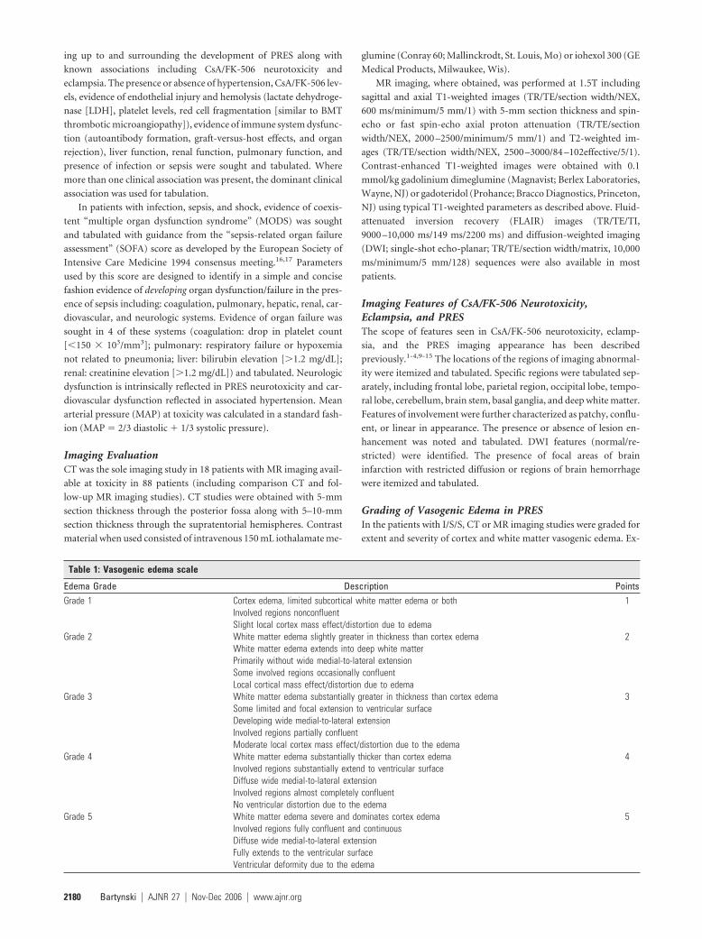
Grading of Vasogenic Edema in PRESIn the patients with I/S/S, CT or MR imaging studies were graded for
extent and severity of cortex and white matter vasogenic edema. Ex-
Table 1: Vasogenic edema scale
Edema Grade Description PointsGrade 1 Cortex edema, limited subcortical white matter edema or both
Involved regions nonconfluentSlight local cortex mass effect/distortion due to edema
1
Grade 2 White matter edema slightly greater in thickness than cortex edemaWhite matter edema extends into deep white matterPrimarily without wide medial-to-lateral extensionSome involved regions occasionally confluentLocal cortical mass effect/distortion due to edema
2
Grade 3 White matter edema substantially greater in thickness than cortex edemaSome limited and focal extension to ventricular surfaceDeveloping wide medial-to-lateral extensionInvolved regions partially confluentModerate local cortex mass effect/distortion due to the edema
3
Grade 4 White matter edema substantially thicker than cortex edemaInvolved regions substantially extend to ventricular surfaceDiffuse wide medial-to-lateral extensionInvolved regions almost completely confluentNo ventricular distortion due to the edema
4
Grade 5 White matter edema severe and dominates cortex edemaInvolved regions fully confluent and continuousDiffuse wide medial-to-lateral extensionFully extends to the ventricular surfaceVentricular deformity due to the edema
5
2180 Bartynski � AJNR 27 � Nov-Dec 2006 � www.ajnr.org
tent and severity of edema in the involved regions were independently
assessed by 2 observers blinded to patients’ blood pressure, studies
were graded on a 5-point scale summarized in Table 1, and results
were tabulated for each patient. Difference in patient grade was agreed
upon by consensus. Edema grade results for patients with severe hy-
pertension and without hypertension were separately averaged and
compared.
Vascular AssessmentIn 11 patients with I/S/S, MRA was available along with MR imaging
in the time frame of neurotoxicity and PRES. MRA was obtained
using 3D time-of-flight (TOF) technique (TR/TE/Flip angle/FOV/
matrix/acquisitions, default/min/45°/18 –22 cm/226 � 224/1) with
multiple overlapping slab reconstruction. In 10 of 11 patients, MRA
was obtained during initial MR assessment of neurotoxicity, and in 1
patient, MRA was obtained on a follow-up study 1 week after toxicity
and after the patient had clinically stabilized.
MRA studies were evaluated for the presence or absence of vascu-
lar abnormality or vasospasm. Studies were blindly and indepen-
dently graded by 2 neuroradiologists, and any differences were re-
solved by consensus. Traditional features of vasospasm or vasculitis
were identified including: significant diffuse constriction of first-, sec-
ond-, and third-order branch vessels, areas of focal vessel narrowing
and constriction, and string-of-bead appearance.
Statistical AssessmentStatistical significance was evaluated using the SAS software package,
PROCAPABILITY statistical analysis software function (SAS release
8.2; SAS Institute, Cary NC). Comparison between hypertensive and
nonhypertensive subsets was performed with Student t test and Wil-
coxon signed rank test. Statistical significance was considered to exist
for P � .05.
ResultsThe clinical background of the 106 patients is summarized inTable 2. Seventy-two patients (67.9%) were female and 34(32.1%) were male; their average age was 42.4 years (range,17–79 years). Headache, vision change, altered mental status,nausea, or aphasia (alone or in combination) was the present-ing symptom in 35 (33%) of patients and seizure (frequentlyaccompanied by or preceded by headache or vision change) in71 (67%). Blood pressure was normal (patient baseline) at
presentation in 32 patients, slightly elevated in 11 patients, andsevere in 63 patients.
Overall Patient Clinical ProfileIn 49 (46.2%) patients, neurotoxicity developed in associationwith cyclosporine/FK-506 immune suppression. Transplanta-tion was present in 46 of 49 patients (solid organ, 20; allo-BMT, 26), and 3 patients received cyclosporine for treatmentof marrow disease (pure red cell aplasia, aplastic anemia).Four (3.8%) patients developed PRES in association with can-cer chemotherapy and in 11 (10.4%) patients, PRES occurredin association with autoimmune disease (SLE, 5 patients and 1patient each with Wegener granulomatosis, scleroderma, poly-arteritis nodosa, psoriasis, Graves disease, and rheumatoidfactor positive arthropathy). PRES was associated witheclampsia or delayed eclampsia in 11 (10.4%) patients.
In 4 patients, PRES developed in association with increas-ing or acute hypertension with either chronic renal disease(nephrosclerosis; chronic renal failure and dialysis) or no ob-vious cause of acute hypertension (chronic drug use; priorrenal cell carcinoma). In 2 patients, no specific cause was iden-tified with known but unchanged chronic hypertension. In 25(23.6%) of 106 patients, neurotoxicity and PRES were notedto occur in association with I/S/S.
PRES in Infection, Sepsis, and ShockIn 23 patients, significant infection and/or bacteremia oc-curred in close association with the development of PRES. In 2additional patients, PRES developed after an episode of severehemorrhagic shock. In 11 of the 23 patients with infection,clinical sepsis (sepsis, severe sepsis, or septic shock) was notedor suspected during their illness before development of PRES.The clinical profile and characteristics of the patients with in-fection, sepsis, and shock are summarized in Table 3.
In 21 of these 23 patients with infection, PRES developedimmediately after or coincident with the severe infection orbacteremia. In 18 of these 21 patients, PRES occurred within 2weeks of the infection and in 3 patients (1 each: abscess,wound infection, blast crisis with sepsis), neurotoxicity devel-oped between 20 and 30 days of infection identification (over-all average, 6.7 days; range, 0 –30 days). In 19 of the 21 patients,organ or tissue infection was present, and in 2 patients, iso-
Table 2: Categories and timing of PRES neurotoxicity
Toxicity CategoryNo. of
Patients SubgroupNo. of
Patients
Time Point of PRES Relative to ‘Association’ Event
0–1 mos(# pts)
1–4 mos(# pts)
4–12 mos(# pts)
� 12 mos(# pts)
Not Known(# pts)
Cyclosporine/FK-506 toxicity 49 allo-BMT 26 16 5 4 1SOT 20 5 3 5 7CsA only 3 3
Postchemotherapy 4 4 3 1Infection/sepsis/shock 25 Infection/Sepsis 23 21 1 1
Hemorrhagic Shock 2 2Eclampsia 11 Intrapartum 3 3
Delayed 8 8Autoimmune 11 11 11Miscellaneous 6 6 6Total 106 106 56 10 9 8 23
Note:—# pts indicates number of patients; mos, months; SOT, solid organ transplant; CsA only, cyclosporine treatment in marrow diseases; PRES, posterior reversible encephalopathysyndrome; allo-BMT, allogenic bone marrow transplantation.
AJNR Am J Neuroradiol 27:2179 –90 � Nov-Dec 2006 � www.ajnr.org 2181
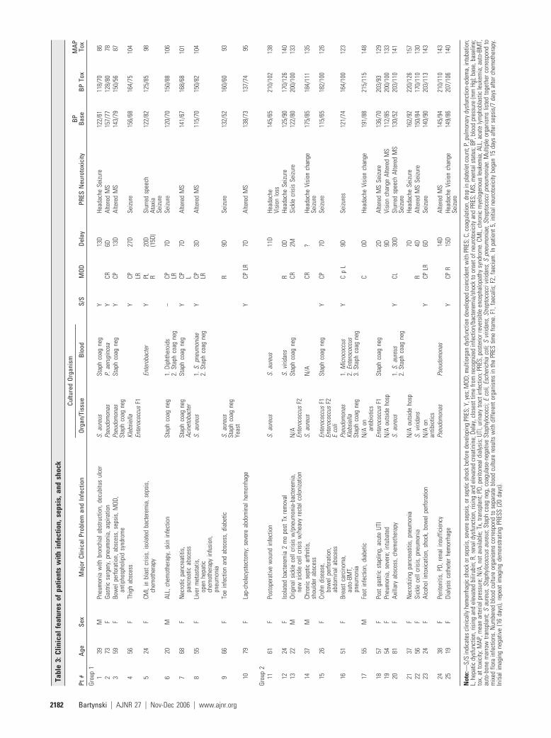
Tabl
e3:
Clin
ical
feat
ures
ofpa
tient
sw
ithin
fect
ion,
seps
is,a
ndsh
ock
Pt#
Age
Sex
Maj
orCl
inic
alPr
oble
man
dIn
fect
ion
Cultu
red
Orga
nism
S/S
MOD
Dela
yPR
ESN
euro
toxi
city
BP Base
BPTo
xM
AP Tox
Orga
n/Ti
ssue
Bloo
dGr
oup
11
39M
Pneu
mon
iaw
ithbr
onch
ialo
bstru
ctio
n,de
cubi
tus
ulce
rS.
aure
usSt
aph
coag
neg
Y13
DHe
adac
heSe
izure
122/
6111
8/70
862
73F
Gast
ricsu
rger
y,pn
eum
onia
,asp
iratio
nPs
eudo
mon
asP.
aeru
gino
saY
CR6D
Alte
red
MS
157/
7712
8/80
783
59F
Bow
elpe
rfora
tion,
absc
ess,
seps
is,M
OD,
antip
hosp
holip
idsy
ndro
me
Pseu
dom
onas
Stap
hco
agne
gSt
aph
coag
neg
YCP
13D
Alte
red
MS
143/
7915
0/56
87
456
FTh
igh
absc
ess
Kleb
siel
laY
CP27
DSe
izure
156/
6816
4/75
104
Ente
roco
ccus
F1LR
524
FCM
Lin
blas
tcr
isis
,iso
late
dba
cter
emia
,sep
sis,
chem
othe
rapy
Ente
roba
cter
YPL R
20D
(15D
)Sl
urre
dsp
eech
Atax
iaSe
izure
122/
8212
5/85
98
620
MAL
L,ch
emot
hera
py,s
kin
infe
ctio
nSt
aph
coag
neg
1.Di
phth
eroi
ds2.
Stap
hco
agne
g–
CP LR7D
Seizu
re12
0/70
150/
8810
6
768
FN
ecro
ticpa
ncre
atiti
s,pa
ncre
atic
absc
ess
Stap
hco
agne
gAc
inet
obac
ter
Stap
hco
agne
gY
CP L7D
Alte
red
MS
141/
6716
8/68
101
855
FLi
ver
met
asta
tis,
open
hepa
ticch
emot
hera
pyin
fusi
on,
pneu
mon
ia
S.au
reus
1.S.
pneu
mon
iae
2.St
aph
coag
neg
YCP LR
3DAl
tere
dM
S11
5/70
150/
8210
4
966
FTo
ein
fect
ion
and
absc
ess,
diab
etic
S.au
reus
Stap
hco
agne
gYe
ast
R9D
Seizu
re13
2/52
160/
6093
1079
FLa
p-ch
olec
yste
ctom
y;se
vere
abdo
min
alhe
mor
rhag
eY
CPLR
7DAl
tere
dM
S13
8/73
137/
7495
Grou
p2
1161
FPo
stop
erat
ive
wou
ndin
fect
ion
S.au
reus
S.au
reus
11D
Head
ache
Visi
onlo
ss14
5/65
210/
102
138
1224
FIs
olat
edba
cter
emia
2m
opo
stTx
rem
oval
S.vi
ridan
sR
0DHe
adac
heSe
izure
125/
9017
0/12
614
013
22M
Orig
inal
sick
lece
llcr
isis
w/p
neum
onia
-bac
tere
mia
,ne
wsi
ckle
cell
cris
isw
/hea
vyre
ctal
colo
niza
tion
N/A
Ente
roco
ccus
F2St
aph
coag
neg
CR2M
Sick
lecr
isis
Seizu
re12
2/80
200/
100
133
1437
MCh
roni
cse
ptic
arth
ritis
,sh
ould
erab
sces
sS.
aure
usN
/ACR
?He
adac
heVi
sion
chan
geSe
izure
175/
8518
4/11
113
5
1526
FCr
ohn
dise
ase,
bow
elpe
rfora
tion,
abdo
min
alab
sces
s
Ente
roco
ccus
F1En
tero
cocc
usF2
Eco
li
Stap
hco
agne
gY
CP7D
Seizu
re11
5/65
182/
100
126
1651
FBr
east
carc
inom
a,au
to-B
MT,
pneu
mon
ia
Pseu
dom
onas
Kleb
siel
laSt
aph
coag
neg
1.M
icro
cocc
us2.
Ente
roco
ccus
3.St
aph
coag
neg
YC
pL
9DSe
izure
s12
1/74
164/
100
123
1755
MFo
otin
fect
ion,
diab
etic
N/A
onan
tibio
tics
C0D
Head
ache
Visi
onch
ange
191/
8821
5/11
514
8
1857
FPo
stga
stric
stap
ling,
acut
eUT
IEn
tero
cocc
usF1
Stap
hco
agne
g2D
Alte
red
MS
Seizu
re13
6/70
203/
9312
919
54F
Pneu
mon
ia,s
ever
e;in
tuba
ted
N/A
outs
ide
hosp
9DVi
sion
chan
geAl
tere
dM
S11
2/85
200/
100
133
2081
FAx
illar
yab
sces
s,ch
emot
hera
pyS.
aure
us1.
S.au
reus
2.St
aph
coag
neg
YCL
30D
Slur
red
spee
chAl
tere
dM
SSe
izure
130/
5220
3/11
014
1
2137
FN
ecro
tizin
gpa
ncre
atiti
s,pn
eum
onia
N/A
outs
ide
hosp
7DHe
adac
heSe
izure
162/
9222
0/12
615
722
56F
Sick
lece
llcr
isis
,pne
umon
iaS.
virid
ans
R4D
Alte
red
MS
Seizu
re15
0/84
170/
110
130
2324
FAl
coho
lint
oxic
atio
n,sh
ock,
bow
elpe
rfora
tion
N/A
onan
tibio
tics
YCP
LR6D
Seizu
re14
0/90
203/
113
143
2438
FPe
riton
itis,
PD,r
enal
insu
ffici
ency
Pseu
dom
onas
Pseu
dom
onas
14D
Alte
red
MS
145/
9421
0/11
014
325
19F
Dial
ysis
cath
eter
hem
orrh
age
YCP
R15
DHe
adac
heVi
sion
chan
geSe
izure
149/
8620
7/10
614
0
Not
e:—
S/S
indi
cate
scl
inic
ally
hem
orrh
agic
shoc
kor
seps
is,s
ever
ese
psis
,ors
eptic
shoc
kbe
fore
deve
lopi
ngPR
ES;Y
,yes
;MOD
;mul
tiorg
andy
sfun
ctio
nde
velo
ped
coin
cide
ntw
ithPR
ES;C
,coa
gula
tion,
drop
inpl
atel
etco
unt;
P,pu
lmon
ary
dysf
unct
ion-
edem
a,in
tuba
tion;
L,he
patic
dysf
unct
ion,
risin
gan
del
evat
edbi
lirub
in;R
,ren
aldy
sfun
ctio
n,ris
ing
and
elev
ated
crea
tinin
e;De
lay,
clos
estt
ime
from
reco
gnize
din
fect
ion/
bact
erem
ia/s
hock
toon
seto
fneu
roto
xici
tyan
dPR
ES;M
S,m
enta
lsta
tus;
BP,b
lood
pres
sure
(mm
Hg);
base
,bas
elin
e;to
x,at
toxi
city
;MAP
,mea
nar
teria
lpre
ssur
e;N
/A,n
otav
aila
ble;
Tx,t
rans
plan
t;PD
,per
itone
aldi
alys
is;U
TI,u
rinar
ytra
ctin
fect
ion;
PRES
,pos
terio
rrev
ersi
ble
ence
phal
opat
hysy
ndro
me.
CML,
chro
nic
mye
loge
nous
leuk
emia
;ALL
,acu
tely
mph
obla
stic
leuk
emia
;aut
o-BM
T,au
to-b
one
mar
row
trans
plan
t;S
aure
us,S
taph
yloc
occu
sau
reus
;Sta
phco
agne
g,co
agul
ase-
nega
tive
Stap
hylo
cocc
i;E
coli,
Esch
eric
hia
coli;
Svi
ridan
s,St
rept
ococ
civi
ridan
s;S
pneu
mon
iae,
Stre
ptoc
occi
pneu
mon
iae.
Mul
tiple
orga
nism
slis
ted
toge
ther
corre
spon
dto
mix
edflo
rain
fect
ions
.Num
bere
dbl
ood
cultu
reor
gani
sms
corre
spon
dto
sepa
rate
bloo
dcu
lture
resu
ltsw
ithdi
ffere
ntor
gani
sms
inth
ePR
EStim
efra
me.
F1,f
aeca
lis;F
2,fa
eciu
m.I
npa
tient
5,in
itial
neur
otox
icity
bega
n15
days
afte
rsep
sis/
7da
ysaf
terc
hem
othe
rapy
.In
itial
imag
ing
nega
tive
(16
days
),re
peat
imag
ing
dem
onst
ratin
gPR
ESS
(20
days
).
2182 Bartynski � AJNR 27 � Nov-Dec 2006 � www.ajnr.org
lated bacteremia and sepsis occurred. Of the 19 patients withorgan/tissue infection, 12 patients also demonstrated bactere-mia coincident with the PRES event. In 4 of the 19 patientswith organ/tissue infection, documented infection waspresent but primary site cultures were not available due toongoing antibiotic treatment. In 2 of the 23 patients with in-fection, timing was difficult to assess because of disease chro-nicity (1 patient with diabetes, chronic septic arthritis [Strep-tococcus aureus] requiring frequent debridement and multipleprior episodes of bacteremia developed neurotoxicity duringrenal dialysis; 1 patient with sickle cell disease where PRESoccurred with high white count, repeat sickle cell crisis, andheavy rectal enterococcal colonization but 10 weeks afterpneumonia with documented bacteremia). Four of 23 patientswith infection/sepsis also received chemotherapy.
Organ or Tissue InfectionOrgan/tissue culture results were available in 16 of 23 patients(15 of 21 patients coincident with PRES; 1 patient with chronicseptic arthritis). These are reviewed in Table 3. In 10 infec-tions, a single organism was isolated, and in 6 infections,mixed flora was present. In single-organism infections, gram-positive cocci were present in 8 of 10 and where mixed florawas cultured, at least one of the organisms was gram-positive.Gram-positive cocci were isolated, therefore, in 14 of 16 pri-mary site infections.
Blood Cultures and SepticemiaBlood cultures were positive in 14 of the 21 patients, whereinfection was coincident with PRES (2 with primary bactere-mia/sepsis, 12 with organ or tissue infection) as well as the 1patient with sickle cell disease, sickle cell crisis, and pneumo-nia. The results are summarized in Table 3. In 10 patients, asingle blood culture was positive coincident with PRES, and in4 patients, multiple blood cultures were positive coincidentwith toxicity. Similar to the results of tissue and organ infec-tion, most organisms identified (12 of 15) were gram-positivecocci.
Multiple Organ Dysfunction Accompanying I/S/Sand PRESIn 18 of the 25 patients with I/S/S, evidence of evolving mul-tiple organ dysfunction was identified coincident with the de-velopment of neurotoxicity/PRES. New or worsening organdysfunction was documented in the systems typically involvedin MODS including: coagulation dysfunction with plateletconsumption (14 patients), pulmonary dysfunction separatefrom pneumonia (11 patients), liver dysfunction (9 patients),and renal dysfunction (12 patients). Two or more systemswere abnormal and changing coincident with PRES in 15 pa-tients (4-system dysfunction: 5 patients; 3-system dysfunc-tion: 4 patients; 2-system dysfunction: 6 patients) with single-system dysfunction noted in 3 patients.
In 5 of 15 patients, manual peripheral smears were avail-able at the time of toxicity and were abnormal demonstratingred cell fragmentation. In 8 of 9 patients, LDH levels obtainedat toxicity were elevated and in 3 of these patients, peripheralsmears were available and demonstrated red cell fragmenta-tion consistent with hemolysis. In 7 of the 8 patients withelevated LDH, a significant decline in platelet count was also
noted suggesting a consumptive coagulopathy and endothelialinjury.
Blood PressureAverage baseline blood pressures and blood pressure at thetime of toxicity are listed in Table 3. Two separate groups ofpatients were identified. Group 1 (referred to as “normoten-sive”): In 10 patients (40%), blood pressure was either nor-mal/patient’s baseline at toxicity (6 patients) or demonstratedonly mild systolic pressure elevation relative to patient base-line at toxicity (4 patients). Group 2 (referred to as “severehypertensive”): In 15 patients (60%), severe hypertension waspresent with significant elevation of systolic pressure (�200mm Hg), diastolic pressure (�100 mm Hg), or both. MAP attoxicity in group 1 was 95 mm Hg (range, 78 –106 mm Hg)and MAP at toxicity in group 2 was 137 mm Hg (range, 123–157 mm Hg).
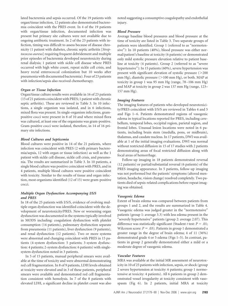
Imaging FeaturesThe imaging features of patients who developed neurotoxici-ty/PRES coincident with I/S/S are reviewed in Tables 4 and 5and Figs 1– 6. Patients demonstrated regions of vasogenicedema in typical locations reported for PRES, including cere-bellum, temporal lobes, occipital region, parietal region, andfrontal lobes. Unusual lesion locations were noted in 6 pa-tients, including brain stem (medulla, pons, or midbrain),thalamus, and caudate nucleus. In 17 patients, DWI was avail-able at 1 of the initial imaging evaluations. DWI was normalwithout restricted diffusion in 15 of 17 studies with 2 patientsdemonstrating areas of focal restricted diffusion along withfocal areas of hemorrhage.
Follow-up imaging in 18 patients demonstrated reversal(12 patients) or partial/substantial reversal (6 patients) of thePRES imaging appearance. In 5 patients, follow-up imagingwas not performed but the patients’ symptoms (altered men-tation, headache, vision change) resolved completely. Two pa-tients died of sepsis-related complications before repeat imag-ing was obtained.
Vasogenic EdemaExtent of brain edema was compared between patients fromgroups 1 and 2, and the results are summarized in Table 4.Vasogenic edema was judged greater in the “normotensive”patients (group 1: average 3.3) with less edema present in the“severely hypertensive” patients (group 2: average 2.07). Thisdifference was statistically significant (Student t test: P � .01;Wilcoxon score: P � .03). Patients in group 1 demonstrated agreater range in the degree of brain edema; 4 of 11 (36%)demonstrated grade 4 or 5 edema (Figs 1–5). In contrast, pa-tients in group 2 generally demonstrated either a mild or amoderate degree of vasogenic edema.
Vascular FeaturesMRA was available at the initial MR assessment of neurotox-icity in 10 of 25 patients with infection, sepsis, or shock (group2 severe hypertension at toxicity: 6 patients; group 1 normo-tensive at toxicity: 4 patients). All 6 patients in group 2 dem-onstrated vessel irregularity at toxicity consistent with vaso-spasm (Fig 6). In 2 patients, initial MRA at toxicity
AJNR Am J Neuroradiol 27:2179 –90 � Nov-Dec 2006 � www.ajnr.org 2183
demonstrated vasospasm, and follow-up MRA demonstratedreversal of spasm with vessel caliber and shape normalization.
In the patients in group 1 (normotensive), MRA at toxicitydemonstrated vessel “pruning” with reduced second- andthird-order branch visualization in 2 patients and vessel prun-ing with reversible spasm on follow-up MRA (Fig 5) in 1 pa-tient. In 1 normotensive patient, MRA appeared normal.
DiscussionThe imaging findings of “PRES” and associated neurotoxicityare recognized.1-15 This imaging pattern is typically seen inpatients who develop eclampsia or cyclosporine/FK-506 neu-rotoxicity after transplantation, but other associations havebeen reported, including autoimmune disease (such as SLE orWegener granulomatosis) and hypertension. These patientstypically present with several days of headache progressing to
visual disturbance and/or grand mal seizure.18 Unstable bloodpressure frequently accompanies toxicity, but significant hy-pertension may be absent (25%–30% of patients).2,19
Table 4: Imaging features in patients with infection, sepsis, and shock
Pt#
Imaging Locations of PRES VasogenicEdema MRA
ImagingModality
DWIF/U
Imaging
Clinicalat
Discharge
EdemaGrade
MAPC T O P F Misc
Group11 x x x MR n/o Resolved 3 862 Spasm-imp MR Neg Res 2 783 x x x x MR Neg Resolving Resolved 5 874 x x x x x t Pruning MR n/o Resolved 5 1045 x x x x CT Res 3 986 x CT Expired 2 1067 Pruning MR Neg Resolving Resolved 2 1018 x x x x CT Res 5 1049 x x x x Normal MR Neg Res 2 9310 x x x MR Neg* Resolving Resolved 4 85
Group 211 x x x x t mb Spasm/
prunMR Neg Normal 2 138
12 x x x x MR Neg Res 3 14013 x x x x x bs
mb cMR Neg Res 1 133
14 x x x x x bs MR Neg Res 3 13515 x x x x CT Resolved 1 12616 x x x CT Expired 2 12317 x x x t Spasm MR Neg Resolving Resolved 2 14818 x x x x Spasm MR Neg Resolving Resolved 3 12919 x x x Spasm-rev MR Neg Res 1 13320 x x x x bs MR Neg Resolving Resolved 2 14121 x x MR Neg — Resolved 1 15722 x x x x Spasm MR Neg Res 4 13023 x x x x MR Neg Res 1 14324 x x x x Spasm-rev MR Neg* Res* 3 14325 x x x x MR n/o Res 2 140
Note:—PRES indicates posterior reversible encephalopathy syndrome; C, cerebellum; T, temporal lobe/inferior temporal-occipital junction; O, occipital lobe; P, parietal region; F, frontallobes; bs, brain stem including medulla and pons; mb, midbrain; t, thalamus; c, caudate nucleus; rev, spasm reverses; imp, spasm improves; spasm/prun, middle crebral artery spasm withposterior cerebral artery pruning; DWI, diffusion-weighted image characteristics; n/o, DWI not obtained with MR imaging study; Neg, no evidence of restricted diffusion to suggest cytotoxicedema; Neg*, no restricted diffusion in the majority of vasogennic edema with focal region of restricted diffusion and hemorrhage noted; Res, vasogenic edema completely resolved onfollow-up imaging studies; Res*, vasogenic edema resolved with minor persistent abnormal signal in region of previously noted restricted diffusion, infarction, or focal hemorrhage;Resolving, resolving vasogenic edema on follow-up imaging studies or resolving clinical symptoms related to PRES; Resolved, clinical symptoms at PRES neurotoxicity resolved; MAP, meanarterial pressure.
Table 5: Vasogenic edema grade in Group 1 (Normotensive) andGroup 2 (Severely Hypertensive) Patients
BloodPressureGroup
Vasogenic Edema Grade (Number of Patients)Group
AverageGrade 1 Grade 2 Grade 3 Grade 4 Grade 5Group 1 0 4 2 1 3 3.3Group 2 5 5 4 1 0 2.07
Fig 1. Patient 1 is a 39-year-old man with baseline blood pressure 122/61 mm Hg who hadsevere pneumonia with bronchial obstruction. Bronchial lavage grew Staphylococcusaureus and blood culture grew coagulase-negative staphylococci. Neurotoxicity developed13 days after positive cultures with severe headache followed by a seizure with bloodpressure at toxicity 118/70 mm Hg.
A–B, Brain MR imaging (FLAIR sequence) demonstrates moderate signal intensity abnor-mality from vasogenic edema in the occipital lobes bilaterally (open arrows) typical of thePRES pattern with full extension to the ventricular surface and moderate local cortical masseffect judged grade 3. Follow up imaging was not obtained, but the patient’s symptomsresolved completely.
2184 Bartynski � AJNR 27 � Nov-Dec 2006 � www.ajnr.org
The cause of PRES is controversial and unproven. Neuro-toxicity and PRES are associated with eclampsia, cyclosporine/FK-506 toxicity (typically after allo-BMT or solid organ trans-plantation), and systemic chemotherapy.1-11 PRES is also seen
in patients with autoimmune disease, medical-renal disease,and severe hypertension.12-15 Isolated studies have suggestedan association with hypomagnesemia, hypercholesterolemia,and human leukocyte antigen mismatch in allo-BMT.20
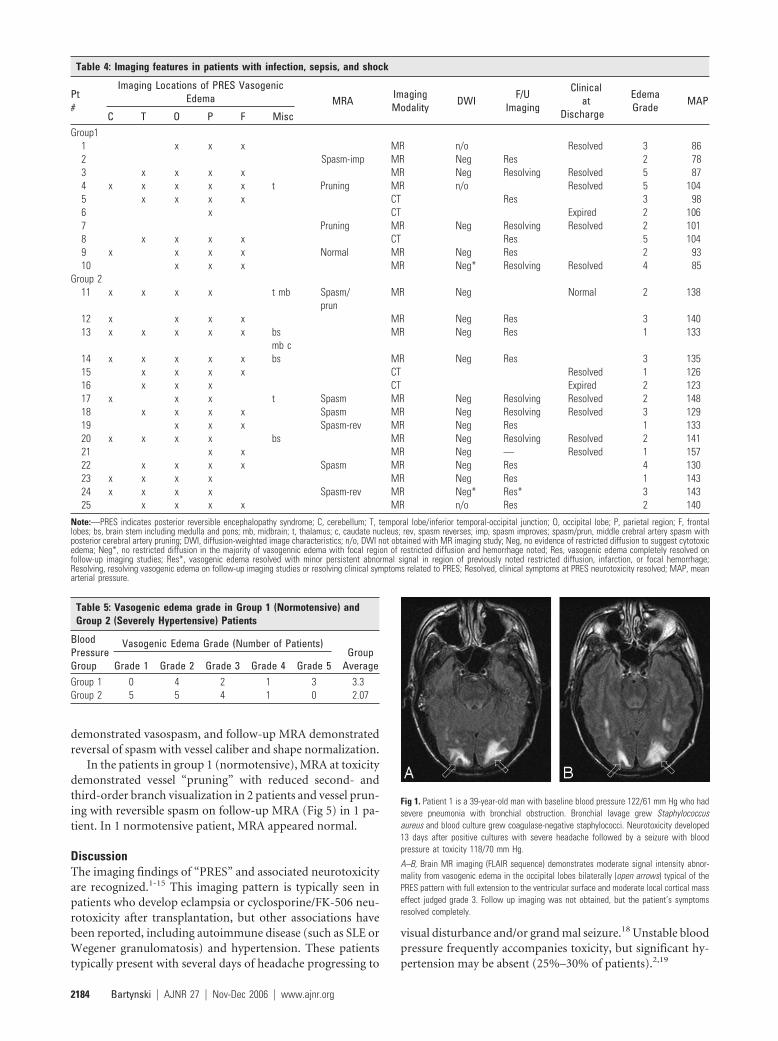
Fig 2. Patient 7 is a 68-year-old woman with necrotic pancreatitis, a pancreatic abscess, and baseline blood pressure of 141/67 mm Hg. Abscess grew mixed flora with coagulase-negativestaphylococci and Acinetobacter baumannii and blood culture was positive for coagulase-negative staphylococci. Altered mental status with PRES developed 7 days after positive cultureswith blood pressure at toxicity of 168/68 mm Hg.
A-B, Brain MR imaging (FLAIR sequence) demonstrates vasogenic edema in the occipital (open arrows) and parietal region (curved arrows) bilaterally typical of PRES with extension intothe deep white matter but no extension to the ventricle surface judged grade 2.
C-D, Brain MR imaging (FLAIR sequence) obtained 1 month after initial imaging and toxicity demonstrates near complete resolution of the edema in the occipital (open arrows) and leftparietal region (curved arrow) with complete resolution in the right parietal area (arrow).
Fig 3. Patient 4 is a 56-year-old woman with a baseline blood pressure of 156/68 who developed a thigh abscess with culture growing mixed flora (Klebsiella pneumonial and enterococci).She developed MOD (coagulopathy, acute respiratory distress syndrome, acute renal failure, liver failure, and shock liver). On day 27 of intensive treatment of her infection and multiorganfailure, the patient developed altered mentation followed by a generalized seizure and blood pressure of 164/75 mm Hg.
A-D, Brain MR imaging (FLAIR sequence) obtained the 1-day after neurotoxicity demonstrates severe and extensive vasogenic edema primarily involving the subcortical white matter ofthe parietal (curved arrows), occipital (open arrows), and temporal lobe regions (arrowheads) bilaterally with ventricular distortion from edema judged grade 5.
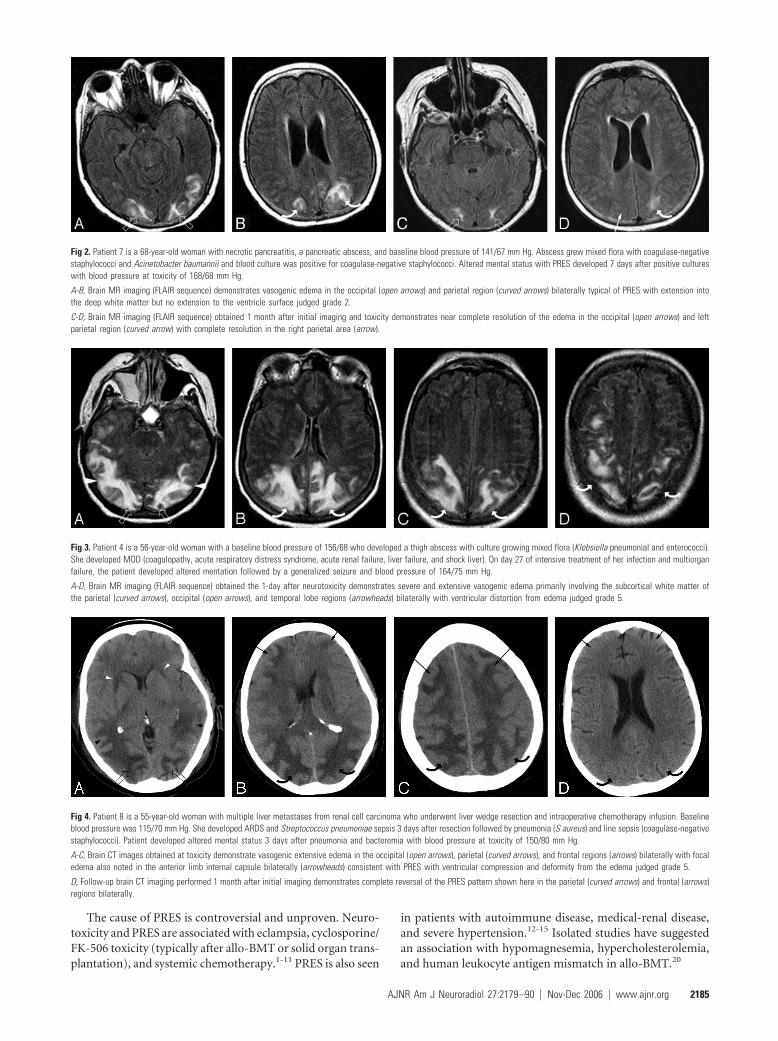
Fig 4. Patient 8 is a 55-year-old woman with multiple liver metastases from renal cell carcinoma who underwent liver wedge resection and intraoperative chemotherapy infusion. Baselineblood pressure was 115/70 mm Hg. She developed ARDS and Streptococcus pneumoniae sepsis 3 days after resection followed by pneumonia (S aureus) and line sepsis (coagulase-negativestaphylococci). Patient developed altered mental status 3 days after pneumonia and bacteremia with blood pressure at toxicity of 150/80 mm Hg.
A-C, Brain CT images obtained at toxicity demonstrate vasogenic extensive edema in the occipital (open arrows), parietal (curved arrows), and frontal regions (arrows) bilaterally with focaledema also noted in the anterior limb internal capsule bilaterally (arrowheads) consistent with PRES with ventricular compression and deformity from the edema judged grade 5.
D, Follow-up brain CT imaging performed 1 month after initial imaging demonstrates complete reversal of the PRES pattern shown here in the parietal (curved arrows) and frontal (arrows)regions bilaterally.
AJNR Am J Neuroradiol 27:2179 –90 � Nov-Dec 2006 � www.ajnr.org 2185
Potential biologic mechanisms for development of PREShave included factors that induce endothelial injury, such asthe immune suppressive drugs cyclosporine and FK-506, en-dothelial activation/injury in eclampsia, endothelial injuryfrom pretransplantation conditioning regimens, or graft-ver-sus-host effects.2,21,22 Hypertension with forced hyperperfu-sion has also remained popular.3,23,24
Our results demonstrate 4 important observations in PRESincluding: (1) association with infection/sepsis/shock, (2) as-sociation with gram-positive organisms, (3) inverse effect ofsevere hypertension on the extent of vasogenic edema, and (4)high frequency of associated cerebral vasospasm.
PRES and Its Association with Infection, Sepsis,and ShockThe first unexpected observation in our study is that PRESseemed to occur in patients with infection, sepsis, and shock.In 18 of 25 patients, PRES occurred within 2 weeks of bacte-remia or organ/tissue infection (abscess, pneumonia, woundinfection), and in 3 patients, PRES developed within 30 days ofabscess drainage or bacteremia. In 2 additional patients, neu-rotoxicity occurred within 2 weeks of severe hemorrhagicshock. If one eliminates patients with transplantation oreclampsia, significant infection, sepsis, or shock was present in25 of 49 patients (51%) in the overall study group. We believe
this is the first report to suggest that PRES may be associatedwith severe infection, sepsis, or shock.
Response to infection is an extremely complex process. Atthe fundamental biologic level, several systemic events occursimultaneously, including anti-infective, acute inflammatory,metabolic, pro-coagulant, and thermoregulatory chang-es.17,25-28 When infection is present, the role of the immunesystem is to 1) contain the infection, 2) target or traffic theinflammatory response to the infected region, and 3) preventthe inflammatory response from becoming systemic.17 If in-fection becomes overwhelming or if the immune response isinadequate, a septic clinical state can develop. The septic re-sponse to infection is known to occur without or with bacte-remia (demonstrating identical morbidity and mortality), andcell-wall antigens of the infective organisms (endotoxin, exo-toxin) are considered likely mediators.17,25-28 Both endotoxin(lipopolysaccharide [LPS] from gram-negative cell wall) andexotoxins/enterotoxins (peptidoglycan and lipoteichoic acidfrom gram-positive cell wall) are well known to be powerfulstimulants of systemic inflammatory response, leukocytes,and endothelial interaction.17,28 It is noteworthy that no obvi-ous source of infection can be identified in 30%–50% of casesthat demonstrate a septic clinical presentation.17
Endothelial activation/injury is considered central to thedevelopment of the primary infection and secondary septic
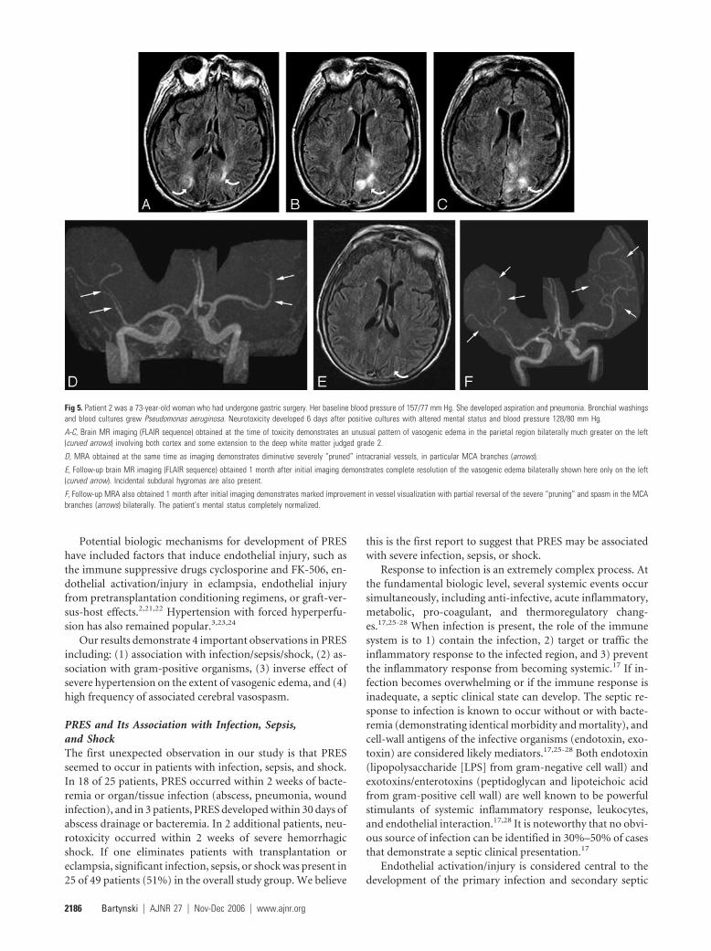
Fig 5. Patient 2 was a 73-year-old woman who had undergone gastric surgery. Her baseline blood pressure of 157/77 mm Hg. She developed aspiration and pneumonia. Bronchial washingsand blood cultures grew Pseudomonas aeruginosa. Neurotoxicity developed 6 days after positive cultures with altered mental status and blood pressure 128/80 mm Hg.
A-C, Brain MR imaging (FLAIR sequence) obtained at the time of toxicity demonstrates an unusual pattern of vasogenic edema in the parietal region bilaterally much greater on the left(curved arrows) involving both cortex and some extension to the deep white matter judged grade 2.
D, MRA obtained at the same time as imaging demonstrates diminutive severely “pruned” intracranial vessels, in particular MCA branches (arrows).
E, Follow-up brain MR imaging (FLAIR sequence) obtained 1 month after initial imaging demonstrates complete resolution of the vasogenic edema bilaterally shown here only on the left(curved arrow). Incidental subdural hygromas are also present.
F, Follow-up MRA also obtained 1 month after initial imaging demonstrates marked improvement in vessel visualization with partial reversal of the severe “pruning” and spasm in the MCAbranches (arrows) bilaterally. The patient’s mental status completely normalized.
2186 Bartynski � AJNR 27 � Nov-Dec 2006 � www.ajnr.org
response.17,29-32 This process (which is mediated by inflam-matory cytokine release [tumor necrosis factor (TNF)�, inter-leukin (IL)-1�, and other cytokines]) leads to up-regulation ofendothelial surface antigens (P-selectin, E-selectin, ICAM-1)with increased white cell adherence, microcirculatory dys-function, and altered vascular tone, vascular permeability, andcoagulation.17,28,31,33,34 Microcirculatory dysfunction devel-ops in part because of leukocyte adherence/trafficking withreduced local tissue blood flow at the capillary/venule level.34
An alteration in vascular tone develops secondary to compet-ing vasoconstrictive (platelet degranulation with thrombox-ane release, endothelin-1, angiotensin, vasopressin, and cen-tral sympathetic stimulation) and vasodilatory (nitric oxide,prostacyclin) effects.17,33 Significant vascular instability hasbeen documented in 50% of septic patients within 28 days ofthe infection.26
The potent vasoconstrictor endothelin-1 is released at itshighest levels in sepsis, and morbidity/mortality in sepsis hasbeen shown to parallel plasma endothelin-1 concentra-
tion.35-37 The inflammatory cytokines TNF-� and IL-1 up-regulate endothelin-1 mRNA production and stimulate its re-lease from endothelial cells.38-40 Endotoxin also promotes therelease of endothelin.41 Perhaps intermittent episodes of hy-pertension occur in sepsis but are difficult to recognize be-cause of confounding factors (pain, intubation) and simplemanagement with antihypertensive agents.
The clinical features of our patients parallel the generalobservations on sepsis with: 1) primary infection most com-monly in the lungs, abdomen, wound or urinary tract, 2)blood culture-positive and blood culture-negative cases iden-tified and 3) vascular instability and PRES developing within14 –30 days of severe infection. Endothelin-1 could contributeto the development of PRES in I/S/S.
Multiple Organ DysfunctionThe decision to group the patients with infection, sepsis,and hemorrhagic shock together is not arbitrary or for mereconvenience. It is recognized that the systemic effects
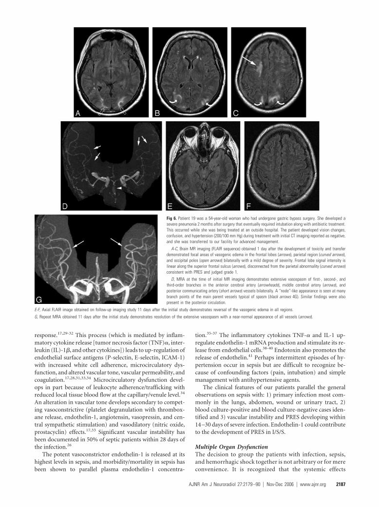
Fig 6. Patient 19 was a 54-year-old woman who had undergone gastric bypass surgery. She developed asevere pneumonia 2 months after surgery that eventually required intubation along with antibiotic treatment.This occurred while she was being treated at an outside hospital. The patient developed vision changes,confusion, and hypertension (200/100 mm Hg) during treatment with initial CT imaging reported as negative,and she was transferred to our facility for advanced management.
A-C, Brain MR imaging (FLAIR sequence) obtained 1 day after the development of toxicity and transferdemonstrated focal areas of vasogenic edema in the frontal lobes (arrows), parietal region (curved arrows),and occipital poles (open arrows) bilaterally with a mild degree of severity. Frontal lobe signal intensity islinear along the superior frontal sulcus (arrows), disconnected from the parietal abnormality (curved arrows)consistent with PRES and judged grade 1.
D, MRA at the time of initial MR imaging demonstrates extensive vasospasm of first-, second-, andthird-order branches in the anterior cerebral artery (arrowheads), middle cerebral artery (arrows), andposterior communicating artery (short arrows) vessels bilaterally. A “node”-like appearance is seen at manybranch points of the main parent vessels typical of spasm (black arrows 4G). Similar findings were alsopresent in the posterior circulation.
E-F, Axial FLAIR image obtained on follow-up imaging study 11 days after the initial study demonstrates reversal of the vasogenic edema in all regions.
G, Repeat MRA obtained 11 days after the initial study demonstrates resolution of the extensive vasospasm with a near-normal appearance of all vessels (arrows).
AJNR Am J Neuroradiol 27:2179 –90 � Nov-Dec 2006 � www.ajnr.org 2187
present in these conditions are similar and that a final com-mon course/end point can occur with multiple organ sys-tem dysfunction and/or failure (MODS) including: coagu-lation, pulmonary, hepatic, renal, cardiovascular, andneurologic.30,42-44 This response to I/S/S seems to representthe effects of systemic toxicity similar to the recently de-scribed “Systemic Inflammatory Response Syndrome”(SIRS) or MODS.27,42-47 Cytokine response (TNF-�, IL-1)is believed to play a critical role in the development of thiseffect.42-45,48
In 18 of our patients with I/S/S, additional multiple organsystem dysfunction (separate from neurologic and cardiovas-cular) was identified coincident with neurotoxicity and PRESsimilar to patterns observed in patients with MODS.
Gram-Positive SepsisThe second important observation in these patients is the un-expectedly high incidence of gram-positive infection. In 16 of19 culture-positive patients (84%), gram-positive organismswere identified in the primary infection site, blood cultures, orboth. Gram-positive organisms are becoming more frequentlyidentified in association with bacteremia and sepsis from bothcommunity-acquired and nosocomial sources with Staphylo-coccus aureus, coagulase-negative staphylococci, and entero-cocci accounting for 30%–50% of cases.49-51
The mechanisms involved in gram-positive sepsis are dif-ferent from gram-negative sepsis with cell surface antigen(exotoxins: peptidoglycan and lipoteichoic acid) and superan-tigen-related T-cell stimulation of cytokine release comparedwith the more limited traditional T cell trigger of inflamma-tion/cytokines as occurs with endotoxin/LPS from gram-neg-ative organisms.17,52-56 Superantigens demonstrate a mark-edly greater interaction rate with the overall T cell population(5%–20% of T cells for superantigens versus 1 in 104-106 Tcells for traditional antigen) with broader T cell stimulationand cytokine response.17,52 This broader, more generalized re-sponse to gram-positive organisms could underlie the onsetand systemic manifestations in patients with PRES.
Although our study was not designed to assess the inci-dence of PRES in infection/sepsis/shock, an approximate ref-erence point can be drawn from the predicted incidence ofseptic shock in patients with sepsis. The incidence of sepsis inthe United States is estimated to be approximately 240 pa-tients/100,000 persons per year; septic shock is estimated tooccur in approximately 7%– 8%.27,57 For our region, thiswould translate into 330 to 380 cases of septic shock per yeardistributed among 20 hospitals (approximately 115–190gram-positive; 5.7–10 cases per year). The number of patientswe identify with infection/sepsis and PRES (approximately2.5–3 patients per year) is similar to the incidence of gram-positive septic shock for a single institution in our geographicregion (excluding transplantation).27,58 It is interesting thatacute graft-versus-host disease (GVHD) seen after allo-BMThas been labeled a “distortion of the cellular response” toinfection.57
PRES Imaging Appearance, MRA, and HypertensionThe third crucial and unexpected observation in our patientswith I/S/S is that the extent of brain edema graded on imagingstudies appears to be inversely related to blood pressure at
toxicity. Normotensive patients (average MAP, 95 mm Hg)demonstrated the greatest degree of vasogenic edema, whereasseverely hypertensive patients (average MAP, 137 mm Hg)demonstrated less brain edema, and this difference is statisti-cally significant (P � .05). Fourth, in those patients for whomMRA was available, clear evidence of vasospasm or vessel“pruning” was observed. In the patients with severe hyperten-sion, typical features of vasospasm were present and reversiblewhere follow-up MRA was available for comparison. In nor-motensive patients, a combination of second- and third-ordervessel “pruning” were noted. The reason behind these obser-vations is not certain.
Although hypertension is frequently cited as the cause ofPRES, blood pressure is not elevated in all cases. In 20%–30%of patients with cyclosporine toxicity or eclampsia, bloodpressure is normal at toxicity.2,19 In preeclampsia/eclampsia,hypertension develops secondary to and along with complexsystemic interactions, including endothelial activation/injury,platelet consumption, hemolysis, resultant vasospasm andvascular constriction, endothelial leakage, and organ hypoper-fusion.59-61 Similar systemic effects are also present in otherconditions, including transplantation and autoimmune dis-ease.57,62-78 These issues suggest that a mechanism other thanhypertension could be responsible for PRES.
Vessel “pruning” in our normotensive patients could re-flect slow or sluggish cerebral blood flow, perhaps related tomicrocirculatory effects, such as enhanced platelet/white celladherence/trafficking at the capillary/venular level.29,34 Theeffects of microcirculatory abnormality might be demon-strated by a flow-sensitive technique such as MR perfusion.
In the “severely” hypertensive patients, recognizable vaso-spasm could reflect involvement of larger cerebral vessels inthe “vasculopathy” process. Alternatively, better vessel visual-ization (lack of pruning), observed vasospasm, and reducedvasogenic edema in these patients could suggest that hyperten-sion is acting in a positive fashion to improve cerebral bloodflow at some point in the toxicity process. Therefore, the roleof hypertension and its contribution to the development ofPRES is unclear.
Major Conditions Associated with PRES and Summationof Causes in TransplantationMost the patients in our overall study population (100 of 106patients [94%]) developed neurotoxicity and PRES in the set-ting of a complex systemic condition/illness, including infec-tion, sepsis, shock: 25, chemotherapy: 4, autoimmune disease:11, CsA/FK-506 (allo-BMT, solid organ transplant, marrowdisorders): 49, and eclampsia: 11. The pathophysiologicmechanisms underlying these conditions are similar and in-clude: immune system dysfunction, endothelial activation/in-jury, and/or a complex cytokine response.57,59-66 In eclampsia,immune challenge by the fetus and placenta is well recognizedand complex immune reaction to fetal antigen is noted, in-cluding T cell, endothelial, and coagulation system activa-tion.59-61 A similar response is noted after transplantation, inparticular after allo-BMT with a T cell and cytokine responsethat has been likened to the response to viral or bacterial in-fection.57 In autoimmune diseases, a complex T cell immuneand antibody response is felt responsible for most observedabnormality.62-66
2188 Bartynski � AJNR 27 � Nov-Dec 2006 � www.ajnr.org
After transplantation, patients are intrinsically exposed tomost of the major risk factors we identify associated withPRES. Opportunistic infection is well recognized after trans-plantation, in particular immediately after transplant.67-69 Theeffects of cyclosporine/FK-506 and complications of trans-plantation often coexist, including endothelial injury with sys-temic effects.70-72 In allo-BMT, a direct effect of chemotherapymay also be present related to local endothelial turnover.21,22 Atissue injury response is also noted within the first 7–10 daysafter preconditioning regimens accompanied by a brief butrecognizable cytokine response (IL-2, TNF�, IL-1).73-77
A markedly altered immune state is present after transplan-tation including GVHD and graft rejection. Acute GVHD af-ter allo-BMT is generally related to either a T cell-mediatedresponse of the graft to the host (in particular to host endo-thelium) or a response to the preconditioning regi-mens.17,18,31,32 Cytokines may play a prominent role in thiseffect (IL-2, IL-6, TNF�, IL-1).22,57,73-76 Graft rejection insolid organ transplant is related to the development of both Tcell activation and anti-vascular/anti-endothelial anti-bodies.77,78
Transplants therefore may be experiencing a summation ofthe fundamental factors that are associated with the develop-ment of neurotoxicity and PRES, including infection (oppor-tunistic), immune-related effects, chemotherapy-related ef-fects, and the effects of cyclosporine or FK-506.
ConclusionPRES occurred after severe infection, sepsis, or shock in 25(23.6%) of 106 patients with other “associations,” includingautoimmune disease, postchemotherapy, cyclosporine/FK-506, and eclampsia. In most patients with available cultures(84%), gram-positive organisms, particularly gram-positivecocci, were identified in organ/tissue culture, blood culture, orboth. Normotensive and severely hypertensive patients werepresent in the I/S/S group. The extent of brain edema gradedon CT/MR imaging studies was greater in the normal MAPgroup and less in patients who were severely hypertensive.Vasospasm was noted at MRA in patients who were severelyhypertensive. A combination of spasm and vessel “pruning”was seen in MRA in the normal MAP group, perhaps related toreduced branch visualization secondary to diminished cere-bral blood flow.
References1. Bartynski WS, Grabb BC, Zeigler Z, et al. Watershed imaging features and
clinical vascular injury in cyclosporin A neurotoxicity. J Comput Assist Tomogr1997;21:872– 80
2. Bartynski WS, Zeigler Z, Spearman MP, et al. Etiology of cortical and whitematter lesions in cyclosporin-A and FK-506 neurotoxicity. AJNR Am J Neuro-radiol 2001;22:1901–14
3. Schwartz RB, Bravo SM, Klufas RA, et al. Cyclosporine neurotoxicity and itsrelationship to hypertensive encephalopathy: CT and MR findings in 16 cases.AJR Am J Roentgenol 1995;165:627–31
4. Sanders TG, Clayman DA, Sanchez-Ramos L, et al. Brain in eclampsia: MRimaging with clinical correlation. Radiology 1991;180:475–78
5. Lin JT, Wang WJ, Fuh JL, et al. Prolonged reversible vasospasm in cyclosporinA-induced encephalopathy. AJNR Am J Neuroradiol 2003;24:102– 04
6. Ito T, Sakai T, Inagawa S, et al. MR angiography of cerebral vasospasm inpreeclampsia. AJNR Am J Neuroradiol 1994;16:1344 – 47
7. Shengar AR, Gupta RK, Dhanuka AK, et al. MR imaging, MR angiography andMR Spectroscopy of the brain in eclampsia. AJNR Am J Neuroradiol 1997;18:1485–90
8. Schwartz RB, Jones KM, Kalina P, et al. Hypertensive encephalopathy: findings
on CT, MR imaging and SPECT imaging in 14 cases. AJR Am J Roentgenol1992;159:379 – 83
9. Rippe DJ, Edwards MK, Schrodt JF, et al. Reversible cerebral lesions associatedwith tiazofurin usage: MR demonstration. J Comput Assist Tomogr 1988;12:1078 – 81
10. Vaughn DJ, Jarvik JG, Hackney D, et al. High-dose cytarabine neurotoxicity:MR findings during acute phase. AJNR Am J Neuroradiol 1993;14:1014 –16
11. Ito Y, Arahata Y, Goto Y, et al. Cisplatin neurotoxicity presenting as reversibleposterior leukoencephalopathy syndrome. AJNR Am J Neuroradiol 1998;19:415–17
12. Hinchey J, Chaves C, Appignani B, et al. A reversible posterior leukoencepha-lopathy syndrome. N Engl J Med 1996;334:494 –500
13. Provenzale JM, Petrella JR, Cruz LC, et al. Quantitative assessment of diffusionabnormalities in posterior reversible encephalopathy syndrome. AJNR Am JNeuroradiol 2001;22:1455– 61
14. Mukherjee P, McKinstry RC. Reversible posterior leukoencephalopathysyndrome: evaluation with diffusion-tensor MR imaging. Radiology 2001;219:756 – 65
15. Covarrubias DJ, Leutmer PH, Campeau NG. Posterior reversible encephalop-athy syndrome: prognostic utility of quantitative diffusion-weighted MR im-ages. AJNR Am J Neuroradiol 2002;23:1038 – 48
16. Vincent JL, Moreno R, Takala J, et al. The SOFA (Sepsis-related Organ FailureAssessment) score to describe organ dysfunction/failure. Intensive Care Med1996;22:707–10
17. Munford RS. Sepsis, severe sepsis, and septic shock. In: Mandell GL, BennettJE, Dolin R eds. Principles and Practice of Infectious Disease. Philadelphia:Elsevier; 2005:906 –26
18. Schwartz RB, Feske SK, Polack JF, et al. Preeclampsia-eclampsia: clinical andneuroradiographic correlates and insights into the pathogenesis of hyperten-sive encephalopathy. Radiology 2000;217:371–76
19. Sibai BM. Eclampsia: maternal-perinatal outcome in 254 consecutive cases.Am J Obstet Gynecol 1990;163:1049 –55
20. Gijtenbeek JMM, van den Bent MJ, Vecht CJ. Cyclosporine neurotoxicity: areview. J Neurol 1999;246:339 – 46
21. Bartynski WS, Zeigler ZR, Shadduck RK, et al. Pretransplantation condition-ing influence on the incidence of cyclosporine and FK-506 neurotoxicity inallogeneic bone marrow transplantation. AJNR Am J Neuroradiol 2004;25:261– 69
22. Bartynski WS, Zeigler RZ, Shadduck RK, et al. Variable incidence of cyclospor-ine and FK-506 neurotoxicity in hematopoietic malignancies and marrowconditions after allogeneic bone marrow transplantation. Neurocritical Care2005;3:33– 45
23. Scherrer U, Vissing SF, Morgan BJ, et al. Cyclosporine-induced sympatheticactivation and hypertension after heart transplantation. N Engl J Med 1990;323:693–99
24. Mark AL. Cyclosporine, sympathetic activity, and hypertension. N Engl J Med1990;323:748 –50
25. Brun-Buisson C, Doyon F, Carlet J, et al. Incidence, risk factors, and outcomeof severe sepsis and septic shock in adults. JAMA 1995;274:968 –74
26. Sands KE, Bates DW, Lanken PN, et al. Epidemiology of sepsis syndrome in 8academic medical centers. JAMA 1997;278:234 – 40
27. Rangel-Frausto MS, Pittet D, Costigan M, et al. The natural history of thesystemic inflammatory response syndrome (SIRS). A prospective study.JAMA 1995;273:117–23
28. Cohen J. The immunopathogenesis of sepsis. Nature 2002;420:885–9129. Mutunga M, Fulton B, Bullock R, et al. Circulating endothelial cells in patients
with septic shock. Am J Respir Crit Care Med 2001;163:195–20030. Aird WC. The role of the endothelium in severe sepsis and multiple organ
dysfunction syndrome. Blood 2003;101:3765–7731. Parent C, Eichacker PQ. Neutrophil and endothelial cell interactions in sepsis.
The role of adhesion molecules. Infect Dis Clin North Am 1999;13:427– 4732. Bannerman DD, Goldblum SE. Mechanisms of bacterial lipopolysaccharide-
induced endothelial apoptosis. Am J Physiol Lung Cell Mol Physiol 2003;284:L899 –914
33. Symeonides S, Balk RA. Nitric oxide in the pathogenesis of sepsis. Infect DisClin North Am 1999;13:449 – 63
34. McCuskey RS, Urbaschek R, Urbaschek B. The microcirculation during endo-toxemia. Cardiovasc Res 1996;752– 63
35. Wanecek M, Weitzberg E, Rudehill A, et al. The endothelin system in septic andendotoxin shock. Eur J Pharmacol 2000;407:1–15
36. Weitzberg E, Lundberg JM, Rudehill A. Elevated plasma levels of endothelin inpatients with sepsis syndrome. Circ Shock 1991;33:222–27
37. Pittet JF, Morel DR, Hemsen A, et al. Elevated plasma endothelin-1 concentra-tions are associated with the severity of illness in patients with sepsis. Ann Surg1991;213:261– 64
38. Marsden PA, Brenner BM. Transcriptional regulation of the endothelin-1gene by TNF-�. Am J Physiol 1992;262:854 – 61
39. Mantovani A, Bussolino F, Dejanna E. Cytokine regulation of endothelial cellfunction. FASEB J 1992;6:2591–99
40. Maemura K, Kurihara H, Morita T, et al. Production of endothelin-1 in vascu-
AJNR Am J Neuroradiol 27:2179 –90 � Nov-Dec 2006 � www.ajnr.org 2189
lar endothelial cells is regulated by factors associated with vascular injury.Gerontology 1992;38(Suppl 1):29 –35
41. Sugiura M, Inagami T, Kon V. Endotoxin stimulates endothelin-release in vivoand in vitro as determined by radioimmunoassay. Biochem Biophys Res Com-mun 1989;161:1220 –27
42. Battistini B, Forget MA, Laight D. Potential roles for endothelins in inflamma-tory response syndrome with a particular relationship to cytokines. Shock1996;5:167– 83
43. Varon J, Marik PE. Multiple organ dysfunction syndrome. In: Irwin RS andRippe JM, eds. Intensive Care Medicine. Philadelphia: Lippincott Williams &Wilkins; 2003:1834 –38
44. Rivers EP, Otero RM, Nguyen HB. Approach to the patient in shock. In: Tin-tinalli JE, Kelen GD, Stapczynski JS, eds. Emergency Medicine: A ComprehensiveStudy Guide. New York: McGraw Hill; 2004:219 –25
45. McGuire TR, Bociek GR, Pavletic SZ, et al. Organ dysfunction following stemcell transplantation: relationship to plasma cytokine concentrations. BoneMarrow Transplant 2001;28:889 –93
46. Gordon B, Lyden E, Lynch J, et al. Central nervous system dysfunction as thefirst manifestation of multiple organ dysfunction syndrome in stem celltransplant patients. Bone Marrow Transplant 2000;25:79 – 83
47. Haire W. The multiple organ dysfunction syndrome in cancer patients under-going hematopoietic stem cell transplantation. Semin Thromb Hemost 1999;25:223–37
48. Gabay C, Kushner I. Acute-phase proteins and other systemic responses toinflammation. N Engl J Med 1999;6:448 –54
49. Eykyn SJ, Gransden WR, Phillips I. The causative organisms of septicaemia andtheir epidemiology. J Antimicrob Chemother 1990;25:41–58
50. Wisplinghoff H, Seifert H, Wenzel RP, et al. Current trends in the epidemiologyof nosocomial bloodstream infections in patients with hematological malig-nancies and solid neoplasms in hospitals in the United States. Clin Infect Dis2003;36:1103–10
51. Spanu T, Sanguinetti M, D’Inzeo T, et al. Identification of methicillin-resistantisolates of staphylococcus aureus and coagulase-negative staphylococci re-sponsible for bloodstream infections with the Phoenix system. Diagn Micro-biol Infect Dis 2004;48:221–27
52. Kotb M. Bacterial pyrogenic exotoxins as superantigens. Clin Microbiol Rev1995;411– 426
53. Sriskandan S, Cohen J. Gram-positive sepsis. Mechanisms and differencesfrom gram-negative sepsis. Infect Dis Clin North Am 1999;13:397– 412
54. Unnikrishnan M, Altmann DM, Proft T, et al. The bacterial superantigen strep-tococcal mitogenic exotoxin Z is the major immunoactive agent of streptococ-cus pyogenes. J Immunol 2002;169:2561– 69
55. Bannan J, Visvanathan K, Zabriskie. Structure and function of streptococcaland staphylococcal superantigens in septic shock. Infect Dis Clin North Am1999;13:387– 463
56. Cohen SS. Gram-positive sepsis. Mechanisms and differences from gram-neg-ative sepsis. Infect Dis Clin North Am 1999;13:397– 412
57. Ferrara JLM. Pathogenesis of acute graft-versus-host disease: cytokines andcellular effectors. J Hematother Stem Cell Res 2000;9:299 –306
58. Martin GS, Mannino DM, Eaton S, et al. The epidemiology of sepsis in theUnited States from 1979 –2000. N Engl J Med 2003;348:1546 –54
59. Hypertensive disorders in pregnancy. In: Cunningham FG, Gant NF, Leveno KJ,et al, eds. Williams Obstetrics, 21st ed. New York: McGraw-Hill; 2001:567– 618
60. Dekker GA, Sibai BM. Etiology and pathogenesis of pre-eclampsia: currentconcepts. Am J Obstet Gynecol 1998;179:1359 –75
61. The placenta and fetal membranes. In: Cunningham FG, Gant NF, Leveno KJ, etal, eds. Williams Obstetrics, 21st ed. New York: McGraw-Hill; 2001:85–108
62. Bradley JR, Lockwood CM, Thiru S. Endothelial cell activation in patients withsystemic vasculitis. Q J Med 1994;87:741– 45
63. Pall AA, Savage COS. Mechanisms of endothelial cell injury in vasculitis.Springer-Verlag Semin Immunopathol 1994;16:23–27
64. Smith EA. Systemic sclerosis: etiology and pathogenesis. In: Hochberg MC, Sil-man AJ, Smolen JS, et al, eds. Rheumatology, 3rd ed. Edinburgh: Mosby; 2003:1481–92
65. Crow MK. Cellular immunity. Systemic lupus erythmatosis. In: Hochberg MC,Silman AJ, Smolen JS, et al, eds. Rheumatology, 3rd ed. Edinburgh: Mosby;2003:1347–58
66. Stone JH, Hoffman GS. Wegener’s granulomatosis and lymphoid granulomato-sis. In: Hochberg MC, Silman AJ, Smolen JS, et al, eds. Rheumatology, 3rd ed.Edinburgh: Mosby; 2003:1624 –34
67. Burt RK, Walsh T. Infection prophylaxis in bone marrow transplant recipi-ents—myths, legends, and microbes. In: On Call in Bone Marrow Transplanta-tion. Burt RK, Deeg HJ, Lothian ST, et al, eds. Georgetown, Tex: LandesBioscience; 1996:439 – 451
68. Burns WH. Virus infections complicating bone marrow translation. In: On Callin Bone Marrow Transplantation. Burt RK, Deeg HJ, Lothian ST, et al, eds.Georgetown, Tex: Landes Bioscience; 1996:439 – 451
69. Stosor V. Infection in transplant recipients. In: On Call in Bone Marrow Trans-plantation. Burt RK, Deeg HJ, Lothian ST, et al, eds. Georgetown, Tex: LandesBioscience; 1996:399 – 425
70. Ramanathan V, Helderman JH. Cyclosporine formulations. In: Sayegh M, Re-muzzi G, eds. Current and Future Immunosuppressive Therapies following Trans-plantation. Netherlands: Kluwer Academic; 2001:111–21
71. Shapiro R. Tacrolimus. In: Sayegh M, Remuzzi G, eds. Current and Future Im-munosuppressive Therapies following Transplantation. Netherlands: KluwerAcademic; 2001:123– 42
72. Holler E, Kolb HJ, Hiller E, et al. Microangiopathy in patients on cyclosporineprophylaxis who developed acute graft-versus-host disease after HLA-identi-cal bone marrow transplantation. Blood 1989;73:2018 –24
73. Hill GR, Crawford JM, Cooke KR, et al. Total body irradiation and acute graft-versus-host disease: the role of gastrointestinal damage and inflammatorycytokines. Blood 1997;90:3204 –13
74. Antin JH, Ferrara JLM. Cytokine dysregulation and acute graft-versus-hostdisease. Blood 1992;80:2964 – 68
75. Holler E, Kolb HJ, Moller A, et al. Increased serum levels of tumor necrosisfactor � precede major complications of bone marrow transplantation. Blood1990;75:1011–16
76. Schots R, Kaufman L, Van Riet I, et al. Proinflammatory cytokines and theirrole in the development of major transplant-related complications in theearly phase after allogeneic bone marrow transplantation. Leukemia 2003;12:1150 –56
77. Halloran PF, Batiuk TD, Goes N, et al. Immunologic concepts. In: Stuart FP,Abecassis MM, Kaufman DB eds. Organ Transplantation. Georgetown, Tex:Landes Bioscience; 2003:1– 44
78. Cerilli J, Brasile L ,Galouzis T, et al. The vascular endothelial cell antigen sys-tem. Transplantation 1985;39:286 – 89
2190 Bartynski � AJNR 27 � Nov-Dec 2006 � www.ajnr.org