POST TRANSCRIPTIONAL SILENCING AND FUNCTIONAL CHARACTERIZATION … · POST TRANSCRIPTIONAL...
49
1 POST TRANSCRIPTIONAL SILENCING AND FUNCTIONAL CHARACTERIZATION OF THE Drosophila melanogaster HOMOLOG OF HUMAN Surf1. Mauro A. Zordan *,2 , Paola Cisotto *,2 , Clara Benna * , Alessandro Agostino § , Giorgia Rizzo * , Alberto Piccin * , Mirko Pegoraro * , Federica Sandrelli * , Giuliana Perini * , Giuseppe Tognon ** , Raffaele De Caro † , Samantha Peron † , Truus te Kronniè †† , Aram Megighian † , Carlo Reggiani † , Massimo Zeviani § , Rodolfo Costa *,1 . * Department of Biology, University of Padova, ITALY. † Department of Human Anatomy and Physiology, University of Padova, ITALY. † † Department of Pediatrics, University of Padova, ITALY. § Division of Molecular Neurogenetics, National Institute of Neurology "C. Besta", Milano, ITALY. ** CNR Institute of Biomedical Technology, University of Padova, ITALY. 2 These authors contributed equally to the work Genetics: Published Articles Ahead of Print, published on September 19, 2005 as 10.1534/genetics.105.049072
Transcript of POST TRANSCRIPTIONAL SILENCING AND FUNCTIONAL CHARACTERIZATION … · POST TRANSCRIPTIONAL...
1
POST TRANSCRIPTIONAL SILENCING AND FUNCTIONAL
CHARACTERIZATION OF THE Drosophila melanogaster HOMOLOG OF
HUMAN Surf1.
Mauro A. Zordan*,2, Paola Cisotto *,2, Clara Benna*, Alessandro Agostino§, Giorgia
Rizzo*, Alberto Piccin*, Mirko Pegoraro*, Federica Sandrelli*, Giuliana Perini*,
Giuseppe Tognon**, Raffaele De Caro†, Samantha Peron†, Truus te Kronni膆, Aram
Megighian†, Carlo Reggiani†, Massimo Zeviani§, Rodolfo Costa*,1.
* Department of Biology, University of Padova, ITALY.
† Department of Human Anatomy and Physiology, University of Padova, ITALY.
† †Department of Pediatrics, University of Padova, ITALY.
§Division of Molecular Neurogenetics, National Institute of Neurology "C. Besta",
Milano, ITALY.
**CNR Institute of Biomedical Technology, University of Padova, ITALY.
2These authors contributed equally to the work
Genetics: Published Articles Ahead of Print, published on September 19, 2005 as 10.1534/genetics.105.049072
2
RUNNING HEAD
surf1 function in D. melanogaster
KEY WORDS
Drosophila melanogaster, dsRNAi, surf1, Leigh syndrome
1Corresponding author address: Department of Biology, University of Padova, Via
U. Bassi 58/B, 35131, Padova, ITALY. Email: [email protected]

3
ABSTRACT
Mutations in Surf1, a human gene involved in the assembly of cytochrome c oxidase
(COX), cause Leigh syndrome, the most common infantile mitochondrial
encephalopathy, characterized by a specific COX deficiency. We report the generation
and characterization of functional knockdown (KD) lines for Surf1 in Drosophila. KD
was produced by post-transcriptional silencing employing a transgene encoding a
dsRNA fragment of the Drosophila homologue of human Surf1, activated by the UAS
transcriptional activator. Two alternative drivers, Actin5C-GAL4 or elav-GAL4, were
used to induce silencing ubiquitously or in the CNS, respectively. Actin5C-GAL4 KD
produced 100% egg-to-adult lethality. Most individuals died as larvae, which were
sluggish and small. The few larvae reaching the pupal stage died as early imagos. elav-
GAL4-driven KD individuals developed to adulthood, although cephalic sections
revealed low COX-specific activity and electron microscopy of larval muscles showed
severely altered mitochondria. Behavioural and electrophysiological abnormalities
were detected, including reduced photoresponsiveness in KD larvae using either
driver, reduced locomotor speed in Actin5C-GAL4 KD larvae, and impaired optomotor
response as well as abnormal electroretinograms in elav-GAL4 KD flies. These results
indicate important functions for SURF1 specifically related to COX activity and
suggest a crucial role of mitochondrial energy pathways in organogenesis and CNS
development and function.
4
INTRODUCTION
In humans, mutations in mitochondrial or nuclear genes encoding mitochondrial
components are associated with a wide range of multisystem disorders affecting
tissues highly dependent on mitochondrial energy, such as brain, muscle, heart, and
the sensorineural epithelia (Fadic and Johns 1996). An example of such a disorder is
Leigh syndrome (LS, OMIM 256000) which is a progressive mitochondrial
encephalopathy characterized by the presence of symmetric necrotizing lesions in
subcortical areas of the CNS, including brainstem, cerebellum, diencephalon and
corpus striatum (Leigh, 1951; Farina et al., 2002). The disease onset is in infancy,
generally after a symptom-free interval of several months. By-and-large, neurological
signs are correlated to the functional anatomy of the brain structures involved in the
lesions, and include generalized hypotonia, dystonia, ataxia, oculomotor
abnormalities, disturbances of swallowing, breathing and other brainstem-related
neurological functions, and, less frequently, seizures, sensory-neural deafness, and
optic atrophy (for a review see Di Mauro and Schon, 2003). The syndrome has a
relentlessly progressive course, death usually occurring within the first decade of life,
although protracted cases are known.
The single most prevalent cause of LS are mutations in the Surf1 gene, which
encodes a 31 kDa protein located in the inner membrane of mitochondria (Zhu et al.,
1998; Tiranti et al., 1998). Mammalian Surf1 belongs to the surfeit locus which
comprises a cluster of six non-homologous housekeeping genes (Surf1-6), (Colombo
et al., 1992), showing a strong conservation from birds to mammals, the function of
which is relatively little known. Surf1 is ubiquitously expressed in human tissues, in
particular in aerobic tissues such as heart, skeletal muscle and kidney (Yao and
Shoubridge, 1999). The protein (SURF1), which is highly conserved from recent
5
prokaryotes to humans (Poyau et al., 1999), is characterized by an N-terminal
mitochondrial targeting sequence and by two trans-membrane domains and it is
actively imported into mitochondria where it localises to the inner membrane (Tiranti
et al., 1999a; Yao and Shoubridge, 1999). More than 50 mutations of human Surf1
have been reported, most of which are nonsense mutations predicting the expression
of a prematurely truncated and/or aberrant protein (Péquignot et al., 2001).
The biochemical hallmark of LSSurf1 mutant patients is a marked decrease in
the activity of mitochondrial respiratory chain complex IV (cytochrome c oxidase,
COX) (Tiranti et al., 1999b). COX is the terminal enzyme in both the eukaryotic and
prokaryotic respiratory chain, catalyzing the reduction of molecular oxygen to water
with concomitant proton pumping from the matrix to the intermembrane space.
Studies in Surf1-/- cells from humans (Tiranti et al., 1999a), mice (Agostino et al.,
2003) and yeast (Nijtmans et al., 2001) indicate that SURF1 is involved in COX
assembly. However, in human Surf1-/- cells low amounts of apparently fully-
assembled COX can still be detected by 2D-blue native electrophoresis, suggesting
redundancy of the SURF1 function via unknown mechanism(s) (Tiranti et al., 1999,
Coenen et al., 1999, Hanson et al., 2001 ).
Since, the molecular mechanism leading to COX deficiency and the
pathogenesis of neurodegeneration which characterize human Leigh syndrome are
still poorly understood, we sought a non-human model of the disease, ideally tractable
at the genetic, biochemical, molecular and physiological levels. We thus set up a
Drosophila melanogaster model, by inducing the post-transcriptional silencing (PTS)
of the human Surf1 homologue by transgenic double-stranded RNA interference. The
Drosophila melanogaster Surf1 homolog (CG9943-PA; Surf1) maps to chromosome
3L 65D4 and while the vertebrate homologue is part of the surfeit gene cluster,
6
Drosophila Surf genes are dispersed throughout the genome. The Surf1 gene spans
1200 bp and the 900 bp coding sequence consists of 4 exons. The inferred 300aa
protein features a 41aa N-terminal mitochondrial targeting sequence and two
transmembrane domains (included between aa 62-80 and 296-314, respectively).
Following Surf1 PTS, our findings show that this gene plays a fundamental role in the
completion of the pupal-to-adult moult during development as well as in the
accomplishment of oculomotor and sensorineural functions in the adult fly.
MATERIALS AND METHODS
Fly strains : Flies were raised on a standard yeast-glucose-agar medium (Roberts and
Standen, 1998) and were maintained at 23°C, 70% relative humidity, in 12-hr
light/12-hr dark cycles. The w1118 strain was microinjected with the transformation
plasmid. The dominantly marked, multiply inverted balancer chromosomes w;
CyO/Sco; MKRS/TM6B stock was used for determining the chromosomal locations of
the transgenes and for subsequent manipulations of the transgenic lines. The
following GAL4 lines were used to drive the expression of the UAS-Surf1-IR
construct: y, w; Act-GAL4/TM6B, Tb (Bloomington Stock Center; Genotype: y[1]
w[*]; Pw[+mC]=Act5C-GAL417bFO1/TM6B, Tb[1]) and elav-GAL4/CyO (a gift
from the Garçia Bellido laboratory). In order to study elav-GAL4 Surf1 KD larvae, a
fluorescent balancer was introduced in the elav-GAL4/CyO strain by crossing it with
the w;L2Pin1/CyO, Kr-GAL4, UAS-GFP strain (Bloomington Stock Center;
Genotype: w[*]; L[2] Pin[1]/CyO, Pw[+mC]=GAL4-Kr.CDC3, Pw[+mC]=UAS-
GFP.S65T DC7) and then selecting green fluorescent individuals with the curly wing
7
phenotype. Observations were made with a Leica MZFLIII stereoscope equipped with
an ebq100 UV source. Wild-type Canton S and UAS-Surf1-IR strains were used as
controls.
Plasmid construction: The procedure adopted is as in Piccin et al., (2001). In brief, a
858 bp fragment from the coding sequence of the Drosophila Surf1 gene (CG9943,
Surf1) was amplified using the pair of primers 5’-ATACAGACTGTATTCCGAAAC-
3’and 5’-GTCAAGAGGATACCCTTCTGA-3’ and cloned in the T vector pDK101
to yield pDK-Surf1. A subfragment included between the endogenous EcoR1 and PstI
sites was then excised and cloned in the pBC KS+ vector (Stratagene). In parallel, a
330 bp fragment of the green fluorescent protein (GPF)-coding sequence was
amplified using primers 5’-ACGGCCTGCAGCTTCAGC-3’ and 5’-
GAGCTGCAGGCTGCCGTCCT-3’. pDK-Surf1 was then linearized and re-
circularised together with the 330 bp PstI-PstI GFP fragment and the Surf1 fragment
included between the SalI and PstI sites excised from pBC. Finally, the EcoRI-EcoRI
fragment containing the two Surf1-IR separated by the GFP spacer was cloned into
the Drosophila transformation vector pUAST to give UAS-Surf1-IR.
P-element mediated transformation: Transformation of Drosophila embryos was
carried out according to Spradling and Rubin, (1982). Three transgenic lines were
obtained, each carrying single UAS-Surf1-IR autosomal insertions. Chromosomes
harbouring the UAS-Surf1-IR insert were balanced and later made homozygous. In
situ hybridization was used to map the position and detect the number of inserts in
each line as described in Piccin et al., 2001.
Egg-to-adult viability: For each of the transgenic lines (23.4, 79.1 and 79.10) and for
strain Canton S, about 300 fertilized eggs were collected on standard yeast-glucose-
agar medium in a Petri dish (60x15mm) and the same procedure was adopted for the

8
corresponding knock-down individuals, where Act-GAL4 was used to induce Surf1
gene silencing. The fertilized eggs were incubated at 23°C and for each experimental
condition the number of individuals reaching third instar larva, pupa, or adult, and the
relative percentages, were calculated.
Whole mount preparation of cuticular structures: Cuticular details were studied in
whole mount larvae preparations as reported in Van Der Meer (1977). Specimens
were analysed with a Leica DMR microscope using 40X DIC optics and images were
taken by a Leica DFC 480 digital camera and “Image Cast imaging software.
Whole mount embryo in situ analysis of Surf1 expression: In situ expression
analysis was conducted essentially as in Lehman and Tautz (1994). The RNA probe
was designed to include the whole Surf1 sequence excluding the mitochondrial target
sequence. The corresponding cDNA was cloned using the PCR II topo-cloning kit
(Invitrogen), transcribed and DIG-labelled with an RNA labeling kit (Roche).
Northern-blot Analysis: Total RNA was extracted from 50 mg of larvae using the
TRIZOL reagent (Life technologies) according to the manufacturer’s instructions.
Total RNA (25 µg and 50 µg for each sample) was subjected to electrophoresis
through a 1% agarose-formaldehyde gel, blotted onto nylon membrane (Magna Nylon
Transfer Membrane; Stepbio) and hybridized with DNA probes labeled with [α-
32P]dCTP by random priming (New England Biolabs). Probes included (1) the full-
length Surf1 cDNA, (2) the full-length cDNA of the rp49 gene (as a standard), and (3)
a 330 bp fragment of the green fluorescent protein coding sequence. Hybridization
conditions were: 0,25M Na2HPO4 pH 7.2 and 7% SDS at 65°C overnight. The blots
were then washed twice for 20 min at 65°C in 3X SSC and 0,5% SDS and exposed at
-80°C to X-ray film with an intensifying screen.
9
RNA extraction and Real Time PCR: RNA was treated with RQ1 RNAse-free
DNAse (Promega) and a cleaning step with 1:1 phenol-chloroform. For each sample,
1µg of RNA was used for the first-strand cDNA synthesis, employing
oligonucleotides dT-20 and M-MLV Reverse Transcriptase Rnase H Minus, Point
Mutant (Promega) according to the manufacturer’s instructions. The primers used for
the Real Time PCR were designed with the Primer 3 Software available at
(http://www.basic.nwu.edu/biotools/Primer3.html). Real Time PCR was performed on
a Rotor Gene 3000 (Corbett Research) by using the following primers: 5’-
TATGATACGTTTGGGGAACC-3’ and 5’-ACCATCCCAAAGGAGCTATC-3’ for
Surf1 mRNA (positions 87-106 and 264-283 respectively) and 5’-
ATCGGTTACGGATCGAACAA-3’ and 5’-GACAATCTCCTTGCGCTTCT-3’ for
the housekeeping rp49 mRNA (positions 169-188 and 314-333 in the sequence,
respectively). The primers used for Surf1, were designed so as not to amplify the ds
interfering RNA. SYBRGreen (Applied Biosystems) fluorescent marker was used
to generate semiquantitative data. PCR premixes containing all reagents except for
target cDNAs were prepared and aliquoted by a Robotic Liquid Handling System
(CAS-1200, Corbett Robotics) into PCR tubes (Corbett Research). The PCR reaction
was carried out following the program: 95° C for 30 sec, 60° C for 30 sec, 72° C for
45 sec, for 40 cycles, followed by a final 10 min at 72° C. The amplification
efficiencies (E=10-1/slope) for each sample were calculated on the basis of the results of
three amplification reactions, each with a different quantity of the template (6, 18, 54
ng of total reverse transcribed RNA), according to Pfaffl (2001). Each reaction was
performed in duplicate. The amplification efficiency, obtained for each sample, was
used to estimate the relative level of expression between the normal sample we chose
as the “calibrator” (w1118 larvae or parental line brains) and the silenced samples

10
(called “X conditions”): R=Et∆Ct/Eh∆Ch with Et=amplification efficiency of Surf1
mRNA; Eh=amplification efficiency of rp49 mRNA; ∆Ct=Ctc-Ctx=difference
between Cycle Threshold (Ct) of the “calibrator” and the Ct of “X condition” for
Surf1 mRNA; ∆Ch=Ctc-Ctx=difference between Ct of the “calibrator” and the Ct of
“X condition” for rp49 mRNA.
Western blot analysis
Antibodies: Polyclonal antibodies against Drosophila melanogaster SURF1 (324
anti-DmSURF1 and 325 anti-DmSURF1) were produced in rabbits using two
synthetic peptides (VNWTTSIPNQAAKDKEKC and RGRFLHDKEMRLGPR)
corresponding to residues 42-60 and 115-131 of the Drosophila putative SURF1
protein (Neosystem, France).
Preparation of mitochondria: Approximately 70 mg of sample were placed in 600µl
of buffer pH 7,4 containing 20mM Hepes, 100mM KCl, 1mM EDTA, 0,3% BSA and
2mM Mercapto-Ethanol and were homogenized with a tight fitting glass potter
homogenizer. The solution was spun at 800 x g for 10 min and the precipitate
discarded. The supernatant was spun at 10000 x g for 15 min and the crude
mitochondrial pellet was suspended in 150µl of MSEM buffer (220mM Mannitol,
70mM Sucrose, 1mM EDTA and 5mM MOPS pH 7.2).
Protein extraction and SDS-PAGE: the mitochondrial suspension was sonicated
twice for 5 sec., frozen and thawed in liquid nitrogen 3 times; a protease inhibitor
cocktail and detergents were added (2% Triton X-100, 0,25% SDS, 1mM DTT, 0.5
mM PMSF, 20 mg/ml Aprotinin, 5 mg/ml Leupeptin, 5 mg/ml Pepstatin A) and the
solution was left 1 hour and 30 min on ice to allow membrane solubilization. Protein
quantification was conducted using the Bradford assay (Bio-Rad), and equal amounts
11
of total proteins (60µg) for each sample were resolved on a 12% SDS-polyacrylamide
gel. Following separation and Western blotting onto nitrocellulose membrane (Trans-
Blot Transfer Medium; Bio-Rad), the membrane was blocked for 1 h in 1 X TBST
(0.01 M Tris-Cl pH 7.5, 0.14 M NaCl, 0.1% Tween-20) with 10% Dry milk
(Carnation NonFat Dry Milk) and incubated overnight at 4°C with rabbit anti-
DmSURF1 antibody (1:500, Neosystem, see above). An anti-rabbit IgG-HRP (1:3000;
Bio-Rad) was used as secondary antibody. Positive immunoreactivity was visualized
using a chemiluminescent system. The immunological specificity of anti-DmSURF1
antibody 325 was confirmed by two types of negative controls: 1) omission of the
primary antibody and 2) performing a peptide/antigen competition test for the
antibody. For the peptide competition experiment the primary antibody (diluted
1:500) was incubated with the peptide (300µM) at 4°C for 30 min before incubation
with the membrane. For quantization of the immunodetected signals, SURF1 signal
was compared with that of an unknown protein signal (about 70 kDa).
Behavioural and physiological analyses
Survival of adult flies: Single adults were placed into polystyrene test tubes (7.5 cm
x 1.2 cm) containing about 1 ml of standard culture medium and plugged with cotton
wool. For each genotype we analyzed the survival of 50 males and 50 virgin females
which were collected (using brief CO2 anesthesia) on the day of eclosion and kept at
23°C under an LD 12:12 cycle. Dead flies were counted each day and all surviving
flies were transferred into clean medium-containing tubes every 14 days without
anesthesia.
Larval locomotor speed: The locomotor speed of third instar larvae was analysed in
100 x 15 mm Petri dishes, with 1% agar covering the base to a depth of 0.5 cm.
12
Individual larvae were placed into a narrow linear groove produced with the aid of a
glass capillary (diameter 1 mm) on the surface of the agar layer and allowed to
acclimatize for 60 sec. Testing was performed using a fiber-optic lamp (100 W) with
a photon throughput of 60 µM x m–2 x s–1 (light intensity was measured by a Basic
Quantum Meter, model QMSW-SS, Apogee Instruments). The time necessary to
cover a 1 cm distance was recorded and larval locomotor speed was expressed as cm
x sec-1. Statistical analyses were done using Student's t-test.
Photobehavioural assay: Measurements of larval photobehaviour were conducted
using the checker assay (Hassan et al., 2000). Petri dishes (140 mm x 15 mm)
containing 30 ml of 1% agar were used. Data were collected excluding the agar
surface area included between a circular boundary at 1cm from the plate edge and the
plate edge itself. Testing was performed using a fiber-optic lamp (100 W) with a
photon throughput of 60 µM x m–2 x s–1. Four-day-old (after egg deposition) larvae
were placed on a pre-test plate in dark conditions (illuminated only by a dark-room
red light) for a period of 30 min. Each larva was then positioned in the center of the
checkered test plate. The test plate was lit from below by positioning on a suitable
light box. Larval movement was checked by eye. Larval behaviour was recorded
either until individuals reached the 1 cm boundary or after a total test time of 180
seconds. For each genotype at least 20-30 larvae were tested. Residence time on the
dark and light quadrants were obtained. A response index (R.I.) [(time on dark
squares – time on light squares)/total time of test], with lights on (R.I. ON) was
calculated on a per larva basis and an average of these individual indices was taken.
Walking optomotor test: The optomotor response was tested as in Sandrelli et al.,
(2001) with modifications as in Zordan et al. (2005). For each genotype 20
individuals (10 for each sex) were tested. The significance of the differences between
13
the behaviour shown by the knock-down individuals and random behaviour (assumed
as 50% of correct turns) was determined by a X2 test.
Physiological investigations
Muscle Force measurement: Muscle force measurement experiments were done at
(20-21°C) as described in Beramendi et al. (2005). Briefly, body-wall preparations of
third instar larvae were cut in order to obtain a narrow strip comprising longitudinal
muscles 6 and 7 on both sides of the midline still attached to the cuticle. The tail and
head ends were tied to the hook of a micromanipulator and the hook of a force
transducer (AME-801 SensorOne, Sausalito, California). After A/D conversion, the
signal from the force transducer was fed into a personal computer. For data storage
and analysis, Spike 2 software (CED, Cambridge, UK) was used. Tension was
calculated by dividing force by cross-sectional area. The latter was approximated by
multiplying the width of the muscle preparation (i.e. the distance between the outer
edges of each of the two muscle 6 fibers present in each segment), by a thickness
value obtained from measurements of transverse sections of fixed tissue. The
contractile performance was analyzed by recording for each preparation: 1) Single
twitch: the preparation was stimulated using two platinum electrodes connected to a
Grass S88 stimulator. Single stimuli (10 ms duration) were delivered by increasing
the voltage until threshold voltage for force development was reached. The
comparison between control and preparations obtained from Surf1 knock-down larvae
was based on force measured at the same voltage in each preparation. 2) Tetanus: the
preparations were stimulated with trains of electrical stimuli (train duration 1.5 s,
frequency 15 Hz, pulse duration 10 ms). Force was measured during the tetanic force
plateau. 3) Caffeine contracture: contraction was induced by quickly immersing the
preparation in physiological saline containing 25mM caffeine. Caffeine causes
14
maximal calcium release from the sarcoplasmic reticulum through the opening of
ryanodine receptors (Vasquez-Martinez et al., 2003). Whereas electrical stimulation
(twitch and tetanus) only induced limited activations, caffeine induced a maximal
contractile response: the amplitude of the caffeine contracture might be limited by the
amount of calcium present in the intracellular stores or by the amount of myofibrillar
proteins. On average, 10 larvae for each genotype were tested.
Evoked neurotransmitter release: Experiments were performed at room
temperature (20-22°C) on third instar larvae body wall as described in Beramendi et
al. (2005). Single segmental nerves were stimulated (square wave stimuli, 0.15ms
duration, x 1.5 threshold voltage) using a suction electrode connected to a Grass S88
stimulator. Excitatory junctional potentials (EJPs, evoked by segmental nerve
stimulation) were recorded intracellularly in muscle fibers 6, 7 of abdominal segments
A2-A4, using glass microelectrodes (0.5 µm tip diameter, 10-15 MΩ resistance).
Single-pulse stimulations were given at 0.3 Hz frequency (0.1 ms duration).
Recordings were amplified with a current-voltage clamp amplifier (NPI, Germany),
conditioned with a signal conditioner (Axon, Cyber Amp.) and fed to a computer for
subsequent analyses via an A/D interface (Digidata 1200, Axon). Digitized data were
analyzed using appropriate software (MiniAnalysis, Synaptosoft; Pclamp6, Axon). An
average of 5 larvae for each genotype was used for these experiments.
Electroretinogram (ERG): All experiments were done as described in Zordan et al.,
(2005). Recorded signals were amplified with an intracellular amplifier (705, WPI
Instruments, USA), fed to a signal conditioner (CyberAmp, Axon Instruments, USA),
lowpass filtered (3 kHz), and then to a PC through an A/D converter (Digidata 1200,
Axon Instruments, USA). For each fly, the amplitude of the first two ERG responses
15
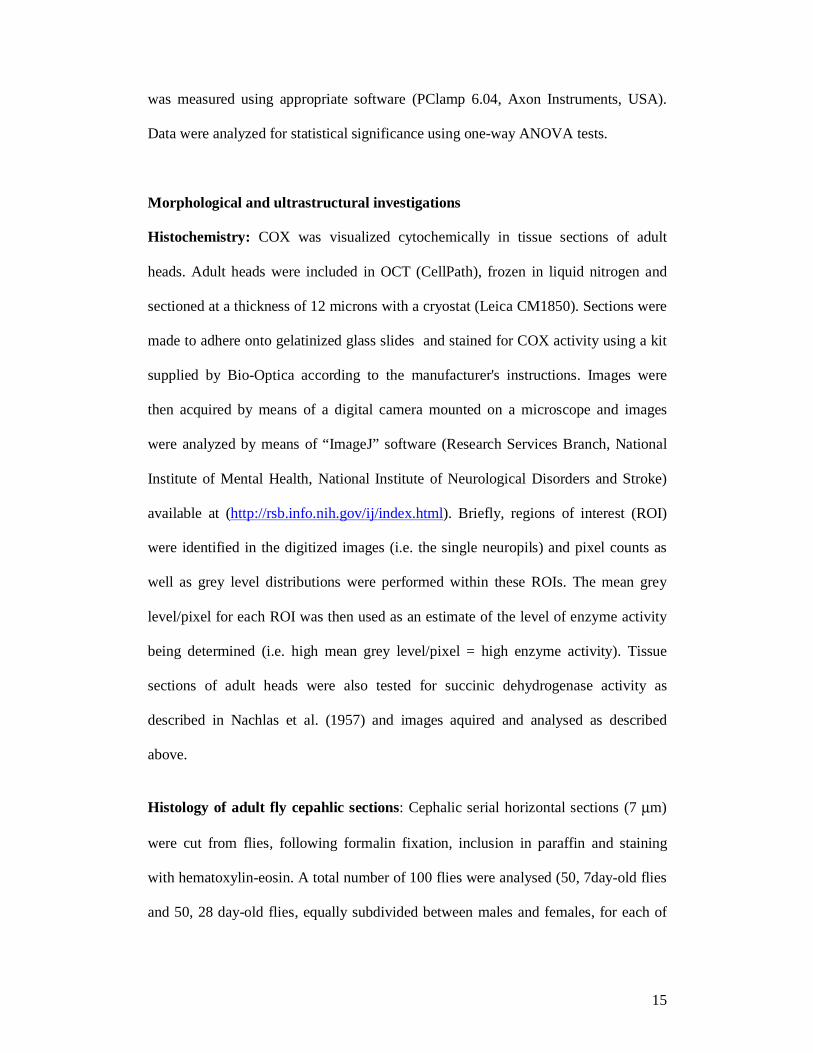
was measured using appropriate software (PClamp 6.04, Axon Instruments, USA).
Data were analyzed for statistical significance using one-way ANOVA tests.
Morphological and ultrastructural investigations
Histochemistry: COX was visualized cytochemically in tissue sections of adult
heads. Adult heads were included in OCT (CellPath), frozen in liquid nitrogen and
sectioned at a thickness of 12 microns with a cryostat (Leica CM1850). Sections were
made to adhere onto gelatinized glass slides and stained for COX activity using a kit
supplied by Bio-Optica according to the manufacturer's instructions. Images were
then acquired by means of a digital camera mounted on a microscope and images
were analyzed by means of “ImageJ” software (Research Services Branch, National
Institute of Mental Health, National Institute of Neurological Disorders and Stroke)
available at (http://rsb.info.nih.gov/ij/index.html). Briefly, regions of interest (ROI)
were identified in the digitized images (i.e. the single neuropils) and pixel counts as
well as grey level distributions were performed within these ROIs. The mean grey
level/pixel for each ROI was then used as an estimate of the level of enzyme activity
being determined (i.e. high mean grey level/pixel = high enzyme activity). Tissue
sections of adult heads were also tested for succinic dehydrogenase activity as
described in Nachlas et al. (1957) and images aquired and analysed as described
above.
Histology of adult fly cepahlic sections: Cephalic serial horizontal sections (7 µm)
were cut from flies, following formalin fixation, inclusion in paraffin and staining
with hematoxylin-eosin. A total number of 100 flies were analysed (50, 7day-old flies
and 50, 28 day-old flies, equally subdivided between males and females, for each of
16
the following genotypes (w1118; elav, Cyo; elav, Cyo/+; UAS-Surf1 23.4 IR and elav-
GAL4-Surf1 KD).
Phalloidine-rhodamine staining: Third instar larvae were dissected and treated as
described in Beramendi et al. (2005). Samples were then observed with a fluorescence
confocal microscope (BioRad, Radiance 2000), and measurements were made on
digitized images using ImageJ software. In control and knock-down larvae 4
parameters relating to muscle fiber 6, were measured: length, width of the two
extremities and of the central region of the fibre.
Electron microscopy: For transmission electron microscopy, Actin5C-GAL4-Surf1
KD and third instar wild type larvae were pierced and immediately transferred to ice-
cold fixation solution containing 3% paraformaldehyde, 2% glutaraldehyde, 100mM
sucrose and 2mM EGTA in 0.1 M sodium phosphate buffer at pH 7.2 . Samples were
fixed for 6 hours and subsequently washed overnight at 4°C in 0.1M phosphate
buffer, pH 7.2. The next day larvae were treated for 2 hours with cold (4°C) post-
fixative solution (1% OsO4 in 0.1M sodium cacodylate buffer pH 7.2), rinsed three-
four times (5 min each) in 0.1M sodium phosphate buffer pH 7.2 and then dehydrated
through ethanol series and finally by three 15 min-washes in Propylene oxide.
Samples were embedded in Epon® resin mixture (14ml EPOXY, 7ml DDSA, 9ml
MNA) additioned with 2% DMP30 solidification accelerator. The following gradual
embedding procedure was utilized: three 45 min infiltrations in resin:propylene oxide
respectively 1:2, 1:1, and 2:1, embedding in Epon® within plastic capsules and
polymerization at 60°C for three days. Ultrathin (400Å) cross sections of larval body
wall muscles were cut with a diamond knife and stained for 20 minutes in 2%

17
aqueous uranyl acetate solution followed by 30 sec staining in 5% aqueous lead
citrate solution and finally rinsed in distilled water. Sections were examined and
photographed with a Philips 200 Electron microscope.
RESULTS
Generation of Surf1 knockdown lines: We generated three independent Drosophila
transgenic lines, (23.4, 79.10, and 79.1). For each line obtained the insert map
position is given in parentheses: 23.4 (2R, 54BC), 79.1 (3R, 88D) and 79.10 (2R,
50C). Each line carried a single UAS-Surf1 Inverted Repeat autosomal insertion,
which allowed the post-transcriptional gene silencing of Surf1 via dsRNA interference
(dsRNAi), following activation by GAL4.
Ubiquitous knockdown of Surf1: The Actin5C (Act-GAL4) driver was used for
early, ubiquitous expression of dsRNAi-mediated knock-down (KD).
Northern blot analysis on the Act-GAL4 KD larvae showed the presence of an
approximately 2kb band, corresponding to the RNAisurf1 transcript, as well as a smear
starting from 0.9kb, the expected size of the Surf1 transcript, (which is clearly visible
in the controls on the left in Figure 1A), corresponding to degradation products
originating from the interference of the Surf1 gene transcript (Figure 1A). Controls
were larvae and adults from strain w1118, which was used to generate the UAS-Surf1
Inverted Repeat transgenic lines. The same 2kb band was visualized by re-
hybridization of the blot with a probe complementary to the “GFP-spacer” used in the
RNAisurf1 construct (data not shown).
18
Real-Time PCR-based (RT-PCR) quantitative analysis confirmed a drastic
reduction of Surf1 mRNA (Figure 1B), which corresponded to the virtual absence of
SURF1 protein, as demonstrated by Western blot analysis (Figure 1C).
In situ analysis on whole mount embryo preparations showed that Surf1
mRNA is expressed ubiquitously from early to late embryonic stages (Figure 2).
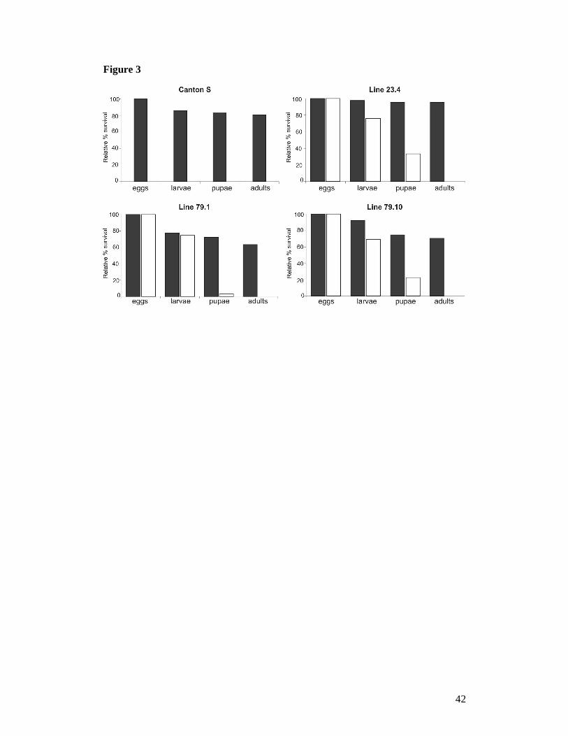
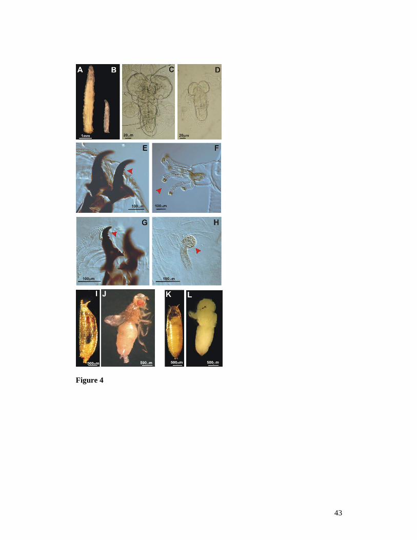
Larval lethality in Act-GAL4 Surf1 KD individuals: All KD individuals derived
from the three transgenic KD lines (Act-GAL4 KD23.4, Act-GAL4 KD79.10, and Act-
GAL4 KD79.1) showed 100% egg-to-adult lethality (Figure 3). Most individuals died
as larvae, which were very sluggish and showed impaired development. In all three
strains, the 7 day-old KD larvae appeared smaller than age-matched controls (Figure
4A and B), with undersized optic lobes (Figure 4C, D). The Act-GAL4 KD23.4 and
Act-GAL4 KD79.10 individuals had the typical cuticular features of third stage larvae
(Figure 4E and F) while, in Act-GAL4 KD79.1 larvae, the morphological features were
typical of the 2nd instar stage (Figure 4G, H). Only a few larvae, in particular Act-
GAL4 KD23.4 and Act-GAL4 KD79.10, reached the pupal stage but they did not progress
any further in development. Dissection of the KD pupae showed that these individuals
died at early imago stages (Figure 4K and L) as compared to controls (4I and J).
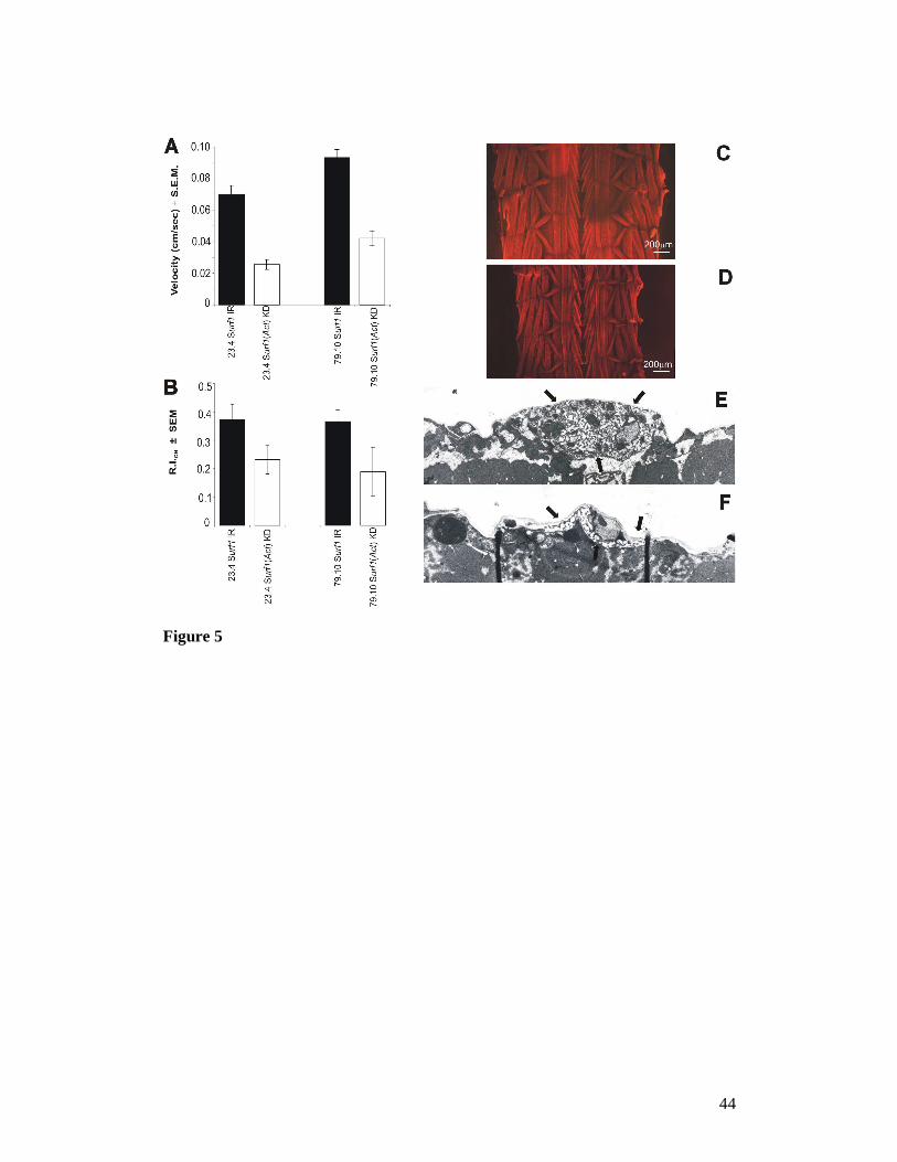
Locomotor and photobehavioural defects in Act-GAL4 Surf1 KD larvae: In order
to better characterize the “sluggishness” observed in Surf1 Act-GAL4 KD larvae, we
applied the “checker test paradigm” (see Methods), which measures both the speed of
spontaneous locomotion (i.e. in the absence of external stimuli) and the locomotor
response to light. The KD larvae showed a highly significant reduction in locomotor
speed (Figure 5A) and a profoundly altered (i.e. less responsive) photobehavioural
response (Figure 5B), which was particularly evident in Act-GAL4 KD79.10
individuals. The photobehavioural test consists in the evaluation of the light-
19
avoidance reaction, which is normally shown by wild type larvae. In the checker test
this is evaluated by the ratio, expressed in the form of a normalized relative index
(R.I.), of the time spent by a larva on the black (opaque to light) squares with respect
to the white (transparent to light) ones of a checkerboard illuminated from beneath.
Morphological and physiological examination of muscle: To understand whether
the altered locomotion of Act-GAL4 KD larvae was due to structural defects of the
muscular apparatus, we examined body wall preparations by fluorescence
microscopy. Rhodamine/phalloidin-stained preparations showed that muscle fibers
appeared normal in structure and arrangement, but they were much smaller in size,
compared to age- and development-matched controls (Figure 5C, D).
Furthermore, the measurement of the specific force output (i.e. force/muscle
fibre unit section area), developed by longitudinal muscles following a single
electrical stimulus, gave similar values in Act-GAL4 KD vs. control UAS-IR23.4 (i.e.
0.397 ± 0.083 vs 0.548 ± 0.085 mN/mm2, determined in 8 and 20 individuals
respectively). Specific force output was also determined, again without significant
differences between KD individuals and the respective controls, following tetanic
stimulation and the observed values were (Act-GAL4 KD =1.97 ± 0.37 vs UAS-IR23.4
= 2.25 ± 0.37 mN/mm2) as well as following caffeine-induced contraction (Act-GAL4
KD = 8.94 ± 1.68 vs UAS-IR23.4 = 7.78 ± 0.73 mN/mm2). Altogether, these results
allow to exclude the existence of muscular impairments involving signal transduction,
calcium release or structural abnormalities of the contractile apparatus.
To further clarify the nature of the larval locomotor defects, we examined the
neuromuscular junction (NMJ) on muscle fibers 6 and 7 by confocal and electron
microscopy. We found no abnormality in the number, size or type of synaptic
boutons, except that the structure of the subsynaptic reticulum (SSR) showed a lower
20
complexity in KD larvae (Figure 5F) than in controls (Figure 5E). Nonetheless, no
abnormality in the evoked junction potentials (EJPs) was found in the KD larvae
compared to age-matched controls (control EJP amplitude, UAS-IR23.4 = 23.01 ± 4.10;
Act-GAL4 KD23.4 EJP amplitude = 25.72 ± 6.57, amplitudes expressed in mV).
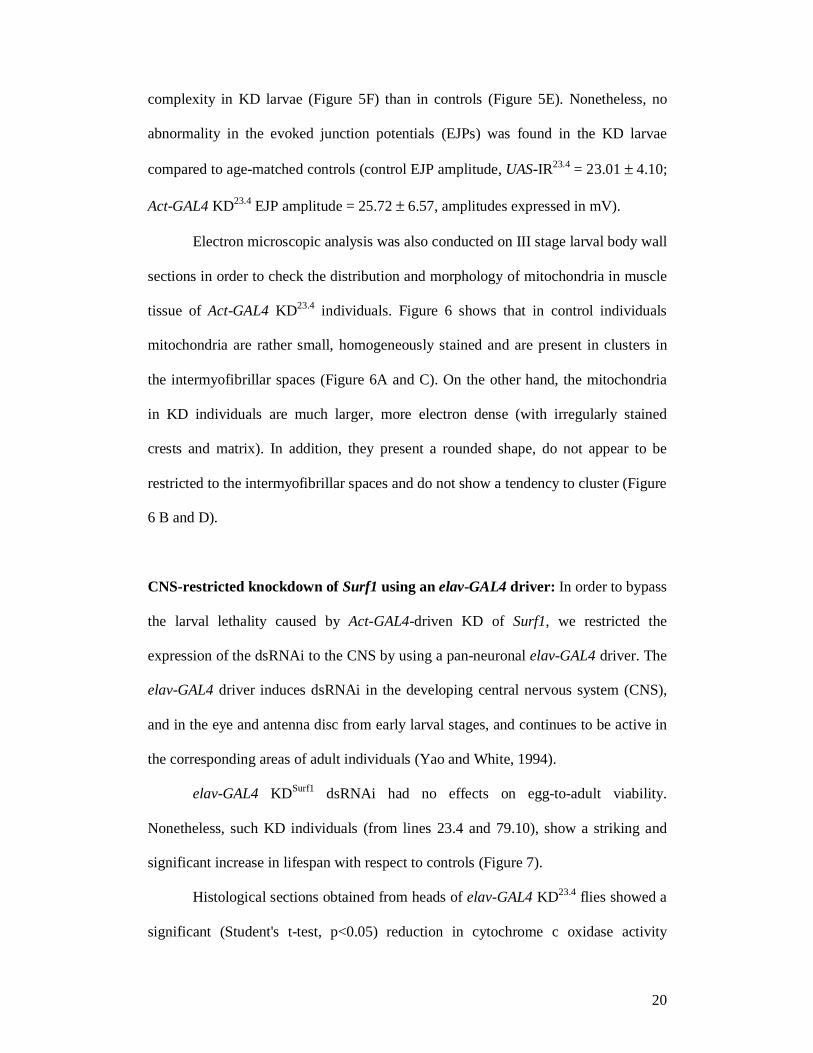
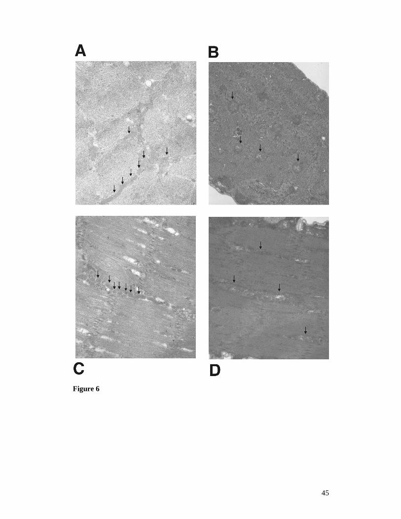
Electron microscopic analysis was also conducted on III stage larval body wall
sections in order to check the distribution and morphology of mitochondria in muscle
tissue of Act-GAL4 KD23.4 individuals. Figure 6 shows that in control individuals
mitochondria are rather small, homogeneously stained and are present in clusters in
the intermyofibrillar spaces (Figure 6A and C). On the other hand, the mitochondria
in KD individuals are much larger, more electron dense (with irregularly stained
crests and matrix). In addition, they present a rounded shape, do not appear to be
restricted to the intermyofibrillar spaces and do not show a tendency to cluster (Figure
6 B and D).
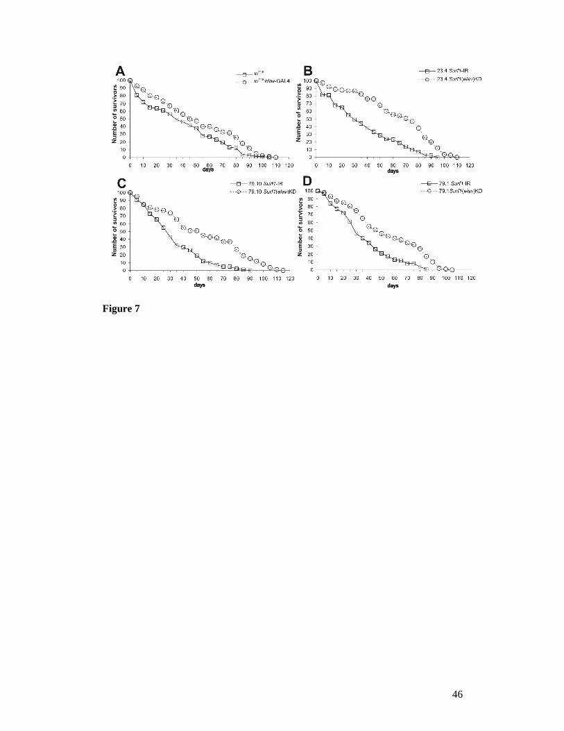
CNS-restricted knockdown of Surf1 using an elav-GAL4 driver: In order to bypass
the larval lethality caused by Act-GAL4-driven KD of Surf1, we restricted the
expression of the dsRNAi to the CNS by using a pan-neuronal elav-GAL4 driver. The
elav-GAL4 driver induces dsRNAi in the developing central nervous system (CNS),
and in the eye and antenna disc from early larval stages, and continues to be active in
the corresponding areas of adult individuals (Yao and White, 1994).
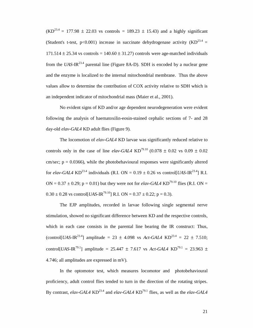
elav-GAL4 KDSurf1 dsRNAi had no effects on egg-to-adult viability.
Nonetheless, such KD individuals (from lines 23.4 and 79.10), show a striking and
significant increase in lifespan with respect to controls (Figure 7).
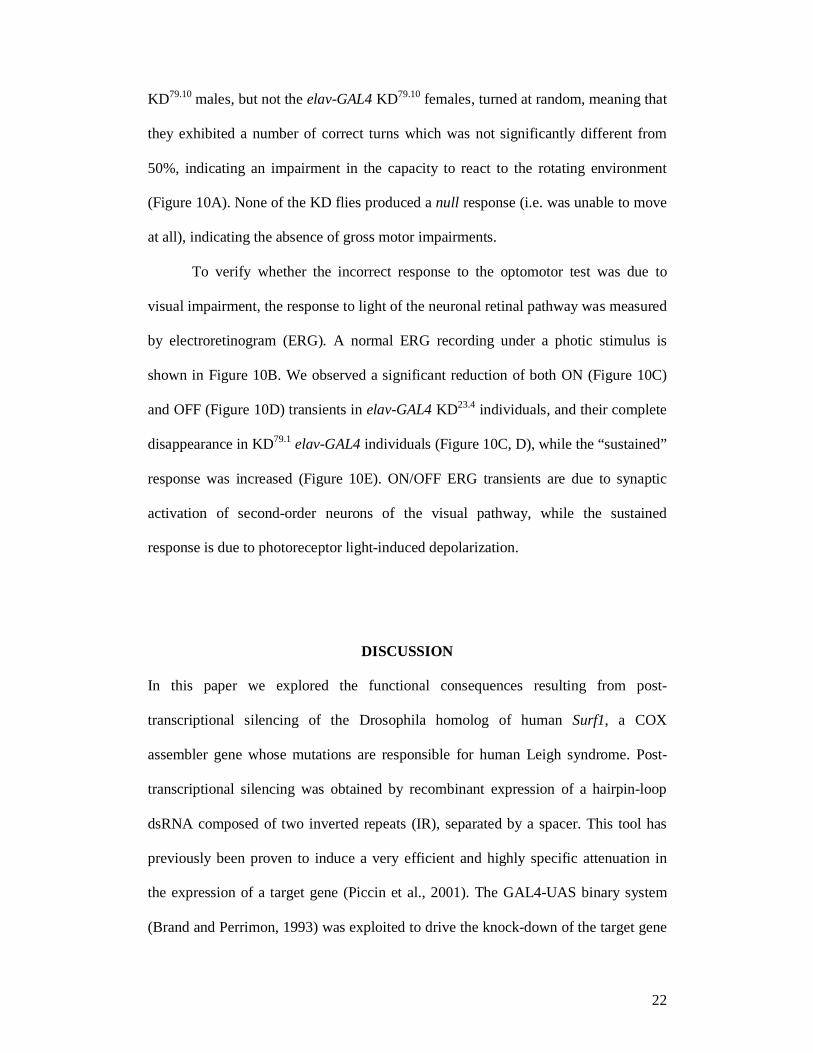
Histological sections obtained from heads of elav-GAL4 KD23.4 flies showed a
significant (Student's t-test, p<0.05) reduction in cytochrome c oxidase activity
21
(KD23.4 = 177.98 ± 22.03 vs controls = 189.23 ± 15.43) and a highly significant
(Student's t-test, p<0.001) increase in succinate dehydrogenase activity (KD23.4 =
171.514 ± 25.34 vs controls = 140.60 ± 31.27) controls were age-matched individuals
from the UAS-IR23.4 parental line (Figure 8A-D). SDH is encoded by a nuclear gene
and the enzyme is localized to the internal mitochondrial membrane. Thus the above
values allow to determine the contribution of COX activity relative to SDH which is
an independent indicator of mitochondrial mass (Maier et al., 2001).
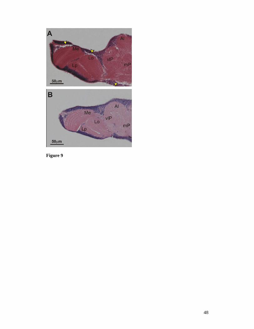
No evident signs of KD and/or age dependent neurodegeneration were evident
following the analysis of haematoxilin-eosin-stained cephalic sections of 7- and 28
day-old elav-GAL4 KD adult flies (Figure 9).
The locomotion of elav-GAL4 KD larvae was significantly reduced relative to
controls only in the case of line elav-GAL4 KD79.10 (0.078 ± 0.02 vs 0.09 ± 0.02
cm/sec; p = 0.0366), while the photobehavioural responses were significantly altered
for elav-GAL4 KD23.4 individuals (R.I. ON = 0.19 ± 0.26 vs control[UAS-IR23.4] R.I.
ON = 0.37 ± 0.29; p = 0.01) but they were not for elav-GAL4 KD79.10 flies (R.I. ON =
0.30 ± 0.28 vs control[UAS-IR79.10] R.I. ON = 0.37 ± 0.22; p = 0.3).
The EJP amplitudes, recorded in larvae following single segmental nerve
stimulation, showed no significant difference between KD and the respective controls,
which in each case consists in the parental line bearing the IR construct: Thus,
(control[UAS-IR23.4] amplitude = 23 ± 4.098 vs Act-GAL4 KD23.4 = 22 ± 7.510;
control[UAS-IR79.1] amplitude = 25.447 ± 7.617 vs Act-GAL4 KD79.1 = 23.963 ±
4.746; all amplitudes are expressed in mV).
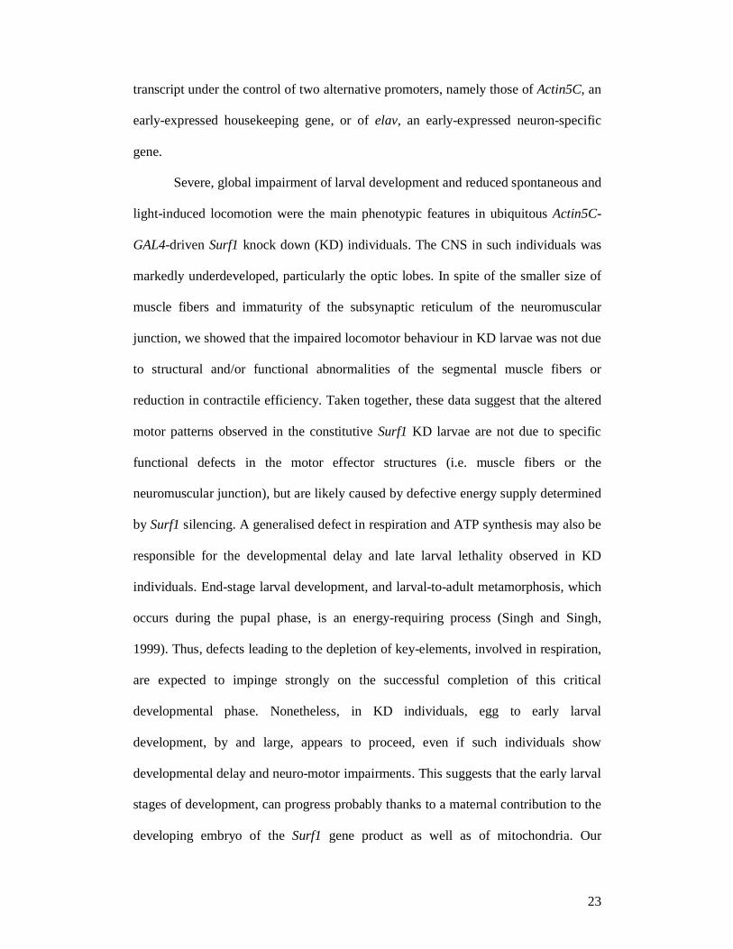
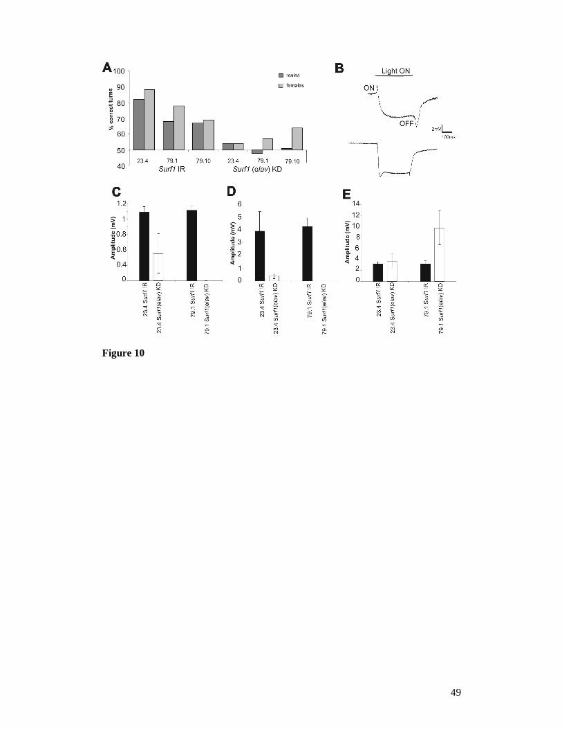
In the optomotor test, which measures locomotor and photobehavioural
proficiency, adult control flies tended to turn in the direction of the rotating stripes.
By contrast, elav-GAL4 KD23.4 and elav-GAL4 KD79.1 flies, as well as the elav-GAL4
22
KD79.10 males, but not the elav-GAL4 KD79.10 females, turned at random, meaning that
they exhibited a number of correct turns which was not significantly different from
50%, indicating an impairment in the capacity to react to the rotating environment
(Figure 10A). None of the KD flies produced a null response (i.e. was unable to move
at all), indicating the absence of gross motor impairments.
To verify whether the incorrect response to the optomotor test was due to
visual impairment, the response to light of the neuronal retinal pathway was measured
by electroretinogram (ERG). A normal ERG recording under a photic stimulus is
shown in Figure 10B. We observed a significant reduction of both ON (Figure 10C)
and OFF (Figure 10D) transients in elav-GAL4 KD23.4 individuals, and their complete
disappearance in KD79.1 elav-GAL4 individuals (Figure 10C, D), while the “sustained”
response was increased (Figure 10E). ON/OFF ERG transients are due to synaptic
activation of second-order neurons of the visual pathway, while the sustained
response is due to photoreceptor light-induced depolarization.
DISCUSSION
In this paper we explored the functional consequences resulting from post-
transcriptional silencing of the Drosophila homolog of human Surf1, a COX
assembler gene whose mutations are responsible for human Leigh syndrome. Post-
transcriptional silencing was obtained by recombinant expression of a hairpin-loop
dsRNA composed of two inverted repeats (IR), separated by a spacer. This tool has
previously been proven to induce a very efficient and highly specific attenuation in
the expression of a target gene (Piccin et al., 2001). The GAL4-UAS binary system
(Brand and Perrimon, 1993) was exploited to drive the knock-down of the target gene
23
transcript under the control of two alternative promoters, namely those of Actin5C, an
early-expressed housekeeping gene, or of elav, an early-expressed neuron-specific
gene.
Severe, global impairment of larval development and reduced spontaneous and
light-induced locomotion were the main phenotypic features in ubiquitous Actin5C-
GAL4-driven Surf1 knock down (KD) individuals. The CNS in such individuals was
markedly underdeveloped, particularly the optic lobes. In spite of the smaller size of
muscle fibers and immaturity of the subsynaptic reticulum of the neuromuscular
junction, we showed that the impaired locomotor behaviour in KD larvae was not due
to structural and/or functional abnormalities of the segmental muscle fibers or
reduction in contractile efficiency. Taken together, these data suggest that the altered
motor patterns observed in the constitutive Surf1 KD larvae are not due to specific
functional defects in the motor effector structures (i.e. muscle fibers or the
neuromuscular junction), but are likely caused by defective energy supply determined
by Surf1 silencing. A generalised defect in respiration and ATP synthesis may also be
responsible for the developmental delay and late larval lethality observed in KD
individuals. End-stage larval development, and larval-to-adult metamorphosis, which
occurs during the pupal phase, is an energy-requiring process (Singh and Singh,
1999). Thus, defects leading to the depletion of key-elements, involved in respiration,
are expected to impinge strongly on the successful completion of this critical
developmental phase. Nonetheless, in KD individuals, egg to early larval
development, by and large, appears to proceed, even if such individuals show
developmental delay and neuro-motor impairments. This suggests that the early larval
stages of development, can progress probably thanks to a maternal contribution to the
developing embryo of the Surf1 gene product as well as of mitochondria. Our
24
conclusions are supported by the phenotype observed in several Drosophila mutants
for genes related to mitochondrial functions. For instance, mutant phenotypes for
either the tamas gene, which encodes for the mitochondrial DNA polymerase catalytic
subunit (Pol-γA) (Iyengar et al., 1999, 2001), or for the gene encoding the Pol-γB
accessory subunits, both cause lethality during early pupation, concomitant with loss
of mtDNA and mitochondrial mass, and reduced cell proliferation in the CNS. A
similar phenotype is presented by Drosophila mutants for the lopo (low power) gene,
which encodes the mitochondrial single-stranded DNA-binding protein (mtSSB), a
key-component in mtDNA replication and maintenance (Maier et al., 2001). The lopo
mutants die late in the third instar before completion of metamorphosis, because of a
failure in cell proliferation. Molecular, histochemical, and physiological experiments
show a drastic decrease in mtDNA content that is coupled with the loss of respiration
in these mutants. Pleiotropic phenotypes are linked to different mutations in the
cyclope gene, which encodes a COX subunit VIc homolog (Szuplewski and Terracol,
2001), including embryonic lethality, several body growth abnormalities and aberrant
development of the eye disc and ommatidia. Larval lethal phenotypes were also
obtained in mutants for the alpha subunit of mitochondrial ATP-synthase (complex V)
(Talamillo et al., 1998) and for subunit 9 of the mitochondrial ubiquinol-cytochrome c
reductase (complex III) (Frolov et al., 2000). Taken together, these studies on
Drosophila mutants show early or late larval lethality often associated with
neuromotor and/or photobehavioural defects, suggesting that these functions are
energy-demanding and are exquisitely sensitive to reductions in the energy supply
provided by mitochondrial oxidative phosphorylation.
CNS-wide silencing of Surf1 was obtained by driving transcription of the target gene-
specific dsRNA with elav-GAL4, and allowed us to bypass the larval lethality
25
observed with the Actin5C-GAL4 driver. In this case, individuals survived to the adult
phase with no overt signs of impairment. Intriguingly, such individuals actually
showed a strikingly enhanced survival with respect to controls. The larval stages of
the elav-GAL4 Surf1 KD individuals were morphologically comparable to the
corresponding parental line controls (i.e. UAS-IR lines), including development of the
CNS, although a slight impairment in locomotor activity and photobehaviour was
noted also in these lines. Once again, electrophysiological analyses produced no
evidence of functional synaptic impairments. However, the photobehaviour of adult
elav-GAL4-KD flies, measured by the optomotor test paradigm, was clearly impaired,
suggesting an abnormality in the visual pathway, which was confirmed by alterations
in the ERG response. These alterations consisted in a reduction or disappearance of
ON and OFF transients and by an increase of the “sustained response”. The ERG
consists in an extracellular recording from the Drosophila eye that measures light-
induced depolarization of photoreceptors (the “sustained response”) and synaptic
activation of second-order neurons in the visual pathway (Hotta and Benzer, 1969;
Pak et al., 1969; Heisenberg, 1971). The latter synaptic events occur at the onset and
termination of a light pulse and are represented by the ON- and OFF-transients, which
are generated at the level of the R1 to L1/L2 histaminergic synapses (Wu and Wong,
1977). Several mutations known to block synaptic transmission, including
synaptotagmin (DiAntonio and Schwarz, 1994), rop (Harrison et al., 1994), and csp
(Zinsmaier et al., 1994), decrease or abolish the ON/OFF transients (Heisenberg,
1971). Taken together, the optomotor and ERG results in our elav-GAL4 KD flies are
likely to be due to complex alterations in the eye and CNS, involving several visual
circuits. This conclusion is also supported by the demonstration that the neural foci
responsible for the ERG transients are different from those mediating the optomotor
26
response (Buchner et al., 1978; Sandrelli et al., 2001). As in the case of the Actin5C-
GAL4 KD larvae, the morphological and functional analysis of the elav-GAL4 KD
larvae and adult flies indicate that the behavioural abnormalities are not due to
functional failure of single or specific components of the neuromotor and/or
photoreceptive structures, but are the consequence of subtle widespread nervous
system defects. Furthermore, following a detailed histological analysis, we were
unable to show any overt signs of neurodegenerative damage in the brains of such
individuals. A direct effect of Surf1 silencing on mitochondrial respiration was
demonstrated by the significant reduction in the histochemical reaction to COX
observed in the optic lobes of the elav-GAL4 KD adult flies. In addition, the parallel
strong increase in the SDH levels mesaured in the same tissues suggests that there is
an increase in mitochondrial mass, which may be tentatively interpreted as a
compensatory reaction to the decrease in COX. Interestingly, electron microscopic
observation of larval body wall muscle fibers, shows that in Act-GAL4 KD
individuals, mitochondria are much larger than normal, with evident morphological
alterations (in particular regarding the organization of the internal membranes) while
also showing an altered distribution within the muscle tissue (i.e. in controls
mitochondria tend to form clusters within the intermyofibrillar spaces, while in KD
individuals they do not). Although we do not have any direct evidence, it is tempting
to speculate that perhaps the increased size of the mitochondria in KD individuals
could be due to mitochondrial fusion, a process which is thought to occur as a
response to mitochondrial damage and which could protect cells from entering the
apoptotic pathway (Meeusen and Nunnari, 2005). Altogether, the above results
strongly support the involvement of respiratory deficiency in determining the
developmental and functional impairments observed in KD individuals.

27
The structural and functional abnormalities at the nervous system level, as well
as the global developmental arrest, observed following Surf1 KD in Drosophila are
largely concordant with the human LSSurf1-/- phenotype, which predominantly affects
the brain and, to a lesser extent, the skeletal muscle apparatus, and is also frequently
associated with poor growth and failure to thrive. In the light of these considerations it
will be of great interest to conduct further studies on the molecular and biochemical
effects of Surf1 KD in the adult fly, as well as at the level of specific organs and
structures. These investigations will contribute to our understanding on the specific
involvement of the SURF1 protein and, more broadly, of mitochondrial energy
metabolism, to body growth, neural development, and neurodegeneration.
ACKNOWLEDGEMENTS
This study was supported by the collaborative programme between CNR and MIUR
"Legge 449/97" (Grant N° CU04.00067 to R.C.), Fondazione Pierfranco e Luisa
Mariani (to M.Z.), Italian Ministry of Health grant RF-2002/158 (to M.Z.),
Fondazione Cariplo (to M.Z.), EUMITOCOMBAT network grant from the EU-FP6
(to M.Z.) and by grant Telethon N. GP0048Y01 (to R.C.). Thanks to Dr. Giuliano
Carlesso (Dept. of Human Anatomy and Physiolology, University of Padova) for
providing haematoxilin/eosin-stained sections of adult fly CNS.
REFERENCES
AGOSTINO, A., INVERNIZZI, F., TIVERON, C., FAGIOLARI, G., PRELLE, A., et al., 2003
Constitutive knockout of Surf1 is associated with high embryonic lethality,

28
mitochondrial disease and cytochrome c oxidase deficiency in mice. Hum. Mol.
Genet. 12: 1-15.
BERAMENDI, A., PERON, S., MEGIGHIAN, A., REGGIANI, C., AND R. CANTERA, 2005
The inhibitor kappaB-ortholog Cactus is necessary for normal neuromuscular
function in Drosophila melanogaster. Neuroscience. 134: 397-406.
BRAND, A. H AND N. PERRIMON, 1993 Targeted gene expression as a means of
altering cell fates and generating dominant phenotypes. Development 118: 401-
15.
BUCHNER, E., GOTZ, K. G. AND C. STRAUB, 1978 Elementary detectors for vertical
movement in the visual system of Drosophila. Biol. Cybern. 31: 235-42.
COENEN, M., VAN DEN HEUVEL, L. P., NIJTMANS, L. G., MORAVA, E., MARQUARDT,
I., et al., 1999 SURFEIT-1 gene analysis and two-dimensional blue native gel
electrophoresis in cytochrome c oxidase deficiency. Biochem. Biophys. Res.
Commun. 265: 339-344.
COLOMBO, P., YON, J., GARSON, K. AND M. FRIED, 1996 Conservation of the
organization of five tightly clustered genes over 600 million years of divergent
evolution. Proc. Natl. Acad. Sci. U S A. 89: 6358-62.
DIANTONIO, A. AND T. L. SCHWARZ, 1994 The effect on synaptic physiology of
synaptotagmin mutations in Drosophila. Neuron 12: 909-20.
DIMAURO, S. AND E. A. SCHON, 2003 Mitochondrial respiratory-chain diseases. New
Eng. J. Med. 348: 2656-2668.
ECHALIER, G., 1997 Drosophila cells in culture. Academic Press, San Diego, USA.
FADIC, R. AND D. R. JOHNS, 1996 Clinical spectrum of mitochondrial diseases.
Semin. Neurol. 16: 11-20.
29
FARINA, L., CHIAPPARINI, L., UZIEL, G., BUGIANI, M. AND M. ZEVIANI, 2002 MR
findings in Leigh syndrome with COX deficiency and Surf-1 mutations. AJNR
23: 1095-1100.
FROLOV, M. V., BENEVOLENSKAYA, E. V. AND J. A. BIRCHLER, 2000 The oxen gene
of Drosophila encodes a homolog of subunit 9 of yeast ubiquinol-cytochrome c
oxidoreductase complex: evidence for modulation of gene expression in
response to mitochondrial activity. Genetics 156: 1727-36.
HANSON, B. J., CARROZZO, R., PIEMONTE, F., TESSA, A., ROBINSON, B. H. AND R. A.
CAPALDI, 2001 Cytochrome c oxidase-deficient patients have distinct subunit
assembly profiles. J. Biol. Chem. 276: 16296-16301.
HARRISON, S. D., BROADIE, K., VAN DE GOOR, J. AND G. M. RUBIN, 1994 Mutations
in the Drosophila rop gene suggest a function in general secretion and synaptic
transmission. Neuron 13: 555-66.
HASSAN, J., BUSTO, M., IYENGAR, B. AND A. R. CAMPOS, 2000 Behavioural
characterization and genetic analysis of the Drosophila melanogaster larval
response to light as revealed by a novel individual assay. Behav. Genet. 30: 59-
69.
HEISENBERG, M., 1971 Separation of receptor and lamina potentials in the
electroretinogram of normal and mutant Drosophila. J. Exp. Biol. 55: 85-100.
HOTTA, Y. AND S. BENZER, 1969 Abnormal electroretinograms in visual mutants of
Drosophila. Nature 222: 354-6.
IYENGAR, B., ROOTE, J. AND A. R. CAMPOS, 1999 The tamas gene, identified as a
mutation that disrupts larval behavior in Drosophila melanogaster, codes for the
mitochondrial DNA polymerase catalytic subunit (DNApol-gamma125).
Genetics 153: 1809-24.
30
IYENGAR, B., ROOTE, J. AND A. R. CAMPOS, 2001 Erratum. Genetics 159: 1867.
KESHISHIAN, H., BROADIE, K., CHIBA, A. AND M. BATE, 1996 The Drosophila
neuromuscular junction: a model system for studying synaptic development and
function. Ann. Rev. Neurosci. 19: 545-75.
LEIGH, D.,1951 Subacute necrotizing encephalomyelopathy in an infant. J. Neurol.
Neurosurg. Psychiat. 14: 216-221.
LEHMANN, R. AND D. TAUTZ 1994 in situ hybridization to RNA. Methods in Cell
Biology 44: 575-598
MAIER, D., FARR, C. L., POECK, B., ALAHARI, A., VOGEL, M., et al., 2001
Mitochondrial single-stranded DNA-binding protein is required for
mitochondrial DNA replication and development in Drosophila melanogaster.
Mol. Biol. Cell. 12: 821-30.
MEEUSEN, S. L., AND J. NUNNARI, 2005 How mitochondria fuse. Curr. Opin. Cell.
Biol. 17:389-94.
NACHLAS, M. M., YOUNG, A. C. AND A. M. SELIGMAN 1957 Problems of enzymatic
localization by chemical reactions applied to tissue sections. J. Histochem.
Cytochem, 5 : 565-583.
NIJTMANS, L. G. J., ARTAL SANZ, M., BUCKO, M., FARHOUD, M. H., FEENSTRA, M.,
et al., 2001 Shy1p occurs in a high molecular weight complex and is required
for efficient assembly of cytochrome c oxidase in yeast. FEBS Lett. 498: 46-51.
O’DONNELL, P. T. AND S. I. BERNSTEIN, 1988 Molecular and ultrastructural defects in
a Drosophila myosin heavy chain mutant: differential effect on muscle function
produced by similar thick filament abnormalities. J. Cell Biol. 107: 2601-2612.
PAK, W. L., GROSSFIELD, J. AND N. V. WHITE, 1969 Nonphototactic mutants in a
study of vision of Drosophila. Nature 222: 351-4.

31
PÉQUIGNOT, M. O., DEY, R., ZEVIANI, M., TIRANTI, V., GODINOT, C., et al., 2001
Mutations in the SURF1 gene associated with Leigh syndrome and cytochrome
c oxidase deficiency. Human Mutation 17: 374-381.
PFAFFL, M. W., 2001 A new mathematical model for relative quantification in real-
time RT-PCR. Nucleic Acids Res. 29: e45.
PICCIN, A., SALAMEH, A., BENNA, C., SANDRELLI, F., MAZZOTTA, G., et al., 2001
Efficient and heritable functional knock-out of an adult phenotype in Drosophila
using a GAL4-driven hairpin RNA incorporating a heterologous spacer. Nucleic
Acids Res. 29: e55-5.
POYAU, A., BUCHET, K., AND C. GODINOT, 1999 Sequence conservation from human
to prokaryotes of Surf1, a protein involved in cytochrome c oxidase assembly,
deficient in Leigh syndrome. FEBS Lett. 462:416-20.
ROBERTS, D. B. AND G. N. STANDEN, 1998 The elements of Drosophila biology and
genetics, pp. 1 - 54 in Drosophila: A practical approach. edited by D.B.
Roberts, IRL Press, Oxford,UK.
SANDRELLI, F., CAMPESAN, S., ROSSETTO, M. G., BENNA, C., ZIEGER, E., et al., 2001
Molecular dissection of the 5’ region of no-on-transientA of Drosophila
melanogaster reveals cis-regulation by adjacent dGpi sequences. Genetics 157:
765-775.
SINGH, K. AND R. N. SINGH, 1999 Metamorphosis of the central nervous system of
Drosophila melanogaster Meigen (Diptera: Drosophilidae) during pupation
Journal of Biosciences 24: 345–360.
SPRADLING, A. C. AND G. M. RUBIN, 1982 Transposition of P elements into
Drosophila germ line chromosomes. Science 218: 341-352.
32
STEWARD, B. A., ATWOOD, H. L., RENGER, J. J., WANG, J. AND C. F. WU, 1994
Improved stability of Drosophila larval neuromuscular preparations in
haemolymph-like physiological solutions. J. Comp. Physiol. 175: 179-191.
SZUPLEWSKI, S. AND R. TERRACOL, 2001 The cyclope gene of Drosophila encodes a
cytochrome c oxidase subunit VIc homolog. Genetics 158: 1629-43.
TALAMILLO, A., CHISHOLM, A. A., GARESSE, R. AND H. T. JACOBS, 1998 Expression
of the nuclear gene encoding mitochondrial ATP synthase subunit alpha in early
development of Drosophila and sea urchin. Mol. Biol. Rep. 25: 87-94.
TIRANTI, V., GALIMBERTI, C., NIJTMANS, L., BOVOLENTA, S., PERINI, M. P. AND M.
ZEVIANI, 1999a Characterization of SURF-1 expression and Surf-1p function in
normal and disease conditions. Hum. Mol. Genet. 8: 2533-2540.
TIRANTI, V., HOERTNAGEL, K., CARROZZO, R., GALIMBERTI, C., MUNARO, M., et al.,
1998 Mutations of SURF-1 in Leigh disease associated with cytochrome c
oxidase deficiency. Am. J. Hum. Genet. 63: 1609-1621.
TIRANTI, V., JAKSH, M., HOFMAN, S., GALIMBERTI, C., HOERTNAGEL, K., et al.,
1999b Loss-of-function mutations of SURF1 are specifically associated with
Leigh syndrome with cytochrome c oxidase deficiency. Ann. Neurol. 46: 161-
166.
VAN DER MEER, J. M., 1977 Optically clean and permanent whole mount preparation
for phase-contrast microscopy of cuticular structures of insect larvae. DIS. 52:
160.
VAZQUEZ-MARTINEZ, O., CANEDO-MERINO, R., DIAZ-MUNOZ, M. AND J. R. RIESGO-
ESCOVAR, 2003 Biochemical characterization, distribution and phylogenetic
analysis of Drosophila melanogaster ryanodine and IP3 receptors, and
thapsigargin sensitive Ca2+ ATPase. J. Cell. Sci. 116: 2483-2494.
33
WU, C. F. AND F. WONG, 1977 Frequency characteristics in the visual system of
Drosophila: genetic dissection of electroretinogram components. J. Gen.
Physiol. 69: 705-24.
YAO, K-M AND K. WHITE, 1994 Neural specificity of elav expression: defining a
Drosophila promoter for directing expression to the nervous system. J.
Neurochem. 63: 41-51.
YAO, J. AND E. A. SHOUBRIDGE 1999 Expression and functional analysis of SURF1 in
Leigh syndrome patients with cytochrome c oxidase deficiency. Hum. Mol.
Genet. 8:2541-9.
ZHU, Z., YAO, J., JOHNS, T., FU, K., DE BIE, I., et al., 1998 SURF1, encoding a factor
involved in the biogenesis of cytochrome c oxidase, is mutated in Leigh
syndrome. Nature Genetics 20: 337-343.
ZINSMAIER, K. E., EBERLE, K. K., BUCHNER, E., WALTER, N. AND S. BENZER, 1994
Paralysis and early death in cysteine string protein mutants of Drosophila.
Science 263: 977-80.
ZORDAN, M. A., MASSIRONI, M., DUCATO, M. G., TE KRONNIE, G., COSTA, R.,
REGGIANI, C., CHAGNEAU, C., MARTIN. J. R. AND A. MEGIGHIAN, 2005
Drosophila CAKI/CMG protein, a homolog of human CASK, is essential for
regulation of neurotransmitter vesicle release. J. Neurophysiol. 94:1074-83.
34
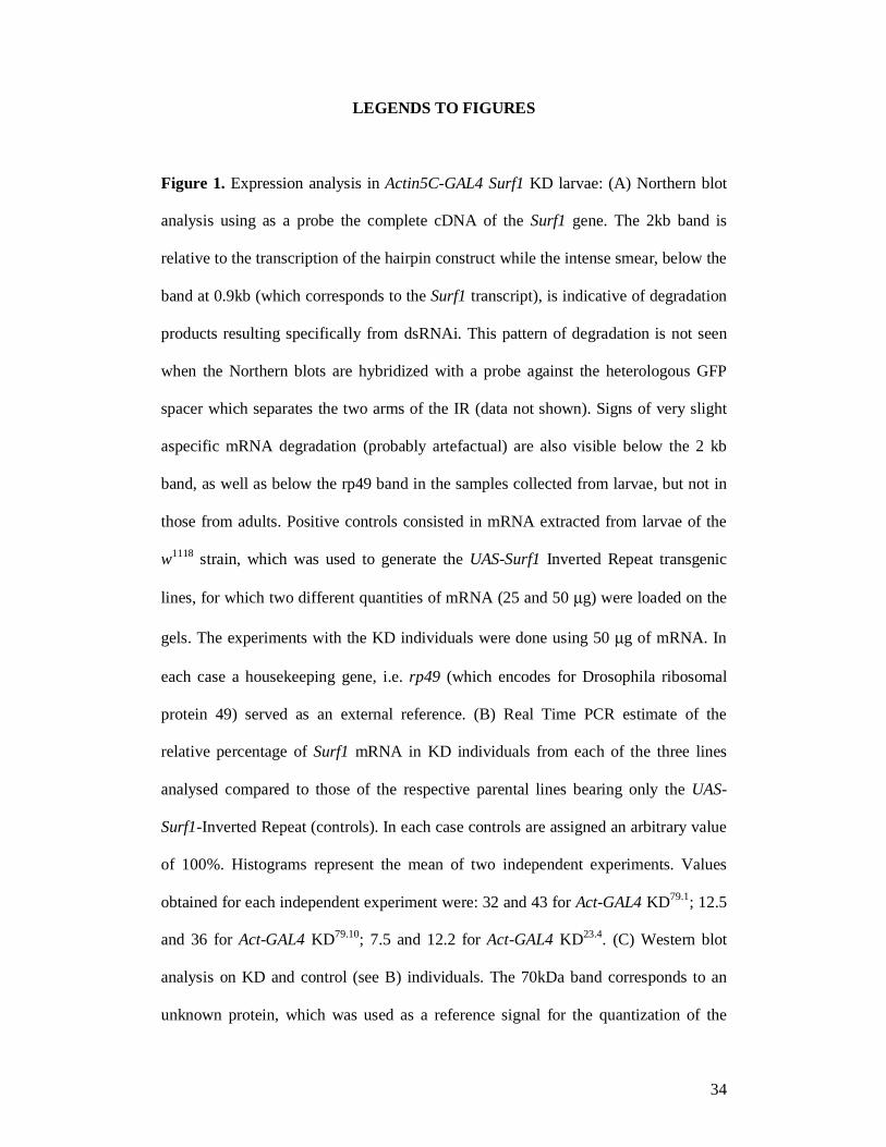
LEGENDS TO FIGURES
Figure 1. Expression analysis in Actin5C-GAL4 Surf1 KD larvae: (A) Northern blot
analysis using as a probe the complete cDNA of the Surf1 gene. The 2kb band is
relative to the transcription of the hairpin construct while the intense smear, below the
band at 0.9kb (which corresponds to the Surf1 transcript), is indicative of degradation
products resulting specifically from dsRNAi. This pattern of degradation is not seen
when the Northern blots are hybridized with a probe against the heterologous GFP
spacer which separates the two arms of the IR (data not shown). Signs of very slight
aspecific mRNA degradation (probably artefactual) are also visible below the 2 kb
band, as well as below the rp49 band in the samples collected from larvae, but not in
those from adults. Positive controls consisted in mRNA extracted from larvae of the
w1118 strain, which was used to generate the UAS-Surf1 Inverted Repeat transgenic
lines, for which two different quantities of mRNA (25 and 50 µg) were loaded on the
gels. The experiments with the KD individuals were done using 50 µg of mRNA. In
each case a housekeeping gene, i.e. rp49 (which encodes for Drosophila ribosomal
protein 49) served as an external reference. (B) Real Time PCR estimate of the
relative percentage of Surf1 mRNA in KD individuals from each of the three lines
analysed compared to those of the respective parental lines bearing only the UAS-
Surf1-Inverted Repeat (controls). In each case controls are assigned an arbitrary value
of 100%. Histograms represent the mean of two independent experiments. Values
obtained for each independent experiment were: 32 and 43 for Act-GAL4 KD79.1; 12.5
and 36 for Act-GAL4 KD79.10; 7.5 and 12.2 for Act-GAL4 KD23.4. (C) Western blot
analysis on KD and control (see B) individuals. The 70kDa band corresponds to an
unknown protein, which was used as a reference signal for the quantization of the
35
SURF1 signal. Controls consisted in mitochondrial protein extracts from larvae,
pupae or adults of the w1118 strain (see above).
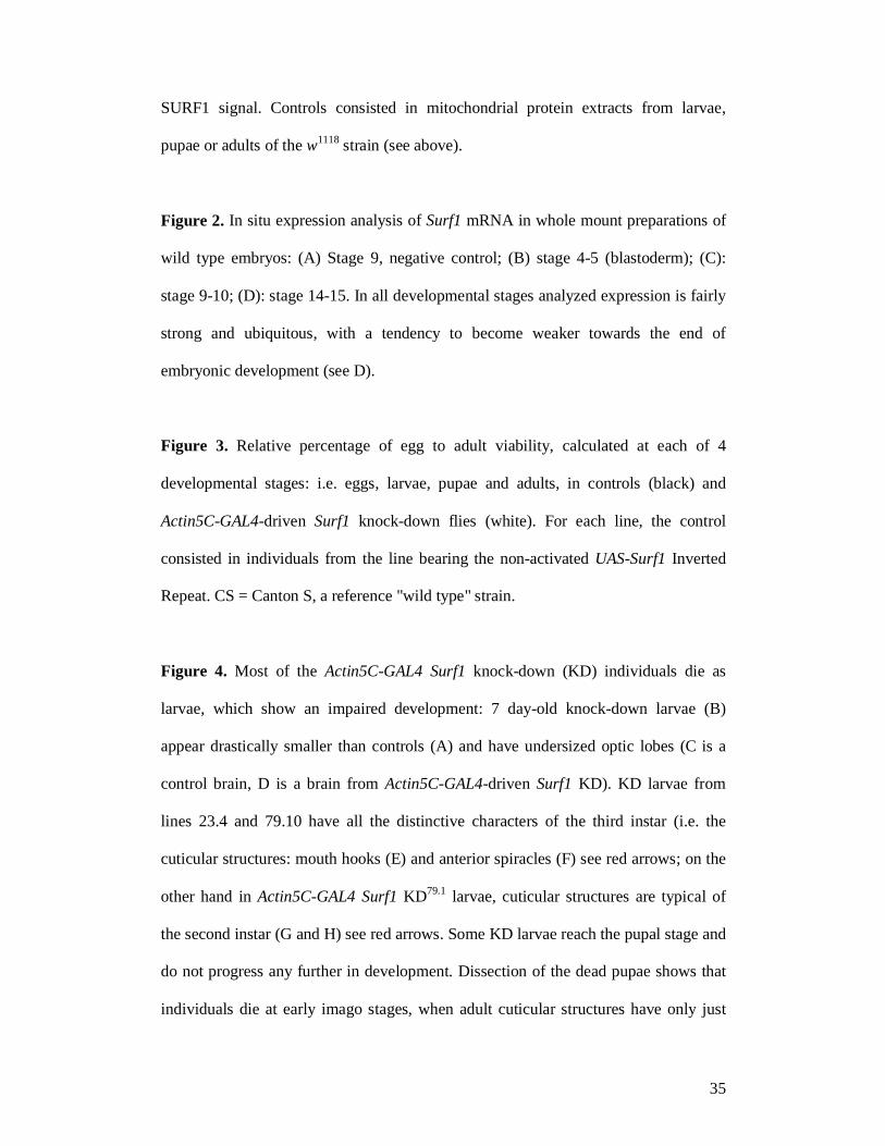
Figure 2. In situ expression analysis of Surf1 mRNA in whole mount preparations of
wild type embryos: (A) Stage 9, negative control; (B) stage 4-5 (blastoderm); (C):
stage 9-10; (D): stage 14-15. In all developmental stages analyzed expression is fairly
strong and ubiquitous, with a tendency to become weaker towards the end of
embryonic development (see D).
Figure 3. Relative percentage of egg to adult viability, calculated at each of 4
developmental stages: i.e. eggs, larvae, pupae and adults, in controls (black) and
Actin5C-GAL4-driven Surf1 knock-down flies (white). For each line, the control
consisted in individuals from the line bearing the non-activated UAS-Surf1 Inverted
Repeat. CS = Canton S, a reference "wild type" strain.
Figure 4. Most of the Actin5C-GAL4 Surf1 knock-down (KD) individuals die as
larvae, which show an impaired development: 7 day-old knock-down larvae (B)
appear drastically smaller than controls (A) and have undersized optic lobes (C is a
control brain, D is a brain from Actin5C-GAL4-driven Surf1 KD). KD larvae from
lines 23.4 and 79.10 have all the distinctive characters of the third instar (i.e. the
cuticular structures: mouth hooks (E) and anterior spiracles (F) see red arrows; on the
other hand in Actin5C-GAL4 Surf1 KD79.1 larvae, cuticular structures are typical of
the second instar (G and H) see red arrows. Some KD larvae reach the pupal stage and
do not progress any further in development. Dissection of the dead pupae shows that
individuals die at early imago stages, when adult cuticular structures have only just

36
everted (I and J are control pupae respectively with and without puparium, K and L
are Actin5C-GAL4 Surf1 KD pupae).
Figure 5. (A) The speed of locomotion (cm/sec) measured in Actin5C-GAL4 Surf1
KD larvae is significantly different from that of the respective controls for both lines
analyzed (23.4 vs control, P<0.0001, n = 23 and 21 respectively; 79.10 vs control, P=
0.0366, n= 20 and 10 respectively). (B) Response in the photobehavioural assay of
Surf1 KD and control larvae tested using the "checker test" (see Materials and
Methods). Briefly, the photobehavioural test consists in the evaluation of the larval
light-avoidance reaction. In the checker test paradigm this is evaluated by the ratio,
expressed in the form of a normalized relative index (R.I.), of the time spent by a
larva on the black (opaque to light) squares with respect to the white ones (transparent
to light) of a checker board illuminated from beneath. KD individuals from both lines
showed a significant decrease in their ability to avoid the light stimulus (Act-GAL4
KD23.4 vs control[UAS-IR23.4]; p = 0.032) and (Act-GAL4 KD79.10 vs control[UAS-
IR79.10]; p = 0.02). (C, D) An example of phalloidin-rhodamine staining of whole
mount larval body wall preparations shows no major anomalies in muscle structure
and arrangement in Actin5C-GAL4 Surf1 KD79.1 individuals (D), although a clear
reduction in size is evident with respect to controls (C). (E, F) Cross sectional
ultrastructure of glutamatergic synapses from third segment muscles 6 or 7 of stage 3
larvae. (E) Electron micrograph of a Type 1 bouton in a control (UAS-IR23.4) larva,
showing the well developed subsynaptic reticulum (SSR) see arrows; (F)
Representative micrograph of a Type 1 bouton in an Actin5C-GAL4 Surf1 KD23.4
larva, showing a reduction in the complexity of the SSR, which is more typical of a
mid 1st to 2nd stage wild type larva. Magnification (E, F) = 2842 X.

37
Figure 6. Electron micrograph of larval body wall muscle fibres showing
mitochondria (arrows) in control larvae bearing only the UAS-Surf1-Inverted Repeat
and in Actin5C-GAL4 KD individuals. Cross section of (A): control and (B): Actin5C-
GAL4 KD larva. Longitudinal section of (C) control and (D) Actin5C-GAL4 KD larva.
Magnification for all figures: 10,000 X. Mitochondria in controls tend to be rather
small, elongated and homogeneously stained, forming clusters within the
intermyofibrillar spaces, whereas mitochondria in KD individuals are much larger and
round with an apparently disorganized internal structure (i.e. matrix and cristae).
Furthermore the latter mitochondria are not typically found in clusters within the
muscle fiber.
Figure 7. Graphs showing the number of adult flies surviving after up to 115 days.
(A): w1118 (the background used for transgenesis) and elav-GAL4 x w1118 (B): 23.4
Surf1 IR and elav-GAL4 KD23.4 (C): 79.10 Surf1 IR and elav-GAL4 KD79.10 (D): 79.1
Surf1 IR and elav-GAL4 KD79.1. For each graph the statistical comparison of the pairs
of curves (i.e. KD vs parental IR line) was done using the Wilcoxon test. A: p=Not
Significant; B: p = 0.008; C: p = 0.02; D: p= Not Significant.
Figure 8. (A) cytochrome c oxidase (COX) activity in adult control flies bearing only
the UAS-Surf1-Inverted Repeat; (B) COX activity in elav-GAL4 Surf1 KD adult flies
from line 23.4; (C) Succinate dehydrogenase (SDH) activity in adult control flies
bearing only the UAS-Surf1-Inverted Repeat; (D) Succinate dehydrogenase activity in
elav-GAL4 Surf1 KD adult flies from line 23.4.Nuclear-encoded SDH is localized to
the internal mitochondrial membrane. Thus comparison of COX levels to those of
38
SDH provide an indication of the contribution of COX activity relative to SDH, an
independent indicator of mitochondrial mass. In each image analyzed, the regions of
interest (ROI) were limited to the optic neuropils (i.e. lamina, medulla, lobula and
lobula plate). ROIs were identified in the digitized images (i.e. the single neuropils)
and pixel counts as well as grey level distributions were performed within these ROIs.
The mean grey level/pixel for each ROI was then used as an estimate of the level of
enzyme activity being determined (i.e. high mean grey level/pixel = high enzyme
activity). Sections obtained from heads of elav-GAL4 KD23.4 flies showed a
significant (Student's t-test, p<0.05) reduction in cytochrome c oxidase activity
(KD23.4 = 177.98 ± 22.03 vs controls[UAS-IR23.4] = 189.23 ± 15.43) and a highly
significant (Student's t-test, p<0.001) increase in succinate dehydrogenase activity
(KD23.4 = 171.514 ± 25.34 vs controls[UAS-IR23.4] = 140.60 ± 31.27). In particular
SDH activity in KD individuals is greater than that observed in controls, suggesting
an increase in mitochondrial mass in the former. La= Lamina, Me=Medulla, Lo=
Lobula, Lp= Lobula plate.
Figure 9. Haematoxilin-eosin stains of paraffin-embedded frontal cephalic sections of
adult flies. (A): control flies bearing only the UAS-Surf1-Inverted Repeat; (B): elav-
GAL4 Surf1 KD adult flies from line 23.4 . Me = medulla, Lo = Lobula, Lp = Lobula
plate, Al = antennal lobe, vlP = ventrolateral protocerbrum, mP = medial
protocerebrum. Arrows = sectioning artifacts.
Figure 10. (A) Optomotor behaviour in elav-GAL4 Surf1 KD and control (UAS-IR)
adults. KD flies (males and females) from lines 23.4 and 79.1 and only KD males
from line 79.10 turned at random, giving mean values of correct turns not

39
significantly different from 50%. On the other hand, controls (UAS-IR23.4, UAS-IR 79.1
and UAS-IR79.10) turned in the direction of the moving environment 70–80% of the
time. For each genotype 20 individuals (10 males and 10 females) were tested. Each
fly was given 10 trials, and each time the direction of rotation of the stripes was
changed; (B) Top: An example of a wild type electroretinographic (ERG) response
showing the major features: the ON and OFF transients with the sustained response
between the two transients. (B) Bottom: Example of an ERG response from an elav-
GAL4 KD individual from line 79.1; in this case the ON and OFF transients are
completely missing. (C, D, E) results of ERGs recorded from elav-GAL4 Surf1 KD
individuals from lines 23.4 and 79.1 and controls (as in A). Graphs represent mean
amplitudes (mV ± SD) of ON (C) and OFF (D) transients (due to synaptic activation
of second order neurons of the visual pathway) and of the sustained response (E) (due
to photoreceptor light-induced depolarization). Mean amplitude of ON and OFF
transients for both lines, of KD vs control(as above) individuals were significantly
different (P<0.05). However, only the sustained response of KD individuals from line
79.1 was significantly different to the relative control (as above). The graphs showing
the ERG data are relative to 16-29 individuals for line 23.4 and 11-14 individuals in
the case of line 79.1.

40
Figure 1

41
Figure 2
42
Figure 3
43
Figure 4
44
Figure 5
45
Figure 6
46
Figure 7

47
Figure 8
48
Figure 9
49
Figure 10