
Characterization of the Transcriptional Properties of ...
169
Louisiana State University LSU Digital Commons LSU Historical Dissertations and eses Graduate School 1989 Characterization of the Transcriptional Properties of Equine Infectious Anemia Virus. Siyamak Rasty Louisiana State University and Agricultural & Mechanical College Follow this and additional works at: hps://digitalcommons.lsu.edu/gradschool_disstheses is Dissertation is brought to you for free and open access by the Graduate School at LSU Digital Commons. It has been accepted for inclusion in LSU Historical Dissertations and eses by an authorized administrator of LSU Digital Commons. For more information, please contact [email protected]. Recommended Citation Rasty, Siyamak, "Characterization of the Transcriptional Properties of Equine Infectious Anemia Virus." (1989). LSU Historical Dissertations and eses. 4871. hps://digitalcommons.lsu.edu/gradschool_disstheses/4871
Transcript of Characterization of the Transcriptional Properties of ...
Louisiana State UniversityLSU Digital Commons
LSU Historical Dissertations and Theses Graduate School
1989
Characterization of the Transcriptional Propertiesof Equine Infectious Anemia Virus.Siyamak RastyLouisiana State University and Agricultural & Mechanical College
Follow this and additional works at: https://digitalcommons.lsu.edu/gradschool_disstheses
This Dissertation is brought to you for free and open access by the Graduate School at LSU Digital Commons. It has been accepted for inclusion inLSU Historical Dissertations and Theses by an authorized administrator of LSU Digital Commons. For more information, please [email protected].
Recommended CitationRasty, Siyamak, "Characterization of the Transcriptional Properties of Equine Infectious Anemia Virus." (1989). LSU HistoricalDissertations and Theses. 4871.https://digitalcommons.lsu.edu/gradschool_disstheses/4871

INFORMATION TO USERS
The most advanced technology has been used to photograph and reproduce this manuscript from the microfilm master. UMI films the text directly from the original or copy submitted. Thus, some thesis and dissertation copies are in typewriter face, while others may be from any type of computer printer.
The quality of this reproduction is dependent upon the quality of the copy submitted. Broken or indistinct print, colored or poor quality illustrations and photographs, print bleedthrough, substandard margins, and improper alignment can adversely affect reproduction.
In the unlikely event that the author did not send UMI a complete manuscript and there are missing pages, these will be noted. Also, if unauthorized copyright material had to be removed, a note will indicate the deletion.
Oversize materials (e.g., maps, drawings, charts) are reproduced by sectioning the original, beginning at the upper left-hand corner and continuing from left to right in equal sections with small overlaps. Each original is also photographed in one exposure and is included in reduced form at the back of the book.
Photographs included in the original manuscript have been reproduced xerographically in this copy. Higher quality 6" x 9" black and white photographic prints are available for any photographs or illustrations appearing in this copy for an additional charge. Contact UMI directly to order.
University Microfilms International A Bell & Howell Information Company
300 North Zeeb Road, Ann Arbor, Ml 48106-1346 USA 313/761-4700 800/521-0600
Order Num ber 9025333
C haracterization of the transcrip tional properties of equine infectious anem ia virus
Rasty, Siyamak, Ph.D.
The Louisiana State University and Agricultural and Mechanical Col., 1989
Copyright © 1990 by Rasty, Siyam ak. A ll rights reserved.
300 N. Zeeb Rd.Ann Arbor, MI 48106


CHARACTERIZATION OF THE TRANSCRIPTIONAL PROPERTIES OF EQUINE INFECTIOUS ANEMIA VIRUS
A Dissertation
Submitted to the Graduate Faculty of the Louisiana State University and
Agricultural and Mechanical College in partial fulfillment of the
requirements for the degree of Doctor of Philosophy
in
The Department of Biochemistry
bySiyamak Rasty
B.S., Louisiana State University, 1984 December 1989
ACKNOWLEDGMENTS
I would like to extend sincere appreciation to my major professor,
Dr. R. C. Montelaro, for his guidance, counsel, encouragement, and supervision
during the span of this research and the preparation of this dissertation.
I would also like to express my sincere thanks to Dr. K. E. Rushlow for
helpful discussions and suggestions throughout the course of this research, for
generous gifts of DNA subclones and various other reagents, and for taking the
time to critically review this manuscript.
Special thanks are due to the members of my graduate advisory committee
for taking the time to serve on the committee.
I am sincerely grateful to Drs. K. G. Kousoulas and A. Baghian of the
LSU Department of Veterinary Microbiology and Parasitology for introducing me
to the COS cell expression system and for allowing me to conduct experiments in
their laboratory.
I am indebted to M. A. Miller for preparing the computer generated
drawings.
I would like to deeply thank my parents, Jamileh and Hossein Ali Rasty,
and my brother, Jahangir Rasty, for their endless love, support, understanding,
and encouragement. Finally, I am thankful to Faranak Naghavi for her patience,
love, and constant words of encouragement throughout the writing of this
dissertation.

FOREWORD
Parts of the data obtained from this work has led to a publication in the
Virus-Cell Interactions section of the January 1990 issue of Journal of Virology
(Rasty, S., Dhruva, B.R., Schiltz, R. L., Shih, D. S., Issel, C. J., and Montelaro, R
C. 1990. Proviral DNA integration and transcriptional patterns of equine
infectious anemia virus during persistent and cytopathic infections. J. Virol., in
press).

TABLE OF CONTENTS
Page
ACKNOWLEDGMENTS................................................................... ii
FOREW ORD...................................................................................... iii
TABLE OF CONTENTS................................................................... iv
LIST OF TABLES.............................................................................. v
LIST OF FIGURES........................................................................... vi
ABSTRACT........................................................................................ ix
INTRODUCTION.............................................................................. 1
MATERIALS AND METHODS...................................................... 44
RESULTS............................................................................................ 57
DISCUSSION..................................................................................... 123
LITERATURE CITED..................................................................... 138
V TIA .................................................................................................... 153
iv

LIST OF TABLES
Table Page
I Taxonomic characteristics common to retroviruses ............... 9
II Panel of EIAV proviral DNA restriction fragments used
as probes in Northern hybridization analyses ........................ 63
HI Nucleotide sequence of the putative splice donor and acceptor
sites located immediately upstream of or within the
EIAV env gene ......................................................................... 91
v

LIST OF FIGURES
Figure Page
1. Schematic representation of the different stages of EIA in
infected horses............................................................................... 3
2. A simplified schematic representation of the various stages
involved in reverse transcription................................................... 18
3. The molecular events involved in the retroviral life cycle 20
4. Anatomy of a genomic RNA subunit of a replication-competent
retrovirus......................................................................................... 23
5. Anatomy of an L T R ..................................................................... 26
6. The genomic organization of representative lentiviruses 33
7. The HTV fra/u-activators.............................................................. 38
8. Northern hybridization analysis of total RNA from EIAV-
infected and uninfected FEK or FDD cells................................ 59
9. Northern hybridization analysis of EIAV-specific poly(A)+
RNA’s in FDD cells infected with FDD-adapted EIA V 64
10. Time-course study on the synthesis of EIAV-specific
transcripts........................................................................................ 69
11. Identification of clones containing the splice-junction cDNA
of the EIAV 3.5-kb m RNA........................................................... 70
vi
12. Nucleotide sequence analysis of the 270-bp Smal-BamWl
splice-junction cDNA of the EIAV 3.5-kb m RNA....................... 79
13. Nucleotide sequence of the EIAV provirus near the 5’-terminus
of the gag gene................................................................................ 83
14. Northern hybridization analysis of poly(A)+ RNA from
EIAV-infected FDD cells using the three oligonucleotide
probes SD-1, SD-2, and SD-3........................................................ 87
15. Schematic diagram of the EIAV genome................................... 90
16. Schematic representation of the probes used in SI nuclease
mapping........................................................................................... 94
17. SI nuclease mapping of splice junctions in spliced mRNA’s
of E IA V ........................................................................................... 98
18. Predicted amino acid sequences of the three short ORF’s
of E IA V ........................................................................................... 105
19. Construction of the S2-env in vitro transcription vector............. 108
20. Analysis of the 35S-labeled in vitro translational products of the
full length and truncated S2-env in vitro transcripts by
SDS-PAGE...................................................................................... I l l
21. The eukaryotic transient expression vector p91023(B)............... 115
22. Immunofluorescence photographs of fixed p9SR426-transfected
COS-7 cell cultures treated with anti-gp90 monoclonal antibody
followed by fluorescein-conjugated goat anti-mouse Ig G 120
vii

23. Immunofluorescence photographs of fixed p9SR426-transfected
COS-7 cell cultures treated with anti-52 rabbit polyclonal
antibody followed by fluorescein-conjugated goat anti-rabbit
Ig G .................................................................................................... 121
24. Summary of the splicing patterns of the EIAV full-length
m RNA............................................................................................. 129

ABSTRACT
The transcriptional properties of equine infectious anemia virus were
examined in two distinct equine cell lines in which the virus establishes either a
persistent or a cytopathic infection. Northern hybridization analyses were
performed to determine the number, sizes, and relative levels of the EIAV
transcripts encoded during persistent or cytopathic infections. Three species of
viral mRNA were detected in infected cells: an 8.2-kb full-length genomic mRNA,
a 3.5-kb single-spliced mRNA, and a low abundance 1.5-kb mRNA, presumably
formed by a double-splicing event of the full-length mRNA. Analysis of the levels
of EIAV-specific RNA’s present during persistent and cytopathic infections has
revealed that quantitative differences characterize the transcriptional patterns of
EIAV in these two infections. In persistently infected FEK cells the 8.2- and 3.5-
kb mRNA’s are the predominant viral transcripts and are detected in
approximately equal concentrations, while the 1.5-kb mRNA is detected at very
low levels. During the cytopathic infection of FDD cells, however, the 3.5-kb
mRNA is the predominant viral transcript, comprising nearly 75% of the total
viral mRNA, while the 8.2- and 1.5-kb mRNA’s constitute the remaining 25% of
viral transcripts. Moreover, the cytopathic infection is characterized by almost a
thirty-fold higher level of viral transcripts than those detected during the
persistent infection.
The splicing patterns of the full-length EIAV mRNA during the cytopathic
infection were determined by cDNA cloning and sequencing, Northern
hybridization analyses using splice donor-specific oligonucleotide probes, and SI

nuclease mapping of RNA from virus-infected cells. The results have identified
the splice donor and acceptor sites used to generate the spliced mRNA’s of EIAV
in infected cells.
The expression of a putative regulatory protein of EIAV from a structural
viral gene was investigated by analysis of in vitro and in vivo expression products.
In vitro transcription and translation along with in vivo expression in transfected
COS-7 cells were used to analyze the expression of the viral env gene. Based on
the results of these studies, a potential mechanism for co-expression of two
separate proteins from the env mRNA is proposed.
x
CHAPTER 1
INTRODUCTION
EQUINE INFECTIOUS ANEMIA: THE DISEASE
Persistent retrovirus infections constitute a major challenge in
contemporary human and veterinary medicine. With the discoveries of
retroviruses associated with human leukemia and acquired immunodeficiency
syndrome (AIDS), the urgency to develop preventative or therapeutic measures
against these types of viral infections has become increasingly more apparent. To
understand the patterns of retrovirus gene expression and to make correlations
with viral pathogenesis, it is essential that animal systems be developed to serve
as models for human retroviruses, where experimental infections are not possible.
Equine infectious anemia (EIA) is a naturally occuring worldwide disease
of horses caused by a nononcogenic retrovirus (Charman et al., 1976; Cheevers et
al., 1977; Issel and Coggins, 1979). EIA, or swamp fever as it is known
colloquially, has been recognized as a viral disease of horses since the early
1900’s when it was shown to be caused by a filterable viral agent (Vallee and
Carre, 1904). Each year in the United States nearly 700,000 horses are tested for
EIA, and the highest incidence of the disease is reported to be in Louisiana.
Although known as a viral disease for over eighty years, significant progress
toward understanding the disease at the cellular and molecular level has only
been accomplished within the last decade.
1

2
Clinical Signs of EIA
One of the features distinguishing EIA from other retrovirus-induced
diseases is its episodic nature and variable clinical course (Crawford et al., 1978;
Issel and Coggins, 1979; Orrego et al., 1982). Following infection, the disease is
characterized by unpredictable bursts of plasma viremia, recurring peaks of fever
above normal animal temperatures, and cycles of clinical symptoms which occur
in sequential episodes that are separated by several weeks or months. Horses
developing the clinical signs of EIA are described to exhibit acute, chronic, or
inapparent cases of the disease (Issel and Coggins, 1979), as depicted in Figure 1.
Acute EIA. This stage of the disease is most often associated with the
animal’s first exposure to the virus and is a result of massive virus replication in
infected macrophages. At this stage of the disease the virus is believed to cause a
lytic infection of macrophages (Kono, 1969; McGuire et al., 1971) leading to their
eventual destruction. Most horses with acute EIA survive the infection and may
go unnoticed since the classical signs of the disease such as weight loss, anemia,
or edema are not usually evident at this stage.
Chronic EIA. The more classic clinical signs of EIA, such as weight loss,
anemia, and ventral edema are exhibited by the infected animal at this stage of
the disease. In these animals the characteristic recurrent cycles of illness are
observed, the frequency and severity of which usually decline with time. In
addition to the above symptoms, infected horses may also exhibit lathargy,
leukopenia, depression of central nervous system, fever, and hemorrhages (Orrego
et al., 1982).

TEM
PERA
TURE
3
C H R O N I C I N A P P A R E N T
l2monthsTIMES T R E S S
Figure 1. Schematic representation of the different stages of EIA in infected
horses.

Inapparent EIA. A high percentage of infected horses exhibit no clinical
illness and are inapparent carriers of virus. These animals, although asymptomatic
and healthy appearing, harbor the virus for life and virus or viral products can be
detected in blood of such animals (Kemen and Coggins, 1972; Issel et al., 1982).
Therefore these inapparent carriers pose a serious threat in spreading the disease
to normal uninfected horses.
The variable clinical course of EIA depends in part on resistance factors of
the host, viral virulence factors, and environmental stresses (Issel and Coggins,
1979). Following the initial infection, the animal may develop chronic EIA with
its characteristic recurrent disease episodes, or in some cases the infection may
lead to an initial febrile attack, after which the animal may remain asymptomatic
for the remainder of its life. Such apparently normal horses may suddenly re
experience an acute febrile episode. Moreover, such inapparently infected animals
may be induced to experience clinical EIA by exposure to certain environmental
stresses such as hard work, or upon injection with immunosuppressive drugs (Issel
and Coggins, 1979).
Transmission
Natural transmission of EIA is mainly horizontal and rarely vertical. It
usually occurs through the transfer of blood from the infected horse by
interrupted blood feeding of hematophagous insects, such as horse flies or deer
flies (Issel and Coggins, 1979; Williams et al., 1981; Foil and Issel, 1982; Foil et
al., 1983, 1984; Issel et al., 1988). Virus may also be transmitted to an uninfected
horse by the transfer of blood or blood products on contaminated instruments or

5
syringes (Issel and Coggins, 1979). Although the virus can be demonstrated in
excretions and secretions of acutely infected horses, there is no evidence of the
natural contact transmission of the virus. In all types of transmission, it appears
that an infected donor exhibiting the clinical signs of EIA has a greater potential
of transmitting the virus when compared to one showing no signs of disease. This
observation is apparently correlated with the level of virus in the donor’s blood
and tissues. The virus can apparently cross the placental barrier and be
transmitted to the fetus. Mares with signs of acute EIA during pregnancy seem to
have the greatest potential for carrying infected fetuses (Kemen and Coggins,
1972), although more than 75% of the foals born from infected mares appear to
be free of EIA (Kemen and Coggins, 1972; Foil and Issel, 1982).
Diagnosis
As a clinical disease, EIA can often be diagnosed on the basis of history
and clinical signs of the affected animal. Since clinical signs of EIA are not
always definitive, specific laboratory tests are helpful in the diagnosis of the
disease. Such tests normally involve the detection of virus or EIA-specific
antibody in the animal.
The horse inoculation test (Issel and Coggins, 1979) is by far the most
direct method developed to date for demonstration of circulating virus in the
blood of an infected horse. It is performed by injection of blood taken from a
suspected horse into an uninfected host. Development of clinical disease in the
recipient horse conclusively demonstrates the presence of virus in the donor. The
main drawback with this procedure, however, is that it is expensive, time

6
consuming, and the results may be unclear as it is often difficult to interpret the
clinical illness in the recipient horse as a definitive sign of EIA in the donor
animal.
A variety of serological tests have also been developed and used for
diagnosis of EIA. These include the complement fixation test (Kono and
Kabayashi, 1966), the serum neutralization test, and the agar gel immunodiffusin
(AGID) test, popularly known as the Coggins test (Roth et al., 1971; Coggins et
al., 1972). The AGID test is based on the detection of EIA-specific antibody in
the serum of the suspected animal and is a reliable and sensitive test for EIA.
The correlation between AGID-seropositivity and the presence of infectious virus
in the blood of the animal being tested has been documented to approach 100%
(Coggins, 1972). However, as with any other serologic test based on antibody
detection, the AGID test does not detect the presence of dormant virus in an
infected animal with very low levels of virus-specific antibody or the phase of
infection prior to antibody production.
Other serological tests, usually more sensitive than the AGID test,
including immunofluorescence (Crawford et al., 1971), radioimmunoassay
(Coggins et al., 1978), enzyme-linked immunosorbent assay (ELISA) (Shane et al.,
1984), and competitive ELISA (C-ELISA) (Hussain et al., 1988) have also been
used for detection of EIA-specific antibody.
The more sensitive serological tests for EIA have become important in
diagnosis of EIA in cases when the AGID titers are extremely low. However, the
guidelines established by the United States Department of Agriculture (USDA)

7
for diagnosis of EIA-positive horses are currently introducing certain barriers in
the use of such tests. The USDA standard for diagnosis of EIA requires that a
horse be either: (1) AGID test-positive, or (2) horse inoculation test-positive, or
(3) both (Issel et al., 1988). There are inherent problems in diagnosis of EIA-
positive horses which do not have a high enough virus titer in their blood to
transfer the infection to a recipient horse in a standard horse inoculation test, but
are diagnosed as EIA-positive using the more sensitive ELISA tests. However,
such antibody-positive "virus-negative" horses are currently being released from
USDA restrictions as "false-positive" animals (Issel et al., 1988). Such potential
transmitters of EIA pose a great threat to the healthy normal horse population.
Therefore, the urgent need for the development of a more accurate set of
standards for EIA detection cannot be overemphasized. Accordingly, more
definitive methods of diagnosis designed to detect the presence of actively
replicating virus in the blood or tissues of a test animal rather than EIA-specific
antibody need to be developed. In this regard diagnostic ELA tests based on the
polymerase chain reaction (PCR) (Scharf et al., 1986; Mullis and Fallona, 1987;
Saiki et al., 1988), which would detect viral genetic material in the suspected
animal, would have a great potential in the near future. To date there has been
no known treatment or preventative measures developed for EIA. Efforts to
control this economically important disease have mainly involved screening for
EIA-positive horses and subsequent spatial separation of these carriers from
normal healthy horses during housing and transportation.

EQUINE INFECTIOUS ANEMIA VIRUS: THE CAUSATIVE AGENT
Equine infectious anemia virus (EIAV), the causative agent of the disease
equine infectious anemia, is a member of the retrovirus subfamily of RNA
viruses. Retroviruses are unusual entities in that they propogate themselves by
taking advantage of the lifestyles of their host animals in unique and
unprecedented ways. They convert their genes from RNA to DNA, incorporate
the DNA intermediate stably into chromosomes of somatic or germ cells, and
often times mutate or even capture cellular genes. Many retroviruses rarely
impair and often potentiate the growth of their host cells, and all retroviruses
express their genes by using host-specific mechanisms and machinery under the
direction of viral genetic control signals. These viruses have been isolated from a
diverse group of vertebrate animals including mammals, birds, and reptiles
(Teich, 1982, 1985), and can cause a wide array of oncogenic, nononcogenic, and
progressive diseases. The common taxonomic features of retroviruses are
summarized in Table I.
Subfamilies of Retroviuses
On the basis of cross-reactivity of the group-specific major antigen within a
subfamily, genomic organization, morphological data, and in part, the
characteristics of the disease the virus induces in the infected animal, the family
of retroviruses are divided into three subfamilies: oncoviruses, spumaviruses, and
lentiviruses.

Table L Taxonomic characteristics common to retroviruses.0
MorphologySpherical enveloped virions (80 to 120 nm in diameter); variable surface projections (8 nm); icosahedral or cubic capsid containing a ribonucleoprotein complex.
Physiochemical propertiesDensity 1.16 to 1.18 g/ml in sucrose, 1.16 to 1.21 g/ml in cesium chloride; sensitive to lipid solvents, detergents, and heat inactivation; highly resistant to UV and X-ray irradiation.
Nucleic acidAbout 1% by weight; linear single-stranded positive-sense RNA (60 to 70 S) composed of two identical subunits (30 to 35 S).
Protein^About 60% by weight; gag-encoded internal structural proteins (3 to 5); po/-encoded reverse transcriptase, protease, and integrase (2 to 3); env-encoded envelope structural proteins (1 to 2).
LipidAbout 35% by weight; derived from the infected cell's membrane.
CarbohydrateAbout 4% by weight; associated predominantly with env proteins.
a. From Teich (1982), and Bharat Parekh, Ph.D. dissertation, Louisiana State University, 1983.
b. The numbers in parentheses indicate the number of corresponding polypeptides.

10
The oncovirus subfamily includes the oncogenic and serologically related
nononcogenic retroviruses and replication-defective non-infectious oncoviruses.
Oncoviruses, recognized for their cancer-causing potential, have been isolated
from a wide variety of vertebrates such as fish, chickens, rodents, cats, and
primates, and induce various tumors of mesenchymal origin (sarcomas),
leukemias, and less often, epithelial malignancies including carcinomas of the
breast, kidney, and liver (Weiss et al., 1982, 1985). Representative members of
oncoviruses include the avian leukemia and sarcoma viruses, the murine leukemia
and sarcoma viruses, bovine and feline leukemia viruses, and Mason Pfizer
monkey virus. In addition, Human T-cell leukemia viruses type I and II (HTLV-I
and HTLV-II) have also been tentatively placed in this subfamily (Poiesz et al.,
1980; Sonigo et al., 1981; Kalyanaraman et al., 1982).
Spumaviruses, commonly referred to as "foamy" viruses, are characterized
by induction of foamy degeneration of infected cells in tissue culture. These
viruses have been isolated from a number of mammalian species such as cattle,
hamsters, rabbits, cats, monkeys, and humans (Gelderblom and Frank, 1987) and
establish persistent infections with no evidence of pathogenesis. Representative
members of this subfamily include simian foamy virus, bovine syncytial virus, and
human syncytium-forming virus.
The lentivirus subfamily derives its name from the slow time course of the
infections its members cause in their hosts. Lentiviruses cause chronic infections
characterized by prolonged incubation periods of several months to many years,
followed by a protracted symptomatic disease phase. The unifying theme among
11
lentivirus infections is the restricted viral gene expression in the host animal and
persistent, latent infections that are not usually apparent for long periods of time
(Haase, 1986). Among the chronic diseases which lentiviruses cause in animals
are those affecting the lungs, joints, nervous, hematopoietic, and immune systems
of humans and animals.
Although the classification of a retrovirus in this subfamily has traditionally
been more heavily based on the progressive nature of the induced disease than
on group-specific major antigen cross-reactivity, more recently retroviruses have
been added to this subfamily based on nucleotide sequence homology and the
organization of their genome (Gonda et al., 1985; Sonigo et al., 1985). Members
of the lentivirus subfamily include the visna-maedi virus, progressive pneumonia
virus (PPV), and caprine arthritis encephalitis virus (CAEV) of sheep and goats,
equine infectious anemia virus (EIAV) of horses, feline immunodeficiency virus
(FIV) of cats, bovine immunodeficiency virus (BIV) of cows, simian
immunodeficiency virus (SIV) of monkeys, and the causative agent of acquired
immunodeficiency syndrome (AIDS) in humans, the human immunodeficiency
virus type 1 and type 2 (HIV-1 and HIV-2). EIAV was originally classified as a
lentivirus due to its morphology and the chronic, persistent nature of its induced
disease in horses (Charman et al., 1976; Parekh et al., 1980; Montelaro et al.,
1982), although no serological relatedness could be documented between this
virus and the prototype lentivirus, visna virus (Stowring et al., 1979). However,
recent determination of the EIAV nucleotide sequence and its genomic
organization has provided clear evidence for the remarkable relatedness of this

12
virus to other lentiviruses such as visna, CAEV, and HTV (Chiu et al., 1985;
Rushlow et al., 1986; Stephens et al., 1986), thus substantiating its classification as
a lentivirus. Moreover, recent serological studies have provided further evidence
for the close relationship among the viruses of this subfamily by demonstrating
that anti-HIV rabbit serum exhibits cross-reactivity with the BIV p26, CAEV p27,
and ELAV p26 core proteins (Casey et al., 1985; Gonda et al., 1987).
Physiochemical Properties of EIAV
ELAV has been shown to possess many of the physiochemical properties
common to enveloped viruses in general and retroviruses in particular. Mature
EIAV particles are enveloped and have a buoyant density of about 1.16 g/ml in
cesium chloride (Matheka et al., 1976; Cheevers et al., 1977). Viral infectivity is
sensitive to reagents which disrupt the lipid envelope such as detergents or ether,
but is resistant to trypsin treatment, ultraviolet, and X-ray irradiation (Nakajima,
1972). Moreover, treatment with certain chemical disinfectants such as sodium
hydroxide, sodium hypochlorite, organic phenolic compounds, and chlorohexidine
has been shown to inactivate virion infectivity (Shen et al., 1977).
The virus particle contains a 60 to 70S RNA genome composed of two
identical single-stranded 34S subunits that are noncovalently bound near their 5’-
ends, a virus-encoded RNA-dependent DNA polymerase (reverse-transcriptase)
with optimal endogenous activity in the presence of Mg+ +, and a host-derived
lysine-specific tRNA molecule whose 3’-end is hydrogen-bonded to an 18-base
sequence (primer binding site, PBS) near the 5’-end of each genomic RNA

subunit (Charman et al., 1976; Archer et al., 1977; Cheevers et al., 1977; Varmus,
1982; Stephens et al., 1986).
Virus Morphology
The morphological properties of retroviruses have been investigated by
electron microscopy (Bernhard et al., 1958; Gross, 1970; Vigier, 1970). By virtue
of minor differences revealed by electron microscopy, retroviruses can be divided
into four morphological groups: type-A, type-B, type-C, and type-D (Bernhard,
1960; Sarkar et al., 1972; Fine and Schochetman, 1978). These differences are
related to the presence or absence of visible intracellular or extracellular viral
particles, location of the inner core within the viral envelope, and changes in the
morphology of the virion particles during the process of budding from the cell
membrane.
Morphologically, EIAV exhibits characteristics consistent with that of most
retroviruses. Electron microscopy studies have revealed spherical virion particles
of 80 to 120 nm in diameter, with a rod- or cube-shaped electron dense nucleoid
of about 40 to 60 nm in length (Weiland et al., 1977; Teich, 1982). The virus
particle is bounded by a double-layered lipid envelope with spike-and-knob
surface projections of about 6 to 8 nm in diameter (Nakajima et al., 1969; Tajima
et al., 1969; Weiland et al., 1977). Mature EIAV particles acquire their envelope
from the plasma membrane of the host cell during the budding process
(McConnell et al., 1977; Gonda et al., 1978).

14
Structural Components of EIAV
Experiments performed to identify viral structural proteins by guanidine
hydrochloride gel filtration (GHCL-GF) and high resolution sodium dodecyl
sulfate polyacrylamide gel electrophoresis (SDS-PAGE), along with localization
studies for determination of the organization of these polypeptides within the
virion have provided the basis for understanding the structure of the intact EIAV
virion (Montelaro et al., 1982; Parekh et al., 1980). In gel filtration
chromatographic analysis of the cell-adapted Wyoming strain of EIAV
radiolabeled with radioactive leucine or glucosamine, six distinct peaks of
radioactivity, representative of virus-associated proteins, have been observed
(Parekh et al., 1980). These proteins exhibit apparent molecular weights of
100,000, 74,000, 26,000, 15,000, 11,000, and 9000. SDS-PAGE analysis of EIAV
has revealed four major low molecular weight polypetides, designated p26, pl5,
p ll , and p9, and six minor components. The minor components have been shown
to include two glycosylated proteins of 90,000 and 45,000 molecular weight,
designated gp90 and gp45, respectively, and four minor nonglycosylated proteins
designated as p70, p61, p30, and p23. The unrelatedness of the four major
nonglycosylated proteins (p26, pl5, p ll, and p9) have been demonstrated by
comparative peptide mapping and enzyme-linked immunosorbent assays
(Montelaro et al., 1982). Furthermore, biochemical characterization of these four
nonglycosylated proteins of EIAV (Montelaro et al., 1982) has revealed that p26
and p9 focus at isoelectric points (pi) of 6.2 and 5.0, respectively, and that they
contain no unusual amino acids. The EIAV pl5 was shown to display a
15
heterogeneous isoelectric focusing pattern with a pi ranging from 5.7 to 8.3, which
could be attributed to different levels of phosphorylated serine or threonine or
both. Finally, p l l was observed to focus at a pi of greater than 10.0, suggesting
its high content of basic amino acids. Among the two major glycoproteins of
EIAV, gp90 is the more glycosylated polypeptide, while gp45 aggregates in 6 M
guanidine hydrochloride, indicative of a hydrophobic nature (Parekh et cd., 1980).
Localization studies aimed at assigning the relative location of the virion
polypeptides have been performed by treatment of purified virus with the
protease bromelain. Such an analysis distinguishes surface proteins from those
sequestered by the viral lipid bilayer from enzymatic digestion (Mosser et cd.,
1975). Applicaton of these studies to EIAV have revealed that gp90 and gp45
constitute the surface proteins of EIAV, whereas p26, pl5, p ll, and p9 form the
internal virion components. Furthermore, treatment of purified EIAV with
Nonidet P-40, which releases the viral ribonucleoprotein complex, has shown that
the highly basic EIAV p l l is closely associated with the viral RNA genome
(Montelaro et al., 1982).
Retroviral Genome: Structure, Integration, and Replication
The single most important feature which separates retroviruses from other
animal viruses is their mode of replication. The very name "retrovirus" embodies
the biological phenomenon which this family of viruses is most famous for: the
ability to copy their single-stranded RNA genome into a double-stranded DNA
intermediate in a multistep process coined as "reverse transcription". In fact, it
was the pioneering work with these viruses which originally convinced the

scientific community of the "provirus hypothesis" (Temin, 1964, 1976); that
transfer of genetic information in biological systems is not limited to the
conventional processes of replication (DNA to DNA or RNA to RNA),
transcription (DNA to RNA), and translation (RNA to protein).
As mentioned before, the viral genome is wrapped and securely protected
in a core of viral protein which, in turn, is surrounded by a lipid envelope derived
from the membrane of the previous host cell and containing viral glycoproteins.
The infecting virion attaches to its target cell through an interaction with cell
surface receptor proteins that are specifically recognized by viral envelope
glycoproteins (Tardieu et al., 1982). Entry of the virus particle into the cell is
probably mediated by receptor-mediated endocytosis, a mechanism that cells have
evolved to ingest beneficial extracellular substances such as growth factors (Mims,
1986).
Although replication of the extracellular virion particle occurs only within
cells and depends on cellular functions and machinery, the infecting virus brings
with it an organized collection of viral enzymes and RNA to direct its replication
process. Following entry into the cell, the quiescent enveloped virion is uncoated
and becomes an enzymatically active nucleoprotein complex capable of
performing its initial programmed functions with little or no help from the host
cell. Each viral genomic RNA molecule is base-paired with a specific, host cell-
derived transfer RNA molecule that primes viral DNA synthesis during reverse
transcription. Once the genomic RNA is copied into DNA by the virus-coded
reverse transcriptase, the nucleoprotein complex migrates into the nucleus and

17
mediates the covalent integration of the double-stranded DNA copy of the viral
genome into the chromosomal DNA of the host cell. A schematic view of this
replication cycle is presented in Figure 2 (Varmus, 1983).
Following the integration of its provirus, the retrovirus becomes totally
dependent on its host cell for replication of the integrated provirus as a part of
the cellular chromosome, for transcription of the proviral DNA by RNA
polymerase II, for post-transcriptional processing of viral RNA transcripts by the
host capping, polyadenylation, and splicing machineiy, and for translation of the
resultant messenger RNA’s by the host’s ribosomes. Recognition signals, one of
which may be localized in the leader sequences near the 5’-end of the viral
genomic RNA (Shank and Linial, 1980; see below), then draw together the newly
synthesized viral genomic RNA and core proteins from the cytoplasm and
envelope glycoproteins embedded in the plasma membrane. A virus-encoded
protease cleaves the viral polyproteins into the smaller functional components,
and mature progeny virions are then released from the plasma membrane of the
host cell. Figure 3 shows a schematic representation of the molecular events
involved during the replication of the virion particle during the retroviral life
cycle (Varmus, 1988).
Retroviral Gene Expression
Within the retrovirus particle, the genome is a complex of two identical
RNA strands. Such a state of diploidy and genetic redundancy is a property not
seen in other animal viruses and is unique to retroviruses, although its advantages
are not well understood (Varmus, 1982, 1983, and 1988). Starting from the 5’-end,

18
Figure 2. A simplified schematic representation of the various stages involved in
reverse transcription. 1. Viral genomic RNA. R5 and R3: 5’- and 3’-direct
terminal repeats; U5 and U3: 5’- and 3’-untranslated regions; PB: primer binding
site; G,P, and E: gag, pol, and env genes, respectively. 2 and 3. First-strand cDNA
and double-stranded DNA synthesis. 4. Circular double-stranded proviral DNA. 5.
Integration of proviral DNA into host genome. 6. Transcription of proviral DNA
to yield viral mRNA’s.

/ / / / / / / / / / .
d D
tzzzzzzzza 5
*n‘y fn 3 d o QdViVn
t
tt
*n *j fn a 'd
qd *n *j cn tt qd sn *j
tT ?-----------y n a "d o Qd 5n *y
61

20
Figure 3. The molecular events involved in the retroviral life cycle. Cap, capped
nucleotide at the 5’-end of viral genomic RNA; A„ , poly(A) tail at the 3’-end of
viral genomic RNA; R, direct repeat sequence at each end of viral RNA; U3 and
U5, unique sequences duplicated during reverse transcription; LTR, long terminal
repeat; gag, coding region for viral nucleocapsid proteins; pol, coding region for
RNA-dependant DNA polymerase (reverse transcriptase); env, coding region for
viral envelope proteins; CJ, circle junction where the ends of linear viral DNA
are joined together; SD and SA , splice donor and acceptor sites, respectively; Psi,
signal for packaging of viral RNA; P and CHO, post-translational modifications of
viral proteins by phosphorylation and glycosylation, respectively. (From Varmus,
1988).

V ir u sp a r t i c l e
gag pol env Cap § a Virion subunit
' IR U5
Free DNA
{ 11=gag pol env
O h — Provirus
S D | SA '>
Psi I ^ envI C a p | A n
vwwvw
W W W AM W W W
*W A MW W W \
S p l i c e
V W W W W A
*AMWA M W
CHO
Genome+
mRNA
Viral h proteins

22
as presented schematically in Figure 4, the arrangement of the noncoding
sequences on the RNA molecule has the following features (Varmus, 1983):
A. The 5 ’-Cap. Similar to the S’-termini of almost all eukaryotic mRNA’s,
an inverted 7-methylguanyl nucleotide (the "cap" nucleotide) is present at the 5’-
end of the viral RNA.
B. The R Sequence. In addition to the genetic redundancy of retroviruses in
having two identical genomic RNA’s, each RNA subunit is itself terminally
redundant and carries at each end a direct repeat sequence of about 10-80
nucleotides sequence (Coffin, 1979; see below). This sequence, termed R, plays
an important role during the process of reverse transcription, and is present as a
component of each long terminal repeat (LTR) sequence at the 5’- and 3’-ends of
the proviral DNA (see below).
C. The U5 Sequence. Following the R sequence, a sequence of about 80-
100 nucleotides, referred to as U5, is present at the 5’-end of the viral RNA. This
sequence occurs only once in the viral RNA, but is present twice in the proviral
DNA as a segment within the 5’- and 3’-LTR’s.
D. The Primer Binding Site (PBS). The initiation site for synthesis of the
first strand of viral DNA during reverse transcription is the 3’-boundary of U5,
which in turn is the site at which the host-derived tRNA primer is hydrogen-
bonded to the viral RNA (Taylor, 1977). This sequence, usually 18 nucleotides
long, is known as the primer binding site (PBS).
E. The Leader Region (L). Following the PBS, and preceding the
translational initiation codon of the first structural gene (gag), is a sequence of a

23
cop 4 l.U5 x l O (♦)P,909 pol
So SA
Figure 4. Anatomy of a genomic RNA subunit of a replication-competent
retrovirus. The important structural and genetic domains of a typical single-
stranded RNA subunit (about 8- to 9-kb) of a replication-competent retrovirus
are indicated by the following notations: cap, 5’-cap nucleotide; R, terminal
redundancy; U5, sequence unique to the 5’-terminus, present twice in vial DNA;
(-)PB, binding site of primer, a host tRNA, for (-)DNA strand synthesis; L, leader
region; Sj), splice donor site; SA, splice acceptor site; gag, pol, and env, coding
regions for viral nucleocapsid proteins, reverse transcriptase, and envelope
proteins, respectively; NT, nontranslated region between env and U3; (+)P,
polypurine tract, primer for (+)DNA strand synthesis; U3, sequence unique to
the 3’-terminus, present twice in viral DNA; A „ , poly(A) tract at the 3’-terminus.
For clarity, terminal regions are shown at a 10X expanded scale. Lengths of the
various regions are approximate and vary among different retroviruses. (From
Varmus, 1983).

24
few hundred nucleotides referred to as the leader (L) region. In addition to
containing splice donor sites for generation of the spliced viral mRNA’s in many
retroviruses, the L region may also contain the signal which facilitates packaging
of viral RNA into virion particles (Shank and Linial, 1980).
F. The 3’-NT. The portion of the noncoding region downstream of the env
gene which does not overlap with the U3 and R regions of the 3’-LTR is termed
3’-NT. This region is variable in length among different retroviruses and lacks
substantial open reading frames. A stretch of purine-rich nucleotides, directly
preceding the U3 boundary and about 10-20 bases long, is located at the 3’-
terminus of the 3’-NT and is a highly conserved feature among retroviruses. This
purine-rich tract plays an important role during reverse transcription and appears
to serve as primer for the synthesis of plus strand viral DNA.
G. The U3 Sequence. Analogous to the U5 sequence at the 5’-end of the
viral RNA, the U3 sequence is the unique sequence which appears once at the 3’-
end of the RNA molecule and twice in the proviral DNA as a component of each
LTR. Within the LTR unit, an inverted repeat is present as a result of the first 5
to 23 nucleotides of U3 being inversely repeated at the 3’-end of U5. Ranging in
size from about 170 to 1250 nucleotides among different retroviruses, the U3
sequence also varies greatly in length among different isolates of a single
retroviral strain (Ju et al., 1980; Shimotohno and Temin, 1982; Van Beveren et
al., 1982). The U3 sequence, although long enough in certain retroviruses to be
considered as a potential protein coding domain, is analogous to the 3’-NT region
in being usually devoid of open reading frames. The major function of the U3
25
sequence involves the regulation of synthesis and processing of viral RNA (see
below).
H. The Poly (A) Tail. Added to the 3’-end of the viral RNA post-
transcriptionally, presumably by cellular polynucleotide synthetases, is a poly(A)
tail of about 100-200 nucleotides long. Since the poly(A) sequence is added to the
RNA after its synthesis, its copy is not present in the proviral DNA template and
is not reverse transcribed during the synthesis of viral DNA.
During the intricate process of reverse transcription, sequences present
once at the 5’- and 3’-ends of the genomic RNA (U5 and U3, respectively) are
duplicated to generate the long terminal repeat (LTR) sequences at the 5’- and
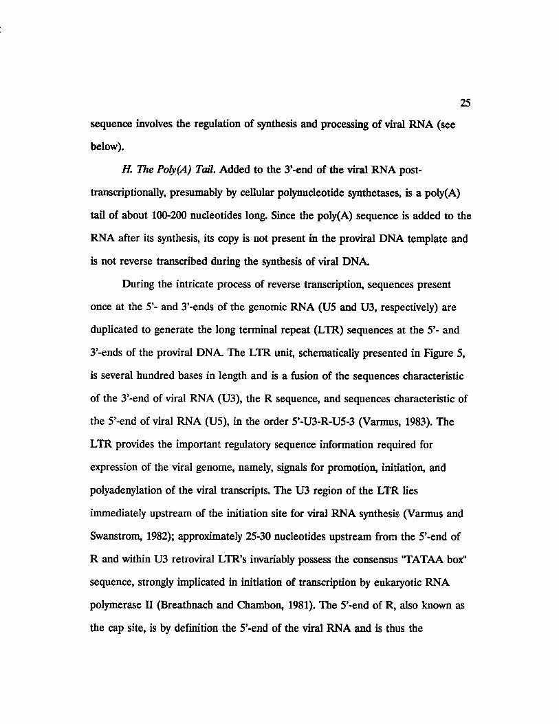
3’-ends of the proviral DNA. The LTR unit, schematically presented in Figure 5,
is several hundred bases in length and is a fusion of the sequences characteristic
of the 3’-end of viral RNA (U3), the R sequence, and sequences characteristic of
the 5’-end of viral RNA (U5), in the order 5’-U3-R-U5-3 (Varmus, 1983). The
LTR provides the important regulatory sequence information required for
expression of the viral genome, namely, signals for promotion, initiation, and
polyadenylation of the viral transcripts. The U3 region of the LTR lies
immediately upstream of the initiation site for viral RNA synthesis (Varmus and
Swanstrom, 1982); approximately 25-30 nucleotides upstream from the 5’-end of
R and within U3 retroviral LTR’s invariably possess the consensus 'TATAA box"
sequence, strongly implicated in initiation of transcription by eukaryotic RNA
polymerase II (Breathnach and Chambon, 1981). The 5’-end of R, also known as
the cap site, is by definition the 5’-end of the viral RNA and is thus the

26
In teg ra tio n•It* Cap s ita
(*)PrimarU3
An li t* In tag ra tion •Ita
Figure 5. Anatomy of an LTR. The figure shows the important features of the
U3, R, and U5 sequences of the retroviral LTR and its flanking viral sequence.
Features pertaining to a 5’-LTR are shown at the top of the figure, and those
relevant to a 3’-LTR are presented at the bottom. The viral sequence at the 5’-
boundary of U3 is the sequence of the putative primer for plus-strand DNA
synthesis, and the sequence at the 3’-boundary of U5 is the binding site for the
tRNA primer for minus-strand DNA synthesis. The LTR terminates with short
inverted repeats (IR). The integration sites are usually 2-bp from each boundary.
The sequences resembling CCAAT and TATAAA regulate the initiation of viral
RNA synthesis from the cap site, while the non-overlapping sequence, usually
AATAAA, determines the polyadenylation (A„) site at the 3’-end of RNA. (From
Varmus, 1983).

27
transcriptional initiation site (Varmus and Swanstrom, 1982; Varmus, 1983). A
sequence resembling CCAAT is also usually found about 30-40 nucleotides
upstream of the 'TATAA box" in the U3 region. The consensus eukaryotic signal
for polyadenylation, AATAAA (Proudfoot and Brownlee, 1974; Fitzgerald and
Shenk, 1981), is usually located near the 3’-end of U3, about 20 nucleotides
upstream from the site of poly(A) additon (Varmus, 1982). In retroviruses with
large R sequences the poly(A) addition signal can occur within R and must
apparently be ignored during transcription from the 5’-LTR (Varmus, 1983). The
dinucleotide CA, usually defining the R-U5 boundary, also serves as the
polyadenylation site on the RNA molecule (Bonner et al., 1982; Chen and Barker,
1984).
The coding sequences within the retroviral genome contain at least three
genes, located in between the two LTR units, which encode the structural and
replicative components of the virus particle. These genes are gag, encoding the
viral core proteins or group-specific antiens; pol, encoding the viral RNA-
dependent DNA polymerase (reverse transcriptase), integrase, and protease; and
env, encoding the viral envelope proteins.
Certain retroviruses possess additional genes which code for proteins with
specialized intracellular functions, such as oncogenes or regulatory genes. A group
of oncoviruses, typified by Rous sarcoma virus (RSV), carry a viral oncogene
whose product is directly involved in the swift induction of tumors in animals and
the efficient transformation of cells grown in culture (Bishop and Varmus, 1982).
On the other hand, HTLV-I and -II, also recognized as oncoviruses, promote
28
their oncogenic action through the product of a small open reading frame that
lies between the viral env gene and the 3’-LTR. This protein acts as a positive
effector of transcription from the viral LTR and from certain cellular promoters,
thus promoting tumorigenesis (Sodroski et al., 1984; Greene et al., 1986; Seiki et
al., 1986). Moreover, a common feature among lentiviruses, which is absent in
oncoviruses, is the presence of several small open reading frames (ORF’s) in their
genomes in addition to the structural and replicative gag, pol, and env genes
(Ratner et al., 1985; Sanchez-Pescador et al., 1985; Sonigo et al., 1985; Wain-
Hobson et al., 1985; Rushlow et al., 1986). These short ORF’s, usually located
between the pol and env genes and near the 3’-end of the env gene, have been
intensely studied within the past few years and have invariably been shown to
encode regulatory proteins which act as either positive or negative effectors of
viral gene expression (see below).
As mobile genetic elements within eukaryotes, retroviruses have evolved
into complex entities which have adopted a combination of devices and
mechanisms to facilitate their expression in host cells that normally operate in a
totally different pattern of genetic organization. In eukaryotic cells, genes are
principally arranged as single coding sequnces that are interrupted by noncoding
regions (introns or intervening sequences). Retroviruses, however, are composed
of multicistronic genomes destined to be expressed in eukaryotic cells, which are
normally not equipped to translate several independent genes from a single
mRNA. To get around this problem, retroviruses have evolved two specific
strategies of gene expression: production of spliced subgenomic mRNA’s from a

29
single full-length viral mRNA in order to provide the coding message for proteins
encoded at internal positions of the viral genome; and cleavage of the primaiy
viral translational products (polyproteins) to generate multiple functional proteins
required for viral replication.
Synthesis and Splicing of the Full-Length Viral mRNA
The primary product of transcription from the proviral DNA template
initiates at the 5’-boundaiy of the R sequence within the 5’-LTR, to which then
the 5’-cap nucleotide is added post-transcriptionally (Varmus, 1983). Although this
primary transcripts, hereafter referred to as the full-length viral mRNA, is
approximately the same length as the subunit of viral genomic RNA, there is
evidence to believe that transcription can extend some distance beyond the 3’-end
of the R sequence, copied from the 3’-LTR and to which the poly(A) tail is
added (Yamamoto et al., 1980; Ucker, 1981; Nevins, 1982).
Since the coding region for the env gene lies in the 3’-half of the genomic
RNA, its translation from the full-length viral mRNA requires a splicing event in
which the gag-pol intron is removed from this primary transcript, joining 5’-leader
sequences to the env coding sequences (Varmus and Swanstrom, 1982). The splice
donor site for this splicing event usually resides in the leader region downstream
of the transcriptional initiation site and upstream of the gag open reading frame,
although there are exceptions to this rule in certain retroviruses, such as RSV, in
which the donor site lies within the gag gene (Hacket et al., 1982). The splice
acceptor site is usually located just upstream of the env open reading frame, thus
30
allowing the fusion of the 5’-leader and env sequences through the two splice
junction signals.
As mentioned above and presented in detail below, relative to other
retroviruses, lentiviruses possess additional smaller regulatory genes near the 3’-
end of the env gene or within the pol-env intergenic region. In many cases studied
to date (Arya et al., 1985; Arya and Gallo, 1986; Davis et al., 1987; Muesing et al.,
1985; Rosenblat et al., 1988), these regulatory genes are encoded by multiple
small exons encoded within short open reading frames (ORFs) in the 3’-half of
the genome. Generation of mRNA’s for these small coding sequence usually
occurs through multiple splicing events by which both the gag-pol and the env
introns are spliced out of the full-length viral mRNA to generate a small mRNA,
usually 1.2-2.0 kb in length and composed of 5’-leader sequences fused to the
small exons of the regulatory gene, to allow for the translation of the regulatory
mRNA.
Translation of the Viral mRNA’s
The full-length viral mRNA serves as the message for synthesis of a gag-
pol precursor polyprotein which is then post-translationally modified and
processed to yield the mature viral core proteins and reverse transcriptase (Vogt
and Eisenman, 1973; Vogt et al., 1975). The mechanism used for the synthesis of
the gag-pol fusion protein illustrates the unique and intricate strategies evolved by
retroviruses to utilize the eukaryotic cellular machinery. The gag and pol genes
are separated by a nonsense termination codon at the 3’-end of gag in MLV; they
read in different and briefly overlapping frames in RSV and all lentiviruses such

31
as HTV, visna, and EIAV; and are separated by a third open reading frame (pro,
which stands for the viral protease) in MMTV, BLV, and HTLV-II (Varmus,
1988). Rather than directing the host cell’s splicing machinery to splice out the
gag stop codon or to create monocistronic messages by splicing out pol sequences
from the full-length viral mRNA, retroviruses instead take advantage of the
potential of eukaryotic ribosomes to occasionally insert an amino acid in place of
the termination codon at the 3’-end of the MLV gag (Yoshinaka et al., 1985) to
suppress translational termination, or to utilize the occasional frameshifting of
eukaryotic ribosomes at defined sites and frequencies in order to bypass the gag
termination codon in RSV, HIV, MMTV, BLV, and HTLV-II (Jacks and Varmus,
1985; Jacks et al., 1987; Moore et al., 1987, Jacks et al., 1988). To date, these
phenomena have not been observed in translation of cellular mRNA’s and would
indeed be deleterious if occurring frequently and without any control mechanism.
However, their occurence have clear advantages for retroviruses in that structural
(gag) proteins can be synthesized in large quantities required for viral replication,
while catalytic (pol and pro) proteins not required to be made in large amounts
can be synthesized in relatively small quantities. With this translational
mechanism, pol products destined to be packaged into progeny virion particles
can also be directly incorporated into the viral core through attached gag
components.

32
The Lentivirus Genomic Organization: Trans-acting Factors and Cis-acting
Sequences
As mentioned before, a common feature among lentiviruses, which is
absent in oncoviruses, is the presence of several small open reading frames
(ORFs) in their genomes in addition to the structural and replicative gag, pol,
and env coding sequences. In general, these short ORFs encode tram-acting
regulatoiy proteins which act as either positive or negative effectors of viral gene
expression. The complex regulation of lentivirus gene expression, brought about
by the fine tuning of these regulatory genes, is believed to enable these viruses to
establish latency within the infected host, then respond rapidly to various signals
to synthesize high levels of viral proteins leading to active viral replication
(Peterlin et al., 1988).
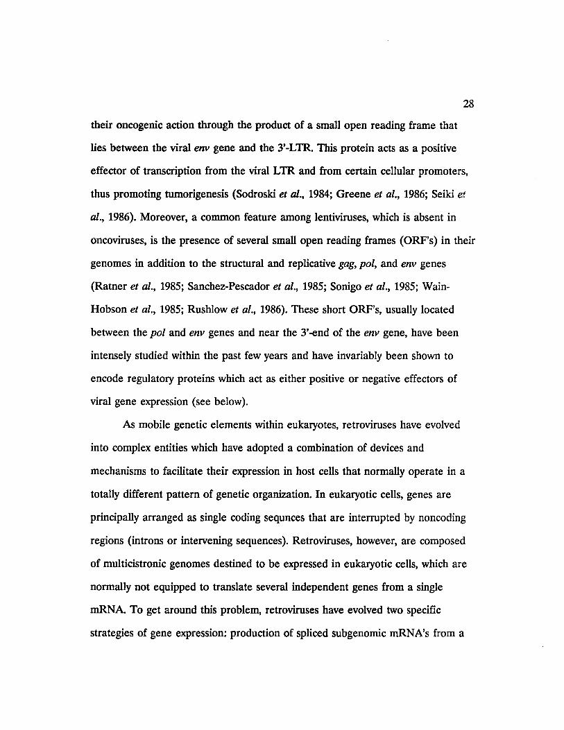
The genomic organization of several representative lentiviruses, including
EIAV, are presented in Figure 6. Among lentiviruses, the most intensely studied
virus within the past five years has been HTV-1, and much has been learned
about the molecular biology and genetic complexity of this virus. HIV-1 and HIV-
2 are very similar and the two viruses share a 50-60% similarity in their primary
proviral DNA sequence (Clavell et al., 1986; Ho et al., 1987; Fauci, 1988).
The HTV-1 genome encodes at least three genes, tat, rev, and nef, whose
products regulate viral gene expression. The coding sequences for tat, or trans-
activator of transcription, are within two separate exons of the HTV genome
(Figure 6) which are brought together a double-splicing event of the full-length
HTV-1 mRNA (Arya et al., 1985; Sodroski et al., 1985). The tat gene product of

33
Figure 6. The genomic organization of representative lentiviruses. In addition to
the gag, pol, and env genes, lentiviruses possess additional short open reading
frames in between pol and env and near the 3’-terminus of env. These additional
ORF’s in HIV-1 code for virion infectivity factor (vif), viral protein R (vpr), trans-
activator of transcription (tat), regulator of expression of virion proteins (rev),
viral protein U (vpu), and negative factor (nef). The HIV-2 and SIV genomes
similarly encode vif (vip or virion infectivity protein in the case of SIV), vpr, tat,
rev, and nef, but do not code for vpu. Instead, HTV-2 and SIV contain an ORF
for viral protein X (vpx). Of the three short ORF’s of EIAV, SI, S2, and S3, only
SI is currently known to encode a regulatory protein (see text).

34
HIV-1
HIV-2
SIV
tat
vpr
c : >
3* LTR
E r ^ ~ ' ' ~ ~ T Lvir JeMSSlyjw- rev nef| / / /'} J I n \ \ \ s i I / / ✓ / J
3* LTR
5* LTR
3’ LTRjr/m
EIAV
S 3 3' LTR

35
HIV-1 is a 14-kda protein containing a stretch of cysteine residues followed by a
stretch of basic amino acids, both of which are required for its activity (Arya et
al., 1985; Sodroski et al., 1985; Arya et al., 1987). Moreover, the tat protein forms
a non-covalently linked dimer which binds divalent cations in solution (Frankel et
al., 1988) and is localized in the nucleus (Hauber et al., 1987). Mutations and
deletions in the tat gene have been shown to abolish viral infectivity, thus tat is
essential for HIV-1 replication (Dayton et al., 1986; Fisher et al., 1986; Luciw et
al., 1987). The cis-acting element on the HIV-1 genome required for tat activity is
a tram-acting response sequence (TAR), originally defined as the region from
positions -17 to +80 within the 5’-LTR (Rosen et al., 1985). Additional
mutational analysis of TAR has revealed that only sequences from +19 to +42
are required for responsiveness to tat (Jacobovits et al., 1988; Peterlin et al.,
1988). Although not precisely defined, the mechanism of fram-activation by tat
has been suggested to involve transcriptional, post-transcriptional, and
translational events. The regulatory function(s) of tat have been related to an
increase in steady-state levels of RNA species containing the TAR element at
their 5’-ends (Cullen, 1986; Peterlin et al., 1986; Wright et al., 1986; Kao et al.,
1987; Muesing et al., 1987), a bimodal mechanism involving two sites of action,
one leading to increased levels of TAR-containing RNA’s and the other to even
higher levels of their encoded protein products (Cullen, 1986; Wright et al., 1986),
and an increase in the rates of initiation (Jacobovits et al., 1988; Laspia et al.,
1989) and elongation (Kao et al., 1987; Laspia et al., 1989) of transcription from
the 5’-LTR. More recently, tat has been shown to interact with the RNA

36
sequence of TAR, rather than its DNA sequence (Berkhout et al., 1989), giving
rise to speculations that it might function as an RNA enhancer (Sharp and
Marciniak, 1989). In general, the interaction of tat with TAR-containing RNA is
believed to facilitate transcriptional elongation, increase mRNA stability, and
enhance translation. The predominance of each effect might then depend on
quantitative differences in tat and qualitative differences in host cellular factors
(Peterlin and Luciw, 1988).
rev, or the regulator of expression of viral proteins, previously known as
art/trs (the anti-repression fro/is-activator/fra/is-regulator of splicing), is another
trans-acting HIV gene product and its coding sequences overlap but are in a
different reading frame from those of tat (Figure 6) (Sodroski et al., 1986;
Feinberg et al., 1986). Similar to tat, mRNA coding sequences for rev are within
two separate exons on the HIV genome which are brought together by RNA
splicing. The rev gene product is a 19-kda protein which, like tat, contains basic
residues and is localized in the nucleus (Cullen et al., 1988). The os-acting
repression sequences (crs) in the full-length and single-spliced HIV-1 mRNA’s,
encoding gag and env structural proteins, respectively, have been shown to render
these long mRNA’s very unstable (Rosen et al., 1988). A separate as-acting anti
repression sequence {car), located within env and non-overlapping with crs, is the
target for the trans-acting rev protein (Felber et al., 1989; Hammarskjold et al.,
1989; Malim et al., 1989; Rosen et al., 1988). By interacting with car, also known
as the rev-response element or RRE (Malim et al., 1989), the rev protein has been
shown to directly effect the post-transcriptional processing of HIV-1 transcripts by
37
activating the transport of full-length and single-spliced HIV-1 mRNA’s to the
cytoplasm and away from the splicing machinery in the nucleus (Felber et al.,
1989; Hammerskjold et al., 1989; Malim et al., 1989). By so doing, at the expense
of viral regulatory proteins being expressed from the double-spliced viral
mRNA’s, including rev itself, rev rescues and exports incompletely spliced HIV-1
gag and env mRNA’s away from the nucleus for translation of viral structural
proteins in the cytoplasm.
nef, or the negative factor, previously known as 3'-orf, orfB, E \ and F,
encodes a 25- to 27-kda protein believed to negatively regulate HIV replication
and expression (Ahmad and Venkatesan, 1988; Luciw et al., 1987). Cloned HIV
proviruses designed to contain mutations or deletions in the nef gene have been
shown to give rise to viruses which replicate to higher titers in tissue culture than
the wild-type virus (Luciw et al., 1987). Moreover, the nef gene product has been
proposed to downregulate HIV-1 transcription by activating tram-acting factors
which in turn interact with the negative regulatory element (NRE) in the
upstream U3 region of the 5’-LTR (Ahmad and Venkatesan, 1988).
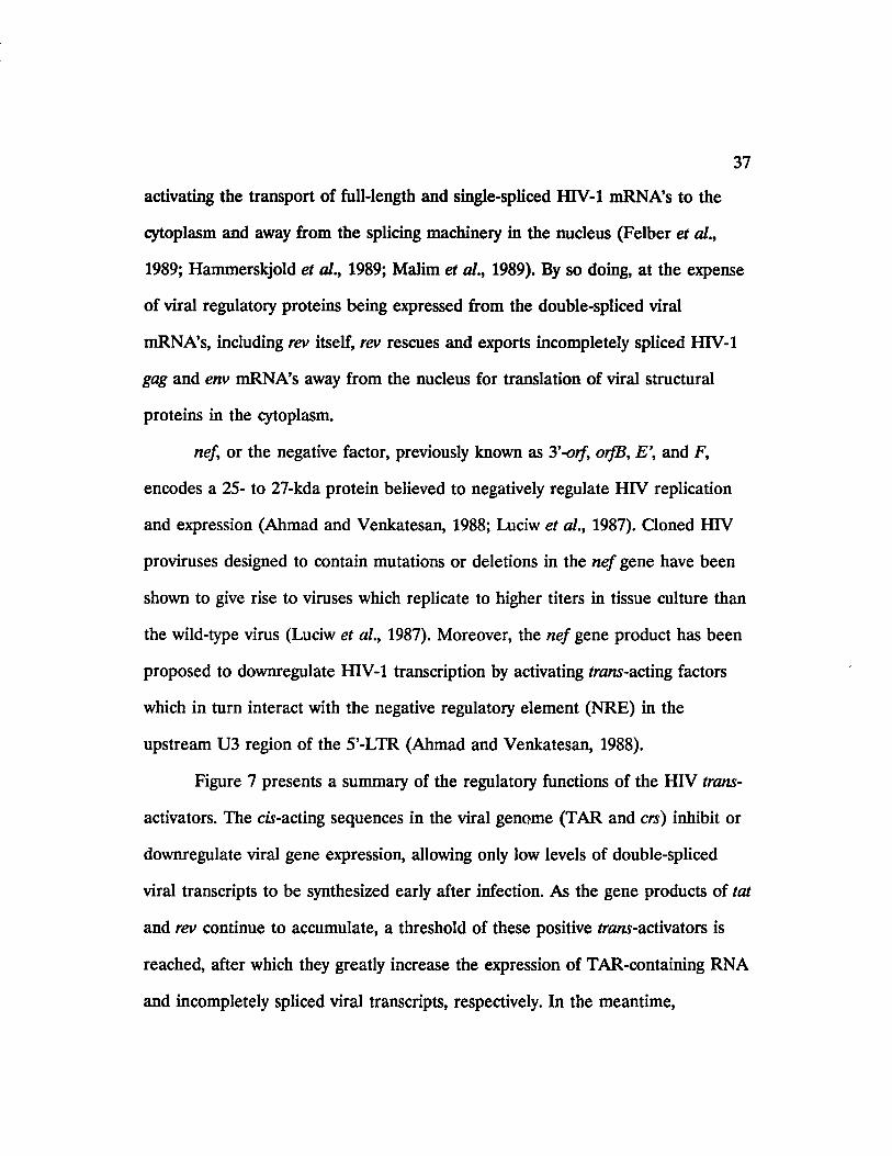
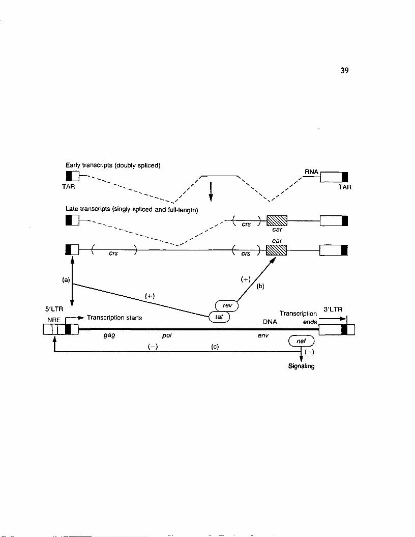
Figure 7 presents a summary of the regulatory functions of the HIV trans-
activators. The cis-acting sequences in the viral genome (TAR and crs) inhibit or
downregulate viral gene expression, allowing only low levels of double-spliced
viral transcripts to be synthesized early after infection. As the gene products of tat
and rev continue to accumulate, a threshold of these positive /ram-activators is
reached, after which they greatly increase the expression of TAR-containing RNA
and incompletely spliced viral transcripts, respectively. In the meantime,

38
Figure 7. The HIV fra/u-activators. tat, rev, and nef are translated from double
spliced viral mRNA, denoted as "early transcripts", containing coding sequences
flanking the env gene. Transcription initiates from the 5’-LTR. (a) tat acts upon
TAR, which is present just 3’ to the initiation site of trancription and which
becomes a part of the 5’-terminal sequences in HTV full-length RNA, to result in
large increases in TAR-containing RNA species, (b) rev acts on car (cw-acting
anti-repression sequence) to increase the expression of full-length and single
spliced HIV RNA, denoted as "late transcripts". In the absence of rev, double
spliced "early transcripts" predominate. Full-length and single-spliced viral RNA
are quickly processed or degraded since they contain crs elements (ds-acting
repression sequence) in gag and env coding regions, (c) nef is encoded by an ORF
located 3’ to the env gene and negatively regulates viral gene expression, either by
decreasing cellular signaling, or by acting on the negative regulatory element
(NRE) in the upstream U3 region of the 5’-LTR. The open boxes at the 3’-end of
viral transcripts represent U3 and R sequences. (From Peterlin and Luciw, 1988).
39
Early transcripts (doubly spliced)
TAR
RNA
/ I TAR
Late transcripts (singly spliced and full-length)
crscar
car
crs crs
(+)
rev5'LTRNRE I Transcription starts
3'LTRTranscriptionendsDNA
pol envG j DnF>Signaling
( - )

40
accumulated levels of nef decrease cellular signaling and viral transcription. The
balance between the function of these viral regulators and their effects within the
infected cell is never completely achieved, and as a result, the infected cell dies
while enough virions infect other cells to insure survival of the virus (Franza et
al., 1988; Peterlin et al., 1988; Peterlin and Luciw, 1989).
Much less is known about the function of the other small genes of
HIV-1 and -2. vif, previously known as sor, orfA, P \ or Q, encodes a 23-kda
protein (Lee et al., 1986; Kan et al., 1986). Because cloned proviral genomes
containing mutations in the vif gene yield low levels of infectious viral particles,
the vif gene product has been suggested to be required for viral infectivity
(Sodroski et al., 1986; Strebel et al., 1987). Precise functions for vpr, vpu, and vpx
have yet to be defined.
Much has been learned in the past five years in understanding the
molecular biology of lentiviruses. Relative to HIV-1, however, progress in
discovering the patterns of gene expression of other lentiviruses has been lagging
behind. In the case of visna virus, a triple-spliced viral mRNA, whose four exons
contain sequences from the 5’-end of the viral genome, the 3’-end of pol, a short
ORF immediately upstream of env (S), and sequences at the 3’-end of env,
respectively, has been shown to encode viral /rani-activating functions (Mazarin et
al., 1988; Davis and Clements, 1989). A similar double-spliced viral mRNA,
whose three exons contain sequences from the 5’-end of the viral genome, SI, and
S3 ORFS of EIAV (Figure 6) has been reported to code for an EIAV trans-
activator (Derse et al., 1989). It is believed that by dissecting the functions of the

41
structural and regulatoiy genes of lentiviruses in general, and the HIV’s in
particular, measures can then be taken to interfere or block the action of such
genes and their encoded products, thus providing therapeutic or preventative
protocols against lentivirus-induced diseases, including AIDS.
The patterns of transcription of lentiviruses in infected cells have proven to
be quite complex, involving multiple splicing events required for the generation of
virus-encoded regulatory frans-acting proteins which, in turn, act on cu-acting
sequences on the viral genome to enhance viral transcription or influence the
ratio of spliced to unspliced RNA’s. The objectives of the current study have been
to answer some of the unanswered, yet fundamental, questions on the molecular
biology of equine infectious anemia virus (EIAV).
RESEARCH OBJECTIVES
The main objective of the body of research presented here has been to
characterize the transcriptional properties of EIAV in virus-infected cells.
Questions regarding different aspects of this objective are listed below, along with
their significance.
(1) How many virus-specific transcripts are encoded by EIAV in virus-
infected cells? What are the relative levels, sizes, and kinetics of synthesis of
EIAV-specific transcripts? Moreover, are there any differences in patterns of
EIAV transcription when comparing persistent to cytopathic EIAV infections?
Answers to these questions are necessary in order to carefully examine the
genetic properties of EIAV and the possibility of differential expression of the
viral genome during persistent and cytopathic infections.

42
(2) What are the splicing patterns utilized in generation of the spliced
EIAV mRNA’s in infected cells? Can the location of the splice donor and
acceptor sites used for generating the spliced viral mRNA’s be mapped on the
viral genome? Characterization of the viral mRNA splicing patterns is a
prerequisite to defining the exact nucleotide sequence of these mRNA’s, which in
turn is required for analysis of the potential of these mRNA’s to encode viral
structural, replicative, and regulatory proteins.
(3) Do the short ORFs on the EIAV genome code for any proteins, such
as the fra/w-acting proteins encoded by HIV-1 or visna virus, and if so what are
the transcriptional and translational mechanisms used for their synthesis? To
better understand the molecular biology of lentiviruses it is important to search
for these regulatory proteins and to understand their function.
(4) Finally, the unique nature of the persistent disease induced by EIAV,
in which the infected horse eventually brings the disease under control, provides
an exciting venue for answering some of the complex questions on the biology of
other important lentiviruses such as HIV-1. Do the determined patterns of EIAV
gene expression correlate with the complex patterns observed in other lentivirus
systems or are they in any way different or unique? Such information is vital to
understanding patterns of lentiviral gene expression and designing diagnostic,
therapeutic, and preventative measures for combating lentiviral diseases.
The first objective has been accomplished by analysis of RNA from EIAV-
infected cells in Northern blot hybridizations using a battery of subcloned, gene-
specific proviral DNA probes obtained from the cloned EIAV provirus. The

43
splicing patterns of EIAV, outlined in objective (2), have been analyzed by
cloning and sequencing of cDNA obtained from poly(A)+ RNA of EIAV-infected
cells, by Northern hybridization analysis using short oligonucleotide DNA probes
complementary to sequences upstream of putative splice donor sites near the 5’-
end of the EIAV gag gene, and also by S 1-nuclease mapping studies of RNA
from virus-infected cells. Characterization of splicing patterns and mapping of
splice donor and acceptor sites on the viral genome has facilitated the
determination of coding sequences within the spliced EIAV mRNA’s, which in
turn has allowed the design of experimental approaches for addressing objective
(3). Subcloning of specific EIAV cDNA fragments encoding these mRNA’s into
plasmid vectors for in vitro transcription/translation or expression in eukaryotic
cells has provided a means for preliminary analysis of the expressed protein
products. Future detailed characterization of the expressed proteins, including an
analysis of their reactivity with appropriate antisera, would then allow their
assignment to specific EIAV genes.

CHAPTER 2
MATERIALS AND METHODS
Cell cultures and virus strains
Primary cultures of fetal equine kidney (FEK) and fetal donkey dermal
(FDD) cells were prepared and maintained as described previously (Orrego et al.,
1982). A prototype stock of EIAV, obtained by propagation of the Wyoming cell-
adapted strain of EIAV (Malmquist et al., 1973) in FEK cells, was used to carry
out infection of confluent monolayers of FEK cells at a multiplicity of infection of
1.0. In addition, an FDD-adapted stock of EIAV, prepared by propagation of
prototype EIAV in FDD cells, was used to infect confluent monolayers of FDD
cells at a multiplicity of infection of 1.0.
Isolation and purification of total and poly(A)+-selected cellular RNA
To inactivate contaminating RNases, all solutions used for preparation of
RNA were prepared using diethylpyrocarbonate (DEPC)-treated double-distilled
water (ddH20). In addition, all solutions were subjected to a second round of
DEPC treatment following their preparation, with the exception of Tris buffers.
For DEPC treatment, 2 ml of DEPC was added to 100 ml of water or solution
and shaken vigorously. After an overnight incubation at room temperature, the
solution was autoclaved to remove remaining traces of DEPC. In addition, all
glassware used for preparation of such solutions wws sterilized by baking at
200°C for 8 hours.
44
45
Total cellular RNA was isolated from EIAV-infected or uninfected cells by
a modified guanidinium thiocyanate extraction method (Chirgwin et al. 1979,
Maniatis et al., 1982). Briefly, using repeated washes with phosphate buffered
saline (PBS) containing 4mM EDTA (pH 7.0), infected FEK cells were harvested
from roller bottles at 3-4 weeks post-infection, and infected FDD cells were
recovered from flasks at the peak of the cytopathic effect (10-12 days after
infection with FDD-adapted EIAV). The cells were pelleted in 50 ml
polypropylene conical centrifuge tubes at 2,000 rpm for five minutes using an IEC
HN-SII table-top clinical centrifuge, and immediately resuspended in 5-10 ml of
guanidinium thiocyanate lysis buffer [4.0 M guanidinium thiocyanate, 0.5%
sodium-N-lauroyl sarcosine, 5.0 mM sodium citrate (pH 7.0), 0.1 M 2-mercapto-
ethanol]. The cells were lysed by repeated passage through a 23-gauge needle,
which also resulted in shearing of the chromosomal DNA Four grams of solid
CsCl was dissolved in each 10.0 ml of homogenate (0.4 g/ml final) and the
homogenate was gently layered over a 3.25 ml cushion of sterile 5.7 M CsCl in
sterilized 13.5 ml Sarstedt polyallomer ultracentrifugation tubes. Following
ultracentrifugation for 18 hours at 28,500 rpm at 20°C in a Beckman SW40Ti
rotor, the cellular homogenate and the CsCl cushion were carefully aspirated out
and the inside walls of the tube were wiped dry by a sterile gauze. The light
brown pellet of RNA was resuspended in 300 ul of 10.0 mM Tris.Cl (pH 7.5), 5.0
mM EDTA (pH 7.0), 1.0% SDS, and extracted twice with phenokchloroform:
isoamyl alcohol (25:24:1). The aqueous supernatant was brought up to 0.3 M in
sodium acetate (pH 5.2) and precipitated by addition of three volumes of ethanol.
46
Poly(A)+ RNA was purified by two cycles of oligo(dT)-cellulose chromatography
of total cellular RNA (Maniatis et al., 1982), or by direct oligo(dT)-cellulose
chromatography of cellular lysates using the Fast Track mRNA Isolation Kit from
Invitrogen, according to the manufacturer’s protocol.
For time-course studies on EIAV RNA synthesis, various time-points after
infection were used for recovery of infected FEK and FDD cells, as described in
Chapter 3.
Subcloning of EIAV proviral DNA fragments
EIAV proviral DNA fragments were obtained by restriction enzyme
digestions of an EIAV proviral DNA clone, lambda 12 (Derse et al., 1987;
Rushlow et al., 1986; Stephens et al., 1986). Restriction digestions were performed
according to standard protocols (Maniatis et al., 1982) using the 10X digestion
buffer provided by the enzyme’s manufacturer and under the manufacturer’s
recommended reaction conditions. Typically, 5 to 10 ug of DNA was digested by
10 to 20 units of enzyme for at least 5 hours at the appropriate reaction
temperature. Restriction fragments of EIAV proviral DNA were subjected to
electrophoresis through 0.8-1.2% low melting-point agarose, normal agarose, or
6.0-8.0% polyacrylamide gels (Maniatis et al., 1982), following which the gels were
stained with 0.5 ug/ml ethidium bromide for visualization of the DNA bands by
UV illumination. After excision of appropriate DNA bands from gels, DNA was
purified by chromatography through a column of NACS-52 resin (BRL) or by the
use of a GeneClean Kit (Bio 101). Purified viral DNA fragments were typically
ligated to appropriately linearized plasmid vectors at a molar ratio of 20:1

47
insert:vector under standard ligation reaction conditions (Maniatis et al., 1982).
Aliquots of the ligatation reactions were used to transform competent E. coli
JM101 or DH5a cells to antibiotic resistance (Hanahan, 1985). Recombinant
clones were identified by restriction enzyme mapping of minilysate plasmid DNA
prepared from 2 ml overnight cultures of antibiotic resistant clones by the
alkaline lysis method (Bimboim and Doly, 1979; Maniatis et al., 1982).
Recombinant plasmids were then prepared on a large scale from 2 liter cultures
of appropriate clones and purified by two cycles of equilibrium density
ultracentrifugation through CsCl-ethidium bromide gradients (Maniatis et al.,
1982).
Labeling of probes for Northern hybridization analysis
Recombinant plasmids containing subcloned EIAV proviral DNA
fragments, which were to be used as probes for Northern hybridizations, were
digested with appropriate restriction enzymes, and the EIAV-specific insert DNA
fragments were purified following gel electrophoresis as described above. 50 to
100 ng of each fragment was then labeled with [a-32P]dCTP using a random
primer labeling kit from BRL (Feinberg and Vogelstein, 1983). Oligonucleotide
probes (see below) were prepared by 5’-end labeling of 200 ng of each
oligonucleotide with [y-32P]ATP and polynucleotide kinase (Maniatis et al., 1982).
Unincorporated 32P-labeled ribo- or deoxyribonucleotides were removed from the
labeling reactions by chromatography through Sephadex G-50 spun columns, as
described (Maniatis et al., 1982). Typically, specific activities greater than 5X10®

48
cpm/ug for restriction DNA fragments and about 1X10⁹ cpm/ug for oligo
nucleotides were obtained.
Northern, Southern, and colony hybridization analyses
For Northern hybridization analysis, 2.5 ug of poly (A)+ RNA or 15.0 ug
of total RNA from infected or uninfected cells was subjected to electrophoresis in
20X20 cm 1.0% or 1.4% agarose-formaldehyde gels in 50 mM MOPS (pH 7.0), 1
mM EDTA buffer (Gerard and Miller, 1986). Electrophoresis was carried out at
100 volts for 1 hour without buffer circulation and at 30 volts for 14 hours with
buffer circulation. To check for the integrity of RNA samples, the gels were
stained by soaking in a 5.0 ug/ml solution of ethidium bromide in water for 5
minutes in the dark and destained for 3 to 8 hours in water in the dark. The
stained RNA bands were then visualized by short-wave UV illumination of the
gel. For transfer of RNA the staining step was omitted, and the gel was washed
immediately following electrophoresis for 1 hour in 10X SSC (IX SSC is 0.15 M
NaCl, 0.015 M sodium citrate) to remove the formaldehyde. The gel was then
transferred overnight to GeneScreen or GeneScreenPlus nylon membranes (New
England Nuclear-DuPont) in 10X SSC using the capillary Southern transfer
method (Maniatis et al., 1982). Following the transfer, the membrane was baked
for 2 hours at 80°C in a vacuum oven and prehybridized at 42°C for 2-4 hours in
a buffer containing 50% formamide, 5X Denhardt (Maniatis et al., 1982), 6X
SSC, 1% SDS, 250 ug/ml denatured salmon sperm DNA and 25 mM sodium
phosphate (pH 6.5). Hybridization was carried out at 42°C for 18-24 hours using
1X10⁶ cpm of 32P-labeled probe per ml of hybridization solution, which contained

49
the same composition as the prehybridization buffer except that IX Denhardt was
used. Following hybridization, the membranes were washed for 30 minutes at
room temperature in 2X SSC, 0.2% SDS, and for 30 minutes at 65°C in 0.2X
SSC, 0.2% SDS. The membranes were then subjected to autoradiography at
-70°C for 12-36 hours.
For Southern hybridization analysis, DNA fragments were fractionated by
electrophoresis through 1.0% agarose gels. Denaturation, neutralization, and
transfer from the gel onto a nitrocellulose membrane was performed according to
standard protocols (Maniatis et al., 1982). Baking, prehybridization, hybridization,
and autoradiography was exactly as described for Northern hybridizations above.
For colony hybridization analysis, individual E. coli colonies were picked by
sterile toothpicks and streaked as short lines (2-3 mm long) on sterile circular
nitrocellulose membranes placed on LB agar plates containing the appropriate
antibiotic. A grid pattern taped to the back of the plate was used to identify the
location of individual colonies on the membrane. The plate was inverted and
incubated at 37°C until colonies had grown to 0.5-1.0 mm in width (6-8 hours).
Lysis of the bacterial colonies on the membrane were performed as described
(Maniatis et al., 1982). Baking, prehybridization, hybridization, and
autoradiography of the membrane was exactly as described for Northern
hybridizations above.
Chemical synthesis and purification of oligonucleotides
The four oligonucleotides used in this study were the generous gift of Dr.
K. E. Rushlow, Battelle Memorial Institute, Columbus, Ohio. The first

50
oligonucleotide, used to prime first-strand cDNA synthesis, was a 30-mer and had
the following sequence:
5’-ACTGAAGTAATTTTGAAATGGTCrACACCC-3’
The sequence of the oligonucleotide is complementary to a 30 nucleotide-long
sequence immediately downstream of the Smal site within the EIAV env gene
(positions 1642-1672 of Rushlow et al., 1986; Figure 11 A). The other three
oligonucleotides (SD-1, SD-2, and SD-3) were used as probes in Northern
hybridization analyses and their primary sequence is shown in Figure 13.
The oligonucleotides were synthesized on an Applied Biosystems 380A
DNA Synthesizer. Following synthesis, the deprotected oligonucleotides were
purified by chromatography through oligonucleotide purification cartridges
(Applied Biosystems) according to the manufacturer’s protocol.
cDNA synthesis and cloning
Double-stranded cDNA was synthesized from 13.0 ug of poly(A)+ RNA
obtained from EIAV-infected FDD cells, using a cDNA Synthesis Kit from BRL.
The manufacturer’s protocol was exactly followed, except that 3.0 ug of the
synthetic 30-mer oligonucleotide described above was used to prime first-strand
cDNA synthesis instead of the oligo(dT) primer provided in the kit. Following the
synthesis of second-strand cDNA the double-stranded cDNA products were
precipitated with ethanol.
For cloning of cDNA the double-stranded cDNA products were tailed
with oligo(dC) and annealed to oligo(dG)-tailed, PstI digested pUC9 DNA.
Following this strategy a PstI site is created at the 5’- and 3’-ends of the cloned
51
cDNA. The ethanol precipitated cDNA products were pelleted, dried, and
resuspended in 10 ul sterile ddH20 . The oligo(dC)-tailing reaction was set up by
addition of 5 ul of 10 mM dCTP (1 mM final concentration) and 10 ul of the
BRL 5X tailing buffer (IX final concentration) to the double-stranded cDNA.
The volume of the mixture was brought up to 48.5 ul with sterile ddH20 , after
which the contents were mixed and the mixture was incubated in a 37°C water
bath for 5 minutes. The tailing reaction was initiated by addition of 20 units (1.5
ul) of BRL Terminal Deoxynucleotidyl Transferase (50 ul final reaction volume)
and the mixture was incubated at 37°C. Five ul aliquots were removed from the
reaction at 30 second intervals (10 aliquots), and to inactivate the enzyme each
aliquot was transferred to individual microfuge tubes pre-incubated in a 70°C
water bath for 10 minutes. The 20 ul annealing reaction for each aliquot was set
up by addition of 2 ul of 10X annealing buffer [100 mM Tris.Cl (pH 7.5), 1,000
mM NaCl, 1 mM EDTA (pH 7.5)], 11 ul of sterile ddHzO, and 2 ul of 11 ng/ul
oligo(dG)-tailed, PstI digested pUC9 (Pharmacia). Annealing was carried out at
58°C for 3 hours. After a brief centrifugation step for collection of residual water
on the inside walls of the tubes, the annealing reaction was continued by
incubation at room temperature overnight.
A 5 ul aliquot from each 20 ul annealing reaction was used to transform
BRL library efficiency competent DH5a E. coli cells (Hanahan, 1985).
Transformants were plated on LB plates (Maniatis et al.) containing 100 ug/ml
ampicillin, 80 ug/ml X-gal, and 10 ug/ml IPTG. Plates were incubated for 12

52
hours at 37°C, and colonies exhibiting the white phenotype were randomly picked
for colony hybridization analysis, as described above.
Nucleotide sequencing
Double-stranded DNA fragments to be sequenced were isolated and
purified on 1.0-1.2% agarose gels, as described above, and subcloned into the
replicating form (RF) DNA of M13mpl8 or M13mpl9 vectors. Single-stranded
DNA from the M13 phage particles was sequenced by the dideoxy chain
termination method (Sanger et al., 1977), following which the sequencing reaction
mixtures were subjected to electrophoresis on 10% polyacrylamide-8% urea
sequencing gels (BRL).
SI nuclease mapping
The four probes used for SI nuclease mapping of RNA from ELAV-
infected and uninfected FDD cells are described in detail in Chapter 3 and
schematically diagrammed in Figure 16. To create the M13 "tag" sequence at the
intronic end of probe pi, a 308-bp Taql-PvuU EIAV proviral DNA restriction
fragment was subcloned into M13mpl8 RF DNA digested with Accl and Smal.
The cloned DNA was then excised out as a 550-bp Narl fragment containing a
300-bp Narl-PvuVL proviral DNA fragment and a 250-bp M13mpl8 Smal-Narl tag
attached from its Smal end to the PvuU site (Figure 16B).
Similarly, probe p2 was constructed by cloning a 450-bp Ncol-BamHl EIAV
proviral DNA restriction fragment, which had been previously blunt-ended at the
Ncol site by treatment with T4 DNA polymerase (Maniatis et al., 1982), into
M13mpl9 RF DNA digested with BamUl and Smal. The cloned DNA was then
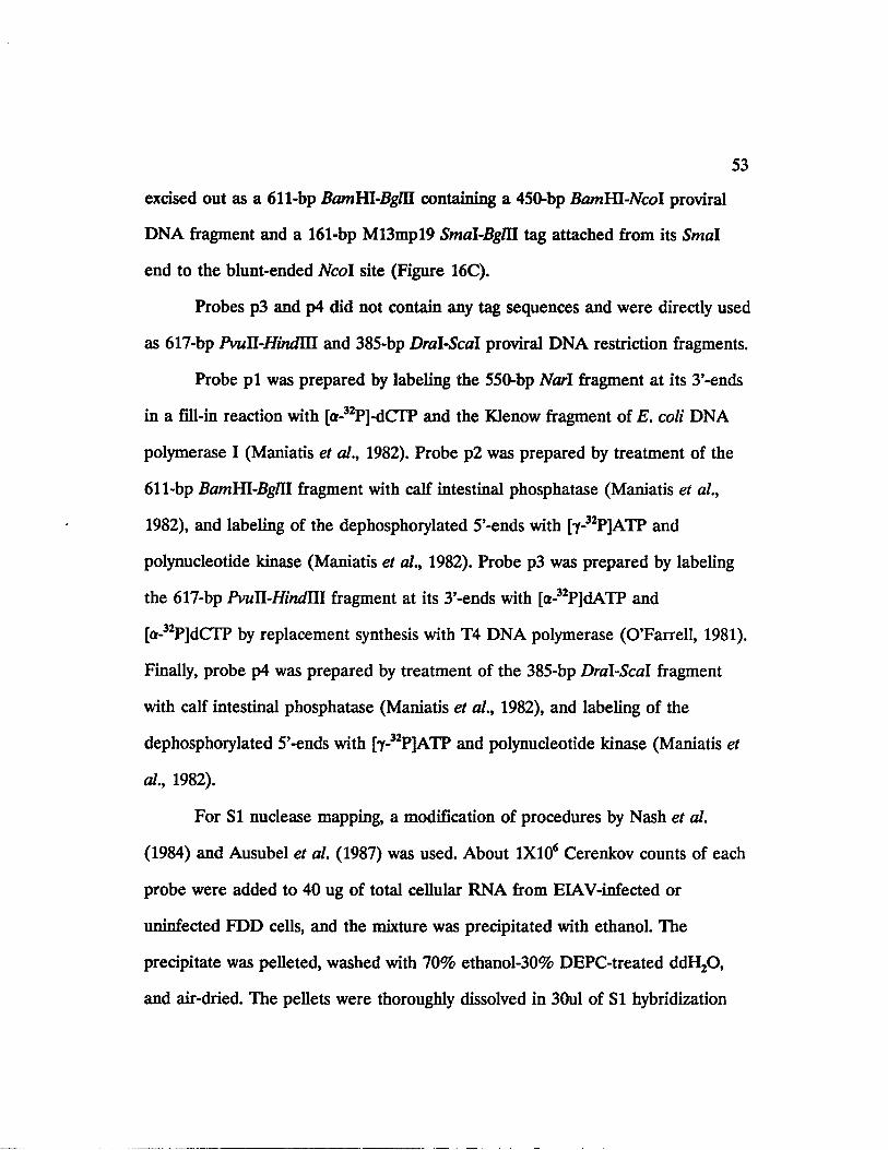
53
excised out as a 611-bp BamHL-BgM containing a 450-bp BamHL-Ncol proviral
DNA fragment and a 161-bp M13mpl9 Smal-BglU tag attached from its Smal
end to the blunt-ended Ncol site (Figure 16C).
Probes p3 and p4 did not contain any tag sequences and were directly used
as 617-bp PvuU-Hindm and 385-bp Dral-Scal proviral DNA restriction fragments.
Probe p i was prepared by labeling the 550-bp Narl fragment at its 3’-ends
in a fill-in reaction with [a-32P]-dCIP and the Klenow fragment of E. coli DNA
polymerase I (Maniatis et al., 1982). Probe p2 was prepared by treatment of the
611-bp BamYR-BgUl fragment with calf intestinal phosphatase (Maniatis et al.,
1982), and labeling of the dephosphorylated 5’-ends with [y-32P]ATP and
polynucleotide kinase (Maniatis et al., 1982). Probe p3 was prepared by labeling
the 617-bp PvuU-Hindlll fragment at its 3’-ends with [a-32P]dATP and
[a-32P]dCTP by replacement synthesis with T4 DNA polymerase (O’Farrell, 1981).
Finally, probe p4 was prepared by treatment of the 385-bp Dral-Scal fragment
with calf intestinal phosphatase (Maniatis et al., 1982), and labeling of the
dephosphorylated 5’-ends with [y-32P]ATP and polynucleotide kinase (Maniatis et
al., 1982).
For SI nuclease mapping, a modification of procedures by Nash et al.
(1984) and Ausubel et al. (1987) was used. About 1X10⁶ Cerenkov counts of each
probe were added to 40 ug of total cellular RNA from ELAV-infected or
uninfected FDD cells, and the mixture was precipitated with ethanol. The
precipitate was pelleted, washed with 70% ethanol-30% DEPC-treated ddH20 ,
and air-dried. The pellets were thoroughly dissolved in 30ul of SI hybridization

54
buffer [80% deionized formamide, 40 mM PIPES (pH 6.4), 400 mM NaCl, 1 mM
EDTA (pH 8.0)], and the resuspended RNA-probe mixture was immersed in an
85°C water bath for 15 minutes. The hybridization mixture was then immediately
transferred to water baths adjusted to 50°C (in the case of probes p i and p3) or
55°C (in the case of probes p2 and p4) and incubated overnight. The following
mix was then added to the hybridization reaction: 150 ul 2X SI nuclease buffer
[0.56 M NaCl, 0.1 M NaOAc (pH 4.5), 9 mM ZnS04], 3 ul 2.0 mg/ml single
stranded calf thymus DNA, 147 ul DEPC-treated ddH20 , and 300 units SI
nuclease (BRL). The contents were mixed and incubated at 30°C for 60 minutes,
after which the SI digestion was stopped by addition of 80 ul of SI stop buffer
[4.0 M ammonium acetate, 20 mM EDTA (pH 8.0), 40 ug/ml yeast tRNA]. The
digest was extracted twice with phenol:chloroform:isoamyl alcohol (25:24:1), and
the aqueous supernatant was precipitated with ethanol. To remove traces of salt,
the ethanol precipitated samples were pelleted, resuspended in sterile ddH20 ,
and the ethanol precipitation was repeated two more times. The final pellet was
washed with 70% ethanol, dried in a Speedvac evaporator, and resuspended in 3
ul of TE buffer (Maniatis et al., 1982). Four ul of formamide loading dye [95%
deionized formamide, 20 mM EDTA (pH 7.5), 0.05% bromophenol blue, 0.05%
xylene cyanol] was added, the sample was boiled for 4 minutes, and quenched
immediately on ice. The entire sample was then subjected to electrophoresis
through a 6.0% polyacrylamide-8.0 M urea sequencing gel. Electrophoresis was
typically stopped just as the bromophenol blue reached the bottom of the gel.

55
Mspl-digested pBR322 DNA, labeled with [a-32P]dCTP by filling-in with Klenow
(Maniatis et al., 1982), was run in parallel lanes as size marker.
In vitro transcription and translation
Construction of the pSP65 plasmid containing the 2980-bp BamHl-Pstl S2-
env cDNA insert is diagrammed in Figure 19. Briefly, a ligation reaction
containing the 270-bp Smal-BamHL splice-junction cDNA, a 2710-bp BamHl-Pstl
cDNA fragment from a separate cDNA clone (#3006), and pUC19 DNA digested
with Smal and PstI was carried out [The PstI end of the second cDNA fragment
had been originally created through cloning of that cDNA into pUC9 by
oligo(dC)-(dG) tailing]. As a result, a 2980-bp Smal-Pstl hybrid cDNA insert was
cloned into pUC19. Appropriate recombinants were identified by restriction
enzyme mapping and the 2980-bp insert was excised out of pUC19 by digestions
with Sacl and PstI. The 2980-bp Sacl-PstI fragment was then cloned into pSP65
DNA which had been digested by Sacl and PstI. Appropriate recombinants were
again identified by restriction enzyme mapping.
To prepare templates for in vitro transcription, the pSP65 plasmid
containing the 2980-bp Sad-Pstl cDNA insert was then linearized by either
HindUl or Mlul (Figure 19). Capped transcripts were synthesized in 50 ul
reactions, containing the cap analog GpppG (Pharmacia), using an in vitro
transcription kit (Promega Biotec) and according to the manufacturer’s protocol,
based on previously published procedures (Krieg and Melton, 1984). In vitro
translation reactions of the synthetic transcripts were performed in wheat germ
extracts in the presence of 35S-methionine, as described previously (Bartlett et al.,

56
1982). Incorporation of 35S-methionine was determined by assaying an aliquot of
the translation reaction for acid-insoluble radioactivity using Whatman 3mm filter
paper discs.
Construction of the p9SR426 expression plasmid, transfections, and indirect
immunofluorescence assays
A 426-bp Sspl-Smal restriction fragment of the EIAV proviral DNA,
containing the entire S2 coding sequence and the the first 379 nucleotides of the
env gene, was cloned into the p91023(B) expression vector (Figure 21). In
addition to its unique EcoRI cloning site, the p91023(B) vector also contains a
unique BglU cloning site immediately upstream of the FcoRI site. The 426-bp
Sspl-Smal fragment was initially cloned into the Smal site of plasmid TrpSF9,
and the orientation of the insert in the recombinant clones was identified by
restriction enzyme mapping. The insert DNA was then excised as a Bglll-EcoRl
fragment and ligated to p91023(B) DNA digested with BglII and EcoRl. The
recombinant plasmid, termed p9SR426, was then grown on a large scale and
subjected to two cycles of equilibrium density ultracentrifugation through CsCl-
ethidium bromide gradients (Maniatis et al., 1982). COS-7 cells were grown on
sterile tissue culture chamber slides to about 50% confluency. Five ug of the
plasmid was transfected onto COS-7 cells as a calcium phosphate co-precipitate
followed by a 15% glycerol shock, as described previously (Fordis and Howard,
1987). Indirect immunofluorescence assays were performed at 48 hours post-
transfection, using monoclonal antibody #86 or rabbit polyclonal S2b antibody,
according to previously published procedures (Pereira et al., 1980).

CHAPTER 3
RESULTS
I. IDENTIFICATION OF EIAV-SPECIFIC TRANSCRIPTS IN INFECTED
CELLS
The cell-adapted Wyoming (prototype) strain of EIAV establishes a
persistent infection in fetal equine kidney (FEK) cells grown in tissue-culture,
leading to continuous virus production for up to 4-5 weeks after the initial
infection, during which no cytopathic effects are exerted by the replicating virus
on the infected cells. Infection of fetal donkey dermal (FDD) cells by FDD-
adapted EIAV, a strain of the virus obtained by propagation of prototype EIAV
in FDD cells, however, results in a distinct and observable cytopathic effect
characterized by rounding and detachment of infected cells at 10-12 days post
infection.
By comparison to other retroviruses, transcription of the EIAV provirus
initiates at the 5’-terminus of the R region in the 5’-LTR. Since the R-U5-leader
sequences are present in both full-length genomic and spliced viral mRNA’s, a
proviral DNA fragment containing nucleotide sequences from the R-U5 region of
the LTR would share sequence homology with the 5’-ends of all viral mRNA’s.
Such a DNA fragment, hereafter referred to as the "5’-LTR probe", should
hybridize to all virus-specific transcripts and once radiolabeled can be used as a
general probe for detection of all viral mRNA’s actively transcribed in infected
cells.
57

}I
58
A 226-bp restriction fragment of the EIAV provirus, beginning at 50
nucleotides upstream of the R region in the 5’-LTR (within the U3 sequences)
and extending 63 nucleotides downstream of the U5 region, was radiolabeled with
32P and used as the 5’-LTR probe in Northern blot hybridization analysis of
EIAV-specific transcripts during persistent and cytopathic infection of FEK and
FDD cells, respectively. Total cellular RNA was isolated from FEK cells at three
weeks after infection with prototype EIAV, from FDD cells at the peak of
cytopathic effects at twelve days after infection with FDD-adapted EIAV, and
from uninfected FEK and FDD cells as negative controls. Fifteen micrograms of
total RNA from each source was subjected to electrophoresis in a 1.0%
denaturing agarose-formaldehyde gel, transferred to the hybridization membrane,
and hybridized with the 5’-LTR probe. The left panel in Figure 8 shows the
ethidium bromide-stained agarose-formaldehyde gel of total RNA samples, while
an autoradiogram of the Northern blot of the same gel hybridized to the 5’-LTR
probe is presented in the right panel of Figure 8. In both infected cell-lines, two
major species of hybridizing EIAV-specific transcripts are detected: an 8.2-kb
genome-length RNA and a 3.5-kb RNA. Also detected in RNA from both
infected cell lines is a low abundance 1.5-kb RNA species which migrates just
below the 18S rRNA band on the gel. No EIAV-specific transcripts are detected
in RNA from uninfected cells, indicating that there is no endogenous expression
of ELAV-related RNA in these cells and that the hybridizing RNA species
present in RNA from infected cells are a result of EIAV infection.

59
Figure 8. Northern hybridization analysis of total RNA from EIAV-infected and
uninfected FEK or FDD cells. Left. 1.0% Agarose-formaldehyde gel of total
cellular RNA from FEK cells infected with prototype EIAV (lane 1), FDD cells
infected with FDD-adapted EIAV (lane 2), uninfected FEK cells (lane 3), and
uninfected FDD cells (lane 4). Lane M contains molecular weight markers (0.3-
to 9.5-kb RNA ladder of BRL) whose mobilities are indicated on the left. Right.
Northern blot of the gel on the left panel, hybridized to the 226-bp Mlul-BamHl
5’-LTR probe. The position of 28S and 18S rRNA bands on both the gel and the
blot are indicated. The numbers on the right corespond to the molecular weight
values for the three observed species of EIAV mRNA’s.

2 8 s
18 s

61
Densitometric analysis of the Northern blot in Figure 8 reveals that there
is almost a 10-fold higher level of viral RNA’s in infected FDD cells at the peak
of the cytopathic effect relative to that in persistently infected FEK cells. In
addition, the results indicate that the 8.2- and 3.5-kb RNA are present at nearly
equal concentrations in infected FEK cells, while the concentration of the 3.5-kb
RNA is about three-fold greater than that of the full-length transcript.
Immunofluorescence studies have shown the EIAV infection of FDD cells
to be asynchronous, resulting in infected cells comprising about 30% of the total
cell population, while during infection of FEK cells nearly 100% of the cell
population is infected by the virus (Issel and Montelaro, unpublished data).
Taking into account the asynchronous nature of viral infection in FDD cells, it
appears that the levels of EIAV-specific RNA’s are nearly 30-fold higher during
the cytopathic infection of FDD cells relative to the persistent infection of FEK
cells. Moreover, the relatively high levels of the 3.5-kb mRNA in infected FDD
cells suggests enhanced levels of 8.2-kb genome-length viral RNA splicing during
this cytopathic EIAV infection.
The presence of abundant quantities of 28S and 18S rRNA in samples of
total cellular RNA, as clearly seen in Figure 8A, introduces inherent problems in
identification of specific RNA’s present on a hybridization membrane which share
sequence homology with the hybridization probe. In the absence of the rRNA
species, the probe would hybridize to a background "smear" of degraded
homologous RNA on the membrane, in addition to the specific non-degraded
RNA species. However, when the abundant non-hybridizing rRNA species are

62
present on the membrane, they physically block the hybridization of the probe to
the background smear of degraded homologous RNA at the position of each
rRNA species on the membrane, resulting in the re-appearance of the
background immediately below the lower boundary of each rRNA which then
appears as a faint hybridizing band on the Northern blot. This band is present
just below the position of 28S and 18S rRNA in Figure 8B, and has been
observed in repeated experiments involving Northern hybridization of total RNA
samples.
To clarify this inherent problem and also to better detect any EIAV-
specific low molecular transcripts which migrate slightly faster than 18S rRNA
during denaturing gel electrophoresis, poly(A)+ RNA, which contains little or no
rRNA, was isolated and purified from uninfected and EIAV-infected FDD cells
and used as substrate for Northern hybridization analysis. Furthermore, to identify
the gene-specific content of the EIAV-specific mRNA’s in infected cells, a battery
of subgenomic EIAV DNA fragments (Table II), in addition to the 5’-LTR 226-
bp fragment, were used as probes in Northern hybridization analyses. Using
unique restriction sites on the EIAV proviral DNA, the DNA fragments were
individually subcloned so that each, with the exception of the 5’-LTR probe,
represents sequences from each of the seven genes (including the short ORF’s) of
EIAV. By designing the gene-specific probes spanning across the EIAV genome,
separate Northern hybridizations were carried out using each individual probe,
and the identity of viral mRNA’s containing sequences complementary to each
viral gene were identified. Figure 9 shows a composite of the location of the

63
Table II. Panel of EIAV proviral DNA restriction fragments used as probes in Northern hybridization analyses.
Probe Restriction Fragment Specificity within the EIAV genome
EIAV mRNA Specificity
A 226-bp Mlul-Barnm 5’ - LTR All mRNA’s
B 393-bp PvuU-Kpril gag Full-length
C 511-bp Smal-Sstl pol Full-length
D 102-bp Pvull-Sspl SI Full-length, S I , env*
E 114-bp £amHI-7a<7l S2 Full-length, S2, env
F 417-bp Sphl-Xbal env Full-length, env
G 320-bp Avail S3 Full-length, env, S3
* The hybridization of this probe to the env mRNA would be dependent on the env splice acceptor site being located upstream of the SspI site.

64
Figure 9. Northern hybridization analysis of EIAV-specific poly(A)+ RNA’s in
FDD cells infected with FDD-adapted EIAV. A diagram of the EIAV proviral
DNA genome and the location of the six open reading frames is represented on
top of the figure. Based on DNA sequence data (Rushlow et al., 1986; Stephens et
al., 1986) solid triangles represent the location of consensus splice donor sites and
open triangles represent the location of consensus splice acceptor sites. The
stippled bars, labeled A through G, show the location and size of the subgenomic
probes relative to the EIAV genome. The bottom portion of the figure shows the
results of hybridization of 2.5-ug of poly(A)+ RNA from FDD cells infected with
FDD-adapted EIAV, isolated at the peak of the cytopathic effect, to each of the
subgenomic probes A-G. Panel H, RNA from uninfected FDD cells. The
positions of the 8.2-, 3.5-, and 1.5-kb viral mRNA’s are indicated. Panel Al
represents a Northern blot of a 1.5% agarose-formaldehyde gel, while the
remaining panels, including A2, are blots of a 1.0% gel. The molecular weight
markers used in this experiment were the 0.3- to 9.5-kb RNA Ladder (BRL).

65

66
various probes on the EIAV genome and their pattern of hybridization to
Northern blots of poly(A)+ RNA from FDD cells infected with FDD-adapted
EIAV. As shown in Figure 9, panels A l and A2, three species of viral RNA were
identified in poly(A)+ RNA of infected FDD cells following hybridization to the
5’-LTR probe: an 8.2-kb genome-length mRNA, a 3.5-kb mRNA, and a low-
abundance 1.5-kb mRNA. Panel A l is a Northern blot of a 1.4% agarose-
formaldehyde gel, with the higher percentage gel resulting in a sharpened 1.5-kb
hybridizing RNA band, while panel A2 is a blot of a 1.0% agarose-formaldehyde
gel. As demonstrated before, no viral-specific RNA species were detected in RNA
from uninfected cells (Figure 9, panel H).
Northern blots of poly(A)+ RNA from infected FDD cells, run on a 1.0%
agarose-formaldehyde gel, were also hybridized to a panel of probes spanning the
EIAV genome (probes B through G). As shown in Figure 9, the gag- and the pol-
specific probes (B and C) hybridize only to the full-length 8.2-kb viral mRNA,
indicating that this transcript represents both the gag-pol message as well as the
genomic RNA The env-specific probe (F) hybridizes to the full-length viral
mRNA, in addition to the 3.5-kb mRNA, suggesting that the 3.5-kb transcript may
be the single-spliced mRNA encoding the EIAV env gene. Moreover, although
not clearly observed in panel F due to the hybridization of the probe to a smear
of degraded RNA below the 3.5-kb mRNA band, the results of separate
experiments have revealed that probe F doesn’t hybridize to the 1.5-kb mRNA
(data not shown). This indicates that probe F is complementary to an intron
within the env gene which is not present in the 1.5-kb mRNA

67
Each of the subgenomic probes representing sequences from the three
short open reading frames (ORFs) SI, S2, and S3 (Dorn & Derse, 1988; Rushlow
et al., 1986) (probes D, E, and G) hybridizes to all three virus-specific transcripts,
suggesting that the 1.5-kb mRNA is presumably a double-spliced message
containing sequences from the 5’-end of the genome and each of the ORFs SI,
S2, and S3. It should be noted that a low molecular weight hybridizing RNA
band, migrating at about 0.8- to 1.0-kb, is also detected by the 5’-LTR probe and
each of the probes representing sequences from SI, S2, and S3 (probe A in panel
A2, and probes D, E, and G). Because this band is not readily detected in
Northern blots of 1.4% gels (e.g., probe A in panel Al), it is uncertain whether it
actually represents a viral mRNA species or is a degradation product of the
larger mRMA’s. Thus in this cytopathic EIAV infection three distinct virus-
specific RNA species are detected in infected cells. Moreover, the levels of the
3.5-kb viral transcript appears to be much more abundant than that of the 8.2-kb
or the 1.5-kb RNA’s, as observed previously by Northern blot analysis of total
cellular RNA from infected FDD cells (Figure 8).
The differential steady-state levels of the viral RNA’s detected late in this
cytopathic EIAV infection could be attributed to a temporally regulated,
differential splicing of the genome-length RNA, resulting in relatively different
concentrations of the various viral RNA species late during the infection. To
examine this possibility, a time-course study was performed during which total
cellular RNA isolated from FEK cells at 3-, 5-, 7-, and 9-days after infection with
prototype EIAV and from FDD cells at 2-, 4-, 6-, 8-, 10-, and 12-days after

68
infection with FDD-adapted EIAV, was subjected to Northern blot analysis using
a mixture of probes A and G. As shown in Figure 10A, no viral transcripts are
detected in infected FEK cells until 9-days after infection, and the earliest
detectable RNA species, present at almost equal concentrations, are the full-
length 8.2-kb and the spliced 3.5-kb transcripts. In infected FDD cells, however,
viral transcripts are detectable as early as 2-days after infection, and the increase
in their concentrations reaches a plateau at the sixth day of infection (Figure
10B). It is evident that overproduction of the 3.5-kb spliced RNA occurs early
during the cytopathic infection of FDD cells and does not appear to be
temporally regulated. Moreover, it appears that the synthesis of the 1.5-kb mRNA
may be temporally regulated as the levels of this transcript increase during the
course of the cytopathic infection, with the highest level being detected at 12-days
after infection.
II. CHARACTERIZATION OF THE SPLICING PATTERNS OF EIAV-
SPECIFIC mRNA’S IN INFECTED CELLS
cDNA Cloning and Sequencing
Nucleotide sequence analysis of the EIAV proviral DNA (Rushlow et al.,
1986; Stephens et al., 1986) has revealed the presence of putative splice donor
and acceptor sites located throughout the viral genome. A schematic diagram of
the location of the various putative splice donor and acceptor sites in relation to
the six open reading frames of EIAV is presented in Figure 11 A.
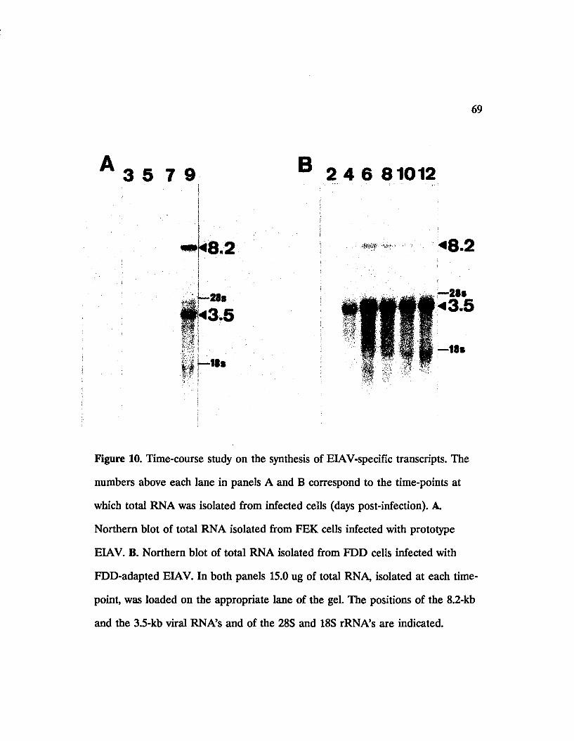
69
3 5 7 9
*8.2
B 2 4 6 81012
* 8.2
Figure 10. Time-course study on the synthesis of EIAV-specific transcripts. The
numbers above each lane in panels A and B correspond to the time-points at
which total RNA was isolated from infected cells (days post-infection). A
Northern blot of total RNA isolated from FEK cells infected with prototype
EIAV. B. Northern blot of total RNA isolated from FDD cells infected with
FDD-adapted EIAV. In both panels 15.0 ug of total RNA, isolated at each time-
point, was loaded on the appropriate lane of the gel. The positions of the 8.2-kb
and the 3.5-kb viral RNA’s and of the 28S and 18S rRNA’s are indicated.
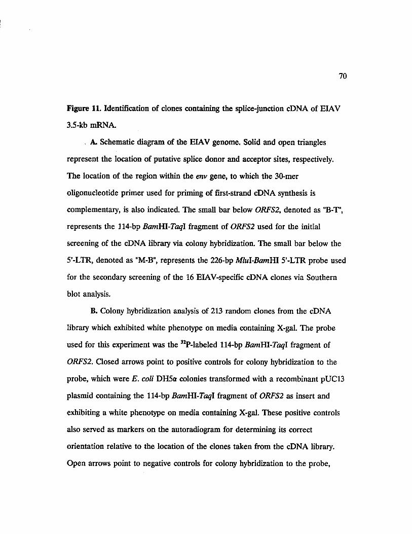
70
Figure 11. Identification of clones containing the splice-junction cDNA of EIAV
3.5-kb mRNA.
A. Schematic diagram of the EIAV genome. Solid and open triangles
represent the location of putative splice donor and acceptor sites, respectively.
The location of the region within the env gene, to which the 30-mer
oligonucleotide primer used for priming of first-strand cDNA synthesis is
complementary, is also indicated. The small bar below ORFS2, denoted as "B-T',
represents the 114-bp BamHL-Taql fragment of ORFS2 used for the initial
screening of the cDNA library via colony hybridization. The small bar below the
5’-LTR, denoted as "M-B", represents the 226-bp Mlul-BamHl 5’-LTR probe used
for the secondary screening of the 16 EIAV-specific cDNA clones via Southern
blot analysis.
B. Colony hybridization analysis of 213 random clones from the cDNA
library which exhibited white phenotype on media containing X-gal. The probe
used for this experiment was the 32P-labeled 114-bp BamHl-Taql fragment of
ORFS2. Closed arrows point to positive controls for colony hybridization to the
probe, which were E. coli DH5a colonies transformed with a recombinant pUC13
plasmid containing the 114-bp BamHl-Taql fragment of ORFS2 as insert and
exhibiting a white phenotype on media containing X-gal. These positive controls
also served as markers on the autoradiogram for determining its correct
orientation relative to the location of the clones taken from the cDNA library.
Open arrows point to negative controls for colony hybridization to the probe,
71
which were E. coli DH5a colonies transformed with pUC19 plasmid and
exhibiting a blue phenotype on media containing X-gal.
C. Ethidium bromide-stained 1.0% agarose gel of Prfl-digested minilysate
plasmid DNA isolated from the 16 EIAV-specific cDNA clones (lanes 1-16)
which were identified in panel B by colony hybridization. Lane C contains a
positive control for Southern blot hybridization of the gel to the 226-bp Mlul-
BamHl probe, which was an Mlul/BamHl double-digested recombinant pIBI76
plasmid containing the 226-bp Mlul-BamHl fragment as insert. Arrows above the
gel point to cDNA clones 4 and 13, whose inserts were not cut out by Pstl.
D. Southern blot of the gel in panel C, hybridized to the 32P-labeled 226-
bp Mlul-BamHl probe. Arrows above the blot point to cDNA clones 4 and 13,
whose plasmid DNA strongly hybridized with the probe.

72
□»• LTR WWWl
B
pol
9 0 9
Sis2 "i^«rp i 30-mcr primer for
Ist-strand cDNA synthesis
* 10
=> * *
■: ' " # •
M C I 2 3 4 5 6 7 8 9 10 1112 13 14 15 16M C 1 2 3 4 5 6 7 8 9 10 11 12 13 14 15 16

73
With the availability of the Northern hybridization data, it was clear that
the single-spliced 3.5-kb env mRNA is the predominant viral transcript during
cytopathic infection of FDD cells. Moreover, since the 102-bp PvuU-Sspl fragment
of ORFS1 was shown to hybridize to the 3.5-kb mRNA (Figure 9, panel D), it
was clear that the splice acceptor site for this mRNA is located upstream of the
Sspl site and thus, as expected, 5’ to the translational initiation codon of the env
gene. Thus the exact splicing pattern of this mRNA was investigated by
identification and sequencing of cDNA clones in a cDNA library constructed
from poly(A)+ RNA of EIAV-infected FDD cells.
The cDNA library was constructed by utilizing a synthetic oligonucleotide,
complementary to a 30-nucleotide-long sequence 400 nucleotides downstream of
the initiation codon of the env gene, as the primer for first-strand cDNA synthesis
(Figure 11A). The primer was designed to hybridize to RNA sequences within the
env reading frame. Thus it was expected to hybridize to both the unspliced, 8.2-kb
full-length mRNA, and the spliced, 3.5-kb env mRNA. Moreover, the 5’-to-3’
direction of first-strand cDNA synthesis, initiated from the primer towards the 5’-
end of its complementary viral mRNA, was expected to generate cDNA copies of
both the 8.2- and the 3.5-kb mRNA’s. Since the 3.5-kb mRNA splice acceptor site
was known to be situated upstream of the env gene initiation codon, therefore
cDNA copies of this mRNA were expected to span the splice junction of this
transcript.
Following the synthesis of both first- and second-strand cDNA products,
the double-stranded cDNA products were cloned into the plasmid vector pUC9

74
and propogated in E. coli DH5a. Previous cDNA cloning experiments from
poly(A)+ RNA of ELAV-infected cells had revealed that the first-strand cDNA
synthesis reaction, under the conditions employed, results in a majority of the
cDNA products being synthesized from cellular rather than viral mRNA’s (data
not shown), most possibly due to non-specific hybridization of the oligonucleotide
primer to cellular mRNA’s. To identify recombinant clones containing viral
cDNA inserts, 213 independent clones of the library, exhibiting a white phenotype
on media containing X-gal, were randomly selected and initially screened by
colony hybridization using the 32P-labeled 114-bp BamHl-Taql DNA fragment of
the env gene as the hybridization probe (Figure 11 A). This fragment contains
DNA sequences immediately upstream of the priming site for first-strand cDNA
synthesis, and the use of it as probe in the initial screening of the library
permitted the identification of those clones which contained cDNA inserts
generated from the priming site on ELAV-specific poly(A)+ RNA’s. The result of
colony hybridization to the 114-bp BamHl-Taql probe is presented in Figure 11B,
showing DNA from 16 of the 213 bacterial clones hybridizing to the probe.
Because each clone was individually picked from the cDNA library and streaked
on the nitrocellulose membrane used for colony hybridization analysis, differential
intensities of the hybridization signal from the various clones are due to
quantitative differences in sizes of the various colonies after their growth on the
membrane, resulting from differences in either the growth rate of the individual
clones or the number of viable bacteria transfered onto the membrane.

75
A splicing event joins the R-U5-leader sequences from the 5’-end of the
full-length genomic RNA to the splice acceptor site upstream of the env gene.
Therefore it was apparent that among the 16 EIAV-specific cDNA clones, shown
by colony hybridization to contain cDNA sequences located between the BamHl
and Taql sites of the env gene, those possessing sequences spanning the splice
junction of the 3.5-kb mRNA must also contain sequences from the 5’-region of
the EIAV genome upstream of the gag gene. Thus the 16 EIAV-specific cDNA
clones were subjected to a second round of screening using the radiolabeled 226-
bp Mlul-BamHl fragment as the hybridization probe (Figure 11 A). The BamHl-
end of the Mlul-BamHl probe is situated upstream of the three putative splice
donor sites near the 5’-end of the gag gene (Figure 11 A), therefore it was clear
that those clones hybridizing to this probe would contain cDNA sequences
extending upstream of the BamHl site and thus would also span the splice
junction of the 3.5-kb mRNA.
The cloning strategy used for the generation of the cDNA library was
based on oligo(dC)-tailing of the double-stranded cDNA products, in the presence
of deoxycytidine and terminal deoxynucleotidyl transferase, followed by annealing
of the oligo(dC)-tailed cDNA to /M-linearized, oligo(dG)-tailed pUC9. In such a
manner, a Pstl site is created at each end of the cDNA insert within the pUC9
vector, allowing recovery of the cDNA insert by a Pstl digestion of the plasmid
DNA. Thus minilysate plasmid DNA, prepared from the 16 positive clones, was
digested with Pstl to release the cDNA inserts, fractionated by electrophoresis
through a 1.0% agarose gel, and Southern blotted using the 226-bp Mlul-BamHl

76
probe. Figure 11C shows the ethidium bromide-stained 1.0% agarose gel of the
Ptfl-digested minilysate plasmid DNA from the 16 EIAV-specific cDNA clones
(lanes 1-16). As a positive control for the hybridization of the Southern blot of
the same gel to the 226-bp Mlul-BamHl probe, the pIBI76 plasmid DNA
containing the 226-bp Mlul-BamHl fragment was digested with Mlul and BamHl
and loaded on lane C of the gel. Digestion of this plasmid with Mlul and BamHl
yields two DNA fragments: a 4200-bp linearized pIBI76 vector DNA and a 226-
bp Mlul-BamHl fragment (Figure 11C, lane C). Digestion of the minilysate
plasmid DNA from the 16 EIAV-specific cDNA clones with Pstl yields a 2700-bp
linearized pUC9 vector DNA, a 1500- to 1700-bp undigested plasmid DNA and
the cDNA inserts of the individual clones, ranging from 400- to 1000-bp in size
(Figure 11C, lanes 1-16). Interestingly, the cDNA inserts in plasmid DNA of
clones 4 and 13 (Figure 11C, lanes 4 and 13, marked by arrows) are not excised
from the vector following the Pstl digestion. Additional restriction enzyme
mapping of plasmid DNA from these two clones indicated that they were
identical and revealed a deletion of one of the two Pstl sites present at each end
of the cDNA insert, thus explaining the inability of the Pstl digestion to release
their cDNA inserts.
The Southern blot of the same gel, hybridized to the 226-bp Mlul-BamHl
probe, is presented in Figure 11D. Out of the 16 EIAV-specific cDNA clones,
only plasmid DNA from clones 4 and 13, which contain a deleted Pstl site,
hybridizes with this probe (Figure 11D, lanes 4 and 13, marked by arrows). As
can be seen, the probe hybridizes with two specific DNA bands in lanes 4 and 13

77
of Figure 11D. The slower migrating band is the Ryfl-linearized plasmid DNA of
each of the two clones, while the faster migrating band is undigested plasmid
DNA. Since the cDNA insert is not excised by Pstl due to a deleted Pstl site in
plasmid DNA of these two clones, the cDNA sequence is therefore present in
both the P&I-linearized and the undigested plasmid DNA bands. Thus the probe,
recognizing the cDNA sequence present in both DNA bands, strongly hybridizes
with both. Moreover, the particular preparation of the 226-bp Mlul-BamHl probe
used in this experiment also contained low amounts of contaminating vector
DNA, as evidenced by the weak hybridization of the probe to the pIBI vector
DNA band in addition to the 226-bp Mlul-BamHl fragment in the control sample
(Figure 11D, lane C). Since the labeled contaminating pDBI vector DNA present
in the probe preparation also contains a pBR322-derived sequence present in
pUC9 DNA, it also recognizes the linearized and undigested pUC9 DNA
fragments of clones 1-3, 5-12, and 14-16 (Figure 11D, lanes 1-3, 5-12, 14-16), thus
explaining the weak hybridization of these fragments to the probe. The
hybridization of plasmid DNA from these latter 14 clones only to the 114-bp
BamHl-Taql probe (Figure 11B), and not to the 226-bp Mlul-BamHl probe
(Figure 11D), indicated that their cDNA inserts had primed from the 8.2-kb full-
length EIAV mRNA and thus had extended into the pol gene instead of spanning
the splice junction of 3.5-kb mRNA.
The hybridization of plasmid DNA from clones 4 and 13 to the 226-bp
Mlul-BamHl probe indicated that they contained cDNA inserts extending
upstream of the gag gene and thus spanning the splice junction of the 3.5-kb

78
mRNA. This finding was further confirmed by restriction enzyme mapping of
plasmid DNA from these two clones using restriction enzymes Smal and BamHl.
The distance between the Smal site upstream of the gag gene and the BamHl site
near the 5’-end of the env gene on the EIAV proviral DNA is 4945-bp. However,
a Smal/BamHl double-digestion of plasmid DNA from both clones had yielded a
270-bp fragment (data not shown), clearly indicating that a splicing event between
the Smal site, upstream of the gag gene, and the BamHl site, near the 5’-end of
the env gene, had reduced the distance separating the two restriction sites from
4945-bp on the provirus to 270-bp on the cDNA.
The 270-bp Smal-BamHl fragment from both cDNA clones was subcloned
into M13mpl8 and M13mpl9 RF DNA, allowing nucleotide sequencing of both
strands of each clone by the dideoxy chain termination method (Sanger et al.,
1977). The two clones were found to contain identical splice sites, and the
nucleotide sequence of the splice junction present in both clones is presented in
Figure 12A The sequencing data identifies the splice donor and acceptor sites, in
the full-length EIAV genomic mRNA, used to generate the spliced mRNA copy
of the two cDNA clones. Of the three potential splice donor sites near the 5’-
terminus of the gag gene (Figure 12B; see below), in this spliced mRNA the 5’-
most splice donor site is spliced to the splice acceptor site near the 5’-end of
ORFS1, 178 nucleotides upstream of the env gene translational initiation ATG
codon. Also shown in Figure 12B are the nucleotide sequences of the identified
splice donor and acceptor sites and the extent of their homology to the consensus
splice donor and acceptor sequences (Mount, 1982; Ohshima and Gotoh, 1987).

79
Figure 12. Nucleotide sequence analysis of the 270-bp Smal-BamHl splice-
junction cDNA of the EIAV 3.5-kb mRNA.
A. Autoradiogram of a sequencing gel showing the region of nucleotides
immediately 5’- and 3’- to the splice junction site, which is pointed out by the
arrow.
B. Schematic diagram of the EIAV genome, showing the location of
putative splice donor sites (solid triangles) and acceptor sites (open triangles) on
the viral genome. The bold-face nucleotide sequences within the left and right
lower brackets are those of the identified splice donor and acceptor site,
respectively, with underlined bases showing perfect homology to the consensus
donor and acceptor site sequences (Mount, 1982; Ohshima and Gotoh, 1987). The
arrows show the site of splicing.

80
B
□ 5* LTR
pot
3' LTR
3
Splice donor
%Splice acceptor
A fifiA C A flflT A A flA
I______ IiTTG AGT ATTGTTGCAGG AAGCAA
I___________ I

81
Northern Hybridization Analysis Using Splice Donor-Specific Oligonucleotide
Probes
As mentioned previously, three potential splice donor sites are located
near the 5’-terminus of the EIAV gag gene (Figure 12B) based on matches to the
consensus sequence for splice donor sites (Mount, 1982; Ohshima and Gotoh,
1987). Based on the utilization of one or more of the three donor sites for
generation of the spliced viral transcripts, a number of differentially spliced
mRNA’s can be generated in EIAV-infected cells with varying coding potentials.
The nucleotide sequence of the three short ORFs in the EIAV genome has
revealed that only S2 contains a translational initiation methionine codon
(Rushlow et al., 1986). Thus for functional expression of SI or S3, an initiation
codon from an upstream gene needs to be spliced, in frame, to the coding
sequences of the two ORF’s.
By comparison to other lentiviruses, which utilize multiple splicing of their
full-length genomic mRNA to generate the small mRNA’s needed to encode their
short ORFs (Arya et al., 1985; Arya and Gallo, 1986; Davis and Clements, 1989;
Mazarin et al., 1988; Muesing et al., 1985), the initiation codon for the EIAV SI
or S3 ORFs would be assumed to be located within the first exon of such multi
spliced mRNA. This exon would contain sequences from the 5’-region of the viral
genome, and the only translational initiation ATG codon in this region is that of
the gag gene.

82
The nucleotide sequence of the EIAV provirus near the 5’-terminus of the
gag gene is presented in Figure 13 (Stephens et al., 1986). The sequence starts
from the Smal site, 71 nucleotides upstream of the gag gene’s ATG translational
initiation codon, and continues for 120 nucleotides (40 codons) into gag.
Moreover, the location of each of the three putative splice donor sites in relation
to the gag ATG codon is shown with underlined bases in each donor site
representing a perfect match to the consensus donor site sequence (Mount, 1982;
Ohshima and Gotoh, 1987). As can be seen, the 5’-most donor site is an excellent
match with the consensus sequence except in the last position; the middle donor
site is a very good match with the consensus sequence with the exception of the
last two nucleotides; and the 3’-most donor site is a good match with the
consensus sequence except in the second and the eighth positions.
Furthermore, as shown in Figure 13, the 5’-most splice donor site is
situated immediately upstream of the gag ATG codon, and its utilization in a
splicing event would remove the gag initiation codon, as part of the intron, from
the spliced mRNA. Since the other two putative splice donor sites are located
downstream of the gag ATG codon, potential splicing events utilizing either one
of these two donor sites, however, would result in the ATG codon of gag being
retained in the spliced mRNA.
Although the use of the 5’-most splice donor site for generation of a
spliced EIAV mRNA was already determined by cDNA cloning and sequencing,
this finding does not rule out the potential utilization of either of the other two
putative splice donor sites to generate additional alternatively spliced
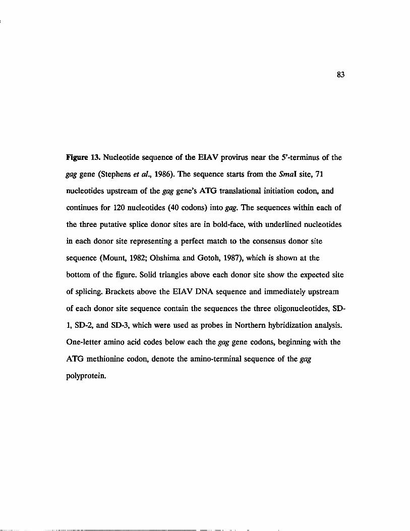
83
Figure 13. Nucleotide sequence of the EIAV provirus near the 5’-terminus of the
gag gene (Stephens et al., 1986). The sequence starts from the Smal site, 71
nucleotides upstream of the gag gene’s ATG translational initiation codon, and
continues for 120 nucleotides (40 codons) into gag. The sequences within each of
the three putative splice donor sites are in bold-face, with underlined nucleotides
in each donor site representing a perfect match to the consensus donor site
sequence (Mount, 1982; Ohshima and Gotoh, 1987), which is shown at the
bottom of the figure. Solid triangles above each donor site show the expected site
of splicing. Brackets above the EIAV DNA sequence and immediately upstream
of each donor site sequence contain the sequences the three oligonucleotides, SD-
1, SD-2, and SD-3, which were used as probes in Northern hybridization analysis.
One-letter amino acid codes below each the gag gene codons, beginning with the
ATG methionine codon, denote the amino-terminal sequence of the gag
polyprotein.

84
SD-1I 3 1-CTCCACAAGGACCGGTCTTGTGI
Steal£££GGGACAGCAGAGGAGAACTTACAGAAGTCTTCTGGAGGTGTTCCTGGCCAGAACAC
_________________ SD-2____________________lTCCTCC-5~n 13 ’ -CCTCTGGGAAACTGTACCTCGTTCCGCG-5'TI
TAGGAGGACAGGTAAGATGGGAGACCCTTTGACATGGAGCAAGGCGCTCAAGAAGTTAGAG
M G D P L T W S K A L K K L E
________________ SD-3____________________| 3 ' -CCATGTTCCCAGAGTCTTTAATTGATG-51 I
▼ ?AAGGTGACGGTACAAGGGTCTCAGAAATTAACTACTGGTAACTGTAATTGGGr.GGTAAGT
K V T V Q G S Q K L T T G N C N W A L S
C TAG TAGAC T TAT T T ____L V D L P
Consensus splice donor sequence: ^ACaGT^AGT

85
EIAV mRNA’s in infected cells. For instance, the 3.5-kb mRNA could represent
a heterogeneous population of mRNA molecules generated by splicing of each of
the three donor sites to the same acceptor site upstream of the env gene, or by
splicing of one donor site to different, but proximally located, acceptor sites
upstream of the env gene. As a direct method for addressing the first possibility,
that is differential splicing of the three donor sites to the same acceptor site,
oligonucleotides complementary to sequences immediately upstream of each of
the three splice donor sites were radiolabeled with 32P and used as probes in
Northern hybridization analysis of poly(A)+ RNA from infected FDD cells. The
sequence of the three oligonucleotides, termed SD-1, SD-2, and SD-3, along with
the location of their complementary region upstream of each splice donor site on
the viral genome is also indicated in Figure 13.
Since all three probes contain complementary sequences to the full-length
genomic mRNA, they would all be expected to hybridize to this viral transcript.
Moreover, because SD-1 is complementary to a region upstream of all three
putative splice donor site, it is expected to also hybridize to all spliced viral
mRNA’s, namely the 3.5- and 1.5-kb mRNA’s, whose production would utilize
either one, two, or all three of the donor sites downstream of SD-1. Furthermore,
the hybridization of SD-2 to either the 3.5- or the 1.5-kb mRNA, or to both,
would indicate that the middle splice donor site, in addition to the 5’-most donor
site, is utilized to generate the corresponding spliced viral mRNA(s). Similarly,
the hybridization of SD-3 to either of the two spliced mRNA’s, or to both, would
reveal the usage of the 3’-most donor site, in additon to the 5’-most donor site,
86
for generation of the corresponding viral mRNA(s). However, the hybridization of
SD-2 and SD-3 to only the full-length 8.2-kb mRNA, and not to any spliced
mRNA, would indicate that only the 5’-most splice donor site is used to generate
the 3.5- and 1.5-kb mRNA’s of EIAV.
The results of the three Northern hybridizations are presented in Figure
14. As expected, SD-1 hybridizes to the three species of EIAV-specific mRNA’s.
However, SD-2 and SD-3 only hybridize to the 8.2-kb full-length mRNA and do
not detect either the 3.5- or the 1.5-kb spliced mRNA’s recognized by SD-1.
These results indicate that the 5’-most splice donor site, already shown to be used
for generating a spliced EIAV mRNA, is the only donor site utilized in EIAV-
infected FDD cells for splicing of the full-length viral genomic mRNA. Thus the
R-U5-leader sequences initiating from the 5’-boundary of the R region and
terminating at the 5’-most splice donor site constitute the first exon of all spliced
EIAV mRNA’s. The consequences of this finding are that spliced EIAV mRNA’s
containing sequences from ORFS1 or ORFS3 lack the gag gene AUG codon, and
if these short ORFs are indeed expressed in EIAV-infected cells, translation of
their corresponding mRNA’s would presumably have to involve the use of non-
AUG initiation codons.

87
Kb
8.2
3.5
1.5-
Figure 14. Northern hybridization analysis of poly(A)+ RNA from EIAV-infected
FDD cells using the three oligonucleotide probes SD-1, SD-2, and SD-3 (lanes 1,
2, and 3, respectively). The electrophoretic mobilities of the three species of
ELAV-specific mRNA’s are indicated on the left.

i :i
88
SI Nuclease Mapping of Viral mRNA’s
The results of Northern hybridizations with the splice donor-specific
oligonucleotide probes indicated that all spliced EIAV mRNA’s utilize the 5’-
most splice donor site immediately proceeding the gag gene. Moreover, the results
obtained from cDNA cloning and sequencing had revealed the splicing of this
donor site to the acceptor site 178-nucleotides upstream of the 5’-boundary of the
env gene to generate the single-spliced 3.5-kb env mRNA.
By analogy to other lentiviruses, it is presumed that the majority of
sequences within the EIAV single-spliced mRNA, lying outside of those within
the short ORFs, would be removed as an intron in a second splicing event to
produce the double-spliced viral mRNA (Aiya et al., 1985; Arya and Gallo, 1986;
Davis and Clements, 1989; Mazarin et al., 1988; Muesing et al., 1985). The
double-spliced message, containing coding sequences of the short ORFs, would
then encode the regulatory proteins of the virus such as the viral trans-
activator(s). Indeed a 1.4-kb cDNA copy of one such double-spliced mRNA,
containing sequences from ORFS1 and ORFS3, has already been isolated and
shown to encode a viral fra/tr-activator (Derse et al., 1989; see below). Moreover,
in agreement with our finding that only the 5’-most splice donor site on the EIAV
genome, upstream of the gag gene AUG codon, is utilized to generate the first
exon of all virus-specific spliced mRNA’s in infected cells, the translation of the
aforementioned double-spliced viral mRNA is proposed to be initiated from a
non-AUG codon (Derse et al., 1989).

89
To map the location of the splice donor site(s) within the EIAV env gene
that are used to produce the double-spliced mRNA species, and also to
determine whether any additional splice acceptor sites, located upstream of and
within the env gene, are used to generate the spliced EIAV mRNA’s, Sl-nuclease
mapping studies were performed using RNA from FDD cells infected with FDD-
adapted EIAV. The studies were designed to also include the 5’-region of the gag
gene to test the validity of the results obtained previously using the splice donor-
specific oligonucleotide probes.
Figure 15 shows the relative location of putative splice donor and acceptor
sites, represented by closed and open triangles, respectively, on the EIAV
genome. In addition to the acceptor site located 178-nucleotides upstream of the
env gene initiation codon and identified by cDNA cloning and sequencing, a
second putative acceptor site is present at 129-nucleotides upstream of the env
initiation codon. Following the 3’-terminus of ORFS1 and extending into the
coding sequences of ORFS2 are four putative donor sites, any one of which could
potentially be utilized in the second splicing event of the single-spliced 3.5-kb
mRNA to generate a double-spliced 1.5-kb mRNA. Moreover, a putative splice
acceptor site is situated immediately at the 5’-boundary of the ORFS3 reading
frame.
The sequence of each putative donor and acceptor site, with the exception
of the three putative donor sites near the 5’-terminus of the gag gene whose
sequence was presented earlier in Figure 13, is presented in Table III. In each
case, underlined bases correspond to a perfect match with the consensus donor

90
gag
5'LTR3'LTR
pol
Figure 15. Schematic diagram of the EIAV genome. The figure shows the
location of putative splice donor and acceptor sites, represented by solid and
open triangles, respectively, on the viral genome.

Table m . Nucleotide sequence of the putative splice donor andacceptor sites located immediately upstream of or within the EIAV env genea.
Nucleotide sequence of putative splice sites
Nature of splice site Position on the EIAV genome^
M.GLrrT<2A Donor 1226
TTG^TAAAG Donor 1248
TGGGGTAACA Donor 1256
ACjJ gTAAGA Donor 1385
VTTG AGTATTGTTGCAGG Acceptor^ 1084
VTTCCTGAGGTCTCT AGG Acceptor 1133
VAACCTCTTCGCCTAAGA Acceptor 3184
a. The splice junction site within the putative donor and acceptor sequences is denoted by closed and open triangles, respectively. Underlined nucleotides are a perfect match with the consensus splice junction sequences (Mount, 1982; Ohshima and Gotoh,1987).
b. Based on the numbering system of Rushlow et al. (1986).
c. The utilization of this acceptor site in prototype EIAV-infected FDD cells has already been confirmed by cDNA cloning and sequencing (Figure 11).

92
and acceptor site sequences (Mount, 1982; Ohshima and Gotoh, 1987). Both
acceptor sites located upstream of the env gene show an excellent match with the
consensus acceptor site sequence. Moreover, the four putative donors which
follow the 3’-end of ORFS1 and extend into ORFS2 exhibit good matches with
the consensus donor sequence. Finally, the acceptor site located immediately at
the 5’-boundary of the ORFS3 reading frame is a good match with the consensus
sequence but contains an A instead of a G in the +1 position and an A instead
of a pyrimidine in the -3 position.
For SI nuclease mapping studies of the splice donor and acceptor sites in
spliced viral mRNA’s, four specific EIAV proviral DNA restriction fragments,
spanning the various putative splice donor and acceptor sites on the viral genome,
were used as probes. Since the entire nucleotide sequence within each of the four
probes comes from the EIAV proviral DNA, the full-length nonsense strand of
each probe is complementary to the sense strand sequence of the unspliced, full-
length 8.2-kb EIAV genomic mRNA. Moreover, by virtue of spanning the
putative splice donor or acceptor sites on the full-length genomic mRNA, only a
segment of the nucleotide sequence within the nonsense strand of each probe is
complementary to the corresponding exon sequence of the full-length viral mRNA
and its spliced mRNA product. The other segment of the nonsense strand of the
respective probe, however, is complementary to the corresponding intron
sequence of the full-length viral mRNA which is not present in its spliced
product. Such differential complementarity of each splice junction probe to the

93
unspliced and spliced viral mRNA’s provides the basis for mapping the exact
location of splice donor and acceptor sites on the viral genome.
Following the hybridization of each denatured probe to RNA from EIAV-
infected cells, a protection assay is performed in which the RNA-DNA hybrid is
treated with the single-strand-specific nuclease SI. Single-stranded probe DNA or
viral RNA is degraded by the nuclease into mononucleotides while double
stranded DNA or RNA-DNA hybrids are left intact (Berk and Sharp, 1977). Thus
in this assay the full-length probe fragment is expected to be protected from SI
nuclease digestion by the full-length viral genomic mRNA, while smaller probe
fragments would be rescued from SI nuclease digestion by the spliced viral
mRNA’s. The size of the smaller probe fragments, determined by denaturing
polyacrylamide gel electrophoresis, would then be equal to the actual distance
between the splice site on the spliced mRNA relative to the radiolabeled end of
the probe.
Figure 16A presents a schematic diagram of the EIAV genome, showing
the location of the four probes used for SI nuclease mapping studies relative to
the putative splice donor and acceptor sites on the viral genome. Because the
probes are radiolabeled double-stranded DNA fragments, in each hybridization
reaction a portion of the denatured nonsense strand input probe molecules re-
hybridize back to their complementary sense DNA strand instead of hybridizing
to viral mRNA’s. Such full-length nonsense strand probe molecules cannot be
distinguished from those that are protected from SI nuclease digestion by the full-
length mRNA. To allow for distinction of the full-length input probe from the

94
Figure 16. Schematic representation of the probes used in SI nuclease mapping.
A. Schematic diagram of the EIAV genome, showing the location of the
four probes used for SI nuclease mapping studies (pi, p2, p3, p4) relative to the
location of putative splice donor (solid triangle) and acceptor sites (open triangle)
on the viral genome. Probe p i consists of a 300-bp Narl-Pvull proviral DNA
fragment and a 250-bp M13mpl8 PvuU-Narl tag fragment at the Pvull site. Probe
p2 consists of a 450-bp NcoI-BamHL proviral DNA fragment and a 161-bp
M13mpl9 Ncol-Bglll tag fragment at the Ncol site. Probes p3 and p4 do not
contain tag sequences and were directly used in S 1-nuclease protection analysis as
617-bp PvuU-HindlU and 385-bp Dral-Scal proviral DNA restriction fragments,
respectively. Abbreviations: N, Nar\\ P, Pvull; G, BglII; C, Ncol; B, BaraHI; P,
Pvull; H, Hindlll; D, Dral; S, Seal; SD, putative splice donor site(s); SA, putative
splice acceptor site(s).
B.-E. Schematic representations of probes pl-p4, respectively. Also shown
in each panel are fragments of the nonsense strand of each respective probe, and
their lengths in nucleotides (nt), which could potentially be protected from SI
nuclease digestion by the unspliced, single-spliced, and double-spliced EIAV
mRNA’s. The asterisk represents the 32P-labeled end of the nonsense strand of
each probe.

II
A.
d5'LTR
N■
B.
Input Probe*
Unspliced *
Spliced *
D.
Input Probe/ Unspliced *
Spliced *
» gag
P N
Pi
N fE/ z z / z z JN \ \ \ \ 1 ' ' '
SD .intron P M13 N'/////////A
c.G M13 C intron SA
3'LTR
7 , ,!/✓✓//✓✓✓✓ /I| \ \ \ \ \ N \ \ \
300-nt
130-, 183-, 217-nt
550-nt 61 l-nt«
450-nt
157-, 206-nt
* Input Probe
* Unspliced
* Spliced
SD intron H
■617-nt
E.intron SA
V____________5h/V/VV^VAl_ K _ N _\ N _S \ N \ \ I
385-nt"Input Probe/
* Unspliced
114- ,136-, 144-, 273-nt 178-nt. * Spliced
\oLn

96
probe fragment rescued by the full-length mRNA, two of the probes were
specifically designed to contain "tag" sequences taken from the double-stranded
replicating form (RF) DNA of either M13mpl8 or M13mpl9 and attached to
their intronic end. As shown in Figure 16B, probe 1 (pi) consisted of a 300-bp
Narl-PvuTL proviral DNA fragment and a 250-bp M13mpl8 Smal-Narl tag at the
Pvull site. Thus following S 1-nuclease protection analysis using this probe, the
input probe and the probe fragment rescued by the full-length mRNA would be
550- and 300-nucleotides (nt) long, respectively. Moreover, smaller probe
fragments, whose size equals the distance between the splice donor site and the
JVorl-end of the probe, would be protected from S 1-nuclease digestion by the
spliced mRNA’s. This distance is 130-nt for the 5’-most donor site, 183-nt for the
middle donor site, and 217-nt for the 3’-most donor site.
Similarly, as shown in Figure 16C, probe 2 (p2) consisted of a 450-bp
Ncol-BamHl proviral DNA fragment and a 161-bp M13mpl9 Smal-BgRl tag at
the repaired Ncol site. Thus following S 1-nuclease protection analysis using this
probe, the expected length of the input probe would be 611-nt while the probe
fragment protected by the full-length mRNA would be 450-nt long. In additon,
the distance between each of the two putative splice acceptor sites in this region
and the BamHl-end of the probe is 157- and 206-nt long, respectively.
The other two probes, probes p3 and p4, did not contain tag sequences
and were directly used in Sl-nuclease protection analysis as 617-bp PvuU-Hindlll
and 385-bp Dral-Scal proviral DNA restriction fragments, respectively. As shown

97
in Figure 16D, the distance between each of the four putative splice donor sites
located within the sequences of probe p3 and the fVuII-end of this probe is 114-,
136-, 144-, and 273-nt long, respectively. Moreover, Figure 16E shows the the
distance between the putative acceptor site located within the sequences of probe
p4 and the Scal-end of the probe, which is 178-nt long.
For SI nuclease protection assays, RNA-DNA hybrids, obtained from
hybridization of each end-labeled probe with total RNA isolated from FDD cells
infected with FDD-adapted EIAV, were digested with SI nuclease and the
digestion products were resolved on 6% polyacrylamide-8M urea sequencing gels.
As negative control, RNA from uninfected FDD cells, lacking EIAV-specific
unspliced and spliced transcripts, was used in parallel experiments. The results of
these studies are presented in Figure 17.
Panels A, B, C, and D in Figure 17 correspond to individual S 1-nuclease
protection assays using probes pi, p2, p3, and p4, respectively, which were
described above. In each panel, lane M contains end-labeled MspI fragments of
plasmid pBR322, while lane 1 or lane 2 contain the corresponding probe
fragments protected from SI nuclease digestion by RNA from EIAV-infected
FDD cells or uninfected FDD cells, respectively. In panel A, three distinct
fragments of probe p i (550-bp Narl\ Figure 16A and 16B), migrating at about
550-nt, 300-nt, and 130-nt are protected following the Sl-nuclease protection assay
utilizing RNA from infected cells (lane 1), while only the 550-nt full-length probe
fragment is present following the assay in which control RNA from uninfected
cells is utilized (lane 2). The 550-nt probe fragment in lanes 1 and 2 corresponds

98
Figure 17. SI nuclease mapping of splice junctions in spliced mRNA’s of EIAV.
Panels A, B, C, and D correspond to SI nuclease protection assays of probes pi,
p2, p3, and p4, respectively, using total cellular RNA from FDD cells infected
with FDD-adapted EIAV (lane 1), or uninfected FDD cells (lane 2), Lane M in
each panel contains 32P-labeled Mspl-digested pBR322 as size marker.

99
Ant
022_527~ ■404-
309- * * *
23217- * » 201- «, 100- «■ 180- * »
160- m
147- m
B C DM l 2 M l 2 M 1 2
m i* * m^Sn& SrI F r « |
• • w
• •
J|4
123- « *
110— <•» W
90- •
76- •
67- • <*

100
to the portion of full-length input probe molecules, containing the M13 tag
sequence, which have escaped degradation by SI nuclease due to re-hybridization
with their complementary DNA strand. Moreover, the additional probe fragments
present in lane 1, but not in lane 2, are those which have been protected from
Sl-nuclease degradation by EIAV-specific mRNA’s. In lane 1, the 300-nt
fragment of the probe is that which is protected by the full-length 8.2-kb EIAV
mRNA (Figure 16B), while the 130-nt probe fragment is that which is protected
by a spliced viral mRNA whose splice junction site is 130-nt downstream of the
Afarl-end of the probe within the EIAV genome. This splice site corresponds
exactly to the 5’-most donor site among the three putative donor sites near the 5’-
end of the EIAV gag gene (Figure 16A). Furthermore, as mentioned above, the
protected probe fragments which would correspond to spliced mRNA’s utilizing
the middle or the 3’-most putative donor sites in this region would be expected to
be 183- or 217-nt long (Figure 16A and 16B). Since these additional bands are
not present in lane 1 of panel A, it appears that the 5’-most splice donor site is
indeed the only donor site near the 5’-end of the gag gene that is utilized in
EIAV-infected FDD cells for splicing of the full-length viral mRNA, confirming
the earlier results obtained using the splice-donor-specific oligonucleotide probes.
In Figure 17, panel B, four distinct fragments of probe p2 (611-bp BamHl-
BglU; Figure 16A and 16C), migrating at about 611-nt, 450-nt, 206-nt, and 155-nt
are protected by RNA from infected cells (lane 1). As expected, the 611-nt full-
length probe fragment, containing the M13 tag sequence, is the only protected
probe fragment in the assay utilizing RNA from uninfected cells (lane 2). In lane

101
1 of panel B, the 450-nt fragment of the probe is that which is protected by the
full-length 8.2-kb EIAV mRNA, while the probe fragments migrating at about
206-nt and 155-nt are those protected by spliced viral mRNA’s whose splice
junction sites are therefore 206-nt and 155-nt, respectively, upstream of the
BamHl-end of the probe. The higher intensity of the 206-nt probe fragment
corresponds to the higher concentration of the viral mRNA which has protected
this fragment relative to the other viral mRNA’s present in infected FDD cells.
The splice junction site located at 206-nt upstream of the BaniHl site corresponds
exactly to the acceptor site, already identified by cDNA cloning and sequencing,
which is located at 178-nt upstream of the env gene initiation codon (position
1084 on the nucleotide sequence of Rushlow et al., 1986; Table IB). Interestingly,
the second splice junction site identified by this assay and located at 155-nt
upstream of the BamHI-end of the probe corresponds to the acceptor site present
at 129-nt upstream of the env gene initiation codon (position 1133 on the
nucleotide sequence of Rushlow et al., 1986; Table III). The length of the probe
fragment protected by a spliced mRNA utilizing this acceptor site was actually
expected to be 157-nt (Figure 16C), although the apparent mobility of this
fragment is closer to 154- or 155-nt during denaturing polyacrylamide gel
electrophoresis (Figure 17, panel B, lane 2).
Panel C in Figure 17 shows the results of SI nuclease protection analysis
of probe p3 (617-bp PvuU-HindUl; Figure 16A and 16D). As expected, the full-
length 617-bp probe fragment is protected by RNA from either infected or
uninfected FDD cells (lanes 1 and 2). Moreover, a protected probe fragment,

102
migrating at about 140- to 144-nt, is protected by RNA from infected cells (lane
1) but not by RNA from uninfected cells (lane 2). This fragment of the probe is
therefore protected by a spliced EIAV mRNA whose splice junction site is
situated at about 140- to 144-nt downstream of the Pvull site on the viral
genome. As mentioned previously, the four putative splice donor sites near the
end of ORFS1 and within the sequences of the Pvull-Hindlll probe (p3) are
located at 114-, 136-, 144-, and 273-nt from the fVnll-end of the probe. Since the
mobility of the protected probe fragment identified in this assay corresponds to a
fragment of about 140- to 144-nt, the donor site located at 144-nt from the Pvull-
end of the probe (position 1256 on the nucleotide sequence of Rushlow et al.,
1986; Table III) appears to be the donor site used to generate a spliced EIAV
mRNA which has protected this fragment of probe p3.
Finally, in Figure 17, panel D, the full-length probe p4 (385-bp Dral-Scal;
Figure 16A and 16E) is protected from SI nuclease digestion by RNA from either
infected or uninfected FDD cells (lanes 1 and 2), while an additional fragment of
the probe migrating at about 180-nt is only protected by RNA from infected cells
(lane 1). Therefore the 180-nt probe fragment is one which is protected from SI
nuclease digestion by a spliced EIAV mRNA whose splice junction site is 180-nt
upstream of the Seal site on the viral genome. The only splice junction site in this
region of the viral genome is a putative splice acceptor site situated at the 5’-
boundary of ORFS3 (Figure 16A; position 3184 on the nucleotide sequence of
Rushlow et al., Table III). This acceptor site is exactly 178-nt upstream of the
Scal-end of the Dral-Scal probe (p4), and a spliced mRNA generated by the

103
utilization of this acceptor site would be expected to protect a 178-nt fragment of
the probe (Figure 16E). The apparent mobility of the protected probe fragment
during denaturing polyacrylamide gel electrophoresis, although closer to 180-nt
than to 178-nt, suggests that this splice acceptor site is indeed utilized in EIAV-
infected FDD cells to generate a spliced viral mRNA which has protected the
180-nt probe fragment.
Longer exposures of the autoradiograms in panels A, B, C, and D of
Figure 17 did not reveal any additional protected probe fragments (data not
shown), suggesting that the detected EIAV mRNA’s constitute the major species
of viral transcripts present in infected FDD cells. The SI nuclease mapping
studies described above have confirmed the earlier results obtained through
Northern hybridizations using the splice donor-specific oligonucleotide probes,
showing that the 5’-most splice donor site, immediately upstream of the gag gene,
is the common donor site to all spliced EIAV mRNA’s. Moreover, in addition to
the splice acceptor site previously identified by sequencing of splice-junction
cDNA obtained from the 3.5-kb mRNA, the SI nuclease mapping studies have
identified and localized a second splice acceptor site upstream of the env gene, a
splice donor site immediately downstream of ORFS1, and a splice acceptor site at
the 5’-boundary of ORFS3.

104
III. The 3.5-kb EIAV mRNA: Potential To Encode The ORFS2 Gene Product In
Addition To The env Proteins
Based on the nucleotide sequence of the EIAV proviral DNA, the
predicted amino acid sequences of the three short ORFs of EIAV is presented in
Figure 18 (Rushlow et al., 1986). As with the short ORFs of other lentiviruses,
such as HTV-1 or visna virus, whose encoded products have been generally shown
to code for tram -acting viral regulatory proteins (Arya et al., 1985; Davis and
Clements, 1989; Mazarin et al., 1988; Sodroski et al., 1986), attempts to delineate
the function of the three short ORFs of EIAV have mainly focused on assessing
the ability of their gene products to fram-activate the viral LTR. Based on the
results of early deletion mutagenesis experiments, by which parts of the coding
sequence within each ORF were removed and the resulting mutant proviruses
were assayed for their capacity to tram-activate the viral LTR, sequences within
the pol-env intergenic region on the viral genome, to which both SI and S2
belong, were shown to be required for fraro-activation of the viral LTR (Sherman
et al., 1988). More recently, Derse et al. (1989) have isolated a cDNA clone of a
double-spliced EIAV mRNA, containing sequences from SI and S3, and have
shown that it can fra/u-activate the viral LTR in transfected cells. Since the
coding sequence of SI is located in the pol-env intergenic region, it is therefore
presently believed that SI encodes a viral tram-activating protein. The function of
the other two ORFs, however, is not known and to date no protein products
from any of the three ORFs have been detected in EIAV-infected cells.

105
ORFS1
V L L Q VE A R P N Y H C Q L C F L R S L VG I D Y L D A S L R K K N K
Q R L K A I Q Q G R Q P Q Y L L
ORFS2
Y M G L F G K G V T W S A S H S M G G S Q G E S Q P L L P N S Q K
N L S V R R T Q C F N L I V I I M T V R T A W Q N R R K Q E T K K
ORFS3
VD P Q G P L E S D Q W C R V L R Q S L P E E K I P S Q T C I A R R H
L G P G P T Q H T P S R R D R W I R G Q I L Q A E V L Q E R L E W R
I R G V Q Q A A K E L G E V N R G I W R E L Y F R E D Q R G D F S
A W G G Y Q R A Q E R L W G E Q S S P R V L R P G D S K R R R K H
L
Figure 18. Predicted amino acid sequences of the three short ORFs of EIAV.
The sequences are "translated" from the nucleotide sequence of EIAV proviral
DNA (Rushlow et al., 1986). The splice acceptor sites in SI and at the 5’-
boundary of S3, identified by sequencing of splice-junction cDNA and SI nuclease
mapping, are indicated by open triangles.

106
A unique property among these short ORFs is that S2 is the only one
which contains a translational initiator ATG codon, located at its second codon
position (Figure 18). Thus, because SI and S3 lack such a codon, their potential
expression in EIAV-infected cells would require the in-frame splicing of an
initiator codon from an upstream reading frame. The results of our SI nuclease
mapping experiments point to the existence of at least one double-spliced mRNA
in EIAV-infected FDD cells whose first exon contains the 5’-leader sequences
upstream of the gag gene, and in its second and third exons it contains the coding
sequences of SI and S3, respectively. Almost the entire coding sequence of S2,
with the exception of its first seven codons, is removed from this mRNA by
splicing, suggesting that potential expression of S2 in virus-infected cells would
require the existence of a separately spliced mRNA.
Nucleotide sequencing of the splice-junction cDNA of the 3.5-kb mRNA
had revealed that the splice acceptor site for this mRNA is located 178
nucleotides upstream of the env gene ATG initiator codon. Because the S2 ATG
codon is 23 nucleotides upstream of the ATG codon of env, and the coding
sequences of the two genes are present on the same spliced 3.5-kb mRNA and in
different reading frames, we postulated that this mRNA could potentially encode
the S2 gene product in addition to the env proteins. To test the hypothsesis, a
transcript designed to contain the coding sequences of both S2 and env identical
to that of the 3.5-kb mRNA was synthesized in vitro and its coding capacity was
evaluated by in vitro translation.

107
The 270-bp Smal-BamYR fragment of the splice junction cDNA, sequenced
earlier for identification of the splice donor and acceptor sites of the 3.5-kb
mRNA, was ligated to a 2710-bp BamHl-Pstl fragment containing the remainder
of the env coding sequence and derived from a separate cDNA clone. The hybrid
Smal-PstI cDNA fragment, containing the entire coding sequences of both S2 and
env genes, was then cloned into the in vitro transcription vector pSP65
downstream of the SP6 promoter (Figure 19). Two separate linear DNA
templates of the plasmid DNA containing the S2-env cDNA insert were prepared
for in vitro transcription by SP6 RNA polymerase. One of the linearized templates
was prepared by digestion of the plasmid with HindUl, an enzyme which cleaves
the env gene at two unique restriction sites, one at about 300-bp and the other at
approximately 1030-bp downstream of the S2 coding sequence (Figure 19). The
second linearized template was prepared by digestion of the plasmid with Mlul,
an enzyme which cuts at a unique restriction site within the 3’-LTR downstream
of the env gene (Figure 19). The design of the two linear templates for in vitro
transcription was based on the premise that if translational initiation from this
mRNA begins at the ATG codons of both S2 and env, then in vitro translation of
the truncated transcript generated from the /fr/u/III-linearized template would
yield a full-length S2 protein and a truncated env protein, while translation of the
full-length transcript generated from the M/uI-linearized template would yield
both S2 and env proteins in their full-length forms.

108
Figure 19. Construction of the S2-env in vitro transcription vector. The 270-bp
Smal-BamHl fragment of the splice junction cDNA was ligated to a 2710-bp
BaniiH-Psil fragment containing the remainder of the env coding sequence and
derived from a separate cDNA clone. The hybrid Smal-Pstl cDNA fragment,
containing the entire coding sequences of both S2 and env genes in their natural
spliced form, was subcloned into pUC19 and taken out as a Sacl-Pstl fragment.
The 2980-bp Sacl-Pstl S2-env hybrid cDNA was then cloned into the in vitro
transcription vector pSP65 downstream of the SP6 promoter. The location of the
HindUl and Mlul restriction sites on the EIAV genome are also indicated.

109
BamlU HindUl HindUl M lu\ Pstl
\
SI S2 env ^3

110
The transcripts derived from the two templates were translated in a cell-
free extract of wheat-germ in the presence of 35S-methionine, and the 35S-labeled
translational products were fractionated by SDS-polyacrylamide gel electro
phoresis (SDS-PAGE). The results, presented in Figure 20, indicate that a lower
molecular weight polypeptide of about 7.5-kda is encoded by both transcripts
(lanes 1 and 2). Moreover, the longer transcript, generated from the Mlul-
linearized template, encodes a second protein of about 27.0- to 28.0-kda (lane 1),
while the second protein expressed by the truncated transcript, produced from the
///nJHI-linearized template, migrates at about 16.0- to 17.0-kda (lane 2).
Based on the nucleotide sequence of 52 and env, the coding potential of
these two genes were calculated to be approximately 7.5- and 97.0-kda,
respectively, from the long transcript. Moreover, the size of the env protein
translated from the truncated transcript was calculated to be around 17.0-kda.
The translation of the smaller protein from both truncated and full-length
transcripts and its apparent molecular weight of 7.5-kda, as expected for the 52
gene product, suggested that it was expressed from the 52 coding sequence in
both transcripts. Moreover, in the case of the truncated transcript, the larger
protein exhibited the 17.0-kda electrophoretic mobility expected from the
truncated env protein (Figure 20, lane 2), whose coding sequence would have had
ended at the HindUl site. This observation suggested that both 52 and env could
have been translated from this transcript. However, in the case of the full-length
transcript, the larger protein exhibited a molecular weight of about 27.0-kda
(Figure 20, lane 1), much smaller than the 97.0-kda full-length protein expected

I l l
1 2 Kda
43- 0 -
29. 0 -
18. 4 -
14. 3 -
■ — w
3- 0 -
Figure 20. Analysis of the 35S-labeled in vitro translational products of the full-
length (lane 1) and truncated (lane 2) S2-env in vitro transcript by SDS-PAGE.
Size markers on the left are the 14C-labeled 3.0- to 43.0-kda molecular weight
standards from BRL.

112
to have been synthesized from the env coding sequences of the full-length
transcript.
Because most plasmid clones of the full-length EIAV env gene, once
propogated in E. coli, are very unstable and contain nucleotide deletions,
insertions, or substitutions (K. Rushlow, unpublished data; see below), it was
suspected that the env coding sequences within the cDNA insert of the
transcription vector, from which the full-length transcript had been transcribed,
had undergone a mutation during the plasmid’s propogation in E. coli. Such
mutation(s) could have potentially introduced premature termination codon(s) in
the env coding sequence of the full-length transcript, resulting in prematurely
terminated env translational products synthesized from this RNA.
To examine this possibility, the env gene of the same plasmid preparation
used for in vitro synthesis of the truncated and full-length transcripts was
subjected to nucleotide sequencing (Sanger et al., 1977). The results indicated that
indeed a deletion had occured within the env coding sequence, 718 nucleotides
downstream of the env ATG initiation codon (K. Rushlow, personal
communication; unpublished data). As a result, a C in a CCT (proline) codon had
been deleted, bringing a TGA termination codon located at 748 nucleotides
downstream of the env ATG codon (30 nucleotides downstream of the mutation)
in frame with the env coding sequence and thus terminating translation. In the
wild-type env sequence, this TGA codon is located in the +1 frame relative to
the env reading frame, thus it does not function to terminate the translation of
the wild-type env mRNA.

113
The expected molecular weight of the terminated env protein, translated
from the mutated full-length env transcript, was then calculated and found to be
27.5-kda, a value matching the observed size of the larger protein translated from
the full-length transcript (Figure 20, lane 1). Thus in vitro translation of
transcripts containing the coding sequences of both S2 and env gives rise to two
distinct proteins having the sizes expected of the gene products of these two
genes, suggesting that translational initiation can occur from each gene’s AUG
codon on the same mRNA. That the two proteins are indeed encoded by env and
S2 have been confirmed by immunoprecipitation analysis of the in vitro
translational products. Polyclonal or monoclonal antibodies against the ELAV
outer envelope glycoprotein, gp90, and a rabbit polyclonal antibody against a
synthetic peptide containing amino acid sequences of S2 have been used in
immunoprecipitation analyses of the translational products. The results indicate
that the individual large and small proteins expressed from the in vitro transcripts
described above are recognized by the anti-gp90 and anti-S2 antibodies,
respectively (R.L. Schiltz, unpublished data).
To examine whether the co-expression of env and S2 proteins from the
same mRNA, which was observed in vitro, can also be shown in vivo, the
expression of the two proteins was investigated by utilizing a eukaryotic transient
expression vector, p91023(B), and a simian cell line, COS-7. In addition to having
the prokaryotic origin of replication of plasmid pBR322, the p91023(B) eukaryotic
expression vector (Figure 21) contains the origin of replication derived from the
small DNA tumor virus, simian virus 40 (SV40). Moreover, COS-7 cells are

114
African green monkey cells which have been transformed by an origin of
replication-defective mutant of SV40 and constitutively express high levels of the
SV40 large T antigen, a viral protein required for initiation of SV40 DNA
synthesis from the SV40 origin of replication (Gluzman, 1981; Cullen, 1987). Thus
the p91023(B) plasmid, once introduced into COS-7 cells, can replicate to a very
high copy number in these cells and efficiently express the protein products of the
cloned gene of interest.
As mentioned previously, most plasmid clones of the full-length EIAV env
gene, once propogated in E. coli, are very unstable and contain nucleotide
deletions, insertions, or substitutions. It is believed that a stretch of nucleotides
near the 5’-terminus of the env gene, containing a sequence very similar to that of
the prokaryotic ribosome binding site (Shine and Dalgamo, 1975), causes cryptic
or "leaky" expression of the env gene in bacteria which harbor plasmids containing
the env coding sequences (K. Rushlow, unpublished data). The expressed env
proteins, most possibly toxic to the bacteria, result in aberrant cell growth
patterns such as extremely slow growth in liquid cultures or small colony
morphology on solid media. Those bacterial clones which grow normally or
exhibit a wild-type phenotype, however, are usually selected for by mutations in
the env gene which block the full expression of env. This phenomenon was
documented earlier by the recombinant pSP65 in vitro transcription vector,
containing the full-length S2-env sequence, which had expressed a truncated env

115
Figure 21. The eukaryotic transient expression vector p91023(B) (Kaufman et a l,
1985; Wong et al., 1985; Sambrook and Gething, 1988). The vector contains the
pBR322 prokaryotic origin of replication and tetracycline resistance gene, but
lacks the "poison" sequences of pBR322 which have been claimed to inhibit DNA
replication in eukaryotic cells (Luskey and Botchan, 1981). In addition, the vector
contains the following eukaryotic regulatory elements: 1) the SV40 origin of
replication and enhancer; 2) the adenovirus major late promoter coupled to a
cDNA copy of the adenovirus tripartite leader; 3) a hybrid intron consisting of
splice donor site from the first exon of the tripartite leader and a splice acceptor
site from a mouse immunoglobulin gene; 4) the SV40 polyadenylation signal; 5)
the adenovirus VAI and VAII genes. The vector also contains the mouse
dihydrofolate reductase (DHFR) coding sequence immediately downstream of the
unique EcoRI cloning site. Both the cloned DNA and the DHFR gene lie within
the same transcription unit, allowing the transcription of a dicistronic mRNA and
translation of the DHFR coding sequence from the 3’-proximal open reading
frame of the dicistronic transcript. In addition, the DHFR gene serves to provide
a marker for selection and amplification of the vector DNA in transformed cells
(Sambrook and Gething, 1988). The adenovirus tripartite leader and the VA
RNA’s have been proposed to increse the efficiency with which polycistronic
mRNA’s are translated (Logan and Shenk, 1984; Kaufman, 1985).

116
SV40 origin
Xha I Xho I
^ adenovirus major late promoter
intron
Eco RI adenovirus late tripartite leader sequence
dhfrp91023 (B)
SV40 poly (A) addition site
adenovirus VA genes
Bam HI
tetracyclineresistance

117
protein as a result of a single nucleotide deletion, and has also been observed
and documented in a variety of EIAV env subclones (K. Rushlow, personal
communication).
To assess the potential of the single-spliced EIAV env mRNA to encode
both env and 52 proteins in vivo, the 2980-bp hybrid cDNA, previously cloned
into the pSP65 in vitro transcription vector and containing the coding sequences
of both env and S2, was cloned into the p91023(B) vector and propogated in E.
coli. All attempts for recovering a recombinant clone containing the full-length
cDNA insert were unsuccessfull, as all recombinant plasmids contained large
deletions, presumably within the env gene. These observations were not
surprising, in light of the problems mentioned above regarding plasmids harboring
full-length EIAV env sequences. These problems are complicated even further
due to the presence of an elaborate series of prokaryotic and eukaryotic
regulatory elements within the p91023(B) expression vector (see below) which
probably enhance the cryptic expression of the cloned EIAV env sequences when
the plasmid is propogated in E. coli.
To obtain a functional expression plasmid lacking major deletions in the
env gene, a 426-bp Sspl-Smal fragment of the EIAV provirus, starting at 21
nucleotides upstream of the S2 ATG codon and extending for 379 nucleotides
downstream of the env ATG codon, was cloned into the p91023(B) vector. The
recombinant plasmid, designated p9SR426 and containing the entire S2
coding sequence followed by the 5’-terminal 379 nucleotides of env, was then
expected to encode the 7.5-kda 52 protein and an approximately 14.0-kda amino-

118
terminal env protein in transfected COS-7 cells, assuming the in vivo expression of
both S2 and env from the same mRNA. Restriction enzyme mapping of
recombinant p9SR426 plasmid DNA from many individual clones had indicated
that the full-length 426-bp insert was present within the plasmid (data not shown).
Thus the putative p9SR426 expression plasmid was transfected into COS-7 cells
for an assessment of its ability to express both the S2 and env coding sequences.
To date no function has been associated with the putative EIAV S2
protein. Therefore detection of any expressed S2 protein in COS-7 cells
transfected with the p9SR426 expression plasmid could not be based on a
functional assay. Instead, assays for detection of S2 and the amino-terminal env
protein, both of which could potentially be expressed from the expression
plasmid, were based on immunological detection methods.
The EIAV env gene, 2578 nucleotides long, codes for the viral envelope
and transmembrane glycoproteins (gp90 and gp45, respectively; Rushlow et al.,
1986). Starting from the 5’-end of env, the first 1330 nucleotides code for gp90,
while its latter 1248 nucleotides code for gp45. The env coding sequences present
in p9SR426, as mentioned previously, contain the first 379 nucleotides of env.
Thus the expressed env protein product of p9SR426 should be the amino-terminal
126 amino acids of gp90.
As an initial approach for detection of the expressed protein products of
p9SR426 in COS-7 cells, indirect immunofluorescence microscopy was used. As
primary antibody for detection of the putative env protein, a monoclonal antibody
recognizing an epitope in the amino-terminal region of gp90 was used. On the
119
other hand, the primary antibody used for detection of the putative 52 protein
was a rabbit polyclonal antibody raised against a 36-residue synthetic peptide
derived from the sequences of S2. Each primary antibody was reacted with a
separate preparation of COS-7 cells transfected with p9SR426. Subsequently, the
cells were incubated with fluorescein-conjugated goat anti-mouse (in the case of
anti-gp90 monoclonal antibody) or goat anti-rabbit IgG (in the case of anti-52
polyclonal antibody), and examined under UV light. As shown in Figure 22, high
level expression of the env coding sequence of p9SR426 can be clearly observed
in 4-6% of COS-7 cells transfected with this plasmid, the percentage of cells
expected to have incorporated the plasmid DNA. The expression of 52 in
p9SR426-transfected cells, however, is not as clearly observed (Figure 23),
although faint yellow-green fluorescent areas can be detected within the cell
population examined. The high level of observed background fluorescence is due
to the polyclonal nature of the anti-52 antibody, making it difficult to distinguish
positive fluorescent cells from the background of non-specific fluorescence.
To better detect the putative expression of 52 from p9SR426 in transfected
COS-7 cells, an alternative approach was taken. Following transfection with
p9SR426, COS-7 cells were labeled with 35S-methionine and -cysteine. The
cellular extract was then immunopredpitated using the anti-52 polyclonal
antibody described above, the predpitated products were fractionated by SDS-
polyacrylamide gel eledrophoresis, and they were deteded by autoradiography.
The results, however, indicated no detectable levels of the 52 protein (data not

Figure 22. Immunofluorescence photographs of fixed p9SR426-transfected COS-7
cell cultures treated with anti-gp90 monoclonal antibody followed by fluorescein-
conjugated goat anti-mouse IgG.

Figure 23. Immunofluorescence photographs of fixed p9SR426-transfected COS-7
cell cultures treated with anti-52 rabbit polyclonal antibody followed by
fluorescein-conjugated goat anti-rabbit IgG.

122
shown), suggesting either a low level or a lack of S2 expression in p9SR426-
transfected COS-7 cells.
The indirect immunofluorescence microscopy technique has allowed us to
conclude that the env coding sequence of p9SR426 is indeed expressed in COS-7
cells, although no such conclusion can be made regarding the expression of the S2
gene from this plasmid. The potential implications of these findings are presented
in Chapter 4.

CHAPTER 4
DISCUSSION
The results of the experiments described above have allowed us to
determine the following:
(1) The number, sizes, and relative levels of the transcripts of EIAV
encoded during persistent and cytopathic infections of equine cell lines.
(2) Quantitative differences in patterns of EIAV transcription during
persistent and cytopathic infections.
(3) The splicing patterns of the full-length EIAV mRNA, including the
splice donor and acceptor sites, used to generate the spliced EIAV mRNA’s in
virus-infected cells.
(4) A potential mechanism for expression of the viral env proteins and a
putative regulatory protein from the same single-spliced viral transcript.
The results of the present investigations have indicated that EIAV encodes
three species of mRNA in infected cells: an 8.2-kb full-length genomic mRNA, a
3.5-kb single-spliced mRNA, and a low abundance 1.5-kb mRNA, presumably
formed by a double-splicing event of the full-length mRNA. Analysis of the levels
of EIAV-specific RNA’s present during persistent and cytopathic infections has
revealed that quantitative differences characterize the transcriptional patterns of
EIAV in these two infections. In persistently infected FEK cells the 8.2- and 3.5-
kb transcripts are the predominant viral transcripts and are detected in
approximately equal concentrations, while the 1.5-kb viral mRNA is detected at
123

124
very low levels. During the cytopathic infection of FDD cells the 3.5-kb mRNA is
the predominant viral transcript, comprising nearly 75% of the total viral mRNA.
Moreover, the cytopathic infection is characterized by almost a thirty-fold higher
level of viral transcripts than those detected during the persistent infection.
It is not known whether the nearly 30-fold higher concentration of viral
transcripts observed in the cytopathic infection of FDD cells, relative to that in
persistently infected FEK cells, is a result of more efficient viral transcription or a
consequence of greater stability of viral RNA’s in infected FDD cells. Moreover,
the differential transcriptional pattern of EIAV in the two cell-lines examined in
these investigations suggests the involvement of a cell type-specific regulatory
mechanism of viral transcription. It is possible that the more efficient splicing of
the full-length EIAV genomic mRNA to the 3.5-kb env mRNA in infected FDD
cells is a cell-specific phenomenon resulting from potential interactions between
certain cellular factor(s) and viral regulatory protein(s). This may in turn affect
the transcriptional initiation or the stability of the full-length viral mRNA, or the
efficiency of its transport from the nucleus to the cytoplasm relative to that of the
3.5-kb mRNA. In the case of HTV-1, the fra/u-activator tat protein has already
been shown to enhance the rate of viral transcription from the 5’-LTR (Jacobovits
et al., 1988; Laspia et al., 1989), and the trans-acting rev protein has already been
reported to have a role in transporting the full-length and single-spliced viral
mRNA’s to the cytoplasm and away from the splicing machinery in the nucleus
(Felber et al., 1989; Hammarskjold et al., 1989; Malim et al., 1989). Furthermore,
the enhanced levels of env mRNA in EIAV-infected FDD cells may result in

125
higher levels of viral glycoprotein production in these cells, thus contributing to
the cytopathic effects of the infection. In this regard, high level expression of the
HIV-1 envelope gene has been shown to have a direct role in syncytium
formation and cytopathicity (Sodroski et al., 1986).
The transcriptional patterns of the lentivirus full-length mRNA are
generally believed to be more complex than those of other retroviruses, and
studies on HIV-1 and visna virus point to the presence of a family of double
spliced viral mRNA’s, 1.5- to 2.0-kb long, which in general encode a variety of
tram-acting viral regulatory proteins (Arya et cd., 1985; Rabson et al., 1985; Arya
and Gallo, 1986; Davis et al., 1987; Vigne et al., 1987; Mazarin et al., 1988; Davis
and Clements, 1989). Through Northern hybridization analysis of RNA from
virus-infected cells, reports by separate groups of investigators on the patterns of
HIV-1 or visna virus transcription in different cell lines have generally revealed
analogously complex but slightly different findings. For instance, in the continuous
virus producer human T-cell line H9 (Popovic et al., 1984), which is relatively
resistant to the cytopathic effects of HIV-1, Arya et al. (1984) have reported the
presence of four virus-specific RNA’s of 9.4-, 4.2-, 2.0-, and 1.8-kb. On the other
hand, using the same infected H9 T-cell line, or phytohemagglutinin (PHA)-
stimulated normal human lymphocytes in which infection by HIV-1 results in
cytopathic effects and cell death, Rabson et al. (1985) have observed five virus-
specific RNA’s of 9.1-, 5.5-, 5.0-, 4.3-, and 1.8-kb. Moreover, in the case of
visna virus, Davis et al. (1987) have reported the presence of viral transcripts of
9.4-, 5.0-, 4.3-, 1.8-, and 1.5-kb in virus-infected primary sheep choroid plexus

126
cells, while a separate report by Vigne et al. (1987) has pointed to the presence
of viral RNA’s of 9.4-, 4.8-, 43-, 3.7-, 1.6-, and 1.2-kb in infected American ovine
fetal trachea cells. Additionally, Davis et al. (1987) have reported a temporal
regulation of visna virus transcription in which the smallest viral transcript, the
1.5-kb RNA species, is detected 6 how’s after the other viral transcripts are
observed. On the other hand, studies by Vigne et al. (1987) have suggested a
different pattern of temporal regulation of visna virus transcription in which the
1.2- and 1.6-kb viral RNA’s are detected early after infection (at 24 hours), while
all RNA’s, including the 1.2- and 1.6-kb species, are observed late (at 72 hours)
after infection. It is apparent that a determination of lentivirus transcriptional
patterns by Northern hybridization analysis yields only an approximation of the
actual mRNA structure. This approximation is partly due to variations in the
length of the electrophoresis gel used by separate investigators to fractionate the
viral mRNA’s prior to Northern blotting, resulting in differences in resolution of
the detected mRNA’s on the blot. Other more vigorous analyses, such as
determination of mRNA splice junctions by SI nuclease mapping, or isolation and
sequencing of cDNA clones of such spliced mRNA’s are required before the
exact structure of lentivirus mRNA’s are determined. In this regard, the
polymerase chain reaction (PCR) (Mullis and Fallona, 1987; Saiki et al., 1988)
would be an extremely useful experimental tool for amplification and cloning of
low abundance spliced viral mRNA’s.
In addition to their complexity, the transcriptional patterns of HIV-1 and
visna virus have a second characteristic in common which is the expression of the

127
family of low molecular weight transcripts of these viruses at concentrations
equivalent to those of the full-length and single-spliced viral RNA species (Aiya
et al., 1985; Davis et al., 1987; Muesing et al., 1985; Rabson et al., 1985; Vigne et
al., 1987). The results of the studies on patterns of EIAV transcription in the
present studies, however, point to low detectable levels of the double-spliced 1.5-
kb viral mRNA in cells of equine origin. Whether this observation is due to
specific differences between the genetic organization of EIAV and the other
lentiviruses, or is a result of the cell-type specific regulation of viral transcription
described above remains to be determined. Future studies on the patterns of
EIAV transcription in cell lines different from those utilized in the present
studies should provide useful information in this regard.
The results of the SI nuclease mapping experiments in the present
investigations have confirmed the results obtained through Northern hybridization
analyses using the splice junction-specific oligonucleotide probes, showing that the
5’-most splice donor site, immediately upstream of the gag gene, is the common
donor site to all spliced EIAV mRNA’s. Moreover, in addition to the splice
acceptor site previously identified by sequencing of splice-junction cDNA
obtained from the 3.5-kb mRNA, the SI nuclease mapping experiments have
identified and localized a second splice acceptor site upstream of the env gene, a
splice donor site immediately downstream of SI, and a splice acceptor site at the
5’-boundary of S3.
Based on the body of information obtained on the splicing patterns of
EIAV mRNA’s through the experimental results, the splicing patterns of the

EIAV full-length 8.2-kb mRNA are summarized in Figure 24. The splice donor
site located immediately upstream of the gag gene, 136-nt from the 5’-boundary of
the primer binding site for reverse transcription, is the common donor site used
to generate the first exon of all EIAV spliced mRNA’s (Figure 24C-F). Following
a single splicing event of the full-length mRNA, by which the donor site at
position 136 is spliced to the acceptor site at position 1084, the gag-pol intron is
removed and a 3.5-kb mRNA is produced whose second exon begins at the fourth
codon of SI (position 1084 of Rushlow et al., 1986), and contains the entire S2
and env coding sequences (Figure 24C). Direct evidence for the existence of this
spliced mRNA was already presented by cloning of cDNA primed within the
coding sequences of the env gene. A second potential single splicing event,
different than that presented in Figure 24C, is depicted in Figure 24D and would
involve the splicing of the donor site at position 136 to the acceptor site at
position 1133. This single-spliced mRNA, while still 3.5-kb long, would have a
first exon identical to that of the former 3.5-kb mRNA (Figure 24C), while its
second exon would start following the twentieth codon of SI and would contain
the entire coding sequences of S2 and env. It should be noted that the results of
SI nuclease mapping studies, as shown in Figure 17B, indicate that the 3.5-kb
mRNA utilizing the splice acceptor site at position 1084 (Figure 24C) is more
abundant than the one using the acceptor site at position 1133 (Figure 24D).
A second splicing event of either or both 3.5-kb mRNA’s, utilizing the
donor site at position 1256 and the acceptor site at position 3184, would then
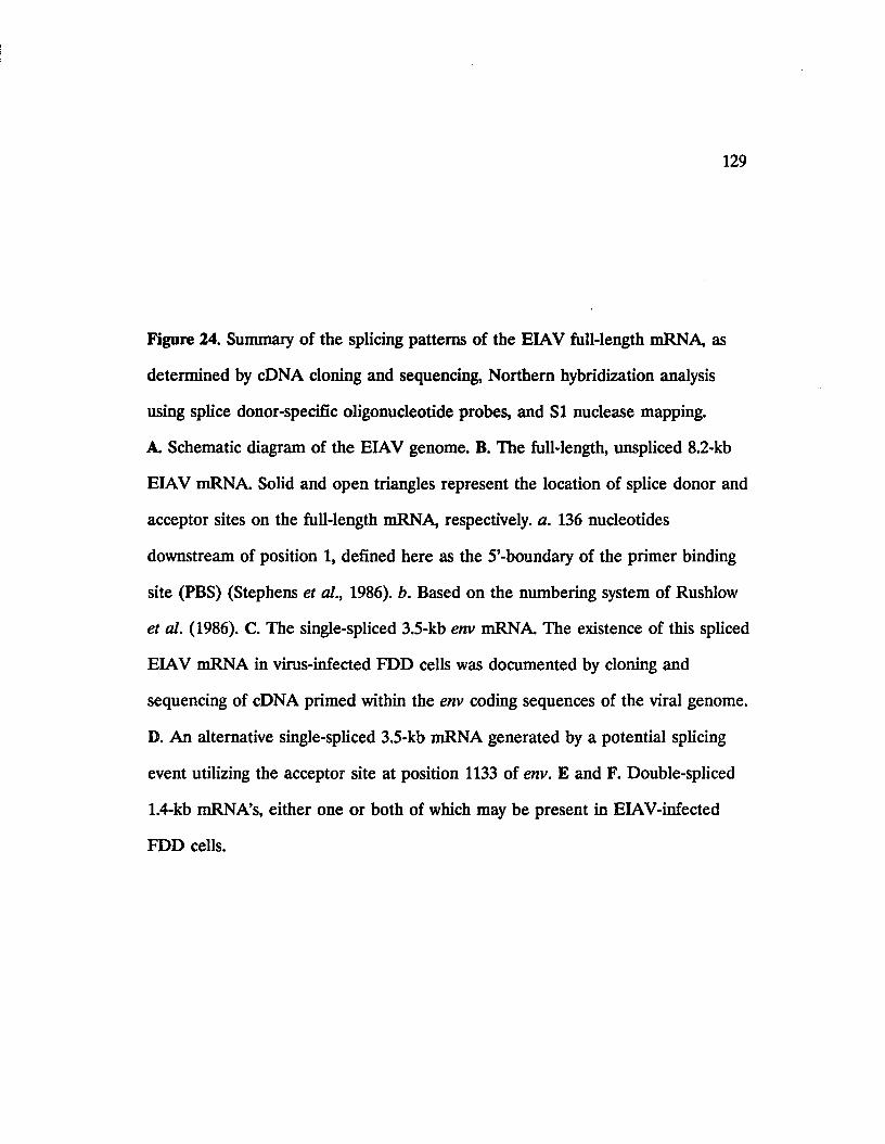
129
Figure 24. Summary of the splicing patterns of the EIAV full-length mRNA, as
determined by cDNA cloning and sequencing, Northern hybridization analysis
using splice donor-specific oligonucleotide probes, and SI nuclease mapping.
A Schematic diagram of the EIAV genome. B. The full-length, unspliced 8.2-kb
EIAV mRNA Solid and open triangles represent the location of splice donor and
acceptor sites on the full-length mRNA, respectively, a. 136 nucleotides
downstream of position 1, defined here as the 5’-boundary of the primer binding
site (PBS) (Stephens et al., 1986). b. Based on the numbering system of Rushlow
et al. (1986). C. The single-spliced 3.5-kb env mRNA The existence of this spliced
EIAV mRNA in virus-infected FDD cells was documented by cloning and
sequencing of cDNA primed within the env coding sequences of the viral genome.
D. An alternative single-spliced 3.5-kb mRNA generated by a potential splicing
event utilizing the acceptor site at position 1133 of env. E and F. Double-spliced
1.4-kb mRNA’s, either one or both of which may be present in EIAV-infected
FDD cells.

A. □gag
5'LTR
pol SI S2 anv S3
3'LTR
B.136°y
gag pol
3184*»y__SI S2 env S3
' poly (A)
C . i poly (A)
D. .poly (A)
E.1084 1256
7 7 .poly (A)
F.1133 1256
7 7 .poly (A)
u>o

131
remove most of the env coding sequences as a second intron and could potentially
generate either one or two double-spliced 1.5-kb mRNA’s (Figure 24E and F).
The first exon of both double-spliced mRNA’s would be identical to that of the
two single-spliced transcripts. However, their second exon, beginning at
position 1084 or 1133 (Figure 24E and F, respectively), would contain different
coding capacities. The mRNA utilizing the acceptor site at position 1084 (Figure
24E) would retain all but the initial four codons of SI; however, in the case of
the mRNA utilizing the acceptor site at position 1133 (Figure 24F), the first
twenty codons of SI would be spliced out and the mRNA would retain only the
latter thirty codons of SI. Following the UAA translational termination codon of
SI, both mRNA’s would contain the AUG initiation codon of S2 and its following
six codons spliced onto the third exon of both mRNA’s at the acceptor site
located at the 5’-boundary of S3.
Although the SI nuclease mapping data have identified the splice donor
and acceptor sites used to generate the spliced mRNA’s of EIAV, direct evidence
for the actual presence of the latter three spliced mRNA’s in EIAV-infected cells,
as depicted in Figure 24D-F, can only be provided by obtaining their respective
cDNA clones, similar to that of the 3.5-kb mRNA shown in Figure 24C.
Moreover, it should be noted that only the most abundant viral mRNA’s would
be expected to be detected by the SI nuclease mapping technique. As mentioned
previously, other low abundance spliced viral mRNA’s would best be detected by
amplification techniques such as the PCR.

Recently, a cDNA clone of a double-spliced 1.4-kb EIAV mRNA has been
isolated from an EIAV-infected feline embryo fibroblast (FEA) cell line (Derse
et al., 1989). The sequence of the 1.4-kb cDNA indicates that its mRNA copy
contains three exons. The first exon, containing the R and U5 regions of the 5’-
LTR and 130 nucleotides of the region preceding the gag gene, is joined to the
second exon using the splice donor site immediately upstream of the gag AUG
codon and a splice acceptor site near the 5’-end of SI. The second exon,
containing SI, is 142 nucleotides long and is joined to the third exon using a
splice donor site immediately following the SI translational termination codon
and a splice acceptor site within the env gene at the 5’-boundary of the S3. The
third exon is 948 nucleotides long and contains S3 followed by the U3 and R
regions of the 3’-LTR. It is interesting to note that this double-spliced mRNA,
and the splice donor and acceptor sites used to generate it, are identical to those
defined by the results of SI nuclease mapping experiments in the present studies
(Figure 24E), with one exception. The splice donor site at the end of the second
exon, as identified by our SI mapping experiments, is located at 31 nucleotides
downstream of the SI termination codon (position 1256 of Rushlow et al., 1986;
Figure 24E; Table III). However, the sequencing of the cDNA has identified this
donor site as the one located at 1 nucleotide downstream of the SI termination
codon (position 1226 of Rushlow et al., 1986; Table III; Derse et al., 1989). It is
uncertain whether the identification of two different splice donor sites by the two
separate studies is a result of differential cell type-specific mechanisms which
control post-transcriptional processing of EIAV mRNA’s in the EIAV-infected
133
FDD cells examined in the present studies, relative to the infected FEA cells
used by Derse et al. (1989).
An additional observation regarding the double-spliced 1.4-kb mRNA
identified by Derse et al. (1989) is that in this mRNA the S3 sequence, lacking an
AUG initiation codon, is immediately preceded by the SI termination codon and
no translational initiation AUG codon is spliced onto the S3 sequence. On the
other hand, the double-spliced mRNA identified by our SI mapping experiments
would have the S2 AUG codon near the 3’-end of its second exon spliced onto
the S3 sequence (Figure 24E). However, this AUG codon would not be spliced in
frame with the S3 coding sequence, thus leaving S3 without an AUG initiation
codon. Therefore, in neither double-spliced mRNA does an AUG codon splice in
frame onto the S3 coding sequence. Whether the S3 gene of EIAV indeed
encodes a protein product remains to be determined, although serological
evidence points to the expression of an Si-related protein in EIAV-infected
horses (J. M. Ball and R. C. Montelaro, unpublished data).
As shown by the results of the present studies, a feature common to the
spliced mRNA’s of EIAV is that they all use the splice donor site located
immediately upstream of the gag AUG codon to generate their first exon. This
splicing pattern of the full-length genomic mRNA, which would generate spliced
mRNA’s sharing similar 5’-ends, has also been observed in other lentiviruses. SI
nuclease mapping studies of the 5’-region of visna virus (Vigne et al., 1987) and
sequencing of cDNA clones obtained from spliced visna virus mRNA’s (Mazarin
et al., 1988; Davis and Clements, 1989) have indicated that all spliced visna virus
134
transcripts use a common donor site located at 188 nucleotides upstream of the
visna virus gag gene (Sonigo et al., 1985). Moreover, sequencing of cDNA clones
obtained from the spliced mRNA’s of HTV-1 have revealed that the splice donor
site located at 45 nucleotides upstream of the HIV-1 gag gene generates the first
exon of these spliced transcripts (Aiya et al., 1985; Muesing et al., 1985).
The interesting finding regarding the spliced transcripts of EIAV, however,
is that following such splicing events the gag AUG codon is removed from all
these mRNA’s. Thus, for instance, in the double-spliced mRNA identified either
by the present studies or by those of Derse et al. (1989), the SI coding sequence
located within the second exon of the mRNA lacks an AUG initiation codon. In
the case of HIV-1 or visna virus, however, the coding sequences of the short
ORFs encoded by these viruses contain their own AUG codons and their
translational initiation is not dependant on the splicing of an AUG codon from
an upstream gene (Arya et al., 1985; Muesing et al., 1985; Mazarin et al., 1988;
Davis and Clements, 1989). In the case of EIAV, it is proposed that the
translation of the SI coding sequence on the double-spliced 1.4-kb mRNA is
initiated from a non-AUG codon (Derse et al., 1989). Although the translational
mechanism of SI from this mRNA is not fully understood yet, the usage of a non-
AUG codon for translational initiation of a viral regulatory gene would
potentially provide an additional control mechanism for fine-tuning of EIAV gene
regulation not previously observed in other lentiviruses. The first two exons of the
double-spliced 1.4-kb mRNA identified by Derse et al. (1989) are identical to the
first two exons of the single-spliced 3.5-kb env mRNA identified in the present

135
studies (Figure 24C). Therefore it is plausible to assume that 52, 52, and env
genes could all be expressed from this single-spliced mRNA. If this phenomenon
indeed occurs in vivo, the efficient expression of the distal 52 and env genes
would require that the proximal 52 coding sequence be poorly expressed. The
utilization of a non-AUG codon, which might be poorly utilized and frequently
passed over by the ribosomes, would potentially provide such a control
mechanism for translation of 52 (Derse et al., 1989).
The results of the present investigations on the co-expression of 52 and env
from the same transcript indicate that expression of both proteins from the same
transcript can be detected in vitro. Studies on the in vivo expression of the two
proteins, however, have not been as conclusive. Although expression of the env
gene from a proviral DNA fragment containing the ATG codons of both 52 and
env can be clearly demonstrated in transfected COS-7 cells, no such conclusion
can be made regarding the expression of 52.
It is possible that the 52 protein is indeed not expressed in these cells.
However, this lack of 52 detection, based solely on results of immunological
detecton methods, could also be due to other factors. It is possible, for instance,
that the anti-52 titer of the polyclonal antibody is perhaps insufficient to detect
any expressed protein in these experiments. In addition, as mentioned before, the
length of the 5’-upstream sequences of the 52 gene in the p9SR426 expression
plasmid have been minimized to avoid the problems associated with cloning of
the env gene. It is possible that additional 5’-untranslated sequences, not present
in p9SR426, are required for efficient translation of the 52 reading frame.
136
An interesting possibility is that the in vivo expression of the EIAV 52
gene occurs at a much lower frequency relative to that of a structural viral gene
such as env, making the 52 protein difficult to detect. In this regard, it should be
noted that the nucleotide sequence surrounding the 52 AUG codon does not
constitute a favorable context for translational initiation at this AUG, as defined
by Kozak (1986a and 1986b). The sequence of nucleotides around the 52 AUG
codon is GAUAUAUGG (Rushlow et al., 1986), while the AUG sequence context
for optimal translational initiation is defined as CCACCAUGG (Kozak, 1986b).
On the other hand, the env AUG codon, present in a GUAACAUGG sequence
context (Rushlow et al., 1986), is much more favored for tanslational initiation. It
has been suggested that positions -3 and +4 in this sequence context, where the
"A" nucleotide of the AUG triplet is defined as the +1 position, are especially
critical; the presence of a pyrimidine instead of a purine at these two positions
can reduce the frequency of translational initiation by 5- to 10-fold (Kozak,
1986a). It is interesting to note that the 52 AUG contains a pyrimidine at the -3
position, while the env AUG contains a purine. This suggests that if indeed
translational initiation does occur from the 52 AUG in vivo, it would happen
much less frequently than from the env AUG. In the in vitro analysis of 52 and
env expression in a wheat-germ extract, however, it was observed that higher
levels of 52 were synthesized relative to env (Figure 20, lanes 1 and 2). In this
regard, it should be noted that in the wheat-germ system translational initiation
occurs much more efficiently from the 5’-most AUG codon (D. Shih, personal
communication), which in this case is that of 52.

137
Taken together with the model of infrequent SI translation from a non-
AUG codon, as discussed above, it is plausible to propose that Si, S2, and env
could all be potentially expressed from the 3.5-kb env transcript of EIAV. The
virus can then be assumed to have evolved the following fine-tuning mechanism
to insure optimal expression of the structural env gene located distal to the 5’-end
of the 3.5-kb mRNA. The first two genes (Si and S2) would be inefficiently
translated from a non-AUG codon and an AUG codon in a non-optimal
sequence context, respectively, while a majority of the ribosomes initiate
translation from the env AUG codon, resulting in efficient synthesis of the viral
envelope proteins.
The co-expression of more than one viral protein from the same mRNA
has been observed in many different animal virus systems, including retroviruses
such as HTLV-I (Nagashima et al., 1986), HTLV-II (Shimotohno et al., 1985),
BLV (Rice et al., 1987), and HIV-1 (Arya and Gallo, 1986; Sodroski et al., 1986).
An ideal means of detecting the potential expression of the 52 gene from the
expression plasmid p9SR426 used in the present studies would be based on a
functional assay by which the function encoded by 52 could be assayed in
transfected cells. Design of such functional assays, however, await the assignment
of a specific function to the putative EIAV 52 protein.

LITERATURE CITED
Ahmad, N., and Venkatesan, S. 1988. N ef protein of HIV-1 is a transcriptional repressor of HIV-1 LTR. Science 241: 1481-1485.
Archer, B. G., Crawford, T. B., McGuire, T. C., Frazier, M. E. 1977. RNA- dependent DNA polymerase associated with equine infectious anemia virus. J. Virot. 22: 16-22.
Arya, S. K., Gallo, R. C., Hahn, B. H., Shaw, G. M., Popovic, M., Salahuddin, S. Z., Wong-Staal, F. 1984. Homology of genome of AIDS-associated virus with genomes of human T-cell leukemia viruses. Science 225: 927-930.
Arya, S. K., Guo, C., Josephs, S. F., Wong-Staal, F. 1985. 7ra/w-activator gene of human T-lymphotropic virus type III (HTXV-III). Science 229: 69-73.
Arya, S. K., and Gallo, R.C. 1986. Three novel genes of human T-lymphotropic virus type III: immune reactivity of their products with sera from acquired immune deficiency syndrome patients. Proc. Natl. Acad. Sci. USA 83: 2209-2213.
Arya, S. K., Beaver, B., Jagodzinski, L., Ensoli, B., Kanki, P. J., Alber, J., Fenyo,E. M., Biberfeld, G., Zagury, J. F., Laure, F., Essex, M., Norrby, E., Wong-Staal,F., Gallo, R. C. 1987. New human and simian HIV-related retroviruses possess functional transactivator (tat) gene. Nature 328: 548-550.
Ausubel, F. M., Brent, R., Kingston, R. E., Moore, D. D., Smith, J. A., Seidman, J. G., and Struhl, K. 1987. Current protocols in molecular biology. John Wiley & Sons, Inc. New York.
Bartlett, S. G., Grossman, A. R., and Chua, N.-H. 1982. In vitro synthesis and uptake of cytoplasmically-synthesized chloroplast proteins. In: "Methods in Chloroplast Molecular Biology" (Edelman, M., Hallick, R. B., and Chua, N.-H., eds.) Elsevier Biomedical Press, Amsterdam-New York-Oxford, pp. 1081-1091.
Berk, A. J., and Sharp, P. A. 1977. Sizing and mapping of early adenovirus mRNAs by gel electrophoresis of SI endonuclease digested hybrids. Cell 12:721- 732.
Berkhout, B., Silverman, R. H., and Jeang, K.-T. 1989. Tat fra/w-activates the human immunodeficiency virus through a nascent RNA target. Cell 59: 273-282.
Bernhard, W. 1958. Electron microscopy of tumor cells and tumoe viruses. A review. Cancer Res. 18: 491-509.
Bernhard, W. 1960. The detection and study of tumor viruses with the electron microscope. Cancer Res. 20: 712-727.
138

139
Bimboim, H. C., and Doly, J. 1979. A rapid alkaline extraction procedure for screening recombinant plasmid DNA. Nucl. Acids Res. 7: 1513-1523.
Bishop, J. M., and Varmus, H. E. 1982. Functions and origins of retroviral transforming genes. Molecular Biology of Tumor Viruses: RNA Tumor Viruses (Cold Spring Harbor Laboratory, Cold Spring Harbor, NY, 1982), pp. 999-1108.
Bonner, T. J., O’Connel, C. and Cohen, M. 1982. Cloned endogenous retroviral sequence from human DNA. Proc. Natl. Acad. Sci. USA 79: 4709-4713.
Breathnach, R., and Chambon, P. 1981. Organization and expression of eucaryotic split genes coding for proteins. Ann. Rev. Biochem. 50: 349-384.
Casey, J. M., Kim, Y., Anderson, P. R., Watson, K. F., Fox, J. L., and Devare, S. G. 1985. Human T-cell lymphotropic virus type HI: Immunologic characterization and primary analysis of the major internal protein p24. J. Virol. 55: 417-421.
Charman, H. P., Bladen, S., Gilden, R. W., and Coggins, L. 1976. Equine infectious anemia virus: Evidence favouring classification as a retrovirus. J.Virol. 19: 1073-1079.
Cheevers, W. P., Archer, B. G., and Crawford, T. B. 1977. Characterization of RNA of equine infectious anemia virus. J. Virol. 24: 489-497.
Chen, H. R., and Barker, W. C. 1984. Nucleotide sequences of the retroviral long terminal repeats and their adjacent region. Nucleic Acids Res. 12: 1767-1788.
Chirgwin, J. M., Przybyl, A. E., MacDonald, R. J., and Rutter, W. J. 1979. Isolation of biologically active ribonucleic acid from sources enriched in ribonuclease. Biochemistry 18: 5249-5299.
Chiu, I. M., Yaniv, A., Dahlberg, J. E., Gazit, A., skuntz, S. F., Tronick, S. R., and Aaronson, A. 1985. Nucleotide sequence evidence for relationship of AIDS retrovirus to lentiviruses. Nature 317: 366-368.
Clavell, F., Guetard, D., Brun-Vezinet, F., Chamaret, S., Rey, M. A., Santos- Ferreira, M. O., Lament, A. G., Dauget, C., Katlama, C., Rouzioux, C., Klatzmann, D., Champalimaud, L., Montagnier, L. 1986. Isolation of a new human retrovirus fron West African patients with AIDS. Science 233: 343-346.
Coffin, J. M. 1979. Structure, replication, and recombination of retrovirus genomes: some unifying hypotheses. J. Gen. Virol. 42: 1-26.
Coggins, L. 1972. Mechanism of viral persistence in equine infectious anemia. Cornell Vet. 65: 143-151.

140
Coggins, L., Norcross, N. L., and Naustraum, S. R. 1972. Diagnosis of equine infectious anemia by immunodiffusion test. Amer. J. Vet. Res. 33: 11-18.
Coggins, L., Matheka, H. P., and Charman, H. P. 1978. Radioimmunoassay for equine infectious anemia virus antigen and antibody. J. Equine Med. and Surg. (Suppl. I): 351-358.
Crawford, T. B., McGuire, T. C., and Henson, J. B. 1971. Detection of equine infectious anemia in vitro by immunofluorescence. Arch. Gesamte. Virusforsch 34: 332-339.
Crawford, T. B., Cheevers, W. P., Klevjer-Anderson, P., and McGuire, T. C. 1978. EIA: Virion Characteristics, virus-cell interaction, and host responses. In: "Persistent Viruses: Symposium in Molecular and Cellular Biology" (Stevens, J., Todaro, G., and Fox, F., eds.) Vol. 11. Academic Press, Inc., New York, pp. 727- 749.
Cullen, B. R. 1986. Trans-activation of human immunodeficiency virus occurs via a bimodal mechanism. Cell 46: 973-982.
Cullen, B. R. 1987. Use of eukaryotic expression technology in the functional analysis of cloned genes. Meth. Enzym. 152: 684-705.
Cullen, B. R., Hauber, J., Campbell, K., Sodroski, J. G., Haseltine, W. A., Rosen,C. A. 1988. Subcellular localization of the human immunodeficiency virus transacting art gene product. J. Virol. 62: 2489-2501.
Davis, J. L., Molineaux, S., and Clements, J. 1987. Visna virus exhibits a complex transcriptional pattern: one aspect of gene expression shared with the acquired immunodeficiency syndrome retrovirus. J. Virol. 61: 1325-1331.
Davis, J. L., and Clements, J. E. 1989. Characterization of a cDNA clone encoding the visna virus transactivating protein. Proc. Natl. Acad. Sci. USA 86: 414-418.
Dayton, A. L., Sodroski, J. G., Rosen, C. A., Goh, W. C., Haseltine, W. A. 1986. The trans-activator gene of the human T cell lymphotropic virus type III is required for replication. Cell 44: 941-947.
Derse, D., Dorn, P., DaSilva, L., and Martarano, L. 1989. Structure and expression of the equine infectious anemia virus transcriptional tram-activator (tat). J. Virol, (in press).
Dorn, p. L., and Derse, D. 1988. cis- and tram-acting regulation of gene expression of equine infectious anemia virus. J. Virol. 62: 3522-3526.

141
Fauci, A. S. 1988. The human immunodeficiency virus: infectivity and mechanisms of pathogenesis. Science 239: 617-622.
Feinberg, A. P., and Vogelstein, B. 1983. A technique for radiolabeling DNA restriction endonuclease fragments to high specific activity. Anal. Biohem. 132: 6- 13.
Feinberg, M. B., Jarrett, R. F., Aldovini, A., Gallo, R. C., Wong-Staal, F. 1986. HTLV-III expression and production involve complex regulation at the levels of splicing and translation of viral RNA. Cell 46: 807-817.
Felber, B. K., Hadzopoulou-Cladaras, M., Cladaras, C. Copeland, T., and Pavlakis, G. N. 1989. rev protein of human immunodeficiency virus type 1 affects the stability and transport of the viral mRNA. Proc. Natl. Acad. Sci. USA 86: 1495-1499.
Fine, D. and Schochetman, G. 1978. Type-D primate retroviruses: A review, cancer Res. 38: 3132-3139.
Fisher, A. G., Feinberg, M. B., Josephs, S. F., Harper, M. E., Marselle. L. M., Reyes, G., Gonda, M. A., Aldovini, A., Debouck, C., Gallo, R. C., Wong-Staal, F. 1986. The trans-activator gene of HTLV-III is essential for virus replication. Nature 320: 367-370.
Fitzgerald, M., and Shenk, T. 1981. The sequence 5’-AAUAAA-3’ forms part of the recognition site for polyadenylation of late SV40 mRNAs. Cell 24: 251-260.
Foil, L. and Issel, C. J. 1982. EIA - living with the disease. Louisiana Agr. 25: 8-9.
Foil, L. D., Meek, L., Adams, W. V., Jr., and Issel, C. J. 1983. Mechanical transmission of equine infectious anemia virus by deer flies (Chrysops flavidus) and stable flies (Stomoxys calcitrans). Am. J. Vet. Res. 44: 155-156.
Foil, L. D., Adams, W. V., Issel, C. J., and Pierce, R. 1984. Tabanid (Diptera) populations associated with an equine infectious anemia outbreak in an inapparently infected herd of horses. J. Med. Entomol. 21: 28-30.
Fordis, C. M., and Howard, B. H. 1987. Use of the CAT reporter gene for optimization of gene transfer into eukaryotic cells. Methods Enzymol. 151: 382- 397.
Frankel, A. D., Bredt, D. S., Pabo, C. O. 1988. tat protein from human immunodeficiency virus forms a metal-linked dimer. Science 240: 70-73.
Franza, B. R., Cullen, B. R., Wong-Staal, F. (eds) 1988. The control of human retrovirus gene expression. Banbury Reports. Cold Spring Harbor, New York: Cold Spring Harbor Laboratory.

142
Gelderblom, H. and Frank, H. 1987. Spumavirinae. In: Animal virus structure (Nermut, M. V. and Steven, A. C. eds.) pp. 305-311 Elservier Science Publ. Co. Inc., New York, New York.
Gerard, G. F., and Miller, K. 1986. Comparison of glyoxal and formaldehyde gels for sizing rRNAs. BRL Focus. 8 (3): 5-6.
Gluzman, Y. 1981. SV40-Transformed simian cells support the replication of early SV40 mutants. Cell 23: 175-182.
Gonda, M. A., Charman, H. P., Walker, H. L., and Coggins, L. 1978. Scanning electron microscopy study of EIAV. Amer. J. Vet. Res. 39: 431-440.
Gonda, M. A., Wong-Staal, F., Gallo, R. C., Clements, J. E., Narayan, O., and Gelden, R. V. 1985. Sequence homology and morphologic similarity of HTLV-III and visna virus, a pathogenic lentivirus. Science 227: 173-177.
Gonda, M. A., Braun, M. J., Carter, S. G., Kost, T. A., Bess, J. W. Jr., Arthur, L. O., and Van Der Maaten, M. J. 1987. Characterization and molecular cloning of a bovine lentivirus related to human immunodeficiency virus. Nature 330: 388- 391.
Greene, W. C., Leonard, W. J., Wano, Y., Svetlik, P. B., Peffer, N. J., Sodroski, J. G., Rosen, C. A., Goh, W. C., Haseltine, W. A. 1986. Trans-activator gene of HTLV-II induces IL-2 receptor and IL-2 cellular gene expression. Science 232: 877-880.
Gross, L. 1970. Oncogenic viruses, second edition, Permagon Press, New York.
Haase, A. 1986. Pathogenesis of lentivirus infections. Nature 322: 130-136.
Hacket, P. B., Swanstrom, R., Varmus, H. E., and Bishop, J. M. 1982. The leader sequence of the subgenomic messenger RNAs of Rous sarcoma virus is approximately 390 nucleotides. J. Virol. 41: 527-534.
Hanahan, D. 1985. Techniques for transformation of E. coli. In: DNA cloning,Vol. 1, D. M. Glover, Ed. (IRL Press, Oxford, England, 1985), pp. 109-135.
Hammarskjold, M. L., Heimer, J., Hammaraskjold, B., Sangwan, I., Albert, L., and Rekosk, D. 1989. Regulation of human immunodeficiency virus env expression by the rev gene product. J. Virol. 63: 1959-1966.
Hauber, J., Perkins, A., Heimer, E. P., Cullen, B.R. 1987. Trans-activation of human immunodeficiency virus gene expression is mediated by nuclear events. Proc. Natl. Acad. Sci. USA 84: 6364-6386.

143
Ho, D. D., Pomerantz, R. J., Kaplan, J. C. 1987. Pathogenesis of infection with human immunodeficiency virus. N. Engl. J. Med. 317: 278-286.
Hussain, K. A., Issel, C. J., Schnorr, K. L., Rwambo, P. M., West, M., and Montelaro, R. C. 1988. Antigenic mapping of the envelope proteins of equine infectious anemia virus: identification of a neutralization domain and a conserved region on glycoprotein 90. Arch. Virol. 98: 213-224.
Issel, C. J. and Coggins, L. 1979. Equine infectious anemia: Current knowledge. J. Aimer. Vet. Med. assoc. 174: 727-733.
Issel, C. J., Adams, W. V., Meek, L., and Ochoa, R. 1982. Transmission of EIAV from horses without clinical signs of disease. J. Amer. Vet. Med. Assoc. 180:272- 275.
Issel. C. J., Rushlow, K., Foil, L. D., and Montelaro, R. C. 1988. A perspective on equine infectious anemia with an emphasis on vector transmission and genetic analysis. Vet. Micro. 17: 251-286.
Jacks, T., and Varmus, H. E. 1985. Expression of the Rous sarcoma virus pol gene by ribosomal frameshifting. Science 230: 1237-1242.
Jacks, T., Townsley, K., Varmus, H. E., and Majors, J. 1987. Two efficient ribosomal frameshifting events are required for synthesis of mouse mammary tumor virus gag-related polyproteins. Proc. Natl. Acad. Sci. USA 84: 4298-4302.
Jacks, T., Power, M. D., Masiarz, F. R., Luciw, P. A., Barr, P. J., & Varmous, H.E. 1988. Characterization of ribosomal ffamesharing in HIV-1 gag-pol expression. Nature 331: 280-283.
Jacobovits, A., Smith, D. A., Jacobovits, E. B., Capon, D. J. 1988. A discrete element of 3’ of human immunodeficiency virus 1 (HIV-1) and HIV-2 mRNA initiation site mediates transcriptional activation by an HIV trans activator. Mol. Cell. Biol. 8: 2555-2561.
Ju, G., Boone, L., and Skalka, A. M. 1980. Isolation and characterization of recombinant DNA clones of avian retroviruses: size heterogeneity and instability of the direct repeat. J. Virol. 33: 1026-1033.
Kalyanaraman, V. C., Sarngadharan, M. G., Robert-Guroff, M., Miyoshi, I., Blaynay, D., Golde, D., and Gallo, R. C. 1982 A new subtype of human T-cell leukemia virus (HTLV-II) associated with a variant of hairy cell leukemia.Science 218: 571-573.
Kan, N. C., Franchini, G., Wong-Staal, F., DuBois, G. C., Robey, W. G., Lautenberger, J. A., Papas, T. S. 1986. Identification of HTLV-III/LTV sor gene product and detection of antibodies in human sera. Science 231: 1553-1555.
144
Kao, S. Y., Caiman, A. F., Luciw, P. A., Peterlin, B. M. 1987. Anti-termination of transcription within the long-terminal repeat of HIV-1 by tat gene product.Nature 330: 489-493.
Kaufman, R. J. 1985. Identification of the components necessary for adenovirus translational control and their utilization in cDNA expression vectors. Proc. Natl. Acad. Sci. USA 82: 689-693.
Kaufman, R. J., Wasley, L. €., Spiliotes, A. J., Gossels. S. D., Latt, S. A., Larsen,G. R., and Kay, R. M. 1985. Coamplification and coexpression of human tissue- type plasminogen activator and murine dihydrofolate reductase sequences in Chinese hamster ovary cells. Mol. Cell. Biol. 5: 1750-1759.
Kemen, M. J. and Coggins, L. 1972. Equine infectious anemia: Transmission from infected mares to foals. J. Amer. Vet. Med. Assoc. 161: 469-499.
Kono, Y. and Kobayashic, K. 1966. Complement-fixation test of equine infectious anemia. I. Specificity of test. Natl. Inst. Animal Health, Q. (Tokyo) 6: 194-203.
Kono, Y. 1969. Viremia and immunological responses in horses infected with equine infectious anemia virus. Natl. Inst. Anim. Hlth. Quart. (Tokyo) 8: 1-9.
Kozak, M. 1986a. Point mutations define a sequence flanking the AUG initiator codon that modulates translation by eukaryotic ribosomes. Cell 44: 283-292.
Kozak, M. 1986b. Bifunctional messenger RNAs in eukaryotes. Cell 47: 481-483.
Krieg, P.A., and Melton, D. A. 1984. Functional messenger RNAs are produced by SP6 in vitro transcription of cloned cDNAs. Nucleic acids Res. 12:7057-7070.
Laspia, M. F., Rice, A. P., and Mathews, M. 1989. HIV-1 Tat protein increases transcriptional initiation and stabilizes elongation. Cell 59: 283-292.
Lee, T. H., Coligan, J. E., Allan, J. S., McLane, M. F., Groopman, J. E., Essex,M. 1986. A new HTLV-III/LAV protein encoded by a gene found in cytopathic retroviruses. Science 231: 1546-1549.
Lo, K.-M., Jones, S. S., Hackett, N. R., and Khorana, H. G. 1984. Specific amino acid substitutions in bacterioopsin: Replacement of a restriction fragment in the structural gene by synthetic DNA fragments containing altered codons. Proc. Natl. Acad. Sci. USA 81: 2285-2289.
Logan, J., and Shenk, T. 1984. Adenovirus tripartite leader sequence enhances translation of mRNAs late after infection. Proc. Natl. Acad. Sci. USA 81:3655- 3659.

145
Luciw, P. A., Chen-Mayer, G , Levy, J. A. 1987. Mutational analysis of the human immunodeficiency virus (HIV): the orf-B region downregulates virus replication. Proc. Natl. Acad. Sci. USA 84: 1434-1438.
Lusky, M. and Botchan, M. 1981. Inhibition of SV40 replication in simian cells by specific pBR322 DNA sequences. Nature 293: 79-81.
Malim, M. H., Hauber, J., Le, S. Y., Maizel, J., & Gillen, B. R. 1989. The HIV-1 rev fra/w-activator acts through a structured target sequence to activate nuclear export of unspliced viral mRNA. Nature 338: 254-257.
Malmquist, W. A., Barnett, D., and Becvar, G S. 1973. Production of equine infectious anemia antigen in a persistently infected cell line. Arch. Virol. 42:361- 370.
Maniatis, T., Fritsch, E. F., and Sambrook, J. V. 1982. Molecular cloning: a laboratory manual. Cold Spring Harbor Laboratory, Cold Spring harbor, N.Y.
Matheka, H. D., Coggins, L., Shively, J. N., Norcross, N. L. 1976. Purification and characterization of equine infectious anemia virus. Arch. Virol. 51: 107-114.
Mazarin, V., Gourdou, I., Querat, G., Sauze, N., and Vigne, R. 1988. Genetic structure and function of an early transcript of visna virus. J. Virol. 62: 48134818.
McConnell, M., Katada, M., McConnell, S. 1977. Demonstration of equine infectious anemia virus in primary leukocytes culture by electron microscopy. Am. J. Vet. Res. 38: 2067-2069.
McGuire, T. C., Crawford, T. B., Henson, J. B. 1971. Immunofluorescent localization of equine infectious anemia virus in tissues. Ame. J. Path. 62:283- 292.
Mims, C. A. 1986. Virus receptors and cell tropisms. J. Infect. 12: 199-203.
Montelaro, R. C., Lohrey, N., Parekh, B., Blakeney, E. W., and Issel, C. J. 1982. Isolation and comparative biochemical properties of the major internal polypeptides of equine infectious anemia virus. J. Virol. 42: 1029-1038.
Montelaro, R. C., Robey, W. G., West, M. D., Issel, C. J., and Fischinger, P. J. 1988. Characterization of the serological cross-reactivity between glycoproteins of the human immunodeficiency virus and equine infectious anemia virus. J. Virol. 69: 1711-1717.
Moore, R., Dixon, M., Smith, R., Peters, G., and Dickson, C. 1987. Complete nucleotide sequence of a milk-transmitted mouse mammary tumor virus: two frameshift suppression events are required for translation of gag and pol. J. Virol. 61: 480-490.

146
Mosser, A., Montelaro, R., and Rueckert. R. 1975. Proteins of Rous-associated virus-61: polypeptide stoichiometry and evidence that glycoprotein gp35 is not a cleavage produce of gp85. J. Virol. 23: 10-19.
Mount, S. M. 1982. A catalogue of splice junction sequences. Nucleic Acids Res. 10: 459-472.
Muesing, M. A., Smith, D. H., Cabradilla, C. D., Benton, C. V., Lasky, L. A., and Capon, D. J. 1985. Nucleic acid structure and expression of the human AIDS/lymphadenopathy retrovirus. Nature 313: 450-458.
Muesing, M. A., Smith, D. H., Capon, D. J. 1987. Regulation of mRNA accumulation by human immunodeficiency virus trans-activator protein. Cell 48: 691-701.
Mullis, K. B. and Fallona, F. A. 1987. Specific synthesis of DNA in vitro via a polymerase-catalyzed chain reaction. Methods Enzymol. 155: 335-350.
Nagashima, K., Yoshida, M., and Seiki, M. 1986. A single species of p X mRNA of human T-cell leukemia virus type I encodes trans-activator p40* and two other phosphoproteins. J. Virol. 60: 394-399.
Nakajima, H., Tajima, M., Tanaka, S., and Ushimi, C. 1969. Physicochemical studies of equine infectious anemia virus. III. Purification and electron microscopic observation of the virus. Arch. Gesamte Virusforsch. 28: 348-360.
Nakajima, H. 1972. Physicochemical and biological characterization of equine infectious anemia virus, pp. 162-174. In Proc. 3rd Int. Conf. Equine Infectious Dis., Paris (Karger, Basel 1973).
Nash, M., Brown, N. V., Wong, J. L., Arlinghaus, R. B., and Murphy, E. C., Jr.1984. SI nuclease mapping of viral RNAs from a temperature-sensitive transformation mutant of murine sarcoma virus. J. Virol. 50: 478-488.
Nevins, J. R. 1982. Adenovirus gene expression: control at multiple steps of mRNA biogenesis. Cell 28: 1-2.
O’Farrell, P. 1981. Replacement synthesis method of labeling DNA fragments. BRL Focus 3 (3): 1-3.
Ohshima, Y., and Gotoh, Y. 1987. Signals for the selection of a splice site in pre- mRNA. Computer analysis of splice junction sequences and like sequences. J. Mol. Biol. 195: 247-259.
Orrego, A., Issel, C. J., Montelaro, R. C., and Adams, W. V. 1982. Virulence and in vitro growth of a cell-adapted strain of equine infectious anemia virus after serial passage in ponies. Amer. J. Vet. Res. 43: 1556-1560.

147
Parekh, B., Issel, C. J., and Montelaro, R. C. 1980. Equine infectious anemia virus, a putative lentivirus, contains polypeptides analogous to prototype-C oncornaviruses. Virology 107: 520-525.
Pereira, L., Ali, M., Kousoulas, K., Huo, B., and Banks, T. 1989. Domain structure of herpes simplex virus 1 glycoprotein B: Neutralizing epitopes map in regions of continuous and discontinuous residues. Virol. 172: 11-24.
Peterlin, B. M., Luciw, P. A., Barr, P. J., Walker, M. D. 1986. Elevated levels of mRNA can account for the trans-activation of the human immunodeficiency virus. Proc. Natl. Acad. Sci. USA 83: 9734-9738.
Peterlin, B. M., and Luciw, P. A 1988. Molecular biology of HIV. AIDS 2 (suppl 1):S29-S40.
Peterlin, B. M., Caiman, A F., Kao, S. Y., Selby, M., Tong-Starksen, S., Luciw, P. A 1988. Activation and trans-activation of human immunodeficiency virus type I. Banbury Report. Cold Spring Harbor, New York: Cold Spring Harbor Press, pp. 45-47.
Peterlin, B. M., Caiman, A F., Kao, S. Y., Selby, M., Tong-Starksen, S., Luciw, P. A 1988. Activation and trans-activation of human immunodeficiency virus type I. Banbury Report 1988. Cold Spring Harbor, New York: Cold Spring Harbor Press, pp. 45-47.
Poiesz, B. J., Ruscetti, F. W., Gazder, A F., Bunn, P. A , Minna, J. D., and Gallo, R. C. 1980. Detection and isolation of type-C retrovirus particles from fresh and cultured lymphocytes of a patient with cutaneous T-cell lymphoma. Proc. Natl. Acad. Sci. USA 77: 7415-7419.
Popovic, M., Samgadharan, M. G., Read, E., and Gallo, R. C. 1984. Detection, isolation, and continuous production of cytopathic retroviruses (HTLV-III) from patients with AIDS and pre-AIDS. Science 224: 497-500.
Proudfoot, N. J., and Brownlee, G. G. 1974. Sequence analysis at the 3’ end of globin mRNA shows homology with immunoglobulin light chain messenger mRNA Nature 252: 359-362.
Rabson, A B., Daugherty, D. F., Venkatesan, S., Boulukos,, K. E., Benn, S. I.1985. Transcription of novel open reading frames of AIDS retrovirus during infection of lymphocytes. Science 229; 1388-1390.
Ratner, L., Haseltine W., Patarca, R., Livak, K. J., Starcich, B., Josephs, S. F., Doran, E. R., Rafalski, J. A , Whitehom, E. A , Baumeister, K., Ivanoff, L., Petteway, S. R. Jr., Pearson, M. L., Lautenberger, J. A , Papas, T. S., Ghrayeb, J., Chang, N. T., Gallo, R. C., and Wong-Staal, F. 1985. Complete nucleotide sequence of the AIDS virus, HTLV-III. Nature 313: 277-284.

148
Rice, N. R., Simek, S. L., Dubois, G. G, Showalter, S. D., Gilden, R. V., and Stephens, R. M. 1987. Expression of the bovine leukemia virus X region in virus- infected cells. J. Virol. 61: 1577-1585.
Rosen, G A., Sodroski, J. G., Haseltine, W. A. 1985. The location of cis-acting regulatory sequences in the human T cell lymphotropic virus type in (HTLV- ffl/LAV) long terminal repeat. Cell 41: 813-823.
Rosen, C. A., Terwilliger, E. F., Dayton, A. L., Sodroski, J. G., Haseltine, W. A. 1988. Intragenic cu-acting art gene responsive sequence of the human immunodeficiency virus. Proc. Natl. Acad. Sci. USA 85: 2071-2075.
Rosenblatt, J. D., Cann, A. J., Slamon, D. J., Smalberg, I. S., Shah, N. P., Fujii, J., Wachsman, W., Chen, I. S. Y. 1988. HTLV-II Transactivation is regulated by the overlapping tax/rex nonstructural genes. Science 240: 916-919.
Roth, E., Coggins, L., and Patten, V. 1971. Evaluation of the immunodiffusion test for the diagnosis of EIA. In: "Proceedings of the 75th Annual Meeting of the U. S. Animal Health Assoc.", pp. 222-231.
Rushlow, K., Olson, K., Stiegler, G., Payne, S. L., Montelaro, R. C., and Issel, C. J. 1986. Lentivirus genomic organization: The complete nucleotide sequence of the env gene region of equine infectious anemia virus. Virology 155: 309-321.
Saiki, R. D., Gelfand, D. D., Stoffel, S., Scharf, S. J., Higuchi, R., Horn, G. G., Mullis, K. B., and Erlich, H. A. 1988. Primer-directed enzymatic amplification of DNA with a thermostable DNA polymerase. Science 239: 487-491.
Sambrook, J., and Gething, M.-J. 1988. Vectors for high level expression of proteins in mammalian cells. BRL Focus: 10 (3): 41-48.
Sanchez-Pescador, R., Power, M. D., Barr, P. J., Steiner, K. S., Stempien, M. M., Brown-Shimer, S. L., Gee, W. W., Renard, A., Randolph, A., Levy, J. A., Dina,D., and Luciw, P. A. 1985. Nucleotide sequence and expression of an AIDS- associated retrovirus (ARV-2). Science 227: 484-492.
Sanger, F., Nicklen, S., and Coulson, A. R. 1977. DNA sequencing with chain- terminating inhibitors. Proc. Natl. Acad. Sci. USA 74: 5463-5467.
Sarkar, N. H., Moore, D. H., and Nowinski, R. C. 1972. Symmetry of the nucleocapsid of the oncornaviruses. In: "RNA Viruses and Host Genome in Oncogenesis" (Emmelot, P. and Bentvelzen, A., eds.) North Holland, Amsterdam, pp. 71-79.
Scharf, S. J., Horn, G. T., and Elrich, H. A. 1986. Direct cloning and sequence analysis of enzymatically amplified genomic sequence. Science 233: 1076-1078.
149
Seiki, M., Inoue, J.-I., Takeda, T., and Yoshida, M. 1986. Direct evidence that p40* of human T-cell leukemia virus type I is a taz/zs-acting transcriptional activator. EMBO J. 5:561-565.
Shane, B. S., Issel, C. J., and Montelaro, R. C. 1984. Enzyme-linked immunosorbent assay for detection of equine infectious anemia p26 antigen and antibody. J. Clin. Microbiol. 19: 351-355.
Shank, P. R., and Linial, M. 1980. Avian oncornavirus mutant (Se21Qlb) deficient in genomic RNA: characterization of a deletion in the provirus. J. Virol. 36: 450-456.
Sharp, P. A., and Marciniak, R. A. 1989. HIV TAR: An RNA enhancer? Cell 59: 229-230.
Shen, D., Crawford, T. B., Gorham, J. R., McGuire, T. C. 1977. Inactivation of equine infectious anemia virus by chemical disinfectants. Ame. J. Vet. Res. 38(8): 1217-1219.
Sherman, L., Gazit, A., Yaniv, A., Kawakami, T., Dahlberg, J. E., and Tronick, S. R. 1988. Localization of sequences responsible for trans-activation of the equine infectious anemia virus long terminal repeat. J. Virol. 62: 120-126.
Shimotohno, K., and Temin, H. M. 1982. Spontaneous variation and synthesis in the U3 region of the long terminal repeat of an avian retrovirus. J. Virol. 41:163- 171.
Shine, J., and Dalgamo, L. 1975. Determinant of cistron specificity in bacterial ribosomes. Nature 254: 34-38.
Sodroski, J. G., Rosen, C. A., Haseltine, W. A. 1984. Trans-acting transcriptional activation of the long terminal repeat of human T lymphtropic viruses in infected cells. Science 225: 381-385.
Sodroski, J., Patarca, R., Rosen, C., Wong-Staal, F., Haseltine, W. 1985. Location of the trans-activating region on the genome of human T-cell lymphotropic virus type HI. Science 229: 74-77.
Sodroski, J. Goh, W. C., Rosen, C., Campbell, K., and Haseltine, W. A. 1986a. Role of the HTLV-m/LAV envelope in syncytium formation and cytopathicity. Nature 322: 470-474.
Sodroski, J., Goh, W. C., Rosen, C., Dayton, A., Terwilliger, E., Haseltine, W.1986. A second post-transcriptional trans-activator gene required for HTLV-III replication. Nature 321: 412-417.

150
Sonigo, P., Poiesz, B. J., Ruscetti, F. W., Reitz, M. S., Kalyanaraman, V. S., and Gallo, R. C. 1981. Isolation of a new type-C retrovirus (HTTLV) in primaiy cultured cell of patient with Sezaiy F cell leukemia. Nature 294: 268-271.
Sonigo, P., Alizon, M., Staskus, K , Klatzmann, D., Cole, S., Danos, O., Retzel, E., Tiollais, P., Haase A., and Wain-Hobson, S. 1985. Nucleotide sequence of the visna lentivirus. Relationship to the AIDS virus. Cell 42: 369-382.
Stephens, R. M., Casey, J. W., and Rice, N. R. 1986. Equine infectious anemia virus gag and pol genes: Relatedness to visna and AIDS virus. Science 231:589- 594.
Stowring, L., Haase, A. T., and Charman, H. P. 1979. Serological definition of the lentivirus group of retroviruses. J. Virol. 29: 523-528.
Strebel, K., Daugherty, D. Clouse, K., Cohen, D., Folks, T., Martin, M. A. 1987. The HTV ’A’ (sor) gene product is essential for virus infectivity. Nature 328:728- 730.
Tajima, M., Nakajima, H., Ito, Y. 1969. Electron microscopy of equine infectious anemia virus. J. Virol. 4: 521-527.
Tardieu, M., Epstein, R. L., Weiner, H. L. 1982. Interaction of viruses with cell surface receptors. Int. Rev. Cytol. 80: 27-61.
Taylor, J. M. 1977. An analysis of the role of tRNA species as primers for the transcription into DNA of RNA tumor virus genomes. Biochim. Biophys. Acta 473: 57-71.
Teich, N. 1982. Taxonomy of retroviruses. In: "RNA Tumor Viruses" (Weiss, R., Teich, N., Varmus, H., and Coffin, J., eds.), Cold Spring Harbor Laboratory, New York, pp. 25-208.
Teich, N. 1985. Taxonomy of retroviruses. In: "RNA Tumor Viruses" (Weiss, R., Teich, N., Varmus, H., and Coffin, J., eds.), Cold Spring Harbor Laboratory, Cold Spring Harbor, New York, pp. 1-16.
Temin, H. M. 1964. Nature of the provirus of Rous sarcoma. Natl. Cancer Inst. Monogr. 17: 557-570.
Temin, 1976. The DNA provirus hypothesis: The establishment and implications of RNA-directed DNA synthesis. Science 192: 1075-1080.
Valle, H. and Carre, H. 1904. Nature infectieuse de l’anemie du cheval. C. R. Hebd. Seances. Acad. Sci. 139: 331-333.

151
Van Beveren, C., Rands, E., Chattopadhyay, S. K., Lowy, D. R., and Verma, L. M. 1982. Long terminal repeat of murine retroviral DNAs: sequence analysis, host-proviral junctions and preintegration site. J. Virol. 41: 542-556.
Varmus, H. 1982. Form and function of retroviral proviruses. Science 216: 812- 820.
Varmus, H. and Swanstrom, R. 1982. Replication of retroviruses. In: "RNA Tumor Viruses" (Weiss, R., Teich, N., Varmus, H., and Coffin, J., eds.) Cold Spring Harbor Laboratory, New York, pp. 369-512.
Varmus, H. 1983. Retroviruses. In: Mobile Genetic Elements, J. Shapiro, Ed. (Academic Press, New York, 1983), pp. 411-503.
Varmus, H. 1988. Retroviruses. Science 240: 1427-1435.
Vigier, P. 1970. RNA oncogenic viruses: Structure, replication, and oncogenicity. Prog. Med. Virol. 12: 240-283.
Vigne, R., Barban, V., Querat, G., Mazarin, I., Gourdou, I., and Sauze, N. 1987. Transcription of visna virus during its lytic cycle: evidence for a sequential early and late gene expression. Virology 161: 218-227.
Vogt, V. M., and Eisenman, R. 1973. Identification of a large polypeptide precursor of avian oncornavirus proteins. Proc. Natl. Acad. Sci. USA 70:1734- 1738.
Vogt, V. M., Eisenman, R., and Diggelmann, H. 1975. Generation of avian myeloblastosis virus structural proteins by proteolytic cleavage of a precursor polypeptide. J. Mol. Biol. 96: 471-493.
Wain-Hobson, S., Sonigo, P., Danos, O., Cole, S., and Alizon, M. 1985. Nucleotide sequence of the AIDS virus, LAV. Cell 40: 9-17.
Weiland, F., Matheka, H. D., Coggins, L., and Hartner, D. 1977. Electron microscopic studies on EIAV. Arch. Virol. 55: 335-340.
Weiss, R., Teich, N., Varmus, H., and Coffin, J., eds. 1982, 1985. RNA Tumor Viruses. Cold Spring Harbor Laboratory, Cold Spring Harbor, New York. Vol. 1 and Vol. 2, respectively.
Williams, D. L., Issel, C. J., Steelman, C. D., Adams, W. V., and Benton, C. V. 1981. Studies with equine infectious anemia virus: Transmission attempts by mosquitos and survival of virus on vector mouth parts and hypodermic needles, and in mosquito tissue culture. Amer. J. Vet. Res. 42: 1469-1473.

152
Wong, G. G., Witek, J. S., Temple, P. A., Wilkens, K. M., Leary, A. C., Luxenberg, D. P., Jones, S. S., Brown, E. L., Kay, R. M., Orr, E. G, Shoemaker , G , Golde, D. W., Kaufman, R. J., Hewick, R. M., Wang, E. A., Clark, S. C. 1985. Human GM-CSF: molecular cloning of the complementary DNA and purification of the natural and recombinant proteins. Science 228: 810-815.
Wright, C. M., Felber, B. K., Paskalis, H., Pavlakis, H., Pavlakis, G. N. 1986. Expression and characterization of the trans-activator of HTLV-III/LAV virus. Science 234: 988-992.
Yamamoto, T., Jay, G., and Pastan, I. 1980b. Unusual features in the nucleotide sequence of a cDNA clone derived from the common region of avian sarcoma virus messenger RNA. Proc. Natl. Acad. Sci. USA 77: 176-180.
Yoshinaka, Y., Katoh, I., Copeland, T. D., Oroszlan, S. 1985. Murine leukemia virus protease is encoded by the gag-pol gene and is synthesized through suppression of an amber termination codon. Proc. Natl. Acad. Sci. USA 82: 1618- 1622.

VITA
December 15, 1963 Born, Tehran, Iran.
1969-1975 Attended Mahboubi Grammar School, Tehran, Iran.
1975-1979 Attended Kharazmi High School, Tehran, Iran.
1979-1980 Attended Holy Rosaiy Institute, Lafayette, Louisiana.
1980-1984 Attended Louisiana State University.
1984 Received B. S. in Biochemistry.
1989 Candidate for Ph. D. in Biochemistiy, Louisiana
State University, Baton Rouge, Louisiana.
153

D O CTO RA L EX AM IN A TIO N AND D ISSER TATIO N R E PO R T
Candidate: S iy a m a k R a s t y
Major Field: B i o c h e m i s t r y
Title o f Dissertation: C h a r a c t e r i z a t i o n o f t h e T r a n s c r i p t i o n a l P r o p e r t i e so f E q u i n e I n f e c t i o u s A n e m i a V i r u s
A p p ro v ed :
M ajor Professor and C hairm an
D ean of the G raduate^School
EXAMINING COMMITTEE:
I
Date of Examination:
N o v e m b e r 2 7 , 1 9 8 9