PONTIFÍCIA UNIVERSIDADE CATÓLICA DO RIO GRANDE DO … · vinculados ao Instituto de Toxicologia e...
92
PONTIFÍCIA UNIVERSIDADE CATÓLICA DO RIO GRANDE DO SUL FACULDADE DE ODONTOLOGIA PROGRAMA DE PÓS-GRADUAÇÃO - NÍVEL: DOUTORADO ÁREA DE CONCENTRAÇÃO: ENDODONTIA EFEITO BIOLÓGICO DO BIODENTINE ® E DO MTA SOBRE EXPOSIÇÃO DE TECIDO PULPAR E PERIODONTAL DA FURCA: ESTUDO EM RATOS BIOLOGICAL EFFECT OF BIODENTINE ® AND MTA ON RATS´ DENTAL PULP AND FURCAL PERIODONTAL TISSUES MAGDA DE SOUSA REIS PORTO ALEGRE, 2015
Transcript of PONTIFÍCIA UNIVERSIDADE CATÓLICA DO RIO GRANDE DO … · vinculados ao Instituto de Toxicologia e...

0
PONTIFÍCIA UNIVERSIDADE CATÓLICA DO RIO GRANDE DO SUL
FACULDADE DE ODONTOLOGIA
PROGRAMA DE PÓS-GRADUAÇÃO - NÍVEL: DOUTORADO
ÁREA DE CONCENTRAÇÃO: ENDODONTIA
EFEITO BIOLÓGICO DO BIODENTINE® E DO MTA SOBRE EXPOSIÇÃO DE
TECIDO PULPAR E PERIODONTAL DA FURCA: ESTUDO EM RATOS
BIOLOGICAL EFFECT OF BIODENTINE® AND MTA ON RATS´ DENTAL PULP
AND FURCAL PERIODONTAL TISSUES
MAGDA DE SOUSA REIS
PORTO ALEGRE, 2015

1
PONTIFÍCIA UNIVERSIDADE CATÓLICA DO RIO GRANDE DO SUL
FACULDADE DE ODONTOLOGIA
PROGRAMA DE PÓS-GRADUAÇÃO EM ODONTOLOGIA - NÍVEL: DOUTORADO
ÁREA DE CONCENTRAÇÃO: ENDODONTIA
EFEITO BIOLÓGICO DO BIODENTINE® E DO MTA SOBRE EXPOSIÇÃO DE
TECIDO PULPAR E PERIODONTAL DA FURCA: ESTUDO EM RATOS
Linha de pesquisa
Etiopatogênese e tratamento das doenças periodontais e periapicais
MAGDA DE SOUSA REIS
Orientador: Prof. Dr. José Antonio Poli de Figueiredo
Co-orientadora: Profa. Dra. Roberta Kochenborger Scarparo
Porto Alegre, setembro de 2015
Tese apresentada ao Programa de Pós-Graduação em
Odontologia da Faculdade de Odontologia da Pontifícia
Universidade Católica do Rio Grande do Sul como
requisito para a obtenção do título de Doutora em
Odontologia, na área de concentração de Endodontia.

DADOS INTERNACIONAIS DE CATALOGAÇÃO NA PUBLICAÇÃO (CIP)
Alessandra Pinto Fagundes Bibliotecária CRB10/1244
R375e Reis, Magda de Sousa Efeito biológico do biodentine® e do MTA sobre exposição de tecido pulpar e periodontal da furca : estudo em ratos / Magda
de Sousa Reis. Porto Alegre, 2015. 90 f. : il.
Tese (Doutorado) – Programa de Pós-Graduação em Odontologia, Área de concentração em Endodontia, Faculdade de Odontologia, PUCRS, 2015.
Orientador: Prof. Dr. José Antonio Poli de Figueiredo. Co-orientador: Profa. Dra. Roberta Kochenborger Scarparo.
1. Endodontia. 2. Cimentos Dentários. 3. Biodentine. 4. Silicato Tricálcio. 5. Agregado Trióxido de Mineral (MTA). 6. Perfurações de Furca (Odontologia). 7. Agentes de Capeamento da Polpa Dentária e Pulpectomia. 8. Ratos Wistar. I. Figueiredo, José Antonio Poli de. II. Scarparo, Roberta Kochenborger. III. Título.
CDD: 617.643

2
Eu fico com a pureza da resposta das crianças
É a vida. É bonita e é bonita
Viver e não ter a vergonha de ser feliz
Cantar a beleza de ser um eterno aprendiz
Trechos de: O que é, o que é? - Gonzaguinha

3
Dedicatória
Aos que vivenciaram e me encorajaram nesta jornada.
Querido filho Vitor Arthur Schmidt, o tempo passou e você tornou-se exemplo vivo
da construção do conhecimento passo a passo. Obrigada por suportar minhas
ausências. Amo você!
Amado João Pedro Schmidt, meu grande companheiro de bons e maus momentos.
Obrigada pela convivência harmoniosa mesmo diante de tanta ansiedade, e por me
incentivar e relembrar o objetivo principal deste trabalho.
Minha amada mãe Vilma. Minha grande amiga e meu braço direito. Sua coragem e
fé diante das dificuldades é exemplo de vida.
Querido pai Agostinho. Meu eterno incentivador, sua torcida foi importante mesmo
que distante. ―Minha herança‖ está garantida: tive acesso ao caminho do saber.
A todos meus familiares e amigos, pelo incentivo e compreensão inesgotáveis.
Cada um, do seu jeito, contribuiu para que eu chegasse a este momento.
Recebam meu reconhecimento, gratidão e carinho.

4
AGRADECIMENTOS
Companheirismo e amizade são valores inestimáveis. Agradeço aos colegas
da Endodontia, com quem compartilhei momentos importantes: Carolina Cucco,
Cauana Oliva Tavares, Gabriela Xavier Fagundes, Daiana Flores Gonçalves
Giannastasio, Luiza Wessel, Grasiela Sabrina Longhi Grundling, Gustavo
Golgo Kunert, Flávia Vilella Laurindo, Tiago André Fontoura de Melo, Helena
Fetter Fellipini e Rafael Chies Hartmann. Valeu estar com vocês. Obrigada pelo
carinho.
A todos os professores do Programa de Pós-Graduação da PUCRS pelo
estímulo a permanente formação científica calcada numa reflexão crítica. Através de
vocês pude oxigenar minha prática docente na Universidade de Santa Cruz do Sul
(UNISC). Recebam meu agradeçimento a todos vocês, ao lembrar a simplicidade e a
alegria da professora Maria Martha Campos.
Para desenvolver a pesquisa, foram necessárias muitas pessoas alocadas em
diferentes espaços. Agradeço os vários professores do Curso de Odontologia da
PUCRS por onde circulei em diversos momentos no trasncorrer do doutoramento.
De forma especial aos colegas e professores das disciplinas de Endodontia:
Maristela Gutierrez de Borba; Alexandre Correa Ghisi, Silvana Beltrami
Gonçalves Waltrick, Nicole de Mello Rahde, Maximiliano Schunke Gomes e
José Antônio Poli de Figueiredo. Meu agradecimento à profª Sandra Delgado
Pagnocelli e demais professores do Estágio em Clínica Integrada Infantil e
Adolescente, por experienciar as atividades ali desenvolvidas. Minha lembrança e
agradecimento às colegas Fabiana Viera Vier-Pelisser, Renata Dornelles
Morgental e Daiana Elisabeth Bottcher pela convivência e contribuições para este
trabalho.
Aos funcionários, técnicos, bolsistas, mestrandos e doutorandos
vinculados ao Instituto de Toxicologia e Farmacologia (INTOX) e Laboratório de
Farmacologia Aplicada 01. Todos muito prestativos ao dividir o ambiente de
trabalho e auxiliar na aquisição, recepção e cuidados para o modelo experimental
deste trabalho. Destaco entre eles: Isaque de Sousa Maciel, Caroline Klein e
Keisiane Mayra da Costa. Muito obrigada pela confiança e desprendimento.
Ao longo do trabalho cultivei a paciência para acompanhar as mudanças dos
ambientes do Biotério e do local de execução do experimento. A ansiedade foi

5
minimizada por entender fluxos burocráticos institucionais e por ser bem recebida
por onde passei. Agradeço às funcionárias do Biotério da PUCRS pela dedicação
e cuidado com os animais.
Por fim, reconheço e agradeço muitíssimo aos colegas que diretamente me
auxiliaram durante o experimento em diferentes etapas: Carolina Cucco, Gabriela
Xavier Fagundes, Grasiela Sabrina Longhi Grundling, Cauana Oliva Tavares,
Rafael Chies Hartmann e à bolsista de iniciação científica Juliana Bolzan, agora
colega cirurgiã-dentista.
Obrigada professora Maria Ivete Bolzan e equipe da radiologia (professoras
e funcionárias) do Laboratório de Radiologia da PUCRS, no auxílio direto da
captura e processamento das imagens radiográficas.
Muito obrigada Luciana Adolfo Ferreira e professores responsáveis pelo
Laboratório de Patologia da PUCRS. A disponibilidade e a divisão do espaço
foram fundamentais para o acervo de imagens histológicas que compuseram este
trabalho.
Agradeço a gentileza de todos os funcionários dos diversos setores do
Curso de Odontologia da PUCRS. Vocês tornaram o meu dia-a-dia nesta intituição
ainda mais leve. Ao Programa de Pós-Graduação da PUCRS pelo incentivo
através da bolsa de estudo.
Meu agradecimento à Universidade de Santa Cruz Do Sul (UNISC), pelo
apoio e incentivo à minha formação. Ao Laboratório de Histologia da UNISC, pela
recepção e acolhimento, em especial ao Joel Ellwanger, Danieli Rosane
Dallemole e à bolsista e estudante do curso de Odontologia da UNISC, Manoela
Gonçalves da Silva, pelo processamento histológico e organização das amostras
desta pesquisa. Ao cuidado de Patrícia Molz e Vanessa Kannenberg para os
ajustes técnicos desta tese. Muito obrigada. A trajetória de vocês está marcada pelo
profissionalismo, competência e com certeza muito sucesso.
A todos meus colegas, professores do Departamento de Enfermagem e
Odontologia, em especial meus parceiros da Endodontia: Márcia Helena Wagner,
Ronise Ferreira Dotto e Daniel Renner, pela torcida e companheirismo na divisão
das atividades. Agradeço muito ao colega Fernando Branco Barletta pelo incentivo
e motivação para a contínua formação da nossa equipe.
Obrigada queridas amigas (gurias) Beatriz Baldo Marques, Gladis
Benjamina Grazziotin e Renita Baldo Moraes, pelo apoio reforçado e constante.

6
A todos os estudantes e funcionários da Odontologia da UNISC. Que a
pesquisa seja também minha fonte de inspiração no processo de ensino e
aprendizagem.
E assim, de Minas Gerais (MG) para o Rio Grande do Sul (RS), vou
transitando do uai ao tchê. O RS se tornou meu lar e posso agora parafrasear os
gaúchos Kleiton e Kledir: andei pelas ruas de um porto e experimentei novos ares.
Porto Alegre é demais! Obrigada meu Deus por mais esta experiência de vida.

7
AGRADECIMENTO ESPECIAL
Agradeço ao meu orientador José Antônio Poli de Figueiredo, pela
convivência fraterna e motivadora. Você é um eterno entusiasta do ensino e da
Odontologia. Defensor do foco e da persistência. Sorriso nos lábios é o seu lema.
Simples assim! Muitíssimo obrigada pela orientação e permanente disposição.
Jamais esquecerei.
À minha co-orientadora Roberta Kochenborger Scarparo, obrigada pela
dedicação, parceria, apoio científico e estímulo para aprender coisas novas. Foi um
prazer vivenciar ao longo destes anos os enfrentamentos e desafios de sua vida
profissional. Parabéns pelas conquistas.
Vocês compuseram uma interessante dupla para minha orientação. Muito
obrigada pelo apoio quando precisei tomar importantes decisões que envolviam o
valor das coisas e das pessoas.

8
SUMÁRIO
1 INTRODUÇÃO ....................................................................................................... 16
2 OBJETIVOS ........................................................................................................... 22
2.1 Geral .................................................................................................................... 22
2.2 Específicos .......................................................................................................... 22
3 HIPÓTESE EXPERIMENTAL ................................................................................ 22
3.1 Hipótese de nulidade ........................................................................................... 22
4 ARTIGO O1 ............................................................................................................ 23 Rats’ pulpal response after capping with conventional mineral trioxide aggregate (MTA) or a tricalcium silicate based-material (Biodentine™) 5 ARTIGO 02 ............................................................................................................ 45 Rat’s periodontal response after furcal perfuration using conventional mineral trioxide aggregate (MTA) or a tricalcium silicate based-material (Biodentine™)
6 DISCUSSÃO GERAL............................................................................................. 63
7 CONCLUSÃO ........................................................................................................ 72
REFERÊNCIAS ......................................................................................................... 74
ANEXOS.....................................................................................................................83

9
Há duas formas para viver a vida.
Uma é acreditar que não existe milagre.
Outra é acreditar que todas as coisas são um milagre.
Fernando Pessoa

10
RESUMO
O objetivo deste estudo foi comparar a resposta tecidual, após pulpotomias e
perfurações de furca, utilizando agregado trióxido de mineral (MTA) e o material à
base de silicato tricálcico (Biodentine®). Os primeiros molares inferiores de 140 ratos
Wistar machos foram utilizados em dois tempos (14 e 21 dias) e divididos em grupos
conforme o experimento e materiais utilizados (pulpotomia n=8 e perfuração de furca
n=6). Nos dois experimentos, grupos capeados e selados com MTA e Biodentine®
foram restaurados com amálgama de prata; um grupo adicional utilizou somente
Biodentine® e o controle positivo recebeu guta-percha e restauração de amálgama;
dentes intactos serviram de controle negativo. No experimento de pulpotomias,
realizou-se exame macroscópico e radiográfico do selamento coronário e exame
histológico dos eventos inflamatórios (intensidade e extensão), degenerativos e de
formação de barreira mineralizada. No experimento de perfurações de furca, foram
avaliados histologicamente os eventos inflamatórios (intensidade e extensão), de
reabsorção óssea e reparo de cemento. Os dados foram comparados através da
análise de variância (ANOVA) e posthoc de Tukey (p<.05). Nas pulpotomias, o
Biodentine® e MTA apresentaram resultados satisfatórios, com menor resposta
inflamatória (p<.0001) e pronunciada formação de barreira mineralizada (p<.0001)
comparada aos dentes do controle positivo. O Biodentine®, utilizado como
restauração, manteve o selamento coronário em apenas 37.5% das amostras. Nas
perfurações de furca, o Biodentine e MTA apresentaram resultados satisfatórios,
caracterizados por uma resposta inflamatória mais suave em comparação ao
controle positivo, independentemente do material utilizado para o selamento
coronário e período experimental avaliado (p<.0001). Todos os grupos testes
mostraram menor reabsorção óssea do que o controle positivo após 21 dias (p<.05),
sendo esta diferença mais acentuada em dentes restaurados com amálgama de
prata. A reparação de cemento ocorreu em 30% das amostras de MTA e Biodentine,
e não foi detectada em qualquer amostra do grupo do controle positivo. Concluiu-se
que houve respostas similares após o uso de Biodentine® e MTA no capeamento da
polpa exposta e selamento de perfurações de furca. Por outro lado, o uso do
Biodentine® como material restaurador provisório não promoveu selamento coronário
eficiente. Embora sejam necessárias novas investigações, o Biodentine deve ser
considerado como uma alternativa frente aos tratamentos propostos.

11
Palavras-chave: Biodentine, material à base de silicato tricálcico, agregado trióxido
de mineral (MTA), materiais bioativos, materiais capeadores pulpares, terapia da
polpa vital, perfurações de furca, reparo.

12
ABSTRACT
The aim of this study was to evaluate dental pulp and furcal periodontal tissue
responses to mineral trioxide aggregate (MTA) and to a calcium silicate-based
material (Biodentine™). The lower first molars in hundred forty (140) male Wistar rats
were divided into groups according to the experimental site and period (14 and 21
days) testing two materials (n=8 per group/period in pulpotomy and n=6 per
group/period in furcal perforation). Groups capped and sealed with MTA or
Biodentine™ were restored with silver amalgam; in an additional test group, teeth
were capped and restored with Biodentine™. Teeth capped with gutta-percha and
restored with silver amalgam served as a positive control and untouched teeth were
negative control. Histological evaluation of pulp responses and macroscopic
examination of coronal sealing were performed. Inflammation extension and intensity,
degenerative events and formation of mineralized tissue barrier were assessed. In
the experiment of furcal perforations, inflammation extension and intensity, bone
resorption and cement formation were assessed histologically. All results underwent
ANOVA and Tukey post hoc tests (p<.05). In pulpotomy, Biodentine™ and MTA
presented satisfactory results, which were characterized by a mild inflammatory
response (p<.0001) and more pronounced formation of mineralized barrier (p<.0001)
compared to teeth capped with gutta-percha. When used as a restorative material,
Biodentine™ kept coronal sealing in only 37.5% of the samples. As to furcal
perforations, Biodentine™ and MTA presented satisfactory results, which were
characterized by a milder inflammatory response compared to the control, regardless
of the material used for coronal sealing and of the experimental period evaluated
(p<.0001). All test groups showed less bone resorption than the positive control after
21 days (p<.05), being this difference more pronounced in teeth restored with silver
amalgam. Cement repair occurred in 30% of MTA and Biodentine™ samples, and
was not detected in any specimen of the positive control group. In conclusion,
Biodentine™ and MTA promoted similar responses when used to pulp capping and
furcal perforation sealing. On the other hand, the use of Biodentine™ as temporary
restorative material did not promote efficient coronal sealing. Although further
investigations are required, Biodentine™ can be considered an alternative for
pulpotomy and furcal perfuration.

13
Keywords. Biodentine, Tricalcium silicate, Mineral trioxide aggregate (MTA),
Bioactive materials, Endodontic materials, Pulp capping materials, Vital pulp therapy,
Furcal perfuration, repair.

14
LISTA DE ABREVIATURAS E SÍMBOLOS
™ – Trademark
® – Marca registrada
MTA – Mineral Trioxide Agregate
EDTA – ácido etileno-diamino-tetracético
DPSCs – Células-tronco da polpa dental
TGF-B1 – Fator transformador de crescimento - Beta1
cm – centímetro
g – gramas
HE – Hematoxilina e Eosina
kg – quilograma
mg – miligrama
m – micrômetro
PUCRS – Pontifícia Universidade Católica do Rio Grande do Sul
CEUA – Comissão de ética para uso de animais
RS – Rio Grande do Sul
15
LISTA DE ANEXOS
ANEXO A – Aprovação da Comissão Científica e de Ética da Faculdade de Odontologia da PUCRS ............................................................................................ 83
ANEXO B – Aprovação da Comissão de Ética no Uso de Animais da PUCRS ........ 84
ANEXO C – Carta de submissão ao periódico Dental Materials ............................... 85
ANEXO D – Carta de submissão ao periódico Journal of Endodontics ..................... 86
ANEXO E – Imagens clínicas e radiográficas ........................................................... 87
ANEXO F – Imagens histológicas ............................................................................. 88
ANEXO G - Imagens do Biodentine® e modo de preparo ……………………..…90
16
1 INTRODUÇÃO
A ocorrência das exposições pulpares e perfurações endodônticas constituem
desafios da endodontia, que busca preservação, reparação e regeneração dos
tecidos envolvidos nestas situações. A investigação acerca de materiais e protocolos
mais eficazes é foco de crescente interesse na endodontia.
Manter a fisiologia pulpar e preservar sua vitalidade garante longevidade ao
elemento dentário e a saúde dos tecidos periapicais em qualquer idade (TZIAFAS,
2004; ASGARY et al., 2008). Além disso, terapias que mantêm a vitalidade pulpar
são mais fáceis, rápidas e econômicas se comparadas a tratamentos convencionais.
A remoção completa da polpa enfraquece a estrutura dentária, deixando-a mais
suscetível a trincas e fraturas. Pode ainda ocorrer infiltração microbiana devido a
falhas no selamento coronário ou, por deficiência da obturação dos canais
radiculares. Tais complicações levam ao insucesso endodôntico ou perda do
elemento dentário. Riscos semelhantes se aplicam aos dentes cuja integridade dos
tecidos na área da furca esteja comprometida por perfurações de diferentes causas.
Nestes casos, o selamento rápido com materiais que estimulem o reparo dos tecidos
contribui para minimizar os danos e melhorar o prognóstico dos tratamentos
(TZIAFAS et al., 2004; ASGARY et al., 2008; TROPE, 2008; AGUILAR &
LINSUWANONT, 2011;).
Uma breve consideração sobre a constituição e as funções da polpa e do
periodonto favorece o entendimento da dinâmica e da importância das terapias
conservadoras da polpa e das perfurações de furca.
O tecido da polpa dentária é de origem mesenquimal. Nele se encontram
células, substância fundamental amorfa, fibras, vasos sanguíneos, vasos linfáticos e
nervos, distribuídos nas seguintes zonas: odontoblástica, acelular, rica em células e
central. Estes componentes, através de um processo dinâmico e interligado,
respondem pelas funções de nutrição, defesa, inervação e formação do dente.
Comumente, os odontoblastos da periferia da polpa promovem a dentinogênese e,
ao longo da vida clínica do dente, formam dentina terciária em defesa a estímulos
diversos. Quando diante de uma rápida agressão bacteriana, que pode levar à morte
de odontoblastos pré-existentes, as células indiferenciadas da polpa se transformam
em odontoblastóides (odontoblastos de reposição) e promovem o reparo e a
17
mineralização (TZIAFAS, SMITH, LESOT, 2000; COOPER et al., 2010). A
intensidade, tempo e frequência do agente irritante interferem na resposta pulpar,
determinada pela sua capacidade de ativar e responder aos eventos inflamatórios e
imunopatológicos que se instalam em sua defesa. Podem ocorrer desde uma
simples deposição de dentina, modificações de natureza degenerativa e inflamatória,
até a necrose pulpar (SELTZER, BENDER & ZIONTZ, 1963; COOPER et al., 2010).
Na terapia da polpa vital, busca-se induzir, por meio da aplicação de materiais
biocompatíveis, a diferenciação de células produtoras de tecido calcificado,
formando uma barreira mineralizada que protege a polpa de infiltrações microbianas
(DAMMASCHKE, et al., 2010; PARANJPE et al., 2011;OBEID et al., 2013). Nesse
aspecto, têm sido realizadas investigações acerca da capacidade de diversos
materiais em promover o reparo e a regeneração dos tecidos do complexo dentino-
pulpar (COOPER et al., 2010; PRATI & GANDOLFI, 2015).
O periodonto é formado por estruturas que participam da sustentação dos
dentes na maxila e na mandíbula. Cemento, ligamento periodontal e osso alveolar
diferenciam o periodonto de sustentação ou inserção, possibilitando a ancoragem
estrutural e funcional entre o dente e o alvéolo. Enquanto a gengiva caracteriza o
periodonto marginal ou de proteção. A gengiva recobre a crista do processo alveolar
e estabelece a continuidade do epitélio da mucosa oral com o colo do dente através
do epitélio juncional (SEO et al., 2004; NANCI & BOSSHARDT, 2006).
As células que compõem o periodonto de inserção incluem os fibroblastos,
cementoblastos, osteoblastos e células mesenquimais indiferenciadas (SEO et al.,
2004; NANCI & BOSSHARDT, 2006). Perfurações de furca agridem os tecidos do
periodonto de inserção que podem provocar desde eventos inflamatórios em
diferentes níveis até a perda óssea e o comprometimento do dente.
O selamento das perfurações também é importante para o sucesso do
tratamento. As perfurações endodônticas advindas de processos reabsortivos,
cariosos ou da associação de fatores de risco (localização do dente, anatomia dental
e experiência do operador) geram um processo inflamatório de graus variáveis,
dependendo da localização, da extensão do dano e do tempo decorrido da
perfuração até o seu selamento (FUSS, 1995; YILDIRIM & DALCI, 2006; TSESIS et
al., 2010). O prognóstico desses acidentes está diretamente relacionado também ao
tratamento, materiais e técnicas adequadas. Quanto mais rápida a perfuração for
selada, maior é a probabilidade de reparo, principalmente nas perfurações de furca,

18
quando a proximidade do sulco gengival com a coroa clínica permite uma irritação
contínua. Nestes casos, reabsorções de cemento e osso dificultam o reparo e podem
comprometer o prognóstico em longo prazo (SELTZER, SINAI, AUGUST, 1970).
Através do selamento das perfurações procura-se estimular a diferenciação das
células mesenquimais indiferenciadas do ligamento periodontal em cementoblastos e
osteoblastos. Isso contribuirá para o vedamento da área afetada e o selamento
biológico da perfuração (MAEDA et al., 2010)
As terapias de polpa vital e de perfurações endodônticas requerem materiais
que apresentem baixa toxicidade, ausência de potencial carcinogênico e genotóxico,
estabilidade dimensional e de selamento. Outras características clínicas importantes
estão relacionadas à facilidade de manuseio e radiopacidade (PARIROKH &
TORABINEJAD, 2010; DARVELL & WU, 2011; MENTE et al., 2014; PRATI &
GANDOLFI, 2015). No caso das perfurações, o material selecionado não deve ser
afetado pela presença de contaminação sanguínea. Além disso, é desejável que
promova uma boa vedação contra bactérias e fluidos, endureça em ambiente úmido,
e tenha suficiente força de compressão e dureza (SHOKOUHINEJAD et al., 2010;
MA et al., 2011; HASHEM & WANEES, 2012; HAAPASALO et al., 2015; CAMILLERI,
2015a). Reunir várias destas particularidades num só material continua sendo o
maior desafio.
Por várias décadas, o material que melhor satisfez os requisitos de
revestimento pulpar foi o pó de hidróxido de cálcio. Sua capacidade de manter a
polpa vital sob uma barreira calcificada é defendida e confirmada em pesquisas
(HOLLAND, 1971; ACCORINTE et al., 2008; DAMMASCHKE, et al., 2010; TABARSI
et al., 2010; MENTE et al., 2014). Tal vantagem, associada ao fácil manuseio e à
acessibilidade, faz o contraponto às diferentes críticas apontadas para este material,
visto que sua composição misturada com água forma uma pasta que favorece sua
desintegração com o tempo. As formulações em forma de cimento não são puras, e
a liberação de íons hidroxila torna-se comprometida (TABARSI et al., 2010;
ESKANDARIZADEH, et al., 2011; MENTE et al., 2014). Também para o selamento
das perfurações endodônticas, diversos materiais foram sugeridos, entre eles
amálgama, cimentos de óxido de zinco e eugenol, material restaurador intermediário
(IRM), Super EBA, resina composta e, ionômeros de vidro comum ou modificado
(DAOUDI & SAUNDERS, 2002; HAAPASALO et al., 2015). Diferentes investigações

19
têm o intuito de encontrar um material que apresente maior número de
características positivas.
Em 1993, o Agregado Trióxido de Mineral (MTA) foi introduzido com objetivo de
selar comunicações entre o sistema de canais e os tecidos periodontais de suporte,
em especial as retro-obturações (LEE, MONSEF, TORABINEJAD, 1993). Ele é um
pó hidrofílico, que consiste na mistura de silicato tricálcico, óxido tricálcio, óxido de
silicato e aluminato tricálcio. A parte do pó do MTA é acondicionada em embalagens
de um grama, acompanhado da parte líquida (água destilada). O MTA deve ser
espatulado numa placa de vidro na proporção de pó/líquido de 3:1, até adquirir
consistência arenosa. Para manter sua umidade durante o manuseio, é
recomendado manter uma gaze umedecida sobre o cimento. Sua cor original é
cinza, mas foi lançado em branco, pela exclusão dos componentes de ferro, o que
forneceu um matiz mais parecido com a cor dos dentes.
As características positivas do MTA, como elevada biocompatibilidade;
estímulo à neoformação de dentina, cemento e osso; atividade antimicrobiana; alta
alcalinidade; tomada de presa na presença de umidade e, adequado selamento,
levaram à utilização para as perfurações endodônticas, além dos tratamentos
conservadores da polpa, tornando-o popular entre clínicos e especialistas em
endodontia (PITT FORD et al., 1995; DAOUDI & SAUNDERS, 2002; HAKKI et al.,
2009; PERINPANAYAGAM & AL-RABEAH, 2009; PARIROKH & TORABINEJAD,
2010a; DAMMASCHKE, et al., 2010; PARANJPE et al., 2011; MENTE et al., 2014).
O MTA possui propriedades biológicas similares à pasta de hidróxido de cálcio, mas
suas propriedades físicas são reconhecidamente superiores. Todavia, desvantagens,
como dificuldade na manipulação e na aplicação, longo tempo de presa (3 a 4
horas), possibilidade de descoloração da estrutura dentária e custo elevado
(DARWELL & WU, 2011; MA et al., 2011; VALLÉS et al., 2013; CAMILLERI et al.,
2015a) têm estimulado à investigação de novos materiais para aplicação sobre a
polpa e perfurações.
Biodentine® (Septodont, Sair Maur de Fossés, France) é um cimento de
silicato tricálcico, que vem sendo utilizado desde 2009. O produto é indicado pelo
fabricante para revestimento pulpar, selamento de perfurações e reabsorções,
apicificação, obturações retrógradas e como material restaurador. Biodentine® é
baseado em um policarboxilato modificado e pertence à mesma classe do MTA, mas
com aparentes vantagens químicas, mecânicas e de manuseio. Sua forma de

20
apresentação é em pó (cápsula) e líquido (pipeta). A parte em pó, acondicionada em
cápsula, é constituída principalmente por silicato tricálcico (tornando-o similar ao
MTA). Também contém pequenas proporções de silicato dicálcico, carbonato de
cálcio e óxido de zircônio, usado como radiopacificador. O líquido, mantido numa
pipeta, é composto de água, polímero hidrossolúvel e cloreto de cálcio, que funciona
como um agente acelerador de presa da mistura. A dispensação do Biodentine® é
feita através de cinco (05) gotas do líquido sobre o pó. Em seguida, a cápsula é
colocada num misturador (amalgamador) durante 30 segundos. A mistura obtida
após o preparo apresenta brilho e consistência similar ao cimento de fosfato, fácil
manuseio, rapidez no tempo de presa (12 minutos) e alta resistência em curto prazo
(CAMILLERI, 2011; GRECH, MALLIA, CAMILLERI, 2013; CAMILLERI,
SORRENTINO, DAMIDOT, 2013; RAJASEKHARAN et al., 2014).
A elevada pureza dos componentes do Biodentine®, associados à maior
capacidade de absorção e concentração de íons de cálcio na dentina, menor tempo
de presa, resistência à força de compressão e microdureza, são algumas das
vantagens ressaltadas (GRECH, MALLIA, CAMILLERI, 2013; HAN & OKIJI, 2013).
Os estudos acerca da sua citoxicidade, atividade antimicrobiana, genotoxicidade e
características físicas são favoráveis e o recomendam no tratamento de exposições
pulpares (PENG et al., 2011; CAMILLERI, SORRENTINO, DAMIDOT, 2013;
GRECH, MALLIA, CAMILLERI, 2013; CHANG et al., 2014; CORRAL NUNEZ et al.,
2014; MORI et al., 2014). Também foi confirmada sua capacidade de induzir a
aposição de dentina terciária reacional e reparadora, via estímulo da atividade
odontoblástica, com influência positiva no reparo através da diferenciação das
células pulpares (LAURENT, CAMPS, ABOUT, 2012; HAN & OKIJI, 2013; LUO et
al., 2014) e da biomineralização em estudos in vitro e após capeamento pulpar direto
(ZANINI et al., 2012; TRAN et al., 2012; HAN & OKIJI, 2013; NOWICKA et al., 2013).
Avaliações recentes demonstraram bons resultados do Biodentine® na manutenção
da estabilidade da coloração da estrutura dentária (VALLÉS, et al., 2015; KOHLI, et
al., 2015). As propriedades físicas supracitadas foram referenciadas para que o
Biodentine® fosse indicado como material restaurador (KOUBI et al., 2012; RASKIN,
et al., 2012; KOUBI et al., 2013).
Apesar de bons indicativos do uso do Biodentine® na prática endodôntica,
ainda são consideradas incipientes as evidências produzidas sobre sua
potencialidade como revestimento biológico pulpar, periodontal e restaurador. Os

21
resultados da aplicação desse material sobre o tecido pulpar e sobre as áreas de
perfuração merecem ser investigados, a fim de avaliar suas vantagens, limitações e
segurança.
É reconhecido que materiais aplicados sobre o tecido pulpar e periodontal
podem determinar reações que influenciam no tratamento e no prognóstico,
alterando o processo de reparação tecidual. Tal fato justifica a importância de
estudos de biocompatibilidade em modelos animais que contemplem a avaliação
clínica, radiográfica e histológica da aplicacão de biomateriais nestas situações.
Existe, na atualidade, expectativa dos clínicos, que fazem tratamentos endodônticos
e conservadores da polpa dentária, em relação ao Biodentine® e o desempenho
deste produto como material de substituição da dentina e, ao mesmo tempo,
restaurador. Essa última característica seria própria do Biodentine®, não sendo
encontrada no MTA. No entanto, inúmeros são os materiais lançados no mercado
odontológico com aludidas propriedades que acabam não se confirmando após
extenso processo avaliativo sem interesses comerciais.
A avaliação das características químicas, físicas e biológicas de um novo
material endodôntico fornece base científica para sua aplicação nas práticas
terapêuticas, respaldando a eficácia clínica. Biologicamente, ainda que técnicas
laboratoriais com culturas celulares avaliem diferentes aspectos dos biomateriais, é
importante que haja a complementação por estudos in vivo. Frente ao exposto, a
presente investigação teve por objetivo verificar a resposta tecidual ao Biodentine®
em pulpotomias e perfurações de furca realizadas em molares de ratos, avaliando
também sua capacidade restauradora como material de selamento coronário,
comparando seus efeitos com aqueles observados para materiais com mais tempo
de mercado, como MTA e restaurações convencionais com amálgama.

22
2 OBJETIVOS
2.1 Geral
Avaliar, in vivo, o efeito biológico do cimento de silicato tricálcico (Biodentine®),
sobre a exposição de polpa e os tecidos periodontais da furca de dentes de ratos,
comparando ao cimento de agregado de trióxido mineral (MTA).
2.2 Específicos
Comparar histologicamente a reação do tecido pulpar de dentes de ratos após
exposição e aplicação de Biodentine® e MTA.
Comparar histologicamente a reação dos tecidos periodontais da furca de
dentes de ratos após perfuração e selamento com Biodentine® e MTA.
Avaliar a capacidade de selamento do Biodentine® quando utilizado como
material restaurador.
3 HIPÓTESE EXPERIMENTAL
3.1 Hipótese de nulidade
Em situacões de exposicão pulpar e perfuracões radiculares de furca, não há
diferença entre a reação produzida pela ação do Biodentine® nem do MTA sobre os
tecidos avaliados, quando comparados aos seus controles sem tratamento.

23
4 ARTIGO 01
Rats’ pulpal response after capping with conventional mineral trioxide
aggregate (MTA) or a tricalcium silicate based-material (Biodentine™)
Submetido ao periódico Dental Materials
Fator de impacto: 3,76

24
Rats’ pulpal response after capping with conventional mineral trioxide
aggregate (MTA) or a tricalcium silicate based-material (Biodentine™).
Magda S. Reisa,c, Roberta K. Scarparob, Juliana T. Bolzana, Liviu Steierd, José A. P.
Figueiredoa,*
a Clinical Department, Pontifical Catholic University of Rio Grande do Sul (PUCRS),
Porto Alegre, Brazil.
b Department of Conservative Dentistry, Federal University of Rio Grande do Sul
(UFRGS), Porto Alegre, Brazil.
c Department of Nursing and Dentistry, University of Santa Cruz do Sul (UNISC),
Santa Cruz do Sul, RS, Brazil
d MSc, PhD in Endodontology Director, Dental Education Unit, University of Warwick ,
Warwick University Medical School, Coventry, United Kingdom
*Corresponding author
Roberta Kochenborger Scarparo
Federal University of Rio Grande do Sul - UFRGS
Rua Ramiro Barcelos, 2492
CEP 90035-003
Porto Alegre – RS – Brazil
Phone: 55 51 3308 5010
25
Highlights
- Dental pulp responses to mineral trioxide aggregate (MTA) and to a calcium
silicate-based material (Biodentine™) were compared in a rat experimental
model.
- Biodentine™ showed favorable biological properties in vital pulp therapy,
similar to MTA.
-Biodentine™ did not provide efficient coronal sealing as a temporary restorative
material.

26
Rats’ Pulpal response after capping with conventional mineral trioxide
aggregate (MTA) or a tricalcium silicate based-material (Biodentine™).
Abstract
Objectives. The aim of this study was to evaluate pulpal responses to mineral
trioxide aggregate (MTA) and to a calcium silicate-based material (Biodentine™).
Methods. Eighty (80) male Wistar rats were divided into five groups, according to the
materials used for pulp capping and coronal sealing (n=8 per group/period). The
lower first molars were mechanically exposed and capped with either MTA or
Biodentine™, and restored with silver amalgam. In an additional test group, teeth
were capped and restored with Biodentine™. Teeth capped with gutta-percha and
restored with silver amalgam served as a positive control and untouched teeth were
negative control. Histological evaluation of pulp responses and macroscopic
examination of coronal sealing were performed after experimental periods of 14 or 21
days. Inflammation extension and intensity, degenerative events and formation of
mineralized tissue barrier were statiscally analysed by ANOVA and Tukey post hoc
tests (p<0.05).
Results. Biodentine™ and MTA presented satisfactory results, which were
characterized by a milder inflammatory response (p<0.0001) and more pronounced
formation of mineralized barrier (p<0.0001) compared to teeth capped with gutta-
percha. When used as a restorative material, Biodentine™ kept coronal sealing in
only 37.5% of the samples.
Significance. Biodentine™ showed favorable properties in vital pulp therapy, being
similar to MTA. However, its use as temporary restorative material did not promote
efficient coronal sealing.
Keywords: Biodentine, Tricalcium silicate-based material, Mineral trioxide aggregate
(MTA), pulp capping materials, vital pulp therapy, Bioactive materials, Endodontic
materials.

27
1. Introduction
Vital pulp therapy aims to induce, through the application of biocompatible
materials, the differentiation of mineralized tissue producing cells [1-2] and the
formation of a calcified barrier, thus protecting pulp from microleakage and favoring
healing [3-4]. To achieve these goals, a number of properties, including low toxicity,
dimensional stability, sealing ability, ease of handling, radiopacity and absence of
carcinogenic and genotoxic potential, are required [5-8].
For decades, the capping material that best met these criteria was calcium
hydroxide, which exerts biological activity through the dissociation of calcium and
hydroxyl ions [7-9]. However, when used as a powder or mixed with aquous vehicles,
it presented poor stability, affecting sealing ability. Besides, cement formulations of
calcium hydroxide are not pure, and the release of ions is compromised [7-8].
Mineral trioxide aggregate (MTA) was first suggested for sealing
communications between the root canal system and periodontal tissues [10]. Latter,
the material sealing ability, added to its biocompatibility, antibacterial activity and
capacity in stimulating dentin neoformation justified its use in vital pulp therapy [2, 4-
5, 9]. Nevertheless, disadvantages such as difficulties in handling, long setting time,
high cost and tooth discoloration were pointed out [6, 11], encouraging the
development of alternative materials.
Biodentine™ (Septodont, Saint Maur-des-Fossées, France) is a recent tricalcium
silicate based material. According to the manufacturer, it presents a composition that
resembles MTA, but exhibits further advantages regarding handling, chemical and
mechanical properties. The higher purity of Biodentine, the hability to provide greater
absorption, the concentration of calcium ions on dentin, the lower setting time, and
the greater microhardness and resistance to compressive force are some of the
advantages highlighted [12-13].
Added to its indications as a root-end filling material and for sealing root
perforations and resorptions, Biodentine™ can be used and as a pulp capping
material and for dental restoration [14]. Studies evaluating citotoxicity, antimicrobial
activity, genotoxicity and physical characteristics showed favorable outcomes [12, 15-
17]. The ability of Biodentine™ in inducing odontoblast-like cells differentiation, and
thus stimulating the formation of tertiary dentin, has been demonstrated [13, 18-19],
which justify the perspective of using this material as a pulp capping agent [13, 20-

28
22]. Moreover, the physical properties of this material appear to be adequate in
dental restorations [23-25].
The use of Biodentine™ in endodontic practice is still considered inceptive, and
more clinical insights are needed to provide consistency to recent findings. Using
biological test models may provide evidence to ensure that the material presents
advantages related to other available alternatives for vital pulp therapy and coronal
sealing. Thus, the present investigation aimed to evaluate, in Wistar rats, the pulpal
tissue responses after capping procedures using Biodentine™ or MTA. Moreover, the
ability of Biodentine™ in keeping coronal sealing when used as a temporary
restoration will be tested and related to the histological findings.
2. Material and Methods
2.1. Materials
For the pulp capping
- Mineral Trioxide aggregate – MTA – (Angelus, Londrina, PR, Brazil).
- Calcium Silicate Cement Biodentine - Biodentine™ - (Septodont, Saint Maur-des-
Fosses, France).
- White Guta-Percha- Gutta- (Dentsply, Petrópolis, RJ, Brasil).
In the sealing of cavities:
- Silver Amalgam – SA - (DFL, Rio de Janeiro, RJ, Brasil).
-Calcium Silicate Cement Biodentine - Biodentine™ - (Septodont, Saint Maur-des-
Fosses, France).
All materials were used in accordance with the manufacturers’ recommended
time and instructions. A flowchart was provided with materials and techniques used
within the study (Fig.1)
2.2. Experimental Design
This study was approved by the local Animal Ethics Committee (Protocol:
12/00320). Eighty (80) male Wistar rats weighing approximately 250g were employed
(n=8 per group/period).
29
The right lower first molars of the animals were mechanically exposed and pulp
capped/sealed using the materials mentioned above. The negative group was
accomplished by untouched healthy teeth. Prior to experimental procedures, the
animals were anesthetized intraperitoneally with Ketamine (Dopalen, Ceva –
Paulínea, SP – Brazil) and Xylazine (Rompum, Bayer – São Paulo – SP – Brazil) with
80 and 20mg/kg body weight, respectively [26]. Mouth opening was achieved by
using a previously designed device, and the tissues were kept away with the aid of
dental pliers [26].
Dental pulp exposures were performed in the center of occlusal surface, using a
new round diamond bur (ISO # 1011 HL, KG Sorensen, Cotia, SP, Brasil) for each
tooth, at high speed and under water cooling. The resulting cavities presented nearly
the same size (about 1mm) of the bur tip. Pulp expositions were confirmed by visual
examination and clinical inspection with Rhein probes (Odous de Deus, Belo
Horizonte, Brazil). Bleeding was controlled through copious irrigation with distilled
water and by using wet cotton pellets with light pressure.
After that, in samples of the test and positive control groups, teeth were capped
and sealed, being manipulated as instructed by the manufacturers, as follows:
MTA/silver amalgam (MTA/SA); Biodentine™/silver amalgam (Biodentine/SA);
Biodentine™/Biodentine™ (Biodentine) and Gutta-percha/silver amalgam (Gutta/SA)
(Fig.1).
Biodentine™ powder was added five drops of liquid provided in single dose
units and mixing 30s at 4,000rpm with an amalgam mixing machine (Ultramat 2 - SDI,
São Paulo, Brazil). The sachet containing MTA powder was added 1 drop of distilled
water and mixing was performed on a glass slab and spatulation during 30s to obtain
an arenous consistency. Silver amalgam was mixed during 15s in the same machine
as for Biodentine™. Gutta-percha of control group was plasticized with heat.
After experimental periods of 14 and 21 days, the animals were euthanized by
inhalation of isoflurane, and the jaws were dissected for photographic, radiographic
and histological evaluation.
2.3. Photographic and radiographic analysis of coronal sealing
The marginal sealing produced by silver amalgam and Biodentine™ was
macroscopically evaluated through photographs and radiographies. After jaws
dissection, the specimens were fixed in a table with utility wax stripes. Then, they

30
were photographed using a camera (Canon EOS 500 D – Rebel T1I Tóquio, Japão)
adjusted to manual mode (1/200 and F/32 with 2s temporization) attached to a tripod
distant 30cm from the sample. The camera was positioned in such a way that
enabled the acquisition of images of the entire occlusal surface.
Radiographs were taken using an x-ray unit (Gnatus, Ribeirão Preto, SP, Brazil)
operated at 70 kVp. A size-2 phosphor plate (Gendex, Chicago, IL, USA), and an
exposure time of 0.2 s was employed. Samples were positioned so as to achieve a
perpendicular angle between the buccal surface of teeth and the x-ray exposure. A
focal distance of 30 cm was established. A Digital x-ray system (Denoptix/Gendex, IL,
USA) was used to capture images, which were scanned at 300 dpi resolution and
saved in TIFF format.
Radiographic and photographic images were analyzed by a blinded calibrated
examiner (ICC = 0.90), which classified the coronal sealing of each sample according
to criteria described in Table 1.
2.4. Histological processing and analysis
After photographic and radiographic acquisitions, samples were fixed with 10%
buffered paraformaldehyde for 24 hours. Then, the specimens were decalcified with
17% EDTA for 5 weeks. Finally, they were dehydrated in ascending concentrations of
ethanol and embedded in paraffin. 5 µm-thick sections were obtained in a bucco-
lingual direction and stained with hematoxylin and eosin (HE). For each sample, the
three sections that better allowed the observation of pulp exposure area were
selected for histological analysis. A capture system (Motic Plus Moticam- 5
megapixels) was used for images storage. A histological descriptive analysis of
samples was performed. Additionally, samples were classified in scores by a blinded
calibrated examiner (Kappa> 0.90 for all variables) according to inflammation
extension and intensity and to pulp degenerative events (Table 2).
2.5. Statistical analysis
Comparisons of histological features among the groups were performed using
2-way analysis of variance followed by Tukey post-hoc test, using Graphpad Prism
6.0 Statistical Software (Graph Pad Software Inc. San Diego, CA). Differences were
regarded when the p< .05.

31
3. Results
3.1. Coronal sealing
Photographic and radiographic evaluation of coronal sealing showed that teeth
restored with amalgam remained intact in 91.66% of samples. In contrast, only
18.75% of teeth sealed with Biodentine™ presented intact restoration or mild
abrasion; 43.75% showed material degradation/fracture; and in 37.5%, the complete
loss of restoration was noted after the experimental periods (Table 3 and Fig. 2G).
3.2. Histological analysis
Histological analysis of pulp tissue reaction to the tested biomaterials (MTA and
Biodentine™) showed significant differences regarding inflammatory events (intensity
and extent) (p<0.0001) (Fig. 2A), degenerative events (p<0.05 and p<0.01, at 14 and
21 days respectively) (Fig. 3A) and formation of dentin bridge (p<0.0001) (Fig. 4A)
compared to the positive control group (in which teeth were capped with gutta-
percha). Samples of the negative control group (healthy teeth) served as a parameter
of pulp tissue homeostasis and revealed absence of inflammation and degenerative
events (Fig. 3B).
When gutta-percha was placed in contact with the dental pulp, severe
inflammatory reaction, abscess formation and necrosis were observed, whereas most
of the samples capped with the biomaterials showed a slight inflammatory infiltration,
predominantly comprised of mononuclear cells (Fig. 2B-F).
Some samples in contact with MTA or Biodentine™ showed moderate
inflammation with neutrophils and / or sparse eosinophils. When they occurred in the
test groups, these reactions were frequently limited by a fibrous condensation, and
remained restricted to the area of pulp exposure or next to the entrance of root
canals. On the other hand, in the positive control group, inflammatory reaction
reached the middle and apical third of root canals in approximately half of the
samples (Fig. 2 B-F).
Regarding the occurrence of degenerative processes in response to the
materials (MTA and Biodentine™), moderate fiber condensation, hyalinization areas
and calcifications were observed at the pulp cervical third. Severe pulp degeneration
was noted throughout the root canal length in the positive control group (Fig. 3B-F).

32
Dentin bridge formation (partial or complete) in the exposure site was observed
in 14 and 21 days in all groups with one of the biomaterials (MTA/SA, Biodentine/SA
and Biodentine) and none found at positive control samples (Fig. 4B-D).
4. Discussion
The present investigation verified that Biodentine™ and MTA present similar
abilities in promoting hard tissue barrier, in producing acceptable levels of
inflammatory response and in maintaining pulp vitality when used as capping agents.
These outcomes are in accordance with other studies testing MTA in vital pulp
therapy [4-6], and enabled the comparison of tissue responses of well-established
and recently introduced materials. Moreover, the current study showed some
difficulties regarding Biodentine™ ability to produce a long-term coronal seal when it is
used as a restorative material.
The adequacy of using rat molars to evaluate the results of vital pulp therapy, as
adopted herein, has been confirmed in several studies [27-28], considering
similarities to human teeth regarding anatomical, physiological, biological and
histological characteristics. Moreover, it allows the analysis of pulp and periapical
tissue responses in shorter periods because rats present a faster metabolism in
comparison to other animals [9, 26, 29-30]. As previously demonstrated [21, 29] and
confirmed herein, post-experimental periods of 14 days are adequate to the
evaluation of dental pulp inflammatory and degenerative responses to biomaterials,
while 21-days experimental periods are usually suitable for observing the formation of
hard tissue barrier, although some samples displayed hard tissue already at 14 days.
Reports of longer periods to obtain the same outcomes are available in miniature
swines, dogs and humans [20, 31-32].
The simultaneous occurrence of inflammatory and degenerative events after
pulp exposition and placement of biomaterials was assessed through the comparison
of pulp characteristics of negative control samples (healthy teeth), and confirms
previous descriptions of this tissue reactions to external stimulus [3, 33]. The
presence of inflammatory cells and abscess together with fibrous condensation,
hyalinization and calcified areas, characterize pulp diseases as dynamic processes.
Even though, inflammation intensity is often used as the only measure of damage,
and comprehensive histological descriptions of pulp responses [28] are not covered
in most of the studies on vital pulp therapy [20].

33
The positive control group showed the greatest occurrence of accentuated pulp
degeneration and tissue necrosis. Most of the specimens in contact with MTA or
Biodentine™ displayed a moderate fibrous condensation, as well as small areas of
hyalinization and calcific degeneration. In some samples, there was extensive calcific
degeneration or large areas of intrapulpal calcifications. Biodentine™ tended to favor
the occurrence of pulp calcifications, although the differences related to the other
groups were not statistically significant. Similarly, Tziafa et al. 2014 [32] observed
higher occurrence of pulp ectopic calcification in samples treated with this material.
MTA and Biodentine™ produced transient inflammatory reactions, which took place in
a limited extent and in levels that positively influenced pulp repair and the formation
of hard tissue barriers, differently from the positive control group. In agreement,
Nowicka et al. 2013 [20] observed reduced inflammation and formation of dentin-like
tissue barriers in long postoperative periods after using MTA or Biodentine™ as
capping agents. In this regard, the understanding of a mild pulp inflammation as a
part of the process that leads to stem cells proliferation and differentiation [3] was
confirmed. The inflammation that precedes the formation of calcified barriers
stimulate molecular signaling pathways of capable to induce dental pulp stem cells
(DPSCs) proliferation and differentiation into odontoblast-like cells, leading to the
synthesis and secretion of a dentin matrix and its mineralization [1-2, 22]. Thus, the
formation hard tissue barriers in the test groups can be partially explained by the
mineralization potential of MTA [4, 6-9] and Biodentine™ [14, 21, 31-32], which are
started by these materials effects on pulp tissue. Moreover, a previous in vitro
investigation revealed that, even when prepared in different concentrations,
Biodentine™ is able to increase stem cells proliferation, migration and adhesion [18],
which could have favored the biological responses observed herein. The ability of
Biodentine™ in promoting pulp mineralization in shorter periods than other materials
has been suggested in an entire human tooth culture model [19]. Dental pulp cells in
contact with this material showed to promote transforming growth factor, beta1 (TGF-
B1) release and the formation of mineralized foci in just 2 days, being an osteodentin
appearance observed after 14 days.
In the current in vivo investigation, although Biodentine™ was able to favor the
formation of hard tissue barriers, this outcome did not occur earlier than observed to
MTA samples. Moreover, although osteodentine zones have been previously
described as a characteristic of dental bridges induced by Biodentine™ [32], the same

34
was not confirmed in the current outcomes, mainly because the rat model does not
allow observations at exactly the same sites to infer differences within dentin bridge
characteristics. The results presented herein confirmed the potentialities of
Biodentine™ in inducing mineralization [17, 19-22] and in maintaining pulp tissue
health [31-32]. The loss of seal in some samples did not affect this potential. The
lower pressure needed to insert Biodentine™ as a temporary restoration in rats may
have influenced this outcome.
As well as in other investigations [20-21, 31] MTA and Biodentine™ capped
samples promoted higher formation of incomplete and complete hard tissue barriers
than the positive control group. It was notable, although not measured, that
Biodentine™ induced more consistent and thicker bridges compared to MTA, which
confirms findings from other studies [31-32].
The capacity of the restorative materials in keeping an efficient coronal seal is of
paramount importance for avoiding infection and allowing vital pulp therapy success
[7, 20, 28, 30]. As demonstrated in the present study, some samples sealed with
Biodentine showed degradation or total loss of the restorative material in 21 days.
Even though, in only one sample, the formation of mineralized tissue barrier did not
occur, and the earlier establishment of the biological seal in most of the test groups
samples may explain this outcome. Nevertheless, gaps in the coronal seal shall
prevent long-term success of treatment, through reinfection and reestablishment of
pulp disease, which would probably occur if longer experimental periods were
included. Although the rat model produces higher masticatory stress that
characterizes rodents [21], the erosion of Biodentine™ restorations can occur in
human posterior teeth [23]. These findings, together with evidence of the material
disintegration in the presence of blood or other fluids [34], contradict reports of
resistance to compression [12, 15] and of the suitable of using Biodentine™ as a
restorative material [24-25]. Thus, this application requires caution before deciding
upon its clinical use.
The present results confirmed that Biodentine™ present interesting properties in
vital pulp therapy, especially due to its potential to induce hard tissue barriers.
Although biological advantages compared to MTA were not clearly evidenced, easier
handling and the perspective of avoiding teeth discoloration [20, 23-24] are favorable
to Biodentine™ application in dental practice. On the other hand, the material showed

35
limitations regarding coronal sealing ability, and should be associated with a more
efficient restorative material to ensure long-term positive outcomes.
5 Conclusion
Within the limitations of this study, it can be concluded that Biodentine™
presents favorable tissue response, comparable to MTA, when used as a dental pulp
capping agent. However, its use as a restorative material was not advantageous.
Acknowledgments
M.S.R. is a PhD student in Dentistry supported by Pro bolsa 2 (PUCRS).
Thanks Ms. Joel Henrique Ellwanger for technical assistance. The authors deny any
conflicts of interest related to this study.

36
REFERENCES
[1] Obeid M, Saber SEDM, Ismael AED, Hassanien E. Mesenchymal stem cells
promote hard-tissue repair after direct pulp capping. J Endod 2013;39:626-31.
[2] Paranjpe A, Smoot T, Zhang H, Johnson JD. Direct contact with mineral trioxide
aggregate activates and differentiates human dental pulp cells. J Endod
2011;37:1691-5.
[3] Cooper PR, Takahashi Y, Graham LW, Simon S, Imazato S, Smith AJ.
Inflammation-regeneration interplay in the dentine-pulp complex. J Dent 2010;38:687-
97.
[4] Prati C, Gandolfi MG. Calcium silicate bioactive cements: Biological perspectives
and clinical applications. Dent Mater 2015;31:351-70.
[5] Parirokh M, Torabinejad M. Mineral Trioxide Aggregate: A Comprehensive
Literature Review—Part I: Chemical, Physical, and Antibacterial Properties. J Endod
2010;36:16-27.
[6] Darvell BW, Wu RCT. ―MTA‖—An Hydraulic Silicate Cement: Review update and
setting reaction. Dent Mater 2011;27:407-22.
[7] Mente J, Hufnagel S, Leo M, Michel A, Gehrig H, Panagidis D, et al. Treatment
Outcome of Mineral Trioxide Aggregate or Calcium Hydroxide Direct Pulp Capping:
Long-term Results. J Endod 2014;40:1746-51.
[8] Tabarsi B, Parirokh M, Eghbal MJ, Haghdoost AA, Torabzadeh H, Asgary S. A
comparative study of dental pulp response to several pulpotomy agents. Int Endod J
2010;43:565-71.
[9] Dammaschke T, Stratmann U, Wolff P, Sagheri D, Schafer E. Direct pulp capping
with mineral trioxide aggregate: an immunohistologic comparison with calcium
hydroxide in rodents. J Endod 2010;36:814-9.
[10] Lee SJ, Monsef M, Torabinejad M. Sealing ability of a mineral trioxide aggregate
for repair of lateral root perforations. J Endod 1993;19:541-4.
[11] Kang S-H, Shin Y-S, Lee H-S, Kim S-O, Shin Y, Jung I-Y, et al. Color Changes
of Teeth after Treatment with Various Mineral Trioxide Aggregate–based Materials:
An Ex Vivo Study. J Endod 2015;41:737-41.
[12] Grech L, Mallia B, Camilleri J. Investigation of the physical properties of
tricalcium silicate cement-based root-end filling materials. Dent Mater 2013;29:e20-8.
[13] Han L, Okiji T. Bioactivity evaluation of three calcium silicate-based endodontic
materials. Int Endod J 2013;46:808-14.

37
[14] Rajasekharan S, Martens LC, Cauwels RG, Verbeeck RM. Biodentine material
characteristics and clinical applications: a review of the literature. Eur Arch Paediatr
Dent 2014;15:147-58.
[15] Camilleri J, Sorrentino F, Damidot D. Investigation of the hydration and
bioactivity of radiopacified tricalcium silicate cement, Biodentine and MTA Angelus.
Dent Mater 2013;29:580-93.
[16] Formosa LM, Mallia B, Camilleri J. The effect of curing conditions on the physical
properties of tricalcium silicate cement for use as a dental biomaterial. Int Endod J
2012;45:326-36.
[17] Chang SW, Lee SY, Ann HJ, Kum KY, Kim EC. Effects of calcium silicate
endodontic cements on biocompatibility and mineralization-inducing potentials in
human dental pulp cells. J Endod 2014;40:1194-200.
[18] Luo Z, Li D, Kohli MR, Yu Q, Kim S, He W-x. Effect of Biodentine™ on the
proliferation, migration and adhesion of human dental pulp stem cells. J Dent
2014;42:490-7.
[19] Laurent P, Camps J, About I. Biodentine™ induces TGF-beta1 release from
human pulp cells and early dental pulp mineralization. Int Endod J 2012;45:439-48.
[20] Nowicka A, Lipski M, Parafiniuk M, Sporniak-Tutak K, Lichota D, Kosierkiewicz
A, et al. Response of human dental pulp capped with biodentine and mineral trioxide
aggregate. J Endod 2013;39:743-7.
[21] Tran XV, Gorin C, Willig C, Baroukh B, Pellat B, Decup F, et al. Effect of a
calcium-silicate-based restorative cement on pulp repair. J Dent Res 2012;91:1166-
71.
[22] Zanini M, Sautier JM, Berdal A, Simon S. Biodentine Induces Immortalized
Murine Pulp Cell Differentiation into Odontoblast-like Cells and Stimulates
Biomineralization. J Endod 2012;38:1220-6.
[23] Koubi G, Colon P, Franquin JC, Hartmann A, Richard G, Faure MO, et al.
Clinical evaluation of the performance and safety of a new dentine substitute,
Biodentine, in the restoration of posterior teeth - a prospective study. Clin Oral
Investig 2013;17:243-9.
[24] Raskin A, Eschrich G, Dejou J, About I. In vitro microleakage of Biodentine as a
dentin substitute compared to Fuji II LC in cervical lining restorations. J Adhes Dent
2012;14:535-42.

38
[25] Koubi S, Elmerini H, Koubi G, Tassery H, Camps J. Quantitative evaluation by
glucose diffusion of microleakage in aged calcium silicate-based open-sandwich
restorations. Int J Dent 2012;2012:105863.
[26] Scarparo RK, Dondoni L, Bottcher DE, Grecca FS, Rockenbach MI, Batista EL.
Response to intracanal medication in immature teeth with pulp necrosis: an
experimental model in rat molars. J Endod 2011;37:1069-73.
[27] Dammaschke T. Rat molar teeth as a study model for direct pulp capping
research in dentistry. Lab Anim 2010;44:1-6.
[28] Dondoni L, Scarparo RK, Kantarci A, Van Dyke TE, Figueiredo JAP, Batista EL.
Effect of the pro-resolution lipid mediator Resolvin E1 (RvE1) on pulp tissues
exposed to the oral environment. Int Endod J 2014;47:827-34.
[29] Salako N, Joseph B, Ritwik P, Salonen J, John P, Junaid TA. Comparison of
bioactive glass, mineral trioxide aggregate, ferric sulfate, and formocresol as
pulpotomy agents in rat molar. Dent Traumatol 2003;19:314-20.
[30] Kakehashi S, Stanley HR, Fitzgerald RJ. The Effects of Surgical Exposures of
Dental Pulps in Germ-Free and Conventional Laboratory Rats. Oral Surg Oral Med
Oral Pathol 1965;20:340-9.
[31] De Rossi A, Silva LAB, Gatón-Hernández P, Sousa-Neto MD, Nelson-Filho P,
Silva RAB, et al. Comparison of Pulpal Responses to Pulpotomy and Pulp Capping
with Biodentine and Mineral Trioxide Aggregate in Dogs. J Endod 2014;40:1362-9.
[32] Tziafa C, Koliniotou-Koumpia E, Papadimitriou S, Tziafas D. Dentinogenic
responses after direct pulp capping of miniature swine teeth with Biodentine. J Endod
2014;40:1967-71.
[33] Seltzer S, Bender IB, Ziontz M. The dynamics of pulp inflammation: Correlations
between diagnostic data and actual histologic findings in the pulp. Oral Surg Oral
Med Oral Pathol 1963;16:846-71.
[34] Grech L, Mallia B, Camilleri J. Characterization of set Intermediate Restorative
Material, Biodentine, Bioaggregate and a prototype calcium silicate cement for use as
root-end filling materials. Int Endod J 2013;46:632-41.

39
Tables:

40
Table 2. Scoring system for pulpal procedures and tissue response.
Scores Inflammation pulp intensity
1 Absence of inflammation.
2 Mild inflammatory infiltration, predominantly mononuclear, restricted by
fibercondensation; underlying tissue with sparse inflammatory cells.
3 Moderate inflammatory infiltrate, with infiltrates of mononuclear cells, or with sparse
neutrophils and/or eosinophils, restricted by condensation fibers.
4 Moderate inflammatory infiltrate, with infiltrates of mononuclear cells, or with sparse
neutrophils and/or eosinophils, not restricted by condensation fibers.
5
Severe inflammatory infiltrate, with tissue necrosis or abscess, with predominance of
neutrophils and/or eosinophils, or a mononuclear infiltrate extending towards the whole
pulp tissue.
Scores Inflammation pulp (extension)
1 Absence of inflammation.
2 Inflammation restricted to area of exposed pulp.
3 Inflammation restricted the access of the root canals.
4 Inflammation affecting the cervical third of the root canals.
5 Inflammation affecting the middle and apical third.
Scores Degenerative events
1 Slight fiber condensation; underlying tissue with slightly condensed extracellular matrix.
2 Moderate fiber condensation; underlying tissue with presence of slightly condensed
extracellular matrix, displaying small areas of hyalinization.
3
Moderate fiber condensation; underlying tissue altered by condensed extracellular
matrix (with small areas of hyalinization) and small clusters of calcium degeneration (not
related to odontoblastic layer).
4 Extensive hyalinization areas and/or calcific degeneration towards pulp chamber and
radicular pulp.
5 Total or partial pulp degradation
Scores Dentin bridge
1 Complete dentin bridge formation (regular and thick) at the exposure area.
2 Incomplete dentin bridge formation (irregular or thin) at the exposure area.
3 Complete or incomplete dentin bridge together with osteodentin at the exposure area
4 Complete or incomplete dentin bridge together with osteodentin at the exposure area
and invading root canal.
5 Absence of dentin bridge

41
Table 3. Evaluation of restorative material (Silver amalgam and Biodentine) following
capping procedures.

42
Figures:
Figure 1. Flowchart of study experimental design.

43
Figure 2. Inflammatory event means and standard deviations after 14 and 21 days of
pulp capping. (A) Significant differences of inflammation intensity and (B) extension
were observed comparing all test groups with the positive control (P< 0.0001). In the
21 days experimental period, samples capped with Biodentine alone showed more
intense inflammatory infiltrate compared to the ones capped with Biodentine and
sealed with silver amalgam (SA) (P< 0.01). (C1- C2) Sample capped with gutta-
percha and sealed with SA showing abscess formation (*) and intense inflammatory
infiltrate that extends up to the root canal middle third (arrow) after 21 days (H/E -
original magnification 40x and 100x). Samples capped with (D) MTA and (E)
Biodentine and sealed with SA 14 days after treatment. Note that the inflammatory
infiltrate is limited to the coronal portion of the root (arrows) (H/E - original
magnification 40x). (F1-F2) Sample capped and sealed with Biodentine showing
intense inflammatory infiltrate (*) (H/E - original magnification 40x and 100x)
associated to (G) the loss of coronal seal.

44
Figure 3. Degenerative event mean and standard deviation scores that took place
after capping procedures. (A) Significant differences were observed comparing all
test groups with the positive control in both 14 (P< 0.05) and 21 days (P< 0.01)
experimental periods. (B) Untouched teeth served as the control for the absence of
pulp inflammation and/or degeneration, being featured by abundant cellular content
(H/E - original magnification 100x). (C) Sample capped with gutta-percha and sealed
with silver amalgam 14 days after the procedures showing hyalinization area and
reduction of dental pulp cellular content (arrows) (H/E - original magnification 100x).
(D) Specimen capped with MTA and sealed with silver amalgam at the 21 days
experimental period presenting pulp calcification (*) and fibrous condensation
(arrows) (H/E - original magnification 100x). (E) Pulp Stones (∆) and tissue
hyalinization in a sample capped with Biodentine and sealed with silver amalgam 21
days after treatment (H/E - original magnification 100x). (F) Specimen capped and
sealed with Biodentine alone presenting both inflammatory infiltrate (*) and
degenerative events (arrows) associated to the loss of coronal seal after 21 days
(H/E - original magnification 100x).

45
Figure 4. (A) Graph displaying mean and standard deviation scores for pulp bridge
formation after capping procedures amongst groups. All test groups showed
significant differences related to the control (P< 0.0001); (B) Sample capped with
gutta-percha showing pulp necrosis (*) and absence of hard tissue barrier after 21
days (H/E - original magnification 100x); (C) healed and well-vascularized tissue (∆)
associated with pulp bridge formation (arrows) 21 days after pulp capping with
MTA/SA (H/E - original magnification 100x); (D) Sample capped with Biodentine and
sealed with silver amalgam at the 14 days experimental period presenting thick and
completely formed hard tissue barrier (arrows) (H/E - original magnification 100x); (E)
Sample capped and sealed with Biodentine showing almost complete hard tissue
barrier (arrows) after 14 days (H/E - original magnification 100x).

46
5 ARTIGO 02
Rats’ periodontal response after furcal perfuration using conventional mineral
trioxide aggregate (MTA) or a tricalcium silicate based-material (Biodentine™).
Submetido ao periódico Journal of Endodontics
Fator de impacto: 3,35

47
Rat’s periodontal response after furcal perfuration using conventional mineral
trioxide aggregate (MTA) or a tricalcium silicate based-material (Biodentine™).
Magda S. Reis 1,3 DDS, MSc
Roberta K. Scarparo 2, DDS, MSc, PhD
Liviu Steier 4 DDS, MSc, PhD
José A. P. Figueiredo 1 DDS, MSc, PhD
1 Clinical Department, Pontifical Catholic University of Rio Grande do Sul (PUCRS),
Porto Alegre, Brazil.
2 Department of Conservative Dentistry, Federal University of Rio Grande do Sul
(UFRGS), Porto Alegre, Brazil.
3 Department of Nursing and Dentistry, University of Santa Cruz do Sul (UNISC),
Santa Cruz do Sul, RS, Brazil
4 MSc in Endodontology Director, Dental Education Unit, University of Warwick,
Warwick University Medical School, Coventry, United Kingdom
*Corresponding author
Roberta Kochenborger Scarparo
Federal University of Rio Grande do Sul - UFRGS
Rua Ramiro Barcelos, 2492 - CEP 90035-003
Porto Alegre – RS – Brazil
Phone: 55 51 3308 5010
48
Highlights
Tissue responses after sealing furcal perforations with mineral trioxide
aggregate (MTA) or with a silicate-based containing material (Biodentine™)
were compared in a rat experimental model.
Biodentine™ and MTA promoted similar responses when used in furcal
perforations.
Biodentine™ reduced inflammation intensity and extent at furcation area,
similarly to MTA.

49
Rats’ periodontal response after furcal perfuration using conventional mineral
trioxide aggregate (MTA) or a tricalcium silicate based-material (Biodentine™).
Abstract
Introduction: This study assessed tissue responses to furcal exposure and sealing
with either Biodentine™ or MTA. Methods: Sixty (60) male Wistar rats were divided
into five groups, according to the materials (n=6 per group/period). The mandibular
first molars had furcation mechanically exposed and sealed with either MTA or
Biodentine™, and restored with silver amalgam. In an additional test group, teeth
were both sealed and restored with Biodentine™. Furcal sealing with gutta-percha
and silver amalgam restoration served as a positive control, and untouched teeth
were the negative control. Histological evaluation was performed at experimental
periods of 14 or 21 days. The results were subjected to ANOVA and Tukey post hoc
tests (p< 0.05). Results: Biodentine™ and MTA presented satisfactory results, which
were characterized by a mild inflammatory response compared to the control,
regardless of the material used for coronal sealing and of the experimental period
evaluated (p<0.0001). All the test groups showed less bone resorption than the
positive control after 21 days (p<0. 05), being this difference more pronounced in
teeth restored with silver amalgam. Cement repair occurred in 30% of MTA and
Biodentine™ samples, and was not detected in any specimen of the positive control
group. Conclusions: Biodentine™ and MTA promoted similar responses when used
to seal furcal perforations. Although further investigations are required, Biodentine
should be considered a promising alternative. As a definitive restorative material it did
not provide efficient seal.
Key Words: Furcal perforation, Repair, Biodentine, Mineral trioxide aggregate.

50
Introduction
Root perforations which communicate pulpal space with periodontal tissues may
have pathological (i.e. extensive caries, resorption) or iatrogenic (i.e. procedural
errors or accidents) origin. The affected tissues respond with various levels of
inflammation and success or failure depend on site, extent of perforation and time
taken to perform the sealing (1-2). This may cause secondary periodontal
involvement and eventual loss of the tooth (3).
Difficulties with furcal perforations are related to the risk of contamination of the
site of exposure. This risk is increased due to the possibility of epithelial proliferation
and periodontitis, which could markedly worsen the prognosis of the tooth (3).
Cement and bone resorption may jeopardize repair and long term prognosis (2). A
shift in this paradigm was established when proper biomaterials, such as mineral
trioxide aggregate (MTA), started to be used to seal the perforation and stimulate
tissue repair (4-10).
MTA is composed of Portland cement and bismuth oxide, and was firstly
advocated for use as a root-end filling material and for repair of root perforations (6).
Several studies confirmed its biocompatibility, dental tissue repair and sealing
abilities over root perforations (4-5, 7-10).
Despite the aforementioned properties, MTA imposes difficulties with
manipulation and applicability, requiring proper training and ability (4, 8, 10). The long
setting time (about 3 hours) results in the need for moist, which may request a
second visit (4-5, 10). Discoloration is another important issue, and even white MTA
formulation is not able to avoid areas to discolor and display a brown-grey precipitate
deriving from the reaction of bismuth oxide and sodium hypochlorite, thus altering
tooth color (11). There should be a call for action towards the release of novel
materials that overcome such inconveniences.
Biodentine™ (Septodont, Sair Maur de Fossés, France) was introduced in 2009
firstly to pulp capping and pulpotomy (12-13) as well as root perforations (8, 14). It
belongs to the same family of MTA, with higher control over hazardous by-products
(10, 15). It is composed of tricalcium silicate, dicalcium silicate, calcium carbonate,
iron oxide and zirconium oxide. Its liquid contains calcium chloride (accelerator)
hydro-soluble polymer and water reduction agent (8, 15).
Positive aspects of Biodentine™ have been pointed out, such as low cytotoxicity
(16), good biocompatibility, bioactivity (17) and biomineralizing properties (18). Easy

51
manipulation, good consistency and reduced setting time (10-15 minutes) add to the
advantages reported (8, 12-15). Zyrconium oxide, the radio-pacifier (15), did not
exhibit dentin discoloration (19).
Some in vitro studies showed high mechanic compression resistance and micro
hardness (14, 17). However, ―in vivo‖ studies with Biodentine™ on perforations are
scarce and not enough evidence has confirmed such potential. Thus, the present
investigation aims to assess tissue response to sealing of rats´ furcal perforations
with Biodentine™ and compare with currently used MTA.
Materials and Methods
This study was approved by the local Animal Ethics Committee (Protocol:
12/00320). Eighty (60) male Wistar rats weighing approximately 250 g (n=6 per
group/period) were anesthetized intraperitoneally with Ketamine (Dopalen, Ceva –
Paulínea, SP – Brazil) and Xylazine Rompum, Bayer – São Paulo – SP – Brazil) with
80 and 20 mg/kg body weight, respectively. Mouth opening was achieved by using a
previously designed device (20), and the tissues were kept away with the aid of
dental pliers. Perforations at furcation area of mandibular first molars were performed
in the center of occlusal surface, using round diamond burs (ISO # 1011 HL, KG
Sorensen, Cotia, SP, Brasil) at high speed and under water-cooling. The resulting
cavities presented nearly the same diameter (about 1mm) of the bur tip. Visual
examination and clinical inspection with Rhein probes (Odous de Deus, Belo
Horizonte, Brazil) confirmed perforations. Copious irrigation with distilled water and
wet cotton pellets with light pressure controlled the bleeding. Sealing was achieved
with either MTA (Angelus, Londrina, PR, Brazil) or Calcium Silicate Cement
Biodentine - Biodentine™ - (Septodont, Saint Maur-des-Fosses, France), and restored
with silver amalgam (DFL, Rio de Janeiro, RJ, Brasil). In an additional test group,
teeth were both sealed and restored with Biodentine. Furcal sealing with white gutta-
percha (Dentsply, Petrópolis, RJ, Brasil) and silver amalgam restoration served as a
positive control, and untouched teeth were the negative control (Figure 1).
Manipulation of all materials followed manufacturers` instructions. After experimental
periods of 14 and 21 days, the animals were euthanized by inhalation of isoflurane,
and the jaws were dissected for histological evaluation.

52
Histological processing and analysis
The samples were fixed with 10% buffered paraformaldehyde for 24 hours,
decalcified with 17% EDTA for 5 weeks, and dehydrated in ascending concentrations
of ethanol and embedded in paraffin. Five µm-thick sections in a bucco-lingual
direction were stained with hematoxylin and eosin (HE). For each sample, the three
sections that better allowed the observation of furcal perforation area were selected
for histological analysis. A capture system (Motic Plus Moticam- 5 megapixels) was
used for images storage. A histological descriptive analysis of samples was
performed and classified in scores by a blinded calibrated examiner (Kappa> 0.90 for
all variables) according to inflammation extension and intensity and events of bone
resorption and cementum repair in the furcal area (table 1).
Comparisons of histological features amongst groups were performed by 2-way
analysis of variance and Tukey post-hoc test, using Graphpad Prism 6.0 Statistical
Software (Graph Pad Software Inc. San Diego, CA). Differences were regarded when
p<.05.
Results
Biodentine™ and MTA presented satisfactory results, characterized by a milder
inflammatory response compared to the control, regardless of the material and
experimental period. Extension of inflammation was similar amongst groups the at 14
days, but all test groups presented more restricted damage compared to control after
21 days. Biodentine and MTA presented less bone resorption at 21 days compared to
control (p<0. 01), but when Biodentine™ was used as restorative material this
difference was less visible (p<0. 05). Cement repair occurred only in 30% of
Biodentine™ and MTA samples, and no repair occurred in control group. Although not
significant, this was a finding worth of note (Figure 2 and 3). Samples of negative
control group (healthy teeth) served as a parameter of periodontal tissues
homeostasis and revealed absence of inflammation, regular bone and intact cement.
Gutta-percha (positive control) caused severe inflammatory reaction and abscess
formation (Figure 1).
Discussion
The choice of a proper sealing material for root perforation should always take
into consideration its ability to respect, or not be hazardous, to the tissues.

53
Depending on the case, these materials may determine repair, which is desirable, or
bone loss and other sequels, which interfere with treatment prognosis (1-2).
Clinical studies are important as they allow inferences about the symptoms and
radiographic observation of repair areas (or its lack of repair) surrounding the
periodontal support tissues (21-24). On the other hand, they do not assess tissue
responses, which should be the base for accurate material testing, helping to
determine the best treatment protocols (1-2, 7, 25-26). Several authors, assessing
dental materials´ periodontal, pulpal and periapical tissue responses have used
animal models such as rats (20, 27-28). Although dogs and monkeys present size
and morphology that facilitates access, visualization and control of the material to
seal perforations (7, 25-26, 29-32), rats contain biological similarities with humans
with a faster metabolism, which reduces the time of research (20, 27-28).
MTA and Biodentine™ performed better than the control in reducing the
inflammatory response at the damaged site. This occurred both in intensity and in
extent in up to 21 days. Positive characteristics of Biodentine™, such as
biocompatibility, lack of citotoxicity or genotoxicity, antimicrobial effects and low
potential for tooth discoloration (15-16, 19) helps us speculate that this material could
be of use at the site of the perforation as an alternative to MTA.
The analysis of cement after the perforations sealing has found repair in 30% of
Biodentine™ and MTA samples against none of the positive control. Although not
statistically significant, these data are worth of note. In agreement, previous studies
(7, 29, 32) observed cement repair following dog teeth perforation and sealing with
MTA, with or without the use of an internal matrix, differently from the detected when
testing different materials (25, 30). The current research showed that Biodentine
present similar behavior than MTA in stimulating cementum repair.
A possible explanation for the absence of cementum repair in some samples
treated with either MTA or Biodentine could be the hypothesis that furcation behaves
differently from apical area, as furcal cement is not cellular and contains a very thin
layer. In cemental repair process, periodontal ligament cells turn into cementoblasts,
generating a cementoid tissue that resembles bone. However, this is more sensitive
in furcal perforations, because of proximity with gingival sulcus, leading to exposure
to oral environment and its bacterial contamination. This reduces chances of repair,
as opposed to the apical region (20).

54
The period of 21 days could be short to display significant cementogenesis.
There are studies with rats, using 28 days analyses, but authors did not assess
cementum formation (28). The downside of longer evaluation periods is the risks of
dental structure wear, typical of rodents (20).
Bone resorptions are commonly present in furcal perforations, due to
contamination or reaction to the material. If the material is adequate for use, proper
sealing may allow osteogenesis (7, 26, 29). Reduction of bone resorption was
significant following 21 days with the test materials. A study with MTA showed this
trend with primary human cell culture (33). Other studies, not using histological
criteria, found similar results with MTA (21-24). Clinical studies generally follow up
using radiological criteria of reduction in radiolucency. The osteoblastic activity could
explain these findings.
Flow and setting time of sealing materials may influence tissue repair, as long
as there is no overflow (29, 34). MTA has challenging characteristics from its
manipulation to setting. This may jeopardize furcal seal as it may be removed with
irrigation or early occlusal load (4, 8, 10). Biodentine™ has better flow and lower
setting time when compared to MTA (8, 14, 19) as well as better resistance to
occlusal load (14). Operators also noticed that in this experiment. A previous
investigation also reported advantages of Biodentine against MTA on exposure to
irrigating solutions (sodium hypochlorite, chlorhexidine, saline). MTA behaved worse
when in contact with chlorhexidine (14). Apparently, Biodentine™ forms dentinal tags
(17) which allow mechanical anchorage to surface, demanding higher strength for it
to dislocate from radicular dentine. On the other hand, tissue response was similar
among test groups, which could be explained by MTA biocompatibility. Other
materials were tested in previous studies, such as glass ionomer and dental
amalgam, with mechanical and chemical advantages over MTA (14, 34).
Contamination, lack of adequate seal and coronal leakage cause poor
prognosis to furcal perforation treatment (7-8, 26, 29, 31). The ability of Biodentine™
as a restorative material was tested in one of the groups. Although significant
differences were not detected amongst the test groups, a slightly higher bone
resorption was detected when Biodentine was used as a restoration. It may be due to
contamination, and one can speculate that, if periods of assessment were longer,
there would be biofilm formation and lack of adequate restoration would exert further
tissue damage.

55
Conclusions
Biodentine™ showed favorable properties in furcal perforations, being similar to
MTA. However, cementum repair should be object of further analysis. Although the
study aids to the knowledge about Biodentine™, more needs to be studied for its
implementation with lack of doubt in such perforations. As a definitive restorative
material it did not provide efficient seal.
Acknowledgments
M.S.R. is a PhD student in Dentistry supported by Pro bolsa 2 (PUCRS).
Thanks Ms. Joel Henrique Ellwanger for technical assistance. The authors deny any
conflicts of interest related to this study.

56
References
1. Fuss Z. Management of root perforations: Classification, diagnosis, prevention,
and treatment. J Endod 1995;21:442.
2. Seltzer S, Sinai I, August D. Periodontal Effects of Root Perforations Before and
During Endodontic Procedures. J Dent Res. 1970;49:332-339.
3. Alhadainy HA. Root perforations. A review of literature. Oral Surc Oral Med Oral
Pathol. 1994;78:368-374.
4. Darvell BW, Wu RCT. ―MTA‖—An Hydraulic Silicate Cement: Review update and
setting reaction. Dent Mater. 2011;27:407-422.
5. Parirokh M, Torabinejad M. Mineral Trioxide Aggregate: A Comprehensive
Literature Review—Part III: Clinical Applications, Drawbacks, and Mechanism of
Action. J Endod. 2010;36:400-413.
6. Lee SJ, Monsef M, Torabinejad M. Sealing ability of a mineral trioxide aggregate
for repair of lateral root perforations. J Endod. 1993;19:541-544.
7. Pitt Ford TR, Torabinejad M, McKendry DJ, et al. Use of mineral trioxide aggregate
for repair of furcal perforations. Oral Surg Oral Med Oral Pathol Oral Radiol Endod.
1995;79:756-763.
8. Haapasalo M, Parhar M, Huang X, et al. Clinical use of bioceramic materials.
Endod Topics 2015;32:97-117.
9. Parirokh M, Torabinejad M. Mineral Trioxide Aggregate: A Comprehensive
Literature Review—Part I: Chemical, Physical, and Antibacterial Properties. J Endod.
2010;36:16-27.
10. Camilleri J. Mineral trioxide aggregate: present and future developments. Endod
Topics 2015;32:31-46.
11. Camilleri J. Color stability of white mineral trioxide aggregate in contact with
hypochlorite solution. J Endod. 2014;40:436-440.
12. Tran XV, Gorin C, Willig C, et al. Effect of a calcium-silicate-based restorative
cement on pulp repair. J Dent Res. 2012;91:1166-1171.
13. Nowicka A, Lipski M, Parafiniuk M, et al. Response of human dental pulp capped
with biodentine and mineral trioxide aggregate. J Endod. 2013;39:743-747.
14. Guneser MB, Akbulut MB, Eldeniz AU. Effect of Various Endodontic Irrigants on
the Push-out Bond Strength of Biodentine and Conventional Root Perforation Repair
Materials. J Endod. 2013;39:380-384.

57
15. Grech L, Mallia B, Camilleri J. Investigation of the physical properties of tricalcium
silicate cement-based root-end filling materials. Dent Mater. 2013;29:e20-28.
16. Zhou HM, Shen Y, Wang ZJ, et al. In vitro cytotoxicity evaluation of a novel root
repair material. J Endod. 2013;39:478-483.
17. Han L, Okiji T. Uptake of calcium and silicon released from calcium silicate-based
endodontic materials into root canal dentine. Int Endod J. 2011;44:1081-1087.
18. Zanini M, Sautier JM, Berdal A, et al. Biodentine Induces Immortalized Murine
Pulp Cell Differentiation into Odontoblast-like Cells and Stimulates Biomineralization.
J Endod. 2012;38:1220-1226.
19. Camilleri J. Staining Potential of Neo MTA Plus, MTA Plus, and Biodentine Used
for Pulpotomy Procedures. J Endod. 2015;41:1139-1145.
20. Scarparo RK, Dondoni L, Bottcher DE, et al. Response to intracanal medication in
immature teeth with pulp necrosis: an experimental model in rat molars. J Endod.
2011;37:1069-1073.
21. Arens DE, Torabinejad M. Repair of furcal perforations with mineral trioxide
aggregate: Two case reports. Oral Surg Oral Med Oral Pathol Oral Radiol Endod.
1996;82:84-88.
22. Pace R, Giuliani V, Pagavino G. Mineral Trioxide Aggregate as Repair Material
for Furcal Perforation: Case Series. J Endod. 2008;34:1130-1133.
23. Ibarrola JL, Biggs SG, Beeson TJ. Repair of a Large Furcation Perforation: A
Four-Year Follow-Up. J Endod. 2008;34:617-619.
24. Main C, Mirzayan N, Shabahang S, et al. Repair of Root Perforations Using
Mineral Trioxide Aggregate: A Long-term Study. J Endod. 2004;30:80-83.
25. Balla R, LoMonaco CJ, Skribner J, et al. Histological study of furcation
perforations treated with Tricalcium phosphate, hydroxylapatite, amalgam, and Life. J
Endod. 1991;17:234-238.
26. Beavers RA, Bergenholatz G, Cox CF. Periodontal wound healing following
intentional root perforations in permanent teeth of Macaca mulatta. Int Endod J.
1986;19:36-44.
27. Dammaschke T. Rat molar teeth as a study model for direct pulp capping
research in dentistry. Lab Anim. 2010;44:1-6.
28. Silva MJB, Caliari MV, Sobrinho APR, et al. An in vivo experimental model to
assess furcal lesions as a result of perforation. Int Endod J. 2009;42:922-929.

58
29. Al-Daafas A, Al-Nazhan S. Histological evaluation of contaminated furcal
perforation in dogs’ teeth repaired by MTA with or without internal matrix. Oral Surg
Oral Med Oral Pathol Oral Radiol Endod. 2007;103:e92-e99.
30. Hartwell GR, England MC. Healing of furcation perforations in primate teeth after
repair with decalcified freeze-dried bone: A longitudinal study. J Endod. 1993;19:357-
361.
31. Vanni JR, Della-Bona Á, Figueiredo JAPD, et al. Radiographic evaluation of furcal
perforations sealed with different materials in dogs' teeth. J Appl Oral Sci.
2011;19:421-425.
32. Samiee M, Eghbal M, Parirokh M, et al. Repair of furcal perforation using a new
endodontic cement. Clin Oral Invest. 2010;14:653-658.
33. Perinpanayagam H, Al-Rabeah E. Osteoblasts interact with MTA surfaces and
express Runx2. Oral Surg Oral Med Oral Pathol Oral Radiol Endod. 2009;107:590-
596.
34. Alhadainy HA, Abdalla AI. Artificial floor technique used for the repair of furcation
perforations: A microleakage study. J Endod. 1998;24:33-35.

59
Table 1: Scoring system for furcal perfurations procedures and tissue response.
Intensity of inflammatory infiltrate
1: Absence of inflammation
2: Presence of sparse mononuclear cells
3: Presence of mononuclear cells infiltrate and/or sparse neutrophils and eosinophils
4: Neutrophilic and/or eosinophilic infiltrate, and areas of abscess
Extent of inflammatory infiltrate
1: Absence of inflammation
2: Inflammation restricted to furcal exposure
3: Inflammation occupying up to half of furcal bone area
4: Inflammation occupying more than half of furcal bone area
Bone resorption
1: Totally repaired
2: Resorption restricted to perforation area
3: Resorption occupying up to half of furcal bone area
4: Resorption occupying more than half of furcal bone area
Cement repair at furcal area
1: Totally repaired
2: Repair until half of furcation cemental area
3: Repair until a quarter of furcation cemental area
4: No repair
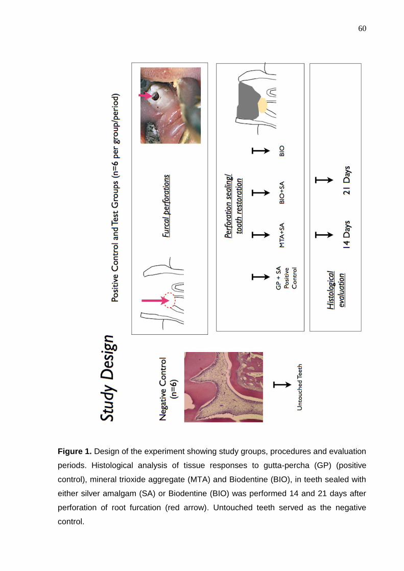
60
Figure 1. Design of the experiment showing study groups, procedures and evaluation
periods. Histological analysis of tissue responses to gutta-percha (GP) (positive
control), mineral trioxide aggregate (MTA) and Biodentine (BIO), in teeth sealed with
either silver amalgam (SA) or Biodentine (BIO) was performed 14 and 21 days after
perforation of root furcation (red arrow). Untouched teeth served as the negative
control.

61
Figure 2. Mean scores and standard deviation of inflammatory events (extension and
intensity), bone resorption and cementum repair after 14 and 21 days of furcal
perfuration and sealing. After 21 days, inflammatory extension as significantly greater
in the control group compared to MTA/SA, Biodentine alone (p<.01) and
Biodentine/SA (p<.0001). After 14 days the positive control presented more intense
inflammation compared to MTA/SA, Biodentine/SA (p<.0001) and Biodentine alone
(p<0.1). After 21 days, bone resorption was greater in the positive control group
compared to MTA/SA, Biodentine/SA (p<.01) and Biodentine alone (p<.05).
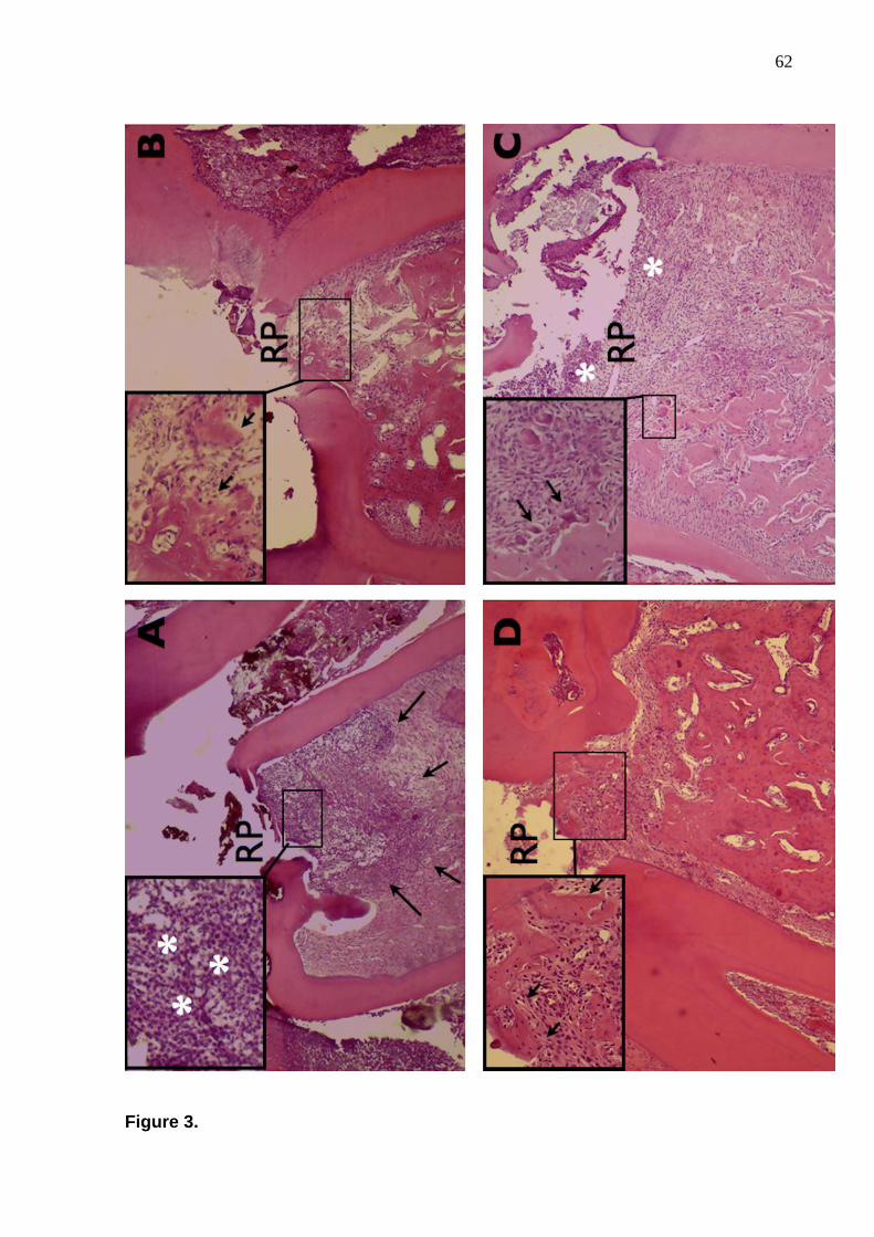
62
Figure 3.

63
Figure 3. Histological events observed after root perforation (RP) and sealing. (A)
Sample of the positive control group showing abscess formation (*) and extent
inflammatory infiltrate (arrows) after 14 days (H/E - original magnification 40x and
100x); (B) Samples sealed with MTA in the 21 days experimental period presenting
osteoblastic activity and bone formation (arrows) (H/E - original magnification 40x and
100x); (C) samples sealed with Biodentine alone 14 days after treatment. Note bone
resorption and osteoclastic activity (arrows), probably influenced by the inflammatory
events (*) started by the loss of coronal sealing (H/E - original magnification 40x and
100x) (D) Sample sealed with Biodentine and restored with silver amalgam showing
bone formation (arrows) 21 days after treatment (H/E - original magnification 40x and
100x).

64
6 DISCUSSÃO GERAL
Uma parte dos avanços da pesquisa odontológica está relacionada a novos
materiais e, neste caso, é fundamental que a tecnologia esteja aliada às bases
biológicas dos tecidos. Em terapias onde os tecidos vivos estabelecem contato direto
com os materiais, estes devem apresentar qualidades próximas às ideais e
possibilitar o reparo e a regeneração dos tecidos afetados, restabelecendo as
funções do dente (TROPE, 2008; COOPER et al., 2010; PRATI & GANDOLFI, 2015).
Muitos materiais à base de silicatos de cálcio possuem reconhecidas
propriedades biológicas, mas apresentam limitações, como longo tempo de presa,
baixas propriedades mecânicas e difícil manipulação. O mais conhecido é o
Agregado Trióxido de Mineral (MTA), que desde a década de 1990 tornou-se uma
referência para diversas terapias endodônticas (LEE & TORABINEJAD, 1993;
PARANJPE et al., 2011; PARIROKH & TORABINEJAD, 2010b; CAMILLERI, 2015a;
DARVEL & WU, 2011; MENTE et al., 2014).
A partir de 2009, outro material à base de silicato de cálcio, o Biodentine®, foi
introduzido como um substituto ao MTA. Desde então, alguns estudos comparativos
têm apontado a superioridade do Biodentine®, devido à indução de dentina terciária
reacional e reparadora; maior resistência à força de compressão e microdureza;
menor tempo de presa; e facilidade de manuseio (LAURENT, CAMPS, ABOUT,
2012; ZANINI et al, 2012; HAN & OKIJI, 2013; GRECH, MALLIA, CAMILLERI, 2013;
LUO et al., 2014; RAJASEKHARAN et al., 2014). A capacidade restauradora
também foi investigada como uma característica do Biodentine® (KOUBI et al., 2012;
RASKIN et al., 2012; CAMILLERI, 2013; KOUBI et al., 2013). No entanto, a indicação
do Biodentine® era para restauração temporária, e não definitiva.
O presente estudo buscou, em ratos wistar, compreender as respostas
teciduais mediante o uso do MTA e do Biodentine® sobre a polpa (exposições
pulpares) e tecidos periodontais de suporte (perfurações de furca). Além disso,
avaliou o comportamento do Biodentine® como material restaurador e sua influência
nas respostas teciduais, mesmo tendo ciência de que esta não é sua indicação
precípua.
As pesquisas relativas a exposições pulpares e perfurações de furca
costumavam utilizar cães e macacos. No entanto, o modelo em ratos tem
apresentado uma série de argumentos positivos relacionados aos aspectos técnicos

65
e financeiros, por se tratar de animais menores (DAMMASCHKE, 2010). O
conhecimento da idade e da genética dos ratos elimina interferências amostrais. A
microbiota oral e as respostas do hospedeiro são similares às observadas em
humanos (KAKEHASHI et al., 1965), e o metabolismo mais acelerado deste modelo
proporciona rapidez na progressão das respostas histológicas pulpares e
periodontais (SILVA et al., 2009; DAMMASCHKE, 2010). Os incisivos de ratos são
de fácil acesso e muito utilizados em pesquisas moleculares por apresentarem
crescimento contínuo. No entanto, os molares dos ratos apresentam similaridades
anatômicas e histológicas com molares humanos (SILVA et al., 2009;
DAMMASCHKE, 2010; SCARPARO et al., 2011; TRAN et al., 2012; DONDONI et al.,
2014).
Diante do pequeno porte dos animais, foi preciso gerenciar o seu manuseio.
Neste estudo, optou-se por procedimentos a quatro mãos. Após o efetivo controle
anestésico e posicionamento correto dos animais, utilizou-se um aparato
(SCARPARO et al., 2011) para driblar a pequena dimensão e o posicionamento
anatômico dos molares. O dispositivo ampliou o campo de ação e facilitou as etapas
operatórias das exposições pulpares e das perfurações de furca.
Para a seleção do outro material utilizado sobre a polpa e tecido periodontal de
suporte no comparativo com o Biodentine optou-se pelo MTA como padrão-ouro,
pois é também um cimento capaz de tomar presa, diferentemente do hidróxido de
cálcio. Para restaurar as cavidades buscou-se um material de fácil aplicação e
resistente à força da mastigação empregada pelos roedores. O amálgama
apresentou estas características. O uso da resina composta requereria preparo
prévio da cavidade com condicionamento ácido e adesivo, comprometendo o tempo
operacional de trabalho nestes animais.
A determinação do tempo experimental (14 e 21 dias) considerou estudos
prévios, que apontaram para a fragilização da estrutura dentária após a execução
dos procedimentos, pois podem comprometer as amostras em períodos de maior
tempo observacional (DAMMASCHKE, 2010; SCARPARO et al., 2011; TRAN et al.,
2012). Tal fato ficou evidenciado neste estudo durante a análise clínica, fotográfica e
radiográfica, que mostrou fraturas de cúspides em determinadas amostras. Ainda
assim, foi possível aferir respostas para as análises histológicas propostas.
Os resultados apontaram que o Biodentine® e o MTA apresentaram capacidade
de promover tecido duro, produzindo níveis aceitáveis de resposta inflamatória e

66
mantendo a vitalidade pulpar quando usados como agentes capeadores. Esta
pesquisa está em concordância com outros estudos que testaram o MTA em
terapias de polpa vital (TORABINEJAD & PARIROKH, 2010; DAMMASCHKE, 2010;
TABARSI et al., 2010; ESKANDARIZADEH et al., 2011). Entretanto, mostrou
algumas dificuldades a respeito da capacidade do Biodentine® em produzir
selamento coronal quando utilizado como um material restaurador.
Em relação as níveis de inflamação pulpar e alterações degenerativas, o tempo
experimental de 14 dias mostrou-se adequado para avaliar respostas frente aos
biomateriais. Para observar formação de barreira de tecido mineralizado, a maioria
das amostras necessitou de 21 dias. Relatos de períodos mais longos para obter os
mesmos resultados estão disponíveis com outros modelos (porquinhos-da-índia,
cães e humanos) (NOWICKA, et al., 2013; DE ROSSI et al., 2014; TZIAFA et al.,
2014).
A simultânea ocorrência de eventos inflamatórios e degenerativos, após
exposição pulpar e aplicação de biomaterias, foi avaliada através da comparação
das características pulpares de amostras do controle negativo (dentes saudáveis).
Estes resultados confirmam as descrições anteriores destas reações teciduais a
estímulos externos (SELTZER, BENDER, ZIONTZ, 1963; TROPE, 2008; COOPER,
et al., 2010). A presença de células inflamatórias e de abcesso, juntamente com
condensação fibrosa, hialinização e áreas calcificadas, caracteriza as alterações
pulpares como processos dinâmicos. Embora, a intensidade da inflamação seja
frequentemente utilizada para medir o dano, as descrições histológicas detalhadas
das respostas pulpares (DONDONI et al., 2014) não são utilizadas na maior parte
dos estudos relativos à manutenção da polpa vital (ACCORINTE et al; 2008;
NOWICKA et al., 2013).
O grupo controle positivo mostrou acentuada degeneração e necrose tecidual.
Muitas amostras em contato com MTA ou Biodentine® apresentaram uma moderada
condensação fibrosa, áreas de hialinização, degeneração cálcica e amplas áreas de
calcificações intrapulpares. Biodentine® favoreceu a ocorrência de calcificações de
polpa, embora comparada aos outros grupos não tenham sido estatisticamente
significativas. Da mesma forma, Tziafa et al. (2014) observaram maior ocorrência de
calcificação ectópica da polpa em amostras tratadas com este material.
MTA e Biodentine® produziram reações inflamatórias transitórias com extensão
limitada, contribuindo no reparo pulpar e na formação de barreiras de tecido duro,

67
diferentemente do grupo controle positivo. Em concordância, Nowicka et al. (2013)
observaram o mesmo em períodos mais longos. Assim, confirmou-se que, uma leve
inflamação, faz parte do processo que leva à proliferação e diferenciação de células-
tronco (TROPE, 2008; COOPER et al., 2010). Os eventos inflamatórios estimulam as
vias de sinalização moleculares capazes de induzir a proliferação e a diferenciação
das células-tronco da polpa dentária (DPSC) em odontoblastóides, levando à
síntese, à secreção e à mineralização da matriz de dentina (OBEID et al., 2013;
PARANJPE et al., 2011; ZANINI et al., 2012).
As barreiras mineralizadas formadas nos grupos de biomateriais podem ser
parcialmente explicadas pelo potencial de mineralização do MTA (TABARSI et al.,
2010; DAMMASCHKE et al., 2010; DARVELL & WU, 2011; MENTE et al., 2014) e
Biodentine® (TRAN et al., 2012; RAJASEKHARAN et al., 2014; DE ROSSI et al.,
2014; TZIAFA et al., 2014) devido ao efeito desses materiais sobre o tecido pulpar.
Além disso, uma investigação prévia in vitro revelou que, mesmo quando preparado
em diferentes concentrações, Biodentine® é capaz de aumentar a proliferação, a
migração e a adesão de células tronco (LUO et al., 2014), o que também pode ter
favorecido as respostas biológicas observadas neste estudo.
A capacidade do Biodentine® em promover a mineralização pulpar em períodos
mais curtos do que outros materiais foi sugerida em estudo in vitro com dentes
humanos (LAURENT, CAMPS, ABOUT, 2012). Células pulpares em contato com
Biodentine® mostraram capacidade de promover liberação de TGF-B1, formação de
focos mineralizados em apenas 2 dias e aparência de osteodentina após 14 dias. Na
presente investigação in vivo, embora Biodentine® tenha sido capaz de favorecer a
formação de barreiras de tecido duro, o processo não foi mais rápido do que nas
amostras de MTA.
Ainda que zonas de osteodentina tenham sido previamente descritas como
características de pontes dentinárias induzidas por Biodentine® (TZIAFA et al.,
2014), o mesmo não foi confirmado neste estudo. Isso pode ter acontecido,
principalmente, porque o modelo em rato não permite observações exatas nos
mesmos locais que possibilitem inferir diferenças dentro das características das
pontes de dentina. Os resultados aqui apresentados são categóricos e confirmam as
potencialidades de Biodentine® na indução de mineralização (TRAN et al., 2012;
ZANINI et al., 2012; LAURENT, CAMPS, ABOUT, 2012; NOWICKA et al., 2013;
CHANG, et al., 2014) e na manutenção da saúde do tecido pulpar (DE ROSSI et al.,

68
2014; TZIAFA et al., 2014). Por outro lado, a perda de selamento em algumas
amostras não afetou esse potencial. Provavelmente, a necessidade de menor
pressão para inserir o Biodentine®como material restaurador nas cavidades dos
molares de ratos pode ter influenciado neste resultado.
Assim como em outras investigações (TRAN et al., 2012; NOWICKA et al.,
2013; DE ROSSI et al., 2014), amostras capeadas com MTA e Biodentine®
promoveram maior formação de barreiras de tecido duro incompletas e completas do
que o grupo controle positivo. Foi notável, embora não mensurado, que Biodentine®
induziu pontes mais consistentes e mais grossas em comparação com MTA, o que
confirma as conclusões de outros estudos (DE ROSSI et al., 2014; TZIAFA et al.,
2014).
A capacidade dos materiais restauradores em manter um vedamento coronal
eficiente é de suma importância para evitar a infecção e permitir o sucesso da
terapia da polpa vital (KAKEHASHI, STANLEY, FITZGERALD, 1965; NOWICKA et
al., 2013; MENTE et al., 2014; DONDONI et al., 2014;). Como demonstrado no
presente estudo, algumas amostras seladas com Biodentine® mostraram
degradação ou perda total do material restaurador em 21 dias, ainda que apenas em
uma amostra não tenha ocorrido a formação de barreira de tecido mineralizado. A
explicação pode ser a ocorrência precoce do selamento biológico. No entanto, falhas
no selamento coronal podem impedir o sucesso do tratamento a longo prazo, através
de re-infecção e do restabelecimento da doença pulpar, o que provavelmente
ocorreria se períodos experimentais mais longos tivessem sido incluídos.
Importante aspecto a ser notado é que, embora o modelo de rato produza
maior tensão mastigatória característica dos roedores (SCARPARO et al., 2011;
TRAN et al., 2012), a erosão das restaurações de Biodentine® foi verificada também
em dentes posteriores humanos (KOUBI et al., 2013). Estes resultados, juntamente
com evidências de alteração qúimica e estrutural na presença de ácido, sangue e
outros fluidos (CAMILLERI, 2013; GRECH, MALLIA, CAMILLERI, 2013), contradizem
os relatos de maior resistência à compressão (CAMILLERI, SORRENTINO,
DAMIDOT, 2013) e do adequado uso do Biodentine® como um material restaurador
temporário (RASKIN et al., 2012; KOUBI et al., 2012). Assim, a aplicação clínica para
esse fim requer cautela.
A escolha do material para selamento de perfurações de furca deve levar em
consideração a sua capacidade de não ser agressivo aos tecidos. Dependendo do

69
caso, o agente pode tanto determinar reparação, o que é desejável, como perda
óssea e outras consequências, que interferem no tratamento (SELTZER, SINAI,
AUGUST, 1970; FUSS, 1995).
Estudos clínicos que avaliam resultados perante selamento de áreas
perfuradas são importantes, pois permitem inferências sobre os sintomas e
observação radiográfica de áreas de reparação (ou sua falta de reparação) em torno
dos tecidos periodontais de suporte (ALHADAINY, 1994; LEE, MONSEF,
TORABINEJAD, 1993; PARIROKH & TORABINEJAD, 2010a; PARIROKH &
TORABINEJAD, 2010b; DARVELL & WU, 2011; HAAPASALO et al., 2015). Por outro
lado, eles não avaliam as respostas teciduais que ajudam a determinar os melhores
protocolos de tratamento (SELTZER, SINAI, AUGUST, 1970; FUSS, 1995;
CAMILLERI, 2015a).
Na comparação do selamento de perfurações, MTA e Biodentine® tiveram
melhor desempenho em reduzir a resposta inflamatória no local agredido do que o
controle positivo em intensidade e em extensão quando atingiu 21 dias. A redução
da inflamação, em conjunto com as características positivas de Biodentine®
(GRECH, MALLIA, CAMILLERI, 2013; ZHOU et al., 2013), contribui para especular
que este material pode ser utilizado no local da perfuração, como uma alternativa ao
MTA.
A análise do tecido cementário, após selamento das perfurações, evidenciou
a reparação do cemento em 30% das amostras dos grupos que utilizaram
Biodentine® e MTA contra nenhuma do controle positivo. Apesar de não serem
estatisticamente significativos, estes dados são dignos de nota, pois concordam com
diferentes estudos (PITT FORD et al., 1995; AL DAAFAS & AL-NAZHAN, 2007;
HAKKI, et al., 2009; SAMIEE et al., 2010). Neles, foi detectada reparação cementária
em dentes selados com MTA, diferentemente do que foi observado com outros
materiais (BALLA et al., 1991; HARTWELL & ENGLAND, 1993). A atual pesquisa
mostrou que Biodentine® tem comportamento semelhante ao MTA para estimular o
reparo do cemento.
Uma hipótese para a ausência de reparação do cemento em algumas
amostras tratadas com MTA ou Biodentine® é que o cemento da furca se comporta
de forma diferente da região apical, pois o cemento não é celular e contém uma
camada muito fina. No processo de reparação de cemento, as células do ligamento
periodontal se transformam em cementoblastos, gerando um tecido ósseo com

70
aspecto cementóide. No entanto, isto é mais sensível em perfurações de furca, por
causa da proximidade do sulco gengival, levando à exposição ao ambiente bucal e à
sua contaminação bacteriana. Tal aspecto reduz as possibilidades de reparação,
diferentemente do que ocorre na região apical (SCARPARO et al., 2011). Além
disso, o período de 21 dias pode ter sido curto para exibir cementogênese
significativa. Existem estudos de 28 dias com ratos, mas os autores não avaliaram o
critério de formação de cemento (SILVA et al., 2009). Como anteriormente apontado,
períodos maiores de avaliação aumentam o risco de desgaste da estrutura dental de
ratos (SCARPARO et al., 2011; TRAN et al. 2012). Futuras investigações podem
incluir diferentes níveis de perfuração, semelhante a estudos clínicos, mas com
número compatível de amostras para permitir inferências adequadas.
Reabsorções ósseas aparecem com frequência em perfurações de furca,
devido à contaminação ou reação ao material. Se este for adequado para utilização
sobre os tecidos, há uma redução na reabsorção óssea e estímulo à osteogênese
(BEAVERS, BERGENHOLTZ, COX, 1986; PITT FORD et al., 1995; AL-DAAFAS &
AL-NAZHAN, 2007). Neste estudo, a redução da reabsorção óssea foi significativa
após 21 dias com MTA e Biodentine®, sugerindo osteogênese. Pesquisa prévia com
MTA mostrou esta tendência com a cultura de células primárias humanas
(PERINPNAYAGAN & AL-RABEAH, 2009). Outras investigações clínicas, que não
utilizaram critérios histológicos, também encontraram resultados semelhantes com
MTA (ARENS & TORABINEJAD, 1996; MAIN et al., 2004; YILDIRIM & DALCI, 2006;
PACE, GIULIANI, PAGAVINI, 2008; IBARROLA, BIGGS, BEESON, 2008;). Esse tipo
de estudo geralmente utiliza critérios radiológicos que avaliam redução de
radiolucência. Nestes casos, a atividade osteoblástica poderia explicar esses
achados.
Escoamento e tempo de presa dos materiais de selamento podem influenciar
a reparação dos tecidos quando ocorre extravazamento (ALHADAINY & ABDALLA,
1998; AL-DAAFAS & AL-NAZHAN, 2007). O MTA tem características difíceis de
manipulação e elevado tempo de presa. Isso pode colocar em risco o selamento da
furca, visto que ele pode ser removido ou deslocado do local com a irrigação ou a
carga oclusal (DARVELL & WU, 2011; HAAPASALO et al., 2015; CAMILLERI,
2015a). O Biodentine® apresentou melhor escoamento e menor tempo de presa
quando comparado com o MTA (GUNESER, AKBULUT, ELDENIZ, 2013;
HAAPASALO et al., 2015; CAMILLERI, 2015b), bem como uma melhor resistência à

71
carga de oclusão (GUNESER, AKBULUT, ELDENIZ, 2013). Isto foi notado pelos
operadores também nesta experiência.
Um estudo prévio relatou as vantagens do Biodentine® em relação ao MTA
quando ambos foram expostos a substâncias irrigantes (hipoclorito de sódio,
clorexidina, soro fiosiológico). MTA apresentou comportamento aquém quando em
contato com a clorexidina (GUNESER, AKBULUT, ELDENIZ, 2013). Aparentemente,
o Biodentine®, por formar tags com a dentina (HAN & OKIJI, 2011), permite maior
ancoragem mecânica à superfície, exigindo maior força para seu deslocamento da
dentina radicular. Por outro lado, a resposta do tecido foi semelhante entre os grupos
teste, que poderia ser explicada pela biocompatibilidade do MTA. Outros materiais
foram testados em estudos anteriores, como o ionômero de vidro e o amálgama,
com maiores vantagens mecânicas e químicas sobre o MTA (ALHADAINY &
ABDALLA, 1998; GUNESER, AKBULUT, ELDENIZ, 2013,).
Contaminação da área de furca, ausência de selamento adequado e
infiltração coronária contribuem para um prognóstico ruim do tratamento de
perfurações de furca (BEAVERS, BORGENHOLTZ, COX, 1986; PITT FORD et al.,
1995, AL-DAAFAS & AL-NAZHAN, 2007; VANNI et al., 2011; HAAPASALO, et al.,
2015). A capacidade restauradora do Biodentine® também foi testada em um dos
grupos do experimento de perfuração. Embora diferenças significativas não tenham
sido detectadas entre os grupos teste, uma reabsorção óssea ligeiramente mais
elevada foi observada quando Biodentine® foi usado como material de restauração.
Isso pode ter ocorrido devido à contaminação. Além disso, pode-se especular que,
se os períodos de avaliação fossem maiores, haveria a formação de biofilme e a falta
de restauração adequada exerceriam danos aos tecidos.
Embora os resultados do presente estudo tenham possibilitado conhecer mais
o Biodentine®, averiguou-se a similaridade com o MTA quando aplicado sobre os
tecidos pulpar e periodontal nas terapias de polpa vital e de perfurações de furca.
Como os resultados de reparação do cemento não foram evidentes, uma análise
mais aprofundada é necessária. Também o uso do Biodentine® como restauração
mostrou limitações em relação à capacidade de vedamento coronal, e deve,
portanto, estar associado com um material restaurador mais eficiente para assegurar
resultados positivos ao longo do tempo. As vantagens do Biodentine® que
corroboram para a escolha do produto na prática odontológica estão na facilidade do
manuseio e na perspectiva de evitar a descoloração dos dentes. Cabe verificar, em

72
longo prazo, o comportamento clínico deste material em humanos. Para vencer a
prova do tempo, no entanto, é preciso que seu uso seja testado e propriedades e
limitações melhor entendidos. Assim, poder-se-á desvelar o real alcance deste
promissor material.

73
7 CONCLUSÃO
A partir dos resultados do presente estudo, pode-se concluir que:
● O Biodentine® e o MTA mostraram propriedades similares favoráveis nas terapias
de polpa vital e de perfurações de furca.
● Nas pulpotomias, Biodentine® e MTA apresentaram diminuição significativa da
resposta inflamatória (intensidade e extensão) e dos eventos degenerativos. A
formação de tecido mineralizado foi confirmada. Quando Biodentine® foi usado como
material restaurador, essa diferença foi menos visível.
● Nas perfurações de furca, Biodentine® e MTA apresentaram redução da resposta
inflamatória (intensidade e extensão) e dos eventos de reabsorção óssea. Porém,
quando Biodentine® foi usado como material restaurador, essa diferença foi menos
visível.
● O uso do Biodentine® como material restaurador não promoveu selamento
coronário eficiente, não sendo recomendável para este fim.

74
REFERÊNCIAS
Accorinte MdLR, Holland R, Reis A, et al. Evaluation of Mineral Trioxide Aggregate
and Calcium Hydroxide Cement as Pulp-capping Agents in Human Teeth. J Endod.
2008;34:1-6.
Aguilar P, Linsuwanont P. Vital Pulp Therapy in Vital Permanent Teeth with Cariously
Exposed Pulp: A Systematic Review. J Endod. 2011;37:581-587.
Al-Daafas A, Al-Nazhan S. Histological evaluation of contaminated furcal perforation
in dogs’ teeth repaired by MTA with or without internal matrix. Oral Surg Oral Med
Oral Pathol Oral Radiol Endod. 2007;103:e92-e99.
Alhadainy HA, Abdalla AI. Artificial floor technique used for the repair of furcation
perforations: A microleakage study. J Endod. 1998;24:33-35.
Alhadainy HA. Root perforations. A review of literature. Oral Surc Oral Med Oral
Pathol. 1994;78:368-374.
Arens DE, Torabinejad M. Repair of furcal perforations with mineral trioxide
aggregate: Two case reports. Oral Surg Oral Med Oral Pathol Oral Radiol Endod.
1996;82:84-88.
Asgary S, Eghbal MJ, Parirokh M, et al. A comparative study of histologic response to
different pulp capping materials and a novel endodontic cement. Oral Surg Oral Med
Oral Pathol Oral Radiol Endod. 2008;106:609-14.
Balla R, LoMonaco CJ, Skribner J, et al. Histological study of furcation perforations
treated with Tricalcium phosphate, hydroxylapatite, amalgam, and Life. J Endod.
1991;17:234-238.
Beavers RA, Bergenholatz G, Cox CF. Periodontal wound healing following
intentional root perforations in permanent teeth of Macaca mulatta. Int Endod J.
1986;19: 36-44.

75
Camilleri J, Sorrentino F, Damidot D. Investigation of the hydration and bioactivity of
radiopacified tricalcium silicate cement, Biodentine and MTA Angelus. Dent Mater.
2013;29:580-93.
Camilleri J. Color stability of white mineral trioxide aggregate in contact with
hypochlorite solution. J Endod. 2014;40:436-440.
Camilleri J. Investigation of Biodentine as dentine replacement material. J Dent.
2013;41:600-610.
Camilleri J. Mineral trioxide aggregate: present and future developments. Endod
Topics. 2015a;32:31-46.
Camilleri J. Characterization of modified calcium-silicate cements exposed to acidic
environment. Materials Characterization 2011;62(1):70-75.
Camilleri J. Staining Potential of Neo MTA Plus, MTA Plus, and Biodentine Used for
Pulpotomy Procedures. J Endod. 2015b;41:1139-1145.
Chang SW, Lee SY, Ann HJ, et al. Effects of calcium silicate endodontic cements on
biocompatibility and mineralization-inducing potentials in human dental pulp cells. J
Endod. 2014;40:1194-200.
Cooper PR, Takahashi Y, Graham LW, et al. Inflammation-regeneration interplay in
the dentine-pulp complex. J Dent. 2010;38:687-97.
Corral Nuñez CM, Bosomworth HJ, Field C, et al. Biodentine and Mineral Trioxide
Aggregate Induce Similar Cellular Responses in a Fibroblast Cell Line. J Endod.
2014;40:406-411.
Dammaschke T, Stratmann U, Wolff P, et al. Direct pulp capping with mineral trioxide
aggregate: an immunohistologic comparison with calcium hydroxide in rodents. J
Endod. 2010;36:814-9.

76
Dammaschke T. Rat molar teeth as a study model for direct pulp capping research in
dentistry. Lab Anim. 2010;44:1-6.
Daoudi MF, Saunders WP. In Vitro Evaluation of Furcal Perforation Repair Using
Mineral Trioxide Aggregate or Resin Modified Glass Ionomer Cement with and
without the Use of the Operating Microscope. Journal of endodontics 2002;28(7):512-
515.
Darvell BW, Wu RCT. ―MTA‖—An Hydraulic Silicate Cement: Review update and
setting reaction. Dent Mater. 2011;27:407-22.
De Rossi A, Silva LAB, Gatón-Hernández P, et al. Comparison of Pulpal Responses
to Pulpotomy and Pulp Capping with Biodentine and Mineral Trioxide Aggregate in
Dogs. J Endod. 2014;40:1362-9.
Dondoni L, Scarparo RK, Kantarci A, et al. Effect of the pro-resolution lipid mediator
Resolvin E1 (RvE1) on pulp tissues exposed to the oral environment. Int Endod J.
2014;47:827-34.
Eskandarizadeh A, Parirokh M, Shahpasandzadeh M, et al. A comparative study on
dental pulp response to calcium hydroxide, white and grey mineral trioxide aggregate
as pulp capping agents. J Conserv Dent. 2011;14:351.
Formosa LM, Mallia B, Camilleri J. The effect of curing conditions on the physical
properties of tricalcium silicate cement for use as a dental biomaterial. Int Endod J.
2012;45:326-36.
Fuss Z. Management of root perforations: Classification, diagnosis, prevention, and
treatment. J Endod. 1995;21:442.
Grech L, Mallia B, Camilleri J. Characterization of set Intermediate Restorative
Material, Biodentine, Bioaggregate and a prototype calcium silicate cement for use as
root-end filling materials. Int Endod J. 2013;46:632-41.
77
Grech L, Mallia B, Camilleri J. Investigation of the physical properties of tricalcium
silicate cement-based root-end filling materials. Dent Mater. 2013;29:e20-8.
Guneser MB, Akbulut MB, Eldeniz AU. Effect of Various Endodontic Irrigants on the
Push-out Bond Strength of Biodentine and Conventional Root Perforation Repair
Materials. J Endod. 2013;39:380-384.
Haapasalo M, Parhar M, Huang X, et al. Clinical use of bioceramic materials. Endod
Topics. 2015;32:97-117.
Hakki SS, Bozkurt SB, Hakki EE, et al. Effects of Mineral Trioxide Aggregate on Cell
Survival, Gene Expression Associated with Mineralized Tissues, and
Biomineralization of Cementoblasts. J Endod. 2009;35:513-519.
Han L, Okiji T. Bioactivity evaluation of three calcium silicate-based endodontic
materials. Int Endod J. 2013;46:808-14.
Hartwell GR, England MC. Healing of furcation perforations in primate teeth after
repair with decalcified freeze-dried bone: A longitudinal study. J Endod. 1993;19:357-
361.
Hashem AAR, Wanees ASA. The Effect of Acidity on Dislodgment Resistance of
Mineral Trioxide Aggregate and Bioaggregate in Furcation Perforations: An In Vitro
Comparative Study. J Endod. 2012;38:245-249.
Holland R. Histochemical response of amputed pulps to calcium hydroxide. Rev Bras
Pesq Med Biol 1971;4(1-2):83-95.
Ibarrola JL, Biggs SG, Beeson TJ. Repair of a Large Furcation Perforation: A Four-
Year Follow-Up. J Endod. 2008;34:617-619.

78
Kakehashi S, Stanley HR, Fitzgerald RJ. The Effects of Surgical Exposures of Dental
Pulps in Germ-Free and Conventional Laboratory Rats. Oral Surg Oral Med Oral
Pathol. 1965;20:340-9.
Kang S-H, Shin Y-S, Lee H-S, et al. Color Changes of Teeth after Treatment with
Various Mineral Trioxide Aggregate–based Materials: An Ex Vivo Study. J Endod.
2015;41:737-41.
Kohli MR, Yamaguchi M, Setzer FC, Karabucak B. Spectrophotometric Analysis of
Coronal Tooth Discoloration Induced by Various Bioceramic Cements and Other
Endodontic Materials. Journal of endodontics 2015;41(11):1862-1866.
Koubi G, Colon P, Franquin JC, et al. Clinical evaluation of the performance and
safety of a new dentine substitute, Biodentine, in the restoration of posterior teeth - a
prospective study. Clin Oral Investig. 2013;17:243-9.
Koubi S, Elmerini H, Koubi G, et al. Quantitative evaluation by glucose diffusion of
microleakage in aged calcium silicate-based open-sandwich restorations. Int J Dent.
2012;2012:105863.
Laurent P, Camps J, About I. Biodentine(TM) induces TGF-beta1 release from
human pulp cells and early dental pulp mineralization. Int Endod J. 2012;45:439-48.
Lee SJ, Monsef M, Torabinejad M. Sealing ability of a mineral trioxide aggregate for
repair of lateral root perforations. J Endod. 1993;19:541-4.
Luo Z, Li D, Kohli MR, et al. Effect of Biodentine™ on the proliferation, migration and
adhesion of human dental pulp stem cells. J Dent. 2014;42:490-7.
Ma J, Shen Y, Stojicic S, et al. Biocompatibility of Two Novel Root Repair Materials.
Journal of endodontics 2011;37(6):793-798.

79
Maeda H, Nakano T, Tomokiyo A, et al. Mineral Trioxide Aggregate Induces Bone
Morphogenetic Protein-2 Expression and Calcification in Human Periodontal
Ligament Cells. Journal of endodontics 2010;36(4):647-652.
Main C, Mirzayan N, Shabahang S, et al. Repair of Root Perforations Using Mineral
Trioxide Aggregate: A Long-term Study. J Endod. 2004;30:80-83.
Mente J, Hufnagel S, Leo M, et al. Treatment Outcome of Mineral Trioxide Aggregate
or Calcium Hydroxide Direct Pulp Capping: Long-term Results. J Endod.
2014;40:1746-51.
Mori GG, Teixeira LM, Oliveira DL, et al. Biocompatibility Evaluation of Biodentine in
Subcutaneous Tissue of Rats. J Endod. 2014;40:1485-1488.
Nanci A, Bosshardt, DD. Structure of periodontal tissues in health and disease.
Peridontol. 2000. 2006; 40: 112-8
Nowicka A, Lipski M, Parafiniuk M, et al. Response of human dental pulp capped with
biodentine and mineral trioxide aggregate. J Endod. 2013;39:743-7.
Obeid M, Saber SEDM, Ismael AED, et al. Mesenchymal stem cells promote hard-
tissue repair after direct pulp capping. J Endod. 2013;39:626-31.
Pace R, Giuliani V, Pagavino G. Mineral Trioxide Aggregate as Repair Material for
Furcal Perforation: J Endod. 2008;34:1130-1133.
Paranjpe A, Smoot T, Zhang H, et al. Direct contact with mineral trioxide aggregate
activates and differentiates human dental pulp cells. J Endod. 2011;37:1691-5.
Parirokh M, Torabinejad M. Mineral Trioxide Aggregate: A Comprehensive Literature
Review—Part I: Chemical, Physical, and Antibacterial Properties. J Endod.
2010a;36:16-27.

80
Parirokh M, Torabinejad M. Mineral Trioxide Aggregate: A Comprehensive Literature
Review—Part III: Clinical Applications, Drawbacks, and Mechanism of Action. J
Endod. 2010b;36:400-413.
Peng W, Liu W, Zhai W, et al. Effect of Tricalcium Silicate on the Proliferation and
Odontogenic Differentiation of Human Dental Pulp Cells. J Endod. 2011;37:1240-
1246.
Perinpanayagam H, Al-Rabeah E. Osteoblasts interact with MTA surfaces and
express Runx2. Oral Surg Oral Med Oral Pathol Oral Radiol Endod. 2009;107:590-
596.
Pitt Ford TR, Torabinejad M, McKendry DJ, et al. Use of mineral trioxide aggregate
for repair of furcal perforations. Oral Surg Oral Med Oral Pathol Oral Radiol Endod.
1995;79:756-763.
Prati C, Gandolfi MG. Calcium silicate bioactive cements: Biological perspectives and
clinical applications. Dent Mater. 2015;31:351-70.
Rajasekharan S, Martens LC, Cauwels RG, et al. Biodentine material characteristics
and clinical applications: a review of the literature. Eur Arch Paediatr Dent.
2014;15:147-58.
Raskin A, Eschrich G, Dejou J, et al. In vitro microleakage of Biodentine as a dentin
substitute compared to Fuji II LC in cervical lining restorations. J Adhes Dent.
2012;14:535-42.
Salako N, Joseph B, Ritwik P, et al. Comparison of bioactive glass, mineral trioxide
aggregate, ferric sulfate, and formocresol as pulpotomy agents in rat molar. Dent
Traumatol. 2003;19:314-20.
Samiee M, Eghbal M, Parirokh M, et al. Repair of furcal perforation using a new
endodontic cement. Clin Oral Invest. 2010;14:653-658.
81
Scarparo RK, Dondoni L, Bottcher DE, et al. Response to intracanal medication in
immature teeth with pulp necrosis: an experimental model in rat molars. J Endod.
2011;37:1069-73.
Seltzer S, Bender IB, Ziontz M. The dynamics of pulp inflammation: Correlations
between diagnostic data and actual histologic findings in the pulp. Oral Surg Oral
Med Oral Pathol. 1963;16:846-71.
Seltzer S, Sinai I, August D. Periodontal Effects of Root Perforations Before and
During Endodontic Procedures. J Dent Res. 1970;49:332-339.
Seo BM, Miura M, Gronthos S, et al. Investigation of multipotent postnatal stem cells
from human periodontal ligament. Lancet. 2004; 364: 149-55.
Shokouhinejad N, Nekoofar MH, Iravani A, et al. Effect of Acidic Environment on the
Push-out Bond Strength of Mineral Trioxide Aggregate. J Endod. 2010;36:871-874.
Silva MJB, Caliari MV, Sobrinho APR, et al. An in vivo experimental model to assess
furcal lesions as a result of perforation. Int Endod J. 2009;42:922-929.
Tabarsi B, Parirokh M, Eghbal MJ, et al. A comparative study of dental pulp response
to several pulpotomy agents. Int Endod J. 2010;43:565-71.
Torabinejad M, Parirokh M. Mineral Trioxide Aggregate: A Comprehensive Literature
Review—Part II: Leakage and Biocompatibility Investigations. J Endod. 2010;36:190-
202.
Tran XV, Gorin C, Willig C, et al. Effect of a calcium-silicate-based restorative cement
on pulp repair. J Dent Res. 2012;91:1166-71.
Trope M. Regenerative Potential of Dental Pulp. J Endod. 2008;34:S13-S17.

82
Tsesis I, Rosenberg E, Faivishevsky V, et al. Prevalence and Associated Periodontal
Status of Teeth with Root Perforation: A Retrospective Study of 2,002 Patients'
Medical Records. Journal of endodontics 2010;36(5):797-800
Tziafa C, Koliniotou-Koumpia E, Papadimitriou S, et al. Dentinogenic responses after
direct pulp capping of miniature swine teeth with Biodentine. J Endod. 2014;40:1967-
71.
Tziafas D, Smith AJ, Lesot H. Designing new treatment strategies in vital pulp
therapy. J Dent. 2000;28:77-92.
Tziafas D. The future role of a molecular approach to pulp-dentinal regeneration.
Caries Res. 2004;38:314-20.
Vallés M, Mercadé M, Duran-Sindreu F, et al. Influence of Light and Oxygen on the
Color Stability of Five Calcium Silicate–based Materials. J Endod. 2013;39:525-528.
Vallés M, Roig M, Duran-Sindreu F, et al. Color Stability of Teeth Restored with
Biodentine: A 6-month In Vitro Study. Journal of endodontics 2015;41(7):1157-1160.
Vanni JR, Della-Bona Á, Figueiredo JAPD, et al. Radiographic evaluation of furcal
perforations sealed with different materials in dogs' teeth. J Appl Oral Sci.
2011;19:421-425.
Yildirim G, Dalci K. Treatment of lateral root perforation with mineral trioxide
aggregate: a case report. Oral Surg Oral Med Oral Pathol Oral Radiol Endod.
2006;102:e55-e58.
Zanini M, Sautier JM, Berdal A, et al. Biodentine Induces Immortalized Murine Pulp
Cell Differentiation into Odontoblast-like Cells and Stimulates Biomineralization. J
Endod. 2012;38:1220-6.
Zhou HM, Shen Y, Wang ZJ, et al. In vitro cytotoxicity evaluation of a novel root
repair material. J Endod. 2013;39:478-483.

83
ANEXO A – Aprovação da Comissão Científica e de Ética da Faculdade de
Odontologia da PUCRS
84
ANEXO B – Aprovação da Comissão de Ética no Uso de Animais da PUCRS

85
ANEXO C – Carta de submissão ao periódico Dental Materials
From: [email protected]
To: [email protected]; [email protected]
Date: Thu, 3 Sep 2015 20:09:35 +0100
Subject: Submission Confirmation for your paper
Full Length Article
Dear Roberta,
Your submission entitled "Pulpal response after capping with conventional mineral
trioxide aggregate (MTA) or a tricalcium silicate based-material (Biodentine)" has
been received by Dental Materials.
You may check on the progress of your paper by logging on to the Elsevier Editorial
System as an author. The URL is http://ees.elsevier.com/dema/.
Your username is: [email protected]
If you need to retrieve password details, please go to:
http://ees.elsevier.com/dema/automail_query.asp.
Your manuscript will be given a reference number once an Editor has been assigned.
Thank you for submitting your work to this journal.
Kind regards,
Elsevier Editorial System Dental Materials

86
ANEXO D – Carta de submissão ao periódico Journal of Endodontics
From: [email protected]
To: [email protected]; [email protected]
Date: Thu, 17 Sep 2015 20:57:13 +0100
Subject: Submission Confirmation for Periodontal response after furcal perfuration
using conventional mineral trioxide aggregate (MTA) or a tricalcium silicate based-
material (Biodentine)
Dear Dr. Scarparo,
Your submission entitled "Periodontal response after furcal perfuration using
conventional mineral trioxide aggregate (MTA) or a tricalcium silicate based-material
(Biodentine)" has been received by the Journal of Endodontics.
You will be able to check on the progress of your paper by logging on to the Journal
of Endodontics web site as an author.
The URL is http://ees.elsevier.com/joe/
Your username is: [email protected]
If you need to retrieve password details, please go to:
http://ees.elsevier.com/joe/automail_query.asp
Your manuscript will be given a reference number once an Editor has been assigned.
Thank you for submitting your work to the Journal of Endodontics.
Kind regards,
Journal of Endodontics
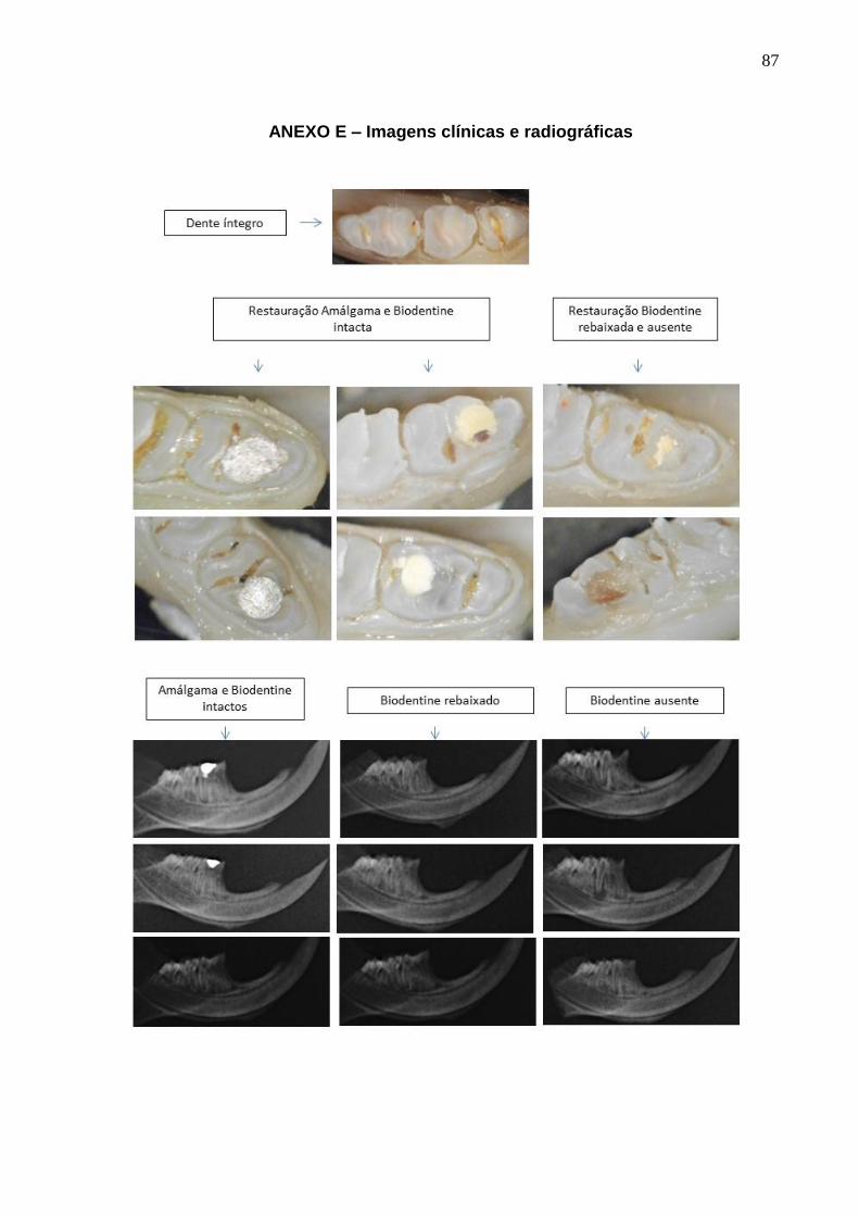
87
ANEXO E – Imagens clínicas e radiográficas

88
ANEXO F – Imagens histológicas

89

90
ANEXO G – Imagens BiodentineTM e modo de preparo


![Manual de Toxicologia Amup 2010[1]](https://static.fdocuments.in/doc/165x107/5695d4941a28ab9b02a1fa77/manual-de-toxicologia-amup-20101.jpg)