Polyaniline-CdS Quantum Dots Composite for Mediator Free Biosensing
9
Volume 3 • Issue 1 • 1000112 J Biosens Bioelectron ISSN:2155-6210 JBSBE an open access journal Research Article Open Access Biosensors & Bioelectronics Dhyani et al. J Biosens Bioelectron 2011, 3:1 http://dx.doi.org/10.4172/2155-6210.1000112 *Corresponding author: Hemant Dhyani, School of Physical Sciences, Jawaharlal Nehru University, New Delhi-110067, India, E-mail: [email protected] Received October 10, 2011; Accepted December 12, 2011; Published December 16, 2011 Citation: Dhyani H, Dhand C, Malhotra BD, Sen P (2011) Polyaniline-CdS Quantum Dots Composite for Mediator Free Biosensing. J Biosens Bioelectron 3:112. doi:10.4172/2155-6210.1000112 Copyright: © 2011 Dhyani H, et al. This is an open-access article distributed under the terms of the Creative Commons Attribution License, which permits unrestricted use, distribution, and reproduction in any medium, provided the original author and source are credited. Polyaniline-CdS Quantum Dots Composite for Mediator Free Biosensing Hemant Dhyani 1 *, Chetna Dhand 2 , B. D. Malhotra 2 and P. Sen 1 1 School of Physical Sciences, Jawaharlal Nehru University, New Delhi-110067, India 2 Department of Science and Technology Centre on Biomolecular Electronics, National Physical Laboratory, Dr. K. S. Krishnan Marg, New Delhi-110012, India Abstract A novel route has been introduced to fabricate the composite of polyaniline (PANI) and cadmium sulphide quantum dots (CdS-QD) using electrochemical polymerization technique for mediator free biosensing. The synthesis process involves in situ formation of CdS quantum dots that provide template for eletro-polymerization of aniline resulting in nanostructured PANI-(CdS-QD) film deposition on the indium-tin-oxide (ITO) coated glass plate. Transmission electron microscopy and scanning electron microscopy have been used to reveal the formation of CdS-QD and morphological changes involved during incorporation of CdS in PANI matrix and while immobilization of cholesterol oxidase (ChOx). The UV–visible and FT-IR investigations show the formation of PANI-(CdS-QD) composite at the molecular level. This matrix has been utilized for the covalent immobilization of cholesterol oxidase to explore its application for cholesterol sensing. The results of the CV and EIS studies indicate enhanced electrochemical and charge transfer behaviour of the composite. The response studies, carried out using CV technique, reveal this ChOx/ PANI-(CdS-QD)/ITO bioelectrode to detect cholesterol in the concentration range of 50 to 500 mgdL -1 with good detection limit (47.8 mgdL -1 ) and low app m K value (0.82 mM). Keywords: Polyaniline; Cadmium Sulphide; Quantum Dots; Conducting Polymers; Biosensor Introduction Conducting polymers are known to be compatible with biological molecules and also have the quality to efficiently transfer the electric charges produced during the biochemical reactions through their conjugated backbone. In the past few years many conducting polymers have been used with the different prospects for biosensing applications [1]. Among all these polymers, polyaniline is known to be semi-flexible rod and has been found to provide the efficient medium for electron transfer. e chemical and structural flexibility surrounding its amine nitrogen linkages for the immobilization of desired biomolecules has attracted much efforts towards the various applications in the field of biosensing due to its electrochemical, electronic, optical and electro- optical properties [2,3]. Synthesis and functionalization of nanomaterials have aroused much interest due to large surface-to-volume ratio and the quantum confinement effect in semiconductor nanomaterials in which appropriate surface modification by stabilizers can remove localized surface-trap states resulting in increased quantum yield of the excitonic emission. Besides this, quantum dots are known to have extraordinary electronic and optical properties due to their size-tunability, surface capping chemistry, high quantum yield, broad absorption with narrow and symmetric spectra and easy association with the other molecular groups [4]. e semiconductor nanocrystals have been used as fluorescent probes and as active components in nanostructure- biomolecule complexes for biosensor applications [5,6]. In recent years, quantum dots have been used for biosensing due to the electrochemical as well as optical properties [8,9]. e bare quantum dots have been used with the principle of fluorescence resonance energy transfer (FRET) and photo-luminescence (PL) quenching treatment to design biosensing assay system for the determination of glucose [10], and the capped QDs have been synthesized in a stable aqueous medium and this system has been used as a fluorescence probe for the mercury ions wherein the optimum fluorescence intensity is proportional to the ion concentration [11]. Furthermore the functional quantum dots have recently been introduced for molecular sieve sensors application [12]. Since, the majority of biosensing applications of quantum dots have been focused using their optical properties and very few results for quantum dots modified electrochemical sensors have been reported till date. Modifying the electrodes by the incorporation of quantum dots promotes direct electron transfer between the biomolecules and electrode surface [13], and also the direct electrochemistry has been reported by preparing the quantum dots/CNT electrode [7]. Among the various quantum dots cadmium sulphide (CdS) (band gap of 2.4 eV) has been studied with its excellent photoelectrochemical and optical properties [14-16]. Conjugated polymers specially like polyaniline combined with semiconductor nanoparticles (CdS, CdSe and TiO 2 ) are widely studied with the remarkable physical, optical, electronic and photoelectric properties [17,22,24]. It has been observed that introducing nanocrystalline CdS on polymer enhances the charges on polymer backbone thus resulting conformational changes in structure of PANI which becomes expanded from its compact coiled form [18]. PANI-CdS composite not only exhibits the enhanced photo and electrical activity but it also increases the life of polymer and have good stability in ambient condition [21,23]. ese properties of polymer-nanocrystal composite have led to increased interest to explore this PANI-CdS composite for biosensing purpose. erefore on this way we have devised a novel electrode of PANI-(CdS-QD) composite for biosensing application. Herein, we report results of the studies relating to the fabrication of PANI-(CdS-QD)/ITO electrode by electrochemical polymerization technique. e composite electrode is shown to have improved responce as well as electrochemical stability due to in situ formation
-
Upload
prakushprakush -
Category
Documents
-
view
46 -
download
4
Transcript of Polyaniline-CdS Quantum Dots Composite for Mediator Free Biosensing

Volume 3 • Issue 1 • 1000112J Biosens BioelectronISSN:2155-6210 JBSBE an open access journal
Research Article Open Access
Biosensors & BioelectronicsDhyani et al. J Biosens Bioelectron 2011, 3:1
http://dx.doi.org/10.4172/2155-6210.1000112
*Corresponding author: Hemant Dhyani, School of Physical Sciences, Jawaharlal Nehru University, New Delhi-110067, India, E-mail: [email protected]
Received October 10, 2011; Accepted December 12, 2011; Published December 16, 2011
Citation: Dhyani H, Dhand C, Malhotra BD, Sen P (2011) Polyaniline-CdS Quantum Dots Composite for Mediator Free Biosensing. J Biosens Bioelectron 3:112. doi:10.4172/2155-6210.1000112
Copyright: © 2011 Dhyani H, et al. This is an open-access article distributed under the terms of the Creative Commons Attribution License, which permits unrestricted use, distribution, and reproduction in any medium, provided the original author and source are credited.
Polyaniline-CdS Quantum Dots Composite for Mediator Free BiosensingHemant Dhyani1*, Chetna Dhand2, B. D. Malhotra2 and P. Sen1
1School of Physical Sciences, Jawaharlal Nehru University, New Delhi-110067, India2Department of Science and Technology Centre on Biomolecular Electronics, National Physical Laboratory, Dr. K. S. Krishnan Marg, New Delhi-110012, India
AbstractA novel route has been introduced to fabricate the composite of polyaniline (PANI) and cadmium sulphide quantum
dots (CdS-QD) using electrochemical polymerization technique for mediator free biosensing. The synthesis process involves in situ formation of CdS quantum dots that provide template for eletro-polymerization of aniline resulting in nanostructured PANI-(CdS-QD) film deposition on the indium-tin-oxide (ITO) coated glass plate. Transmission electron microscopy and scanning electron microscopy have been used to reveal the formation of CdS-QD and morphological changes involved during incorporation of CdS in PANI matrix and while immobilization of cholesterol oxidase (ChOx). The UV–visible and FT-IR investigations show the formation of PANI-(CdS-QD) composite at the molecular level. This matrix has been utilized for the covalent immobilization of cholesterol oxidase to explore its application for cholesterol sensing. The results of the CV and EIS studies indicate enhanced electrochemical and charge transfer behaviour of the composite. The response studies, carried out using CV technique, reveal this ChOx/PANI-(CdS-QD)/ITO bioelectrode to detect cholesterol in the concentration range of 50 to 500 mgdL-1 with good detection limit (47.8 mgdL-1) and low app
mK value (0.82 mM).
Keywords: Polyaniline; Cadmium Sulphide; Quantum Dots; Conducting Polymers; Biosensor
IntroductionConducting polymers are known to be compatible with biological
molecules and also have the quality to efficiently transfer the electric charges produced during the biochemical reactions through their conjugated backbone. In the past few years many conducting polymers have been used with the different prospects for biosensing applications [1]. Among all these polymers, polyaniline is known to be semi-flexible rod and has been found to provide the efficient medium for electron transfer. The chemical and structural flexibility surrounding its amine nitrogen linkages for the immobilization of desired biomolecules has attracted much efforts towards the various applications in the field of biosensing due to its electrochemical, electronic, optical and electro-optical properties [2,3].
Synthesis and functionalization of nanomaterials have aroused much interest due to large surface-to-volume ratio and the quantum confinement effect in semiconductor nanomaterials in which appropriate surface modification by stabilizers can remove localized surface-trap states resulting in increased quantum yield of the excitonic emission. Besides this, quantum dots are known to have extraordinary electronic and optical properties due to their size-tunability, surface capping chemistry, high quantum yield, broad absorption with narrow and symmetric spectra and easy association with the other molecular groups [4]. The semiconductor nanocrystals have been used as fluorescent probes and as active components in nanostructure-biomolecule complexes for biosensor applications [5,6].
In recent years, quantum dots have been used for biosensing due to the electrochemical as well as optical properties [8,9]. The bare quantum dots have been used with the principle of fluorescence resonance energy transfer (FRET) and photo-luminescence (PL) quenching treatment to design biosensing assay system for the determination of glucose [10], and the capped QDs have been synthesized in a stable aqueous medium and this system has been used as a fluorescence probe for the mercury ions wherein the optimum fluorescence intensity is proportional to the ion concentration [11]. Furthermore the functional quantum dots have recently been introduced for molecular sieve sensors application [12]. Since, the majority of biosensing applications of quantum dots
have been focused using their optical properties and very few results for quantum dots modified electrochemical sensors have been reported till date. Modifying the electrodes by the incorporation of quantum dots promotes direct electron transfer between the biomolecules and electrode surface [13], and also the direct electrochemistry has been reported by preparing the quantum dots/CNT electrode [7].
Among the various quantum dots cadmium sulphide (CdS) (band gap of 2.4 eV) has been studied with its excellent photoelectrochemical and optical properties [14-16]. Conjugated polymers specially like polyaniline combined with semiconductor nanoparticles (CdS, CdSe and TiO2) are widely studied with the remarkable physical, optical, electronic and photoelectric properties [17,22,24]. It has been observed that introducing nanocrystalline CdS on polymer enhances the charges on polymer backbone thus resulting conformational changes in structure of PANI which becomes expanded from its compact coiled form [18]. PANI-CdS composite not only exhibits the enhanced photo and electrical activity but it also increases the life of polymer and have good stability in ambient condition [21,23]. These properties of polymer-nanocrystal composite have led to increased interest to explore this PANI-CdS composite for biosensing purpose. Therefore on this way we have devised a novel electrode of PANI-(CdS-QD) composite for biosensing application.
Herein, we report results of the studies relating to the fabrication of PANI-(CdS-QD)/ITO electrode by electrochemical polymerization technique. The composite electrode is shown to have improved responce as well as electrochemical stability due to in situ formation

Citation: Dhyani H, Dhand C, Malhotra BD, Sen P (2011) Polyaniline-CdS Quantum Dots Composite for Mediator Free Biosensing. J Biosens Bioelectron 3:112. doi:10.4172/2155-6210.1000112
Page 2 of 9
Volume 3 • Issue 1 • 1000112J Biosens BioelectronISSN:2155-6210 JBSBE an open access journal
of CdS on the PANI backbone which has significantly increased the charge concentration. The novel synthesized PANI-CdS electrode doped with H2SO4 offers direct electrochemistry in phosphate buffer saline (PBS, 50mM, pH 7.0, 0.9% NaCl) for cholesterol sensing.
Experimental SectionMaterials and reagents
Cholesterol oxidase (ChOx, EC 1.1.3.6, from Pseudomonas fluorescens) with specific activity of 24 U mg-1, potassium mono-hydrogen phosphate, potassium dihydrogen phosphate, N-hydroxysuccinimide (NHS), N-ethyl-N0-(3-dimethylaminopropyl carbodiimide) (EDC) has been purchased from Sigma–Aldrich (USA). Aniline was distilled before electropolymerization. Distilled water is from Millipore water purification system. Cadmium chloride (CdCl2) and sodium sulfide (Na2S) have been purchased from Fisher scientific qualizens fine chemicals. Pre-cleaned ITO plates have been used as substrates for the electrochemical deposition of PANI-(CdS-QD) composite.
Synthesis of CdS quantum dots
For the synthesis of CdS quantum dots, 87.4 mg CdCl2 is dissolved in 50 ml methanol followed by sonication for about 10 mins. 7 mg Na2S is dissolved in 5 ml methanol separately and sonicated. Now Na2S solution is added into the CdCl2 solution with continuous vigorous stirring till the solution turns yellow. The resultant solution is then centrifuged and powder of CdS quantum dots has been obtained.
Fabrication of H2SO4 doped PANI-(CdS-QD) electrode
PANI-(CdS-QD) composite film has been fabricated onto ITO glass plates by electrochemical deposition technique in a three electrode cell having Pt foil as counter electrode and Ag/AgCl as the reference electrode. The 50 µl distilled aniline, 0.5 ml H2SO4 and 17.5 mg CdCl2 are mixed in 9 ml double distilled water and the solution is placed in three electrode glass cell for electrochemical treatment. Synthesis has
been carried out in two steps, firstly a current of 60 µA has been kept constant for 300 s (schematic is shown in Figure 1). The electrode is then taken out and washed properly with distilled water. Again putting electrode into the shell and before starting the electro polymerization we injected 1.4 mg/ml solution of Na2S into the cell which forms the suspensions of CdS nanoparticles in the mixture uniformly, and the similar treatment is done with 120 µA current for next 300 s for the formation of PANI-(CdS-QD) composite on ITO. After synthesis electrode is rinsed thoroughly in distilled water, dried and stored at 4°C.
Immobilization of Cholesterol oxidase (ChOx) onto electrodes
Cholesterol oxidase is covalently immobilized on the surface of prepared electrodes. For this purpose, 50 mg EDC and 10 mg NHS are mixed in 200 µl ChOx added with 400 µl distilled water. 30 µl of this solution is spread on the surface of prepared electrode and then left the electrode inside a humid chamber for about four hours for the proper immobilization of enzyme on electrode surface.
Photometric studies
Photometric experiments have been carried out as a function of cholesterol concentration using PBS buffer (50 mM, pH 7.4, 0.9% NaCl). These measurements have been made to estimate the enzyme activity, reproducibility and the interferent studies of the bioelectrode. To carry out photometric enzymatic assay of the immobilized ChOx, ChOx/PANI-(CdS-QD)/ITO bioelectrode has been dipped in 3 ml PBS solution containing 20 µl HRP (1 mg dl-1), 20 µl o-dianisidine dye and 100 µl of cholesterol. The difference between the initial and final absorbance value at 500 nm after 3 min incubation of cholesterol has been recorded and plotted.
Instrumentation
Scanning electron microscopy (SEM, Zeiss EVO 40) and transmission electron microscopy (JEOL-2100F) have been used to reveal the formation of CdS-QD, PANI/ITO, PANI-(CdS-QD)
Figure 1: Fabrication of PANI-(CdS-QD)/ITO electrode by electropolymerization technique and ChOx immobilization by EDC-NHS chemistry.
120
ITO
Step-160 µA, 300sAniline+H2SO4
PANI/ITO
Polyanilne Chains
Step-2
PenultimatePANILayer
CdS QD
120 µA, 300sAniline+H2SO4+CdCl2 +Na2s
PANI-Cds QD Comp-osite
Step-3
HN
HN
+NH
+NH
NH
HN+
HN+ N
HNH
O
C
NH
NH
+NH
+NH
NH
NH
+NH
+NH
NH
HN
HN
PANI-Cds/ITo+NH
+NH
HN
HN+
HN+
NH2
HN
+NH
+NH
NH
NH
+NH
+NH
NH
NH
EnzymeEDC-NHS
π-Stacking

Citation: Dhyani H, Dhand C, Malhotra BD, Sen P (2011) Polyaniline-CdS Quantum Dots Composite for Mediator Free Biosensing. J Biosens Bioelectron 3:112. doi:10.4172/2155-6210.1000112
Page 3 of 9
Volume 3 • Issue 1 • 1000112J Biosens BioelectronISSN:2155-6210 JBSBE an open access journal
electrode and the immobilization of ChOx onto the PANI-(CdS-QD) matrix. Structural characterization of the prepared samples has been accomplised by UV–visible spectrophotometer (Model 2200DPCV, Phoenix) and Fourier transform infra-red (FT-IR) spectroscopy using Perkin–Elmer Spectrophotometer (model ‘‘Spectrum BX’’ using ATR accessory. Electrochemical investigations (CV and EIS) of the prepared electrodes have been carried out using an Autolab Potentiostat/Galvanostat (Eco Chemie, Netherlands) in a conventional three-electrode electrochemical cell consisting of Ag/AgCl as reference electrode and platinum foil as the counter electrode. Electrochemical impedance spectroscopy (EIS) studies have been performed in the frequency range, 0.01–105 Hz with amplitude of 5 mV in PBS solution. Photometric measurements have been conducted using UV-Vis spectrophotometer, Phoenix-2200DPCV.
Results and DiscussionTEM analysis of CdS quantum dots
The morphology of the prepared CdS studied by the high resolution transmission electron microscopy (HRTEM) is shown in Figure 2. The existence of lattice planes clearly visible in the HRTEM image are indicative of crystallinity in CdS quantum dots. The TEM image shows the well dispersed CdS QDs and most of the dots have been examined to have the elliptical shape with diameter ranging from 4 to 7 nm. The inset shows the HRTEM image of a single quantum dot with clearly visible lattice planes having the inter-planar distance around 0.133 nm between the two consecutive planes.
UV-visible analysis
Figure 3 shows the UV-Vis absorption spectra of CdS quantum dots (i), PANI/ITO electrode (ii) and PANI-(CdS-QD)/ITO electrode (iii), respectively. In the UV-Vis spectra of CdS solution in methanol, the absorption shoulder peak at 450 nm is assigned to the optical transition of the “exciton” in the first excitonic state and the significant blue shift in this peak with respect to bulk CdS (520 nm) corresponds to the formation of CdS within the diameter range of 4-5 nm due to quantum confinement effect [25]. The absorption spectrum of PANI shows bands around 355 nm and 420 nm corresponding to π- π*
transition within the benzenoid moieties and due to the formation of a doping level owing to the ‘exciton’ transition, caused by inter-band charge transfer from benzenoid to quinoid moieties, respectively. The localized polaron peak around 807 nm indicates a compact coiled (tightly coiled chains) conformation of PANI revealing the presence of PANI in its conducting form. The in situ formation of PANI-(CdS-QD) composite during electropolymerization is indicated by the appearance of additional hump around 450 nm (corresponds to absorption of CdS). Furthermore the broadening and red shifting of the peak at 812 nm with remarkable decrease in the overall intensity of polaron absorption band implies the improved doping state of PANI-(CdS-QD) composite. The broadening of this peak also reveals the transformation of PANI from its coiled conformation to the extended coil form [18].
Fourier Transform Infra-red spectroscopy
FT-IR spectra of PANI, PANI-(CdS-QD) composite and PANI-(CdS-QD)-ChOx are depicted in figure 4. The presence of peaks at 1490 and 1555 cm-1 are attributed to C=C stretching of benzenoid and quinoid rings, respectively [19]. The appearance of peak around 1072 cm-1 (due to C-H in plane bending vibration), 1302 cm-1 (corresponds
to C-N+ stretching indicating secondary aromatic amine group in polymer main chain) and 3230 cm-1 attributed to the N-H stretching vibrations [20], indicates the formation of PANI. The formation of the composite is indicated by the broadening of the C-N+ peak shows the interaction of CdS quantum dots with the –NH2 and –NH- groups of PANI. The FT-IR spectrum of the PANI-(CdS-QD) composite film exhibits enhanced quinoid (Q) band intensity with respect to benzenoid (B) band when compared to that of PANI without CdS. The observed changes reveal the richness of the composite with quinoid units that can be assigned to the interaction of positively charged -+NH=C groups of quinoid units with the negatively charged skeleton of CdS quantum dots that are likely to promote the stabilization of the quinoid ring structure in PANI. The peaks around 1570 (60% N-H Bending, 40% C-N stretching) and 1657 (80% C=O stretching + 10% C-N stretching + 10% N-H Bending) in the FT-IR spectra of ChOx/PANI-(CdS-QD)/ITO electrode reveals the successful immobilization of ChOx on this nanocomposite matrix.
Figure 2: TEM micrograph of CdS quantum dots; inset shows magnified view of lattice spacing.
3.18nm
4.88 nm
0.133 nm
Mag : 41,00000X5 nm
Figure 3: UV-Vis absorption spectra of (i)CdS-QDs in water, (ii)PANI film and (iii) PANI-(CdS-QD) electrode film.
300 400 500 600 700 800 9000.0
0.2
0.4
0.6
0.8
(iii)
(ii)
(i)
Abso
rban
ce
Wavelength(nm)
Citation: Dhyani H, Dhand C, Malhotra BD, Sen P (2011) Polyaniline-CdS Quantum Dots Composite for Mediator Free Biosensing. J Biosens Bioelectron 3:112. doi:10.4172/2155-6210.1000112
Page 4 of 9
Volume 3 • Issue 1 • 1000112J Biosens BioelectronISSN:2155-6210 JBSBE an open access journal
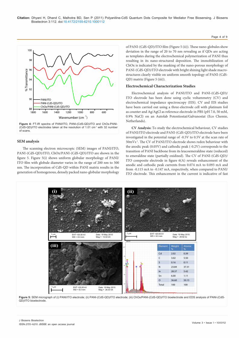
SEM analysis
The scanning electron microscopic (SEM) images of PANI/ITO, PANI-(CdS-QD)/ITO, ChOx/PANI-(CdS-QD)/ITO are shown in the figure 5. Figure 5(i) shows uniform globular morphology of PANI/ITO film with globule diameter varies in the range of 200 nm to 500 nm. The incorporation of CdS-QD within PANI matrix results in the generation of homogenous, densely packed nano-globular morphology
of PANI-(CdS-QD)/ITO film (Figure 5 (ii)). These nano-globules show deviation in the range of 20 to 70 nm revealing as if QDs are acting as templates during the electrochemical polymerization of PANI thus resulting in its nano-structured deposition. The immobilization of ChOx is indicated by the masking of the nano-porous morphology of PANI-(CdS-QD)/ITO electrode with bright shining light shade macro-structures clearly visible on uniform smooth topology of PANI-(CdS-QD) matrix (Figure 5 (iii)).
Electrochemical Characterization Studies
Electrochemical analysis of PANI/ITO and PANI-(CdS-QD)/ITO electrode has been done using cyclic voltammetry (CV) and electrochemical impedance spectroscopy (EIS). CV and EIS studies have been carried out using a three-electrode cell with platinum foil as counter and Ag/AgCl as reference electrode in PBS (pH 7.0, 50 mM, 0.9% NaCl) on an Autolab Potentiostat/Galvanostat (Eco Chemie, Netherlands).
CV Analysis: To study the electrochemical behaviour, CV studies of PANI/ITO electrode and PANI-(CdS-QD)/ITO electrode have been investigated in the potential range of -0.5V to 0.5V at the scan rate of 50mVs-1. The CV of PANI/ITO electrode shows redox behaviour with the anodic peak (0.05V) and cathodic peak (-0.2V) corresponds to the transition of PANI backbone from its leucoemeraldine state (reduced) to emeraldine state (partially oxidized). The CV of PANI-(CdS-QD)/ITO composite electrode in figure 6(A) reveals enhancement of the anodic and cathodic peak currents from 0.074 mA to 0.093 mA and from -0.115 mA to -0.147 mA, respectively, when compared to PANI/ITO electrode. This enhancement in the current is indicative of fast
Figure 4: FT-IR spectra of PANI/ITO, PANI-(CdS-QD)/ITO and ChOx-PANI-(CdS-QD)/ITO electrodes taken at the resolution of 1.01 cm-1 with 32 number of scans.
1800 1600 1400 1200 1000 800 60088
90
92
94
96
98
100
PANI/ITO PANI-(CdS-QD)/ITO ChOx/PANI-(CdS-QD)/ITO
% T
rans
mitt
ance
Wavenumber (cm -1)
Figure 5: SEM micrograph of (i) PANI/ITO electrode; (ii) PANI-(CdS-QD)/ITO electrode; (iii) ChOx/PANI-(CdS-QD)/ITO bioelectrode and EDS analysis of PANI-(CdS-QD)/ITO bioelectrode.
2 4 6 8 10 12 14keV
0
2
4
6
8
10
cps/eV
Cd Cd C
S
S N
In
In
Sn
Sn
O
(i) (ii)
(iii)
(ii)
(iii) Element Weight %
Atomic %
Cd 2.02 0.39
C 3.02 5.50
S 0.16 0.11
N 23.84 37.31
In 28.37 5.42
Sn 6.00 1.11
O 36.60 50.15
Total 100 100
(i) (ii)
(iii)
1 µm EHT =20.00 kVWD = 8.0 mm
EHT =20.00 kVWD = 8.5 mm
Date: 19 May 2010Mag = 14.02 kX
Date: 19 May 2010Mag = 28.82 kx
1 µm
1 µm EHT =20.00 kVWD = 8.0 mm
Date: 19 May 2010Mag = 28.03 kX

Citation: Dhyani H, Dhand C, Malhotra BD, Sen P (2011) Polyaniline-CdS Quantum Dots Composite for Mediator Free Biosensing. J Biosens Bioelectron 3:112. doi:10.4172/2155-6210.1000112
Page 5 of 9
Volume 3 • Issue 1 • 1000112J Biosens BioelectronISSN:2155-6210 JBSBE an open access journal
electron transport and increased charge transport in the parallel interface of electrolyte solution and PANI-(CdS-QD)/ITO electrode. This result can be attributed to the enhanced surface concentration of redox species (Γ) in the composite. To verify this, concentration of the redox species has been calculated using the Laviron’s equation [26] as given below
ln lnpaRT RTE E KnF nF
οο ν
α α = + −
(1)
According to the above equation, the value of 2.303RT/αnF, where α is the transfer coefficient, F is the Faraday constant (96485.34 C mol-
1), R is the gas constant (8.314 J mol-1 K-1), n is the number of electrons transferred and T is the temperature, for PANI/ITO and PANI-(CdS-QD)/ITO can be given by the slope of the plot of log scan rate (ν) versus Epa at different ν values.
For PANI/ITO and PANI-(CdS-QD)/ITO electrodes, the anodic peak potential (Epa) varies linearly with logarithm of ν (scan rate, data not shown) and follows the equation 2nd & 3rd, respectively.
Epa (PANI/ITO) = 0.092 log ν (Vs-1) + 0.0404; R = 0.92; SD = 0.0094 (2)
Epa (PANI-(CdS-QD)/ITO) = 0.1127 log ν (Vs-1) + 0.0348; R = 0.996; SD = 0.00372 (3)
Considering equation (1), (2) and (3), the value of 2.303RT/αnF for PANI/ITO and PANI-(CdS-QD)/ITO can be given as
2.303 0.19192RTnFα
= (For PANI/ITO) (4)
2.303 0.92RTnFα
= (For PANI-(CdS-QD)/ITO) (5)
The value of 2.303RT/αnF can be used to calculate the total surface concentration of the redox species (Γ) onto PANI/ITO and PANI-SWCNT/ITO electrode by using equation given below
2 2
4pn F AI
RTνΓ
= (6)
where Ip/ν can be calculated from the slop of Ip vs. ν plot [27].
The total surface concentration of redox species is found to be 3.211×10⁻⁵ mol m-2 for PANI-(CdS-QD)/ITO composite electrode, which is ~ 16 times to that of concentration for PANI/ITO electrode (2.047 × 10⁻⁶ mol m-2).
This increase in the redox species supports strong interactions between the negatively charged quantum structures of CdS with the aromatic structures of PANI that helps in uncoiling of the otherwise folded chains of PANI also supported by UV-V is spectroscopy. In the extended open conformation of PANI, it is likely that more number of exposed moieties are available for oxidation leading to enhanced faradic current. Figure 6 (B & C) shows CV of PANI/ITO and PANI-(CdS-QD)/ITO electrode recorded at different scan rates (40–110 mV
Figure 6: (A) Cyclic voltammogram of (i) PANI/ITO and (ii) PANI-(CdS-QD)/ITO electrode; (B & C) CV of PANI/ITO electrode and PANI-(CdS-QD)/ITO electrode in PBS buffer at the scan rate from 40 mVs-1 to 110 mVs-1. Inset of fig 6 (B & C) shows the variation of anodic peak current with respect to the square root of scan rate; (D) Electrochemical impedance spectra of (i) PANI/ITO (ii) PANI-(CdS-QD)/ITO electrode in PBS (50 mM, pH 7.0, 0.9% NaCl) solution.
-0.6 -0.4 -0.2 0.0 0.2 0.4 0.6
-0.15
-0.10
-0.05
0.00
0.05
0.10
(ii)(i)
Curr
ent (
mA)
Voltage (V)
0 3000 6000 9000 120000
2000
4000
6000
(ii)
(i)
-Z'' (
ohm
s)
Z' (ohms)
-0.6 -0.4 -0.2 0.0 0.2 0.4 0.6-0.25
-0.20
-0.15
-0.10
-0.05
0.00
0.05
0.10
0.15
R= 0.975
7.0 7.5 8.0 8.5 9.0 9.5 10.0 10.5 11.0
0.06
0.07
0.08
0.09
0.10
0.11
Curr
ent (
mA)
1/2 (mVs -1)
Curr
ent (
mA)
Voltage (V)
-0.6 -0.4 -0.2 0.0 0.2 0.4 0.6-0.30
-0.25
-0.20
-0.15
-0.10
-0.05
0.00
0.05
0.10
0.15
R= 0.99
7.0 7.5 8.0 8.5 9.0 9.5 10.0 10.5 11.00.08
0.09
0.10
0.11
0.12
0.13
Curr
ent (
mA)
1/2 (mVs -1)
Curr
ent (
mA)
Potential (V)
(A)(B)
(C) (D)

Citation: Dhyani H, Dhand C, Malhotra BD, Sen P (2011) Polyaniline-CdS Quantum Dots Composite for Mediator Free Biosensing. J Biosens Bioelectron 3:112. doi:10.4172/2155-6210.1000112
Page 6 of 9
Volume 3 • Issue 1 • 1000112J Biosens BioelectronISSN:2155-6210 JBSBE an open access journal
s-1). Careful examination shows that while moving towards higher scan rate, there is a shift of the anodic peak towards more positive peak potential and cathodic peak in the reverse direction in case of PANI/ITO electrode. However, negligible or no shift is observed in PANI-(CdS-QD)/ITO system which is one of the primary criteria for the system to be reversible. Besides this, anodic and cathodic peak currents (Ipa and Ipc) show improved linear behaviour with respect to square root of scan rate (ν1/2) in case of PANI-(CdS-QD) composite matrix (Regression coefficient (r) = 0.99) (inset of Figure 6 (B)) than that of pure PANI matrix (r= 0.975) (inset of Figure 6(C)) revealing that the electron-transfer processes are more diffusion controlled in case of PANI-(CdS-QD)/ITO electrode. The linear regression equations for PANI/ITO and PANI-(CdS-QD) electrode are given as Equation (7-10).
Ipa (PANI/ITO) = 0.014 ν1/2 (Vs-1) - 0.04; r = 0.975; SD = 0.0046 (7)
Ipc (PANI/ITO) = -0.00159 ν1/2 (Vs-1) + 0.0206; r = -0.985; SD = 0.00709 (8)Ipa (PANI-(CdS-QD)/ITO) = 0.012 ν1/2 (Vs-1) + 0.0198; r = 0.99; SD = 0.00224 (9)Ipc (PANI-(CdS-QD)/ITO) = 0.00166 ν1/2 (Vs-1) + 0.074; r = -0.996; SD = 0.0037 (10)
The above discussion indicates that the incorporation of CdS quantum dots not only enhances the charge transfer properties of PANI matrix but leads to overall improvement in the electrochemical behavior of the system.
Electrochemical impedance spectroscopy (EIS) studies: EIS studies have been done to further support the improved electrochemical response of PANI matrix on the incorporation of CdS-QDs. Figure 6 (D) shows that, in spite of similar shapes of the two Nyquist plots there is remarkable decrease in the diameter of the semicircle (indicative of charge transfer resistance) for the PANI-(CdS-QD) composite film. The smaller semicircle indicates that the charge transfer resistance (6.32 KΩ) for PANI-(CdS-QD) composite film is nearly half to that of the PANI film (12.6 KΩ). This enhanced charge transfer is likely to be due to the presence of hopping centres created by the semiconducting CdS-QD in the PANI matrix that helps in faster charge transfer via electrons from one polymer chain to the other and finally to the electrode (ITO) surface. Moreover, incorporation of QDs facilitate the open conformation of PANI resulting in easy charge transport.
Response studies using cyclic voltammetric technique
To show the potential application of observed enhanced
Figure 7: (A) CV recorded for ChOx/PANI-(CdS-QD)/ITO bioelectrode as a function of concentration of cholesterol (50 to 500 mg dL-1) in PBS (50 mM, pH 7.4, 0.9% NaCl) solution at the scan rate of 30 mV/S; (B) calibration plots derived from CV measurements at 0.03 V as a function of cholesterol concentration; (C) Hanes plot between the substrate concentration and substrate concentration/current value obtained from plot A.
0 50 100 150 200 250 300 350 400 4500.024
0.026
0.028
0.030
0.032
0.034
0.036
Curr
ent (
mA)
Cholesterol Conc (mgdL -1)-0.6 -0.4 -0.2 0.0 0.2 0.4 0.6
-0.04
-0.03
-0.02
-0.01
0.00
0.01
0.02
0.03
0.04
Curr
ent (
mA)
Potential (V)
50 mgdL -1
100 mgdL -1
200 mgdL -1
300 mgdL -1
500 mgdL -1
0 50 100 150 200 250 300 350 400 450
2.0x10 6
4.0x10 6
6.0x10 6
8.0x10 6
1.0x10 7
1.2x10 7
[S]/
Curr
ent
Cholesterol Concenteration [S]
(A) ( B)
(C)
Citation: Dhyani H, Dhand C, Malhotra BD, Sen P (2011) Polyaniline-CdS Quantum Dots Composite for Mediator Free Biosensing. J Biosens Bioelectron 3:112. doi:10.4172/2155-6210.1000112
Page 7 of 9
Volume 3 • Issue 1 • 1000112J Biosens BioelectronISSN:2155-6210 JBSBE an open access journal
electrochemical properties of the PANI-(CdS-QD) composite, amperometric response studies of fabricated ChOx/PANI-(CdS-QD)/ITO bioelectrode have been carried out using CV studies in PBS in the range from -0.5 to 0.5 V (Figure 7(A)). During the response measurement, an anodic peak is observed around 0.03 V in CV which shows continuous increase in its intensity with increasing cholesterol concentration. The anodic peak seen around 0.03 V corresponds to the oxidation of PANI integrated within the matrix, and its increase with increased cholesterol concentration suggests that ChOx gets electrically contacted by PANI-(CdS-QD) modified ITO electrode. It may be noted that no peak is observed relating to the oxidation of H2O2 in the range of 0.5 to 0.7 V indicating that there is direct transfer of electrons from the reduced ChOx enzyme to the PANI–(CdS-QD) matrix during the biochemical reaction. Figure 7(B) shows plot of anodic peak current at 0.03 V for ChOx/PANI-(CdS-QD)/ITO bioelectrode as a function of cholesterol concentration. It can be seen that ChOx/PANI-(CdS-QD)/ITO bioelectrode can detect cholesterol in the range of 50 to 500 mg dL-1 with detection limit of 47.9 mg dL-1 and response time of 20 s. The value of sensitivity calculated from linear region of the calibration curve is 2.63 × 10⁻⁵ mA mg-1 dL-1, with linear regression coefficient (r) as 0.995. The equation for the line of regression is given by:
Current (mA) = 0.0247mA + [2.63 × 10⁻⁵ (mAmg-1dL-1)] × [Cholesterol Conc (mgdL-1)] (11)
This high sensitivity of 2.63 × 10⁻⁵ mAmg-1dL-1 and low response time of 20 s reveals enhanced bioelectrocatalytic activity of the enzyme on PANI-(CdS-QD)/ITO electrode due to the enhanced charge transfer properties of the composite matrix.
The apparent Michaelis-Menten kinetic parameter ( appmK ) of the
enzymatic reaction, that determines the affinity of enzyme with the bio-analyte, has been estimated using Hanes plot i.e. a plot between analyte concentration [S] and [S]/Current (Figure 7(C)). The app
mK value for the immobilized enzyme has been found to be 0.82 mM. The observed lower value of app
mK shows enhanced affinity of the enzyme with the cholesterol on this nanocomposite matrix. Also, this PANI-(CdS-QD) matrix prepared by electrochemical technique may be helping the immobilized enzyme to achieve better conformation for faster enzymatic reaction resulting in enhancement of enzymatic activity.
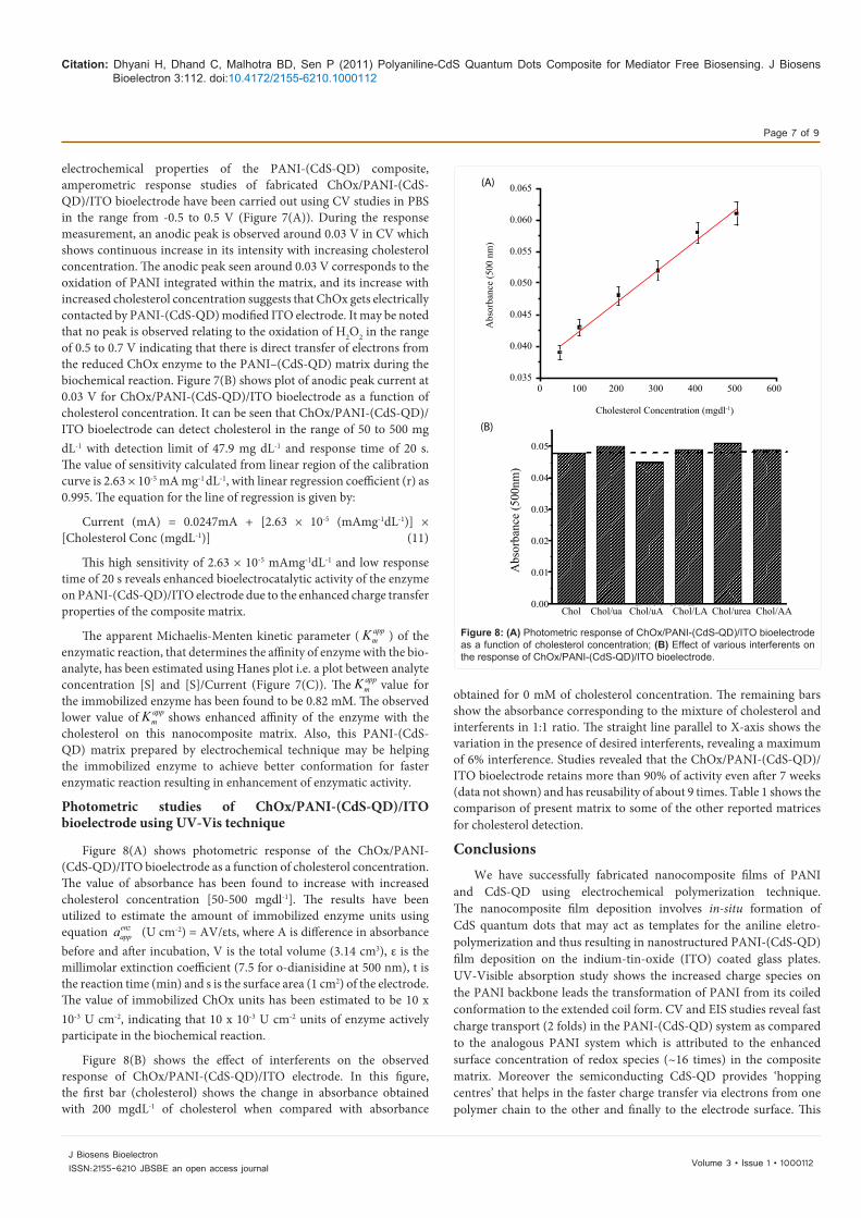
Photometric studies of ChOx/PANI-(CdS-QD)/ITO bioelectrode using UV-Vis technique
Figure 8(A) shows photometric response of the ChOx/PANI-(CdS-QD)/ITO bioelectrode as a function of cholesterol concentration. The value of absorbance has been found to increase with increased cholesterol concentration [50-500 mgdl-1]. The results have been utilized to estimate the amount of immobilized enzyme units using equation enz
appa (U cm-2) = AV/εts, where A is difference in absorbance before and after incubation, V is the total volume (3.14 cm3), ε is the millimolar extinction coefficient (7.5 for o-dianisidine at 500 nm), t is the reaction time (min) and s is the surface area (1 cm2) of the electrode. The value of immobilized ChOx units has been estimated to be 10 x 10-3 U cm-2, indicating that 10 x 10-3 U cm-2 units of enzyme actively participate in the biochemical reaction.
Figure 8(B) shows the effect of interferents on the observed response of ChOx/PANI-(CdS-QD)/ITO electrode. In this figure, the first bar (cholesterol) shows the change in absorbance obtained with 200 mgdL-1 of cholesterol when compared with absorbance
obtained for 0 mM of cholesterol concentration. The remaining bars show the absorbance corresponding to the mixture of cholesterol and interferents in 1:1 ratio. The straight line parallel to X-axis shows the variation in the presence of desired interferents, revealing a maximum of 6% interference. Studies revealed that the ChOx/PANI-(CdS-QD)/ITO bioelectrode retains more than 90% of activity even after 7 weeks (data not shown) and has reusability of about 9 times. Table 1 shows the comparison of present matrix to some of the other reported matrices for cholesterol detection.
ConclusionsWe have successfully fabricated nanocomposite films of PANI
and CdS-QD using electrochemical polymerization technique. The nanocomposite film deposition involves in-situ formation of CdS quantum dots that may act as templates for the aniline eletro-polymerization and thus resulting in nanostructured PANI-(CdS-QD) film deposition on the indium-tin-oxide (ITO) coated glass plates. UV-Visible absorption study shows the increased charge species on the PANI backbone leads the transformation of PANI from its coiled conformation to the extended coil form. CV and EIS studies reveal fast charge transport (2 folds) in the PANI-(CdS-QD) system as compared to the analogous PANI system which is attributed to the enhanced surface concentration of redox species (~16 times) in the composite matrix. Moreover the semiconducting CdS-QD provides ‘hopping centres’ that helps in the faster charge transfer via electrons from one polymer chain to the other and finally to the electrode surface. This
Figure 8: (A) Photometric response of ChOx/PANI-(CdS-QD)/ITO bioelectrode as a function of cholesterol concentration; (B) Effect of various interferents on the response of ChOx/PANI-(CdS-QD)/ITO bioelectrode.
0 100 200 300 400 500 600
Abs
orba
nce
(500
nm
)
0.065
0.060
0.055
0.050
0.045
0.040
0.035
Cholesterol Concentration (mgdl-1)
0.05
0.04
0.03
0.02
0.01
0.00
Abs
orba
nce
(500
nm)
Chol Chol/ua Chol/uA Chol/LA Chol/urea Chol/AA
(A)
(B)

Citation: Dhyani H, Dhand C, Malhotra BD, Sen P (2011) Polyaniline-CdS Quantum Dots Composite for Mediator Free Biosensing. J Biosens Bioelectron 3:112. doi:10.4172/2155-6210.1000112
Page 8 of 9
Volume 3 • Issue 1 • 1000112J Biosens BioelectronISSN:2155-6210 JBSBE an open access journal
improved electrochemical behavior has been used to enhance the bioelectrocatalytic activity of the enzymes to obtain better biosensing characteristics. The response studies, carried out using CV technique, reveal that this ChOx/PANI-(CdS-QD)/ITO bioelectrode can be used to detect cholesterol in the concentration range of 50 to 500 mgdL-1
with good detection limit (47.8 mgdL-1) and low appmK value (0.82
mM). This matrix can further be explored for the detection of other analytes (e.g. glucose, lactic acid, urea, antigens, antibodies etc.).
Acknowledgements
The authors thank Director NPL, New Delhi India for providing the facilities. We also thank Advanced instrumentation and research facility (AIRF), JNU, New Delhi for the TEM, XRD and SEM analysis. Hemant Dhyani & Chetna Dhand thank CSIR-India for award of Research Fellowships.
References
1. Malhotra BD, Chaubey A, Singh SP (2006) Prospects of conducting polymers in biosensors. Analytica Chimica Acta 578: 59-74.
2. Sai VVR, Mahajan S, Contractor AQ, Mukherji S (2006) Immobilization of antibodies on polyaniline films and its application in a piezoelectric immunosensor. Anal Chem 78: 8368-8373.
3. Dhand C, Arya SK, Datta M, Malhotra BD (2008) Polyaniline–carbon nanotube composite film for cholesterol biosensor. Anal Biochem 383: 194–199.
4. Resch-Genger U, Grabolle M, Cavaliere-Jaricot S, Nitschke R, Nann T (2008) Quantum dots versus organic dyes as fluorescent labels. Nature Methods 5: 763-775.
5. Palma RM, Manso M, Costa VT (2009) Optical Biosensors Based on Semiconductor Nanostructures. Sensors 9: 5149-5172.
6. Frasco MF, Chaniotakis N (2009) Semiconductor Quantum Dots in Chemical Sensors and Biosensors. Sensors 9: 7266-7286.
7. Liu Q, Lu X, Li L, Yao X, Li J (2007) Direct electrochemistry of glucose oxidase and electrochemical biosensing of glucose on quantum dots/carbon nanotubes electrodes. Biosens Bioelectron 22: 3203-3209.
8. Tang L, Zhu Y, Yang X, Sun J, Li C (2008) Self-assembled CNTs/CdS/dehydrogenase hybrid-based amperometric biosensor triggered by photovoltaic effect. Biosens Bioelectron 24: 319-323.
9. Yuan J, Guo W, Wang E (2008) Quantum dots–bienzyme hybrid system for the sensitive determination of glucose. Biosensors & Bioelectronics 23: 1567-1571.
10. Huang CP, Liu SW, Chen TM, Li YK (2008) A new approach for quantitative determination of glucose by using CdSe/ZnS quantum dots. Sensors and Actuators B: Chemical 130: 338-342.
11. Koneswaran M, Narayanaswamy R (2009) Mercaptoacetic acid capped CdS quantum dots as fluorescence single shot probe for mercury(II). Sensors and Actuators B 139: 91-96.
S. No. Matrix Linearity Sensitivity Respon-se Time Kmapp
(mM) Shelf Life Reference
1 Electrochemically deposited PANI 100-400 mg/dl 0.042 µAmg-1 dL-1
240s 1.94 6 weeks [28]
2 MWNTs 19.3-231.6 mg/dl 0.559µA cm–2 mM–1 - 7.71 - [29]
3MWCNT/Screen Printed
Carbon Electrode 99.5-399 mg/dl 0.0059 mAmg-1 dL-1- - 9 weeks [30]
4 Gold electrode 0-80 mg/dl 0.13 µA/mM 15 s 2.94 - [31]
5 ZnO Nanoparticles 0.001 – 0.5µM 23.7µA/µM - 4.7 - [32]
6 Chitosan-SnO2 nanocomposite 0.26-10.36 mM 34.7 µAmg-1 dL-1cm–2- 3.8 12 weeks [33]
7 PANI-(CdS-QD)Nanocomposite 50-500 2.63 × 10⁻⁵ mA mg-1 dL-1 20s 0.82 7 weeks Present Work
Table 1: Characteristics of PANI-(CdS-QD) electrode along with those reported in literature for cholesterol detection.
12. Falcaro P, Hill AJ, Nairn KM, Jasieniak J, Mardel JI, et al. (2011) A new method to position and functionalize metal-organic framework crystals. Nat Commun 2: 237.
13. Huang Y, Zhang W, Xiao H, Li G (2005) An electrochemical investigation of glucose oxidase at a CdS nanoparticles modified electrode. Biosensors & Bioelectronics 21: 817-821.
14. Hickey SG, Riley DJ (1999) Distance Dependent Electron Transfer in Gold/Spacer/Q-CdSe-Assemblies. J Phys Chem B 103: 4599-4602.
15. Li Z, Du Y (2003) Biomimic synthesis of CdS nanoparticles with enhanced luminescence. Materials Letters 57: 2480-2484.
16. Gao Y, Tonizzo A, Walser A, Potasek M, Dorsinville R (2008) Enhanced optical nonlinearity of surfactant-capped CdS quantum dots embedded in an optically transparent polystyrene thin film. Appl Phys Lett 92: 033106.
17. Seoudi R, Kamal M, Shabaka AA, Abdelrazek EM, Eisa W (2010) Synthesis, characterization and spectroscopic studies of CdS/polyaniline core/shell nanocomposite. Synthetic Metals 160: 479-484.
18. Khiew PS, Huang NM, Radiman S, Ahmad MS (2004) Synthesis and characterization of conducting polyaniline-coated cadmium sulphide nanocomposites in reverse microemulsion. Materials Letters 58: 516- 521.
19. Kulkarni MV, Viswanath AK, Marimuthu R, Seth T (2004) Synthesis and characterization of polyaniline doped with organic acids. J Polym Sci A Polym Chem 42: 2043–2049.
20. Versiane O, Rodrigueb BL, Ramos JM, Téllez CA, Felcman J (2006) Synthesis, molecular structure and vibrational spectra of a dimeric complex formed by cobalt and glycine. Spectrochim Acta A 65: 1112-1119.
21. Singh N, Kulkarni MV, Lonkar SP, Viswanath AK, Khanna PK (2007) CdS/Polyaniline Nanocomposites: Synthesis and Characterization. Synthesis and Reactivity in Inorganic, Metal-Organic, and Nano-Metal Chemistry 37: 153-159.
22. Chandrakanthi RLN, Careem MA (2002) Preparation and characterization of CdS and Cu2S nanoparticle/polyaniline composite films. Thin Solid Films 417: 51-56.
23. Godovsky DY, Varfolomeev AE, Zaretsky DF, Chandrakanthi RLN, Ku¨ndig A, et al. (2001) Preparation of nanocomposites of polyaniline and inorganic semiconductors. J Mater Chem 11: 2465-2469.
24. Khanna PK, Kulkarni MV, Singh N, Lonkar SP, Subbarao VVVS, et al. (2006) Synthesis of HCl doped polyaniline–CdS nanocomposite by use of organometallic cadmium precursor. Materials Chemistry and Physics 95: 24-28.
25. Nakanishi T, Ohtani B, Uosaki K (1998) Distance Dependent Electron Transfer in Gold/Spacer/Q-CdSe-Assemblies. J Phys Chem B 102: 1571-1577.
26. Zhuang Q, Chen J, Chen J, Lin X (2008) Electrocatalytical properties of bergenin on a multi-wall carbon nanotubes modified carbon paste electrode and its determination in tablets. Sensors and Actuators B 128: 500-506.
27. Hu G, Zhang D, Wu W, Yang Z (2008) Selective determination of dopamine in the presence of high concentration of ascorbic acid using nano-Au self-assembly glassy carbon electrode. Colloids Surf B Biointerf 62: 199-205.

Citation: Dhyani H, Dhand C, Malhotra BD, Sen P (2011) Polyaniline-CdS Quantum Dots Composite for Mediator Free Biosensing. J Biosens Bioelectron 3:112. doi:10.4172/2155-6210.1000112
Page 9 of 9
Volume 3 • Issue 1 • 1000112J Biosens BioelectronISSN:2155-6210 JBSBE an open access journal
28. Singh S, Solanki PR, Pandey MK, Malhotra BD (2006) Cholesterol biosensor based on cholesterol esterase, cholesterol oxidase and peroxidase immobilized onto conducting polyaniline films. Sens Actuators B 115: 534-541.
29. Guo M, Chen J, Li J, Nie L, Yao S (2004) Carbon Nanotubes-Based Amperometric Cholesterol Biosensor Fabricated Through Layer-by-Layer Technique. Electroanalysis 16: 1992-1998.
30. Li G, Liao JM, Hu GQ, Ma NZ, Wu PJ (2005) Study of carbon nanotube modified biosensor for monitoring total cholesterol in blood. Biosens Bioelectron 20: 2140-2144.
31. Parra A, Casero E, Pariente F, Vazquez L, Lorenzo E (2007) Sens Actuators B 124: 30-37.
32. Umar A, Rahman MM, Vaseem M, Hahn YB (2008) Ultra-sensitive cholesterol biosensor based on low-temperature grown ZnO nanoparticles. Electrochem Commun 11: 118-121.
33. Ansari AA, Kaushik A, Solanki PR, Malhotra BD (2009) Electrochemical Cholesterol Sensor Based on Tin Oxide-Chitosan Nanobiocomposite Film. Electroanalysis 21: 965-972.
Submit your next manuscript and get advantages of OMICS Group submissionsUnique features:
• Userfriendly/feasiblewebsite-translationofyourpaperto50world’sleadinglanguages• AudioVersionofpublishedpaper• Digitalarticlestoshareandexplore
Special features:
• 200OpenAccessJournals• 15,000editorialteam• 21daysrapidreviewprocess• Qualityandquickeditorial,reviewandpublicationprocessing• IndexingatPubMed(partial),Scopus,DOAJ,EBSCO,IndexCopernicusandGoogleScholaretc• SharingOption:SocialNetworkingEnabled• Authors,ReviewersandEditorsrewardedwithonlineScientificCredits• Betterdiscountforyoursubsequentarticles
Submityourmanuscriptat:http://www.omicsonline.org/submission