Low-Potential Photoelectrochemical Biosensing Using...
6
Low-Potential Photoelectrochemical Biosensing Using Porphyrin-Functionalized TiO 2 Nanoparticles Wenwen Tu, Yitong Dong, Jianping Lei,* and Huangxian Ju* Key Laboratory of Analytical Chemistry for Life Science (Ministry of Education of China), Department of Chemistry, Nanjing University, Nanjing 210093, P. R. China A novel photoelectrochemical biosensing platform for the detection of biomolecules at relatively low applied poten- tials was constructed using porphyrin-functionalized TiO 2 nanoparticles. The functional TiO 2 nanoparticles were prepared by dentate binding of TiO 2 with sulfonic groups of water-soluble [meso-tetrakis(4-sulfonatophe- nyl)porphyrin] iron(III) monochloride (FeTPPS) and characterized by transmission electron microscopy; contact angle measurement; and Raman, X-ray photo- electron, and ultraviolet-visible absorption spec- troscopies. The functional nanoparticles showed good dispersion in water and on indium tin oxide (ITO) surface. The resulting FeTPPS-TiO 2 -modified ITO elec- trode showed a photocurrent response at +0.2 V to a light excitation at 380 nm, which could be further sensitized through an oxidation process of biomol- ecules by the hole-injected FeTPPS. Using glutathione as a model, a methodology for sensitive photoelectro- chemical biosensing at low potential was thus devel- oped. Under optimal conditions, the proposed photo- electrochemical method could detect glutathione ranging from 0.05 to 2.4 mmol L -1 with a detection limit of 0.03 mmol L -1 at a signal-to-noise ratio of 3. The photoelectrochemical biosensor had an excellent speci- ficity against anticancer drugs and could be success- fully applied to the detection of reduced glutathione in gluthion injection, showing a promising application in photoelectrochemical biosensing. Photoelectrochemical measurement is a newly developed technique for the detection of biomolecules. 1,2 Coupling photoir- radiation with electrochemical detection, photoelectrochemical sensors have the advantages of both optical methods and electrochemical sensors. 3,4 Thus, this technique shows promising analytical applications and has attracted considerable interest. Some metal oxide semiconductor nanoparticles such as ZnO, ZrO 2 , and TiO 2 have been used as significant photoelectrochemical materials. Among them, TiO 2 nanoparticles have been exten- sively investigated for various photoelectrochemical applications because of their wide band gap, high photoelectrochemical activity under UV irradiation, and strong oxidizing power when illuminated. 1 The photoelectrochemical response from il- luminated TiO 2 has been used for the design of photoelectro- chemical sensors. 5,6 The integration of Au-doped TiO 2 nanotubes with acetylcholinesterase has led to a rapid and valid photo- electrochemical approach for the determination of acetylcho- linesterase inhibition with illumination at 253.7 nm. 7 A dopamine- coordinated photoactive TiO 2 nanoporous film has also been used successfully for the sensitive detection of dihydronicoti- namide adenine dinucleotide. 8 Recently, porphyrins, an impor- tant class of conjugated organic molecules for light harvest- ing, 9-11 have been widely used for improving the photo-current conversion efficiency of TiO 2 nanoparticles in molecular elec- tronics and photoelectrochemical devices, especially for solar cells. 12-20 However, the improved photoelectrochemical efficiency has not yet been applied to the construction of a photoelectro- chemical biosensing platform. Porphyrin molecules exhibit ultrafast electron injection, slow charge-recombination kinetics, high absorption coefficients, and * Corresponding authors. Phone and Fax: +86-25-83593593. E-mail: hxju@ nju.edu.cn (H.J.), [email protected]. (1) Wang, G.-L.; Xu, J.-J.; Chen, H.-Y.; Fu, S.-Z. Biosens. Bioelectron. 2009, 25, 791–796. (2) Wang, G.-L.; Yu, P.-P.; Xu, J.-J.; Chen, H.-Y. J. Phys. Chem. C 2009, 113, 11142–11148. (3) Haddour, N.; Chauvin, J.; Gondran, C.; Cosnier, S. J. Am. Chem. Soc. 2006, 128, 9693–9698. (4) Ikeda, A.; Nakasu, M.; Ogasawara, S.; Nakanishi, H.; Nakamura, M.; Kikuchi, J. Org. Lett. 2009, 11, 1163–1166. (5) Zhao, H. J.; Jiang, D. L.; Zhang, S. Q.; Catterall, K.; John, R. Anal. Chem. 2004, 76, 155–160. (6) Zhou, H.; Gan, X.; Wang, J.; Zhu, X. L.; Li, G. X. Anal. Chem. 2005, 77, 6102–6104. (7) Zhu, W.; An, Y.-R.; Luo, X.-M.; Wang, F.; Zheng, J.-H.; Tang, L.-L.; Wang, Q.-J.; Zhang, Z.-H.; Zhang, W.; Jin., L.-T. Chem. Commun. 2009, 2682– 2684. (8) Wang, G.-L.; Xu, J.-J.; Chen, H.-Y. Biosens. Bioelectron. 2009, 24, 2494– 2498. (9) Sgobba, V.; Guldi, D. M. Chem. Soc. Rev. 2009, 38, 165–184. (10) Baskaran, D.; Mays, J. W.; Zhang, X. P.; Bratcher, M. S. J. Am. Chem. Soc. 2005, 127, 6916–6917. (11) Guldi, D. M.; Rahman, G. M. A.; Zerbetto, F.; Prato, M. Acc. Chem. Res. 2005, 38, 871–878. (12) Rochford, J.; Chu, D.; Hagfeldt, A.; Galoppini, E. J. Am. Chem. Soc. 2007, 129, 4655–4665. (13) Mozer, A. J.; Wagner, P.; Officer, D. L.; Wallace, G. G.; Campbell, W. M.; Miyashita, M.; Sunahara, K.; Mori, S. Chem. Commun. 2008, 4741–4743. (14) Tanaka, M.; Hayashi, S.; Eu, S.; Umeyama, T.; Matano, Y.; Imahori, H. Chem. Commun. 2007, 2069–2071. (15) Kathiravan, A.; Kumar, P. S.; Renganathan, R.; Anandan, S. Colloids Surf. A: Physicochem. Eng. Aspects 2009, 333, 175–181. (16) Eu, S.; Hayashi, S.; Umeyama, T.; Oguro, A.; Kawasaki, M.; Kadota, N.; Matano, Y.; Imahori, H. J. Phys. Chem. C 2007, 111, 3528–3537. (17) Ikeda, A.; Tsuchiya, Y.; Konishi, T.; Ogasawara, S.; Kikuchi, J. Chem. Mater. 2005, 17, 4018–4022. (18) Hayashi, S.; Tanaka, M.; Hayashi, H.; Eu, S.; Umeyama, T.; Matano, Y.; Araki, Y.; Imahori, H. J. Phys. Chem. C 2008, 112, 15576–15585. (19) Rochford, J.; Galoppini, E. Langmuir 2008, 24, 5366–5374. (20) Yu, J. H.; Chen, J. R.; Wang, X. S.; Zhang, B. W.; Cao, Y. Chem. Commun. 2003, 1856–1857. Anal. Chem. 2010, 82, 8711–8716 10.1021/ac102070f 2010 American Chemical Society 8711 Analytical Chemistry, Vol. 82, No. 20, October 15, 2010 Published on Web 09/21/2010
Transcript of Low-Potential Photoelectrochemical Biosensing Using...

Low-Potential Photoelectrochemical BiosensingUsing Porphyrin-Functionalized TiO2 Nanoparticles
Wenwen Tu, Yitong Dong, Jianping Lei,* and Huangxian Ju*
Key Laboratory of Analytical Chemistry for Life Science (Ministry of Education of China), Department of Chemistry,Nanjing University, Nanjing 210093, P. R. China
A novel photoelectrochemical biosensing platform for thedetection of biomolecules at relatively low applied poten-tials was constructed using porphyrin-functionalized TiO2
nanoparticles. The functional TiO2 nanoparticles wereprepared by dentate binding of TiO2 with sulfonicgroups of water-soluble [meso-tetrakis(4-sulfonatophe-nyl)porphyrin] iron(III) monochloride (FeTPPS) andcharacterized by transmission electron microscopy;contact angle measurement; and Raman, X-ray photo-electron, and ultraviolet-visible absorption spec-troscopies. The functional nanoparticles showed gooddispersion in water and on indium tin oxide (ITO)surface. The resulting FeTPPS-TiO2-modified ITO elec-trode showed a photocurrent response at +0.2 V to alight excitation at 380 nm, which could be furthersensitized through an oxidation process of biomol-ecules by the hole-injected FeTPPS. Using glutathioneas a model, a methodology for sensitive photoelectro-chemical biosensing at low potential was thus devel-oped. Under optimal conditions, the proposed photo-electrochemical method could detect glutathione rangingfrom 0.05 to 2.4 mmol L-1 with a detection limit of0.03 mmol L-1 at a signal-to-noise ratio of 3. Thephotoelectrochemical biosensor had an excellent speci-ficity against anticancer drugs and could be success-fully applied to the detection of reduced glutathionein gluthion injection, showing a promising applicationin photoelectrochemical biosensing.
Photoelectrochemical measurement is a newly developedtechnique for the detection of biomolecules.1,2 Coupling photoir-radiation with electrochemical detection, photoelectrochemicalsensors have the advantages of both optical methods andelectrochemical sensors.3,4 Thus, this technique shows promisinganalytical applications and has attracted considerable interest.Some metal oxide semiconductor nanoparticles such as ZnO, ZrO2,and TiO2 have been used as significant photoelectrochemical
materials. Among them, TiO2 nanoparticles have been exten-sively investigated for various photoelectrochemical applicationsbecause of their wide band gap, high photoelectrochemicalactivity under UV irradiation, and strong oxidizing power whenilluminated.1 The photoelectrochemical response from il-luminated TiO2 has been used for the design of photoelectro-chemical sensors.5,6 The integration of Au-doped TiO2 nanotubeswith acetylcholinesterase has led to a rapid and valid photo-electrochemical approach for the determination of acetylcho-linesterase inhibition with illumination at 253.7 nm.7 A dopamine-coordinated photoactive TiO2 nanoporous film has also beenused successfully for the sensitive detection of dihydronicoti-namide adenine dinucleotide.8 Recently, porphyrins, an impor-tant class of conjugated organic molecules for light harvest-ing,9-11 have been widely used for improving the photo-currentconversion efficiency of TiO2 nanoparticles in molecular elec-tronics and photoelectrochemical devices, especially for solarcells.12-20 However, the improved photoelectrochemical efficiencyhas not yet been applied to the construction of a photoelectro-chemical biosensing platform.
Porphyrin molecules exhibit ultrafast electron injection, slowcharge-recombination kinetics, high absorption coefficients, and
* Corresponding authors. Phone and Fax: +86-25-83593593. E-mail: [email protected] (H.J.), [email protected].
(1) Wang, G.-L.; Xu, J.-J.; Chen, H.-Y.; Fu, S.-Z. Biosens. Bioelectron. 2009,25, 791–796.
(2) Wang, G.-L.; Yu, P.-P.; Xu, J.-J.; Chen, H.-Y. J. Phys. Chem. C 2009, 113,11142–11148.
(3) Haddour, N.; Chauvin, J.; Gondran, C.; Cosnier, S. J. Am. Chem. Soc. 2006,128, 9693–9698.
(4) Ikeda, A.; Nakasu, M.; Ogasawara, S.; Nakanishi, H.; Nakamura, M.;Kikuchi, J. Org. Lett. 2009, 11, 1163–1166.
(5) Zhao, H. J.; Jiang, D. L.; Zhang, S. Q.; Catterall, K.; John, R. Anal. Chem.2004, 76, 155–160.
(6) Zhou, H.; Gan, X.; Wang, J.; Zhu, X. L.; Li, G. X. Anal. Chem. 2005, 77,6102–6104.
(7) Zhu, W.; An, Y.-R.; Luo, X.-M.; Wang, F.; Zheng, J.-H.; Tang, L.-L.; Wang,Q.-J.; Zhang, Z.-H.; Zhang, W.; Jin., L.-T. Chem. Commun. 2009, 2682–2684.
(8) Wang, G.-L.; Xu, J.-J.; Chen, H.-Y. Biosens. Bioelectron. 2009, 24, 2494–2498.
(9) Sgobba, V.; Guldi, D. M. Chem. Soc. Rev. 2009, 38, 165–184.(10) Baskaran, D.; Mays, J. W.; Zhang, X. P.; Bratcher, M. S. J. Am. Chem. Soc.
2005, 127, 6916–6917.(11) Guldi, D. M.; Rahman, G. M. A.; Zerbetto, F.; Prato, M. Acc. Chem. Res.
2005, 38, 871–878.(12) Rochford, J.; Chu, D.; Hagfeldt, A.; Galoppini, E. J. Am. Chem. Soc. 2007,
129, 4655–4665.(13) Mozer, A. J.; Wagner, P.; Officer, D. L.; Wallace, G. G.; Campbell, W. M.;
Miyashita, M.; Sunahara, K.; Mori, S. Chem. Commun. 2008, 4741–4743.(14) Tanaka, M.; Hayashi, S.; Eu, S.; Umeyama, T.; Matano, Y.; Imahori, H. Chem.
Commun. 2007, 2069–2071.(15) Kathiravan, A.; Kumar, P. S.; Renganathan, R.; Anandan, S. Colloids Surf.
A: Physicochem. Eng. Aspects 2009, 333, 175–181.(16) Eu, S.; Hayashi, S.; Umeyama, T.; Oguro, A.; Kawasaki, M.; Kadota, N.;
Matano, Y.; Imahori, H. J. Phys. Chem. C 2007, 111, 3528–3537.(17) Ikeda, A.; Tsuchiya, Y.; Konishi, T.; Ogasawara, S.; Kikuchi, J. Chem. Mater.
2005, 17, 4018–4022.(18) Hayashi, S.; Tanaka, M.; Hayashi, H.; Eu, S.; Umeyama, T.; Matano, Y.;
Araki, Y.; Imahori, H. J. Phys. Chem. C 2008, 112, 15576–15585.(19) Rochford, J.; Galoppini, E. Langmuir 2008, 24, 5366–5374.(20) Yu, J. H.; Chen, J. R.; Wang, X. S.; Zhang, B. W.; Cao, Y. Chem. Commun.
2003, 1856–1857.
Anal. Chem. 2010, 82, 8711–8716
10.1021/ac102070f 2010 American Chemical Society 8711Analytical Chemistry, Vol. 82, No. 20, October 15, 2010Published on Web 09/21/2010

good chemical stability. The carboxylic or sulfonic groups on someporphyrins can spontaneously bind to TiO2 nanoparticles bybridging ester-like or bidentate interactions.12-16,18,21 Theresulting hybrid of porphyrin-TiO2 nanoparticles shows reducedelectron lifetime and, consequently, lower photoinduced elec-tron density under illumination compared to commonly usedruthenium-dye- (N719-) sensitized solar cells,13 and they canimprove by a factor of 2 the power conversion efficiency ofdye-sensitized TiO2 solar cells.14 The photoelectrochemicalbehavior of para- and meta-substitued porphyrin sensitizerssuggests that both the binding geometry and the distance ofthe sensitizer from the metal oxide surface dramaticallyinfluence the sensitizing effciency.12 A porphyrinatozinc(II)-(Zn5S-) sensitized TiO2 solar cell can produce a maximumincident photocurrent efficiency of 65% and a maximum powerconversion efficiency of 3.1%.16 Furthermore, a strong elec-tronic coupling between porphyrin and the TiO2 surface inporphyrin-sensitized cells has been observed because of theefficient electron injection from the porphyrin excited singlestate to the conduction band of the illuminated TiO2.18 Basedon these advantages of porphyrin-functionalized TiO2 nanopar-ticles in photoelectrochemistry, this work involved the synthe-sis of novel functional TiO2 nanoparticles using water-soluble[meso-tetrakis(4-sulfonatophenyl) porphyrin] iron(III) monochlo-ride (FeTPPS) through the dentate binding of TiO2 nanopar-ticles with sulfonic groups of FeTPPS. The nanoparticlesshowed a stable photoelectrochemical response that could befurther sensitized through an electron-transfer process frombiomolecule to porphyrin and then to the illuminated TiO2,leading to a novel application of porphyrin-functionalized TiO2
nanoparticles as a photoelectrochemical biosensing platform.Using glutathione (GSH) as a model, a novel method for thedetection of GSH was thus developed.
GSH, as the most abundant cellular thiol, plays an importantrole in many biological functions involving antioxidant defense,signal transduction, and cell proliferation.22 Its level is directlyrelated to many diseases such as cancers, AIDs, neurodegenera-tive disease, and Alzheimer’s and Parkinson’s diseases.23-25
Therefore, the monitoring of GSH in physiological media hasgained considerable attention. Several strategies, including mi-crochip electrophoresis-laser induced fluorescence,26 electrogen-erated chemiluminescence of quantum dots,27 absorption28 andfluorescent spectroscopies,29-33 surface-enhanced Raman scat-tering,34 and laser desorption/ionization mass spectrometry,35
have been proposed for this purpose. Several electrochemicalmethods have also been developed for in situ or online monitoring
of GSH.36-39 However, the high overpotential for the oxidationof GSH limits the application of these electrochemical methods.38,39
In this work, the photoelectrochemical response of FeTPPS-TiO2
could be produced at +0.2 V, at which the sensitizing effect ofGSH could be observed, leading to the low-potential biosensingof GSH. The proposed photoelectrochemical biosensor showedgood performance in the monitoring of GSH with a rapidresponse, wide concentration range, low applied potential, andgood selectivity and could successfully be applied to thedetection of reduced glutathione in gluthion injection (Scheme1). The porphyrin-functionalized TiO2 nanoparticles provide arobust approach for the photoelectrochemical detection ofbiomolecules.
EXPERIMENTAL SECTIONMaterials and Reagents. FeTPPS was a gift from Professor
Osamu Ikeda at Kanazawa University (Kanazawa, Japan). TiO2
nanopowder (anatase, <25 nm, 99.7%) and reduced L-glutathione(g99%) were purchased from Sigma-Aldrich (St. Louis, MO).All other chemicals were of analytical grade. In this work, 0.1mol L-1 phosphate buffer solution (PBS) was always employedas the supporting electrolyte after being deaerated with high-purity nitrogen. Reduced glutathione sodium for injection wasproduced by Pharminvest SPA (Milano, Italy). Aqueous solu-tions were prepared with twice-distilled water, and the pH valueof PBS was 7.0 unless indicated otherwise.
Apparatus. Transmission electron microscopy (TEM) wasperformed using a JEM-2100 transmission electron microscope
(21) Tu, W. W.; Lei, J. P.; Ding, L.; Ju, H. X. Chem. Commun. 2009, 4227–4229.
(22) Franco, R.; Panayiotidis, M. I.; Cidlowski, J. A. J. Biol. Chem. 2007, 282,30452–30465.
(23) Wu, G. Y.; Fang, Y.-Z.; Yang, S.; Lupton, J. R.; Turner, N. D. J. Nutr. 2004,134, 489–492.
(24) Roederer, M.; Staal, F. J.; Anderson, M.; Rabin, R.; Raju, P. A.; Herzenberg,L. A.; Herzenberg, L. A. Ann. N.Y. Acad. Sci. 1993, 667, 113–125.
(25) Bains, J. S.; Shaw, C. A. Brain Res. Rev. 1997, 25, 335–358.(26) Chen, Z. Z.; Li, Q. L.; Wang, X.; Wang, Z. Y.; Zhang, R. R.; Yin, M.; Yin,
L. L.; Xu, K. H.; Tang, B. Anal. Chem. 2010, 82, 2006–2012.(27) Wang, Y.; Lu, J.; Tang, L. H.; Chang, H. X.; Li, J. H. Anal. Chem. 2009, 81,
9710–9715.(28) Sudeep, P. K.; Joseph, S. T. S.; Thomas, K. G. J. Am. Chem. Soc. 2005,
127, 6516–6517.
(29) Ahn, Y.-H.; Lee, J.-S.; Chang, Y.-T. J. Am. Chem. Soc. 2007, 129, 4510–4511.
(30) Zhang, Y.; Li, Y.; Yan, X.-P. Anal. Chem. 2009, 81, 5001–5007.(31) Yao, Z. Y.; Feng, X. L.; Li, C.; Shi, G. Q. Chem. Commun. 2009, 5886–
5888.(32) Liu, J. F.; Bao, C. Y.; Zhong, X. H.; Zhao, C. C.; Zhu, L. Y. Chem. Commun.
2010, 2971–2973.(33) Raththagala, M.; Root, P. D.; Spence, D. M. Anal. Chem. 2006, 78, 8556–
8560.(34) Huang, G. G.; Han, X. X.; Hossain, M. K.; Ozaki, Y. Anal. Chem. 2009, 81,
5881–5888.(35) Huang, Y.-F.; Chang, H.-T. Anal. Chem. 2007, 79, 4852–4859.(36) Safavi, A.; Maleki, N.; Farjami, E.; Mahyari, F. A. Anal. Chem. 2009, 81,
7538–7543.(37) Pacsial-Ong, E. J.; McCarley, R. L.; Wang, W. H.; Strongin, R. M. Anal.
Chem. 2006, 78, 7577–7581.(38) Pournaghi-Azar, M. H.; Ahour, F. J. Electroanal. Chem. 2008, 622, 22–28.(39) Kumar, S. M. S.; Pillai, K. C. Electrochim. Acta 2009, 54, 7374–7381.
Scheme 1. Schematic Illustration ofPhotoelectrochemical Process for Oxidation of GSH atFeTPPS-TiO2-Modified ITO Electrode
8712 Analytical Chemistry, Vol. 82, No. 20, October 15, 2010

(JEOL, Japan). Scanning electron microscopy (SEM) was per-formed using a Hitachi S-4800 scanning electron microscope(Hitachi, Tokyo, Japan). Resonance Raman spectra were recordedon a Renishaw-inVia Raman microscope (Renishaw, Gloucester-shire, U.K.). X-ray photoelectron spectroscopy (XPS) measure-ments were performed using an ESCALAB 250 spectrometer(Thermo-VG Scientific, Waltham, MA) with ultrahigh vacuumgenerators. Ultraviolet-visible absorption spectra were recordedwith a Lambda 35 UV/vis spectrometer (Perkin-Elmer Instru-ments, Wellesley, MA). Static water contact angles were measuredwith a contact angle meter (Rame-Hart-100, Rame-Hart, Netcong,NJ) using droplets of deionized water at 25 °C. Photoelectro-chemical measurements were performed with a home-built pho-toelectrochemical system. A 500-W Xe lamp equipped with amonochromator was used as the irradiation source. Photocurrentwas measured on a CHI 660D electrochemical workstation (CHInstruments, Austin, TX). All experiments were carried out atroom temperature using a conventional three-electrode systemwith a modified indium tin oxide (ITO) electrode as the workingelectrode, a platinum wire as the auxiliary electrode, and asaturated calomel electrode as the reference electrode.
Preparation of FeTPPS-TiO2 and Modified ITO Elec-trodes. TiO2 (100 mg) was calcined in a muffle furnace at 450°C for 30 min. After being allowed to cool to 50-80 °C, thecalcined TiO2 was added to 5 mL of FeTPPS solution (1.5 mgmL-1). The suspension was sonicated for 30 min and kept atroom temperature for 24 h. Then, the resulting suspension wascentrifuged at 8000 rpm for 15 min to remove free FeTPPS.After being washed twice with twice-distilled water, the sedi-ment was dried at 70 °C to obtain FeTPPS-TiO2 nanoparticles.
After an ITO electrode had been cleaned with NaOH (1 molL-1) and H2O2 (30%), washed with acetone and twice-distilledwater, and dried at room temperature, 10 µL of the FeTPPS-TiO2 suspension (1 mg mL-1) was coated onto the ITOelectrode and dried at room temperature to obtain an FeTPPS-TiO2-modified ITO electrode. A TiO2-modified ITO electrodewas prepared similarly.
RESULTS AND DISCUSSIONCharacterization of FeTPPS-TiO2. The morphologies of TiO2
and FeTPPS-TiO2 were observed by transmission electronmicroscopy. Compared with the aggregation of TiO2 (Figure1A), the TEM image of FeTPPS-TiO2 in Figure 1B shows a morediscernible and better dispersion, which is advantageous for theformation of a robust homogeneous film for the construction of abiosensor. The average diameter of the FeTPPS-TiO2 nanopar-ticles was estimated to be 15 nm. The thickness of the FeTPPS-TiO2 film on an ITO surface was estimated by SEM to be 0.85µm (Figure 1C). The homogeneous film was beneficial forphotoelectron transfer between FeTPPS-TiO2 and the electrode.
The Raman spectrum of TiO2 nanoparticles (Figure 2A, curvea) shows the characteristics of the anatase nanocrystallinestructure at 393, 513, and 636 cm-1, whereas that of FeTPPS(Figure 2A, curve b) does not show any obvious Raman peakbetween 300 and 740 cm-1. After FeTPPS had been bonded toTiO2, these characteristic bands of TiO2 shifted slightly to 395,514, and 638 cm-1 with strengths enhanced by 27.5%, 25.1%,and 33.0%, respectively (Figure 2A, curve c), which can beattributed to the charge-transfer mechanism of FeTPPS-TiO2
related to the surface-state energy levels of the TiO2 semicon-ductor.40 In addition, no nanocrystalline structure of TiO2 otherthan anatase was detected by Raman spectroscopic measure-ments.41 This fact is of great importance for photocurrentgeneration in photovoltaic applications, because the anatasephase of TiO2 has a larger surface area and faster electrontransport than the rutile phase.42 Moreover, after FeTPPS hadbeen assembled on the surface of TiO2, the TiO2 anatase
(40) Yang, L. B.; Jiang, X.; Ruan, W. D.; Zhao, B.; Xu, W. Q.; Lombardi, J. R. J.Phys. Chem. C 2008, 112, 20095–20098.
(41) Imahori, H.; Hayashi, S.; Umeyama, T.; Eu, S.; Oguro, A.; Kang, S.; Matano,Y.; Shishido, T.; Ngamsinlapasathian, S.; Yoshikawa, S. Langmuir 2006,22, 11405–11411.
(42) Park, N.-G.; Lagemaat, J.; Frank, A. J. J. Phys. Chem. B 2000, 104, 8989–8994.
Figure 1. TEM images of (A) TiO2 and (B) FeTPPS-TiO2 nanopar-ticle suspensions at 1 mg mL-1 and (C) SEM section image ofFeTPPS-TiO2-modified ITO electrode.
Figure 2. (A) Raman and (B) UV-vis absorption spectra of (a) TiO2,(b) FeTPPS, and (c) FeTPPS-TiO2 powders. Inset: amplified UV-visabsorption spectra.
8713Analytical Chemistry, Vol. 82, No. 20, October 15, 2010

nanocrystalline structure retained the three characteristicsbands, indicating that the functionalization of TiO2 nanoparticleswith FeTPPS did not damage the conjugation of the TiO2
nanoparticles.41
The UV-vis absorption spectra of TiO2, FeTPPS, and FeT-PPS-TiO2 are shown in Figure 2B. TiO2 did not show anyUV-vis absorption above 400 nm (curve a), whereas FeTPPSexhibited a typical Soret band absorption at 392 nm and a weakQ-band absorption at 529 nm (curve b). In the presence of TiO2,the Soret band of FeTPPS-TiO2 showed a decrease of intensitywith a red shift from 392 to 415 nm, and the Q-band of FeTPPS-TiO2 split into two adsorption peaks at 575 and 617 nm (curvec), which can be attributed to the dentate binding of TiO2 withsulfonic groups of FeTPPS. Otherwise, no obvious change ofthe absorption spectrum could be observed, as reported forthe physical absorption of meso-tetrakis(4-phenyl)porphyrin onTiO2 nanoparticles in a previous work.15
The XP spectrum of Ti 2p was recorded to further characterizethe formation of FeTPPS-TiO2 nanoparticles (Figure 3). The Ti2p XP spectrum of TiO2 consisted of two peaks assigned to Ti2p1/2 at 465.32 eV and Ti 2p3/2 at 459.49 eV (curve a). Comparedwith these peaks, the Ti 2p1/2 and Ti 2p3/2 peaks of FeTPPS-TiO2 shifted to lower binding energies of 464.75 and 459.04eV, respectively (curve b). These changes were attributed tothe coordination of Ti atom as the acceptor by oxygen atom inFeTPPS-TiO2 as an electron donor,43 confirming the dentatebinding of TiO2 nanoparticles with the sulfonic groups ofFeTPPS.
The biocompatibility of the novel functional TiO2 nanoparticleswas characterized by contact angle measurements. The contactangles of a bare glass slide and TiO2 and FeTPPS-TiO2 filmswere measured to be 32.3°, 21.0°, and 6.8°, respectively. Thesmallest contact angle of the FeTPPS-TiO2 film indicates thebest hydrophilicity, which is attributed to more hydrophilicgroups introduced by water-soluble FeTPPS. The good bio-compatibility of FeTPPS-TiO2 could greatly improve the bio-activity of immobilized FeTPPS for photoelectrochemicalbiosensing.
Photoelectrochemical Oxidation of GSH. Upon photoexci-tation at a wavelength of 380 nm, the TiO2-modified ITO electrodeshowed a photocurrent of 5.6 nA at an applied potential of +0.2V (Figure 4, curve a), whereas the FeTPPS-TiO2-modified ITO
electrode showed a photocurrent of 25.2 nA (Figure 4, curvec), indicating the improvement of the photo-current conversionefficiency of TiO2 by the addition of FeTPPS because of thestrong electronic coupling between the excited-state FeTPPSand the conduction band of TiO2. Furthermore, the improvedphoto-current conversion efficiency could be further amplifiedby an electron-transfer process from other biomolecules toFeTPPS. Using GSH as a model, upon addition of 800 µmolL-1 GSH, the photocurrent of FeTPPS-TiO2-modified ITOelectrode increased by 50.1 nA (Figure 4, curves c and d), whichwas 17.3 times the photocurrent increment of 2.9 nA observed atthe TiO2-modified ITO electrode (Figure 4, curves a and b). Theincrease of 52% at the TiO2-modified ITO electrode is attributedto the oxidization of GSH by the TiO2 holes.8 The differentsensitizing effect at FeTPPS-TiO2-modified ITO electrode withan increase of 199% resulted from the efficient charge separa-tion of the FeTPPS-TiO2 system to form electron-hole pairsfor the photoelectrochemical oxidation of GSH.
The photoelectrochemical process of FeTPPS-TiO2 for theGSH oxidation is proposed in Scheme 1. The oxidation potentialof the excited state of FeTPPS is -0.82 V,44 which is lower thanthe -0.1 V of the conduction band energy level of TiO2.15 Thus,the electron transfer from the excited state of FeTPPS to theconduction band of TiO2 is thermodynamically favorable forgenerating electron-hole pairs under irradiation. In the pres-ence of GSH, GSH as an electron donor and sacrificial reagentcan transfer electrons to the holes located on the excited stateof FeTPPS, and these electrons can then quickly inject intothe conduction band of the TiO2 nanoparticles. Finally, photo-excitation electrons transfer to the ITO electrode, leading to asharp increase of the photocurrent. During this process, GSHwas oxidized to glutathione disulfide (GSSG). Moreover, thecurrent was relatively stable over time and could be turned onand off by controlling the light.
Condition Optimization. As shown in Figure 5A, uponaddition of 800 µmol L-1 GSH, the photocurrent incrementdecreased at the applied potential of +0.2 V as the excitingwavelength increased from 350 to 400 nm. The photocurrentincrement at 380 nm was 62% of that at 350 nm, showingenough sensitivity for photoelectrochemical detection of GSH.
(43) Li, D.; Dong, W. J.; Sun, S. M.; Shi, Z.; Feng, S. H. J. Phys. Chem. C 2008,112, 14878–14882.
(44) Kalyanasundaram, K.; Neumann-Spallart, M. J. Phys. Chem. 1982, 86, 5163–5169.
Figure 3. Ti 2p XP spectra of (a) TiO2 and (b) FeTPPS-TiO2
powders. Figure 4. Photocurrent responses of (a,b) TiO2- and (c,d) FeTPPS-TiO2-modified ITO electrodes in 0.1 M pH 7.0 PBS in the (a,c)absence and (b,d) presence of 800 µmol L-1 GSH at +0.2 V to alight excitation at 380 nm.
8714 Analytical Chemistry, Vol. 82, No. 20, October 15, 2010
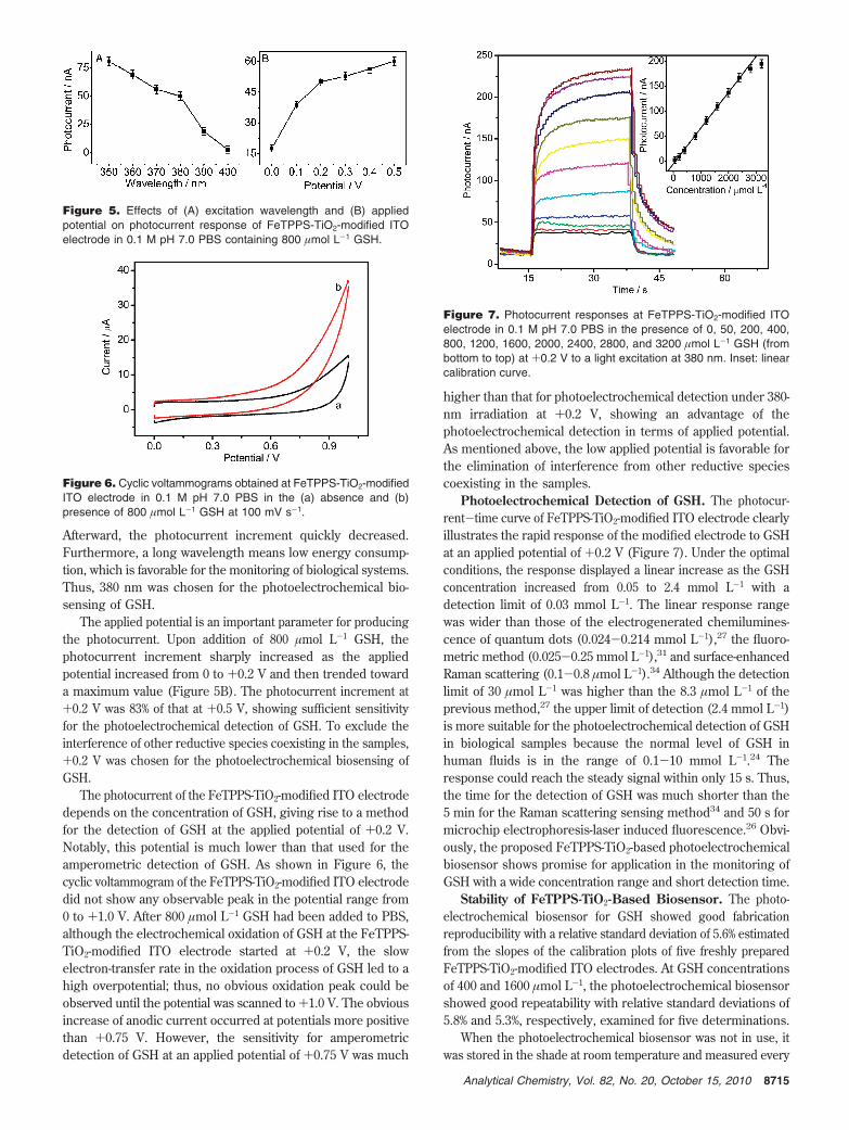
Afterward, the photocurrent increment quickly decreased.Furthermore, a long wavelength means low energy consump-tion, which is favorable for the monitoring of biological systems.Thus, 380 nm was chosen for the photoelectrochemical bio-sensing of GSH.
The applied potential is an important parameter for producingthe photocurrent. Upon addition of 800 µmol L-1 GSH, thephotocurrent increment sharply increased as the appliedpotential increased from 0 to +0.2 V and then trended towarda maximum value (Figure 5B). The photocurrent increment at+0.2 V was 83% of that at +0.5 V, showing sufficient sensitivityfor the photoelectrochemical detection of GSH. To exclude theinterference of other reductive species coexisting in the samples,+0.2 V was chosen for the photoelectrochemical biosensing ofGSH.
The photocurrent of the FeTPPS-TiO2-modified ITO electrodedepends on the concentration of GSH, giving rise to a methodfor the detection of GSH at the applied potential of +0.2 V.Notably, this potential is much lower than that used for theamperometric detection of GSH. As shown in Figure 6, thecyclic voltammogram of the FeTPPS-TiO2-modified ITO electrodedid not show any observable peak in the potential range from0 to +1.0 V. After 800 µmol L-1 GSH had been added to PBS,although the electrochemical oxidation of GSH at the FeTPPS-TiO2-modified ITO electrode started at +0.2 V, the slowelectron-transfer rate in the oxidation process of GSH led to ahigh overpotential; thus, no obvious oxidation peak could beobserved until the potential was scanned to +1.0 V. The obviousincrease of anodic current occurred at potentials more positivethan +0.75 V. However, the sensitivity for amperometricdetection of GSH at an applied potential of +0.75 V was much
higher than that for photoelectrochemical detection under 380-nm irradiation at +0.2 V, showing an advantage of thephotoelectrochemical detection in terms of applied potential.As mentioned above, the low applied potential is favorable forthe elimination of interference from other reductive speciescoexisting in the samples.
Photoelectrochemical Detection of GSH. The photocur-rent-time curve of FeTPPS-TiO2-modified ITO electrode clearlyillustrates the rapid response of the modified electrode to GSHat an applied potential of +0.2 V (Figure 7). Under the optimalconditions, the response displayed a linear increase as the GSHconcentration increased from 0.05 to 2.4 mmol L-1 with adetection limit of 0.03 mmol L-1. The linear response rangewas wider than those of the electrogenerated chemilumines-cence of quantum dots (0.024-0.214 mmol L-1),27 the fluoro-metric method (0.025-0.25 mmol L-1),31 and surface-enhancedRaman scattering (0.1-0.8 µmol L-1).34 Although the detectionlimit of 30 µmol L-1 was higher than the 8.3 µmol L-1 of theprevious method,27 the upper limit of detection (2.4 mmol L-1)is more suitable for the photoelectrochemical detection of GSHin biological samples because the normal level of GSH inhuman fluids is in the range of 0.1-10 mmol L-1.24 Theresponse could reach the steady signal within only 15 s. Thus,the time for the detection of GSH was much shorter than the5 min for the Raman scattering sensing method34 and 50 s formicrochip electrophoresis-laser induced fluorescence.26 Obvi-ously, the proposed FeTPPS-TiO2-based photoelectrochemicalbiosensor shows promise for application in the monitoring ofGSH with a wide concentration range and short detection time.
Stability of FeTPPS-TiO2-Based Biosensor. The photo-electrochemical biosensor for GSH showed good fabricationreproducibility with a relative standard deviation of 5.6% estimatedfrom the slopes of the calibration plots of five freshly preparedFeTPPS-TiO2-modified ITO electrodes. At GSH concentrationsof 400 and 1600 µmol L-1, the photoelectrochemical biosensorshowed good repeatability with relative standard deviations of5.8% and 5.3%, respectively, examined for five determinations.
When the photoelectrochemical biosensor was not in use, itwas stored in the shade at room temperature and measured every
Figure 5. Effects of (A) excitation wavelength and (B) appliedpotential on photocurrent response of FeTPPS-TiO2-modified ITOelectrode in 0.1 M pH 7.0 PBS containing 800 µmol L-1 GSH.
Figure 6. Cyclic voltammograms obtained at FeTPPS-TiO2-modifiedITO electrode in 0.1 M pH 7.0 PBS in the (a) absence and (b)presence of 800 µmol L-1 GSH at 100 mV s-1.
Figure 7. Photocurrent responses at FeTPPS-TiO2-modified ITOelectrode in 0.1 M pH 7.0 PBS in the presence of 0, 50, 200, 400,800, 1200, 1600, 2000, 2400, 2800, and 3200 µmol L-1 GSH (frombottom to top) at +0.2 V to a light excitation at 380 nm. Inset: linearcalibration curve.
8715Analytical Chemistry, Vol. 82, No. 20, October 15, 2010

few days. No obvious decrease in the photocurrent response toGSH was observed after 10 days, and 94.6% of the initialphotocurrent response was maintained after 4 weeks. This impliesthat the structure of FeTPPS-TiO2 is efficient for retaining theactivity of FeTPPS and preventing it from leaking out of thephotoelectrochemical biosensor.
Interference and Application in Real-Life Samples. Be-cause GSH is the auxiliary for the chemotherapy of cancer, theeffects of anticancer drugs as interfering species on the photo-electrochemical biosensing response were examined. For ex-ample, cisplatin, fluorouracil, and adriamycin at 10 times theconcentration of GSH did not interfere with the photoelectro-chemical response of GSH. Thus, the photoelectrochemicalbiosensor had an excellent specificity for the detection of GSH insamples with anticancer drugs.
The concentration of reduced glutathione in gluthion injectionwas detected with the proposed photoelectrochemical biosensorto be 0.018 ± 0.002 mol L-1 (five measurements) without anyneed of sample pretreatment except for appropriate dilution.This value was consistent with the 0.017 mol L-1 value givenin the instruction on glutathione injection, indicating acceptableaccuracy of the photoelectrochemical biosensor.
CONCLUSIONSA novel photoelectrochemical biosensing platform was devel-
oped using newly synthesized FeTPPS-TiO2 nanoparticles, whichwere prepared by the dentate binding of TiO2 nanoparticleswith sulfonic groups of FeTPPS and exhibited good biocom-patibility and dispersion in water. FeTPPS could efficientlyimprove the photo-current conversion efficiency of the TiO2
nanoparticles, which could be further amplified by an electron-transfer process from biomolecules to FeTPPS. Using GSH asa model, the fast photoelectronic communication among GSH,FeTPPS, TiO2, and ITO electrode led to a novel method forthe photoelectrochemical detection of GSH with good analyticalperformance, such as low applied potential, rapid response,wide linear range, and good reproducibility and repeatability.The low-potential detection produced excellent specificity forexcluding the interference of other reductive species andanticancer drugs in real samples. Taking into account that ironporphyrin can mimic peroxidase, this approach could be usedfor hydrogen peroxide detection. The proposed photoelectro-chemical biosensor showed promising application in themonitoring of biomolecules that could donate electrons to hole-injected FeTPPS. These porphyrin-functionalized TiO2 nano-particles open a new avenue for the construction of photoelec-trochemical biosensors.
ACKNOWLEDGMENTThis work was financially supported by the National Basic
Research Program of China (2010CB732400); the National ScienceFunds for Creative Research Groups (20821063); the MajorResearch Plan (90713015), Key (20835006), and General Programs(20875044, 20705012, 21075060) from the National Natural ScienceFoundation of China; and the Ph.D. Fund for Young Teachers(20070284052).
Received for review August 5, 2010. Accepted September10, 2010.
AC102070F
8716 Analytical Chemistry, Vol. 82, No. 20, October 15, 2010