POLITECNICO DI TORINO Repository ISTITUZIONALE · and with reference to the 2p3/2 and 3s levels of...
30
27 March 2020 POLITECNICO DI TORINO Repository ISTITUZIONALE Surface modification of Ti-6Al-4V alloy for biomineralization and specific biological response: Part I, inorganic modification / Ferraris S.; Spriano S.; Pan G.; Venturello A.; Bianchi C.L.; Chiesa R.; Faga M.G.; Maina G.; Vernè E.. - In: JOURNAL OF MATERIALS SCIENCE. MATERIALS IN MEDICINE. - ISSN 0957-4530. - STAMPA. - 22(2011), pp. 533-545. Original Surface modification of Ti-6Al-4V alloy for biomineralization and specific biological response: Part I, inorganic modification Publisher: Published DOI:10.1007/s10856-011-4246-2 Terms of use: openAccess Publisher copyright (Article begins on next page) This article is made available under terms and conditions as specified in the corresponding bibliographic description in the repository Availability: This version is available at: 11583/2381149 since: Springer Link
Transcript of POLITECNICO DI TORINO Repository ISTITUZIONALE · and with reference to the 2p3/2 and 3s levels of...
27 March 2020
POLITECNICO DI TORINORepository ISTITUZIONALE
Surface modification of Ti-6Al-4V alloy for biomineralization and specific biological response: Part I, inorganicmodification / Ferraris S.; Spriano S.; Pan G.; Venturello A.; Bianchi C.L.; Chiesa R.; Faga M.G.; Maina G.; Vernè E.. -In: JOURNAL OF MATERIALS SCIENCE. MATERIALS IN MEDICINE. - ISSN 0957-4530. - STAMPA. - 22(2011), pp.533-545.
Original
Surface modification of Ti-6Al-4V alloy for biomineralization and specific biological response: Part I,inorganic modification
Publisher:
PublishedDOI:10.1007/s10856-011-4246-2
Terms of use:openAccess
Publisher copyright
(Article begins on next page)
This article is made available under terms and conditions as specified in the corresponding bibliographic description inthe repository
Availability:This version is available at: 11583/2381149 since:
Springer Link
1
Surface modification of Ti-6Al-4V alloy for biomineralization and specific biological
response: Part I, inorganic modification
S. Ferraris*, S. Spriano*, G. Pan*, A. Venturello*, C.L. Bianchi**, R. Chiesa***, M.G. Faga****,
G. Maina*****, E. Vernè*
* DISMIC Department, Politecnico di Torino – C.so Duca degli Abruzzi, 24 - 10129 TORINO
(Italy)
** Physical Chemistry and Electrochemistry Department, University of Milano - Via Golgi 19 -
20133 MILANO (Italy)
*** Chemistry, Materials and Chemical Engineering Department G. Natta, Politecnico di Milano –
Via Mancinelli 7, 20131 Milano (Italy)
**** ISTEC-CNR - Strada delle Cacce 73 - 10135 TORINO (Italy)
***** Traumatology Orthopaedics and Occupational Medicine Department, University of Turin -
Via Zuretti, 29 - 10126 TORINO (Italy)
This is the author post-print version of an article published on Journal of Materials Science: Materials in Medicine, Vol. 22, pp. 533-545, 2011 (ISSN 0957-4530). The final publication is available at http://link.springer.com/article/10.1007%2Fs10856-011-4246-2 http://dx.doi.org/10.1007/s10856-011-4246-2 This version does not contain journal formatting and may contain minor changes with respect to the published edition. The present version is accessible on PORTO, the Open Access Repository of the Politecnico of Torino, in compliance with the publisher’s copyright policy. Copyright owner: Springer

2
Corresponding author:
Sara Ferraris
e-mail: [email protected]
Phone: 0039-0115644717
Fax: 0039-0115644699

3
Abstract
Titanium and its alloys represent the gold standard for orthopaedic and dental prosthetic devices,
because of their good mechanical properties and biocompatibility. Recent research has been focused
on surface treatments designed to promote their rapid osteointegration also in case of poor bone
quality. A new surface treatment has been investigated in this research work, in order to improve
tissue integration of titanium based implants. The surface treatment is able to induce a bioactive
behaviour, without the introduction of a coating, and preserving mechanical properties of Ti6Al4V
substrates (fatigue resistance). The application of the proposed technique results in a complex
surface topography, characterized by the combination of a micro-roughness and a nanotexture,
which can be coupled with the conventional macro-roughness induced by blasting. Modified
metallic surfaces are rich in hydroxyls groups: this feature is extremely important for inorganic
bioactivity (in vitro and in vivo apatite precipitation) and also for further functionalization
procedures (grafting of biomolecules). Modified Ti6Al4V induced hydroxyapatite precipitation
after 15 days soaking in simulated body fluid (SBF). The process was optimised in order to not
induce cracks or damages on the surface. The surface oxide layer presents high scratch resistance.
Keywords: titanium alloys, surface modification, bioactivity, nanotextured surface, osteointegration
Introduction
The goal of prosthetic surgery is to obtain implants able to reproduce the natural functions of
healthy tissues, with adequate mechanical properties, stability, reliability, good bone integration and
regeneration of health tissue at the damaged site. Titanium is the most widespread metal for
orthopaedic implants intended for bone integration. It presents high fatigue strength (600- 700 MPa)
and a comparatively low modulus of elasticity (70-130 GPa), respect to other metals [1, 2]. So it is
able to support loads and distribute them to bone, limiting stress shielding. Besides titanium and its
alloys are characterized by a thin natural oxide layer on the surface that limits ion release and
reactivity, making the surface almost inert and biocompatible. On the other hand this surface
inactivity often causes the development of a layer of fibrous tissue, around the implanted device,
hampering bone integration [3].
Several surface modifications have been proposed in literature in order to promote
osteointegration of titanium implants. The easiest strategy is to modify the surface morphology in
order to improve mechanical adhesion between implant and bone. In this field chemical etching,
blasting, Ti particles sintering, Ti plasma spray coatings and micro-patterning could be listed [2, 4].
On the other hand the deposition of a bioactive coating (for example of hydroxyapatite or bioactive

4
glass) could be employed in order to improve the adhesion between implant and bone [2, 5, 6].
Coatings are very effective in terms of bioactivity improvement, but some problems related to their
adhesion could thwart their utility [5, 6]. Different treatments have been proposed in literature, in
order to induce a bioactive behaviour directly on the metal surface, without the deposition of a
coating on it. One of these is the Kokubo treatment that provides samples soaking in NaOH solution
and eventually a subsequent thermal treatment [7-10]. On the other hand Osaka, Tsuru et al.
proposed a series of possible processes based on hydrogen peroxide, eventually added with TaCl,
HCl or TiOSO4 , in order to induce bioactive behaviour onto titanium surfaces [11-15]. These
processes are not easily applicable on industrial scale, they sometimes induce surface cracking and
low fatigue resistance of the treated materials is reported [16]. So actually there is still the need to
develop new processes for this purpose.
An innovative patented [17] modification treatment for bioactive titanium surfaces is
discussed in this research work. It is a thermo-chemical process and it includes firstly an acid
etching in diluted hydrofluoric acid, subsequently a controlled oxidation in hydrogen peroxide and a
thermal treatment. Results of the modification are both a micro and nano-patterned surface and a
modified surface chemistry. A tight control of the treatment is required in order to get a surface free
of cracks and a high adhesion of the modified surface to the substrate. The treatment was tested also
on sandblasted surfaces in order to get a micro and nano-texture superimposed onto a macro-
roughness. The modified titanium alloy promotes in-vitro hydroxyapatite precipitation and it is
suitable for further biological functionalization [18, 19] by grafting biomolecules on it, so it is
promising in order to promote healing by stimulating the growth of both inorganic and organic
component of bone. The inorganic thermo-chemical treatment is essential in order to obtain an
active surface for the direct grafting of biomolecules on the metal, so polymeric coatings and toxic
spacer (such as glutaraldehyde) can be avoided.
Materials and methods
Ti6Al4V has been employed as substrate. Specimens with a thickness of 2mm have been cut from
cylindrical bars (10mm in diameter) with an automatic cutter (Struers Accutom 5) provided with an
alumina blade (356 CA). Each sample has been signed with a number on one side and polished on
the opposite one with abrasive SiC papers (up to 4000 grit).
Some samples have been blasted with corundum for one minute at different pressures (4-6-8 atm) in
a blaster (BLAST0/f, Blastline) and named SB4, SB6 and SB8 respectively. Samples have been
twice ultrasonically washed in double distilled water for 15 minutes after blasting. No organic
solvents have been employed for cleaning them, in order to avoid surface contamination.

5
The thermo-chemical treatment includes a first acid etching in hydrofluoric acid, intended for the
removal of the native oxide layer, and a subsequent controlled oxidation in hydrogen peroxide [17],
so treated samples will be named HF+H2O2. Some samples have been treated only with acid
(named HF samples) and others only with hydrogen peroxide (named H2O2) for comparison.
Different times of acid etching have been tested for the treatment of 6 atm blasted surfaces (SB6), in
order to evaluate the opportunity to preserve sandblasting macro-roughness after the
thermochemical treatment. The blasting pressure for this test has been selected as the most
commonly used in industrial production of medical devices. Increasing etching time are indicated as
HF(1), HF(2) and HF(3) respectively.
Some samples have been thermally treated at 300°C (TT300 samples), 400°C (TT400 samples),
500°C (TT500) and 600°C (TT600 samples) for 1 hour in air and 400°C under vacuum (TT400v),
in order to stabilize the oxide layer generated on the surface. Samples have been posed in an
alumina holder and closed with an aluminium foil in order to prevent contaminations. Samples have
been let cool gradually to room temperature in the furnace at the end of treatments.
Samples morphology has been investigated by means of Scanning Electron Microscopy (SEM –
FEI, QUANTA INSPECT 200, EDS - EDAX PV 9900) and Field Emission Scanning Electron
Microscopy analysis (FESEM - SUPRATM 40, Zeiss)
Crystallographic structure has been analysed by means of X-ray diffraction measurements (XRD -
Philips X’Pert) in parallel beam configuration (with an incident angle fixed to 1°) in order to
analyze only the outermost layer of the sample.
Hydroxyls exposition has been studied by means of a FTIR microscope (IR Hyperion 2000, Tensor
27 - Bruker S.p.A) and X-ray Photoelectron Spectroscopy - XPS (Al source, Surface Science
Instruments, M-Probe) analysis. In this work vibrations of hydroxyls groups at 3000-3600 cm-1 and
of adsorbed water at 1600 cm-1 have been investigated by FTIR in reflection configuration [20, 21] .
Oxygen region (524-540 eV) and titanium region (457-459 eV) have been analysed by XPS in order
to evaluate hydroxyls exposition at different steps of the treatment. The XPS energy scale was
calibrated with reference to the 4f7/2 level of a freshly evaporated gold sample, at 84,00 +/- 0,1 eV,
and with reference to the 2p3/2 and 3s levels of copper at 932,47+/- 0,1 and 122,39 +/- 0,15 eV,
respectively.
The thickness of the modified layer has been measured by means of XPS analysis equipped with Ar
gun. Surface elements have been monitored in function of the etching time in order to detect the
transition point between metallic substrate and surface oxide layer. Some samples have also been
sent to Filmetrics (San Diego –CA- USA) for measurement of the oxide layer thickness by means of
spectral reflectance technique (Filmetrics F20 instrument).
6
Scratch tests have been performed in Revetest mode (CSM Revetest machine) in order to study the
mechanical adhesion of the modified oxide. A load has been applied to the sample surface through a
Rockwell C diamond stylus with 200 µm diameter, increasing progressively from 1N to 50N along
a 4.91 mm track. Scratch test parameters have been set according to the standard [22]. Scratch track
has been optically observed and critical load determined as the ones at which coating damages
appears.
Fatigue tests were carried out by rotating bending tests (2600 rpm – room temperature) and by
applying a simplified stair-case method (6 samples).
Surface wettability has been determined by means of static contact angle measurements with water
before and after the thermo-chemical process.
Samples have been analysed by means of a contact profiler (KLA-Tencor P15) at different steps of
the process (polished, blasted, acid etched, chemically oxidised, thermally treated samples), in order
to determine the surface roughness. Scan length has been set to 500 µm and 2 µm and cantilever
speed at 50 and 2 µm/s respectively. Three measurements have been carried out on each sample in
three different points. Average roughness (Ra) has been evaluated.
Atomic Force Microscopy observations (AFM – XE-100, Park Systems) have been carried out in
order to better characterize surface topography and roughness at the nano-scale.
Inorganic bioactivity has been investigated in-vitro by soaking samples in simulated body fluid
(SBF) for 2 weeks at 37°C. SBF has been prepared according to Kokubo protocol [23]. Solution
refresh has been performed every two days, in order to mimic physiological turnover of body fluids.
The pH has been measured after each refresh. Samples have been gently washed with double
distilled water at the end of the soaking time and let to dry at room temperature. Finally they have
been observed at SEM to look for hydroxyapatite precipitation onto their surface.
Ion release from Ti6Al4V surfaces before and after HF+H2O2 treatment has been evaluated in
simulated body fluid up to 1 week, in order to investigate whether the proposed treatment can
reduce metallic ion release from Ti6Al4V surfaces. In fact ion release from metallic surface reaches
its maximum value after few days in simulated physiological fluids [16]. Three just polished
samples and three modified samples with the optimized treatment (HF+H2O2) and thermally treated
at 300°C for 1h have been soaked in each in 30 ml of SBF at 37°C up to 1 week. 1 ml of the
solution has been spiked up at several time steps (3h, 1, 3, 7days) and analysed by means of
graphite furnace atomic absorption spectroscopy (GFAAS). Any solution refresh have been
performed during the soaking period, a cumulative release has been finally obtained. All values
have been referred to sample surface area, only the modified surface has been considered.

7
Results and discussion
Surface morphology
The interaction between implant materials and the physiological environment play mainly at the
interface. Surface topography and chemistry are the most important characteristics that affect cell
behaviour on artificial materials and that, at the end, pilot the entire tissue integration process [24].
A lot of literature works underline that both shape and dimensions of topographical features can
affect cell adhesion, proliferation and differentiation onto material surface [24, 25]. So the peculiar
surface topography (multiscale roughness at the micro- and nano- levels) and chemistry
(hydroxylation), of the treated and modified materials analysed in this work, have been investigated
in details by means of several techniques (SEM, FESEM, contact profilometry, AFM, XPS and
FTIR).
Polished and treated surfaces
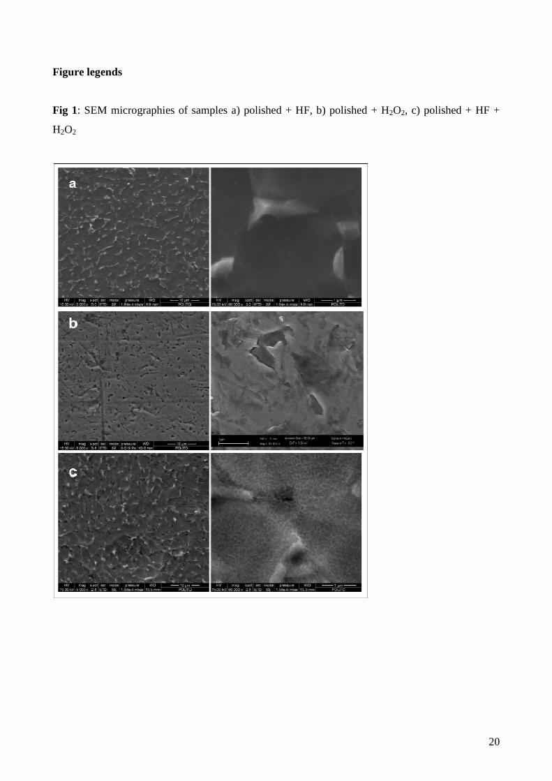
Figure 1 shows the surface morphology of samples treated by acid etching (Figure 1a), by
oxidation in hydrogen peroxide (Figure 1b) or by the complete chemical treatment (etching in HF as
first and then oxidation in H2O2) (Figure 1c). The etching in hydrofluoric acid readily removes the
natural oxide from the all the surface and dissolves titanium according to the reaction (1):
Ti + 3HF � Ti 3+ + 3/2H2 + 3F- (1)
Ti 3+ can be further oxidized by atmospheric oxygen or it can be complexed by F- [26]. This
treatment induces a micro rough surface, due to the preferential dissolution of the alpha phase of the
biphasic alloy by HF (figure 1.a).
On the contrary, hydrogen peroxide causes pitting corrosion on the untreated alloy, where it
acts on the native passivation layer that is not removed by H2O2. The TiO2-H2O2 interaction was
widely studied in literature [27, 28] and it can be described as follows. Hydrogen peroxide is
decomposed to oxygen and water; Ti oxidation and eventually corrosion take place according to the
pH and concentration of the solutions. The formation of Ti(IV)-H2O2 complex occurs in case of
surface corrosion with the appearance of a typical yellow colour of the solution. It should be
underlined that, considering the Ti-Al-V alloy, the alpha phase (rich in Al) is covered by a more
efficient passivation film (since both Ti and Al occur in its formation) while the beta phase (rich in
V) is passivated by a simple TiO2 film. A localized corrosion at the grain boundary between the
alpha phase and beta one can be noticed (figure 1.b), causing surface beta crystal detachment and
pitting effect.
8
A peculiar surface can be obtained by the complete chemical treatment, etching in
hydrofluoric acid and then controlled oxidation in hydrogen peroxide. The natural oxide is firstly
removed by acid etching and the nude titanium alloy is then exposed to the action of hydrogen
peroxide, which causes a re-oxidation of the metal surface at the nanoscale. The reaction between
the metal and the hydrogen peroxide is different respect to what previously described for the titania
layer. The treated surface presents a micro rough aspect and a nanoporous pattern (figure 1 c.).
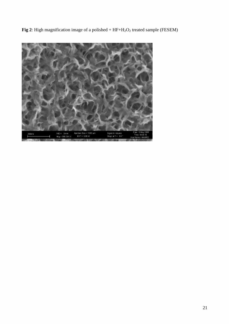
Figure 2 shows a high magnification image of the spongy structure on a nanoscale of the oxide
layer produced by the complete treatment. Corrosion of the titanium alloy does not occur in all the
tested conditions as it can be deduced from the colourless appearance of the hydrogen peroxide
solution at the end of the treatment. The last step of the treatment is a thermal consolidation of the
surface oxide layer (300°C-1h in air). It does not cause any variation of the surface morphology.
SEM observations (not reported) verified the maintenance of nanostructured spongy surface also
after thermal treatments.
Surface roughness of the treated surfaces has been evaluated using a contact profiler. Results
are reported in figure 3a. The acid etching induces a micro roughness (about 0.2 micron) as
observed by SEM observations. Chemical oxidation in hydrogen peroxide and the following
thermal treatment do not alter the surface micro roughness. On the other hand the roughness on a
nanoscale, induced by the chemical oxidation in hydrogen peroxide, can not be detected by this
technique
So AFM analysis was employed in order to measure characteristic parameters for the nanotextured
surfaces. In particular it has been observed that the nano-scale roughness Ra can be estimated as
about 10 nm and that the surface phase is homogeneous.
Several studies dealing with cells interactions with random or ordered nano-patterned
surfaces (produced by different methods, such as photolithography or chemical etchings) are
reported in literature [29] and they are confirming that cells are sensitive to nano-textures and that
osteoblast cells show a lot of filopodia protrusion into the direction of motility over a nanopattern.
Moreover, nano-pores are reported as a determining factor for the chemical equilibrium needed for
biomineralization and apatite precipitation [24, 30, 31] and it was reported that micro and nano-
patterns improve mechanical adhesion of precipitated hydroxyapatite on metallic surface [32]. So
the presence of this morphology is quite relevant for the purpose of this study.
It must be underlined that a crack free surface is obtained at the end of the process by a
severe control of the chemical oxidation of the metallic surface. This is quite relevant in order to
maintain the good fatigue resistance of the titanium alloy and to increase the adhesion of the
9
oxidised surface layer to the substrate. So this treatment seems to be very promising compared to
other bioactive surfaces described in literature [13, 16].
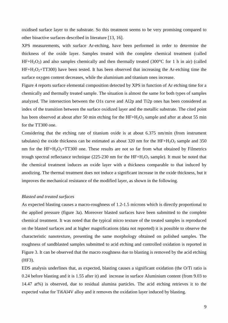
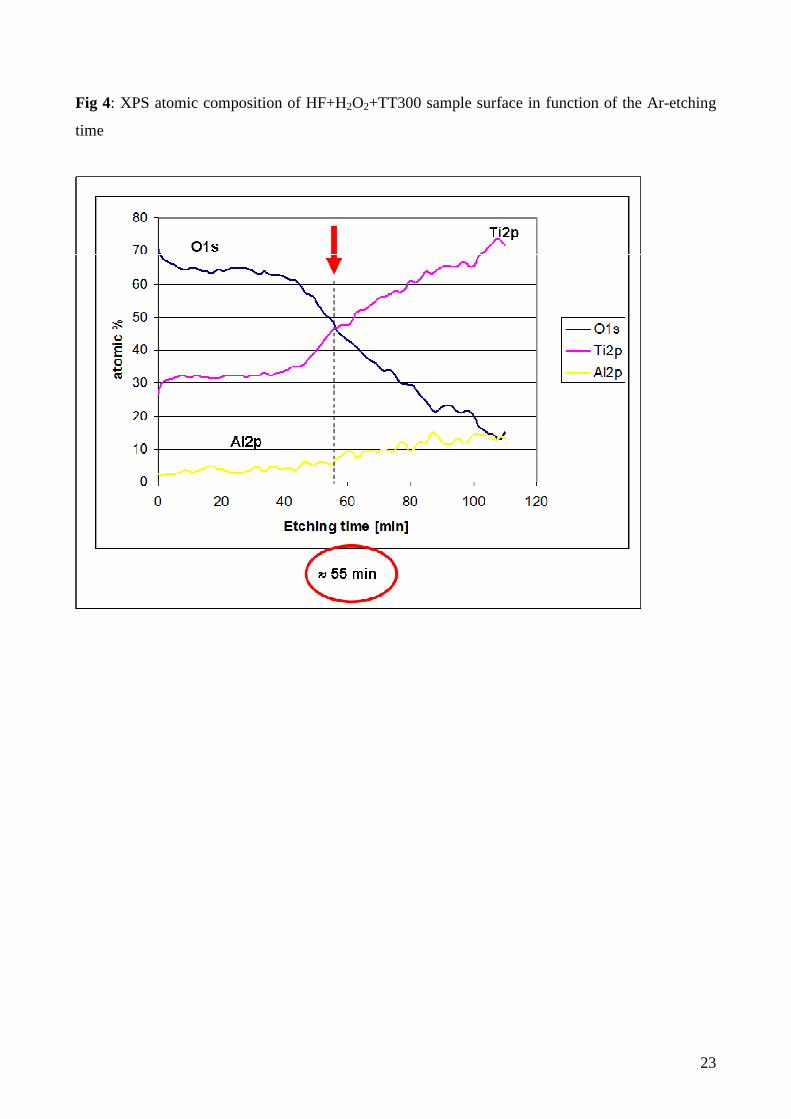
XPS measurements, with surface Ar-etching, have been performed in order to determine the
thickness of the oxide layer. Samples treated with the complete chemical treatment (called
HF+H2O2) and also samples chemically and then thermally treated (300°C for 1 h in air) (called
HF+H2O2+TT300) have been tested. It has been observed that increasing the Ar-etching time the
surface oxygen content decreases, while the aluminium and titanium ones increase.
Figure 4 reports surface elemental composition detected by XPS in function of Ar etching time for a
chemically and thermally treated sample. The situation is almost the same for both types of samples
analyzed. The intersection between the O1s curve and Al2p and Ti2p ones has been considered as
index of the transition between the surface oxidized layer and the metallic substrate. The cited point
has been observed at about after 50 min etching for the HF+H2O2 sample and after at about 55 min
for the TT300 one.
Considering that the etching rate of titanium oxide is at about 6.375 nm/min (from instrument
tabulates) the oxide thickness can be estimated as about 320 nm for the HF+H2O2 sample and 350
nm for the HF+H2O2+TT300 one. These results are not so far from what obtained by Filmetrics
trough spectral reflectance technique (225-230 nm for the HF+H2O2 sample). It must be noted that
the chemical treatment induces an oxide layer with a thickness comparable to that induced by
anodizing. The thermal treatment does not induce a significant increase in the oxide thickness, but it
improves the mechanical resistance of the modified layer, as shown in the following.
Blasted and treated surfaces
As expected blasting causes a macro-roughness of 1.2-1.5 microns which is directly proportional to
the applied pressure (figure 3a). Moreover blasted surfaces have been submitted to the complete
chemical treatment. It was noted that the typical micro texture of the treated samples is reproduced
on the blasted surfaces and at higher magnifications (data not reported) it is possible to observe the
characteristic nanotexture, presenting the same morphology obtained on polished samples. The
roughness of sandblasted samples submitted to acid etching and controlled oxidation is reported in
Figure 3. It can be observed that the macro roughness due to blasting is removed by the acid etching
(HF3).
EDS analysis underlines that, as expected, blasting causes a significant oxidation (the O/Ti ratio is
0.24 before blasting and it is 1.55 after it) and increase in surface Aluminium content (from 9.03 to
14.47 at%) is observed, due to residual alumina particles. The acid etching retrieves it to the
expected value for Ti6Al4V alloy and it removes the oxidation layer induced by blasting.
10
So, it can be concluded that the complete chemical treatment removes alumina contamination from
blasted surfaces and it induces surface micro and nano-structures, but reducing macro-roughness.
Moreover it can be suggested that the treatment removes the atomic disordered layer induced by
blasting, in fact it is thinner than the roughness induced dimension [33]
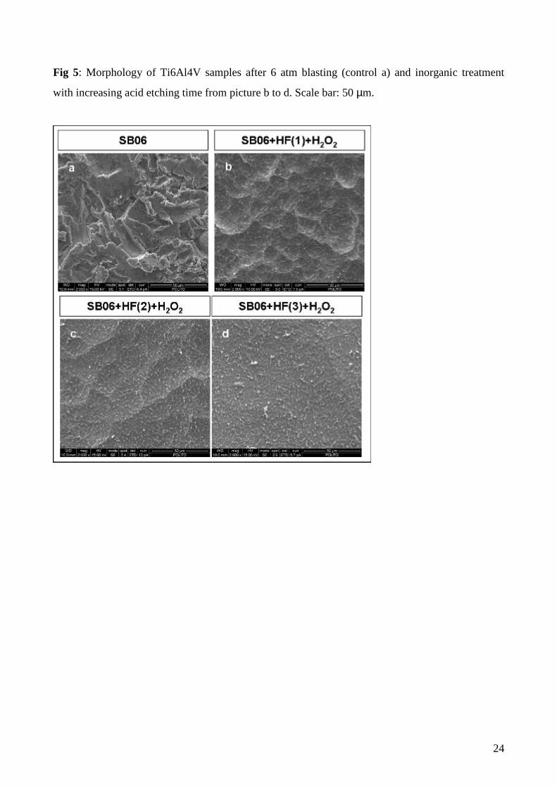
Figure 3b reports surface roughness of 6 atm blasted samples treated with different acid etching
times, increasing from HF(1) to HF(3), and then oxidized in hydrogen peroxide. It can be noted that
the reduction in roughness is proportional to the acid etching time and that it is possible to properly
preserve macroroughness by reducing the duration of acid etching. The micro and nanotextures are
obtained on all these samples.
SEM observations (figure 5) confirm these results.
Hydroxyls exposition
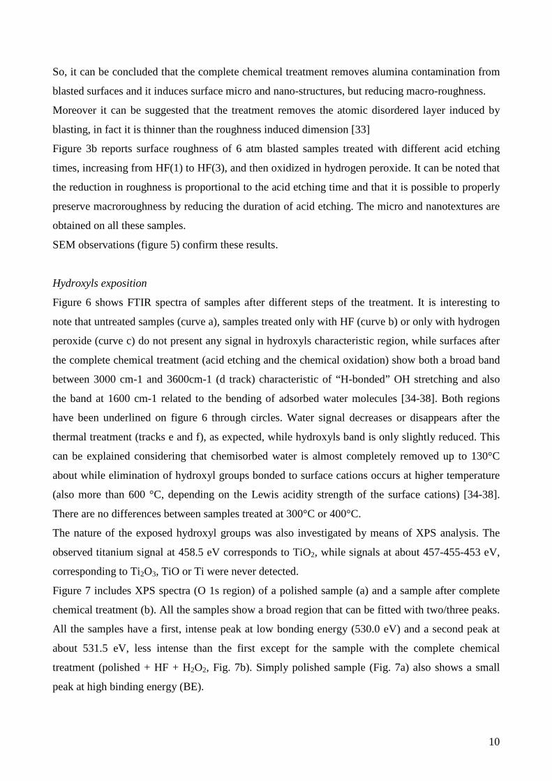
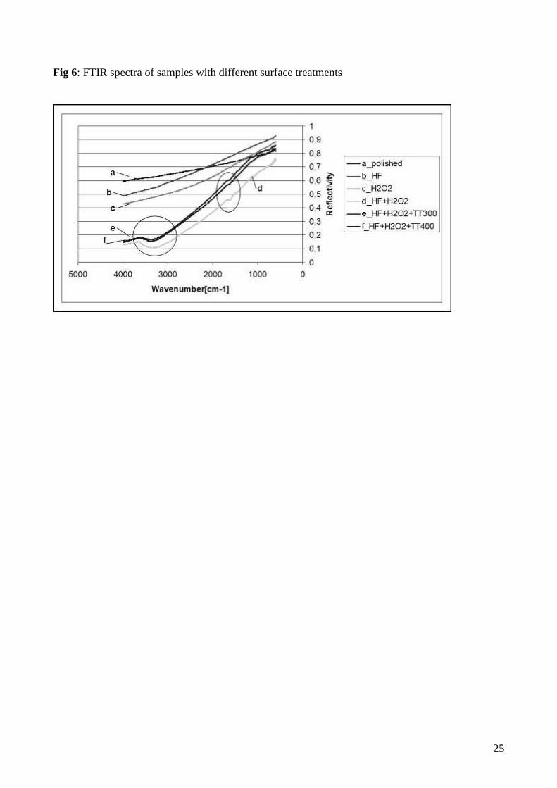
Figure 6 shows FTIR spectra of samples after different steps of the treatment. It is interesting to
note that untreated samples (curve a), samples treated only with HF (curve b) or only with hydrogen
peroxide (curve c) do not present any signal in hydroxyls characteristic region, while surfaces after
the complete chemical treatment (acid etching and the chemical oxidation) show both a broad band
between 3000 cm-1 and 3600cm-1 (d track) characteristic of “H-bonded” OH stretching and also
the band at 1600 cm-1 related to the bending of adsorbed water molecules [34-38]. Both regions
have been underlined on figure 6 through circles. Water signal decreases or disappears after the
thermal treatment (tracks e and f), as expected, while hydroxyls band is only slightly reduced. This
can be explained considering that chemisorbed water is almost completely removed up to 130°C
about while elimination of hydroxyl groups bonded to surface cations occurs at higher temperature
(also more than 600 °C, depending on the Lewis acidity strength of the surface cations) [34-38].
There are no differences between samples treated at 300°C or 400°C.
The nature of the exposed hydroxyl groups was also investigated by means of XPS analysis. The
observed titanium signal at 458.5 eV corresponds to TiO2, while signals at about 457-455-453 eV,
corresponding to Ti2O3, TiO or Ti were never detected.
Figure 7 includes XPS spectra (O 1s region) of a polished sample (a) and a sample after complete
chemical treatment (b). All the samples show a broad region that can be fitted with two/three peaks.
All the samples have a first, intense peak at low bonding energy (530.0 eV) and a second peak at
about 531.5 eV, less intense than the first except for the sample with the complete chemical
treatment (polished + HF + H2O2, Fig. 7b). Simply polished sample (Fig. 7a) also shows a small
peak at high binding energy (BE).

11
It is difficult to precisely attribute XPS peaks to oxygen species except for the one at about 530 eV,
attributable to bulk oxygen in titanium oxide. A debate is present in literature on the attribution of
the other two peaks at about 531.5 eV and at BE higher than 532 eV. In fact the first peak at about
531.5 eV can be attributed to generic surface OH groups [39] and ref [40] therein or to acidic
hydroxyl groups together with adsorbed water [41]. Last peak at BE higher than 532 eV is
attributed to adsorbed water [40] or to basic hydroxyl groups [41], respectively.
Notwithstanding this uncertainty, it is possible to observe unequivocally that the sample treated
with both HF and H2O2 shows a very intense peak related to hydroxyl groups at 531.5 eV. (figure
7b) which is less intense on the untreated one (figure 7a), as also shown by FTIR. A significant
reduction in OH signal is also observed after thermal treatments, both at 300°C (figure 7c) and
400°C (data not shown). In this case, the reduction underlined by XPS analysis is higher than the
one detected by FTIR measurements, this phenomenon could be explained considering that the two
techniques explore different depth of the surface. XPS analyses only the very first surface layer (2-
10 nm) [42] while FTIR goes more in depth (some microns) [42].
The reduction of OH signals on thermally treated samples has been widely investigated in literature.
it is well known that OH disappearing at lower temperature are only the weakly adsorbed ones,
while the OH strongly bound with Ti can remain on the surface up to 600°C or more [34-38].
Moreover it has been observed that only strongly bound hydroxyls groups are effectively
responsible of titanium bioactivity [40, 43].
Observing FTIR and XPS data, it can be concluded that the thermo-chemical treatment modifies
also the chemistry of the surface. The presence of a high density of OH groups on the surface is
interesting also for its further functionalization with biomolecules [18] in order to combine
inorganic and biological bioactivity. The hydroxylated surface is reactive and biomolecules can be
directly linked to it, without the use of a polymeric coating or toxic spacers.
Crystallographic structure
XRD measurements have been carried out in order to determine the crystallographic structure of the
formed oxide layer. In fact it has been suggested in literature that the nature of titanium oxide can
affect both bioactivity and cellular interaction [39].
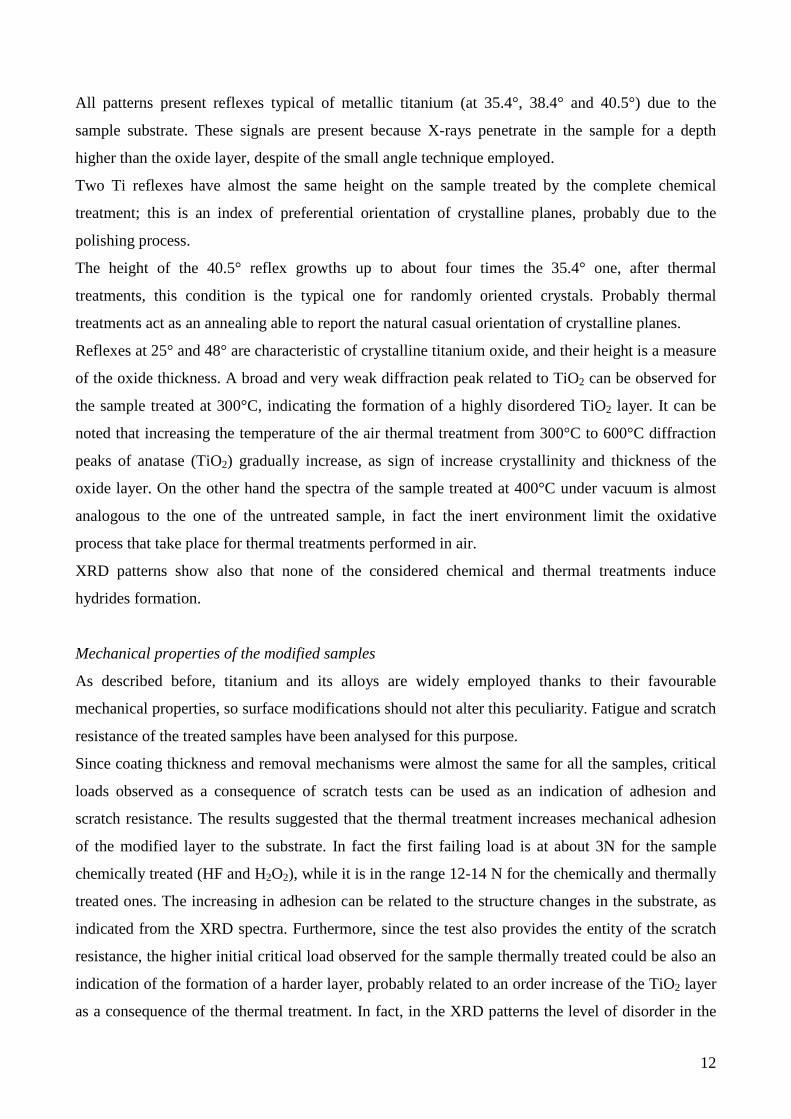
Figure 8 reports XRD patterns of samples treated with the complete chemical treatment and then
thermally treated at different temperatures (from 300°C to 600°C) in air and at 400°C under
vacuum.
12
All patterns present reflexes typical of metallic titanium (at 35.4°, 38.4° and 40.5°) due to the
sample substrate. These signals are present because X-rays penetrate in the sample for a depth
higher than the oxide layer, despite of the small angle technique employed.
Two Ti reflexes have almost the same height on the sample treated by the complete chemical
treatment; this is an index of preferential orientation of crystalline planes, probably due to the
polishing process.
The height of the 40.5° reflex growths up to about four times the 35.4° one, after thermal
treatments, this condition is the typical one for randomly oriented crystals. Probably thermal
treatments act as an annealing able to report the natural casual orientation of crystalline planes.
Reflexes at 25° and 48° are characteristic of crystalline titanium oxide, and their height is a measure
of the oxide thickness. A broad and very weak diffraction peak related to TiO2 can be observed for
the sample treated at 300°C, indicating the formation of a highly disordered TiO2 layer. It can be
noted that increasing the temperature of the air thermal treatment from 300°C to 600°C diffraction
peaks of anatase (TiO2) gradually increase, as sign of increase crystallinity and thickness of the
oxide layer. On the other hand the spectra of the sample treated at 400°C under vacuum is almost
analogous to the one of the untreated sample, in fact the inert environment limit the oxidative
process that take place for thermal treatments performed in air.
XRD patterns show also that none of the considered chemical and thermal treatments induce
hydrides formation.
Mechanical properties of the modified samples
As described before, titanium and its alloys are widely employed thanks to their favourable
mechanical properties, so surface modifications should not alter this peculiarity. Fatigue and scratch
resistance of the treated samples have been analysed for this purpose.
Since coating thickness and removal mechanisms were almost the same for all the samples, critical
loads observed as a consequence of scratch tests can be used as an indication of adhesion and
scratch resistance. The results suggested that the thermal treatment increases mechanical adhesion
of the modified layer to the substrate. In fact the first failing load is at about 3N for the sample
chemically treated (HF and H2O2), while it is in the range 12-14 N for the chemically and thermally
treated ones. The increasing in adhesion can be related to the structure changes in the substrate, as
indicated from the XRD spectra. Furthermore, since the test also provides the entity of the scratch
resistance, the higher initial critical load observed for the sample thermally treated could be also an
indication of the formation of a harder layer, probably related to an order increase of the TiO2 layer
as a consequence of the thermal treatment. In fact, in the XRD patterns the level of disorder in the
13
film only chemically treated is so high that it is really hard to observe the related peak, while it can
be slightly distinguished in the case of the material thermal treated at 300°C. There are not
significant differences in adhesion between samples treated at 300°C or 400°C. The thermal
treatment at 300°C was selected for the following tests, considering that it is able to assure a good
adhesion and that treatments at lower temperature have less effect on the fatigue resistance.
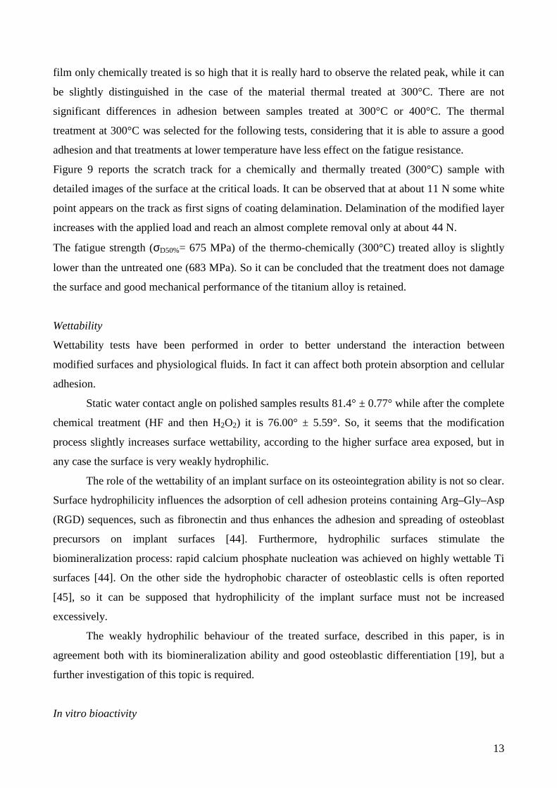
Figure 9 reports the scratch track for a chemically and thermally treated (300°C) sample with
detailed images of the surface at the critical loads. It can be observed that at about 11 N some white
point appears on the track as first signs of coating delamination. Delamination of the modified layer
increases with the applied load and reach an almost complete removal only at about 44 N.
The fatigue strength (σD50%= 675 MPa) of the thermo-chemically (300°C) treated alloy is slightly
lower than the untreated one (683 MPa). So it can be concluded that the treatment does not damage
the surface and good mechanical performance of the titanium alloy is retained.
Wettability
Wettability tests have been performed in order to better understand the interaction between
modified surfaces and physiological fluids. In fact it can affect both protein absorption and cellular
adhesion.
Static water contact angle on polished samples results 81.4° ± 0.77° while after the complete
chemical treatment (HF and then H2O2) it is 76.00° ± 5.59°. So, it seems that the modification
process slightly increases surface wettability, according to the higher surface area exposed, but in
any case the surface is very weakly hydrophilic.
The role of the wettability of an implant surface on its osteointegration ability is not so clear.
Surface hydrophilicity influences the adsorption of cell adhesion proteins containing Arg–Gly–Asp
(RGD) sequences, such as fibronectin and thus enhances the adhesion and spreading of osteoblast
precursors on implant surfaces [44]. Furthermore, hydrophilic surfaces stimulate the
biomineralization process: rapid calcium phosphate nucleation was achieved on highly wettable Ti
surfaces [44]. On the other side the hydrophobic character of osteoblastic cells is often reported
[45], so it can be supposed that hydrophilicity of the implant surface must not be increased
excessively.
The weakly hydrophilic behaviour of the treated surface, described in this paper, is in
agreement both with its biomineralization ability and good osteoblastic differentiation [19], but a
further investigation of this topic is required.
In vitro bioactivity
14
Finally inorganic bioactivity of Ti6Al4V alloy, in simulated body fluid, has been investigated in
order to verify the effectiveness of the proposed treatment.
Samples have been observed at SEM after 15 day soaking in SBF. Figure 10 shows micrographies
and EDS spectra of sample area and of precipitates on different samples: HF+H2O2 (figure 10a),
HF+H2O2+TT300 (figure 10b) and HF+H2O2+TT400 (figure 10c), after 15 days in SBF. Particles
with dimension of about 5µm and the typical morphology of hydroxyapatite precipitate can be
noted on all the considered samples (figure 10 a2, 10 b2, 10 c2). EDS analysis on particles (figure
10 a3, 10 b3, 10 c3) confirm that they are rich in calcium and phosphorous in a ratio close to the
one of hydroxiapatite (at about 1.7). Sodium and chlorine traces derive from SBF soaking that
contains NaCl. XRD analysis does not detect these precipitates because of their reduced dimension
and amount. EDS analysis on the whole surface (figure 10 a1, 10 b1, 10 c1) underlines that the
bioactivity of all the treated surfaces is comparable. An enrichment in Ca and P was also detected
on the surface free from precipiated, showing a diffuse interaction of the treated surfaces with
physiological fluids.
The pH measurements of the SBF soaking solution show that values remain in the range 7.1 – 7.6
during the entire soaking time for all tested samples. These variations are included in physiological
tolerability range (7.00 – 7.80).
So it can be concluded that the proposed process is able to induce inorganic bioactivity on titanium
alloy and it is very promising in order to induce in-vivo biomineralization on the implant.
The thermal treatment does not alter this property despite of the apparent reduction in OH amount
on the surface.
Figure 7d reports XPS detailed analysis of the oxygen region for a sample HF+H2O2+TT400 after
15 days in SBF. It could be noted that the oxygen region is modified and an OH signal attributable
to hydroxyapatite appears [46]
The inorganic bioactivity must be coupled with a good cell response of a biomaterial in order to be
really profitable. A synergistic effect between inorganic and biological bioactivity of the titanium
surface after the described thermo-chemical treatment was previously reported by the authors [19,
47]. The thermo-chemically treated surface shows a better cell response, with a higher early and late
differentiation of osteoblastic cells. Furthermore, a higher and specific biological response of the
cells to the surface can be stimulated by grafting biomolecules on it, as reported in the part II of this
work [18].
Ion release evaluation
15
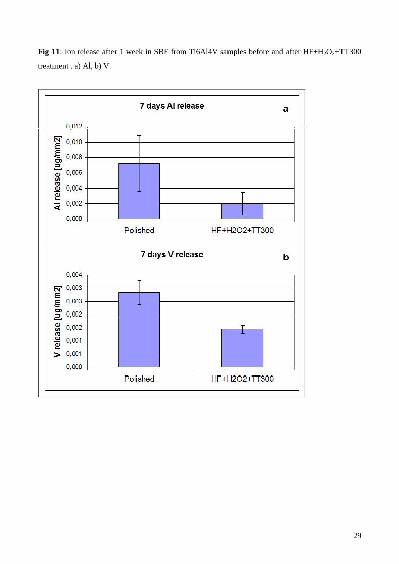
The amount of Al and V ions released from treated (HF+H2O2+TT300) Ti6Al4V samples after 7
days soaking in simulated body fluid is reported in figure 11. A reduction in their release can be
observed for surface modified samples. So it can be assumed that the proposed inorganic treatment
is able to reduce metallic ion release, due to the higher thickness of the formed oxide layer,
compared to the native one, improving biocompatibility.
Conclusions
The application of a new patented thermo-chemical treatment, based on hydrofluoric acid etching,
hydrogen peroxide controlled oxidation and a subsequent thermal treatment allows the preparation
of a bioactive Ti6Al4V alloy. The modified surface induces in-vitro apatite precipitation, so it is
promising for the promotion of in-vivo biomineralization processes. It shows a particular surface
chemistry (high amount of hydroxyls groups exposed), so it is suitable for functionalization with
biomolecules, in order to induce a specific cell response. It has a characteristic surface morphology
(a micrometric roughness coupled with a nano-metric texture). It can be applied also to blasted
surfaces, resulting in a multiscale topography (macro, micro and nano-levels) So these materials
combine inorganic bioactivity with a multiscale texture on the same surface. Finally titanium
modified with the described process possesses is crack free, it has good scratch resistance and the
starting fatigue resistance of the metal is maintained. This feature is often compromised in others
treatments reported in literature.
Acknowledgements
Regione Piemonte is acknowledged for its funding to the BIOSURF project.
Dr. Chiara Manfredotti is acknowledged for AFM measurements kindly performed.
Filmetrics (San Diego –CA- USA) is acknowledged for measurements by means of spectral
reflectance technique
16
References
[1] Pietrabissa R Biomateriali per protesi e organi artificiali Bologna: Patron Editore; 1996
[2] Xiao S-J, Kenausis G, Textor M, Biochemical modification of titanium surfaces, in: Berunette
DM, Tengvall P, Textor M, Thomsen P, Titanium in Medicine, Springler-Verlag, Berlin Heidelberg
New York; 2001, pp. 417-453
[3] Ratner BD, Replacing and Renewing: Synthetic Materials, Biomimetics, and Tissue Engineering
in Implant Dentistry. J Dent Educ 2001; 65:1340-1347
[4] Lausmaa J Mechanical, Thermal, Chemical and Electrochemical Surface Treatment of Titanium
in: Berunette DM, Tengvall P, Textor M, Thomsen P, Titanium in Medicine, Springler-Verlag,
Berlin Heidelberg New York; 2001, pp. 231-256
[5] De Jonge LT, Leeuwenburgh SCG, Wolke JGC, Jansen JA, Organic-Inorganic Surface
Modifications for Titanium Implant Surfaces. Pharm Res 2008; 25:2357-2369
[6] Rizzi G, Scrivani A, Fini M, Giardino R, Biomedical coatings to improve the tissue-biomaterial
interface. Int J Artif Organs, 2004; 27:649-657
[7] Kokubo T, Formation of biologically active bone-like apatite on metals and polymers by a
biomimetic process. Termochim. Acta 1996; 280-281:479-490
[8] Kokubo T, Apatite formation on surfaces of ceramics metals and polymers in body
environment., Acta Mater 1998; 46: 2519-2527
[9] Nishiguchi S, Nakamura T, Kobayashi M, . KimHM, Miyaji F, Kokubo T, The effect of heat
treatment on bone-bonding ability of alkali-treated titanium. Biomaterials 1999; 20:491-500
[10] Kokubo T, Matsushita T, Takadama H, Titania-based bioactive materials. J Eur Cer Soc 2007;
27:1553-1558
[11] Wang XX, Hayakawa S, Tsuru K, Osaka A, Improvement of bioactivity of H2O2/TaCl5-treated
titanium after subsequent heat treatments. J. Biomed. Mater. Res. 2000; 52:171-176
[12] Kaneko S, Tsuru K, Hayakawa S, Takemoto S, Ohtsuki C, Ozaki T, Inoue H, Osaka A, In vivo
evaluation of bone-bonding of titanium metal chemically treated with hydrogen peroxide solution
containing tantalum chloride. Biomaterials 2001; 22: 875-881
[13] Wang XX, Hayakawa S, Tsuru K, Osaka A, Bioactive titania gel layers formed by chemical
treatment of Ti substrate with H2O2/HCl solution. Biomaterials 2002; 23:1353-1357
[14] Xiao F, Tsuru K, Hayakawa S, Osaka A, In vitro apatite deposition on titania film derived from
chemical treatment of Ti substrates with oxysulfate solution containing hydrogen peroxide at low
temperature. Thin solid films 2003; 441:271-276
17
[15] Hayakawa S, Tsuru K, Osaka A, Low-temperature preparation of anatase and rutile layers on
titanium substrates and their ability to induce in vitro apatite deposition. J. Am. Ceram. Soc. 2004;
87:1635-1642
[16] Spriano S, Bronzoni M, Vernè E, Maina G, Bergo V, Windler M, Characterization of surface
modified Ti-6Al-7Nb alloy. J. Mater. Sci.: Mater. Med. 2005; 16:301-312
[17] Patent – EP2214732 (TO2007A000719) S.Spriano,E.Vernè, S. Ferraris “Multifunctional
titanium surfaces for bone integration”
[18] Ferraris S, Spriano S, Bianchi CL, Cassinelli C, Vernè E, Surface modification of Ti-6Al-4V
alloy for biomineralization and specific biological response: Part II, Alkaline phosphatase grafting.
submitted to J Mater Sci Mater Med
[19] Spriano S, Ferraris S, Bianchi CL, Cassinelli C, Torricelli P, Fini M, Rimondini L, Giardino
R, Synergistic effect between inorganic and biological bioactivity of modified titanium surfaces.
Mater Sci Res J 2009; 3:203-222
[20] Kumar PM, Badrinarayanan S, Sastry M, Nanocrystalline TiO2 studied by optical, FTIR and
X-ray photoelectron spectroscopy: correlation to presence of surface states. Thin Solid Films 2000;
358:122-130
[21] Gora-Marek K, Datka J, IR studies of OH groups in mesoporous aluminosilicates. Appl. Catal.,
A 2006; 302:104-109
[22] European Standard prEN 1071-3:2000 (established by the European Standards Committee
CEN TC184 WG5)
[23] Kokubo T, Takadama H, How useful is SBF in predicting in vivo bone bioactivity.
Biomaterials 2006; 27:2907-2915
[24] Boyan BD, Dean DD, Lohmann CH, Cochran DL, Sylvia VL, Schwartz Z, The titanium-Bone
Cell Interface in vitro: the role of the surface in promoting osteointegration, in: Brunette DM,
Tengvall P, Textor M, Thomsen P, Titanium in Medicine, Springler-Verlag, Berlin Heidelberg New
York 2001, pp. 561-585
[25] Boyan BD,. Hummert TW,. Dean DD, Schwartz Z, Role of material surfaces in regulating
bone and cartilage cell response. Biomaterials 1996; 17:137-146
[26] Sutter EMM, Goetz-Grandmont GJ, The behaviour of titanium in nitric-hydrofluoric acid
solutions. Corros. Sci. 1990; 30:461-476
[27] Tengvall P, Elwing H, Lundstrom I, Titanium gel made from metallic titanium and hydrogen
peroxide. J. Colloid Interface Sci. 1989; 130: 405-413
[28] Mao C, Li H, Cui F, Ma C, Feng Q, Oriented growth of phosphates on polycrystalline titanium
in a process mimicking biomineralization. J. Cryst. Growth 1999; 206:308-321
18
[29] Proceedings of the 3rd IEEE Int. Conf. On Nano/Micro Engineered and Molecular Systems
January 6-9, 2008, Sanya, China, Biomaterial 26, 1837 (2005)
[30] Anselme K, Bigerelle M, Noel B, Dufresne E, Judas D, Iost A, Hardouin P, Qualitative and
quantitative study of human osteoblast adhesion on materials with various surface roughness. J.
Biomed. Mater. Res. 2000; 49: 155-166
[31] Tambasco de Oliveira P, Nanci A, Nanotexturing of titanium-based surfaces upregulates
expression of bone sialoprotein and osteopontin by cultured osteogenic cells. Biomaterials 2004;
25: 403-413
[32] Pattanayak DK, Kawai T, Matsushita T, Takadama H, Nakamura T, Kokubo T, Effect of HCl
concentrations on apatite-forming ability of NaOH-HCl- and heat treatment titanium metal. J.
Mater. Sci.: Mater. Med. 2009; 20:2401-2411
[33] Anselme K., Linez P, Bigerelle M, LeMaguer D,.LeMaguer A,.Hardouin P, Hildebrand
HF..Iost A,.Leroy J.M, The relative influence of the topography and chemistry of Ti6Al4V surfaces
on osteoblastic cell behaviour. Biomat. 2000; 21, 1567
[34] Munuera, G.; Rives-Arnau, V.; Saucedo, Photo-adsorption and photo-desorption of oxygen on
highly hydroxylated TiO2 surfaces. Part 1.—Role of hydroxyl groups in photo-adsorption. A. J.
Chem. Soc. Faraday Trans. 1 1979, 75, 736-747
[35] Primet, M.; Pichat, P.P.; Mathieu, M.V, Infrared study of the surface of titanium dioxides. I.
Hydroxyl groups. J. Phys. Chem. 1971, 75, 1216-1220
[36] Tsyganenko, A.A.; Filimonov, V.N. Infrared spectra of surface hydroxyl groups and crystalline
structure of oxides. J. Mol. Struct. 1973, 19, 579-589
[37] Busca, G.; Sausey, H.; Saur, O.; Lavalley, J.C.; Lorenzelli, V, FT-IR characterization of the
surface acidity of different titanium dioxide anatase preparations. Appl. Catal. 1985, 14, 245-260, ,
[38] Martra, G, Lewis acid and base sites at the surface of microcristalline TiO2 anatase:
relationships between surface morphology and chemical behaviour. Appl. Catal., A 2000, 200, 275-
285
[39] Textor M, Sittig C, Frauchiger V, Tosetti S, Properties and biological significance of natural
oxide films on titanium and its alloys in: Berunette DM, Tengvall P, Textor M, Thomsen P,
Titanium in Medicine, Springler-Verlag, Berlin Heidelberg New York; 2001, pp. 171-230
[40] Lu X, Wang Y, Yang X, Zhang Q, Zhao Z, Wenig LT,. Leng Y Spectroscopic analysis of
titanium surface functional groups under various surface modification and their behaviours in vitro
and in vivo. J Biomed Mater Res, Part A 2008; 84:523-534
[41] Febg B, Chen JY, Qi SK, He L,.Zhao JZ, Zhang X, Characterization of surface oxide films on
titanium and bioactivity. Journal of Materials Science: Materials in Medicine 2002; 13:457-464
19
[42] Vӧrӧs J, Wieland M, Taylor LR, Textor M, Brunette DM, Characterization of Titanium
Surfaces, in: Berunette DM, Tengvall P, Textor M, Thomsen P, Titanium in Medicine, Springler-
Verlag, Berlin Heidelberg New York; 2001, pp. 87-144
[43] Lu X, Zhang HP, Leng Y, Fang L, Qu S, Feng B, Weng J, Huang N, The effects of hydroxyl
groups on Ca adsorption on rutile surface: a first-principles study. J. Mater. Sci.: Mater. Med. 2010;
21:1-10
[44] Park JW,. Jang JH, Lee CS, Hanawa T, Osteoconductivity of hydrophilic microstructured
titanium implants with phosphate ion chemistry. Acta Biomater 2009;.5:2311–2321
[45] Zanchetta P, Guezennec J, Surface thermodynamics of osteoblasts: relation between
hydrophobicity and bone active biomaterials. Colloids Surf., B 2001; 22:301-307
[46] Chusuei CC, Goodman DW, Van Stipdonk MJ, Justes DR, Schweikert EA, Van Stipdonk MJ,
Justes DR, Calcium Phosphate Phase Identification Using XPS and Time-of-Flight Cluster SIM.,
Anal. Chem.1999,;71: 149-153
[47] Spriano S, Ferraris S, Bianchi CL, Cassinelli C, Torricelli P, Fini M, Rimondini L, Giardino
R, Bioactive titanium surfaces, in: Titanium alloys: preparation, properties and applications, ISBN:
978-1-60876-151-7 Editor: Pedro N. Sanchez 2010 Nova Science Publishers, Inc.
20
Figure legends
Fig 1: SEM micrographies of samples a) polished + HF, b) polished + H2O2, c) polished + HF +
H2O2
21
Fig 2: High magnification image of a polished + HF+H2O2 treated sample (FESEM)

22
Fig 3: Surface roughness of samples with different surface treatments
23
Fig 4: XPS atomic composition of HF+H2O2+TT300 sample surface in function of the Ar-etching
time
24
Fig 5: Morphology of Ti6Al4V samples after 6 atm blasting (control a) and inorganic treatment
with increasing acid etching time from picture b to d. Scale bar: 50 µm.
25
Fig 6: FTIR spectra of samples with different surface treatments

26
Fig 7: XPS spectra of a) polished, b) polished+HF+H2O2 sample; c) polished+HF+H2O2+TT300
sample and d) polished+HF+H2O2+TT400 sample after 15 days in SBF

27
Fig 8: XRD spectra of Ti6Al4V samples treated with HF and H2O2 and with different thermal
treatments

28
Fig 9: Scratch track and magnification of critical loads point for Ti6Al4V+ HF+H2O2+TT300
sample.
Fig 10: SEM images and EDS spectra of a) polished + HF + H2O2, b) polished + HF + H2O2 +
TT300, c) polished + HF + H2O2 + TT400 samples after 15 days in SBF
29
Fig 11: Ion release after 1 week in SBF from Ti6Al4V samples before and after HF+H2O2+TT300
treatment . a) Al, b) V.