Plant Biochemistry Hans-Walter Heldt in cooperation with Fiona Heldt 1.
64
Plant Biochemis try Hans-Walter Heldt in cooperation with Fiona Heldt 1
-
Upload
amos-nicholson -
Category
Documents
-
view
218 -
download
0
Transcript of Plant Biochemistry Hans-Walter Heldt in cooperation with Fiona Heldt 1.

PlantBiochemistry
Hans-Walter Heldtin cooperation with Fiona Heldt
1

A leaf cell consists of severalmetabolic compartments
1
2

Figure 1.1 Electronmicrograph of mesophylltissue from tobacco. Inmost cells the large centralvacuole is to be seen (v).
Between the cells are theintercellular gas spaces
)ig ,(which are somewhatenlarged by the fixationprocess. c: chloroplast, cw:cell wall, n: nucleus, m:mitochondrion. (By D. G.Robinson, Heidelberg(.
3
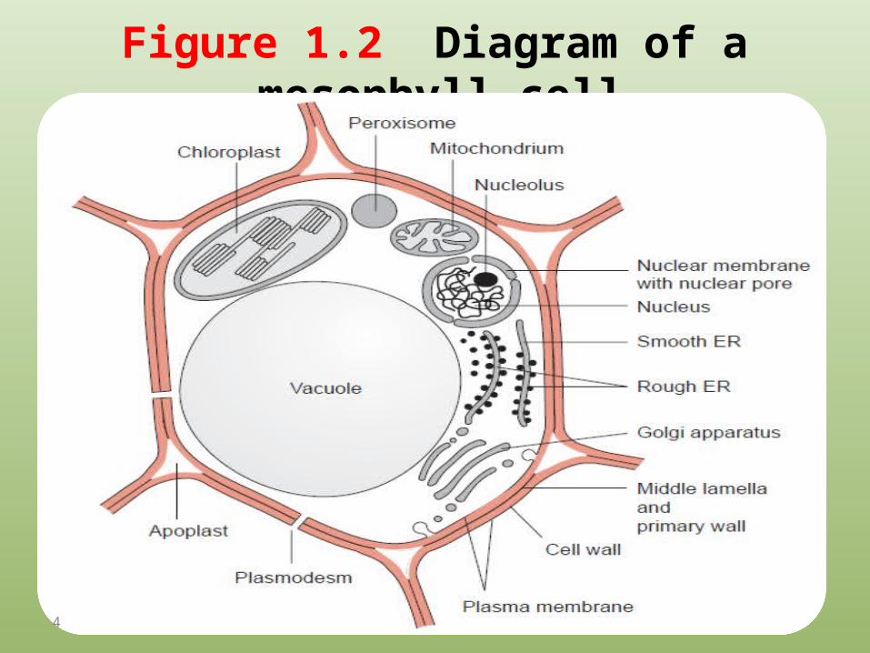
Figure 1.2 Diagram of a mesophyll cell.
4
5

1.1 The cell wall gives the plant cellmechanical stability
Figure 1.3 Main constituents of the cell wall.1.3A. Cellulose
6

1.3B. A hemicellulose
7

1.3C. Constituent of pectin
8

Figure 1.4 Ca++ and Mg++ ions mediate electrostatic interactions between pectin strands.
9
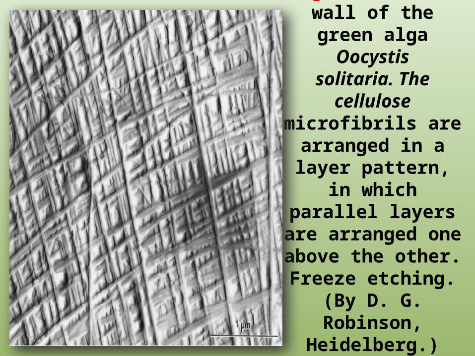
Figure 1.5 Cell wall of the green alga Oocystissolitaria. The cellulose
microfibrils are arranged in a layer pattern, in which parallel layers are
arranged one above the other. Freeze etching. (By D. G.
Robinson, Heidelberg.)
10

Figure 1.6 Plasmodesmata connect neighboring cells to form a symplast . The extracellular spaces between the cell walls form the
apoplast . Schematic representation. Each of the connections shown actually consists of very many neighboring plasmodesmata.
11

Figure 1.7 Diagram of a plasmodesm. The plasma membrane of the neighboring cells is connected by a tubelike membrane invagination. Inside this tube is a continuation of the endoplasmic reticulum. Embedded in the membrane of the ER and the plasma membrane are protein particles that are connected to each other. The spaces between the particles form the diffusion path of the plasmodesm. It is controversial whether a diffusion between the neighboring cells also takes place via the ER lumen. A. cross-sectional view of the membrane B. vertical view
12

1.2 Vacuoles have multiple functions
13
1. An important function of the vacuole is to maintain cell turgor.
Salts, mainly from inorganic and organic acids, are accumulated in the vacuole.
The accumulation of these osmotically active substances draws water into the vacuole.
14

2. recycling those cellular constituents that are defective or no longer required.
Vacuoles contain hydrolytic enzymes for degrading various macromolecules such as proteins, nucleic acids, and many polysaccharides.
Structures, such as mitochondria, can be transferred by endocytosis to the vacuole and are digested there.
The resulting degradation products, such as amino acids and carbohydrates are made available to the cell.
15

3. waste deposits With the exception of gaseous substances,
leaves are unable to rid themselves of waste products or xenobiotics such as herbicides. These are ultimately deposited in the vacuole
16
4. a storage function Many plants use the vacuole to store reserves
of nitrate and phosphate.
17

• The storage function of vacuoles plays a role when utilizing plants as natural protein factories.
• By genetic engineering economically important proteins (e.g., antibodies) could be expressed in plants.
• Since normal techniques could be used for the cultivation and harvest of the plants, this method has the advantage that large amounts of proteins can be produced at low costs.
18
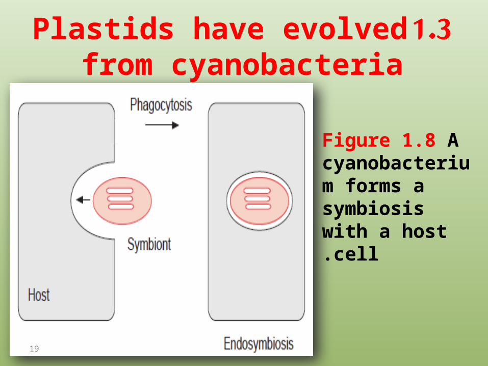
1.3 Plastids have evolved from cyanobacteria
Figure 1.8 Acyanobacterium forms a symbiosis with a host cell.
19

Figure 1.9 Plastids occur in various differentiated forms. A. Proplastid from young primary leaves of Cucurbita pepo (courgette);B. Chloroplast from mesophyll cell of tobaccoleaf fixed at the end of the dark period;
20

C. Leucoplast: amyloplast from the root of Cestrum auranticum; D. Chromoplast from petals also of C. auranticum.
(By D. G. Robinson, Heidelberg.)
21

Figure 1.10 Scheme of
the differentiation of a proplastid
to a chloroplast
22
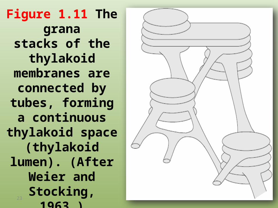
Figure 1.11 The granastacks of the thylakoid
membranes are connected by tubes,
forming a continuous thylakoid space
(thylakoid lumen). (After Weier and Stocking, 1963.)
23
1.4 Mitochondria also result fromendosymbionts
24
Mitochondria are the site of cellular respiration where substrates are oxidized for generating ATP

Figure 1.12 Diagram of the structure of a mitochondrion.
25
1.5 Peroxisomes are the site of reactionsin which toxic intermediates are formed
26

• Peroxisomes contain enzymes catalyzing the oxidation of substances accompanied by the formation of H2O2, and also contain catalase, which immediately degrades this H2O2.
27
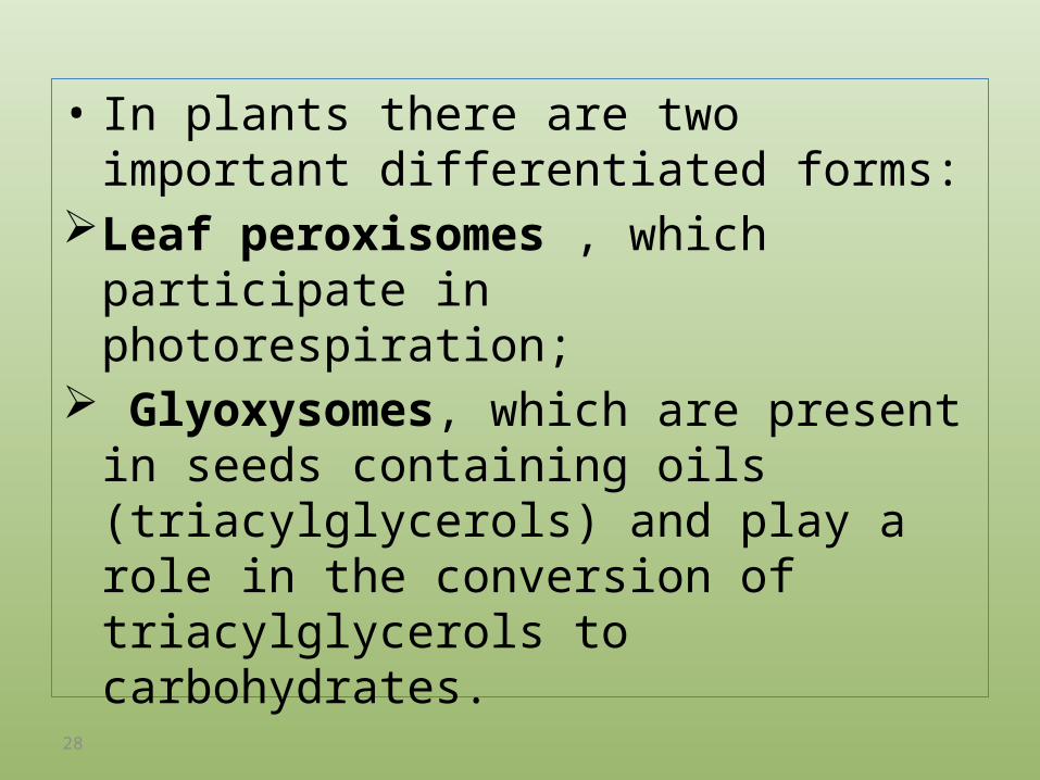
• In plants there are two important differentiated forms:
Leaf peroxisomes , which participate in photorespiration;
Glyoxysomes, which are present in seeds containing oils (triacylglycerols) and play a role in the conversion of triacylglycerols to carbohydrates.
28
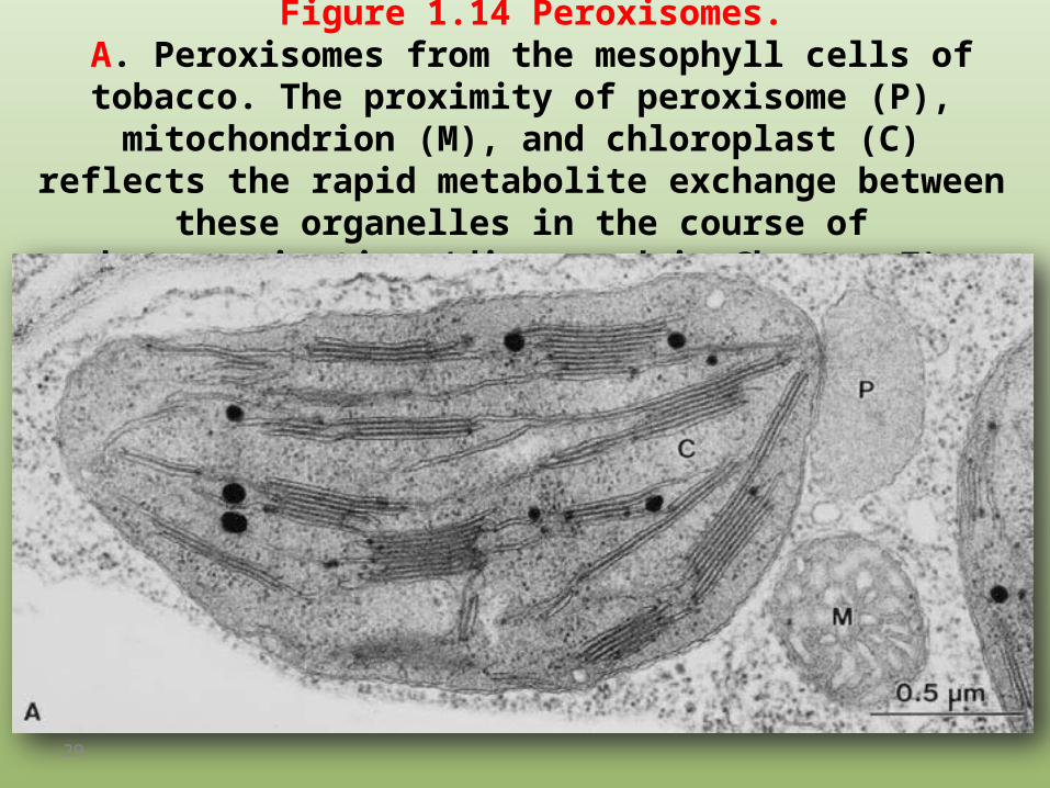
Figure 1.14 Peroxisomes. A. Peroxisomes from the mesophyll cells of tobacco. The proximity of peroxisome (P), mitochondrion (M), and chloroplast (C) reflects
the rapid metabolite exchange between these organelles in the course of photorespiration (discussed in Chapter 7).
29

B. Glyoxysomes from germinating cotyledons of Cucurbita pepo (courgette).
The lipid degradation described in section 15.6 and the accompanying gluconeogenesis require a close contact between lipid droplets (L),
glyoxysome (G), and mitochondrion (M). (By D. G. Robinson, Heidelberg.)
30

1.6 The endoplasmic reticulum and Golgi apparatus form a
network for the distribution of biosynthesis products
31

Figure 1.15 Roughendoplasmic reticulum, cross section (arrows) and tangential
sections (arrowheads). The ribosomes temporarily attached to the membrane occur as polysome complexes (ribosome + mRNA).
Section from the cell of a maturing peacotyledon. (By D. G. Robinson, Heidelberg.)
32

Figure 1.16 Scheme of the interplay between the endoplasmic reticulum and the Golgi apparatus in the transfer of proteins from the ER to the vacuoles and in the secretion of proteins from the cell.
33

Figure 1.17 Golgi apparatus (dictyosome) in the green alga Chlamydomonas reinhardii. C = cis side, t = trans side. Arrowheads point to the trans Golgi network. The swollen endoplasmatic reticulum (ER) is typical for this cell. On the ER, ribosomes can be
recognized, except in the area where vesicles bud off. (By D. G. Robinson, Heidelberg.)
34

• The proteins destined for the lytic vacuoles are transferred in clathrin-coated vesicles.
• Clathrin is a protein consisting of two different subunits (α- 180,000 Dalton, β- 35,000 to 40,000 Dalton).
• Both 3α - and 3β -subunits form a complex with three arms (Triskelion), which polymerizes to a hexagonal latticed structure surrounding the vesicle
35

Figure 1.18 Model of the structure of clathrin coated vesicles.. (A) 3α and 3β subunits of clathrin form a complex with three arms. (B) From this a hexagonal and pentagonal lattice (the latter not
shown here) is formed by polymerization and this forms (C) the coat. (From Kleinig and Sitte.)
36

1.7 Functionally intact cell organelles can
be isolated from plant cells
37

Figure 1.19 Protocol
for the isolation of functionally
intact chloroplasts.
38
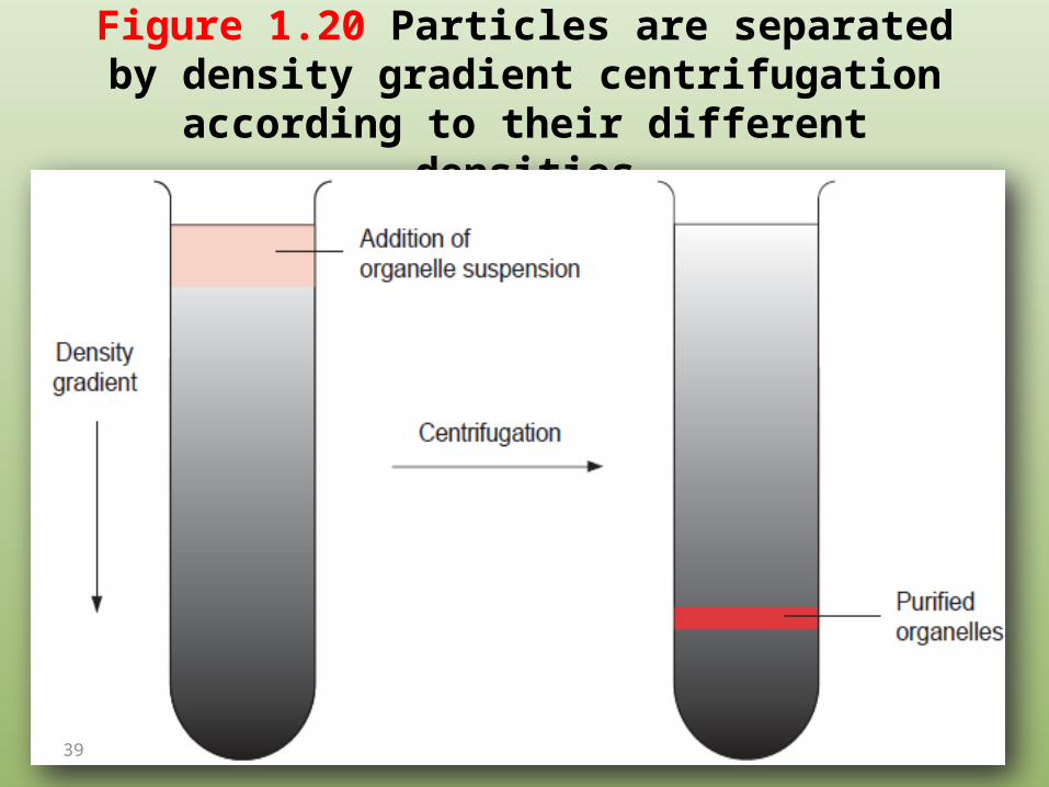
Figure 1.20 Particles are separated by density gradient centrifugation according to their different densities
39
1.8 Various transport processes facilitate the exchange of
metabolites between different compartments
40

Figure 1.21 Classification of membrane transport processes.
41
1.9 Translocators catalyze the specific transport of substrates
and products of metabolism
42
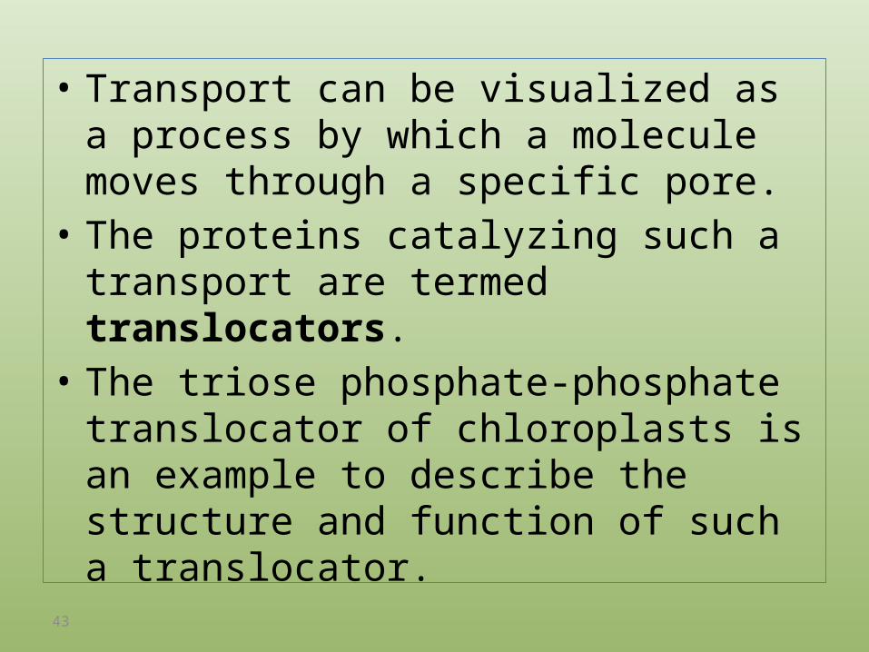
• Transport can be visualized as a process by which a molecule moves through a specific pore.
• The proteins catalyzing such a transport are termed translocators.
• The triose phosphate-phosphate translocator of chloroplasts is an example to describe the structure and function of such a translocator.
43

• Silicone layer filtering centrifugation is a very useful tool (Fig. 1.22) for measuring the uptake of substrates into chloroplasts or other cell organelles.
• The corresponding substrate is added to a suspension of isolated chloroplasts and is terminated by separating the chloroplasts from the surrounding medium by centrif-ugation through a silicone layer.
• The amount of substrate taken up into the separated chloroplasts is then quantitatively analyzed.
44

Figure 1.22 Silicone oil filtering centrifugation
45
The substance to betransported is added to the chloroplast suspension above the silicone layer using asmall spatula. To simplify detection, metabolites labeled with radio isotopes (e.g.,32P or 14C ) are usually used. The uptake of metabolites into the chloroplasts is terminated by centrif-ugation in a rapidly accelerating centrifuge.
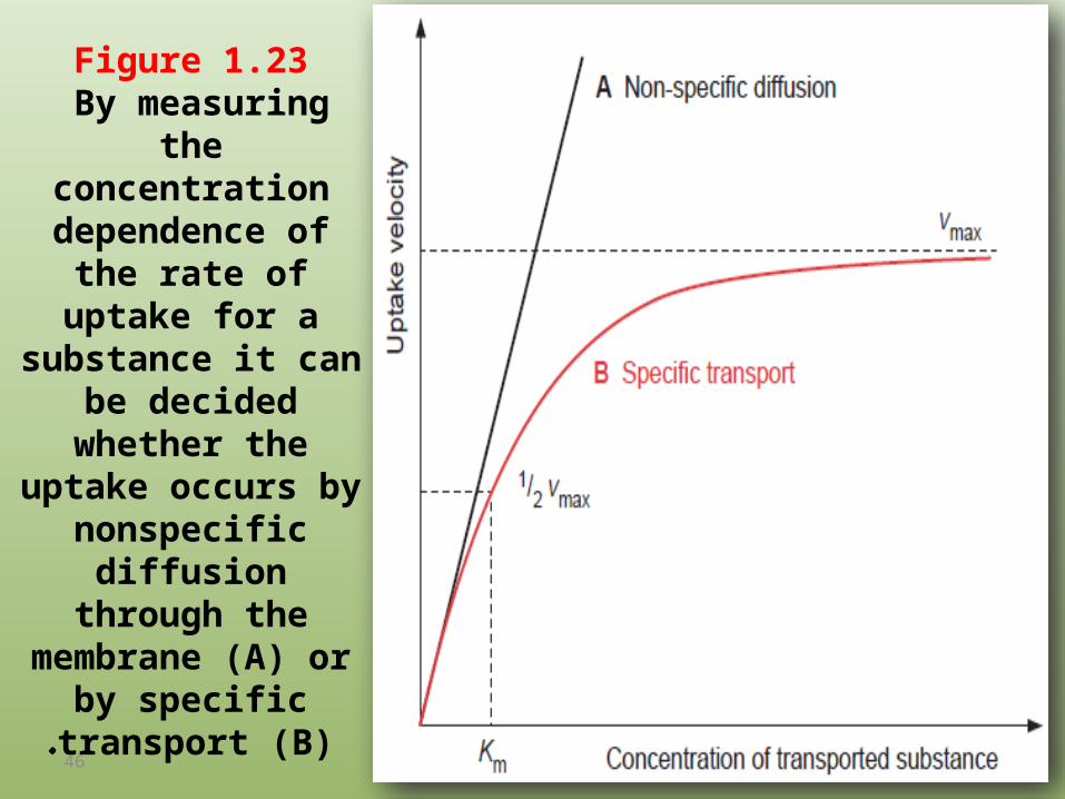
Figure 1.23 By measuring the
concentration dependence of the rate
of uptake for a substance it can be
decided whether the uptake occurs by
nonspecific diffusion through the membrane
(A) or by specific transport (B).
46
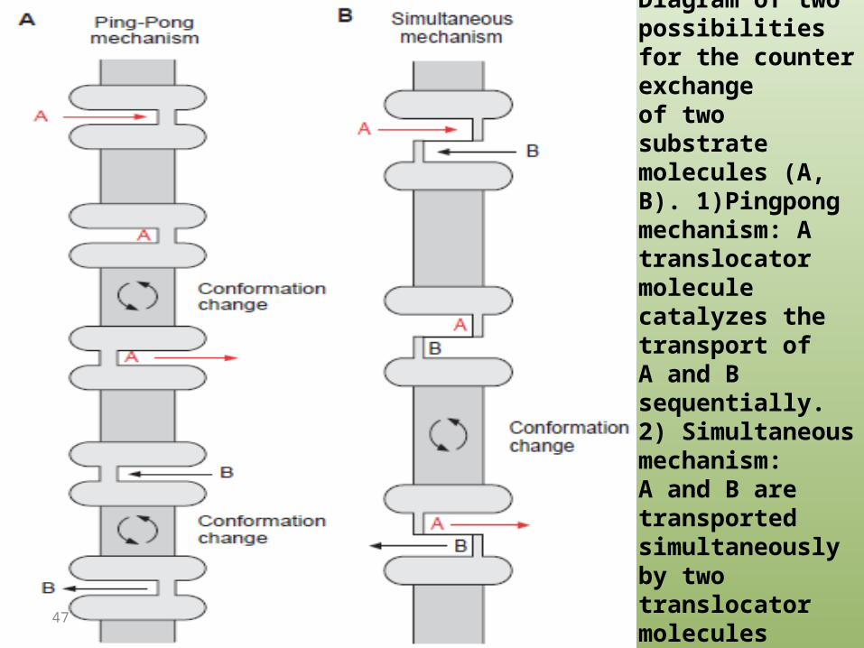
Figure 1.24 Antiport.Diagram of twopossibilities for the counter exchangeof two substratemolecules (A, B). 1)Pingpongmechanism: Atranslocator molecule catalyzes the transport ofA and B sequentially. 2) Simultaneous mechanism:A and B are transportedsimultaneously by two translocator moleculestightly coupled to each other.
47

Translocators have a common basic structure
• Translocators are integral membrane proteins. They traverse the lipid bilayer of the membrane in the form of α-helices. In such transmembrane α-helices the side chains have to be hydrophobic;
• therefore they contain hydrophobic amino acids such as alanine, valine, leucine, isoleucine, or phenylalanine.
48

• Mild nonionic detergents, such as octylglucoside (Fig. 1.25), can be used to dissolve these proteins from the membrane. The hydrophobic carbon chain of the detergent binds to the hydrophobic protein and, due to the glucose residues, the micelle thus formed is water-soluble.
• If the detergent is then removed, the membrane proteins aggregate to a sticky mass that cannot be solubilized again.
49

Figure 1.25 Octylglucoside, a glycoside composed from a-Dglucose and octyl alcohol, is a mild nonionic detergent that allows
membrane proteins to be solubilized from the membranes without being denatured.
50

Figure 1.26 The triosephosphate-phosphate
translocator from spinach forms six transmembrane
helices. Each circlerepresents one amino
acid. The likely positions of the transmembrane helices were evaluated
from the hydrophobicity of the single amino acid
residues. The amino acids, marked
with red, containing apositive charge in helix 5, represent an arginine and
a lysine. These amino acids probably provide the binding sites for the anionic substrates of the
triose phosphate-phosphate translocator. (Data from Flügge et al.,
1989.)51

1.10 Ion channels have a very high transport capacity
52

• The chloroplast triose P-P translocator mentioned previously has a turnover number of 80 s-1 at 25°C, which means that it transports 80 substrate molecules per second.
• The turnover numbers of other translocators are in the range of 10 to 1,000 s-1.
• Membranes also contain proteins which form ion channels that transport various ions at least three orders of magnitude faster than translocators (106 –108 ions per second).
53
• They differ from the translocators in having a pore open to both sides at the same time.
• The flux of ions through the ion channel is so large that it is possible to determine the transport capacity of a single channel from the measurement of electrical conductivity.
• The procedure for such single channel measurements, called the patch clamp technique
54

Figure 1.27 Measurement of ion channel currents by the “patch clamp” technique
55
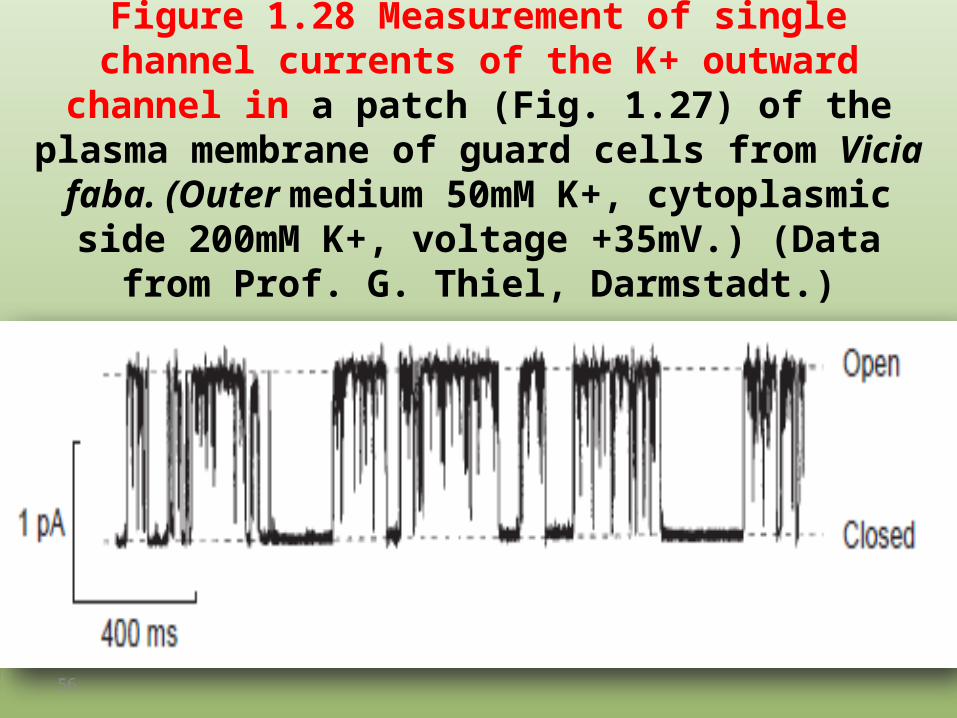
Figure 1.28 Measurement of single channel currents of the K+ outward channel in a patch (Fig. 1.27) of the plasma
membrane of guard cells from Vicia faba. (Outer medium 50mM K+, cytoplasmic side 200mM K+, voltage +35mV.)
(Data from Prof. G. Thiel, Darmstadt.)
56

It has long been known that the channel protein is built from two identical subunits, each of which has two transmembrane helices connected by
a sequence of about 30 amino acids (loop) (Fig. 1.29A).
57

Structure analysis showed that a K+ channel is built of four of thesesubunits (Fig. 1.29B, C).
58

1.11 Porins consist of β-sheet structures
59

• The outer membranes of chloroplasts and mitochondria appear to be unspecifically permeable to metabolites such as, for instance, nucleotides and sugar phosphates. This relatively unspecific permeability is due to pore-forming proteins named porins.
• The size of the aperture of the pore formed by a porin can be determined by incorporating porins into an artificial lipid membrane that separates two chambers filled with an electrolyte (Fig. 1.30).
60

Figure 1.30 Measurement of the size of a porin aperture
61
Membrane proteins, which have been solubilized in a detergent, are added to one of the two chambers.Because of their hydrophobicity, the porin molecules incorporate, one after another, into the artificial lipid membrane, each time forming a new channel, which can be seen in the stepwise increase in conductivity.

Figure 1.31 With β-sheet
conformation the amino acid residues
of a peptide chain are arranged alternately
in front of and behind the surface of the
sheet.
62

Figure 1.32 Diagram of the structure of a membrane pore formed by a porin.
Figure A shows the view from above
63

figure B shows a cross section through the membrane. Sixteen β-sheet sequences of the porin molecules, each 13 amino
acids long, form the pore. The amino acid residues directed toward the membrane side of the pore have hydrophobic character; those
directed to the aqueous pore are hydrophilic.
64