Pineal region masses
22
PINEAL REGION MASS Dr Mohit Goel JR 1 15 Dec. 2012
-
Upload
anish-choudhary -
Category
Health & Medicine
-
view
178 -
download
1
Transcript of Pineal region masses

PINEAL REGION MASS
Dr Mohit Goel
JR 1
15 Dec. 2012

Pineal Region Mass
Signs and symptoms of pineal region masses are most often related to mass
effect on the adjacent structures, but higher-grade structures, such as a pineoblastoma,
may also invade the surrounding tissue.
These signs and symptoms include Parinaud syndrome (it consists of a failure of
conjugate vertical eye movement, mydriasis, failed ocular convergence, and
blepharospasm),
precocious puberty,
and, rarely, pineal apoplexy.
Hydrocephalus results from obstruction of the aqueduct of Sylvius; patients may also
develop headache, nausea, and vomiting as a result of increased intracranial pressure.
Precocious puberty is more commonly associated with germ cell tumors (GCTs) and
may be related to increased human chorionic gonadotropin (hCG) secreted by the
tumor.
Hemorrhage into a pineal tumor or cyst is referred to as pineal apoplexy; the most
common presenting symptom is a sudden decrease in consciousness associated with
headache.
Tumors of Pineal Parenchymal Origin
Pineal parenchymal tumors are rare lesions, accounting for less than 0.2% of
intracranial neoplasms.
These lesions include
• the low-grade pineocytoma,
• the intermediate-grade pineal parenchymal tumor of intermediate differentiation
(PPTID), and
• the highly malignant pineoblastoma.
Pineocytoma
Pineocytoma is a slow-growing lesion that accounts for 14%–60% of pineal parenchymal
neoplasms.
Imaging Findings.—
At computed tomography (CT), pineocytomas are well demarcated, usually less than 3
cm, and iso- to hyperattenuating.
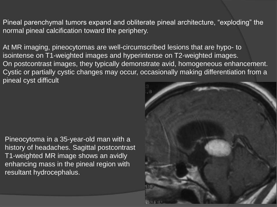
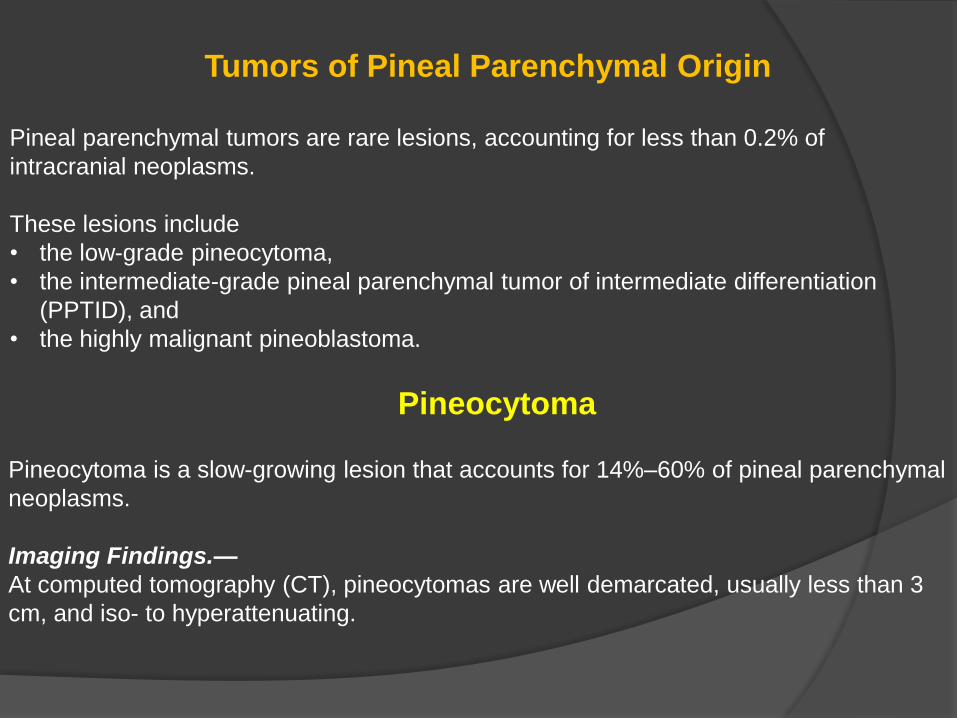
Pineal parenchymal tumors expand and obliterate pineal architecture, “exploding” the
normal pineal calcification toward the periphery.
At MR imaging, pineocytomas are well-circumscribed lesions that are hypo- to
isointense on T1-weighted images and hyperintense on T2-weighted images.
On postcontrast images, they typically demonstrate avid, homogeneous enhancement.
Cystic or partially cystic changes may occur, occasionally making differentiation from a
pineal cyst difficult
Pineocytoma in a 35-year-old man with a
history of headaches. Sagittal postcontrast
T1-weighted MR image shows an avidly
enhancing mass in the pineal region with
resultant hydrocephalus.
Pineal Parenchymal Tumor of Intermediate Differentiation
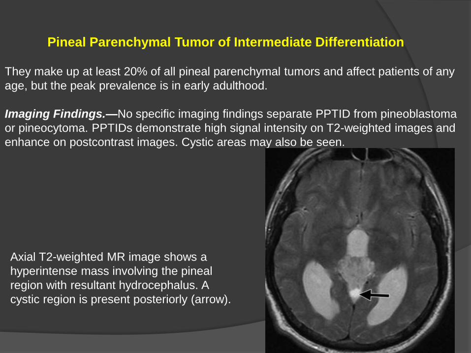
They make up at least 20% of all pineal parenchymal tumors and affect patients of any
age, but the peak prevalence is in early adulthood.
Imaging Findings.—No specific imaging findings separate PPTID from pineoblastoma
or pineocytoma. PPTIDs demonstrate high signal intensity on T2-weighted images and
enhance on postcontrast images. Cystic areas may also be seen.
Axial T2-weighted MR image shows a
hyperintense mass involving the pineal
region with resultant hydrocephalus. A
cystic region is present posteriorly (arrow).

Pineoblastoma
Pineoblastomas are highly malignant lesions that represent the most primitive form of
pineal parenchymal tumors and account for 40% of pineal parenchymal tumors.
They are embryonal tumors described as a primitive neuroectodermal tumor of the
pineal gland.
They most commonly occur in the first 2 decades but can occur at any age, and there is
no gender predilection.
CSF dissemination is a common finding and necessitates imaging of the entire
craniospinal axis and it is the most common cause of death.
Imaging Findings.—
CT reveals a large (typically ≥3 cm), lobulated, typically hyperattenuating mass, an
appearance that reflects its highly cellular histologic features.
The pineal calcifications, if seen, may appear exploded at the periphery of the lesion.
Nearly 100% of patients have obstructive hydrocephalus.

At MR imaging, pineoblastomas are heterogeneous in appearance, with the solid
portion appearing hypo- to isointense on T1-weighted images and iso- to mildly
hyperintense to the cortex on T2-weighted images. Pineoblastomas demonstrate
heterogeneous enhancement on postcontrast images.
Pineoblastoma in a 10-year-old girl with
lethargy, emesis, and downward gaze.
Axial nonenhanced CT image shows a
large pineal region mass with resultant
hydrocephalus. The pineal calcifications
are exploded toward the periphery
(arrows).

Pineoblastoma in a 4-year-old boy with nausea and vomiting.
(a) Sagittal T2-weighted MR image shows a large mass in the pineal region with
resultant hydrocephalus. The mass is hyperintense relative to gray matter.
(b) Postcontrast T1-weighted MR image shows heterogeneous enhancement within the
mass.

Germ Cell Tumors
Central nervous system GCTs are most commonly located in the pineal and suprasellar
regions.
The WHO classifies them into
• germinomas and
• nongerminomatous GCTs.
The nongerminomatous GCTs include
• teratomas,
• embryonal carcinoma,
• yolk sac tumor,
• choriocarcinoma, and
• the mixed GCTs.
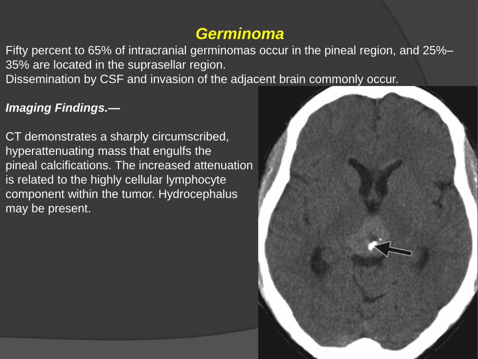
GerminomaFifty percent to 65% of intracranial germinomas occur in the pineal region, and 25%–
35% are located in the suprasellar region.
Dissemination by CSF and invasion of the adjacent brain commonly occur.
Imaging Findings.—
CT demonstrates a sharply circumscribed,
hyperattenuating mass that engulfs the
pineal calcifications. The increased attenuation
is related to the highly cellular lymphocyte
component within the tumor. Hydrocephalus
may be present.
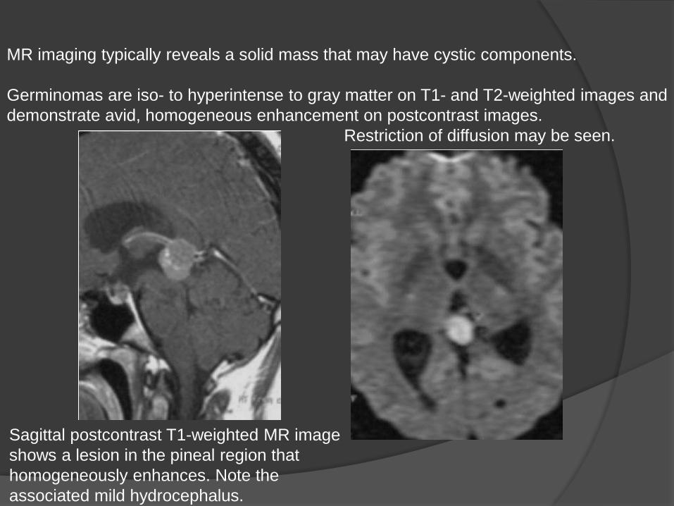
MR imaging typically reveals a solid mass that may have cystic components.
Germinomas are iso- to hyperintense to gray matter on T1- and T2-weighted images and
demonstrate avid, homogeneous enhancement on postcontrast images.
Restriction of diffusion may be seen.
Sagittal postcontrast T1-weighted MR image
shows a lesion in the pineal region that
homogeneously enhances. Note the
associated mild hydrocephalus.
Teratoma
There are three types of teratoma: mature teratoma (fully differentiated tissue), immature
teratoma (complex mixture of fetal-type tissues from all three germ layers and mature
tissue elements), and teratoma with malignant transformation.
Imaging Findings.—
CT reveals a multiloculated, lobulated lesion with foci of fat attenuation, calcification, and
cystic regions.
T1-weighted MR images may show foci of T1 shortening due to fat and variable signal
intensity related to calcification.
On T2-weighted images, the soft-tissue component is iso- to hypointense. The soft-
tissue component demonstrates enhancement on postcontrast images. The malignant
form may have a more homogeneous imaging appearance (fewer cysts and
calcifications).

Axial T1-weighted MR image shows a
lobulated, heterogeneous lesion that
contains an area of hyperintensity (arrow),
a finding consistent with fat.
Postcontrast MR image shows
enhancement of the soft-tissue portions
of the lesion.

Trilateral Retinoblastoma
Trilateral retinoblastoma refers to the presence of bilateral ocular retinoblastoma and an
intracranial, typically midline, small cell tumor. Intracranial tumors associated with
retinoblastoma occur most frequently in the region of the pineal gland (pineoblastoma).
The second most common location is the suprasellar region.
Trilateral retinoblastoma in a 2-year-old girl with a history of enucleation for retinoblastoma. (a) Axial
postcontrast fat-saturated T1-weighted MR image shows a focus of enhancement along the medial
wall of the left globe (arrow), a finding consistent with retinoblastoma. The right globe was removed
due to retinoblastoma, and a prosthesis is in place. (b) Axial postcontrast T1-weighted MR image
shows an associated enhancing pineoblastoma with resultant hydrocephalus.

Pineal Cyst
Pineal cysts occur in all age ranges but are most predominant in adults 40–49
years of age.
Their origin is debated, with some suggesting they result from degenerative
changes in the gland.
These lesions are typically asymptomatic and are usually 2–15 mm in size.
Follow-up studies have indicated that these lesions remain stable in size over
time. When they exceed 15 mm, patients may become symptomatic, typically
with headache or visual changes.
Intracystic hemorrhage (“pineal apoplexy”) and acute hydrocephalus rarely
occur; resultant death has been reported.
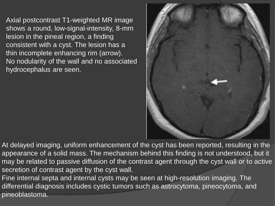
Axial postcontrast T1-weighted MR image
shows a round, low-signal-intensity, 8-mm
lesion in the pineal region, a finding
consistent with a cyst. The lesion has a
thin incomplete enhancing rim (arrow).
No nodularity of the wall and no associated
hydrocephalus are seen.
At delayed imaging, uniform enhancement of the cyst has been reported, resulting in the
appearance of a solid mass. The mechanism behind this finding is not understood, but it
may be related to passive diffusion of the contrast agent through the cyst wall or to active
secretion of contrast agent by the cyst wall.
Fine internal septa and internal cysts may be seen at high-resolution imaging. The
differential diagnosis includes cystic tumors such as astrocytoma, pineocytoma, and
pineoblastoma.
Astrocytoma
Astrocytomas arising in the pineal region are uncommon. They derive from stromal
astrocytes, and in the pineal region they arise from the splenium of the corpus callosum,
the thalamus, or the tectum of the midbrain. Rarely, they may arise from the neuronal
elements within the pineal gland.
Those that occur in the region of the tectum are usually low grade (WHO grade I or II)
and result in enlargement of the tectum with secondary obstruction of the aqueduct.
At MR imaging, bulbous enlargement of the tectal plate is noted. The lesion is typically
isointense on T1-weighted images and hyperintense on T2-weighted images with no to
minimal enhancement on postcontrast images.
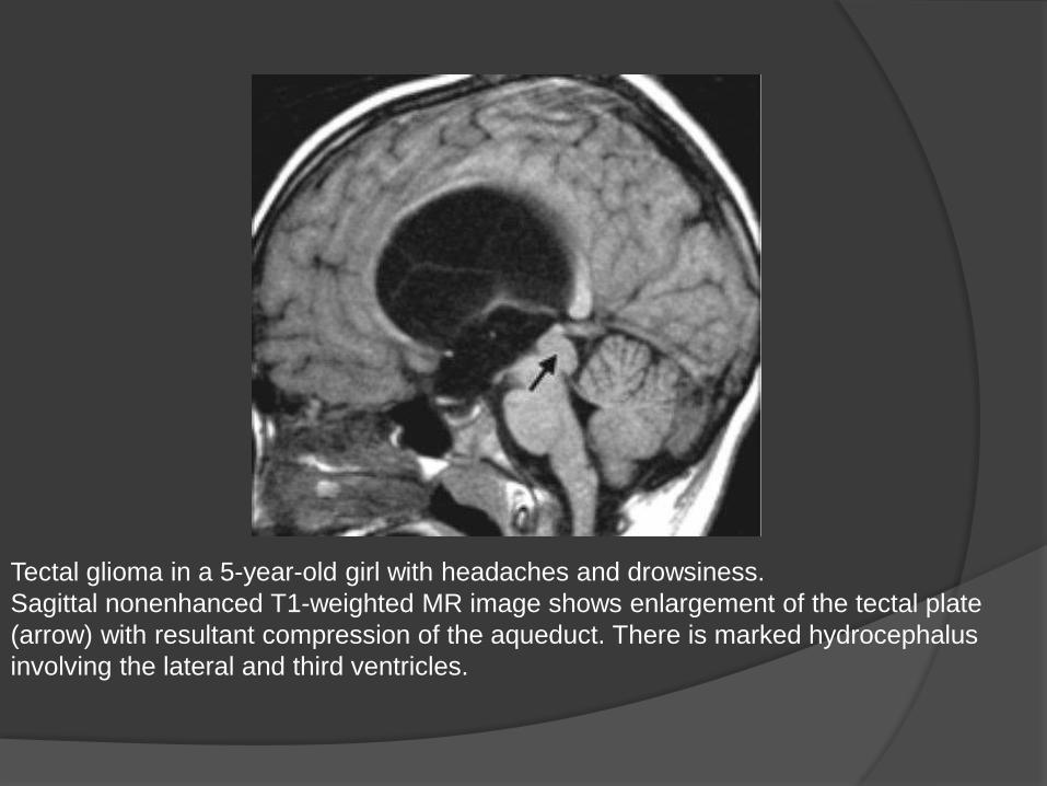
Tectal glioma in a 5-year-old girl with headaches and drowsiness.
Sagittal nonenhanced T1-weighted MR image shows enlargement of the tectal plate
(arrow) with resultant compression of the aqueduct. There is marked hydrocephalus
involving the lateral and third ventricles.
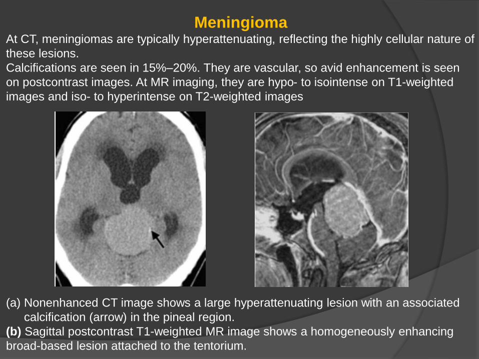
MeningiomaAt CT, meningiomas are typically hyperattenuating, reflecting the highly cellular nature of
these lesions.
Calcifications are seen in 15%–20%. They are vascular, so avid enhancement is seen
on postcontrast images. At MR imaging, they are hypo- to isointense on T1-weighted
images and iso- to hyperintense on T2-weighted images
(a) Nonenhanced CT image shows a large hyperattenuating lesion with an associated
calcification (arrow) in the pineal region.
(b) Sagittal postcontrast T1-weighted MR image shows a homogeneously enhancing
broad-based lesion attached to the tentorium.

Metastasis
Metastases to the pineal gland are rare.
The most common tumors to spread to the pineal region are those of the lung (most
frequent), breast, kidney, esophagus, stomach, and colon.
Pineal metastases may be present without metastases to the brain parenchyma.

Thank you