Physics SP Skills Unit - Wikispacesbradyfonnesbeck.wikispaces.com/file/view/MaP Unit 6... · Unit...
74
Day 1 (10/31/11)
Transcript of Physics SP Skills Unit - Wikispacesbradyfonnesbeck.wikispaces.com/file/view/MaP Unit 6... · Unit...

Day 1 (10/31/11)
Your personalized sheet will be forthcoming! (It has mistakes though )
Play the hand slap game while you are waiting!

Unit Cat Title Page #
Comp Check
Due Date
6 Act General Functions & Structure of the Nervous System (pgs 356-361)
11/10
•Nerve cells that are specialized to react to physical and chemical changes in their surroundings.
Neurons
Nerve Impulse
•Bioelectric signal sent by neurons.

(Cut –n-paste) •Main group of Nervous system consisting of the brain and spinal cord. (CNS)
Central Nervous System
Peripheral Nervous System
•Main group of Nervous system consisting of cranial and spinal branches and nerves. (PNS)

Divison of the PNS that receives and sends information to the CNS.
Sensory Division of
PNS
Motor Division of
the PNS
•Somatic Nervous System – oversees voluntary activities (ex. Skeletal muscle)
•Autonomic Nervous System – oversees involuntary activites (ex. Heart beat, digestion)

•Sensory – taking information back to the CNS
•Integrative – direct sensory information to appropriate areas for processing.
•Motor – conduct impulses from the CNS to effector organs (ex. Glands, muscles).
3 general functions of the nervous
system

•Dendrites – receptors surrounding the cell body of neuron.
•Cell Body – processing center of neuron; contains nucleus.
•Axon – conducts nerve impulses away from the cell body.
•Axon Terminal – axon ending that releases neurotransmitters into the synaptic cleft.
•Myelin – insulating material that speeds up transmission.
Typical Neuron
Anatomy (cut-n-paste)

1. Dendrites (Arms up)
2. Cell Body (Halo over head)
3. Axon (Arm out)
4. Myelin Sheath (rotate arm)
5. Axon Terminals (Wave fingers)
6. Neurtransmission (Flick fingers in and out)
7. Synapse (Jump to the side)
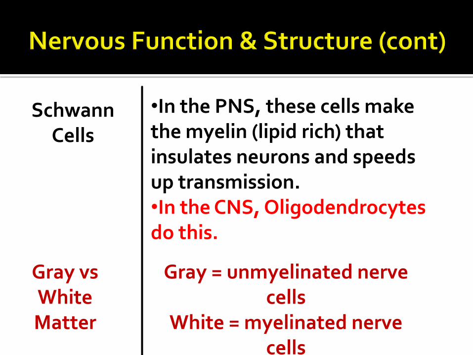
•In the PNS, these cells make the myelin (lipid rich) that insulates neurons and speeds up transmission. •In the CNS, Oligodendrocytes do this.
Schwann Cells
Gray vs White Matter
Gray = unmyelinated nerve cells
White = myelinated nerve cells
Migraines! http://www.youtube.com/watch?v=yL22X5opG
hY&safety_mode=true&persist_safety_mode=1
Cluster Headache Treatment? (Does he need
one?) http://www.youtube.com/watch?NR=1&v=YAb
sDPGwExc&safety_mode=true&persist_safety_mode=1

Neuroglial Cells = cells that help support neurons in the nervous system.
In your group, come up with a charade for
each of the different types of neuron and neuroglial cells listed in the table that you cut and pasted in.
We will be using these as part of the payday
next class!
Unit Cat Title Page #
Comp Check
Due Date
6 Act Neuron and Neuroglial Charades 11/10

Study your notes from today for next class’ payday!
Unit Cat Title Page #
Due Date
2&3 HW Properties of Water 9/17
Unit Cat Title Page #
Comp Check
Due Date
6 HW Study your notes from today for the Payday!
11/10

Day 2 (11/2/11)

Today’s Payday will be a combination of charades and a review using the Power Point of what we did last time!

Network of connected cells, tissue, and organs
Controls thoughts, movement, life processes
Quick responses
Ex: Sunny day pupils shrinking


Also known as Nerve Cells Transfer electrical impulses to/from the brain Three Main Parts
1) Cell body: contains nucleus and organelles
2) Dendrites: branches that receive messages from neighboring cells
3) Axon: extension that carries messages away from the cell body
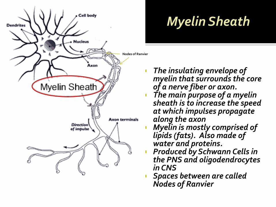
The insulating envelope of myelin that surrounds the core of a nerve fiber or axon.
The main purpose of a myelin sheath is to increase the speed at which impulses propagate along the axon
Myelin is mostly comprised of lipids (fats). Also made of water and proteins.
Produced by Schwann Cells in the PNS and oligodendrocytes in CNS
Spaces between are called Nodes of Ranvier
Myelin Sheath
Nodes of Ranvier

•White matter contains myelinated nerves, while gray matter does not. •Why do we have gray and white matter? •What difference does that make in nerve impulses.

Today you won’t be given organized notes! You will be on your own. I am going to go through the slides quickly so get the main points!
Unit Cat Title Page #
Comp Check
Due Date
6 Act Nerve Impulse Transmission 11/10

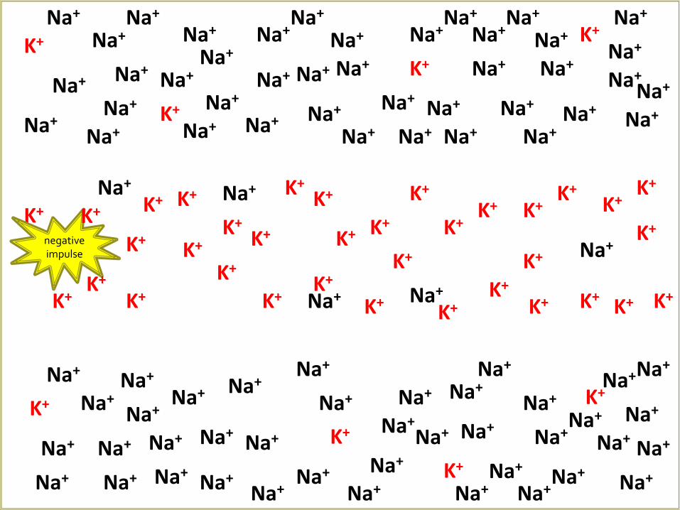
Neuron at rest Neuron Exterior:
Positive charge
Mainly Na+ outside the cell
Neuron Interior:
Negative charge (from various proteins)
K+ inside the cell

Electrical impulse is triggered…
Front end of impulse
Channels open and allow Na+ to enter the cell
Negative impulse attracted towards positive Na+ (High + area)
Back end of impulse
Channels open to allow K+ to exit the cell
Interior returns to normal (- charge, bc of proteins)
Impulse pulled along by the changing of electrical charges

impulse
Action Potential Video
Na+ Na+
Na+ Na+
Na+
Na+
Na+ Na+
Na+ Na+
Na+
Na+
Na+
Na+
Na+
Na+
Na+
Na+ Na+
Na+ Na+
Na+
Na+
Na+ Na+
Na+ Na+
Na+
Na+
Na+
Na+
Na+
Na+
Na+
K+
K+
K+
K+
K+
K+
K+
K+
K+
K+
K+
K+
K+
K+
Na+
Na+
Na+
Na+
Na+
Na+
Na+ K+
K+
K+
K+
K+
K+
K+
K+
K+
K+
K+
K+
K+
K+
K+ K+
Na+ Na+
Na+ Na+
Na+
Na+
Na+
Na+
Na+
Na+ Na+
Na+
Na+
Na+
Na+ Na+
Na+
Na+ Na+
Na+
Na+
Na+ Na+
Na+
Na+
Na+ Na+ Na+ Na+
Na+
Na+ Na+
Na+
Na+
Na+
Na+
Na+
Na+ Na+
Na+
Na+
Na+ Na+
Na+ Na+
Na+
K+ K+
K+
K+ negative impulse
K+ K+
K+
K+
K+
K+
K+ K+
K+
impulse

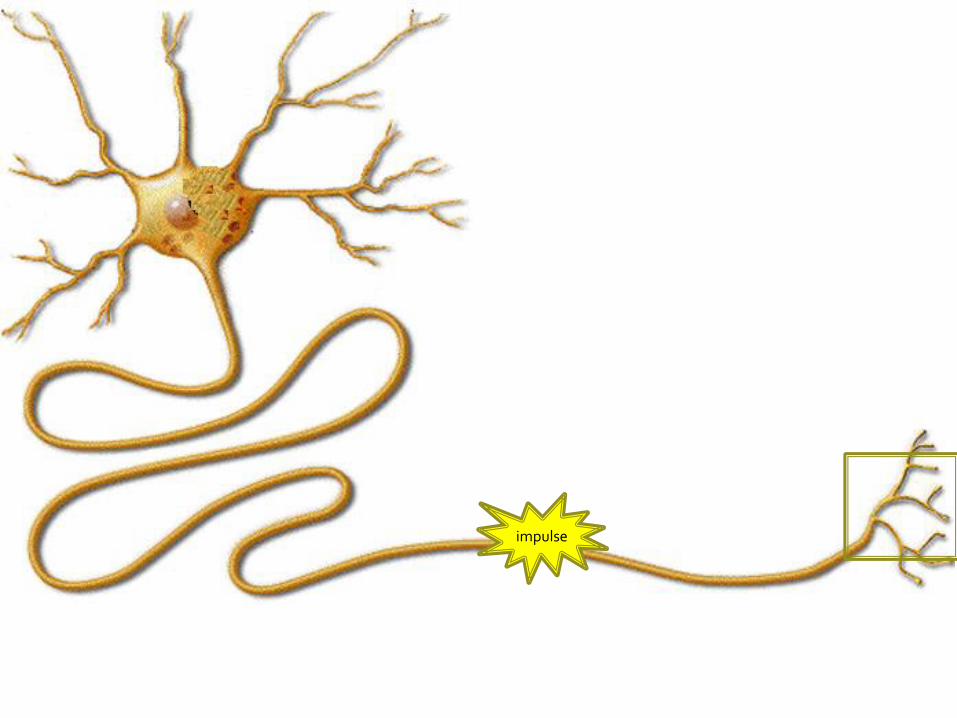
The gap between neurons
Problem: Impulse cannot
cross the gap Solution: Impulse
converted into chemical molecules (neurotransmitters)
Steps:
Impulse reaches neuron’s end
Vesicle releases neurotransmitters
Neurotransmitters attach to neighboring neuron
New impulse created

Feet
Brain

Impulse causes the muscles to contract…this causes MOVEMENT!


Groups of 3 only
Roles: Pricker, Patient, Measurer
8 Different Places, 3 trials, average of trials.
Must present findings visually in lab book.
Answer the guiding questions, use them to prepare a 2 min presentation explaining your results!
*Rules for where you can test!

Read the “Meninges” and “Ventricles & Cerebrospinal Fluid” Sections from page 386-389 in your text and answer the following questions(write the questions with the answer and draw a figure if it provides a good answer also):
1. Describe the Meninges.
2. Name the layers of the meninges.
3. Explain where cerebrospinal fluid is located.
4. Where are the ventricles of the brain located?
5. How does CSF form?
6. What is the function of CSF?
Unit Cat Title Page #
Comp Check
Due Date
6 HW Meninges, Ventricles, & Cerebrospinal Fluid 11/4 11/10

Day 3 (11/4/11)
I will be checking the completion of your Meninges, Ventricles, & Cerebrospinal Fluid
Homework from last time during the quiz!

You had a substitute in class today and after the Payday Quiz you were given 30 minutes to work on the study guide (attached below) before we started watching the “Inside the Living Body” video some more.
This study guide needs to be complete by the day
the book is turned in on exam day (this coming Thursday)!
Unit Cat Title Page #
Comp Check
Due Date
6 Act The Brain: Diencephalon, Brain Stem, & Diseases and DO Study Guide
11/10

Day 4 (11/8/11)

Sit next to someone you want to partner with, just one other person – no groups of three!
Write the following (individually):
In 10-15 words, describe the most beautiful thing you have ever seen.
In 10-15 words, describe the most beautiful thing you have ever heard.
When I tell you to, share the things you wrote with your partner.

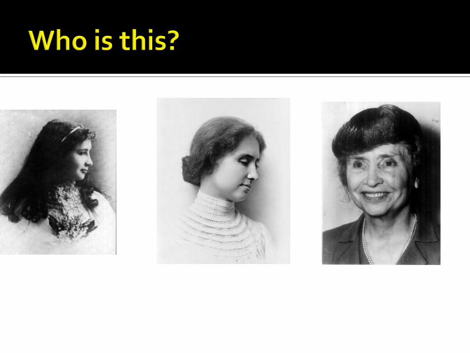
Lived from 1880 – 1968
Born in Alabama
Lost her sense of sight and hearing after contracting scarlet fever or meningitis at 19 months old.
Was taught by Anne Sullivan how to communicate and eventually learned how to speak and became a well known writer.
Awarded Presidential Medal of Freedom and was a political activist for Women’s suffrage, Workers Rights, and even Socialism.
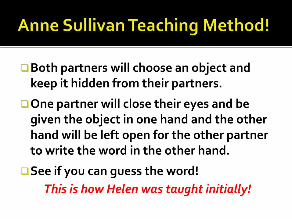
Both partners will choose an object and keep it hidden from their partners.
One partner will close their eyes and be given the object in one hand and the other hand will be left open for the other partner to write the word in the other hand.
See if you can guess the word!
This is how Helen was taught initially!

How did she learn to speak?
http://www.youtube.com/watch?v=Gv1uLfF35Uw&safety_mode=true&persist_safety_mode=1

Explain how the eye works to give us sight in 15-30 words after watching the you tube clip.
Explain how the ear works to allow us to hear in 15-30 words after watching the you tube clip.
Sight
Unit Cat Title Page #
Comp Check
Due Date
6 Act Eye and Ear Notes 11/10
Hearing

Eye http://www.youtube.com/watch?v=gvozcv8pS3
c&safety_mode=true&persist_safety_mode=1
Ear http://www.youtube.com/watch?v=7O-adw-
HyrQ&safety_mode=true&persist_safety_mode=1
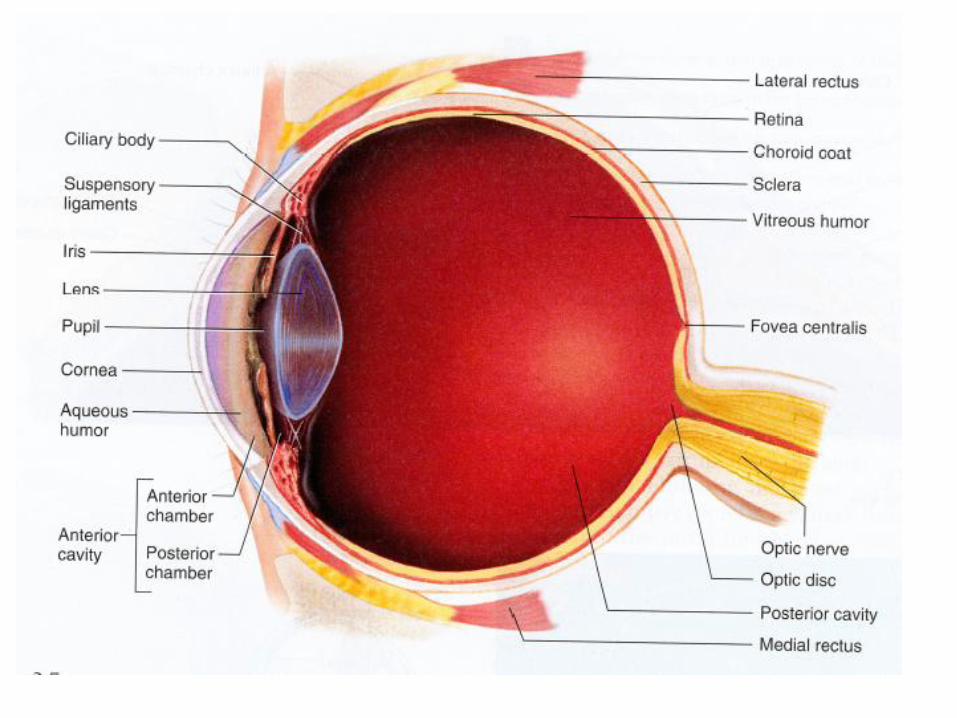
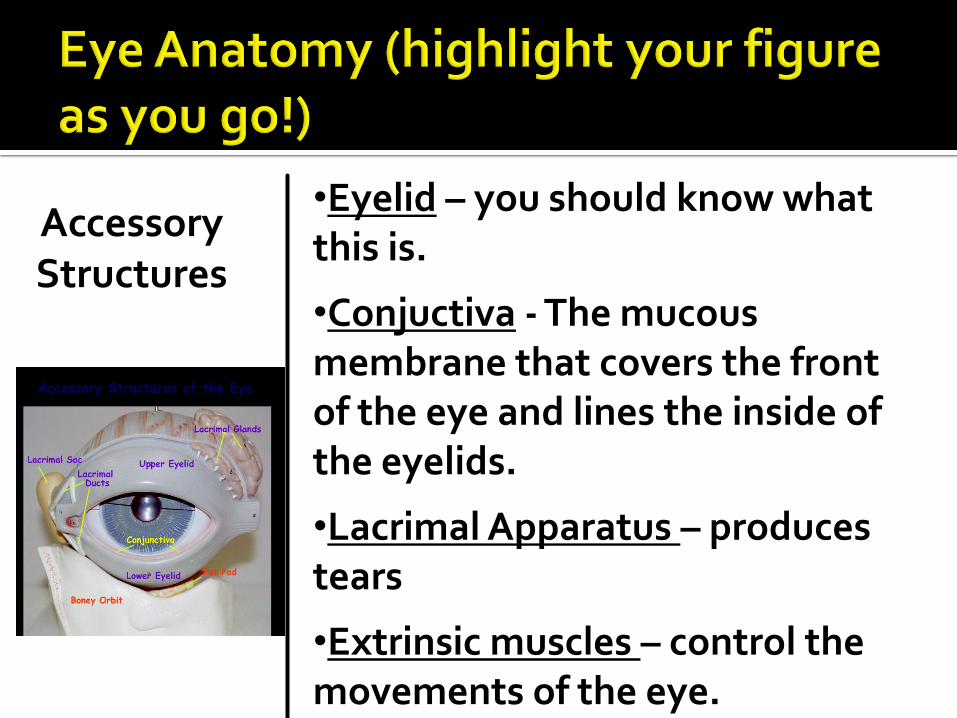
•Eyelid – you should know what this is.
•Conjuctiva - The mucous membrane that covers the front of the eye and lines the inside of the eyelids.
•Lacrimal Apparatus – produces tears
•Extrinsic muscles – control the movements of the eye.
Accessory Structures

Fibrous Tunic
Sclera – white part of your eye
Cornea – where the sclera becomes clear over your pupil and iris.
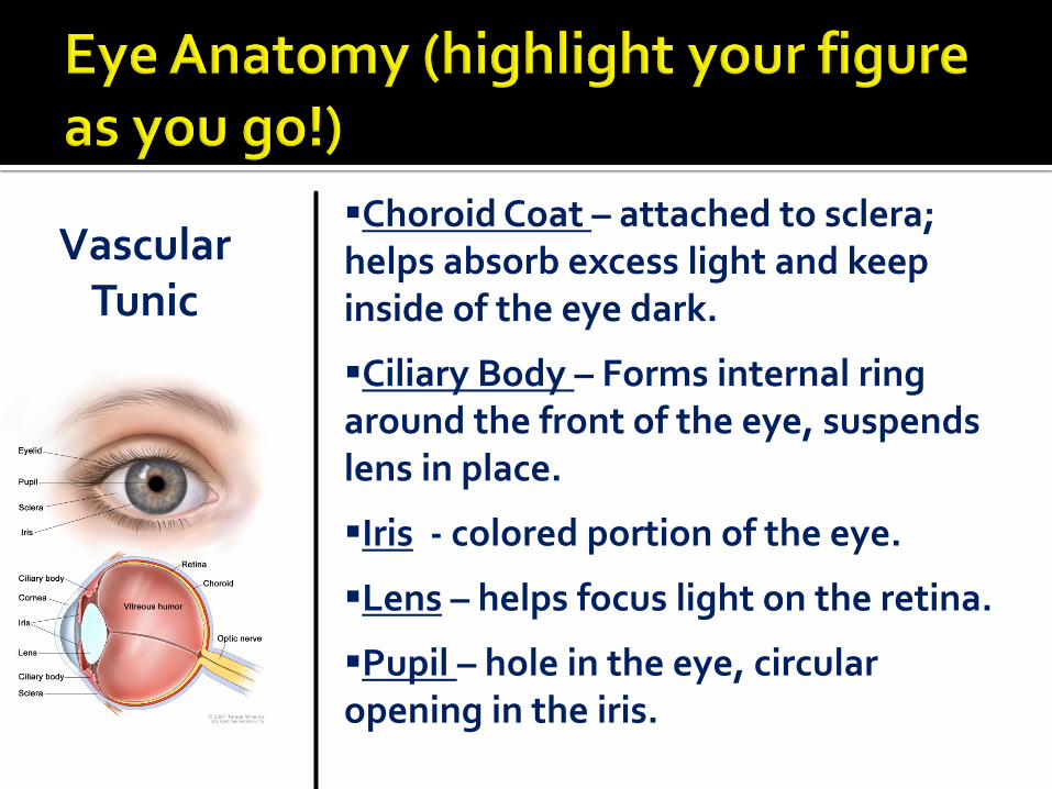
Vascular Tunic
Choroid Coat – attached to sclera; helps absorb excess light and keep inside of the eye dark.
Ciliary Body – Forms internal ring around the front of the eye, suspends lens in place.
Iris - colored portion of the eye.
Lens – helps focus light on the retina.
Pupil – hole in the eye, circular opening in the iris.

Nervous Tunic
Retina – contain visual receptor cells for image processing.
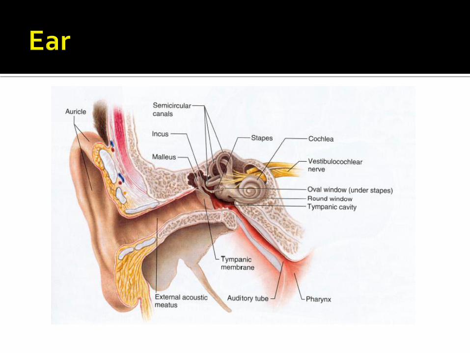

Outer Ear Auricle – Outer funnel like structure that helps collect sound waves.
External Auditory Meatus – tube that leads to the tympanic membrane.
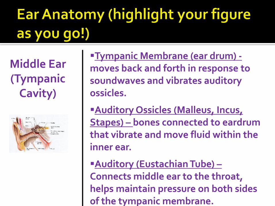
Middle Ear (Tympanic
Cavity)
Tympanic Membrane (ear drum) -moves back and forth in response to soundwaves and vibrates auditory ossicles.
Auditory Ossicles (Malleus, Incus, Stapes) – bones connected to eardrum that vibrate and move fluid within the inner ear.
Auditory (Eustachian Tube) – Connects middle ear to the throat, helps maintain pressure on both sides of the tympanic membrane.

Inner Ear Bony Labyrinth – outer layer of tubes comprising middle ear.
Membranous labryrinth – Tube that lies within the Bony Labyrinth
Semicircular Canals– provide your sense of equilibrium.
Vestibule – bony chamber that houses structures that serve both hearing and equilibrium.
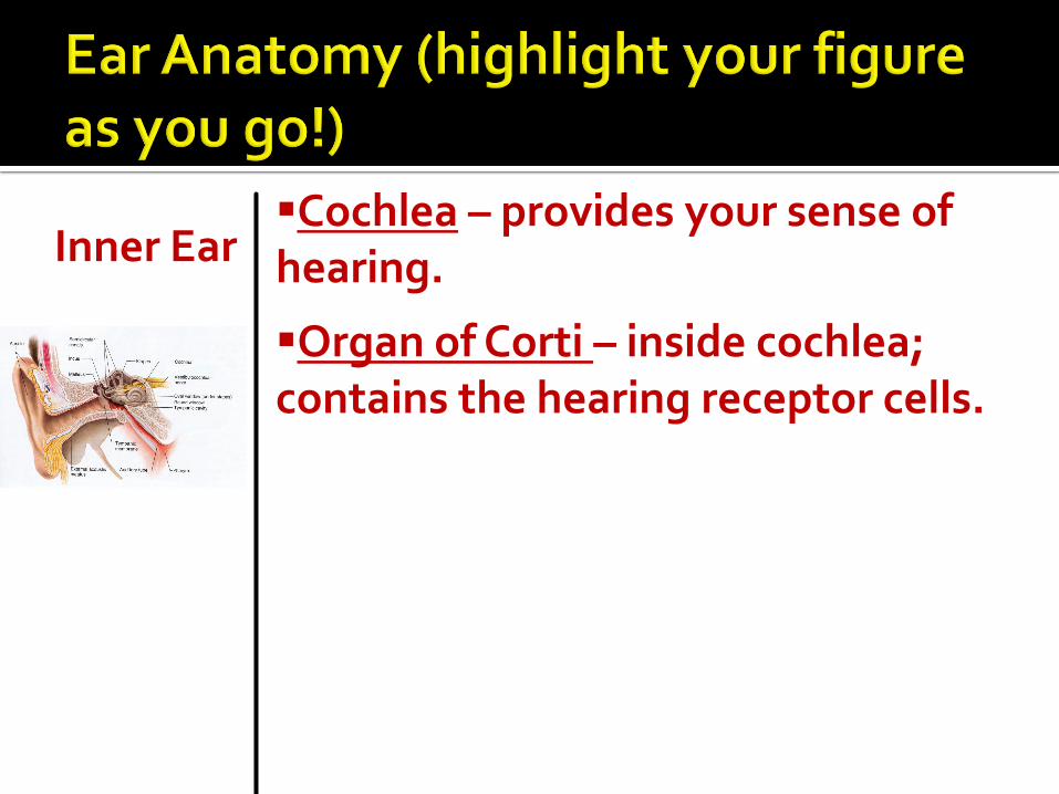
Inner Ear Cochlea – provides your sense of hearing.
Organ of Corti – inside cochlea; contains the hearing receptor cells.
Unit 6 Nervous Exam next time! Study your lab books and Chapters 11 and 12 in your text!
Unit Cat Title Page #
Comp Check
Due Date
6 HW Study for Unit 6 Nervous Exam 11/10

Day 5 (11/10/11)

We have a guest presentation on Nursing today, therefore you will be taking your exam online!
We will go over the Special Senses Disease and Disorders and Review if we have time!
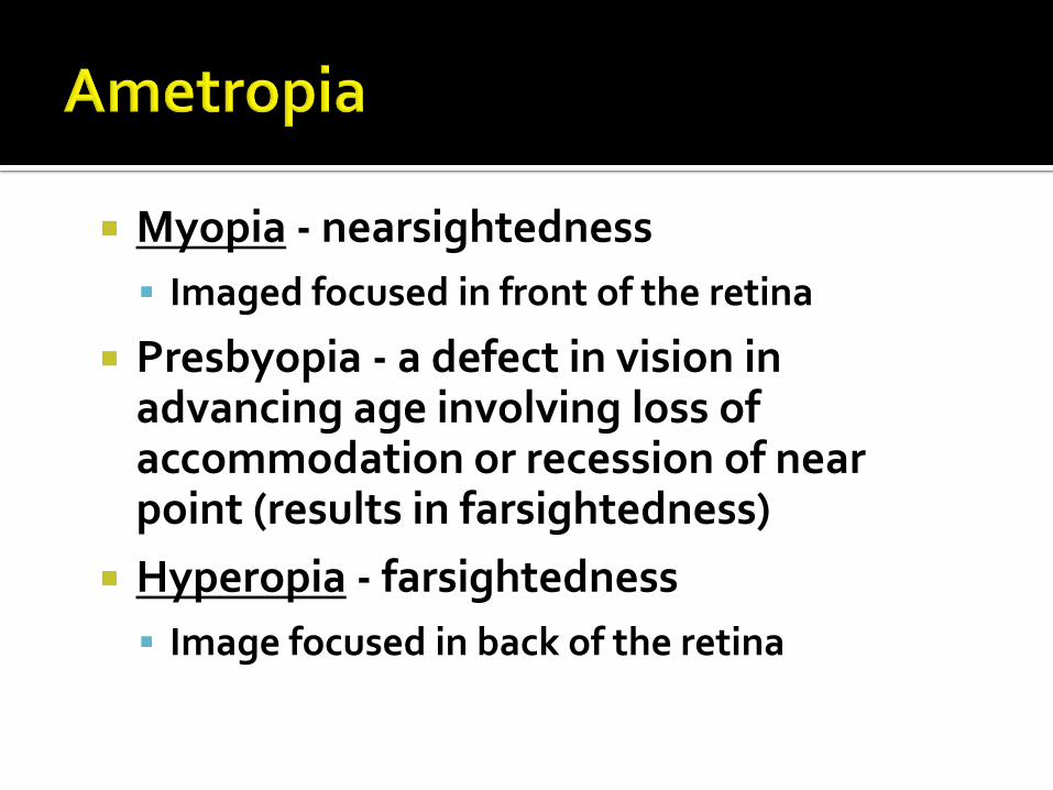
Myopia - nearsightedness
Imaged focused in front of the retina
Presbyopia - a defect in vision in advancing age involving loss of accommodation or recession of near point (results in farsightedness)
Hyperopia - farsightedness
Image focused in back of the retina


Abnormal loss of transparency of the lens Vision becomes blurry or cloudy Can be removed and have an artificial lens
inserted Most often occurs to individuals over the age
of 50. Exposure to sunlight and smoking increases the risk.


Conjunctivitis - inflammation of the
conjunctiva, the mucous membrane that lines the eyelid and is reflected to the eyeball. Also known as “Pink Eye”
Strabismus – “cross-eyed”


A group of eye diseases characterized by elevated intraocular pressure in the eye resulting in atrophy of the optic nerve which may lead to blindness
Caused by an obstruction of the outflow of the aqueous and vitreous humor
Minor cases can be treated with eye drops More severe cases may require a surgical incision
into the iris of the eye


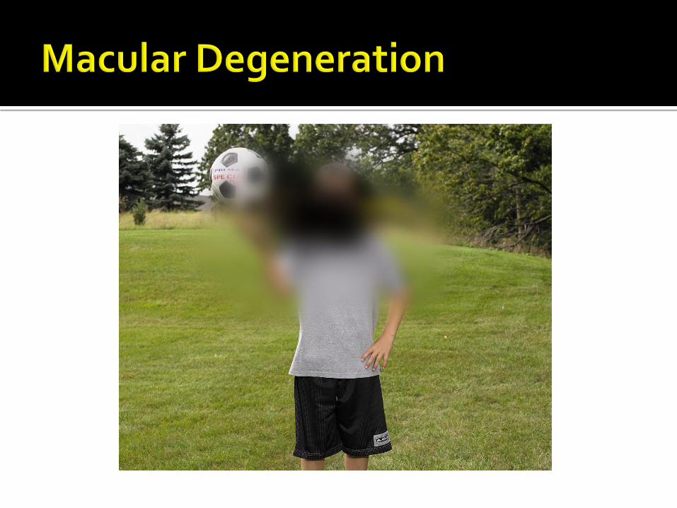
The destruction or tearing away of the retina from the back of the eye
Commonly occurs in the region of the retina known as the macula lutea
Can be caused by:
Vascular diseases (diabetes)
Chronic increased pressure (glaucoma)
Sudden blow or impact to the head or eye (Detached Retina)

A condition of dizziness and spatial disorientation
In some individuals it is due to heights or fear of high places
A spinning sensation that may result in loss of balance and equilibrium


Ringing or tinkling sounds or sensations in the ear

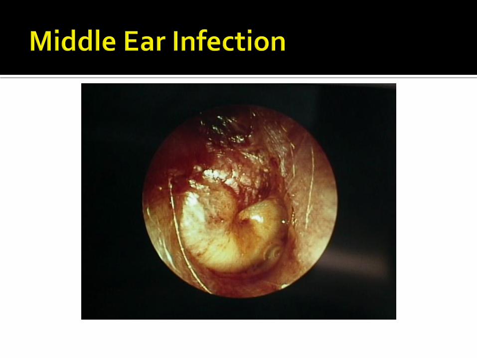
Infection of the tympanic membrane or other structures associated with the middle ear (Otitis Media)

Loss of the ability to hear Conductive Deafness: deafness resulting
from any condition that prevents sound waves from being transmitted to the auditory receptors (ex?)
Sensorineural Deafness: deafness due to defective function of the cochlea, organ of Corti, or the auditory nerve (ex?)


![UNIT 6 – Nervous System · Web view[UNIT 6 – Nervous System] Notes Outline 1 Functions of the nervous system Detection Integration Coordination Central Nervous System Peripheral](https://static.fdocuments.in/doc/165x107/5f051a7f7e708231d41147ca/unit-6-a-nervous-system-web-view-unit-6-a-nervous-system-notes-outline-1-functions.jpg)