Physical Rehabilitation After Total Joint Arthroplasty in Companion Animals
21
Physical Rehabilitation After Total Joint Arthroplasty in Companion Animals Denis J. Marcellin-Little, DEDV a, *, Nancy D. Doyle, MPT b , Joanna Freeman Pyke, PT, BSc.KINE c Osteoarthritis (OA) affects many dogs and cats. Even though global statistics on the prevalence of OA in dogs and cats are lacking, statistics from the Orthopedic Founda- tion for Animals (www.OFFA.org) for the 50 most affected dog breeds indicate that hip dysplasia is present in 21% dogs, based on 430,000 evaluations, and that elbow dysplasia is present in 16% of dogs, based on 180,000 evaluations. When medical management fails to control the pain and disability resulting from OA, total joint a Department of Clinical Sciences, College of Veterinary Medicine, North Carolina State Univer- sity, NCSU CVM VHC #2563, 1052 William Moore Drive, Raleigh, NC 27607-4065, USA; b Gulf Coast Veterinary Specialists, 1111 West Loop South, Houston, TX 77027, USA; c Private Practice, 2285 Bristol Circle, Oakville, Ontario L6H 6P8, Canada * Corresponding author. E-mail address: [email protected] KEYWORDS Animal rehabilitation Ambulation assistance Controlled exercise Total hip replacement Total knee replacement Total elbow replacement Complications KEY POINTS The goal of rehabilitation after total joint arthroplasty (TJA) is the lifelong restoration of a pain-free, functional limb. Rehabilitation after TJA includes controlling postoperative pain, minimizing complications, increasing function, restoring range of motion, and increasing strength of the surrounding musculature. Rehabilitation after TJA is particularly important in patients at high risk of limb disuse or complication. Rehabilitation provides tools for the management of cases experiencing postoperative complications. Patients receiving total elbow replacement or total knee replacement typically require much more extensive rehabilitation for optimal functional outcomes than those receiving total hip replacement. Vet Clin Small Anim 45 (2015) 145–165 http://dx.doi.org/10.1016/j.cvsm.2014.09.008 vetsmall.theclinics.com 0195-5616/15/$ – see front matter Ó 2015 Elsevier Inc. All rights reserved.
-
Upload
joanna-freeman -
Category
Documents
-
view
214 -
download
0
Transcript of Physical Rehabilitation After Total Joint Arthroplasty in Companion Animals

Physical Rehabil itationAfter Total Joint
Arthroplasty in CompanionAnimalsDenis J. Marcellin-Little, DEDVa,*, Nancy D. Doyle, MPTb,Joanna Freeman Pyke, PT, BSc.KINEc
KEYWORDS
� Animal rehabilitation � Ambulation assistance � Controlled exercise� Total hip replacement � Total knee replacement � Total elbow replacement� Complications
KEY POINTS
� The goal of rehabilitation after total joint arthroplasty (TJA) is the lifelong restoration of apain-free, functional limb.
� Rehabilitation after TJA includes controlling postoperative pain, minimizing complications,increasing function, restoring range of motion, and increasing strength of the surroundingmusculature.
� Rehabilitation after TJA is particularly important in patients at high risk of limb disuse orcomplication.
� Rehabilitation provides tools for the management of cases experiencing postoperativecomplications.
� Patients receiving total elbow replacement or total knee replacement typically requiremuch more extensive rehabilitation for optimal functional outcomes than those receivingtotal hip replacement.
Osteoarthritis (OA) affects many dogs and cats. Even though global statistics on theprevalence of OA in dogs and cats are lacking, statistics from the Orthopedic Founda-tion for Animals (www.OFFA.org) for the 50 most affected dog breeds indicate that hipdysplasia is present in 21% dogs, based on 430,000 evaluations, and that elbowdysplasia is present in 16% of dogs, based on 180,000 evaluations. When medicalmanagement fails to control the pain and disability resulting from OA, total joint
a Department of Clinical Sciences, College of Veterinary Medicine, North Carolina State Univer-sity, NCSU CVM VHC #2563, 1052 William Moore Drive, Raleigh, NC 27607-4065, USA; b GulfCoast Veterinary Specialists, 1111 West Loop South, Houston, TX 77027, USA; c Private Practice,2285 Bristol Circle, Oakville, Ontario L6H 6P8, Canada* Corresponding author.E-mail address: [email protected]
Vet Clin Small Anim 45 (2015) 145–165http://dx.doi.org/10.1016/j.cvsm.2014.09.008 vetsmall.theclinics.com0195-5616/15/$ – see front matter � 2015 Elsevier Inc. All rights reserved.

Marcellin-Little et al146
arthroplasties (TJA) can restore lost function and, ultimately, offer pain relief from debil-itating OA. However, the impact of OA on the periarticular structures persists during themonths that follow surgery, making physical rehabilitation an important aspect of thepostoperativemanagementof TJA.Periarticular fibrosisandassociated lossof jointmo-tion, muscle atrophy, particularly of type II fibers that provide rapid dynamic support forjoint protection,decreasedvoluntarymuscle recruitment, impairedproprioception, oste-oporosis, chronic pain, and general physical deconditioning are all associated withchronic arthritic changes in companion animals. Impaired preoperative limb use andmobility in turn compound thepostoperative challenges facedduring recovery after TJA.In the weeks that follow surgery, total joint prostheses are vulnerable to complica-
tions, requiring skilled care for protection and optimal functional recovery. The reha-bilitation environment is ideally suited for patients who are recovering from TJA.Rehabilitation clinicians possess the expertise to safely and effectively restore func-tional use of patients’ operated limbs and ensure maximal recoveries. Rehabilitationclinicians are also uniquely qualified to coordinate client education and communica-tion regarding housing modifications, activity restrictions, and home rehabilitationcare for patients recovering from surgery. This article presents rehabilitation consider-ations for companion animals undergoing total hip replacement (THR), total kneereplacement (TKR) and total elbow replacement (TER), postoperative complicationsand how to mitigate risks, and anticipated patient outcomes. Comparisons aremade to physical therapy procedures and expectations after TJA in humans, as thebody of clinical research relating to TJA in humans is much larger and richer in scien-tific evidence than that in companion animals. Because humans and companion ani-mals generally have parallel physiology, for example as it relates to joint motion andbone metabolism, many clinical findings in humans can be judiciously extrapolatedto companion animals. However, differences related to joint function must be consid-ered, such as that the elbow is a weight-bearing joint in dogs but not in humans. Mostof the companion animal information included in the text refers to TJA for dogs. Wheninformation relates to other nonhuman animal species (ie, cats), the species ismentioned specifically in the text.
GOALS OF REHABILITATION AFTER TOTAL JOINT ARTHROPLASTY
The goal of rehabilitation after TJA is the lifelong restoration of a pain-free, functionallimb after implantation of the prosthesis. Specific rehabilitation goals include control-ling postoperative pain, minimizing complications, increasing function, restoring pas-sive and active range of motion (PROM and AROM), increasing strength of thesurrounding musculature, restoring function, and ultimately improving patients’ qualityof life. Rehabilitation after TJA generally takes 3 months, but adherence to an individ-ualized home exercise program may be warranted for up to 6 months or more,depending on each patient’s needs. Most primary soft tissue healing takes place dur-ing the first month, with maturation continuing for months beyond that point. Thebone-implant interface of cemented implants reaches maximal strength 1 day aftersurgery, whereas the bone-implant interface of cementless implants relies on boneingrowth. Most of the bone ingrowth occurs during the first 2 months; afterward,bone remodels slowly during the first year after surgery. Many patients use their oper-ated limb consistently by the end of the first month, walk without lameness by the endof the second month, and can engage in any activity, including training for specificsporting activities by the end of the third month after surgery, depending on patientprofile and client goals. These timelines serve to guide rehabilitation and patient activ-ity levels to avoid complications.

Total Joint Arthroplasty in Companion Animals 147
A key goal of rehabilitation after TJA is to manage postoperative pain effectively.Many patients undergoing TJA have chronic pain as a result of severe OA. Painmanagement relies on medications (nonsteroidal anti-inflammatory drugs, opiates,N-methyl-D-aspartate receptor antagonists), on electrophysical modalities andmanual therapy, on optimal housing, and on safe transportation (described later inthis article). Chronic pain should subside within 3 months after TJA.1,2 Chronic painand limb disuse can be present over the long term after TJA but, in the absence ofclear complications, they are unusual. If chronic pain or limb disuse persists beyondthis normal recovery time period, the presence of an undetected complication shouldbe suspected.Minimizing the likelihood of complications is another key goal of rehabilitation after
TJA. High-risk patients and living situations must be identified, and confinement andtherapy must be adapted to mitigate these risk factors. Patients may be at riskbecause of their profile, including age, size, behavior, training, and conditioning. Olderdogs generally have more advanced disease (eg, increased bone loss, chronic jointluxation, severe periarticular fibrosis) and frequently have comorbidities (see nextparagraph). They may be more deconditioned, prone to falls due to weakness or bal-ance impairment, and not easy to motivate during recovery. Younger dogs may be un-ruly, not well trained or socialized, and also prone to falls due to exuberance. Patientsthat are very large may be at risk of complications because of increased forces placedon implants and implant-bone interfaces, potential clumsiness, and impingement(described later in this article). Fearful or aggressive dogs could be more prone toshort-term postoperative complications due to uncontrollable or unexpected burstsand ballistic movements. Patients also may be at risk because of their owners’ pro-files. Owners may be unwilling to modify the patient’s living environment or be unableto care for their companions because of their health, occupation, or motivations.Comorbidities also increase risk of complications. Classic comorbidities in dogs
include concurrent orthopedic diseases that cause dogs to offload and use abnormalgait patterns; examples include OA of the contralateral joint, cranial cruciate ligamentinjuries, and lumbosacral disease. The obesity epidemic in America extends to ourTJA patients, increasing the loads borne by the implanted prostheses. Patients’ pre-surgical lifestyles, preoperative levels of function, strength of associated musclegroups, and endurance have a direct effect on patient outcomes. In dogs, the preva-lence of comorbidities and their influence on complications and outcomes of TJA havenot been evaluated. Several dog-specific scoring systems evaluating orthopedicdisability and mobility3–5 and a patient-fracture assessment scoring system havebeen reported.6 A similar patient-disease index would be beneficial to identify factorsthat negatively impact the outcome of TJA. In humans, comorbidities in patients un-dergoing TJA and their influence on functional outcome, implant survival, mortality,and length of hospital stay have been described.7 Several instruments are used toquantify comorbidity, including comorbidities indices (eg, Charlson index, index ofcoexistent disease, functional comorbidity index), other health or quality-of-life indices(eg, Charnley index, American Society of Anesthesiologists physical status classifica-tion, SF-36), and osteoarthritis indices (eg, Western Ontario and McMaster Osteoar-thritis Index [WOMAC], hip osteoarthritis outcomes scale).8 Other instruments areused to evaluate functional outcomes (eg, lower extremity functional scale [LEFS],timed up and go [TUG test], and range of motion [ROM] measurements [flexion andextension]).9 Although analogous instruments are not available for assessment ofour animal patients with total joint replacement (TJR), it is still important for rehabilita-tion clinicians to clearly identify and communicate similar functional characteristics ofeach TJA patient with the surgeon to facilitate detection of potential complications as

Marcellin-Little et al148
early as possible in the treatment process. This discussion among all members of thepatient’s health care team identifies the duration of hospitalization that is safest, thelevel of nursing care required, and the strategies to minimize complications.
COMPLICATIONS OF TOTAL JOINT ARTHROPLASTY
THR is by far the most common TJA in dogs. Recognized complications of THRsinclude luxations, fractures, neurapraxia, implant loosening, and infection. The overallcomplication rate of THR in dogs is approximately 10%.10
Luxation of Prosthetic Joints
Luxations are the most common short-term complications of THR, with an incidenceranging from 8% to 12% of THRs.10,11 Most luxations occur within the first 3 monthsafter surgery. Luxations can result from suboptimal implant positioning; for example,when a cup is too open the prosthesis is predisposed to dorsal luxation, and when thecup is too closed, the prosthesis is predisposed to ventral luxation.12 The risk of luxa-tion also increases when the stem is implanted too recessed in the femoral canal or thefemoral neck is too short.13 The stem also can become recessed as a consequence ofstem subsidence postoperatively. Luxations also can occur when the cup and stemcontact each other during activities of daily living, a phenomenon named impinge-ment. In humans, impingement is more likely when the ratio of cup size/prosthetichead size increases.14 Impingement is most likely a predominant cause of THR luxa-tion in giant-breed dogs, which seemingly are at increased risk (Fig. 1).13 Most femoralheads have diameters ranging from 16 to 18 mm, but one manufacturer (BioMedtrix,Boonton, NJ, USA) recently introduced a larger femoral head measuring 22 mm todecrease the risk of impingement in giant-breed dogs. Luxations can result fromimpingement of the femur and ischium or from a thick, fibrous joint capsule, some-times seen in older German shepherd dogs and other breeds.10 In all cases in whichluxations occur due to suboptimal implant positioning, surgical revisions are indicatedfor successful reduction and prevention of recurrence.
Fig. 1. A 4-year-old neutered male Newfoundland weighing 104 kg (230 lb) is learning towalk on the day after a surgery that reduced a ventral luxation of a total hip implant placed2 years earlier. The dog is controlled and supported by a check harness and 2 slings. Hobblesmade of adhesive tape prevent excessive abduction of the operated limb. One week later,the dog could walk independently without support. The pelvic limbs were hobbled for5 months. The hip did not re-luxate afterward.

Total Joint Arthroplasty in Companion Animals 149
Pelvic limb amputees undergoing THR on the remaining pelvic limb also are atincreased risk of luxation because they stand and walk with their pelvis tilted towardthe side of amputation, placing the THR in relative adduction and decreasing dorsalcoverage of the femoral head.15 The posture of these patients may be improvedwith exercise.Traumatic luxationscanoccurwhenpatients fall intoasplayedposture (ventral luxation)
or onto the hip (dorsal luxation), underscoring the need for supported ambulation andmeasures toprevent slipsor lossesofbalance.Patientswith ventral hip luxations resultingfrom excessive limb abduction (eg, slipping and doing the splits) can be managed withclosed reduction and placement of hobbles that limit abduction (see Fig. 1), providedthat the luxation is not the result of impingement of the femoral neck on the acetabularcup. Joints with impingement are likely to re-luxate during normal activities and requiresurgical repositioning of the implants for successful outcomes. Dorsal luxations alsomay be treated with closed reduction and temporary immobilization with an Ehmer slingor they may require surgical revision. Several investigators proposed that nontraumaticluxations occurring 6 weeks to 4 months after surgery may be the consequence of inad-equate recovery of muscle support and periarticular soft tissue healing.12,16 Althoughnot yet validated through research, rehabilitation of patients identified to be at increasedrisk of luxation could potentially decrease the occurrence of luxation in this population.
Femoral Fracture
Femoral fractures are a common complication of THR in dogs, occurring both intrao-peratively and postoperatively at rates ranging from 2% to 8%.10,17 A femur could beat risk of fracture because of thin cortices. The femoral fractures that occur during sur-gery are often stabilized with cerclage wires placed around the femoral shaft. Afterappropriate cerclage placement, prosthetic stems resist subsidence as much asstems placed in nonfractured femurs, thus not placing the patient at an increasedrisk of luxation from subsidence.18 Postoperatively, fractures can occur when unde-tected surgically induced fissures propagate, when excessive loading occurs beforeadequate cortical hypertrophy has developed, or as a result of trauma. These fracturesusually necessitate surgical stabilization. The rehabilitation plan for patients with THRsand fractures should include a focus on maintaining length/flexibility of the quadricepsand hamstrings due to the impact that fractures and fixation have on these muscles.
Sciatic Neurapraxia
Sciatic nerve neurapraxia is an infrequent complication of THR, occurring in 1.6% to1.9% of patients.19,20 Sciatic neurapraxia can occur after THR due to compression ofthe nerve intraoperatively, in the vicinity of the ischiatic spine during gluteal retractionor between the caudal joint capsule and the ischiatic tuberosity. Patients with sciaticnerve palsy have an abnormal gait. They have impaired active stifle and tarsal flexion,scuff or knuckle their paws, and use increased hip flexion to advance the limb andclear the ground when walking. Sciatic nerve neurapraxia is generally transient, lastingfor a few weeks to a few months, but can be permanent. Geriatric dogs and those un-dergoing longer surgeries have increased risk of postoperative sciatic neurapraxia.20
Infection
Infection can occur after THR.10 Presentation may be acute, with limb swelling and hy-perthermia, or chronic (low-grade infection) without swelling or systemic signs. MostTHR infections are low-grade infections developing weeks to months after surgery,most often from bacteria introduced at the time of surgery or possibly as the resultof hematogenous infection. Infection should be suspected in patients that exhibit

Marcellin-Little et al150
progressively poorer limb use (because of persistent pain) after an initially uneventfulrecovery in the absence of radiographic evidence of fracture or loosening. Infectioncan be confirmed if specific bone changes are visible on radiographs and a bacterialculture of the joint space or regional tissues is positive. Infection is often a devastatingcomplication because antibiotic therapy is unlikely to sterilize the bone-implant inter-face. Explantation of the THR prosthesis may be required, leaving the patient withwhat is essentially a femoral head ostectomy. These patients require extensive reha-bilitation and more aggressive pain management to restore functional use of the limb.
Failure of Fixation
Stems or cups used in THR can be loose either because of lack of bone ingrowth intocementless implants or because of aseptic loosening over time. The acetabular cupmay at increased risk of failure of fixation because of a defect to the medial acetabularwall (protrusio) or dorsal acetabular rim. The bone ingrowth that occurs in the first 6 to8 weeks after surgery is critical to long-term prosthesis stability. Excessive activityduring this time period causes a disruption of the healing due to the development ofa fibrous membrane between the implant and the bone, described as fibrous ingrowth.Loose implants generate an inflammatory response in the bone (visible on radio-graphs) that progresses slowly over time. Like low-grade infections, their presenceleads to poor limb use and progressive loss of muscle mass.
Complications of Total Knee Replacement (TKR) and Total Elbow Replacement (TER)
The complications of other total joints are similar to the complications of THR. AfterTKR, collateral ligament damage (and secondary stifle subluxation), infection, frac-tures, and luxation have been encountered.21 The medial or lateral collateral ligamentscan be damaged during surgery, usually during the proximal tibial osteotomy, or canrupture after surgery, particularly if early exuberant activity is permitted, leading to jointinstability.22 Ruptured ligaments must be repaired and protected for several weeks af-ter surgery. Custom orthoses can effectively shield the stifle from varus or valgusforces to facilitate healing, but require significant owner compliance for effectiveuse. Strengthening of the hip stabilizers during recovery ensures the limb is kept innormal alignment and avoids postures that place the stifle under increased varus orvalgus loads. Similarly, collateral ligament instability, infection, and fractures canoccur after TER.23 Because osteotomies of the medial or the lateral epicondyle maybe performed during implantation (based on the implant systems used), collateral lig-ament instability may result from avulsion of an epicondylar osteotomy site or epicon-dylar screw loosening. Ulnar fractures adjacent to the trochlear notch have beenreported after TER.23 Limb amputation may be required for resolution of infectionbecause TERs and TKRs do not have the option of explantation because unarticulatedelbow or stifle joints are completely dysfunctional.
MANAGEMENT AFTER TOTAL JOINT ARTHROPLASTYHospitalization and Aftercare
Patients recovering from TJA are most often managed as inpatients in the early post-operative period. Inpatient management allows the dogs to be handled by a limitednumber of caregivers who are familiar with the specific needs related to each patient’spersonality, pain levels, mobility, and risk profile. These needs are described in thefollowing text. Inpatient management can last a few days for low-risk patients andcan last several weeks for high-risk patients or in instances in which the owners areunwilling or unable to care for their pet during recovery.

Total Joint Arthroplasty in Companion Animals 151
After discharge from the hospital, patients recovering from TJA are either treated asrehabilitation outpatients or are solely managed at home. Some THR patients aretreated at home because transportation to and from the rehabilitation facility presentsrisks associated with getting in and out of motor vehicles, walking on slippery clinicflooring, and encountering uncontrolled environmental challenges, such as other pa-tients at the facility. Risks are minimized when dogs are walked at a slow, controlledspeed using appropriate equipment, thus increasing their safe, functional mobility inthe early days after surgery. A chest harness (Step In Harness TEC; Canine Equip-ment, Vancouver, Canada) provides more control than a collar and allows weight-bearing support of the forelimbs after TER. A thin support sling (Fig. 2) should beused to provide additional weight-bearing support of the pelvic limbs for dogs needingambulation assistance and prevent falls. Commercial support systems (Help’Em Up;Blue Dog Designs, Denver, CO, USA) also can be used. Even if donning and doffingthem require manipulation, dogs usually tolerate harnesses well and can wear themall day. Owners remove harnesses at night and inspect the skin for hair loss, redness,and abrasions. Harnesses also are removed and cleaned when soiled (eg, by urine orfeces). These support measures also should be used at home when dogs are takenoutside to relieve themselves and during the brief periods of exercise permitted in
Fig. 2. A 1-year-old neutered male Labrador mix with a mid-femoral amputation is learningto walk the day after a contralateral THR done to manage a chronic untreated acetabularfracture. The dog is uncoordinated. He is supported with a chest harness and a sling at alltimes. Gait training includes slow walks (top) and walking on a treadmill, 10 days aftersurgery (bottom).

Marcellin-Little et al152
the early postoperative period. A leash measuring less than 1 m (approximately 3 feet)should be used. Support slings and harness supporting the abdomen and pelvic limbsenhance control of the patient and decrease the likelihood of falls. Assistance duringtransitions also should be provided when needed (eg, sit to stand, controlled loweringto sit or lie down).Care is needed for safe transport. Lighter dogs can be securely lifted in and out of
motor vehicles. Heavy dogs can use ramps to get in and out of motor vehicles with thesupport and protection of slings. Ramps should be rigid and stable and long enough tocreate a gradual slope. Ramps are more manageable for owners to set up and disas-semble when they are telescoping (Deluxe XL Telescoping Pet Ramp; Solvit, Arlington,TX, USA) or can be folded (UltraLite Bi-fold Pet Ramp; Solvit) or rolled (Ramp4Paws,Potomac, MD, USA). In cars or trucks, dogs should be allowed to rest on flat surfaces,ideally secured through their harness/seatbelt or accompanied by a second attendantto ensure they do not attempt to move about while the vehicle is in motion. Tightspaces and gaps present risks of limb entrapment and injury. Smaller dogs are safesttraveling in crates and also can be transported in the facility while in this confinement.At home, dogs should be confined to a large exercise pen or small single roomwhen
unsupervised. The patient should feel comfortable in that room and not exhibit unduestress. Room modifications should include secure footing (carpeting or rugs), a dogbed at floor level, and removal of furniture that the dog may attempt to climb on (ie,couches and chairs). The room should be away from windows or doors that maytrigger explosive responses (eg, the postal carrier stops by or a guest knocks onthe door). When directly supervised, patients can be kept in larger rooms, providedthey have acceptable mobility and the same precautions are taken. Stairs pose spe-cific challenges, particularly when they cannot be avoided (eg, the owner lives in anapartment on an upper level or has a multilevel home with living spaces upstairs). Stra-tegies should be developed with the owner to use the aforementioned support de-vices, enlisting help of neighbors or family members if needed (in the acute phasesof recovery for larger dogs), or make temporary alternative living arrangements. Aslong as these safeguards are in place, stairs can be negotiated as soon as patients re-turn home.Owners often request oral medications for active, boisterous patients that may help
keep them calm during confinement and exercise restriction after TJA. Acepromazinemaleate (Boehringer Ingelheim, Ridgefield, CT, USA) is used for its sedative propertiesbut is rarely used in the long term because its safety profile is considered suboptimal.Side effects include hypotension, excessive or prolonged sedation, ataxia, loweredseizure threshold, and dysphoria, all of which may increase the risk for falls and injury.Trazodone hydrochloride (Desyrel; Bristol-Myers Squibb, New York City, NY, USA), aserotonin antagonist and reuptake inhibitor, has been given to dogs after orthopedicsurgery and appears to have acceptable safety.24 In an open trial, most owners (32 of36, 89%) reported that trazodone improved confinement tolerance moderately orextremely.25 Current research assessing the efficacy of trazodone after THR suggeststhat some dogs may be rated as calmer even when owners give placebo (Dr BL Sher-man, North Carolina State University, personal communication, 2014). The belief ofgiving a medication to improve confinement tolerance might lead to behavior modifi-cation by owners that would promote calmness. To help ease patient “boredom” andrestlessness, gentle play (eg, gentle tug-of-war) is safe; instruction in simple tricks andcommands also serves as fun interactive time with the owner and provides mentalstimulation for patients during convalescence. Puzzle toys that dogs manipulate to ac-cess food also may be provided for supervised distractions. Protected movement,albeit controlled, should be encouraged.

Total Joint Arthroplasty in Companion Animals 153
Wound Management
Wound healing after TJA is important. There is no report of skin infection progressingto implant infection in companion animals, to our knowledge. Nevertheless, woundinfections can cause dehiscence of the surgical incision and could lead to deep tissueinfections (as they do in humans). Wound management includes covering the incisionwith an adhesive bandage until the incision is sealed, approximately 5 days after sur-gery, or until suture removal 10 to 14 days after surgery. If the wound is not covered,disinfecting the skin incision periodically (0.05% chlorhexidine diacetate, Nolvasan;Zoetis, Florham Park, NJ, USA) or placing a thin layer of triple antibiotic ointmentcould be beneficial. Elizabethan collars or alternative donut-style collars may beused in dogs showing signs of self-mutilation. Dogs generally dislike Elizabethan col-lars and may struggle to remove them, potentially putting the procedure at risk forcomplications.
REHABILITATION METHODS AFTER TOTAL JOINT ARTHROPLASTY
Electrophysical modalities, manual therapies, and therapeutic exercises are used forthe rehabilitation of patients undergoing TJA. Cryotherapy (cold packs) begins imme-diately postoperatively and continues through the inflammatory phase to decreasepostoperative pain and swelling. Cryotherapy is safe, well tolerated, and technicallysimple. Cold packs can be used several times daily for approximately 10 to 15minutes,until the skin is cold to the touch. In humans, cryotherapy decreases blood loss afterTJA in general (but not after THR), provides pain relief on the day after surgery (but noton day 1 or 3) and improves quadriceps muscle activation after TKR.26,27 Nontoxic gelpacks should be used and patients should be carefully monitored throughout admin-istration to ensure the packs are not chewed or ingested.Manual therapy plays an important role in recovery for many patients after TJA,
particularly those with ROM restrictions. In humans, restoration of flexion and exten-sion ROM is imperative after TKR surgery and is generally pursued with physical ther-apy interventions beginning as soon as the first day after surgery. Specific motionrestrictions in human patients with THR are more commonly addressed in physio-therapy practices 6 weeks after surgery, once the surgeon lifts the protective motionrestrictions. Massage may be used to decrease pain and edema and to promote mo-tion between tissue planes. In humans, back massage reportedly decreases pain afterTHR and TKR.28 Passive ROM completed in the early phases of recovery helps avoidpostoperative stiffness, improve comfort, and begin restoration of joint motion. Inhumans, the use of a continuous passive motion machine (CPM) decreases lengthof stay after TJA because of improved active joint motion but its long-term benefitsare unclear.29 Active, active assisted, and passive ROM exercises are an alternativeto CPM and are implemented immediately after surgery. These exercises continuevia a home exercise program after discharge from active physiotherapy and shouldcontinue up to 12 weeks after surgery or until specific goals are reached.In dogs, the same goals and interventions apply. Therefore, early interventions to
increase ROM and influence the organization of scar tissue are key to regaining lostmotion and decreasing pain (Fig. 3). If early intervention is not provided, unnecessarystiffness sets in and, in turn, is harder to reverse. In some human patients, manipula-tion under anesthesia is warranted if exuberant adhesions or fibrosis preclude resto-ration of ROM at an expected rate postoperatively. Preventing complications andsetting a path for recovery based on proven techniques delivered by trained therapistsis paramount. Heat therapy may be combined with ROM and passive stretching tech-niques to improve the pliability of the target tissues, but this should not be done until

Fig. 3. A patient with THR is held in lateral recumbency for a massage and stretching sessiona few days after surgery. The hip region lacked extension, and a severe pain response waspresent on extension.
Marcellin-Little et al154
the active, acute inflammation has subsided. More advanced stretching (particularlyjoint flexion, as in the case of TKR) can also be performed during specific active ther-apeutic exercises that stimulate the patient to move the affected joint through the tar-geted ROM. For example, when the patient sits, the hip and stifle are normally placedin flexion. If the patient lacks full flexion, the patient may compensate by sitting “sideswept” (both limbs positioned to one side via spinal rotation) or in a “lazy sit” (abduct-ing the affected limb or sitting back on the ischii to position the limb cranially). Modi-fications can be made to accommodate the restrictions and allow the patient tomaximally flex and stretch, such as cuing the dog to sit on the handler’s leg or platformor minimally abducting the affected limb (Fig. 4). Loss of extension ROM can be morefunctionally debilitating and interfere with proper limb use and weight bearing than lossof flexion ROM. If full extension is not restored, then the loss of ROM at end range maymimic a leg length discrepancy and ultimately impair weight bearing of the affectedlimb. Exercises that cue active stifle extension include sit to stands, uphill walking,walking backward, and reaching for a treat with the forelimbs elevated (Fig. 5). Ideally,both flexion and extension ROM should be restored to normal limits after TJR to pro-vide the patient the best functional outcome.
SPECIFIC REHABILITATION PROGRAMS FOR DOGS UNDERGOING TOTAL JOINTARTHROPLASTY
Completion of therapeutic exercises while hospitalized and at home optimizes limbuse after TJA in humans and companion animals. Large dogs and high-risk patients

Fig. 4. A patient is doing a modified sit to stand exercise that is aimed at strengthening hispelvic limbs and stretching his hip and stifle in flexion. The dog sits on the handler’s knee.That elevated position requires less hip and knee flexion. A harness provides control and anyneeded assistance.
Total Joint Arthroplasty in Companion Animals 155
may need training to learn to get up and walk (see Fig. 1). Patients with TKR and TERneed exercise to initiate and promote use of their operated limbs, particularly if theyhad very limited use of the limbs preoperatively. All patients with TJR benefit fromincreased strength (strengthening their operated limb). In the long term, patientswith TJR should remain active and strong through exercise. This increased strengthand conditioning improves the patient’s quality of life and thus, the owners’ satisfac-tion with the surgical outcome. Specific therapeutic exercises for THR, TKR, and TERare described in the following sections.
Total Hip Replacement
Dog are most often hospitalized for 1 to 3 days after THR. A middle-age large-breeddog with good mobility and good limb use can safely be discharged the day after sur-gery if pain is controlled on oral and patch analgesics. Hospitalization is prolongedwhen the perceived risk of complication is higher, when postoperative limb use is
Fig. 5. A patient is actively reaching for a treat that is offered in a way that promotes push-ing with his pelvic limbs into stifle and hip extension. The sling under his belly (The SoftQuick Lift, Four Flags Over Aspen, St Clair, MN, USA), is used to protect against falls andto maintain control. This exercise can be modulated in difficulty by varying the height ofthe step or by progressing to an unsteady “step,” such as a balance disc or Physioroll.
Marcellin-Little et al156
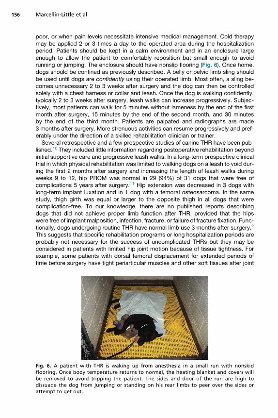
poor, or when pain levels necessitate intensive medical management. Cold therapymay be applied 2 or 3 times a day to the operated area during the hospitalizationperiod. Patients should be kept in a calm environment and in an enclosure largeenough to allow the patient to comfortably reposition but small enough to avoidrunning or jumping. The enclosure should have nonslip flooring (Fig. 6). Once home,dogs should be confined as previously described. A belly or pelvic limb sling shouldbe used until dogs are confidently using their operated limb. Most often, a sling be-comes unnecessary 2 to 3 weeks after surgery and the dog can then be controlledsolely with a chest harness or collar and leash. Once the dog is walking confidently,typically 2 to 3 weeks after surgery, leash walks can increase progressively. Subjec-tively, most patients can walk for 5 minutes without lameness by the end of the firstmonth after surgery, 15 minutes by the end of the second month, and 30 minutesby the end of the third month. Patients are palpated and radiographs are made3 months after surgery. More strenuous activities can resume progressively and pref-erably under the direction of a skilled rehabilitation clinician or trainer.Several retrospective and a few prospective studies of canine THR have been pub-
lished.10 They included little information regarding postoperative rehabilitation beyondinitial supportive care and progressive leash walks. In a long-term prospective clinicaltrial in which physical rehabilitation was limited to walking dogs on a leash to void dur-ing the first 2 months after surgery and increasing the length of leash walks duringweeks 9 to 12, hip PROM was normal in 29 (94%) of 31 dogs that were free ofcomplications 5 years after surgery.11 Hip extension was decreased in 3 dogs withlong-term implant luxation and in 1 dog with a femoral osteosarcoma. In the samestudy, thigh girth was equal or larger to the opposite thigh in all dogs that werecomplication-free. To our knowledge, there are no published reports describingdogs that did not achieve proper limb function after THR, provided that the hipswere free of implant malposition, infection, fracture, or failure of fracture fixation. Func-tionally, dogs undergoing routine THR have normal limb use 3 months after surgery.2
This suggests that specific rehabilitation programs or long hospitalization periods areprobably not necessary for the success of uncomplicated THRs but they may beconsidered in patients with limited hip joint motion because of tissue tightness. Forexample, some patients with dorsal femoral displacement for extended periods oftime before surgery have tight periarticular muscles and other soft tissues after joint
Fig. 6. A patient with THR is waking up from anesthesia in a small run with nonskidflooring. Once body temperature returns to normal, the heating blanket and covers willbe removed to avoid tripping the patient. The sides and door of the run are high todissuade the dog from jumping or standing on his rear limbs to peer over the sides orattempt to get out.

Total Joint Arthroplasty in Companion Animals 157
reduction during surgery, including external rotators, gluteal muscles, and rectus fem-oris. These tight muscles interfere with comfortable locomotion and may result in acharacteristic “hockey stick” posture of hip external rotation and abduction, posi-tioning the paw laterally. Subjectively, periarticular restrictions due to chronic OA typi-cally result in decreased hip extension, internal rotation, and abduction. Of thesemovement planes, extension most impacts the patient’s mobility and function. Humanpatients with THR work to restore lost hip flexion, extension, internal and external rota-tion lost during the 12 weeks of contraindicated movements placed on them. Hipabduction weakness is one of the most measurable impairments in human patientswith THR, but is likely of lesser importance in dogs, considering their quadrupedal gait.Dogs with limited hip extension will benefit from a stretching program. When the
loss of hip extension is severe, moist heat and manual stretching techniques areused. Extension uses a spinning motion of the femoral head on the acetabulum andthus tightens the joint capsule at end ranges. This is a safe direction for stretchingwith regard to very limited possibility for luxating the hip with overzealous motion,but care must be exercised to not cause patient pain with this stretch. Following thestretching session, active hip extension exercises should be performed to retrainthe patient to use the increased ROM. When the loss of hip extension is modestand the patient’s limb use is acceptable, manual stretching may not be critical.Some patients are not receptive to stretching techniques and owners cannot safelyperform stretching at home. For both of these populations, targeted therapeutic exer-cises alone can be used instead to gain hip extension for a more normal gait patternand better function. Walking up a gentle incline, stepping up a single step or a series ofsteps with adequate traction, and stepping over objects all place the trailing limb inincreased hip extension (Fig. 7).Dogs experiencing complications following THR have additional rehabilitation
needs. Following the acute management of a luxation (with reduction/hobbles and/or surgical revision), targeted strengthening of the appropriate muscle groups pro-vides improved dynamic joint support to help prevent a recurrence. Dogs that expe-rienced a dorsal luxation need additional strengthening of the muscles lying on thedorsal aspect of the hip. Suggested exercises include 3-legged standing (lifting the
Fig. 7. A patient is doing a step up exercise in which the dog repeatedly steps up on a mat,promoting extension in the operated left hip. A harness (Web Master Harness, Ruffwear,Bend, OR, USA) is used to provide support against falling and to control speed.

Marcellin-Little et al158
unaffected pelvic limb and cuing the dog to shift weight onto the operative limb whilemaintaining a level pelvis), balancing on a soft or unsteady surface (commercial bal-ance discs or an air mattress), walking perpendicular to an incline with the operativelimb “downhill,” and the previously mentioned hip extension exercises. Dogs experi-encing a ventral luxation require strengthening of the adductors. Suggested exercisesinclude resisted TheraBand exercise (TheraBand, Akron, OH, USA) while walking on atreadmill or alongside the handler (pull the hip into abduction with the band wrappedaround the thigh to stimulate a contraction of the adductors), walking sideways, orwalking perpendicular to an incline with the operative limb “uphill.” Underwater tread-mill walking also can effectively and safely target the desired muscle group in bothcases, particularly in the earlier phases of recovery. Proprioceptive retraining alsoshould be used to improve body awareness and coordination for decreased risk offuture falls.Patients with sciatic neurapraxia typically present with knuckling and weakness of
the muscles in the sciatic distribution, including the hamstrings and crus musculature.Dogs exhibiting deficits due to sciatic neurapraxia after THR need rehabilitation fordays to months, depending on the severity of the deficits.20 Rehabilitation focuseson minimizing hip complications due to decreased active muscular stabilization andprotection (eg, luxation), avoiding skin abrasions resulting from scuffing or knuckling,decreasing the loss of muscle mass in muscles innervated by branches of the sciaticnerve, and strengthening the affected muscle groups. Neuromuscular electrical stim-ulation can be used to elicit muscle contractions of the affected muscles to attenuateatrophy but is not universally well accepted by patients, particularly when sensation isintact. If active hock extension is absent for weight bearing, the hock can be stabilizedby an orthosis during therapeutic exercises (Fig. 8). Once hock extension improves,the dog can exercise without an orthosis. To avoid abrasions, affected dogs shouldavoid walking on abrasive surfaces and metacarpals and toes should be protectedby a thin bootie or bandage. If the patient frequently knuckles, bootie systems withsupport straps that pull the hock into flexion and the digits into extension (TheraPaw,Lebanon, NJ, USA) can be used during ambulation and therapeutic exercise sessionsto create more normal posture for functional limb use while simultaneously protectingthe skin from abrasions. In dogs with weak hock flexion, an exercise band or rubbertraction band (Anti-Knuckling Device; Canine Mobility, Seattle, WA, USA or BikoMobility, Raleigh, NC, USA) can be used to facilitate more normal flexion ROM duringexercise. Exercises to strengthen hock flexion include stepping over progressivelytaller objects, such as segments of PVC pipe, walking in water at the height of thehock, and elicitation of a flexor withdraw reflex by pinching the digits. Most dogs fullyrecover from sciatic neurapraxia.20
Total Knee Replacement
There are few studies reporting the results of TKR in dogs. In 2 experimental studies,TKR was performed in purpose-bred dogs that did not undergo any physical rehabil-itation in the postoperative period.22,30 In one of these, operated stifles lost approxi-mately 32� of ROM excursion in the long term compared with opposite normalstifles (w80� vs w112�), even though the prostheses were implanted into normal,healthy stifles without preoperative osteoarthritic changes.22
The rehabilitation protocol of a prospective clinical study in 6 clinical patients un-dergoing TKR has been reported.21 The ROM of the operated knee and thigh girtharound the joint line were recorded preoperatively and immediately after surgery forcomparison. An ice pack was then applied to the cranial, medial, and lateral aspectsof the stifle for 12 minutes. Passive ROM followed by cold pack application was

Fig. 8. A patient with THR with sciatic neurapraxia is walking with an orthosis on his leftpelvic limb (top). The dog can exercise without scuffing or knuckling in an underwatertreadmill (bottom). Atrophy of the thigh muscles is visible.
Total Joint Arthroplasty in Companion Animals 159
repeated 3 times daily while the dog was hospitalized (duration of hospitalization wasnot specified). After discharge, owners were provided with written and verbal instruc-tions to complete the following sequential activities: short, slow leash walks to stim-ulate use of the operated limb when outside to void, PROM flexion and extension ofthe joint as tolerated, and cold pack application for 12 minutes. Two weeks postop-eratively, the dogs returned for suture removal and examination by the surgeon andphysical therapist. At that time, outpatient rehabilitation was begun and focused onunderwater treadmill walking for gait retraining and strengthening, manual therapiesto restore stifle PROM and to address concurrent compensatory changes in otherjoints, and cold packs after exercise to control inflammation. Low-level LASER alsocould be safely implemented to the stifle to decrease pain and inflammation of thesoft tissues and to support the cascade of cellular healing that occurs. Therapywas typically continued 2 to 3 times per week. Four weeks postoperatively, activehome exercises were started to target each patient’s individual needs, includingAROM, strengthening, proprioceptive retraining, and functional retraining. JointPROM, kinematic gait analysis using a force plate, and limb girth of affected andcontralateral limbs were assessed 2 weeks, 6 weeks, 3 months, 6 months, and12 months after surgery. Stifle joint motion improved within 3 months and remainedwithin normal limits afterward. Mean stifle flexion was 42� before surgery and 37�
1 year after surgery. Mean stifle extension was 143� before surgery and 152� 1 yearafter surgery. Kinetic gait variables (eg, peak vertical force [PVF], vertical impulse)did not differ statistically after 3 months but were larger after 6 months, indicating

Marcellin-Little et al160
that recovery after TKR in dogs is much slower than after THR and more closely mir-rors the recovery exhibited by human patients with TKR. One year after TKR, kineticvariables did not equal the contralateral limb if it was normal (PVF improved from 53%before surgery to 82% 1 year after surgery) but exceeded the contralateral limb whenOA was present. However, favorable functional improvements and high client satis-faction with outcomes were reported. These outcomes are consistent with the im-provements expected after a human TKR.Although purpose-bred dogs with normal stifle joint may independently restore
appropriate limb use after TKR, clinical patients with severe preexisting joint diseasealways need rehabilitation to normalize limb use and gait mechanics, address comor-bidities, and restore lost strength and conditioning, preferably with a focus on function.Patients are frequently non–weight bearing before surgery and have extensive periar-ticular changes, including fibrosis and weakness that require skilled intervention toelicit positive changes and aid recovery after surgery (Fig. 9).
Fig. 9. An 8-year-old spayed female Australian shepherd mix is seen before (top) and2 weeks after TKR (bottom). Before surgery, the stifles are held in excessive flexion andthe dog is shifting weight forward. After surgery, the dog’s posture and weight distributionare improved.

Total Joint Arthroplasty in Companion Animals 161
Total Elbow Replacement
Several clinical studies describe the outcome of TER.23 In the initial study describingimplantation of the Iowa State University TER system, experimental dogs were immo-bilized for 2 weeks by use of a spica splint and therapeutic exercises were initiated6 weeks after surgery.23 In the clinical study of the Iowa State University TER system,dogs were bandaged for 1 to 3 days, rested for 2 weeks in a kennel, then leash walkedfor 15 minutes twice daily, and swum for 15 minutes 3 times per week for 4 weeks. Thedogs continued the leash walks for 6 additional weeks. The dogs had unrestricted ac-tivity after 12 weeks.31 In a more recent report, dogs receiving TATE prostheses (Bio-Medtrix, Boonton, NJ, USA) were protected with a soft, padded bandage for a fewdays. Dogs were typically discharged 2 days after surgery with instructions to performregular PROM and massage and to limit exercise for 4 to 6 weeks.23 Implant stabilitywas confirmed clinically and radiographically 6 weeks after surgery. With confirmationof stability, active therapeutic exercises were begun, including underwater treadmillwalks, swimming, and walking across and squatting under Cavaletti rails. PVFs afterTER continued to improve over a 12-month period, indicating that functional recoveryafter TER is a very lengthy process, much slower than recovery after THR and poten-tially slower than recovery after TKR. Just like patients with TKR, all patients with TERneed rehabilitation for maximal recovery (Fig. 10).
Fig. 10. A 10-year-old spayed female Labrador retriever with severe OA of the left elbowjoint is seen before (top) and 6 weeks after TER (bottom). Before surgery, the dog is non–weight bearing on the left forelimb. After surgery, the dog’s use of the operated limb isimproved.

Marcellin-Little et al162
Most current TER systems for dogs are bicompartmental; they include a humero-radial and humero-ulnar joint but do not include a radio-ulnar joint.32 These TERs elim-inate radio-ulnar motion. Functionally, the antebrachium is fixed in a pronated position,eliminating supination. The rotational demands placed on the shoulder are increasedto compensate for the lack of supination. This may predispose to overuse injuries(bicipital tenosynovitis, medial glenohumeral ligament sprain). Therapy after TERshould strengthen the stabilizing rotator cuff muscles surrounding the shoulder joint.
Rehabilitation After Total Joint Arthroplasty in Cats
THR has been performed in cats.33 TKR also has been performed in a few cats.34 In arecent report describing THR in 3 cats,33 patients were discharged the day after sur-gery and the owners were instructed to prevent jumping and unsupervised activity; ifnecessary, confinement to a cage for 6 weeks afterward was recommended. Rehabil-itation exercises were not recommended. Excellent functional outcomes, defined asbeing able to sit, stand, walk normally, and jump comfortably without the use of anyanalgesic medications, were reported in all 3 cats at the final reevaluation (meanfollow-up, 11 months).The goals of rehabilitation after TJA are similar in cats and dogs: pain control, resto-
ration of joint motion and strength, and return to maximal function. Cats presentunique challenges during recovery after TJA. Cats tend to be more independentand harder to motivate than dogs, but most can successfully undergo rehabilitationprograms.35,36 Cats have the tendency to jump and climb and should avoid these ac-tivities during recovery. Homemodifications to prevent jumping during early convales-cence are even more important for cats than dogs. Cats can be housed in a large cageduring that period. Cats’ tolerance of stretches is more variable than dogs’ so ownersafety must be kept in mind. Home exercise programs are usually less structured thanfor dogs and involve setting up “equipment” for the cat to encounter in the course ofthe day (eg, Cavaletti rails across hallways, ramps or steps to access favoriteperches). In patients with THR and femoral head ostectomy, strengthening of hipextensor muscles (gluteal and hamstring muscles) is important for jumping. Active ex-ercises under controlled condition (eg, batting a toy with the forelimbs, or following aLASER pointer light on nonslip surfaces) are most engaging to cats and can providestrengthening of the hip extensors through stimulation of antigravity positioning intoprogressive hip extension. The report of cat THR suggested that it is safe for cats toresume normal activity after 6 weeks of confinement.33
ASSESSING PATIENT PROGRESS AND OUTCOMES
Patient progress after TJA guides the rehabilitation. Although patients progress at in-dividual rates, previous sections of this article have given anticipated timelines for thereaching of recovery milestones. These milestones include adequate PROM, AROM,acceptable limb use during activities of daily living, return of muscle mass, and returnto full function. With very few exceptions, AROM and limb use start within a few daysafter THR but return is much slower after TKR and TER. If use of the surgical limb is notseen at the 2-week reevaluation, the patient should be assessed for the previously dis-cussed complications that can occur. Once mechanical and biological causes ofdisuse are ruled out, pain must be managed effectively using medications and non-pharmaceutical strategies (eg, ice, massage, PROM, transcutaneous electrical nervestimulation, low-level LASER). When successful pain management results in limb use,active rehabilitation can begin. As with most conditions, the recovery from TJA is notcompletely linear, but significant declines are not anticipated. If at any time the patient
Total Joint Arthroplasty in Companion Animals 163
exhibits a decline in function or a loss of the gains previously attained, that should beseen as a “red flag” and investigation for a cause should commence.Multiple objective measures are used to evaluate the clinical, functional, and eco-
nomic impacts of TJA in humans. Functional scores include knee society scores,Harris hip score, TUG test, WOMAC, SF-12 and SF-36, 6-Minute Walk Test, andothers.8,37 In companion animals, objective clinical assessments include zonal radio-graphic analysis, goniometric measures of joint ROM, muscle mass (clinically deter-mined via circumferential limb measures using a tape measure and compared withthe contralateral limb), kinetic assessment using a force plate or pressure sensitivewalkway, and kinematic analysis. Descriptive measures of functional abilities andlimb usage, such as transfers to/from the ground and stair usage, and owner reportsof functional changes at home (eg, ability to get on and off furniture, navigate thedoggy door, posture to void, including standing on an operative limb while hikingthe contralateral one) also serve as meaningful outcome assessments. Althoughseveral functional scales have been proposed,3,5 validated joint scores or functionalscores have not been used to assess the outcomes of TJA in companion animals.Owners are most often satisfied with clinical and functional outcomes of TJA. THR is
more commonly performed than TKR, TER, and other total joints (eg, custom TJR).THR appears to have the highest success rate of all TJA, ranging from 80% to96%, based on evaluation criteria and methods and on length of follow-up.10 Owners’expectations for high levels of function are consistently met, with our patients routinelyreturning to active, sporting lives. The prosthesis is expected to last the lifetime of thepatient, even when implanted at a young age. Comparatively, if a salvage procedure,such as a femoral head ostectomy, is elected, expectations are modulated to func-tional, but not normal, limb use with relative patient comfort that may necessitate inter-mittent use of pain medications, as the biomechanics of the joint have been disruptedand full limb use is rarely restored.Owner expectations for their dog’s recovery after TER or TKR also remain high but
are more modest. Case selection for these implants focuses on dogs with profoundjoint dysfunction and end-stage joint disease where other surgical options wouldhave limited or no benefits (eg, arthroscopy, sliding humeral osteotomy, tibial plateauleveling osteotomy or tibial tuberosity advancement). As such, most of these patientsare older and less active. Restoration of a functional limb improves quality of life, alle-viates pain, and helps prevent overuse injuries in the other limbs. Published outcomesindicate these goals are consistently met following TER and TKR.21,23,31 Although a re-turn to normal life for some patients encompasses higher-level activities, such aschasing a flying disc and ball play, many patients return to being “couch potatoes”and family members, with fewer physical demands. As use of these implants expandsand prosthetic design advances, case selection also may grow to encompass increas-ingly young, active dogs with higher physical demands and expectations. The role ofrehabilitation in successfully meeting these expectations will remain critical.In conclusion, patients with TJA have varying needs related to rehabilitation. Reha-
bilitation should be used in all dogs to identify high-risk patients and to minimize thelikelihood of postoperative complications. Many patients undergoing THR recover un-eventfully without needing long-term physiotherapy. All patients undergoing TKR andTER need rehabilitation to restore limb use, and maximize their functional recovery.
REFERENCES
1. Tomas A, Marcellin-Little DJ, Roe SC, et al. Relationship between mechanicalthresholds and limb use in dogs with coxofemoral joint OA-associated pain

Marcellin-Little et al164
and the modulating effects of pain alleviation from total hip replacement on me-chanical thresholds. Vet Surg 2014;43:542–8.
2. Lascelles BD, Freire M, Roe SC, et al. Evaluation of functional outcome after BFXtotal hip replacement using a pressure sensitive walkway. Vet Surg 2010;39:71–7.
3. Brown DC. The canine orthopedic index. Step 1: devising the items. Vet Surg2014;43:232–40.
4. Valentin S. Cincinnati orthopaedic disability index in canines. Aust J Physiother2009;55:288.
5. Gingerich DA, Strobel JD. Use of client-specific outcome measures to assesstreatment effects in geriatric, arthritic dogs: controlled clinical evaluation of a nu-traceutical. Vet Ther 2003;4:376–86.
6. Palmer RH. External fixators and minimally invasive osteosynthesis in small ani-mal veterinary medicine. Vet Clin North Am Small Anim Pract 2012;42:913–34,v–vi.
7. Hawker GA, Badley EM, Borkhoff CM, et al. Which patients are most likely tobenefit from total joint arthroplasty? Arthritis Rheum 2013;65:1243–52.
8. Bjorgul K, Novicoff WM, Saleh KJ. Evaluating comorbidities in total hip and kneearthroplasty: available instruments. J Orthop Traumatol 2010;11:203–9.
9. Slaven EJ. Prediction of functional outcome at six months following total hip ar-throplasty. Phys Ther 2012;92:1386–94.
10. Peck JN, Liska WD, DeYoung DJ, et al. Clinical application of total hip replace-ment. In: Peck JN, Marcellin-Little DM, editors. Advances in small animal totaljoint replacement. Ames (IA): Wiley-Blackwell; 2013. p. 69–107.
11. Marcellin-Little DJ, DeYoung BA, Doyens DH, et al. Canine uncemented porous-coated anatomic total hip arthroplasty: results of a long-term prospective evalu-ation of 50 consecutive cases. Vet Surg 1999;28:10–20.
12. Dyce J, Wisner ER, Wang Q, et al. Evaluation of risk factors for luxation after totalhip replacement in dogs. Vet Surg 2000;29:524–32.
13. Nelson LL, Dyce J, Shott S. Risk factors for ventral luxation in canine total hipreplacement. Vet Surg 2007;36:644–53.
14. Malik A, Maheshwari A, Dorr LD. Impingement with total hip replacement. J BoneJoint Surg Am 2007;89:1832–42.
15. Preston CA, Schulz KS, Vasseur PB. Total hip arthroplasty in nine canine hind limbamputees: a retrospective study. Vet Surg 1999;28:341–7.
16. Bergh MS, Gilley RS, Shofer FS, et al. Complications and radiographic findingsfollowing cemented total hip replacement: a retrospective evaluation of 97dogs. Vet Comp Orthop Traumatol 2006;19:172–9.
17. Liska WD. Femur fractures associated with canine total hip replacement. Vet Surg2004;33:164–72.
18. McCulloch RS, Roe SC, Marcellin-Little DJ, et al. Resistance to subsidence of anuncemented femoral stem after cerclage wiring of a fissure. Vet Surg 2012;41:163–7.
19. Montgomery RD, Milton JL, Pernell R, et al. Total hip arthroplasty for treatment ofcanine hip dysplasia. Vet Clin North Am Small Anim Pract 1992;22:703–19.
20. Andrews CM, Liska WD, Roberts DJ. Sciatic neurapraxia as a complication in1000 consecutive canine total hip replacements. Vet Surg 2008;37:254–62.
21. Liska WD, Doyle ND. Canine total knee replacement: surgical technique and one-year outcome. Vet Surg 2009;38:568–82.
22. Allen MJ, Leone KA, Lamonte K, et al. Cemented total knee replacement in 24dogs: surgical technique, clinical results, and complications. Vet Surg 2009;38:555–67.

Total Joint Arthroplasty in Companion Animals 165
23. Dejardin LM, Guillou RP, Conzemius M. Clinical application of total elbow replace-ment in dogs. In: Peck JN, Marcellin-Little DJ, editors. Advances in small animaltotal joint replacement. Ames (IA): Wiley-Blackwell; 2013. p. 179–98.
24. Jay AR, Krotscheck U, Parsley E, et al. Pharmacokinetics, bioavailability, and he-modynamic effects of trazodone after intravenous and oral administration of a sin-gle dose to dogs. Am J Vet Res 2013;74:1450–6.
25. Gruen ME, Roe SC, Griffith E, et al. Use of trazodone to facilitate postsurgicalconfinement in dogs. J Am Vet Med Assoc 2014;245:296–301.
26. Ewell M, Griffin C, Hull J. The use of focal knee joint cryotherapy to improve func-tional outcomes after total knee arthroplasty: review article. PM R 2014;6:729–38.
27. Ni SH, Jiang WT, Guo L, et al. Cryotherapy on postoperative rehabilitation of jointarthroplasty. Knee Surg Sports Traumatol Arthrosc 2014. [Epub ahead of print].http://dx.doi.org/10.1007/s00167-014-3135-x.
28. Buyukyilmaz F, Asti T. The effect of relaxation techniques and back massage onpain and anxiety in Turkish total hip or knee arthroplasty patients. Pain ManagNurs 2013;14:143–54.
29. Glassner PJ, Slover JD, Bosco JA 3rd, et al. Blood, bugs, and motion—what dowe really know in regard to total joint arthroplasty? Bull NYU Hosp Jt Dis 2011;69:73–80.
30. Turner TM, Urban RM, Sumner DR, et al. Bone ingrowth into the tibial componentof a canine total condylar knee replacement prosthesis. J Orthop Res 1989;7:893–901.
31. Conzemius MG, Aper RL, Corti LB. Short-term outcome after total elbow arthro-plasty in dogs with severe, naturally occurring osteoarthritis. Vet Surg 2003;32:545–52.
32. Van Der Meulen G. Biomechanical considerations in total elbow development. In:Peck JN, Marcellin-Little DJ, editors. Advances in small animal total joint replace-ment. Ames (IA): Wiley-Blackwell; 2013. p. 164–78.
33. Liska WD, Doyle N, Marcellin-Little DJ, et al. Total hip replacement in three cats:surgical technique, short-term outcome and comparison to femoral head ostec-tomy. Vet Comp Orthop Traumatol 2009;22:505–10.
34. Woodman JL, Black J, Nunamaker DM. Release of cobalt and nickel from a newtotal finger joint prosthesis made of vitallium. J Biomed Mater Res 1983;17:655–68.
35. Sharp B. Feline physiotherapy and rehabilitation: 2. clinical application. J FelineMed Surg 2012;14:633–45.
36. Sharp B. Feline physiotherapy and rehabilitation: 1. principles and potential.J Feline Med Surg 2012;14:622–32.
37. Kramer JF, Speechley M, Bourne R, et al. Comparison of clinic- and home-basedrehabilitation programs after total knee arthroplasty. Clin Orthop Relat Res2003;225–34.


![WELCOME [arthroplasty-conference.org]arthroplasty-conference.org/pdf/(IAC-2020)ARTHROPLASTY-PROGRA… · KEYNOTE LECTURERS: Wael Barsoum President of Cleveland Clinic, Florida, USA](https://static.fdocuments.in/doc/165x107/5edc4a09ad6a402d6666e51c/welcome-arthroplasty-arthroplasty-iac-2020arthroplasty-progra-keynote-lecturers.jpg)