Phylogenetic characterization, fermentation and biological ... M. Atta and Ayman M. Yas… ·...
16
Int.J.Curr.Microbiol.App.Sci (2014) 3(12) 519-534 519 Original Research Article Phylogenetic characterization, fermentation and biological activities of an antibiotic producing Streptomyces clavuligerus isolated from KSA Houssam M. Atta 1,2 * and Ayman M. Yassen 2 1 Botany and Microbiology Department, Faculty of Science (Boys), Al-Azhar University, Cairo, Egypt 2 Biotechnology Department, Faculty of Education and Science, Taif University, Al-Khurmah branch, KSA *Corresponding author ABSTRACT Introduction Antibiotics are complex chemical secondary metabolites, which are produced by microorganisms and acts against other microorganisms (Singh et al., 2014). Antibiotics are used to prevent infections after surgery or at open wounded areas (Cimochowski et al., 2001). Actinomycetes are a major source of bioactive natural International Journal of Current Microbiology and Applied Sciences ISSN: 2319-7706 Volume 3 Number 12 (2014) pp. 519-534 http://www.ijcmas.com Keywords Actinomycetes, Conventional taxonomy, Phylogenetic analysis, Fermentation, Biological activities and clavulanic acid This work was carried out for the biosynthesis of antimicrobial substance that demonstrated inhibitory effects against pathogenic microorganisms from Streptomyces sp. The KSA-T180 isolate has been considered the most potent, this was identified by biochemical, chemotaxonomic, morphological and physiological properties consistent with classification in the genus Streptomyces, with the nearest species being Streptomyces clavuligerus. Furthermore, a phylogenetic analysis of the 16S rDNA gene sequence and ribosomal database project consistent with conventional taxonomy confirmed that strain KSA-T180 was most similar to Streptomyces clavuligerus (98%). The active metabolite was extracted using n- Butanol (1:1, v/v) at pH 7.0. The separation of the active ingredient of the antibacterial agent and its purification was performed using both thin layer chromatography (TLC) and column chromatography (CC) techniques. The chemical characteristics of the antibacterial agent(s) viz. elemental analysis and spectroscopic characteristics have been investigated. This analysis indicates a suggested empirical formula of C 8 H 8 NO 5 , ultraviolet (UV) absorption spectrum recorded a maximum absorption peak at 285 nm, Infra-red (IR) spectrum showed characteristic twenty-three bands and Mass spectrum showed that the molecular weight at 200.0. The minimum inhibition concentrations "MICs" of the antibiotic were also determined. The collected data emphasized that the antibiotic was characterized as clavulanic acid
Transcript of Phylogenetic characterization, fermentation and biological ... M. Atta and Ayman M. Yas… ·...

Int.J.Curr.Microbiol.App.Sci (2014) 3(12) 519-534
519
Original Research Article
Phylogenetic characterization, fermentation and biological activities of an
antibiotic producing Streptomyces clavuligerus isolated from KSA
Houssam M. Atta
1,2* and Ayman M. Yassen
2
1Botany and Microbiology Department, Faculty of Science (Boys),
Al-Azhar University, Cairo, Egypt 2Biotechnology Department, Faculty of Education and Science, Taif University,
Al-Khurmah branch, KSA
*Corresponding author
A B S T R A C T
Introduction
Antibiotics are complex chemical secondary
metabolites, which are produced by
microorganisms and acts against other
microorganisms (Singh et al., 2014).
Antibiotics are used to prevent infections
after surgery or at open wounded areas
(Cimochowski et al., 2001). Actinomycetes
are a major source of bioactive natural
International Journal of Current Microbiology and Applied Sciences ISSN: 2319-7706 Volume 3 Number 12 (2014) pp. 519-534
http://www.ijcmas.com
K e y w o r d s
Actinomycetes,
Conventional
taxonomy,
Phylogenetic
analysis,
Fermentation,
Biological
activities and
clavulanic acid
This work was carried out for the biosynthesis of antimicrobial substance that
demonstrated inhibitory effects against pathogenic microorganisms from
Streptomyces sp. The KSA-T180 isolate has been considered the most potent, this
was identified by biochemical, chemotaxonomic, morphological and physiological
properties consistent with classification in the genus Streptomyces, with the nearest
species being Streptomyces clavuligerus. Furthermore, a phylogenetic analysis of
the 16S rDNA gene sequence and ribosomal database project consistent with
conventional taxonomy confirmed that strain KSA-T180 was most similar to
Streptomyces clavuligerus (98%). The active metabolite was extracted using n-
Butanol (1:1, v/v) at pH 7.0. The separation of the active ingredient of the
antibacterial agent and its purification was performed using both thin layer
chromatography (TLC) and column chromatography (CC) techniques. The
chemical characteristics of the antibacterial agent(s) viz. elemental analysis and
spectroscopic characteristics have been investigated. This analysis indicates a
suggested empirical formula of C8H8NO5, ultraviolet (UV) absorption spectrum
recorded a maximum absorption peak at 285 nm, Infra-red (IR) spectrum showed
characteristic twenty-three bands and Mass spectrum showed that the molecular
weight at 200.0. The minimum inhibition concentrations "MICs" of the antibiotic
were also determined. The collected data emphasized that the antibiotic was
characterized as clavulanic acid

Int.J.Curr.Microbiol.App.Sci (2014) 3(12) 519-534
520
products. More than 10,000 substances with
bioactivity have been isolated so far from
terrestrial and marine actinomycetes (Berdy
2005, 2012) and many are clinically used as
antitumor agents, antibiotics, or
immunosuppressants. Members of
Actinomycete genera are gram positive
bacteria with high GC content in their DNA
(Kieser et al., 2000). They are well known
for production of a wide range of secondary
metabolites like antibiotics, antitumor
compounds, immunosuppressants,
herbicides, antiviral and antiparasitic agents.
There are 23,000 biologically active
secondary metabolites produced by
microorganisms has been identified up to
now, and 10,000 of them are produced by
the order of Actinomycetales. Streptomyces
spp. produce 7,600 of these 10,000
secondary metabolites (Sacramento et al.,
2004 and Olano et al., 2008). Streptomyces
spp. are filamentous, spore forming and
strictly aerobic bacteria which belong to
Actinomycetes order (Paradkar et al., 2003).
High GC content genome (more than 70
mole %) and large linear plasmids (10-600
kb) are distinctive features of Streptomyces
species (Kieser et al., 2000). Mona-Ibrahim
(2012) reported that, the Streptomyces
generally synthesis a sizeable number of
diverse natural secondary metabolites
(Onaka et al., 2001), such as antibiotics,
insecticides, herbicides, immunosuppressive
actions (Mao et al., 2007), vitamins,
alkaloids, plant growth factor, enzymes and
enzyme inhibitors (Augustine et al., 2005).
Secondary metabolite production of
Streptomyces is strictly related and regulated
with morphological changes (Paradkar et al.,
2003). Streptomyces clavuligerus has been
the subject of extensive research in the last
30 years because of its ability to produce β-
lactam metabolites with antibiotic,
antifungal and β-lactamase-inhibitory
activities (Thai et al., 2001 and Bibb, 2005).
Streptomyces clavuligerus is known to
produce 21 secondary metabolites (Gouveia
et al., 2001; Ortiz et al., 2007 and Rodríguez
et al., 2008) including holomycin; a member
of the pyrrothine class antibiotics, an
antibiotic related to tunicamycin; a
glucosamine-containing antibiotic (Kenig
and Reading, 1979), and β-lactam
metabolites with antibiotic, antifungal
activities, which is why it has been studied
over three decades (Thai et al., 2001). In the
present study were describe the isolation of
an actinomycete strain KSA-T180 from Taif
city, KSA, which generates a production the
bioactive substances that demonstrated
inhibitory affects against microbial
pathogenic. The identification of this strain
based on the cultural, morphology,
physiology and biochemical characteristics,
as well as 16s rDNA methodology. The
primary bioactive substances were tested
against Gram positive and Gram negative
bacteria and unicellular and filamentous
fungi. One major active compound was
extracted from the purified fermented broth
and chemically characterized as clavulanic
acid, based on the elemental analysis and
spectroscopic data obtained from the
application of UV, FT-IR and Mass
Spectrum and by comparison with published
data.
Materials and Methods
Actinomycete isolate: The actinomycete
isolate KSA-T180 was isolated from soil
sample collected from Taif city, Saudi
Arabia kingdom. It was purified using the
soil dilution plate technique described by
(Williams and Davis, 1965).
Test organisms: The test strains
Staphylococcus aureus, NCTC 7447;
Bacillus subtilis, NCTC 1040 ; Bacillus
pumilus, NCTC 8214 ; Micrococcus luteus,
ATCC 9341.Escherichia coli, NCTC 10416;
Klebsiella pneumonia, NCIMB 9111;
Int.J.Curr.Microbiol.App.Sci (2014) 3(12) 519-534
521
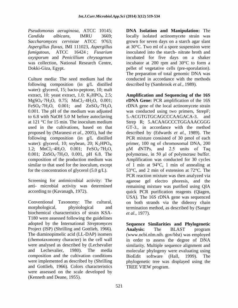
Pseudomonas aeruginosa, ATCC 10145;
Candida albicans, IMRU 3669;
Saccharomyces cervisiae ATCC 9763;
Aspergillus flavus, IMI 111023, Aspergillus
fumigatous, ATCC 16424 ; Fusarium
oxysporum and Penicillium chrysogenum
was collection, National Research Centre,
Dokki-Giza, Egypt.
Culture media: The seed medium had the
following composition (in g/L distilled
water): glycerol, 15; bacto-peptone, 10; malt
extract, 10; yeast extract, 1.0; K2HPO4, 2.5;
MgSO4·7H2O, 0.75; MnCl2·4H2O, 0.001;
FeSO4·7H2O, 0.001; and ZnSO4·7H2O,
0.001. The pH of the medium was adjusted
to 6.8 with NaOH 5.0 M before autoclaving
at 121 °C for 15 min. The inoculum medium
used in the cultivations, based on that
proposed by (Maranesi et al., 2005), had the
following composition (in g/L distilled
water): glycerol, 10; soybean, 20; K2HPO4,
1.2; MnCl2·4H2O, 0.001; FeSO4·7H2O,
0.001; ZnSO4·7H2O, 0.001, pH 6.8. The
composition of the production medium was
similar to that used for the inoculum, except
for the concentration of glycerol (5.0 g/L).
Screening for antimicrobial activity: The
anti- microbial activity was determined
according to (Kavanagh, 1972).
Conventional Taxonomy: The cultural,
morphological, physiological and
biochemical characteristics of strain KSA-
T180 were assessed following the guidelines
adopted by the International Streptomyces
Project (ISP) (Shrilling and Gottlieb, 1966).
The diaminopimelic acid (LL-DAP) isomers
(chemotaxonomy character) in the cell wall
were analysed as described by (Lechevalier
and Lechevalier, 1980). The media
composition and the cultivation conditions
were implemented as described by (Shrilling
and Gottlieb, 1966). Colors characteristics
were assessed on the scale developed by
(Kenneth and Deane, 1955).
DNA Isolation and Manipulation: The
locally isolated actinomycete strain was
grown for seven days on a starch agar slant
at 30°C. Two ml of a spore suspension were
inoculated into the starch- nitrate broth and
incubated for five days on a shaker
incubator at 200 rpm and 30°C to form a
pellet of vegetative cells (pre-sporulation).
The preparation of total genomic DNA was
conducted in accordance with the methods
described by (Sambrook et al., 1989).
Amplification and Sequencing of the 16S
rDNA Gene: PCR amplification of the 16S
rDNA gene of the local actinomycete strain
was conducted using two primers, StrepF;
5.-ACGTGTGCAGCCCAAGACA-3. and
Strep R; 5.ACAAGCCCTGGAAACGGG
GT-3., in accordance with the method
described by (Edwards et al., 1989). The
PCR mixture consisted of 30 pmol of each
primer, 100 ng of chromosomal DNA, 200
μM dNTPs, and 2.5 units of Taq
polymerase, in 50 μl of polymerase buffer.
Amplification was conducted for 30 cycles
of 1 min at 94°C, 1 min of annealing at
53°C, and 2 min of extension at 72°C. The
PCR reaction mixture was then analyzed via
agarose gel electro phoresis, and the
remaining mixture was purified using QIA
quick PCR purification reagents (Qiagen,
USA). The 16S rDNA gene was sequenced
on both strands via the dideoxy chain
termination method, as described by (Sanger
et al., 1977).
Sequence Similarities and Phylogenetic
Analysis: The BLAST program
(www.ncbi.nlm.nih. gov/blst) was employed
in order to assess the degree of DNA
similarity. Multiple sequence alignment and
molecular phylogeny were evaluating using
BioEdit software (Hall, 1999). The
phylogenetic tree was displayed using the
TREE VIEW program.

Int.J.Curr.Microbiol.App.Sci (2014) 3(12) 519-534
522
Fermentation: A loopful of the,
Streptomyces sp. from the 5-day culture age
was inoculated into 250 ml Erlenmeyer
flasks containing 75 ml of antibiotic
production medium had the following
composition (in g/L distilled water):
glycerol, 5; soybean flour, 20; K2HPO4, 1.2;
MnCl2·4H2O, 0.001; FeSO4·7H2O, 0.001;
ZnSO4·7H2O, 0.001, pH 6.8. The flasks
were incubated on a rotary shaker (200 rpm)
at 30 0C for 5 days. Twenty-liter total
volume was filtered through Whatman No.1
filter paper, followed by centrifugation at
5000 r.p.m for 20 minutes. The clear filtrates
were tested for their activities against the
test organisms (Neto et al., 2005).
Extraction : The clear filtrate was adjusted
at different pH values (4 to 9) and extraction
process was carried out using different
solvents separately at the level of 1:1 (v/v).
The organic phase was concentrated to
dryness under vacuum using a rotary
evaporator (Atta, 2013).
Precipitation: The precipitation process of
the crude compound dissolved in the least
amount of the solvent carried out using
petroleum ether (b.p 60-80 °C) followed by
centrifugation at 5000 r.p.m for 15 min. The
precipitate was tested for its antimicrobial
activities (Atta et al., 2010).
Separation: Separation of the antimicrobial
agent(s) into its individual components was
conducted by thin layer chromatography
using n-Butanol: acetic acid: water (3:1:1
v/v). as a solvent system (Atta et al., 2009).
Purification: The purification of the
antimicrobial agent(s) was carried out using
silica gel column (2 X 25) chromatography.
Chloroform-methanol (10:2, v/v), was used
as an eluting solvent. The column was left
for overnight until the silica gel (Prolabo)
was completely settled. One-ml crude
precipitate to be fractionated was added on
the silica gel column surface and the extract
was adsorbed on top of silica gel. Fifty
fractions were collected (each of 5 ml) and
tested for their antimicrobial activities (Lu et
al., 2008).
Elemental and Spectroscopic analysis:
The elemental analysis C, H, O, and N and
Spectroscopic analysis IR, UV and Mass
spectrum were determined at the micro-
analytical center of Cairo University, Egypt.
Determination of minimum inhibitory
concentration: The minimum inhibitory
concentration (MIC) could be determined by
the cup assay method (Kavanagh, 1972).
Characterization of the antibiotic: The
antibiotic produced by Streptomyces sp. was
identified according to the recommended
international references of (Umezawa, 1977;
Berdy, 1974; Berdy, 1980a b & c and Eric,
1999).
Result and Discussion
Screening for the antimicrobial activities
The metabolites of the Streptomyces sp.
exhibited various degrees of activities
against Gram positive and Gram negative
bacteria viz: Staphylococcus aureus, NCTC
7447; Bacillus subtilis, NCTC 1040 ;
Bacillus pumilus, NCTC 8214 ;
Micrococcus luteus, ATCC
9341.Escherichia coli, NCTC 10416;
Klebsiella pneumonia, NCIMB 9111;
Pseudomonas aeruginosa, ATCC 10145
(Table 1).
Identification of the Most Potent
Actinomycete Isolate
Morphological Characteristics
The vegetative mycelia grew abundantly on
both synthetic and complex media. The

Int.J.Curr.Microbiol.App.Sci (2014) 3(12) 519-534
523
aerial mycelia grew abundantly on Starch-
nitrate agar medium Oat-meal agar medium
(ISP-3) and Inorganic salts-starch agar
medium (ISP-4). The Spore chains were
rectiflexibiles, and had a smooth surface
(Plate 1). Neither both sclerotic granules and
sporangia nor flagellated spores were
observed (Table 2).
Cell Wall Hydrolysate
The cell wall hydrolysate contains LL-
diaminopimelic acid (LL-DAP) and sugar
pattern not detected.
Physiological and Biochemical
Characteristics
The actinomycete isolate KSA-T180 could
hydrolyzes starch, protein, lipid and lecithin,
whereas pectin hydrolysis and catalase test
are negative, melanin pigment is negative,
degradation of esculin & xanthin was
positive, citrate utilization, urea and KCN
utilization were positive, whereas nitrate
reduction, H2S production is negative (Table
2).
The isolate KSA-T180 utilizes meso-
inositol, starch, L-phenylalanine, L-valine,
L-arginine, L-tyrosine and L-histidine, but
do not utilize D-mannose, D-mannitol, D-
glucose, D-fructose, D-xylose, D-galactose,
maltose, lactose, L-rhamnose, sucrose,
raffinose, L-arabinose, and cycteine. Growth
was detected in presence of up to (7%)
NaCl. The isolate KSA-T180 utilizes
sodium azid (0.01%), phenol (0.01%); but
do not utilize in thallous acetate (0.001).
Good growth could be detected within a
temperature range of 30 to 45ºC. Good
growth could be detected within a pH value
range of 5 to 9. Moreover, the actinomycete
isolate KSA-T180 are active against
Bacillus subtilis, NCTC 1040; Micrococcus
luteus, ATCC 9341, but not active against
Saccharomyces cerevisiae ATCC 9763 and
Aspergillus niger IMI 31276 (Table 2).
Color and Culture Characteristics
The actinomycete isolate shows the aerial
mycelium is grayish yellow; substrate
mycelium is light yellowish brown, and the
diffusible pigment moderate yellowish
brown for ISP-2, 6 & 7 (Table 3).
Taxonomy of Actinomycete Isolate
This was performed basically according to
the recommended international Key’s viz.
(Buchanan and Gibsons, 1974; Williams,
1989; and Hensyl, 1994) and Numerical
taxonomy of Streptomyces species program
(PIB WIN). On the basis of the previously
collected data and in view of the
comparative study of the recorded properties
of actinomycete isolate in relation to the
closest reference strain, viz. Streptomyces
clavuligerus it could be stated that the
actinomycetes isolate KSA-T180 is
suggestive of being likely belonging to
Streptomyces clavuligerus, KSA-T180.
Molecular phylogeny of the selected
isolate
The 16S rDNA sequence of the local isolate
was compared to the sequences of
Streptomyces spp. In order to determine the
relatedness of the local isolate to these
Streptomyces strains. The phylogenetic tree
(as displayed by the Tree View program)
revealed that the locally isolated strain is
closely related to Streptomyces sp., the most
potent strain evidenced an 98% similarity
with Streptomyces clavuligerus (Fig. 1).
Fermentation, Extraction and
Purification
The fermentation process was carried out for

Int.J.Curr.Microbiol.App.Sci (2014) 3(12) 519-534
524
five days at 30C. After incubation period,
the filtration was conducted followed by
centrifugation at 4000 r.p.m. for 15 minutes.
The entire culture broth (20 liters) was
centrifuged (4000 rpm, 15 minutes) to
separate the mycelium and the supernatant.
The supernatant was extracted with n-
butanol (1:1, v/v) and the organic layer was
evaporated to give an oily material. The oily
material was then dissolved in 15% aqueous
methanol and defatted by partitioning with
petroleum ether (b.p. 60-80C) to give a
solid extract. Separation of antimicrobial
agent into individual components was
carried out by thin-layer chromatography
using a solvent system composed of n-
Butanol: acetic acid: water (3:1:1 v/v). Only
one band at Rf = 0.6 showed antibacterial
activity. The purification process through
column chromatography packed with silica
gel, revealed that the most active fractions
against the tested organisms ranged
between, 23 to 30.
Physicochemical characteristics
The purified antibacterial agent produced by
Streptomyces clavuligerus, KSA-T180 are
produces characteristic odour, their melting
points are 118C. The compound is freely
soluble in chloroform, ethyl acetate, n-
butanol, acetone, ethyl alcohol, methanol
and 10 % isopropyl alcohol, but insoluble in
petroleum ether, hexan and benzene.
Elemental analysis
The elemental analytical data of β-lactamase
inhibitor compound produced by
Streptomyces clavuligerus, showed the
following: The elemental analytical data of
the antibiotic indicated that: C=45.65;
H=3.8; N= 7.1; O= 43.45 and S= 0.0. This
analysis indicates a suggested empirical
formula of: C8H8NO5.
Spectroscopic Characteristics
The spectroscopic analysis of the purified of
β-lactamase inhibitor compound produced
by Streptomyces clavuligerus, the ultraviolet
(UV) absorption spectrum recorded a
maximum absorption peaks at 285 nm (Fig.
3). The Infra-red (IR) spectrum showed
characteristic bands 589, 600, 650, 708, 734,
800, 850, 880, 900, 950, 976, 1062, 1101,
1224, 1300, 1338, 1550, 1618, 1700, 2800,
2886, 3012 and 3420 (Fig.4). The Mass
spectrum showed that the molecular weight
at 200.0 (Fig.5).
MIC of β-lactamase Inhibitory Protein
The MIC of antibiotic produced by
Streptomyces clavuligerus, KSA-T180 for
Staphylococcus aureus, NCTC 7447 and
Bacillus subtilis, NCTC 1040 was 7.8 µg /
ml, whereas, Bacillus pumilus, NCTC
8214 and Micrococcus luteus, ATCC 9341
was 15.6 µg / ml. Escherichia coli, NCTC
10416 and Klebsiella pneumonia, NCIMB
9111 was 31.25 µg / ml. Moreover,
Pseudomonas aeruginosa was 46.87 µg /
ml.
Identification of the β-lactamase Inhibitor
On the basis of the recommended keys for
the identification of antibiotic, it could be
stated that the antibiotic suggestive of being
belonging to clavulanic acid (Chen, et al.,
2003; Parag et al., 2006 and Awad & El-
Shahed, 2013)
The Streptomyces clavuligerus was isolated
from Taif city, KSA. The isolate was
growing on production medium had the
following composition (in g/L distilled
water): glycerol, 5; soybean flour (SF), 20;
K2HPO4, 1.2; MnCl2·4H2O, 0.001;
FeSO4·7H2O, 0.001; ZnSO4·7H2O, 0.001,
pH 6.8. for investigating its potency to

Int.J.Curr.Microbiol.App.Sci (2014) 3(12) 519-534
525
produce antibacterial agents. The
actinomycete isolate, exhibited a wide
spectrum antibacterial agent (Kavanagh,
1972). Due to the selective isolation of soil
actinomycetes for finding novel strains
which can produce useful bioactive
compounds, thus various culture media and
techniques have been developed (Hozzein et
al., 2008 and Dhananjeyan et al., 2010).
Identification process had been performed
(Williams, 1989; Hensyl, 1994 and Holt et
al., 2000). The morphological
characteristics and microscopic examination
emphasized that the spore chain is spiral.
Spore mass is grayish yellow, while spore
surface is smooth, substrate mycelium is
light yellowish brown and diffusible
pigment moderate yellowish brown. The
results of physiological, biochemical
characteristics (Table 2) and cell wall
hydrolysate of actinomycete isolate,
exhibited that the cell wall containing LL-
diaminopimelic acid (DAP). These results
emphasized that the actinomycetes isolate
related to a group of Streptomyces as
previously studied (Reddy et al., 2011;
Afifi et al., 2012 and Muharram et al.,
2013).
The phylogenetic tree (diagram) revealed
that the local isolate KSA-T180 is closely
related Streptomyces clavuligerus, similarity
matrix is 98% as identified strain
of Streptomyces clavuligerus. Similar result
for identified strain of Streptomyces
plicatus (strain 101) by (Kang et al., 2000;
Anderson & Wellington, 2001and Zamanian
et al., 2005) Streptoverticillium sp. and
two Streptomyces sp. by (Raja et al. 2010).
In view of all the previously recorded data,
the identification of actinomycete isolate
KSA-T180 was suggestive of being
belonging to Streptomyces clavuligerus,
KSA-T180, as previously reported (Ghadin
et al., 2008 and Ubukata et al., 2007).
The active metabolites were extracted by n-
butanol at pH 7. Similar results were
obtained by (Sekiguchi, et al., 2007). The
organic phase was collected and evaporated
under reduced pressure using a rotary
evaporator. The extract was concentrated
and treated with petroleum ether (b.p. 40-
60C) for precipitation process where only
one fraction was obtained in the form of
yellowish ppt. and then tested for their
antibacterial activity. Separation of
antibiotic into individual components has
been tried by thin-layer chromatography
using a solvent system composed n-butanol-
acetic acid -water (3:1:1, v/v) as developing
solvent (Zhang et al, 2007 and Atta et al.,
2009). For the purpose of purification
process, the antibiotic were allowed to pass
through a column chromatography packed
with silica gel and eluting solvent was
composed of chloroform and methanol (10:2
v/v), fifty fractions were collected and tested
for their activities. The most active fractions
against the tested organisms ranged
between, 23 to 30. Similarly, many workers
used a column chromatography packed with
silica gel and an eluting solvent composed
of various ratios of chloroform and methanol
(Criswell et al. 2006; El-Naggar et al., 2006
and Sekiguchi, et al., 2007).
The elemental analytical data of
antibacterial agent produced by
Streptomyces clavuligerus, showed the
following: The elemental analytical data of
the antibiotic indicated that: C=45.65;
H=3.8; N= 7.1; O= 43.45 and S= 0.0. This
analysis indicates a suggested empirical
formula of: C8H8NO5. The spectroscopic
analysis of the purified of antibacterial agent
produced by Streptomyces clavuligerus, the
ultraviolet (UV) absorption spectrum
recorded a maximum absorption peak at 285
nm (Fig. 2).

Int.J.Curr.Microbiol.App.Sci (2014) 3(12) 519-534
526
Table.1 Mean diameters of inhibition zones (mm) caused by 100µl of the antimicrobial
activities produced by KSA-T180 in the agar plate diffusion assay (The diameter of the used
cup assay was 10 mm). Test organism Mean diameters of inhibition zone (mm)
Staphylococcus aureus, NCTC 7447 33.0
Bacillus subtilis, NCTC 1040 31.0
Bacillus pumilus, NCTC 8214 30.0
Micrococcus luteus, ATCC 9341 29.0
Escherichia coli, NCTC 10416 29.0
Klebsiella pneumonia, NCIMB 9111 27.0
Pseudomonas aeruginosa, ATCC 10145 24.0
Candida albicans, IMRU 3669 0.0
Saccharomyces cervisiae ATCC 9763 0.0
Aspergillus flavus, IMI 111023 0.0
Aspergillus fumigatous, ATCC 16424 0.0
Fusarium oxysporum 0.0
Penicillium chrysogenum 0.0
Plate.1 Scanning electron micrograph of the actinomycete isolate KSA-T180 growing on
starch nitrate agar medium showing spore chain Spiral shape and spore surfaces smooth
(X7,500)

Int.J.Curr.Microbiol.App.Sci (2014) 3(12) 519-534
527
Table.2 The morphological, physiological and biochemical characteristics of the
actinomycete isolate KSA-T180
Characteristic Result Characteristic Result
Morphological characteristics: Mannitol -
Spore chains Rectiflexibiles L- Arabinose -
Spore mass grayish yellow meso-Inositol +
Spore surface smooth Lactose -
Color of substrate mycelium Light yellowish brown Maltose -
Motility Non-motile D-fructose -
Cell wall hydrolysate Utilization of amino acids:
Diaminopimelic acid (DAP) LL-DAP L-Cycteine -
Sugar Pattern Not-detected L-Valine +
Physiological and biochemical properties:
Hydrolysis of:-
L-Histidine +
L-Phenylalanine +
Starch + L-Arginine +
Protein + L-Tyrosine +
Lipid + Growth inhibitors
Pectin - Sodium azide (0.01) +
Lecithin + Phenol (0.1) +
Catalase test - Thallous acetate (0.001) -
Production of melanin pigment on: Growth at different temperatures (˚C):
Peptone yeast- extract iron agar - 20 -
Tyrosine agar medium - 25 ±
Tryptone – yeast extract broth - 30-45 +
Degradation of: 50 -
Xanthin + Growth at different pH values:
Esculin + 4 -
H2S Production - 5-9 +
Nitrate reduction - 10 -
Citrate utilization + Growth at different concentration of NaCl (%)
Urea test + 1-7 +
KCN test + 10 -
Utilization of carbon sources Antagonistic Effect:
D-Xylose - Bacillus subtilis +
D- Mannose - Micrococcus luteus +
D- Glucose - Saccharomyces cerevisiae -
D- Galactose - Aspergillus niger - Sucrose -
L-Rhamnose - Raffinose - Starch +++
+Positive, - = Negative and ± = doubtful results, ++ = good growth.

Int.J.Curr.Microbiol.App.Sci (2014) 3(12) 519-534
528
Table.3 Cultural characteristics of the actinomycete isolate KSA-T180
Medium Growth Aerial
mycelium Substrate mycelium Diffusible pigment
1- Starch-nitrate agar medium Good 90-gy-y
grayish yellow 76.1.y Br
light yellowish brown 77 m-y Br
moderate yellowish brown
2- Yeast extract - Malt extract agar medium (ISP-2) No growth - - -
3- Oat-meal agar medium (ISP-3) Good 90-gy-y
grayish yellow 76.1.y Br
light yellowish brown -
4- Inorganic salts-starch agar medium (ISP-4) Good 90-gy-y
grayish yellow 76.1.y Br
light yellowish brown -
5- Glycerol-Asparagine agar medium (ISP-5) Moderate 90-gy-y
grayish yellow
93-y-Gray
yellowish gray -
6- Melanin test:
a- Tryptone-yeast extract broth (ISP-1) No growth - - -
b- Peptone yeast extract-iron agar medium (ISP-6) Good 90-gy-y
grayish yellow 76.1.y Br
light yellowish brown 77 m-y Br
moderate yellowish brown
c- Tyrosine agar (ISP-7) Good 90-gy-y
grayish yellow 76.1.y Br
light yellowish brown 77 m-y Br
moderate yellowish brown
The color of the organism under investigation was consulted using the ISCC-NBS color -
Name charts II illustrated with centroid color.
Fig.1 The phylogenetic position of the local Streptomyces sp. strain among neighboring
species. The phylogenetic tree was based on the multiple alignments options of 16S rDNA
sequences

Int.J.Curr.Microbiol.App.Sci (2014) 3(12) 519-534
529
Fig.2 Ultraviolet absorbance of antibiotic produced by Streptomyces clavuligerus,
KSA-T180
Fig.3 FTIR spectrum of antibiotic produced by Streptomyces clavuligerus, KSA-T180

Int.J.Curr.Microbiol.App.Sci (2014) 3(12) 519-534
530
Fig.4 Mass spectrum of antibiotic produced by Streptomyces clavuligerus, KSA-T180
The Infra-red (IR) spectrum showed
characteristic bands 589, 600, 650, 708,
734, 800, 850, 880, 900, 950, 976, 1062,
1101, 1224, 1300, 1338, 1550, 1618, 1700,
2800, 2886, 3012 and 3420 (Fig.3). The
Mass spectrum showed that the molecular
weight at 200.0. Similar investigations and
results were attained by (Baptista-Neto et
al., 2000; Parag et al., 2006; Awad and El-
Shahed, 2013 and Atta et al., 2013). The
MIC of antibiotic produced by
Streptomyces clavuligerus, KSA-T180 for
Staphylococcus aureus, NCTC 7447 and
Bacillus subtilis, NCTC 1040 was 7.8 µg /
ml, whereas, Bacillus pumilus, NCTC
8214 and Micrococcus luteus, ATCC 9341
was 15.6 µg / ml. Escherichia coli, NCTC
10416 and Klebsiella pneumonia, NCIMB
9111 was 31.25 µg / ml. Moreover,
Pseudomonas aeruginosa was 46.87 µg /
ml. Similar investigations and results were
attained by (Parag et al., 2006 and Awad
et al., 2009). Identification purified
antibiotic according to recommended
international keys indicated that the
antibiotic is suggestive of being belonging
to clavulanic acid antibiotic (Chen, et al.,
2003; Parag et al., 2006 and Awad & El-
Shahed, 2013).
References
Afifi, M.M.; Atta, H.M.; Elshanawany,
A.A.; Abdoul-Raouf, U.M. and El-
Adly, A.M. 2012. Biosynthesis of
hygromycin-B antibiotic by
Streptomyces crystallinus AZ151
isolated from Assuit, Egypt. Bacteriol.
J., 2: 46-65.
Anderson, A.S., and Wellington, E.M.
2001. The taxonomy of Streptomyces
and related genera. Int. J. Syst. Evol.
Microbiol., 51: 797-814.
Atta, H. M. 2010. Production, Purification,
Physico-Chemical Characteristics and
Biological Activities of Antifungal
Antibiotic Produced by Streptomyces
antibioticus, AZ-Z710. American-
Eurasian Journal of Scientific
Research. 5 (1): 39-49, 2010.
Atta, H. M.; A. T. Abul-hamd and H. G.
Radwan, 2009. Production of
Destomycin-A antibiotic by
Streptomyces sp. using rice straw as
Int.J.Curr.Microbiol.App.Sci (2014) 3(12) 519-534
531
fermented substrate. Comm. Appl.
Biol. Sci, Ghent University, 74 (3) :
879-897, 2009.
Atta, H.M.; Bayoumi, R.; El-Sehrawi, M.;
and Selim Sh. M. 2013. Application
Biotechnology of Recycling
Agricultural Waste In Al-Khurmah
Governorate For Production
Antimicrobial Agent(S) By
Actinomycetes Isolates Under Solid
State Fermentation Condition. Life
Science Journal 2013 10 (4):1749-
1761.
Augustine, S.K., S.P. Bhavsar and B.P.
Kapadnis, 2005. A non-polyene
antifungal antibiotic from
Streptomyces albidoflavus PU23. J.
Biosci., 30(2): 201-211.
Awad, H.M. and El-Shahed, K.Y.I. 2013.
A Novel Actinomycete sp. Isolated
from Egyptian Soil has β-Lactamase
Inhibitor Activity and Belongs to the
Streptomyces rochei Phylogenetic
Cluster. World Applied Sciences
Journal 21 (3): 360-370, 2013
Awad, H.M.; El-Shahed, K.Y.I. and El-
Nakkadi, A.E.M. 2009. Isolation,
screening and identification of newly
isolated soil Streptomyces
(Streptomyces sp. NRC-35) for β-
lactamase inhibitor production. World
Applied Science Journal, 7(9): 637-
646.
Baptista-Neto A, Gouveia ER, Badino Jr.
AC, Hokka CO 2000.
Phenomenological model of the
clavulanic acid production process
utilizing Streptomyces clavuligerus,
Braz. J. Chem. Eng. 17: 4-7.
Berdy J 2005. Bioactive microbial
metabolites. J Antibiot 58:1–26
Berdy J 2012. Thoughts and facts about
antibiotics: where we are now and
where we are heading. J Antibiot
65:385–395.
Berdy, J. 1974. Recent development of
antibiotic research and classification
of antibiotic according to chemical
structure. Adv. App. Microbiol., 14:
309-406.
Berdy, J. 1980a. Recent advances in and
prospects of antibiotics research. Proc.
Biochem., 15: 28-35.
Berdy, J. 1980b. CRC Handbook of
antibiotic compounds. Vol I. CRC
Press, Boca Raton, Florida.
Berdy, J. 1980c. CRC Handbook of
antibiotic compounds. Vol II. CRC
Press, Boca Raton, Florida.
Bibb, M. J. (2005). Regulation of
secondary metabolism in
Streptomycetes. Current Opinion in
Microbiology, 8(2):208-215.
Buchanan, R.E. and Gibbons, N.E.
1974. Bergey's Manual of
Determinative Bacteriology. 8th
Edn.,
Williams and Wilkins Co., Baltimore.
Chen, K.C.; Lin, H.Y.; Wu, J.Y. and
Hwang, S.C. 2003. Enhancement of
clavulanic acid production in
Streptomyces clavuligerus with
ornithine feeding. Enzyme Microbial
Technology, 32(1): 152-156.
Cimochowski, G. E., Harostock, M. D.,
Brown, R., Bernardi, M., Alonzo, N.,
Coyle, K. 2001. Intranasal mupirocin
reduces sternal wound infection after
open heart surgery in diabetics and
nondiabetics. The Annals of Thoracic
Surgery, 71(5):1572-1579.
Criswell, D.; V. L.Tobiason; J. S.
Lodmell, and D. S. Samuels, 2006.
Mutations Conferring Aminoglycoside
and Spectinomycin Resistance in
Borrelia burgdorferi. Antimicrob.
Agents Chemother. 50: 445-452.
Dhananjeyan, K.J.; Paramasivan, R.;
Tewari, S.C.; Rajendran, R.;
Thenmozhi, V.; Leo, S.V.J.;
Venkatesh, A. and Tyagi, B.K. 2010.
Molecular identification of mosquito
vectors using genomic DNA isolated

Int.J.Curr.Microbiol.App.Sci (2014) 3(12) 519-534
532
from eggshells, larval and pupal
exuvium. Trop. Biomed. ; 27:47–53.
Edwards, U.; Rogall, T.; Blocker, H.;
Emde, M. and Bottger, E.C.
1989. Isolation and direct complete
nucleotide determination of entire
genes. Characterization of a gene
coding for 16S ribosomal RNA.
Nucleic Acids Res., 17: 7843-7853.
El-Naggar, M.Y., S.A. El-Assar and S.M.
Abdel-Gawad, 2006. Meroparamycin
production by newly isolated
Streptomyces sp. strain MAR01:
Taxonomy, fermentation, purification
and structural elucidation. J.
Microbiol., 44(4): 432-438.
Ghadin, N.; Zin, N.M.; Sabaratnam, V.;
Badya, N.; Basri, D.F.; Lian H. and
Sidik, N.M. 2008. Isolation and
characterization of a novel endophytic
streptomyces SUK 06 with
antimicrobial activity from Malaysian
plant. Asian J. Plant Sci., 7: 189-194.
Gouveia E.R., Neto A.B., Badino A.C.,
Hokka C.O. 2001. Optimization of
medium composition for clavulanic
acid production by Streptomyces
clavuligerus. Biotechnol lett 23: 157-
161.
Hall, T.A. 1999. BioEdit: A user-friendly
biological sequence alignment editor
and analysis program for windows
95/98/NT. Nucleic Acid Symp. Ser.,
41: 95-98.
Hensyl, W.R. 1994. Bergey's Manual of
Systematic Bacteriology. 9th
Edn.,
Williams and Wilkins, Baltimore,
Philadelphia, Hong Kong, London,
Munich.
Holt, J.G.; Krieg, N.R.; Sneath, P.H.A;
Staley J.T. and Williams S.T. 2000.
Bergey's Manual of Determinative
Bacteriology. 9th
Ed. Baltimore:
Williams and Wilkins, London.
Hozzein, W.N.; Ali, M.I.A. and Rabie, W.
2008. A new preferential medium for
enumeration and isolation of desert
actinomycetes. World J. Microbiol.
Biotechnol. 24: 1547-1552.
Kang, S.G.; Park, H.U.; Lee, H.S.; Kim,
H.T. and Lee, KJ. 2000. New beta-
lactamase inhibitory protein (BLIP-I)
from Streptomyces exfoliatus SMF19
and its roles on the morphological
differentiation. J. Biol. Chem.
275:16851–16856.
Kavanagh, F., 1972. Analytical
Microbiology. Vol. 2, Acad. Press,
New York.
Kenig, M., Reading, C. 1979. Holomycin
and an antibiotic (MM 19290) related
to tunicamycin, metabolites of
Streptomyces clavuligerus. The
Journal of Antibiotics, 32:549-554.
Kenneth, L.K. and Deane, B.J. 1955.
Color universal language and
dictionary of names. United States
Department of Commerce. National
Bureau of standards. Washington,
D.C., 20234.
Kieser, T.; Bibb M. J.; Buttner, M. J.;
Chater K. F. and Hopwood, D. A.
2000. Practical Streptomyces genetics.
The John Innes Foundation, Norwich,
United Kingdom.
Lechevalier, M.P. and Lechevalier, H.A.
1980. The chemotaxonomy of
actinomycetes. In: Actinomycete
Taxonomy. A. Dietz and D.W.
Thayer, (Eds.), Special publication.
Arlington S I M, USA, 6: 227-291.
Mao, X., Shen, Y. Yang, L. Chen, S.
Yang, Y. Yang, J. Zhu, H. Deng, Z.
and Wri, D. 2007. Optimizing the
medium composition for accumulation
of the noval FR-008/candicidin
derivatives CS101 by a mutant of
Streptomyces sp. using statistical
experimental methods. Proc.
Biochem., 42: 878-883.
Maranesi, G.L.; Baptista-Neto, A.; Hokka,
C.O. and Badino, A.C. 2005.
Int.J.Curr.Microbiol.App.Sci (2014) 3(12) 519-534
533
Utilisation of vegetable oil in the
production of clavulanic acid by
Streptomyces clavuligerus ATCC
27064. World J Microbiol Biotechnol
21, 509–514.
Mona-Ibrahim M. 2012. Investigation on
some Streptomyces species produce
antibiotic with immobilized cells by
using calcium alginate. Journal of
Applied Sciences Research, 8(3):
1466-1476, 2012
Muharram, M.M., Abdelkader M.S. and
Alqasoumi, S.I. 2013. Antimicrobial
activity of soil actinomycetes isolated
from Alkharj, KSA. Int. Res. J.
Microbiol., 4: 12-20.
Neto, A.B.; Hirata, D.B.; Cassiano Filho,
L.C.M.; Bellao, C.; Badino Junior
A.C. and Hokka, C.O 2005. A study
on clavulanic acid production by
Streptomyces clavuligerus in batch,
fed-batch and continuous processes
Brazilian Journal of Chemical
Engineering, 22(4): 557-563.
Numerical taxonomy program 1989.
Numerical taxonomy of Streptomyces
species program (PIB WIN)
(Streptomyces species J. Gen
Microbiol. 1989 13512-133.
Olano, C., Lombó, F., Méndez, C., Salas,
J. A. 2008. Improving production of
bioactive secondary metabolites in
actinomycetes by metabolic
engineering. Metabolic Engineering,
10(5):281-92.
Onaka, H.; Taniguchi, S.; Igarashi Y. and
Furumai, T. 2002. Cloning of the
staurosporine biosynthetic gene
cluster from Streptomyces sp. TP-
A0274 and its heterologous expression
in Streptomyces lividans . J Antibiot
55:1063–1071
Ortiz, S. C. A., Hokka, C. O., Badino, A.
C. 2007. Utilization of soybean
derivatives on clavulanic acid
production by Streptomyces
clavuligerus. Enzyme and Microbial
Technology, 40:1071-1077.
Paradkar, A. S., Trefzer, A., Chakraburtty,
R., Stassi, D. 2003. Streptomyces
Genetics: A Genomic Perspective.
Critical Reviews in Biotechnology,
23:1–27.
Parag, S.; Saudagar, R.; Singhal, S. 2006.
Optimization of nutritional
requirements and feeding strategies
for clavulanic acid production by
Streptomyces clavuligerus.
Bioresource Technology 98: 2010–
2017
Raja, A.; Prabakaran P. and Gajalakshmi,
P. 2010. Isolation and screening of
antibiotic producing psychrophilic
actinomycetes and its nature from
rothang hill soil against
viridans Streptococcus sp. Res. J.
Microbiol., 5: 44-49.
Reddy, N.G.; Ramakrishna, D.P.N and
Gopal, S.V.R. 2011. A morphological,
physiological and biochemical studies
of marine Streptomyces rochei
(MTCC 10109) showing antagonistic
activity against selective human
pathogenic microorganisms. Asian
Journal of Biological Science, 4(1): 1-
14.
Rodríguez, M.; Núñez, L. E.; Braña, A. F.;
Méndez, C.; Salas, J. A. and Blanco,
G. 2008. Identification of
transcriptional activators for
thienamycin and cephamycin C
biosynthetic genes within the
thienamycin gene cluster from
Streptomyces cattleya. Molecular
Microbiology, 69(3):633-645.
Sacramento, D. R., Coelho, R. R. R., Wigg
M. D., Linhares L. F. T. L., Santos, M.
G. M., Semêdo, L. T. A. S., Silva, A.
J. R. 2004. Antimicrobial and antiviral
activities of an Actinomycetes
(Streptomyces sp.) isolated from a
Brazilian tropical forest soil. World

Int.J.Curr.Microbiol.App.Sci (2014) 3(12) 519-534
534
Journal of Microbiology and
Biotechnology, 20(3):225-229.
Sambrook, J.; Fritsch E.F. and Maniatis,
T.A. 1989. Molecular Cloning: A
Laboratory Manual. 2nd
Edn., Cold
Spring Harbor Laboratory Press, New
York, USA., ISBN-13:
9780879695774, Pages: 397.
Sanger, F.; Nicklen, S. and Coulson, A.R.
1977. DNA sequencing with chain-
terminating inhibitors. Proc. Natl.
Acad. Sci., 74: 5463-5467.
Sekiguchi, M.; Shiraish, N.; Kobinata, K.;
Kudo, T.; Yamaguchi, I.; Osada, H.
and Isono, K. 2007. RS-22A and C:
new macrolide antibiotics from
Streptomyces violaceusniger,
Taxonomy, fermentation, isolation
and biological activities. Journal of
Antibiotics 48(4): 289-292.
Shrilling, E.B. and Gottlieb, D. 1966.
Methods for characterization of
Streptomyces species. International
Journal of Systematic Bacteriology,
16(3): 313-340.
Singh, R.; Pandey, B. and Mathew, C.M.
2014. Production, purification and
optimization of Streptomycin from
isolated strain of Streptomyces griseus
and analysis by HPLC.
IndianJ.Sci.Res.4 (1):149-154, 2014
Thai, W.; Paradkar. A. S. and Jensen, S. E.
2001. Construction and analysis of b-
lactamase inhibitory protein (BLIP)
non-producer mutants of Streptomyces
clavuligerus. Microbiology (2001),
147, 325–335.
Ubukata, M., N.; Shiraishi, K.; Kobinata,
T. and Yamaguchi I. 2007. RS-22A, B
and C: New macrolide antibiotics
from Streptomyces violaceusniger. I.
Taxonomy, fermentation, isolation
and biological activities. J. Antibiot
(Tokyo), 48: 289-292.
Umezawa, H. 1977. Recent advances in
bio-active microbial secondary
metabolites. Jap. J. Antibiotic. Suppl.,
30: 138-163.
Williams, S.T. 1989. Bergey's Manual of
Systematic Bacteriology. Vol. 4,
Williams and Williams, Baltimore,
MD., USA.
Zamanian, S.; Shahidi Bonjar, G.H. and
Saadoun, I. 2005. First report of
antibacterial properties of a new strain
of Streptomyces plicatus (strain 101)
against Erwinia carotovora from Iran.
Biotechnology, 4: 114-120.
Zhang, L.; K. Yan; Y. Zhang; R. Huang; J.
Bian; C. Zheng; H. Sun; Z. Chen; N.
Sun; R. An; F. Min; W. Zhao; Y.
Zhuo; J. You; Y. Song; Z. Yu; Z. Liu;
K. Yang; H. Gao; H. Dai; X. Zhang; J.
Wang; C. Fu; G. Pei; J. Liu; S. Zhang;
M. Goodfellow; Y. Jiang; J. Kuai; G.
Zhou; and X. Chen, 2007. High-
throughput synergy screening
identifies microbial metabolites as
combination agents for the treatment
of microbial infections. Proc. Natl.
Acad. Sci. USA 104: 4606-4611.