Philosophy of voltage-gated proton...
22
rsif.royalsocietypublishing.org Review Cite this article: DeCoursey TE, Hosler J. 2014 Philosophy of voltage-gated proton channels. J. R. Soc. Interface 11: 20130799. http://dx.doi.org/10.1098/rsif.2013.0799 Received: 29 August 2013 Accepted: 22 November 2013 Subject Areas: biophysics, bioenergetics, biochemistry Keywords: H V 1, HVCN1, ion channels, voltage sensing, permeation, selectivity Author for correspondence: Thomas E. DeCoursey e-mail: [email protected] Philosophy of voltage-gated proton channels Thomas E. DeCoursey 1 and Jonathan Hosler 2 1 Department of Molecular Biophysics and Physiology, Rush University, 1750 West Harrison, Chicago, IL 60612, USA 2 Department of Biochemistry, University of Mississippi Medical Center, Jackson, MS 39216, USA In this review, voltage-gated proton channels are considered from a mainly teleological perspective. Why do proton channels exist? What good are they? Why did they go to such lengths to develop several unique hallmark prop- erties such as extreme selectivity and DpH-dependent gating? Why is their current so minuscule? How do they manage to be so selective? What is the basis for our belief that they conduct H þ and not OH – ? Why do they exist in many species as dimers when the monomeric form seems to work quite well? It is hoped that pondering these questions will provide an intro- duction to these channels and a way to logically organize their peculiar properties as well as to understand how they are able to carry out some of their better-established biological functions. 1. Introduction Voltage-gated proton channels (H V ) are membrane proteins that mediate the rapid movement of protons (H þ ) across cell membranes while excluding all other ions. Like other ion channels, the proton channel adopts two functional con- figurations. When ‘open’, each channel allows up to 10 5 H þ to cross the membrane per second but when they are ‘closed’, they do not allow any ions to cross the membrane. They are ‘voltage-gated’ because they open or close in response to changes in the membrane potential of the cell. Because H þ carries a positive charge, its movement across the membrane comprises an electrical current that can be recorded experimentally. A voltage-gated proton channel gene was not identified until 2006 [1,2]. Since then, some of the working parts of the molecule have been described, for example the conduction pathway (figure 1), but no crys- tal structure exists. Although a large number of enzymes, including many crucial to bioenergetics, contain proton pathways (or channels) that are essential to the operation of the molecule, the voltage-gated proton channel is a distinct category of protein and has not been reported to exist in mitochondria or chloroplasts. In many cells, proton channels are present in the plasma membrane; in a few instances (dinoflagellates and phagosomes in white blood cells), they have been shown to exist in organelle membranes. When these words were written, this was the 100th review written entirely (n ¼ 75) or partially (n ¼ 25) about voltage-gated proton channels. The number of original research papers, 157, was less than double that number 1 . From these statistics, one might conclude that this review needs only to discuss the two most recent original papers. Instead, we will step back and attempt to view the subject from a distance. Why do voltage-gated proton channels exist? What good are they? Why do they have so many unique properties? What kind of a world would it be if there were no proton channels? We describe the properties of proton channels teleologically and anthropomorphically; we do not pretend to understand the evolutionary forces that produced this protein, but simply describe it as though it were a sentient being whose properties serve the purposes we impute to it. 1.1. The origin of H V species The abbreviation for voltage-gated proton channels is H V (H for H þ , the con- ducted ionic species, subscript V for voltage-gated). To date no more than & 2013 The Author(s) Published by the Royal Society. All rights reserved. on June 7, 2018 http://rsif.royalsocietypublishing.org/ Downloaded from
Transcript of Philosophy of voltage-gated proton...

on June 7, 2018http://rsif.royalsocietypublishing.org/Downloaded from
rsif.royalsocietypublishing.org
ReviewCite this article: DeCoursey TE, Hosler J. 2014
Philosophy of voltage-gated proton channels.
J. R. Soc. Interface 11: 20130799.
http://dx.doi.org/10.1098/rsif.2013.0799
Received: 29 August 2013
Accepted: 22 November 2013
Subject Areas:biophysics, bioenergetics, biochemistry
Keywords:HV1, HVCN1, ion channels, voltage sensing,
permeation, selectivity
Author for correspondence:Thomas E. DeCoursey
e-mail: [email protected]
& 2013 The Author(s) Published by the Royal Society. All rights reserved.
Philosophy of voltage-gated protonchannels
Thomas E. DeCoursey1 and Jonathan Hosler2
1Department of Molecular Biophysics and Physiology, Rush University, 1750 West Harrison, Chicago,IL 60612, USA2Department of Biochemistry, University of Mississippi Medical Center, Jackson, MS 39216, USA
In this review, voltage-gated proton channels are considered from a mainly
teleological perspective. Why do proton channels exist? What good are they?
Why did they go to such lengths to develop several unique hallmark prop-
erties such as extreme selectivity and DpH-dependent gating? Why is their
current so minuscule? How do they manage to be so selective? What is
the basis for our belief that they conduct Hþ and not OH–? Why do they
exist in many species as dimers when the monomeric form seems to work
quite well? It is hoped that pondering these questions will provide an intro-
duction to these channels and a way to logically organize their peculiar
properties as well as to understand how they are able to carry out some of
their better-established biological functions.
1. IntroductionVoltage-gated proton channels (HV) are membrane proteins that mediate the
rapid movement of protons (Hþ) across cell membranes while excluding all
other ions. Like other ion channels, the proton channel adopts two functional con-
figurations. When ‘open’, each channel allows up to 105 Hþ to cross the membrane
per second but when they are ‘closed’, they do not allow any ions to cross the
membrane. They are ‘voltage-gated’ because they open or close in response to
changes in the membrane potential of the cell. Because Hþ carries a positive
charge, its movement across the membrane comprises an electrical current that
can be recorded experimentally. A voltage-gated proton channel gene was not
identified until 2006 [1,2]. Since then, some of the working parts of the molecule
have been described, for example the conduction pathway (figure 1), but no crys-
tal structure exists. Although a large number of enzymes, including many crucial
to bioenergetics, contain proton pathways (or channels) that are essential to the
operation of the molecule, the voltage-gated proton channel is a distinct category
of protein and has not been reported to exist in mitochondria or chloroplasts. In
many cells, proton channels are present in the plasma membrane; in a few
instances (dinoflagellates and phagosomes in white blood cells), they have been
shown to exist in organelle membranes.
When these words were written, this was the 100th review written entirely
(n ¼ 75) or partially (n ¼ 25) about voltage-gated proton channels. The number
of original research papers, 157, was less than double that number1. From these
statistics, one might conclude that this review needs only to discuss the two
most recent original papers. Instead, we will step back and attempt to view
the subject from a distance. Why do voltage-gated proton channels exist?
What good are they? Why do they have so many unique properties? What
kind of a world would it be if there were no proton channels? We describe
the properties of proton channels teleologically and anthropomorphically; we
do not pretend to understand the evolutionary forces that produced this
protein, but simply describe it as though it were a sentient being whose
properties serve the purposes we impute to it.
1.1. The origin of HV speciesThe abbreviation for voltage-gated proton channels is HV (H for Hþ, the con-
ducted ionic species, subscript V for voltage-gated). To date no more than

Figure 1. A snapshot from a molecular dynamics simulation of the openstate of the human voltage-gated proton channel, hHV1. The four transmem-brane helices are shown as ribbons with the extracellular end of the channelto the right. Charged amino acids (labelled) and water molecules in the poreare shown in licorice representation. This snapshot illustrates a configurationthat is present only 10% of the time, in which there is a continuous waterwire; usually the water wire is interrupted by a D112 – R208 salt bridge. Theexternal and internal charge networks are thought to stabilize the openconformation of the channel. (Adapted from [3].)
rsif.royalsocietypublishing.orgJ.R.Soc.Interface
11:20130799
2
on June 7, 2018http://rsif.royalsocietypublishing.org/Downloaded from
one gene has been identified in any species, thus all HV are
HV1, with a prefix that indicates species (e.g. hHV1 for
human, EhHV1 for the coccolithophore Emiliania huxleyi). The
official numbering of other channels is often more complex,
including subtypes, e.g. KV3.2. The HV family tree (figure 2)
continues to grow, but already includes several kingdoms (or
supergroups, if one prefers this classification system), includ-
ing unconfirmed HV in Chlorella (Protista kingdom) and in
the common wine grape, Vitis vinifera (Plantae kingdom), con-
firming the Bacchanalian proclivities of HV. Taylor et al. [5]
noted that HV appear to be absent in land plants and suggested
an explanation: these species tend to have a large inward
pH gradient, which seems fundamentally incompatible with
the exquisite design of the proton channel (in most species)
as a passive acid extrusion mechanism. This argument may
apply to plasma membranes, but HV could still exist in plant
vacuole membranes. Recent BLAST searches produce a hand-
ful of likely candidates among algae, fungi and a few higher
plants. The branch length in figure 2 reflects the extent of differ-
ence in amino acid sequence between the branch-dweller and
its neighbours. Mammalian HV1 are all very similar to each
other, but as we branch out to invertebrates, diversity blossoms
and single-celled organisms have wildly diverse sequences.
Sequence differences, in turn, might be expected to presage
functional differences, nicely illustrated by the novel properties
of the kHV1 in the dinoflagellate Karlodinium veneficum [4],
which has the longest branch of all. This is the only known
proton channel that opens to allow inward proton current; all
other HV open only when there is an outward electrochemical
proton gradient, and consequently their predominant function
is acid extrusion from cells (see ‘Why do proton channels have
DpH-dependent gating?’). Because kHV1 opens negative to the
Nernst potential for protons, EH, it allows inward current, and
is thus capable of mediating an action potential [4], just as are
voltage-gated sodium channels in more traditional excitable
cells such as nerve and muscle [6]. As the HV family tree
grows, and especially as the properties of HV in the 30 not-
yet-studied species in figure 2 are explored, further diversity
in function will likely be found.
Based on sequence homology analysis [4,7], HV are clo-
sely related to two other groups of proteins that also
contain voltage-sensing domains (VSDs), the voltage-sensing
phosphatases (VSP) and c15orf27 (molecules of unknown
function). These groups are in turn related by common ances-
try to Naþ and Ca2þ channels, and appear to have diverged
from the Kþ channel family over one billion years ago [3].
Figure 3 illustrates the molecular structure and membrane
topology of three classes of VSD-containing molecules.
1.2. Why do voltage-gated proton channels exist?A flippant answer to this question is ‘for the same reason that
other ion channels exist!’ Cells need to regulate the flow of ions
across their membranes, and the most sensible way of doing
this is to produce an array of proteins that selectively transport
individual ionic species in a tightly controlled fashion. Because
the consequences of allowing ions to cross membranes differ
drastically for each species of ion, we must delve a bit deeper
for a more informative answer. Reflecting the importance of
the conducted ion species, traditionally ion channels have
received names that indicate the type of ion they transport,
sometimes modified by a salient characteristic (inwardly recti-
fying Kþ channel; voltage-gated Naþ channel; Ca2þ-activated
Kþ channel, stretch-activated non-selective cation channel).
When a channel opens in a cell membrane, it conducts ions pas-
sively down the electrochemical gradient for the conducted
ion. The chemical part of an electrochemical gradient simply
reflects that the concentration of one ionic species is often
greater on one side of the membrane. Ions tend to flow down
the gradient, from high to low concentration, other things
being equal (‘other things’ being the electrical contribution dis-
cussed in the next paragraph). Usually Kþ current is outward,
because the Kþ concentration is much greater inside the cell,
[Kþ]i, than outside, [Kþ]o. Most cells have large inward Naþ
and Ca2þ gradients, meaning that the concentrations of these
cations are much higher outside the cell than inside; hence
Naþ and Ca2þ channels almost always carry inward current.
1.2.1. A digression on the Nernst potentialIn addition to the chemical gradient, we also need to consi-
der the electrical potential across the cell membrane, the
‘membrane potential’. Because ions are charged, they are
affected by electrical fields. For example, [Kþ] is approximately
160 mM inside cells and 4.5 mM outside, so in the absence of
electrical forces, a Kþ channel would carry outward current.
Electrical current is defined in terms of the movement of posi-
tive charge, so this means Kþ leaves the cell. This definition
makes the behaviour of anions less intuitively accessible;
outward Cl– current is recorded electrically when there is
inward Cl– flux (‘flux’ simply means the physical movement
of ions from one place to another). The effects of a concen-
tration gradient can be modified or overridden if we control
the membrane potential, which can be done experimentally
by means of a circuit called a ‘voltage clamp’. Membrane
potential is defined as the potential inside the cell relative to
outside, which is defined as the reference voltage, 0 mV. As
the membrane is hyperpolarized2, the increasingly negative
charge inside the cell tends to prevent the positively charged
Kþ from leaving, despite the outward concentration gradient.
We eventually reach a special voltage called the Nernst
potential for Kþ, EK, at which the electrical force perfectly
balances the chemical potential and no net current will flow:
EK ¼ 2:303RTzF
log½Kþ�o½Kþ�i
; ð1:1Þ

Sus
Can
is2 C
anis1
Monodelphis
Myotis
Sorex
Hom
o*
Macaca
Oryctolagus
Loxodonta
Equus
Taeniopygia
Gallus
Oryzias
Gasterosteus
Danio
X. tropicalis
T. laevis
Ech
inop
s
Sper
mop
hilu
s
Tupa
ia
BosRattus
*Mus
84
100
88
76
96
86
82
69
99
100 C. intestin
alis*C. savig
ny
Lottia
Stro
ngyl
ocen
trotu
s*
Bra
nchi
osto
maTric
hopl
ax
Tha
lass
iosi
ra
Phae
odac
tylu
m*
*C. pelagicus
*E. huxleyi
*Karlodinium
Polysphondylium
Chlorella
Nem
atos
tella
90
76
67 97
invertebrates
amphibians
fish
birds
mammals
single-celledorganisms
Figure 2. Phylogenetic tree of confirmed and probable voltage-gated proton channels. Branch length is proportional to the degree of difference from neighbouringproteins. For the eight starred species, the gene has been expressed heterologously and confirmed electrophysiologically to encode a voltage-gated proton channel.(Adapted from [4].)
rsif.royalsocietypublishing.orgJ.R.Soc.Interface
11:20130799
3
on June 7, 2018http://rsif.royalsocietypublishing.org/Downloaded from
where R is the gas constant, T is temperature, F is Faraday’s
constant, z is the charge of the ion (þ1 in this case), [Kþ]o is
the extracellular (outside) concentration of Kþ, and [Kþ]i
is the intracellular concentration of Kþ. The constant
2.303(RT/zF) is approximately 58 mV at 208C so when
[Kþ]o ¼ [Kþ]i, EK ¼ 0 mV and when there is a 10-fold outward
Kþ gradient, e.g. [Kþ]i ¼ 160 mM and [Kþ]o ¼ 16 mM, EK is
258 mV. The Nernst potentials for other ions are calculated
similarly, with the caveat that the correct z must be used for
anions or divalent ions. If we consider our favourite channel,
the voltage-gated proton channel, its permeant ion is Hþ
alone. The concentration of Hþ is traditionally presented as
pH, the negative logarithm of the concentration, so 1027 M
Hþ is pH 7. We define the pH gradient DpH as pHo 2 pHi,
and from the Nernst equation (equation (1.1)), we see that for
DpH ¼ 1 unit, EH is 258 mV. A positive DpH means there is
an outward Hþ (chemical) gradient; which means that at
0 mV (no applied voltage), Hþwill tend to exit the cell through
any Hþ selective pathway. The Nernst potential is very useful
in characterizing ion channels. The selectivity of a channel
(which ions it allows to pass) can be determined by varying
ion concentrations and measuring the reversal potential (the
voltage where no net current flows), and comparing this to
the Nernst potential calculated for each ionic species present.

S1 S2 S3 S4 S5 S6 S1 S2 S3 S4 S1 S2 S3 S4
VSP
VSP
H+ channelK+ channelextracellularspace
cytosol
N C
extracellularspace
cytosol
membrane
membrane
K+ channel H+ channel
Cys
PO4
H+K+ H+(b)
(a)
Figure 3. Molecular architecture and orientation in the membrane of three classes of molecules that contain VSDs. (a) Illustrates the monomer of each, with itstransmembrane segments numbered (S1, S2, etc.) starting from the intracellular N terminus. In each, the VSD includes the first four segments, S1 – S4, in particularS4 which contains a series of cationic residues (Arg or Lys) that are thought to move outward upon membrane depolarization. The VSDs from these molecules arehomologous. (b) Each molecule in its functional oligomeric state. The voltage-gated potassium (Kþ) channel is a tetramer, with four VSDs surrounding a singlecentral pore, formed by the four S5 – S6 segments, through which Kþ crosses the membrane. The proton channel is a dimer in mammals and many other species,but each monomer has its own conduction pathway [8,9] and can function independently. Phosphorylation of Thr29 in the intracellular N terminus enhances thegating of HV in phagocytes and other cells [10,11]. The VSP is a monomer whose VSD senses membrane potential and accordingly regulates the activity of theattached intracellular phosphatase [12]. (Adapted from [13].)
rsif.royalsocietypublishing.orgJ.R.Soc.Interface
11:20130799
4
on June 7, 2018http://rsif.royalsocietypublishing.org/Downloaded from
When a channel opens and conducts current, one result is
a change in the membrane potential. The rule is that each
channel drives the membrane potential toward its own
Nernst potential. Thus, when Naþ and Ca2þ channels open,
they depolarize the membrane, whereas open Kþ channels
tend to hyperpolarize the membrane. There are also a number
of non-selective ion channels. When they open, they drive the
membrane potential toward their Nernst potential, which is
0 mV; hence, they usually depolarize the membrane, because
for the most part cells have negative resting membrane
potentials. A cell with a large number of open non-selective
ion channels bears a disturbing resemblance to an electro-
physiologically dead cell, exhibiting a familiar Gestalt that
is witnessed by electrophysiologists at the end of every
experiment—a leaky membrane with no reaction to radical
changes in bath solutions, and a relentless approach toward
no membrane resistance and a 0 mV membrane potential.
Even so, there are situations in which brief opening of non-
selective ion channels serves a useful purpose. For example,
non-selective endplate channels in skeletal muscle open in
response to acetylcholine released from a motor neuron,
depolarizing the muscle membrane enough to initiate an
action potential that propagates along the membrane and
produces contraction [16].
The raison d’etre for proton channels differs in different
species. A clue to their general function in multicellular
species, in fact in all species except dinoflagellates [4], is
that they are uniquely regulated by the pH gradient, DpH
(defined as pHo2 pHi), in such a manner that they open
only when there is an outward electrochemical gradient for
Hþ (i.e. DpH . 0) [17]. As is discussed below in ‘Why do
proton channels have DpH-dependent gating?’, when HV
open, they always carry outward current. As a result, they
also tend to hyperpolarize the membrane. In certain situ-
ations, this effect provides identifiable benefit to the cell,
but perhaps more frequently, the primary purpose appears
to be regulating pH. When HV open, Hþ leave the cell. In
other words, HV extrude acid, thereby increasing pHi. One
may speculate that HV exist in a large number of species
because all cells engage in metabolism, which leads to a net
production of protons that must be extruded to maintain a
pHi compatible with cellular activities. Numerous other func-
tions have been proposed that are specific to particular cells
in particular situations [17]; full discussion of these is
beyond the scope of this review.
Proton channels are extremely efficient proton extruders,
capable of changing pHi an order of magnitude faster than
other Hþ equivalent transporters [18–20], at rates up to
6 pH units min21 in small cells [18], more slowly in large
cells [21,22]. Because they do not require ATP [23], they can
extrude acid at minimal metabolic cost to the cell. Under
some circumstances, the concomitant extrusion of charge
could incur some cost owing to the necessity of charge
compensation. In one intensely studied case, however—the
respiratory burst in phagocytes (figure 4)—the cell is already
enthusiastically extruding negative charge in the form of
superoxide anion produced by NADPH oxidase, so the acti-
vation of HV simultaneously extrudes acid and compensates
charge. During the respiratory burst, proton channels set the
membrane potential to precisely the value at which Hþ cur-
rent is equal and opposite to the electron current [24]. The
extraordinarily efficient activity of HV also includes two
additional simultaneous functions. Hþ efflux compensates
the massive electron flux without causing undue osmotic
imbalance, because the biochemical products of Hþ inside
the phagosome include a variety of membrane permeable

bacterium
Cl–
2 Cl–
2 Cl–antiport
CIC-3
O2
H2O2
HOCl
antiport
protonchannel
NADP+HMS
NADPH
ATPase
NADPHoxidasecomplex
p47
p40
p22
p67Rac
gp91
OCl– H+
2 H+ H+H+
H+
H+
+ H+
CO2
??
Na+
Na+
H+
MPO
2 O2
2 O2
2 e–
H2O + HOCl
12
H+
H+
H+
?
?
Figure 4. Proton pathways and proton transport proteins in phagocytes during the ‘respiratory burst’. Part of a white blood cell ( for example, a neutrophil) isdepicted as its plasma membrane engulfs a bacterium. The enzyme NADPH oxidase (a.k.a. ‘NOX2’) assembles and becomes active in the membrane of the phago-some, the newly forming intracellular compartment enclosing the invader. NOX2 activity is so intense in neutrophils that it consumes orders-of-magnitude moreoxygen as a substrate to form O2. 2 than is used by the rest of cell metabolism combined, hence the misnomer ‘respiratory burst’ [24 – 26]. NOX2 is electrogenic,because it moves electrons from cytoplasmic NADPH across the membrane to reduce O2 to O2. 2 [27]. The prodigious electron flux permits NOX2 activity to bemeasured directly as electron current [28,29], which without compensation would depolarize the membrane by approximately 11 kV min21 [23]. Voltage-gatedproton channels compensate electrically for the electron flux, prevent large pH changes in cytoplasm or phagosome, minimize the osmotic consequences of chargecompensation, and are a necessary substrate for producing H2O2 and HOCl [24]. (Adapted from [13].)
rsif.royalsocietypublishing.orgJ.R.Soc.Interface
11:20130799
5
on June 7, 2018http://rsif.royalsocietypublishing.org/Downloaded from
species (H2O, H2O2 and HOCl) which can dissipate an osmo-
tic gradient simply by diffusing through the membrane.
Finally, Hþ flux into the phagosome is crucial because Hþ
is required in large quantities as a substrate for the major
reaction products, H2O2 and HOCl [24].
The teleological benefits of opening proton channels in
different cells tend to fall into four general categories, each
of which is exemplified in the phagocyte respiratory burst:
(i) electrical—charge compensation, setting the membrane
potential or generating action potentials, (ii) control of pHi
(intracellular pH), (iii) control of pHo (extracellular pH) and
(iv) osmotic—e.g. cell or organelle volume regulation.
(i) Charge compensation or regulating membrane
potential by HV has been proposed for neutrophils
[27,30–35], eosinophils [32,36–41], monocytes [42],
macrophages [20], osteoclasts [43], basophils [44],
microglia [45,46], dendritic cells [47], sperm [48], B
lymphocytes [49,50] and cardiac fibroblasts [51]. Pro-
ducing more dramatic electrical effects than simply
setting the resting membrane potential, proton channels
in dinoflagellates exhibit properties consistent with
the suggestion that they mediate the action potential
that triggers a luciferase-induced bioluminescent flash
[4,52–54].
(ii) Regulating pHi is a general function, but has highly
specific consequences in sperm capacitation [55],
breast cancer cells [56], egg maturation [57,58], baso-
phils during histamine release [44], neutrophils and
eosinophils during phagocytosis [34,41,59–61] and
coccolithophores during calcification [62].
(iii) Regulating pHo is considered an important function of
hHV1 in human airway epithelium [63] and in exacer-
bating the invasiveness of breast cancer cells [64].
In the latter situation, HV benefit the cells to the
detriment of the organism.
(iv) Finally, osmotic effects are thought to be involved in
HV1 activity in microglia [65]. Hþ flux via HV in pha-
gosomes likely minimizes the volume changes that
would occur if charge were compensated by other
means [24].
In summary, the consequences of proton channel acti-
vity are highly diverse and depend strongly on the precise
situation in each type of cell or organelle.
1.3. Why do proton channels need to be so selective?Ion channels are often selective for one ion over others pre-
sent in physiological solutions. That said, biology tends to
be parsimonious—Naþ and Kþ channels are selective, but

rsif.royalsocietypublishing.orgJ.R.Soc.Interface
11:20130799
6
on June 7, 2018http://rsif.royalsocietypublishing.org/Downloaded from
not extremely so, and in fact, they do not need to be. If the
‘wrong’ ion permeates 1% of the time, this will have a negli-
gible impact on the cell. After all, Naþ and Kþ gradients are
being dissipated continually, particularly in excitable cells,
and the Naþ/Kþ-ATPase works continuously to restore
these gradients. Naþ channels still manage to mediate
action potentials even though their error rate is substantial:
PK/PNa is 0.05–0.1 [66–69] and as high as 0.23–0.30 when
Kþ is inside and Naþ outside [70], as usually is the case.
Selectivity is often quantified as relative permeability (for
example PK/PNa is the permeability to Kþ relative to that of
Naþ), which can be calculated from measured reversal poten-
tials (Vrev) using the Goldman–Hodgkin–Katz voltage
equation [71–73]
Vrev ¼RTF
logPCl� ½Cl��i þ PKþ ½Kþ�o þ PNaþ ½Naþ�o þ PHþ ½Hþ�oPCl� ½Cl��o þ PKþ ½Kþ�i þ PNaþ ½Naþ�i þ PHþ ½Hþ�i
:
ð1:2Þ
Permeability means the facility with which a particular ion
crosses the membrane. This will depend mainly on which
ion transport proteins are present, because small ions
cannot permeate biological membranes (lacking proteins) at
physiologically relevant rates. If the membrane contains
only one type of ion channel, permeability indicates how effi-
ciently the ion permeates this type of channel. Note that
permeability is a property of the ion and the channel in com-
bination; increasing the concentration of ion X increases the
conductance (gX) calculated from the measured current (IX)
divided by the driving voltage (V-EX), but does not change
the permeability (PX). The Goldman–Hodgkin–Katz
equation (equation (1.2)) says that the ion with the highest
permeability will dominate Vrev, at least when its concen-
tration is reasonably high. In practice, measuring Vrev in
solutions containing as few ion species as possible simplifies
the calculation. Kþ channels tend to be more selective than
Naþ channels, with PNa/PK approximately 0.001–0.1
[73,74]. On the other hand, many Kþ channels are highly per-
meable to Tlþ and Rbþ, and often PNH4/PK and PCs/PK are a
modest, but respectable 0.1–0.2. Ca2þ channels are more
selective, with a relative permeability to physiological mono-
valent cations, PK/PCa or PNa/PCa of just 1023 to 1024 [73],
although essentially all Ca2þ channels conduct certain other
divalent cations (Sr2þ or Ba2þ) relatively indiscriminately
[73] and often better than Ca2þ. Teleologically, Ca2þ channels
need to be highly selective against monovalent cations
because the Ca2þ concentration is substantially lower than
that of Kþ or Naþ. All of these traditional ion channels can
still do their jobs without being meticulously selective.
Other channels are even less selective. Cl– channels tend to
be very poorly selective and conduct fairly large organic
anions [75,76]; their virtue depends on the virtual absence
of other anions in physiological solutions. Acetylcholine
receptor channels at the neuromuscular junction are altogether
non-selective among cations—their job is to depolarize
the membrane just enough to initiate an action potential
in the skeletal muscle membrane [16]. An open non-selective
channel will drive the membrane potential toward 0 mV, and
this rather imprecise effort is still entirely adequate to depolar-
ize the membrane to the threshold (Vthreshold) for opening
voltage-gated Naþ channels.
Proton channels, on the other hand, must be extra-
ordinarily selective in order to accomplish anything useful.
The proton concentration is literally a million times smaller
than that of other ions, so a truly impressive relative per-
meability of PH/PK ¼ 106 would still allow similar numbers
of Kþ (present inside cells at approx. 1021 M) and Hþ (pre-
sent inside cells at approx. 1027 M) to permeate. In fact
there is no evidence that any other ion permeates at all,
which means HV may be considered to be proton specific.
Replacing the predominant cation or anion in the bath
solution does not change Vrev detectably, once liquid junction
potentials are corrected [23]. In some studies, changes in pH
shift Vrev by an amount indistinguishable from the predicted
shift in EH [4,22,36,77–79]. More frequently, the measured
Vrev deviates from EH, but the explanation is simply that it
is very difficult to control pH perfectly, especially when the
measurement of Vrev itself requires activating large Hþ
fluxes across the membrane. Reducing the ionic strength
(decreasing the concentrations of all cations and anions,
except for Hþ and OH–) by 90%, a strategy intended to dis-
tinguish cation from anion permeable channels [80], does not
change Vrev, independently confirming Hþ selectivity [7]. It is
clear that proton channels both need and exhibit extreme
selectivity. The question is—how do they accomplish this?
One key to understanding proton selectivity lies in the mech-
anism by which protons move from point A to point B within
proteins, and how this differs from the movement of other
ions. This is discussed in the following section, before we
return to the question of selectivity.
1.4. Proton transfer in liquid water and in proteinsThe discussion below seeks to provide a qualitative descrip-
tion of the various chemistries used by nature to transfer
protons within proteins. We purposefully exclude the impor-
tant topics of the energetics and the mechanics of proton
transfer. For entry to these topics, we suggest one of many
reviews [81]. We first consider proton transfer in liquid
(bulk) water, then proton transfer through a single file of
waters and finally the roles of protein side chains.
The key to all of the proton transfer events discussed here
is movement of the proton through a well-aligned hydrogen
bond [82–84]. It seems a loosely kept secret that hydrogen
bonds abound in liquid water, therefore the mechanism of
proton transfer should be simple (!). The ion at the centre of
proton transfer through water is hydronium (H3Oþ), with
the slight complication that hydronium never exists as a
free ion in bulk water, only as an idealized structure [85].
Most of the time, protons in liquid water are contained
within Eigen cations (H9O4þ), composed of a hydronium ion
with hydrogen bonds to a primary hydration sphere of
three waters [86,87]. At any instant, the hydronium is closer
to one of its primary sphere waters than it is to the other
two; the hydronium and its closest water are designated the
‘special pair’ [88]. A new special pair forms, on average,
every 40 fs. After several picoseconds, one of these special
pairs becomes a Zundel cation (H5O2þ; H2O–Hþ . . . OH2)
and achieves the optimum hydrogen bond geometry
that allows the proton to be shared between two waters
(H2O–Hþ–OH2), and then transferred to the acceptor water
(H2O . . . Hþ–OH2) [88]. This event is often termed ‘proton
hopping’ because the proton has, in essence, hopped from a
lone pair of electrons on one oxygen to a lone pair on the
acceptor oxygen. Following the proton transfer event in
the Zundel cation, the Eigen cation re-forms. A recent study
H+ OH–
(a) (b)
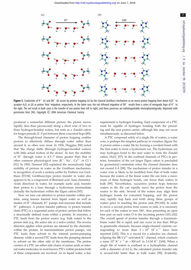
Figure 5. Conduction of Hþ (a) and OH – (b) occurs by protons hopping (a) by the classical Grotthuss mechanism as an excess proton hopping from donor H3Oþ toacceptor H2O, or (b) as proton ‘hole’ migration, respectively. In the latter case, the net leftward migration of OH – results from a series of retrograde hops of Hþ tothe right. The net result in both cases is the transfer of one proton from left to right, and these processes are indistinguishable electrophysiologically. Reprinted withpermission from [96]. Copyright & 2006 American Chemical Society.
rsif.royalsocietypublishing.orgJ.R.Soc.Interface
11:20130799
7
on June 7, 2018http://rsif.royalsocietypublishing.org/Downloaded from
produced a somewhat different picture: the proton moves
rapidly (less than picosecond) along a short wire of two to
three hydrogen-bonded waters, but rests as a Zundel cation
for longer periods (2–5 ps) between these concerted hops [89].
The through-bond character of proton hopping enables
protons to effectively diffuse through water rather than
around it, as other ions must. In 1936, Huggins [90] noted
that ‘the charge shifts (through hydrogen-bonded waters)
with little actual motion of the atoms’. In fact, the mobility
of Hþ through water is 4.5–7 times greater than that of
other common physiological ions (Kþ, Naþ, Ca2þ or Cl–)
[91]. In 1905, Danneel [92] explained the anomalously high
mobility of protons in water as the Grotthuss mechanism,
in recognition of work a century earlier by Freiherr von Grot-
thuss [93,94]. Grotthuss-type proton transfer in water also
appears to be a component of Brønsted acid–base chemistry.
Acids dissolved in water, for example acetic acid, transfer
their proton to a base through a hydronium intermediate
(actually the hydronium within the Eigen cation) [95].
Now we turn our attention to proton transfer within pro-
teins, using lessons learned from liquid water as well as
studies of Hþ channels, Hþ pumps and enzymes that include
Hþ pathways. A proton transfer pathway or proton transfer
chain (PTC) is a sequential series of proton carriers that follow
a structurally defined route within a protein. In enzymes, a
PTC leads from the proton source (e.g. bulk water) to the
proton sink (e.g. the active site or a redox centre). The reverse
direction applies when protons are released by the chemistry
within the protein. In transmembrane proton pumps, one
PTC leads from solvent to the internal proton-pumping
element, while a second PTC leads from the pumping element
to solvent on the other side of the membrane. The proton
carriers of a PTC are either side chains of amino acids or inter-
nal water molecules; in most known PTCs within proteins both
of these components are involved. As in liquid water, a key
requirement is hydrogen bonding. Each component of a PTC
must be capable of hydrogen bonding both the preced-
ing and the next proton carrier, although this may not occur
simultaneously, as discussed below.
A PTC composed solely of a single file of waters, a water
wire, is perhaps the simplest pathway to visualize (figure 5a).
A proton enters a water file by forming a covalent bond with
the first water to form a hydronium ion. The hydronium ion
may hydrogen bond to the next water to form the Zundel
cation, H5O2þ [97]. In the confined channels of PTCs in pro-
teins, formation of the yet larger Eigen cation is precluded
by geometrical constraints when the channel diameter does
not exceed 4 A [98]. The mechanism of proton transfer in a
water wire is likely to be modified from that of bulk water
because the waters of the linear water file can form a maxi-
mum of three hydrogen bonds, one fewer than waters in
bulk [99]. Nevertheless, successive proton hops between
waters in the file can rapidly move the proton from the
source to the sink. Several of the waters may align their
hydrogen bonds for concerted hopping, and the proton
may rapidly hop back and forth along these groups of
waters prior to reaching the proton sink [99,100]. In order
to move a second proton from source to sink, it is necessary
for each of the waters to turn 1808, thus presenting an open
lone pair on each water O to the incoming proton [101,102].
The overall speed of proton transfer through a transmem-
brane water file is rapid, and can occur in less than 500 ps
in gramicidin channels, because single-channel currents cor-
responding to more than 2 � 109 Hþ s21 have been
reported [103]. This is a record for a selective ion channel,
eclipsing the BK Ca2þ-activated Kþ channel, which conducts
a mere 109 Kþ s21 at þ300 mV in 3.4 M Kþ [104]. When a
single file of waters is confined in a hydrophobic channel
with a diameter of 4.0 A, the calculated proton transfer rate
is several-fold faster than in bulk water [98]. Evidently,

rsif.royalsocietypublishing.orgJ.R.Soc.Interface
11:20130799
8
on June 7, 2018http://rsif.royalsocietypublishing.org/Downloaded from
even at atomic dimensions, laminar flow is more efficient
than turbulent flow. In a linear water chain, the proton can
hop forward or backward along the file, but not sideways
or up and down. Moreover, a protein may construct a chan-
nel that optimizes the positions of the waters for rapid proton
transfer [99]; branching in a hydrogen-bonded water network
slows proton conduction [105].
The quintessential example of a purely aqueous proton
pathway through a protein spanning the membrane is the gra-
micidin channel. This short peptide (a dimer of 15-amino acid
monomers) produced by Bacillus brevis forms a cylindrical tube
filled with water [106]. The interior is hydrophilic; peptide
bond carbonyl groups coordinate a single-file row of a dozen
water molecules [107]. Titratable groups, for example carbox-
yls, are absent. Gramicidin is permeable to a variety of
monovalent cations, but it excels at conducting protons [108].
Normalized to the permeant ion concentration, the proton con-
ductance of gramicidin is 100-fold greater than that of other
ions [109]. The facility of proton permeation arises from their
ability to hop along the water file without displacing the
waters, whereas other cations must wait for the water mol-
ecules to diffuse through the pore ahead of them [109]. As
mentioned above, Hþ flux at rates exceeding 2 � 109 s21 have
been recorded in gramicidin channels [110]. The example of
gramicidin shows clearly that a narrow pore filled with water
is capable of conducting protons with high efficiency, but the
transfer is not selective, because other cations pass, albeit
more slowly.
1.4.1. A digression on why proton channels carry suchtiny currents
The human proton channel hHV1, has an estimated con-
ductance of 78 fS at physiological pH and human body
temperature [111]. Given a large driving force, say 100 mV,
one channel would carry 7.8 fA of Hþ current, which trans-
lates into 5 � 104 Hþ s21. Does this mean that HV are not
efficient proton channels, in comparison with gramicidin,
for example? Perish the thought! The gramicidin value men-
tioned above (2 � 109 Hþ s21) was obtained at þ160 mV in
5 M HCl! Our hHV1 can withstand a pH range beyond any-
thing they encounter in the human body, but at pH , 4.5
or pH . 8.5 they, or the membranes they reside in, do not
survive long enough to permit study. By contrast, when
incorporated into synthetic lipid bilayers, gramicidin con-
tinues to function at pH 21, in 10 M HCl [110]! It is
revealing to note that the single-channel proton current
through gramicidin is almost directly proportional to Hþ con-
centration over a wide pH range from 0 to 4.5 [23,110,112].
Extrapolated to the pH range where HV1 single-channel
measurements are possible (pH 5.0–6.5) [111], the gramicidin
Hþ current is actually smaller [23]. We conclude that HV are
very respectable proton channels that carry small currents
simply because the concentration of their permeant ion
(Hþ) is extremely low in physiological solutions.
1.4.2. Involvement of amino acid side chains in proton transferwithin proteins
Long water wires do exist within functional PTCs in proteins.
The ‘D’ pathway of the mitochondrial enzyme cytochrome coxidase (CcO) is proposed to be a file of 8–10 hydrogen-
bonded waters that transfers protons from a surface-exposed
aspartic acid residue to an internal glutamate residue
[113,114]. One fascinating aspect of the D pathway is that
the continuity of the water file is constantly being broken
and restored at one point by an amide side chain that
moves in and out of the water file [115]. Nevertheless,
proton transfer through the waters remains rapid [115,116].
The functional pathway for proton transfer to the quinone-
binding site of Escherichia coli succinate dehydrogenase may
involve up to 13 waters [117].
With the possible exception of the succinate dehydrogen-
ase pathway [117], native proton transfer pathways in proton
pumps and enzymes involve side chains of certain amino
acids (particularly carboxylates) along with internal waters.
The chemistries of the various side chains increase the
number of mechanisms available for proton transfer. For
example, in the 1970s, Nagle and others [101,102,118] recog-
nized that certain side chains of amino acids, for example
hydroxyls, could participate in proton transfer without ioniz-
ation, i.e. without the net dissociation of a proton. In such a
mechanism, the hydroxyl group uses hydrogen bonds to sim-
ultaneously acquire a proton from an upstream proton carrier
and release its proton to a downstream proton carrier. Chemi-
cally, a lone pair of electrons on the hydroxyl O acquires a Hþ
while another pair releases a Hþ. Throughout the process the
hydroxyl remains as –OH, avoiding ionization to either 2O2
or –OH2þ. In theory, a long chain of hydrogen-bonded hydroxyl
groups could carry out concerted proton transfer over a con-
siderable distance. This is not seen in known or proposed
PTCs in proteins, perhaps because the precise alignment of
hydrogen bonds is difficult to sustain over a long series of
hydroxyl groups. In fact, when the hydroxyl groups of serine,
threonine and tyrosine do appear in the proton transfer path-
ways of proteins, they do so individually, and they are
bracketed by carriers capable of forming a stable ion, such as a
carboxyl or water [23]. For example, a recently identified
proton pathway in Clostridium pasteurianum [FeFe]-hydrogenase
comprises a five-member motif with Ser bracketed by two Glu
residues (Glu282, Ser319, Glu279, H2O, Cys299) [119].
The acidic and basic side chains of amino acids add conven-
tional acid–base chemistry to the realm of proton transfer.
Most significantly, the fact that proton acquisition and proton
release are separable events for an acid or base brings the
aspect of rapid side chain movements into play. For example,
at the entrance to the D pathway for proton transfer of CcO,
a carboxylate side chain accepts a proton from solvent
(water), then the protonated carboxyl group rotates 1808about the terminal C–C bond of the side chain, establishes a
hydrogen bond to the oxygen of the first water of the D path-
way and transfers the proton to this water [120]. Just the
rotation of the protonated carboxyl moves the proton approxi-
mately 2.2 A along the trajectory of the transfer path. (The
approximate centre-to-centre distance of the O atoms of a car-
boxyl group is 2.2 A; a proton cannot hop from O to O of the
carboxyl because the geometry of the atoms precludes an intra-
molecular hydrogen bond.) In CcO, the rotation of this D
pathway carboxyl moves the proton from ‘outside’ to ‘inside’
[120]. The crystal structures of CcO show no space for hydro-
nium or other ions to move around the carboxyl, so its
rotation constitutes a proton-specific gate. The chemistry of
such carboxyl rotation has been studied in detail in a bacterial
ferredoxin, where a critical aspartic acid residue also moves a
proton from outside to inside [121]. Examination of other
protein structures suggests that carboxyl rotation may be
common in proton transfer pathways.

rsif.royalsocietypublishing.orgJ.R.Soc.Interface
11:20130799
9
on June 7, 2018http://rsif.royalsocietypublishing.org/Downloaded from
The imidazole group of histidine is analogous to a car-
boxyl, in that it contains two atoms (Nd and N1) that can be
protonated, but a proton cannot hop between these two
sites because there is no intramolecular hydrogen bond. Simi-
larly to carboxyls, neutral imidazole can be protonated on
the open N, rotate 1808, and then deprotonate to the next
proton carrier [122–125]. In this case, the rotation moves
the proton approximately 2.1 A, the centre-to-centre distance
of the N atoms of the imidazole ring.
Transfer of protons can occur via larger, but still rapid,
movements of whole protein side chains (rather than just car-
boxyl or imidazole ends of side chains), e.g. the swinging
motion of a lysine within the K pathway of CcO [126,127].
1.5. How can one design a proton selective channel?With the basics of proton transfer in mind, let us take up the
question how a proton channel may select for protons. A
proton channel requires two features to be selective for pro-
tons. First, there must be a proton transfer pathway, using
any of the options described above for the transfer of protons
through proteins. As we have seen, the mechanisms of proton
transfer in proteins are closely related to proton acid–base
chemistry in solution, and therefore a PTC in a channel
protein is inherently selective for protons. Based on PTCs in
other proteins, we might expect the PTCs of ion channels to
include both waters and amino acid side chains. Although
the channel must supply a pathway for the proton that spans
the entire membrane, the proton selective section of the path-
way may be shorter. In principle, selectivity can be achieved
by directing all of the protons to pass through a single titratable
side chain, such as a carboxyl or an imidazole group, at some
point in the channel. As is the case with many ion channels,
HV1 has large aqueous vestibules (figure 1), separated by a
narrow region approximately 10–12 A long in which water
is mainly single file [3] (figure 1). This short, narrow region is
the section of the transmembrane proton pathway that
is thought to confer selectivity for protons.
Second, proton selectivity requires some mechanism to
prevent other ions from also flowing through that region of
the channel that contains the PTC. A priori, it would seem
that a straightforward design strategy would be close protein
packing. The passage of monovalent cations, for example,
requires an open space, a pore, through which the ions can
move. By contrast, proton transfer requires no open space,
because the Hþ may be transferred as part of the protein.
As it moves from carrier to carrier through the protein via
H-bonds, the Hþ is covalently associated with lone pairs of
electrons on internal waters or protein side chains. Given
this fundamental difference in the structural requirements
for proton transfer versus the transfer of other ions, it
would seem that evolution could produce a highly selective
proton channel simply by filling in the space around the
PTC component of the channel.
1.5.1. Of water wires and Naþ
Water wires (water files) have been proposed for part or all of
the PTC component of some Hþ channels. The Hþ selectivity
of these channels varies. At this time, HV1 is the only one that
appears to be completely proton selective (Table 1 of [17]).
The influenza virus M2 channel is highly proton selective,
but also conducts Kþ and Naþ [128–130]. A synthetic hydro-
phobic a-helix composed of leucine and serine (i.e. lacking
formal charges) oligomerizes to form a channel containing
a water wire that conducts Hþ more than 40-fold better
than Liþ, based on the presence of detectable single-channel
currents [131].
There is no doubt that a water file forms an excellent path-
way for protons; the question is how to enforce proton
selectivity. A relevant paradigm is the transmembrane Hþ
or Naþ channel of the Fo–F1-type ATP synthases. In some
bacterial versions of these enzymes, the transmembrane
channel of the Fo rotor transfers Naþ, but in related enzymes
(including mitochondrial ATP synthase), the channel is
specific for Hþ [132,133]. The transmembrane channel of Fo
is constructed of two water-filled half channels leading to
and from a controlling group located in the middle of the
membrane [134,135]. In the Naþ channels, the central group
is a multi-valent coordination site for Naþ, while in the Hþ
channels the central group appears to be a single carboxyl
side chain [136]. For the Hþ-conducting ATP synthases of
Spirulina platensis and Bacillus pseudofirmus, spectacularly
selective binding (108–109 higher for Hþ than Naþ) is demon-
strated by the Fo rotor being driven by Hþ at pH 9 and
200 mM Naþ [137,138]. This does not prove that Naþ
cannot reach the input half-channel; simply that it does not
bind and catalyze enzyme activity. However, the working
enzyme translocates Hþ across the entire membrane with
superb selectively. Hence, nature appears to have conferred
Hþ selectivity upon the transmembrane channel of many F-
type ATP synthases by replacing a Naþ-binding site with a
single carboxyl side chain. The elucidation of further struc-
tural factors that confer Hþ selectivity in the F-type ATP
synthases should be highly instructive for other Hþ channels,
and vice versa.
Other more subtle and difficult to validate mechanisms
have been proposed for Hþ selectivity by water wires. One
such is the ‘frozen water’ hypothesis, some form of which has
been proposed for the M2 viral proton channel (figure 6b) dis-
cussed below [140–142], for the synthetic proton channel of
Lear et al. mentioned above [131,143], and for HV1 [144,145].
In this mechanism, one or more waters at a narrow region in
an aqueous pore are constrained by the pore walls, essentially
frozen in place in such a way that they can still translocate a
proton by the Grotthuss mechanism, while simultaneously pre-
venting other cations from permeating. Although nominally
frozen, the waters need to retain enough mobility that they
can reorient after each proton conduction event [101,102].
Another way to enforce proton selectivity still envisions
a water wire, but one that is continuous only transiently.
Molecular dynamics simulations of hHV1 in the open state
indicate that a continuous water wire exists just 10% of the
time (figure 1), and it persists only for a nanosecond or so
[3]. One could speculate that protons, speedy little devils
that they are, could zip through such a pathway, whereas
much bulkier and slower ordinary cations could not perme-
ate within such a short-time window. Such a situation is
proposed for the normal function of the D pathway of CcO,
although not as a solution to the problem of ion selectivity.
Although the D pathway transfers protons at rates at least
up to 104 s21 [116], high-resolution structures show a clear
discontinuity in the water file of this pathway in the form
of two amide side chains of Asn139 and Asn121 [127,146].
Molecular dynamics simulations suggest that the amide
side chain of Asn139 spontaneously swings out of the way
to allow formation of a continuous water file [115]. The rate

W41in
H37
G34
e2H37 H37
H
HH H
O
H H
O
H HO
H HO
H HO
H
water wire
out
shuttle hydrogen-bonded dimer
HO
H HO
HH
H +
O
HH
H +
O
H H
O
H HO
H HO
H H
O
H H
O
H+N N
NH
N
N
H
H37N
N-H
H
NNH
N+
H
H
N
N+ +
H H
OH H
O
H H
H H
H+
O
O
H HO
H HO
H HO
(a) (b) (c) (d )
d1
Figure 6. The well-studied M2 proton channel of influenza A virus is a homotetramer; (a) shows the high-resolution crystal structure with key residues labelled.Three proposals for the proton selective conduction are illustrated in (b – d), with only two of the four protomers shown for clarity. In the water wire model (b), theproton pathway is exclusively aqueous, with protonation of multiple His37 serving to open the pore by electrostatic repulsion. Proton selectivity in the shuttle model(c) is achieved by successive protonation and deprotonation of His37, with a ring flip completing each conduction event. In (d ) protonated and unprotonated His37
form hydrogen-bonded dimers that are broken during Hþ conduction. (Adapted from [139].)
rsif.royalsocietypublishing.orgJ.R.Soc.Interface
11:20130799
10
on June 7, 2018http://rsif.royalsocietypublishing.org/Downloaded from
of Hþ transfer remains rapid because the movements of protein
side chains and internal waters are even more rapid.
Yet another hypothetical mechanism for proton selectivity
postulates a continuous water wire that is energetically or elec-
trostatically unfavourable to ions. We have seen how a proton
can move back and forth along a file of waters. This reduces
the effective charge of the proton at any specific location,
i.e. the charge is delocalized [85,97,99,147,148]. By contrast, the
charge on an ion, for example Naþ, exists at a discrete location;
so it becomes possible for a protein structure to purposefully
exclude such a charge. A delocalized-charge mechanism has
been proposed for a synthetic proton channel [149].
1.5.2. Aquaporin: a water pathway that excludes protonsA paradigmatic example of a water-filled channel that
excludes protons is aquaporin (AQP), a membrane protein
that provides a pathway for rapid water permeation across
the membrane [150], but excludes all ions including protons
[151,152]. The structure of AQP revealed a narrow region
20 A long at the centre of which is a pair of NPA (Asn–
Pro–Ala) motifs from nearby short helices [153]. It was
proposed that the two Asn amido groups formed hydrogen
bonds with a single water molecule, preventing the water
from hydrogen bonding with adjacent waters, thereby pre-
cluding proton conduction [153]. Shortly after this, a nearby
‘constriction region’ with conserved Arg, His and Phe resi-
dues was identified [154], later called the ‘selectivity filter’.
A number of molecular dynamics studies ensued to evaluate
the structural and physical basis for blockage. Initial simu-
lations in the absence of Hþ were interpreted to support the
water orientation hypothesis [155], but subsequent calcu-
lations showed that the free energy barrier opposing the
movement of an excess proton through the pore peaks at
the NPA motif [156–159]. Several of the latter studies con-
cluded that ion blockage results from electrostatic effects
combining the ion’s desolvation penalty and interactions
with charged and polar groups of the channel. A simpler
model was proposed by Burykin & Warshel [160,161], who
argued that AQP with all of the ionizable groups in the chan-
nel neutralized, and even a 4 A hole in the membrane
exhibited a large energy barrier to proton permeation, and
concluded that the desolvation penalty for protons to leave
bulk water and cross the centre of the membrane was itself
sufficient to exclude protons. However, point mutations of
the selectivity filter region, R195 V or R195 V/H180A, were
identified that enabled proton conduction through AQP1
[162,163]! These results showed that neutralizing the charge
of R195 in the selectivity filter suffices to confer proton per-
meability, despite the continued presence of the NPA
region and the desolvation penalty for inserting protons
into the narrow pore.
AQP can be mutated to conduct protons, whereas HV1
can be mutated to exclude protons. In the human HV1 chan-
nel (figure 1), the Asp112 crucial to proton selectivity is salt-
bridged with Arg208 about 90% of the time, so their charges
are neutralized. When Asp112 is neutralized by mutation
(e.g. to Ser or Ala) molecular dynamics simulations reveal
that the charge owing to the now-unpaired Arg208 produces
a 10 kcal mol21 barrier to cations [3]. It was shown recently
that Asp112 can be shifted one turn of the helix outward to
position 116 without loss of proton selectivity [164]. Molecu-
lar dynamics simulations reveal that at its new location,
Asp116 is generally paired with Arg208 or Arg205. There is
an electrostatic barrier to cation permeation when Arg208 is
unpaired, but not when it interacts with Asp185 or Asp116
[164]. Evidently, proton exclusion can be achieved by an
unpaired cationic group in a critical location. It is well estab-
lished that the charge selectivity of anion-selective, ligand-
gated channels can be switched to cation selectivity by intro-
ducing negatively charged Glu at key locations [165,166], and
that cation selective channels can be made to conduct anions
by various mutations that include neutralization of Glu
[167,168]. These rules of charge selectivity can be used to
destroy proton selectivity, but producing proton selectivity
is a subtler task.
1.6. The extensively studied M2 viral proton channel asa model
The M2 channel of the influenza A virus [169] is a favourite
example of a highly proton selective ion channel [170–172].
This channel is relatively simple, being a homotetramer of
97-amino acid monomers (figure 6). Four transmembrane
helices of M2 proteins associate to form a single channel that
transfers protons into an endosome containing a virus that
has been taken up by a cell. Proton uptake initiates the loss of

rsif.royalsocietypublishing.orgJ.R.Soc.Interface
11:20130799
11
on June 7, 2018http://rsif.royalsocietypublishing.org/Downloaded from
the viral protein coat. The M2 channel is not as selective for
protons as the HV1 channel, and was shown recently to exhibit
measurable permeability to Naþ and Kþ [128–130,173], which
tarnishes it somewhat as a paradigm of perfect proton per-
meability. Even so, the M2 proton channel selects for Hþ
over Naþ or Kþ by a factor of approximately 106 or more
[128–130,170,173]. As discussed below, this is achieved by
proton transfer through a His37 cluster, combined with phys-
ical occlusion of Naþ or Kþ transfer through the His37
cluster. The rate of Hþ transfer from exterior to interior is
relatively slow at 100 s21 or less by most accounts [128–
130,171,173–176]; a gate composed of a ring of Trp41 side
chains [177,178] slows reverse proton transfer even more.
From the analysis of site-directed mutants and high-
resolution structures, the key to Hþ selective conductance is
His37 [179,180]. As the channel assembles as a tetramer of
four identical a helices, there is a tetrad of His37 whose side
chains face the centre of the pore [181]. His37 plays key
roles both in activating the channel and in proton selectivity.
Protonation of two His37 opens the channel [141,176,182,183].
Hþ conduction is mediated by the third His37 to be proto-
nated [139,169,184], and its relatively high pKa may limit
Hþ flux [139,185]. Mutagenic alteration of His37 (for example
H37G) abolishes proton selectivity [179]. The mechanism by
which His37 imparts proton selectivity (figure 6c) was originally
suggested to be protonation of the imidazole d-nitrogen from
the extracellular side of the membrane, deprotonation of the
imidazole 1-nitrogen to the intracellular side, followed by a
ring flip to restore the orientation of the singly protonated
state [186]. This shuttle mechanism can be described quantitat-
ively by simple mathematical models [184,187,188]. Because
each Hþ conduction event entails conformational changes of
His37 and Trp41, one could reasonably define M2 as a carrier
rather than a channel. The essentials of this model remain,
i.e. protonation and de-protonation of His37 plus a flipping
motion of the imidazole ring. Further details of the mechanism
are currently debated, as discussed below.
High-resolution crystal structures (figure 6a) and NMR
structures have provided considerable structural information
about the rings of His37 and Trp41 residues and the positions
of internal waters [169,176]. In addition, the existence of
hydrogen bonds and the protonation state of the His37 can
be determined using NMR spectroscopy [139,189–191].
These data allow for detailed models of proton transfer
through His37 [169,176,192]. In two current models, the selec-
tivity filter for protons involves just three components and
two proton transfer steps: a hydronium from the exterior aqu-
eous region of the channel transfers a proton to the His37
cluster (which is organized differently in each model), and
then the His37 cluster transfers the proton to a water below
the ring of Trp41 side chains, when this Trp41 gate is open
to allow a hydrogen bond to form between a component of
the His37 cluster and the acceptor water molecule.
In the mechanism proposed by Zhou, Cross and co-
workers [176,183,192], based primarily on calculations on
an NMR structure and further spectroscopy, the deproto-
nated His37 tetrad is organized as two hydrogen-bonded
imidazole–imidazolium dimers (figure 6d ). The introduction
of a proton from an upstream hydronium breaks the H-bond
of one of these dimers (either one, apparently) producing two
imidazolium side chains that rotate and swing away from
each other. One swings down to transfer a proton to a down-
stream water revealed by the opening of the Trp41 gate. The
de-protonation of this His37 allows re-formation of an imida-
zole–imidazolium dimer.
The mechanism (figure 6c) of Hong, DeGrado, and co-
workers [169,193] is primarily based upon a 1.65 A crystal
structure of the transmembrane channel [194] combined
with solid-state NMR dynamics data. In this structure, the
orientation of the side chains of the ring of four His37 roughly
resembles that seen in the NMR structure used by Zhou,
Cross, et al. but the side chains are evenly spaced around
the ring and direct H-bonds between side chains are absent.
Rather, the four imidazole/imidazoliums form a hydrogen-
bonded network with associated waters, which are located
atop the His37 ring and between the histidines and the
Trp41 ring [194]. The histidine side chains and the waters are
capable of extensive delocalization of an added proton; con-
tinuous ring flips of the histidines facilitate the sharing of the
added proton with the waters between the His37 and Trp41
rings. The three components of the PTC in the Hong–DeGrado
model are a hydronium from the aqueous channel above the
His37/water cluster, the His37/water cluster itself and a water
to receive the proton below the Trp41 gate. The rate-limiting
step in Hþ conduction is imidazolium ring reorientation [169].
A principal difference in the two models is the nature of
the His37 structure that is protonated by a single proton from
the exterior side of the channel and then de-protonated by
water beyond the Trp41 gate. In the Zhou–Cross model, the
His37 proton transfer group cycles between an imidazole–
imidazolium dimer and two imidazolium side chains, while
in the Hong–DeGrado model, the His37 structure is a His37/
water cluster in both its protonated and de-protonated forms,
whose degree of hydration increases as the degree of protona-
tion is increased. Evidence that the hydrogen-bonding partner
of His37 is water instead of another His37 was obtained from 1H
NMR chemical shifts [193]. A swinging movement of the
histidine side chains appears less required in the Hong–
DeGrado model, because either of the waters located between
His37 and Trp41 in the His37/water cluster can mediate proton
transfer to the water beyond Trp41.
1.7. What makes hHV1 selective?Returning to our main interest, the voltage-gated proton
channel, our current explanation of proton selectivity consists
mainly of describing which parts of the molecule are
involved. Because no crystal structure exists for the channel
in any species, what we believe about structure is derived
from homology modelling based on crystal structures of
voltage-gated Naþ and Kþ channels (specifically, their hom-
ologous VSDs) and on molecular dynamics simulations in
conjunction with experimental data. As illustrated in the
snapshot in figure 1, hHV1 narrows to a region approxima-
tely 10 A long containing more or less a single file of
waters that most of the time is interrupted at an Asp112–
Arg208 salt bridge [3]. Experimentally, the PTC within HV
appears to consist of more than water. For example, a
number of characteristics of proton permeation through HV
led to the idea that the rate-limiting step occurred inside
the pore, not in the approach through bulk solution
[195–198]. Postulating a PTC including one or more titratable
groups [18,197,198] could explain why the high selectivity,
deuterium isotope effect and temperature dependence of con-
duction differed drastically from proton conduction through
the water-filled gramicidin channel [109,199,200], but were
rsif.royalsocietypublishing.orgJ.R.Soc.Interface
11:20130799
12
on June 7, 2018http://rsif.royalsocietypublishing.org/Downloaded from
similar to the corresponding properties of the M2 channel
[170,171, 201,202], in which proton conduction entails
protonation/deprotonation of His residues [186].
The Hþ selectivity of voltage-gated proton channels in
both humans [7] and dinoflagellates [4] requires an aspartate
residue in the middle of the S1 transmembrane segment, at
a narrow region of the pore (figure 1), Asp112 and Asp51,
respectively. Certainly, Hþ selectivity could be achieved by
a mechanism in which Asp112 is protonated and deproto-
nated by every proton transferred. When Asp112 is replaced
with a neutral amino acid, HV not only lose their proton
specificity but become preferentially permeable to anions.
Phenomenologically speaking, that this Gestalt is identical
in both species indicates that the selectivity mechanism is
strongly conserved evolutionarily, given that there is only
14.6% identity between the two proteins [4,17]. Replacing
Asp with Glu preserved proton specificity, but other mutants,
including the His mutant, D112H or D51H, respectively, were
permeable to anions [4,7]. Recent demonstration that the
selectivity of hHV1 is preserved when Asp is moved from
position 112 to 116, one turn of the S1 helix outwards (but
not to a dozen other positions tested) reiterates the role of
Asp, but indicates some structural latitude in the placement
of the Hþ selective carboxyl group [164].
There exists a clear preference for carboxyl side chains
(Glu or Asp) in proton transfer pathways of pumps and
enzymes [120,121,203–220], often as the group that intro-
duces protons into the pathway. The D pathway of CcO has
been established as an experimental system for examining
the ability of side chains to sustain steady-state proton trans-
fer [120]. In the normal D pathway, the initial proton acceptor
is the carboxyl group of a conserved aspartic acid. Histidine
cannot replace Asp as the initial proton acceptor at the
normal position [221]. The thiol of Cys can function in this
system as an initial proton acceptor, but only when it is
moved to a point in the pathway with significant structural
constraints that limit rotamer possibilities [120,221]. The pre-
ference for a carboxyl group for proton uptake is likely due to
several factors: its structure is analogous to two waters con-
nected by a carbon, its functional pKa is readily modified
by the surrounding protein, and it has the capability to
rapidly rotate end-over-end—like a turnstile—through a com-
pact protein structure in order to transfer a proton to the next
element in the PTC [120].
Nevertheless, simply locating an Asp in a pore-lining
position is not enough to produce proton selectivity. In the
c subunit of H-ATPase, the key carboxyl of Asp61 can be
moved to a different helix (D61G/A24D), but not to nearby
positions 58, 60 or 62 on the same helix [203,222]. Asp185
also faces the pore in hHV1 [3,144,145,223] but can be neutral-
ized by mutation (D185S, D185N, D185V) without affecting
proton selectivity [7]; conversely, Asp185 is still present in
D112x mutants (i.e. mutants in which Asp112 is changed to
His, Lys, Asn, Ser, Ala or Phe) that lack Hþ selectivity [7].
As shown in figure 1, the pore is likely wider at the level of
Asp185 [3]; evidently Asp must reside at a narrow point to
produce selectivity. A compact protein structure around the
carboxyl will prevent the passage of other ions.
That the His mutant of hHV1, D112H, was permeable to
anions [7] was initially surprising, in view of the strong simi-
larity of the transmembrane domain of HV1 to the VSD of Kþ
and Naþ channels [1,2]. The VSD includes the first four trans-
membrane helices (S1–S4), which are thought to act as the
voltage-sensing component of the channel; they mechanically
couple to the pore domain, which comprises the four S5–S6
segments (figure 3). Although the VSD of Kþ or Naþ channels
does not normally conduct current, when the outermost of a
series of Arg residues in the S4 helix is replaced by His, a
voltage-gated proton conductance appears [224–226]. This
result supports the view that the VSD is roughly hourglass-
shaped with aqueous vestibules that access both sides of the
membrane and focus onto a short occluded region [224].
When His is located at this constriction, it selectively translocates
protons [224–228]. Why does His introduced at a comparable
region in hHV1 not shuttle protons? A crucial difference is that
in the mutant Kþ channel VSD, His replaces a cationic Arg,
whereas in the D112H mutant of hHV1, His replaces a negatively
changed Asp. In hHV1, Asp112 normally apposes the cationic
Arg208 (figure 1); mutation of Asp results in uncompensated
positive charge that excludes protons and produces anion selec-
tivity [3]. Intriguingly, in aquaporin, proton exclusion occurs at
the ‘ar/R region’, in which Arg195 apposes His180 [163].
In ATP synthase, the crucial acidic group Asp61 (in E. coli)exists in proximity to a highly conserved Arg210 residue,
called the ‘stator charge’ whose function is thought to be to
ensure release of the proton from the acidic binding site
[133,136,203,229,230]. In an intriguing parallel, the proton
selectivity element in hHV1, both at its native position,
Asp112, and when repositioned to Asp116, is found by MD
simulations to be mainly engaged in salt linkages with one
or more Arg residues in the S4 helix [3,164].
1.8. What is the difference between Hþ and OH –
channels?It turns out that distinguishing Hþ and OH– channels is very dif-
ficult. The electrochemical gradient that drives conduction of
either species is always identical. For example, at pHo 7.5, pHi
6.0, the Nernst potentials are EH ¼ EOH ¼ 287 mV (at 208C).
At symmetrical pH, applying þ100 mV to the inside of a cell
(or an isolated membrane) drives Hþ out and/or OH– in, result-
ing in currents that are indistinguishable to a voltage-clamp
circuit. The effects on pH on either side of the membrane are
also identical; moving Hþ out increases pHi and decreases pHo
by the same amount as moving OH– in. It is likely that most con-
duction of Hþ and OH– occurs by closely related mechanisms
involving proton hopping (figure 5) by a Grotthuss-like mechan-
ism [89,92–94,231–234]. In the chemical systems in figure 5, Hþ
is more likely to carry the current when donor and acceptor are
acidic (pKa, 7), whereas OH– conduction (as a proton ‘hole’) is
more likely when they are basic (pH . 7) [96].
As discussed above, the human voltage-gated proton
channel, hHV1, is proton selective because of a specially
located aspartic acid residue (Asp112) [7], which is almost cer-
tainly negatively charged in situ, with a low pKa. When this
Asp112 is mutated to a neutral amino acid (e.g. Ser, Ala and
Asn), the channel becomes anion selective. This charge rever-
sal is understandable if we consider that, in all likelihood, the
negatively charged Asp was compensated in the WT channel
by one or more nearby cationic charges, so that elimination of
the Asp leaves unpaired cationic charges. However, although
Cl– is clearly permeant in D112x mutants, the preferred
charge carrier appears to be OH– by several orders of magni-
tude (in terms of relative permeability, POH/PCl, although not
necessarily in real numbers) [7]. Why this should be is not
obvious. The pathway in WT hHV1, comprising an aqueous

100
50
0
–50
–100
–150 –100 –50 0Vrev (mV)
Vth
resh
old
(mV
)Vthreshold = 0.79 Vrev + 23 mV
outH+
in
H+
Vthreshold = 0.79 Vrev – 37 mV
Vthreshold = Vrev
50 100
Figure 7. The property of DpH-dependent gating is universally conserved in proton channels, but exists in two forms. In most cells (15 varieties are illustrated hereas various symbols), proton channels open only when there is an outward electrochemical gradient, and this results in acid leaving the cell. This pattern is seen overa very wide range of pHi and pHo; Vthreshold is always above the dashed line of equality. In the single case of the dinoflagellate K. veneficum ( filled circles), Vthreshold
for the kHV1 channel varies with identical slope with changes in pHi and pHo; but at any given DpH, Vthreshold is 60 mV more negative than in other HV1; this meansthat opening kHV1 produces inward proton current under all conditions. Most cells use HV1 to eliminate excess acid, but dinoflagellates have other goals in mind,such as conducting action potentials and triggering a bioluminescent flash. (Adapted from [17].)
rsif.royalsocietypublishing.orgJ.R.Soc.Interface
11:20130799
13
on June 7, 2018http://rsif.royalsocietypublishing.org/Downloaded from
pore that most of the time is interrupted at an Asp112–Arg208
salt bridge [3], supports a high rate of Hþ flux. Mutation of
Asp112 alters the electrostatic environment drastically, by repla-
cing a net-neutral salt bridge with an uncompensated cationic
charge, producing a 10 kcal mole21 barrier for cations [3].
Facile OH– permeation should not be too surprising, because
the mechanism of OH– conduction also involves Hþ hopping
and occurs rapidly. Because OH– permeates by means of
proton hopping (figure 5b), it has tremendous mobility advan-
tages over Cl–, both because of its small effective size and the
rapidity of proton hopping (discussed above).
In several tissues that exhibit Hþ/OH– flux at extremely
high pH, such as Chara australis at pH 10.5 [235,236] or frog
skeletal muscle at pH 10 [237], OH– flux seems more likely
than Hþ flux simply because there are vastly more OH–
than Hþ to act as charge carriers.
In summary, the consequences of Hþ or OH– permeation
are similar, the mechanism of transport is similar, and which
species is transported may depend on the pKa of critical
groups in the pathway or on the net charge at a narrow part
of the channel.
1.9. Why do proton channels have DpH-dependentgating?
A great many ion channels are voltage gated, which means that
they have a high probability of being open (and conducting
their particular favourite ion) at certain membrane voltages
and a high probability of being closed (not conducting any-
thing) at other voltages. Most voltage-gated ion channels,
including our favourite, HV, open when the cell membrane is
depolarized. For most ion channels, the voltage dependence
is absolute, subject to modulation by permeant ion effects dis-
cussed below. A Kþ channel that opens at 240 mV in
physiological solutions will carry outward current. The same
channel exposed to elevated external Kþ concentration will
still open at 240 mV but will carry inward current. Channel
opening depends only on voltage; once a channel is open, the
direction of current flow depends only on the electrochemical
driving force (V 2 Eion). There are rather subtle effects of per-
meant ion species and concentration on the gating of several
channels [238–240], most famously the ‘occupancy hypothesis’
of Clay Armstrong in which occupancy of the pore by a per-
meating ion slows the closing of the channel, also called the
‘foot-in-the-door’ effect [241]. ClC20 Cl– channels [242,243]
and inwardly rectifying Kþ channels [244,245] have more pro-
nounced permeant ion effects on their gating than most other
channels. In contrast with HV, however, these effects are exerted
by permeant ions mainly from just one side of the membrane.
One of the most extraordinary properties of every
voltage-gated proton channel identified to date is DpH-
dependent gating [17,18,23,246,247]. The effect of proton con-
centration (pH) on the gating of proton channels is not only
pronounced, but it occurs equally on both sides of the mem-
brane [248]. The consequence is that the position of the
proton conductance–voltage (gH–V ) relationship is deter-
mined by the pH gradient, DpH, defined as pHo2 pHi [248].
Figure 7 illustrates the practical consequences of this unique
DpH dependence. Here, we indicate the position of the gH–Vrelationship in a convenient format in terms of Vthreshold, the
‘threshold’ voltage at which proton channels first start to
open. Data collected from studies of 15 different cell types are
all described well by the upper line. Vthreshold is plotted as a func-
tion of the reversal potential, Vrev. One could equally replace the
experimentally determined Vrev with the more theoretical value
of the proton Nernst potential, EH, because the perfect selectiv-
ity of HV means that Vrev should be very close to EH. In practice,
however, it is not so easy to establish pH accurately, even when

rsif.royalsocietypublishing.orgJ.R.Soc.Interface
11:20130799
14
on June 7, 2018http://rsif.royalsocietypublishing.org/Downloaded from
one uses such high buffer concentrations that buffer is practi-
cally the only anion in the solution. The proton channel is a
superb pH meter that reports precisely the pH gradient across
the membrane. For this reason, we prefer to believe what HV
tells us about pH, which is contained in Vrev. Returning to
figure 7, we see that Vthreshold is always positive to Vrev—the
dashed line shows the point at which they are equal. Finally,
the punch line: proton channels do not open unless the electro-
chemical gradient is outwards, as it is for all the symbols on the
graph (except the solid circles). Therefore, when the proton
channel first opens, it will conduct acid out of the cell. Voila!
The primary function of proton channels is acid extrusion!
Why do cells need to extrude acid? (Here, we will be king-
domcentric and consider mainly animal cells.) As was cogently
stated by Roos & Boron [249] in their historic 1981 review, ‘the
central problem of pHi regulation [is] the neutralization of
intracellular acid derived from a variety of sources’ [249,
p. 365]. Cells produce acid continuously during metabolism
and are constantly looking for ways to get rid of it. From this
perspective, the worst possible thing is a pathway that would
allow Hþ to leak into cells! The DpH dependence of proton
channels ensures that this disaster will never happen.
Nevertheless, for the proton channel to be most useful, it does
need to open reasonably near EH so that as soon as enough acid
builds up, proton channels will open and release it. A quibbler
might argue that the DpH dependence illustrated in figure 7
results in Vthreshold changing only 40 mV unit21 change in
DpH, insufficient to parallel EH which changes 58 mV unit21.
This point is technically valid, but arguably may not reflect a
design flaw. Perhaps the approach of the upper solid and
dashed lines in figure 7 means that Vthreshold is closest to EH
when there is a large outward gradient, i.e. when the cell has
become very acidic and is in dire need of relief from acid build-up.
A persistent quibbler might point out that Vthreshold is an
artificial concept, because the probability that proton channels
open (Popen) does not have a true threshold, but decreases expo-
nentially with hyperpolarization. From this perspective, a
larger cushion between Vthreshold and EH might be a prudent
strategy in the negative voltage range, where cells may sit at
their resting potential for days on end, and even a tiny Hþ
influx might have untoward cumulative consequences.
Fine, but what about the solid dots in figure 7? In the phy-
logenetic tree in figure 2, we saw that the proton channel that
differs most in amino acid sequence from all the others is
kHV1, a dinoflagellate proton channel. This sequence difference
is reflected in a crucial functional difference, which is obvious
from the dots in figure 7. Although kHV1 has DpH-dependent
gating with precisely the same slope as all other proton chan-
nels, the absolute position is offset by 260 mV [4]. The
consequence of this unique set-point is that when kHV1 first
opens, it conducts inward current. The biological effects of
channel opening could not be more different! Acid enters the
cell instead of leaving. Why would this ever be desirable?
One answer is that this property would allow the proton
channel to mediate an action potential. Conducting an action
potential requires a conductance that activates negative to its
Nernst potential [250,251]. As is discussed above, two familiar
examples are voltage-gated Naþ and Ca2þ channels, both of
which activate negative to their Nernst potentials and do in fact
conduct action potentials [252–254]. The proton channel ident-
ified recently in a non-bioluminescent dinoflagellate species,
K. veneficum, was found to activate well negative to EH over a
wide range of pH (figure 7), precisely the behaviour needed to
mediate an action potential [4]. These properties strongly support
suspicions that proton channels mediate the action potential that
triggers the bioluminescent flash [4,23,53,255].
As it turns out, several bioluminescent dinoflagellates, when
stimulated mechanically by waves or other disturbances of the
seawater, emit a rapid flash that can be very impressive when
plankton blooms concentrate many individuals in one location
[256]. Forty years ago, voltage-gated proton channels were first
conceived by J. Woodland Hastings, who proposed that they
trigger the flash [52]. This response occurs when a stimulus
evokes an action potential in the large intracellular flotation
vacuole [257–260]. The action potential invades the scintillons
[261,262], numerous evaginations of membrane that form
small vesicles [263] that are packed with luciferin, luciferin-
binding protein and luciferase [255,264–266]. Because the vacu-
ole has very low pH of 3.5–4.5 [54,267], an open proton channel
will conduct protons very rapidly into the scintillon, where the
pH is near neutral [268]. Luciferase is quiescent at high pH, but
is activated bya drop in pH to approximately 6.5 [255]. Thus, the
influx of protons through HV rapidly lowers the pH of the scin-
tillon and thereby activates luciferase. An additional parallel
mechanism is that luciferin-binding protein releases luciferin
(the substrate for luciferase) at low pH [255,265,269].
In order for HV to carry out their function, they must
open negative to EH and conduct inward current. Figure 7
shows that this is precisely what they do. Any other proton
channel would just sit there, refusing to conduct inward cur-
rent, because the vacuole interior is the topological equivalent
of extracellular space. The discovery of the unique properties
of kHV1 [4] confirms Woody Hastings’s hypothesis, nearly
40 years later. It turns out that having a Vthreshold negative
to EH is required not only to mediate the action potential,
but also to activate luciferase in the scintillons!
1.10. Why do proton channels in mammals existas dimers?
In most ‘higher’ species, HV1 exist as dimers [270], as illustrated
in figure 3, with the peculiar property that monomeric con-
structs exhibit all of the main physiological features of the
dimer [8,9,223,271,272]. This situation raises the question,
why bother to dimerize? Proteins assemble into multimers for
a variety of reasons. Heteromultimers obviously need to assem-
ble all their parts to make a complete, functioning complex.
Some membrane proteins are more stably expressed when
they form multimers; as monomers they are rapidly degraded.
For example, two key membrane-bound components of the
NADPH oxidase complex, gp91phox and p22phox reside in
the membrane as a heterodimer, but do not remain in the pha-
gocyte membrane when mutation prevents one from being
expressed [273]. However, monomeric HV constructs appear
content to function indefinitely in cell membranes.
The most consistent functional difference between mono-
meric and dimeric HV is in gating kinetics. Monomeric
constructs open approximately five times faster than dimeric
channels [8,223,271,274,275], as is evident by comparison of
figure 8a,b. This distinct difference in speed might seem to
be important in some situations, until we remember that
among species, activation kinetics varies over several orders
of magnitude. HV in snail neurons open within a few millise-
conds [246], whereas mammalian HV require seconds or even
minutes [31]. It would appear that a species with a need for
rapidly opening proton channels would be better served by

Hv1
100 pA 30 pA
1 s3 s
+40 mV1.0
0.5
0
time (ms) time (ms) time (ms)20001000 2000 10001000
norm
. I &
F
+60 mV +80 mV
Hv1DC
(a) (b)
(c)
Figure 8. Kinetics of proton currents generated by the normal dimeric human proton channel, hHV1 (a) and a monomeric construct produced when the C terminuswas truncated, hHV1DC (b). The dimeric channel, which occurs in native cells [276], opens with sigmoid kinetics and approximately five times slower than mono-meric constructs (note different time scales). Conditions are the same, pHi ¼ pHo ¼ 7.5, 238C, voltage pulses to þ50 mV. In (c), the S4 helix of the Cionaintestinalis proton channel was labelled with a fluorescent tag that moves when the channel opens upon depolarization (upper noisy traces), preceding the activationof current (black line). In a Hodgkin – Huxley type cooperative gating mechanism [252], both subunits move before either conducts and the predicted time course issigmoidal, the square of the motion time course (lower noisy traces, superimposed on line) (a,b adapted from [223]; c from [272]).
rsif.royalsocietypublishing.orgJ.R.Soc.Interface
11:20130799
15
on June 7, 2018http://rsif.royalsocietypublishing.org/Downloaded from
making a channel that is intrinsically fast, rather than tinker-
ing with stoichiometry. This perspective reveals no obvious
advantage of dimeric over monomeric architecture.
A more subtle property of dimeric HV1 channels is that they
gate cooperatively [272,275]. Gonzalez et al. [272] attached a
fluorescent tag on the S4 transmembrane helix, which could
be seen to move upon depolarization with an exponential
time course (upper noisy traces in figure 8c) that preceded
the turn-on of Hþ current (smooth lines in figure 8c). The impli-
cation is that both monomers must respond to voltage before
either one can conduct. In such a system, the effective gating
charge that moves when the channel opens is doubled. Open-
ing a tetrameric Kþ channel requires movement of all four of its
VSDs, and the charge moved is quadruple that of a single VSD.
The upshot is that by requiring both protomers to move before
either can conduct, HV1 has twice as steep a dependence on
membrane potential [223,272,277].
What good is this? Consider the plight of a neutrophil
that has just engulfed a bacterium (figure 4). In order to pro-
duce sufficient ROS to kill the microbe, thousands of NADPH
oxidase complexes quickly assemble in the phagosome mem-
brane, and begin to transfer electrons from NADPH in the
cytoplasm across the membrane to reduce O2 to O2.2. The
separation of charge, electrons moving across the membrane
and protons left behind, causes rapid depolarization. In fact,
if this process were not compensated, the membrane poten-
tial of a human eosinophil would depolarize at a rate of
approximately 18 V s21 [23], and membrane dielectric break-
down would occur in a tiny fraction of a second. Clearly,
something must be done.
To limit the depolarization, cations must leave or anions
must enter. As we saw in figure 4, phagocytes have determined
that the best solution to this problem is to open proton channels
and extrude Hþ to balance the transmembrane electron transfer.
It turns out that NADPH oxidase is sensitive to membrane
potential; it is inhibited by depolarization, and turns off
altogether at about þ200 mV [32]. Of course, this problem is
self-imposed—it is the electrogenic activity of NADPH oxidase
[27] that causes the depolarization. Fortunately, depolarization
opens proton channels, and the greater the depolarization, the
more channels open (and the greater the driving force for Hþ
efflux), until the electron efflux is perfectly balanced by proton
efflux. In this remarkable system, NADPH oxidase activity
causes depolarization and lowers pHi and both of these factors
open proton channels, which in turn have the effects of limit-
ing the depolarization as well as minimizing changes in pHi.
Despite the efficiency of HV, the membrane potential still
depolarizes enough, roughly to þ50 mV in human neutrophils
[278–280], to impair NADPH oxidase activity byapproximately
15–20% [24]. Matters would be worse if hHV1 were not dimeric,
however, because the weaker sensitivity to membrane potential
of a monomeric hHV1 would mean that greater depolarization
would be needed to open enough proton channels, and conse-
quently, more self-inhibition of NADPH oxidase activity
would ensue [223]. That hHV1 is dimeric makes NADPH
oxidase more efficient.
Although this explanation applies specifically to HV1 in
phagocytes, it could also apply in a more general way for
other mammalian cells. Steeper voltage dependence means
that a smaller depolarization is required to open enough
Hþ channels to do any job. This makes HV1 more efficient,
and less of a drain on the resources of the cell.
Finally, there are several unicellular species in which HV are
probably monomers [4,270], because the protein lacks the
rsif.royalsocietypublishing.orgJ.R.Soc.Interface
11:20130799
16
on June 7, 2018http://rsif.royalsocietypublishing.org/Downloaded from
coiled-coil domain in the C terminus that is the main enforcer of
dimerization [8,9,223,271,272,281,282]. The original question
was ‘Why are some HV dimers?’ We will ignore the obverse
question ‘Why are some HV monomers?’ because peer pressure
seems inadequate as a basis for evolution.
1.11. Concluding thoughtsThe field of voltage-gated proton channels is expanding
rapidly in all directions: its presence in new species, its pres-
ence in new cell types, novel functions, involvement in
normal physiology as well as in disease, and advances in struc-
ture–function relationships. The function of HV in all situations
depends on its selectivity for protons as well as a unique DpH-
dependent gating mechanism. Both properties are subjects of
intense study. The mechanism of proton selectivity involves
an aspartate in the centre of the pore; further details are
unclear. The mechanism of DpH-dependent gating is remains
obscure. The proton channel field has benefited from work
on other proton-conducting molecules and on VSDs of other
voltage-gated ion channels. As our understanding of HV deve-
lops, one hopes that proton channels will return the favour.
Endnotes1The total includes 10 papers that are only partly about proton chan-nels but excludes seven papers whose authors thought they werestudying proton channels, but in all probability were not.2Since the dawn of electrophysiology over two centuries ago in theheyday of Luigi Galvani, cells have been known to have an electricalpotential across their plasma membranes. During an action potential,or nerve impulse, this ‘polarization’ was reduced or eliminated, a pro-cess described as ‘depolarization’ (literally removal of polarization),which was thought to reflect a transient breakdown of the membranepotential. Similarly, ‘hyperpolarization’ described an increase in polar-ization beyond the normal resting membrane potential. Once it becameclear that the membrane potential transcended 0 mV during the actionpotential [14,15], it was necessary to redefine the terminology. Todaydepolarization simply means making the membrane potential morepositive, and hyperpolarization means making it more negative.
Acknowledgements. The authors appreciate helpful comments and dis-cussions with Timothy A. Cross, William F. DeGrado, Mei Hong,Casey R. Law, Regis Pomes and Susan M.E. Smith. The content issolely the responsibility of the authors and does not necessarily rep-resent the views of the National Institute of General Medical Sciencesor the National Institutes of Health.
Funding statement. This work was supported in part by NSF awardMCB-0943362 and NIH R01-GM087507.
References
1. Ramsey IS, Moran MM, Chong JA, Clapham DE.2006 A voltage-gated proton-selective channellacking the pore domain. Nature 440, 1213 – 1216.(doi:10.1038/nature04700)
2. Sasaki M, Takagi M, Okamura Y. 2006 A voltagesensor-domain protein is a voltage-gated protonchannel. Science 312, 589 – 592. (doi:10.1126/science.1122352)
3. Kulleperuma K, Smith SME, Morgan D, Musset B,Holyoake J, Chakrabarti N, Cherny VV, DeCoursey TE,Pomes R. 2013 Construction and validation of ahomology model of the human voltage-gatedproton channel hHV1. J. Gen. Physiol. 141,445 – 465. (doi:10.1085/jgp.201210856)
4. Smith SME, Morgan D, Musset B, Cherny VV, Place AR,Hastings JW, DeCoursey TE. 2011 Voltage-gated protonchannel in a dinoflagellate. Proc. Natl Acad. Sci. USA108, 18 162 – 18 167. (doi:10.1073/pnas.1115405108)
5. Taylor AR, Brownlee C, Wheeler GL. 2012 Protonchannels in algae: reasons to be excited. Trends PlantSci. 17, 675 – 684. (doi:10.1016/j.tplants.2012.06.009)
6. Hodgkin AL, Huxley AF. 1952 Currents carried bysodium and potassium ions through the membrane ofthe giant axon of Loligo. J. Physiol. 116, 449 – 472.
7. Musset B, Smith SME, Rajan S, Morgan D, Cherny VV,DeCoursey TE. 2011 Aspartate 112 is the selectivity filterof the human voltage-gated proton channel. Nature480, 273 – 277. (doi:10.1038/nature10557)
8. Koch HP, Kurokawa T, Okochi Y, Sasaki M, OkamuraY, Larsson HP. 2008 Multimeric nature of voltage-gated proton channels. Proc. Natl Acad. Sci. USA105, 9111 – 9116. (doi:10.1073/pnas.0801553105)
9. Tombola F, Ulbrich MH, Isacoff EY. 2008 The voltage-gated proton channel Hv1 has two pores, eachcontrolled by one voltage sensor. Neuron 58,546 – 556. (doi:10.1016/j.neuron.2008.03.026)
10. Musset B, Capasso M, Cherny VV, Morgan D,Bhamrah M, Dyer MJS, DeCoursey TE. 2010Identification of Thr29 as a critical phosphorylationsite that activates the human proton channel Hvcn1in leukocytes. J. Biol. Chem. 285, 5117 – 5121.(doi:10.1074/jbc.C109.082727)
11. Morgan D, Cherny VV, Finnegan A, Bollinger J, GelbMH, DeCoursey TE. 2007 Sustained activation ofproton channels and NADPH oxidase in humaneosinophils and murine granulocytes requires PKCbut not cPLA2a activity. J. Physiol. 579, 327 – 344.(doi:10.1113/jphysiol.2006.124248)
12. Murata Y, Iwasaki H, Sasaki M, Inaba K, Okamura Y.2005 Phosphoinositide phosphatase activity coupledto an intrinsic voltage sensor. Nature 435,1239 – 1243. (doi:10.1038/nature03650)
13. DeCoursey TE. 2010 Voltage-gated proton channelsfind their dream job managing the respiratory burstin phagocytes. Physiology 25, 27 – 40. (doi:10.1152/physiol.00039.2009)
14. Hodgkin AL, Huxley AF. 1945 Resting and actionpotentials in single nerve fibres. J. Physiol. 104,176 – 195.
15. Hodgkin AL, Huxley AF. 1939 Action potentialsrecorded from inside a nerve fibre. Nature 144,710 – 711. (doi:10.1038/144710a0)
16. Fatt P, Katz B. 1951 An analysis of the end-platepotential recorded with an intracellular electrode.J. Physiol. 115, 320 – 370.
17. DeCoursey TE. 2013 Voltage-gated proton channels:molecular biology, physiology, and pathophysiologyof the HV family. Physiol. Rev. 93, 599 – 652.(doi:10.1152/physrev.00011.2012)
18. DeCoursey TE, Cherny VV. 1994 Voltage-activatedhydrogen ion currents. J. Membr. Biol. 141,203 – 223. (doi:10.1007/BF00235130)
19. Demaurex N, Grinstein S, Jaconi M, Schlegel W, LewDP, Krause KH. 1993 Proton currents in humangranulocytes: regulation by membrane potentialand intracellular pH. J. Physiol. 466, 329 – 344.
20. Kapus A, Romanek R, Qu AY, Rotstein OD, GrinsteinS. 1993 A pH-sensitive and voltage-dependentproton conductance in the plasma membrane ofmacrophages. J. Gen. Physiol. 102, 729 – 760.(doi:10.1085/jgp.102.4.729)
21. Meech RW, Thomas RC. 1987 Voltage-dependentintracellular pH in Helix aspersa neurones. J. Physiol.390, 433 – 452.
22. Thomas RC, Meech RW. 1982 Hydrogen ion currentsand intracellular pH in depolarized voltage-clampedsnail neurones. Nature 299, 826 – 828. (doi:10.1038/299826a0)
23. DeCoursey TE. 2003 Voltage-gated proton channelsand other proton transfer pathways. Physiol. Rev.83, 475 – 579. (doi:10.1152/physrev.00028.2002)
24. Murphy R, DeCoursey TE. 2006 Chargecompensation during the phagocyte respiratoryburst. Biochim. Biophys. Acta 1757, 996 – 1011.(doi:10.1016/j.bbabio.2006.01.005)
25. Baldridge CW, Gerard RW. 1932 The extra respirationof phagocytosis. Am. J. Physiol. 103, 235 – 236.
26. Sbarra AJ, Karnovsky ML. 1959 The biochemicalbasis of phagocytosis. I. Metabolic changes duringthe ingestion of particles by polymorphonuclearleukocytes. J. Biol. Chem. 234, 1355 – 1362.
27. Henderson LM, Chappell JB, Jones OTG. 1987 Thesuperoxide-generating NADPH oxidase of humanneutrophils is electrogenic and associated with anHþ channel. Biochem. J. 246, 325 – 329.
28. DeCoursey TE, Cherny VV, Zhou W, Thomas LL. 2000Simultaneous activation of NADPH oxidase-relatedproton and electron currents in human neutrophils.

rsif.royalsocietypublishing.orgJ.R.Soc.Interface
11:20130799
17
on June 7, 2018http://rsif.royalsocietypublishing.org/Downloaded from
Proc. Natl Acad. Sci. USA 97, 6885 – 6889. (doi:10.1073/pnas.100047297)
29. Schrenzel J, Serrander L, Banfi B, Nusse O, Fouyouzi R,Lew DP, Demaurex N, Krause KH. 1998 Electroncurrents generated by the human phagocyte NADPHoxidase. Nature 392, 734 – 737. (doi:10.1038/33725)
30. Henderson LM, Chappell JB, Jones OTG. 1988Superoxide generation by the electrogenic NADPHoxidase of human neutrophils is limited by themovement of a compensating charge. Biochem. J.255, 285 – 290.
31. DeCoursey TE, Cherny VV. 1993 Potential, pH, andarachidonate gate hydrogen ion currents in humanneutrophils. Biophys. J. 65, 1590 – 1598. (doi:10.1016/S0006-3495(93)81198-6)
32. DeCoursey TE, Morgan D, Cherny VV. 2003 Thevoltage dependence of NADPH oxidase reveals whyphagocytes need proton channels. Nature 422,531 – 534. (doi:10.1038/nature01523)
33. Ramsey IS, Ruchti E, Kaczmarek JS, Clapham DE.2009 Hv1 proton channels are required for high-level NADPH oxidase-dependent superoxideproduction during the phagocyte respiratory burst.Proc. Natl Acad. Sci. USA 106, 7642 – 7647. (doi:10.1073/pnas.0902761106)
34. El Chemaly A, Okochi Y, Sasaki M, Arnaudeau S,Okamura Y, Demaurex N. 2010 VSOP/Hv1 protonchannels sustain calcium entry, neutrophilmigration, and superoxide production by limitingcell depolarization and acidification. J. Exp. Med.207, 129 – 139. (doi:10.1084/jem.20091837)
35. Okochi Y, Sasaki M, Iwasaki H, Okamura Y. 2009Voltage-gated proton channel is expressed onphagosomes. Biochem. Biophys. Res. Commun. 382,274 – 279. (doi:10.1016/j.bbrc.2009.03.036)
36. Gordienko DV, Tare M, Parveen S, Fenech CJ,Robinson C, Bolton TB. 1996 Voltage-activatedproton current in eosinophils from human blood.J. Physiol. 496, 299 – 316.
37. DeCoursey TE, Cherny VV, DeCoursey AG, Xu W,Thomas LL. 2001 Interactions between NADPHoxidase-related proton and electron currents inhuman eosinophils. J. Physiol. 535, 767 – 781.(doi:10.1111/j.1469-7793.2001.00767.x)
38. Bankers-Fulbright JL, Gleich GJ, Kephart GM, Kita H,O’Grady SM. 2003 Regulation of eosinophilmembrane depolarization during NADPH oxidaseactivation. J. Cell Sci. 116, 3221 – 3226. (doi:10.1242/jcs.00627)
39. Banfi B, Schrenzel J, Nusse O, Lew DP, Ligeti E,Krause KH, Demaurex N. 1999 A novel Hþ
conductance in eosinophils: unique characteristicsand absence in chronic granulomatous disease.J. Exp. Med. 190, 183 – 194. (doi:10.1084/jem.190.2.183)
40. Femling JK et al. 2006 The antibacterial activity ofhuman neutrophils and eosinophils requires protonchannels but not BK channels. J. Gen. Physiol. 127,659 – 672. (doi:10.1085/jgp.200609504)
41. Zhu X, Mose E, Zimmermann N. 2013 Protonchannel HVCN1 is required for effector functionsof mouse eosinophils. BMC Immunol. 14, 24.(doi:10.1186/1471-2172-14-24)
42. Musset B, Cherny VV, DeCoursey TE. 2012 Strongglucose dependence of electron current in humanmonocytes. Am. J. Physiol. Cell Physiol. 302,C286 – C295. (doi:10.1152/ajpcell.00335.2011)
43. Mori H, Sakai H, Morihata H, Kawawaki J, Amano H,Yamano T, Kuno M. 2003 Regulatory mechanismsand physiological relevance of a voltage-gated Hþ
channel in murine osteoclasts: phorbol myristateacetate induces cell acidosis and the channelactivation. J. Bone Miner. Res. 18, 2069 – 2076.(doi:10.1359/jbmr.2003.18.11.2069)
44. Musset B, Morgan D, Cherny VV, MacGlashan Jr DW,Thomas LL, Rıos E, DeCoursey TE. 2008 A pH-stabilizing role of voltage-gated proton channels inIgE-mediated activation of human basophils. Proc.Natl Acad. Sci. USA 105, 11 020 – 11 025. (doi:10.1073/pnas.0800886105)
45. Eder C, DeCoursey TE. 2001 Voltage-gated protonchannels in microglia. Prog. Neurobiol. 64,277 – 305. (doi:10.1016/S0301-0082(00)00062-9)
46. Wu LJ, Wu G, Sharif MR, Baker A, Jia Y, Fahey FH,Luo HR, Feener EP, Clapham DE. 2012 The voltage-gated proton channel Hv1 enhances brain damagefrom ischemic stroke. Nat. Neurosci. 15, 565 – 573.(doi:10.1038/nn.3059)
47. Szteyn K, Yang W, Schmid E, Lang F, Shumilina E.2012 Lipopolysaccharide-sensitive Hþ current indendritic cells. Am. J. Physiol. Cell Physiol. 303,C204 – C212. (doi:10.1074/jbc.M111.275925)
48. Musset B et al. 2012 NOX5 in human spermatozoa:expression, function and regulation. J. Biol. Chem.287, 9376 – 9388. (doi:10.1074/jbc.M111.314955)
49. Capasso M et al. 2010 HVCN1 modulates BCR signalstrength via regulation of BCR-dependentgeneration of reactive oxygen species. Nat.Immunol. 11, 265 – 272. (doi:10.1038/ni.1843)
50. Schilling T, Gratopp A, DeCoursey TE, Eder C. 2002Voltage-activated proton currents in humanlymphocytes. J. Physiol. 545, 93 – 105. (doi:10.1113/jphysiol.2002.028878)
51. El Chemaly A, Guinamard R, Demion M, Fares N,Jebara V, Faivre JF, Bois P. 2006 A voltage-activatedproton current in human cardiac fibroblasts.Biochem. Biophys. Res. Commun. 340, 512 – 516.(doi:10.1016/j.bbrc.2005.12.038)
52. Fogel M, Hastings JW. 1972 Bioluminescence:mechanism and mode of control of scintillonactivity. Proc. Natl Acad. Sci. USA 69, 690 – 693.(doi:10.1073/pnas.69.3.690)
53. Nawata T, Sibaoka T. 1979 Coupling between actionpotential and bioluminescence in Noctiluca: effectsof inorganic ions and pH in vacuolar sap. J. Comp.Physiol. 134, 137 – 149. (doi:10.1007/BF00610472)
54. Nicolas MT, Nicolas G, Johnson CH, Bassot JM,Hastings JW. 1987 Characterization of thebioluminescent organelles in Gonyaulax polyedra(dinoflagellates) after fast-freeze fixation andantiluciferase immunogold staining. J. Cell Biol.105, 723 – 735. (doi:10.1083/jcb.105.2.723)
55. Lishko PV, Botchkina IL, Fedorenko A, Kirichok Y.2010 Acid extrusion from human spermatozoa ismediated by flagellar voltage-gated proton channel.Cell 140, 327 – 337. (doi:10.1016/j.cell.2009.12.053)
56. Wang Y, Li SJ, Pan J, Che Y, Yin J, Zhao Q. 2011Specific expression of the human voltage-gatedproton channel Hv1 in highly metastatic breastcancer cells, promotes tumor progression andmetastasis. Biochem. Biophys. Res. Commun. 412,353 – 359. (doi:10.1016/j.bbrc.2011.07.102)
57. Baud C, Barish ME. 1984 Changes in membranehydrogen and sodium conductances duringprogesterone-induced maturation of Ambystomaoocytes. Dev. Biol. 105, 423 – 434. (doi:10.1016/0012-1606(84)90299-9)
58. Humez S, Fournier F, Guilbault P. 1995 A voltage-dependent and pH-sensitive proton current in Ranaesculenta oocytes. J. Membr. Biol. 147, 207 – 215.(doi:10.1007/BF00233548)
59. Henderson LM, Chappell JB, Jones OTG. 1988Internal pH changes associated with the activity ofNADPH oxidase of human neutrophils. Furtherevidence for the presence of an Hþ conductingchannel. Biochem. J. 251, 563 – 567.
60. Morgan D, Capasso M, Musset B, Cherny VV, Rıos E,Dyer MJS, DeCoursey TE. 2009 Voltage-gated protonchannels maintain pH in human neutrophils duringphagocytosis. Proc. Natl Acad. Sci. USA 106,18 022 – 18 027. (doi:10.1073/pnas.0905565106)
61. Morgan D, Cherny VV, Murphy R, Katz BZ,DeCoursey TE. 2005 The pH dependence of NADPHoxidase in human eosinophils. J. Physiol. 569,419 – 431. (doi:10.1113/jphysiol.2005.094748)
62. Taylor AR, Chrachri A, Wheeler G, Goddard H,Brownlee C. 2011 A voltage-gated Hþ channelunderlying pH homeostasis in calcifyingcoccolithophores. PLoS Biol. 9, e1001085.(doi:10.1371/journal.pbio.1001085)
63. Iovannisci D, Illek B, Fischer H. 2010 Function of theHVCN1 proton channel in airway epithelia and anaturally occurring mutation, M91T. J. Gen. Physiol.136, 35 – 46. (doi:10.1085/jgp.200910379)
64. Wang Y, Li SJ, Wu X, Che Y, Li Q. 2012Clinicopathological and biological significance ofhuman voltage-gated proton channel Hv1 over-expression in breast cancer. J. Biol. Chem. 287,13 877 – 13 888. (doi:10.1074/jbc.M112.345280)
65. Morihata H, Nakamura F, Tsutada T, Kuno M. 2000Potentiation of a voltage-gated proton current inacidosis-induced swelling of rat microglia.J. Neurosci. 20, 7220 – 7227.
66. Hille B. 1972 The permeability of the sodium channelto metal cations in myelinated nerve. J. Gen. Physiol.59, 637 – 658. (doi:10.1085/jgp.59.6.637)
67. Campbell DT. 1976 Ionic selectivity of the sodiumchannel of frog skeletal muscle. J. Gen. Physiol. 67,295 – 307. (doi:10.1085/jgp.67.3.295)
68. Chandler WK, Meves H. 1965 Voltage clampexperiments on internally perfused giant axons.J. Physiol. 180, 788 – 820.
69. Ebert GA, Goldman L. 1976 The permeability of thesodium channel in Myxicola to the alkali cations. J. Gen.Physiol. 68, 327 – 340. (doi:10.1085/jgp.68.3.327)
70. Ravindran A, Kwiecinski H, Alvarez O, Eisenman G,Moczydlowski E. 1992 Modeling ion permeationthrough batrachotoxin-modified Naþ channels fromrat skeletal muscle with a multi-ion pore. Biophys. J.
rsif.royalsocietypublishing.orgJ.R.Soc.Interface
11:20130799
18
on June 7, 2018http://rsif.royalsocietypublishing.org/Downloaded from
61, 494 – 508. (doi:10.1016/S0006-3495(92)81854-4)
71. Goldman DE. 1943 Potential, impedance, andrectification in membranes. J. Gen. Physiol. 27,37 – 60. (doi:10.1085/jgp.27.1.37)
72. Hodgkin AL, Katz B. 1949 The effect of sodium ionson the electrical activity of giant axon of the squid.J. Physiol. 108, 37 – 77.
73. Hille B. 2001 Ion channels of excitable membranes,3rd edn. Sunderland, MA: Sinauer Associates, Inc.
74. LeMasurier M, Heginbotham L, Miller C. 2001 KcsA:it’s a potassium channel. J. Gen. Physiol. 118,303 – 314. (doi:10.1085/jgp.118.3.303)
75. Bretag AH. 1987 Muscle chloride channels. Physiol.Rev. 67, 618 – 724.
76. Vaughan PC, French AS. 1989 Non-ligand-activatedchloride channels of skeletal muscle and epithelia.Prog. Biophys. Mol. Biol. 54, 59 – 79. (doi:10.1016/0079-6107(89)90009-6)
77. Cherny VV, Thomas LL, DeCoursey TE. 2001 Voltage-gated proton currents in human basophils. Biol.Membr. 18, 458 – 465.
78. DeCoursey TE, Cherny VV. 1996 Voltage-activatedproton currents in human THP-1 monocytes.J. Membr. Biol. 152, 131 – 140. (doi:10.1007/s002329900092)
79. Kapus A, Romanek R, Grinstein S. 1994 Arachidonicacid stimulates the plasma membrane Hþ
conductance of macrophages. J. Biol. Chem. 269,4736 – 4745.
80. Barry PH. 2006 The reliability of relative anion –cation permeabilities deduced from reversal(dilution) potential measurements in ion channelstudies. Cell Biochem. Biophys. 46, 143 – 154.(doi:10.1385/CBB:46:2:143)
81. Wraight CA. 2006 Chance and design: protontransfer in water, channels and bioenergeticproteins. Biochim. Biophys. Acta 1757, 886 – 912.(doi:10.1016/j.bbabio.2006.06.017)
82. Huggins ML. 1931 The role of hydrogen bonds inconduction by hydrogen and hydroxyl ions. J. Am.Chem. Soc. 53, 3190 – 3191. (doi:10.1021/ja01359a511)
83. Latimer WM, Rodebush WH. 1920 Polarity andionization from the standpoint of the Lewis theoryof valence. J. Am. Chem. Soc. 42, 1419 – 1433.(doi:10.1021/ja01452a015)
84. Pauling L. 1939 The hydrogen bond. In The natureof the chemical bond and the structure of moleculesand crystals: an introduction to modern structuralchemistry (ed. L Pauling), pp. 264 – 314. Ithaca, NY:Cornell University Press.
85. Marx D, Tuckerman ME, Hutter J, Parrinello M.1999 The nature of the hydrated excess protonin water. Nature 397, 601 – 604. (doi:10.1038/17579)
86. Agmon N. 1995 The Grotthuss mechanism. Chem.Phys. Lett. 244, 456 – 462. (doi:10.1016/0009-2614(95)00905-J)
87. Eigen M. 1964 Proton transfer, acid – base catalysis,and enzymatic hydrolysis. Part I: elementaryprocesses. Angew. Chem. Int. Ed. 3, 1 – 19. (doi:10.1002/anie.196400011)
88. Markovitch O, Chen H, Izvekov S, Paesani F, VothGA, Agmon N. 2008 Special pair dance and partnerselection: elementary steps in proton transport inliquid water. J. Phys. Chem. B. 112, 9456 – 9466.(doi:10.1021/jp804018y)
89. Hassanali A, Giberti F, Cuny J, Kuhne TD, ParrinelloM. 2013 Proton transfer through the watergossamer. Proc. Natl Acad. Sci. USA 110, 13 723 –13 728. (doi:10.1073/pnas.1306642110)
90. Huggins ML. 1936 Hydrogen bridges in ice andliquid water. J. Phys. Chem. 40, 723 – 731. (doi:10.1021/j150375a004)
91. Robinson RA, Stokes RH. 1959 Electrolyte solutions.London, UK: Butterworths.
92. Danneel H. 1905 Notiz uberIonengeschwindigkeiten. Z. Elektrochem. Angew.Phys. Chem. 11, 249 – 252. (doi:10.1002/bbpc.19050111603)
93. de Grotthuss CJT. 1806 Memoire sur ladecomposition de l’eau et des corps qu’elle tient endissolution a l’aide de l’electricite galvanique. Ann.Chim. LVIII, 54 – 74.
94. de Grotthuss CJT. 2006 Memoir on thedecomposition of water and of the bodies that itholds in solution by means of galvanic electricity.1805. Biochim. Biophys. Acta 1757, 871 – 875.(doi:10.1016/j.bbabio.2006.07.004)
95. Mohammed OF, Pines D, Dreyer J, Pines E,Nibbering ET. 2005 Sequential proton transferthrough water bridges in acid – base reactions.Science 310, 83 – 86. (doi:10.1126/science.1117756)
96. Riccardi D, Konig P, Prat-Resina X, Yu H, Elstner M,Frauenheim T, Cui Q. 2006 ‘Proton holes’ in long-range proton transfer reactions in solution andenzymes: a theoretical analysis. J. Am. Chem. Soc.128, 16302 – 16311. (doi:10.1021/ja065451j)
97. Pomes R, Roux B. 1996 Theoretical study of Hþ
translocation along a model proton wire. J. Phys.Chem. 100, 2519 – 2527. (doi:10.1021/jp9525752)
98. Brewer ML, Schmitt UW, Voth GA. 2001 Theformation and dynamics of proton wires in channelenvironments. Biophys. J. 80, 1691 – 1702. (doi:10.1016/S0006-3495(01)76140-1)
99. Shepherd LMS, Morrison CA. 2010 Simulatingproton transport through a simplified model fortransmembrane proteins. J. Phys. Chem. B 114,7047 – 7055. (doi:10.1021/jp910262d)
100. Hassan SA, Hummer G, Lee YS. 2006 Effects ofelectric fields on proton transport through waterchains. J. Chem. Phys. 124, 204510. (doi:10.1063/1.2198820)
101. Nagle JF, Morowitz HJ. 1978 Molecular mechanismsfor proton transport in membranes. Proc. Natl Acad.Sci. USA 75, 298 – 302. (doi:10.1073/pnas.75.1.298)
102. Nagle JF, Tristram-Nagle S. 1983 Hydrogen bondedchain mechanisms for proton conduction and protonpumping. J. Membr. Biol. 74, 1 – 14. (doi:10.1007/BF01870590)
103. Cukierman S, Quigley EP, Crumrine DS. 1997 Protonconduction in gramicidin A and in its dioxolane-linked dimer in different lipid bilayers. Biophys. J.73, 2489 – 2502. (doi:10.1016/S0006-3495(97)78277-8)
104. Brelidze TI, Magleby KL. 2004 Protons block BKchannels by competitive inhibition with Kþ andcontribute to the limits of unitary currents at highvoltages. J. Gen. Physiol. 123, 305 – 319. (doi:10.1085/jgp.200308951)
105. Mikulski R, West D, Sippel KH, Avvaru BS, AggarwalM, Tu C, McKenna R, Silverman DN. 2013 Waternetworks in fast proton transfer during catalysis byhuman carbonic anhydrase II. Biochemistry 52,125 – 131. (doi:10.1021/bi301099k)
106. Wallace BA, Ravikumar K. 1988 The gramicidin pore:crystal structure of a cesium complex. Science 241,182 – 187. (doi:10.1126/science.2455344)
107. Levitt DG, Elias SR, Hautman JM. 1978 Number ofwater molecules coupled to the transport ofsodium, potassium and hydrogen ions viagramicidin, nonactin or valinomycin. Biochim.Biophys. Acta 512, 436 – 451. (doi:10.1016/0005-2736(78)90266-3)
108. Hladky SB, Haydon DA. 1972 Ion transfer across lipidmembranes in the presence of gramicidinA. I. Studies of the unit conductance channel.Biochim. Biophys. Acta 274, 294 – 312. (doi:10.1016/0005-2736(72)90178-2)
109. Myers VB, Haydon DA. 1972 Ion transfer across lipidmembranes in the presence of gramicidin A. II.The ion selectivity. Biochim. Biophys. Acta 274,313 – 322. (doi:10.1016/0005-2736(72)90179-4)
110. Cukierman S. 2000 Proton mobilities in water andin different stereoisomers of covalently linkedgramicidin A channels. Biophys. J. 78, 1825 – 1834.(doi:10.1016/S0006-3495(00)76732-4)
111. Cherny VV, Murphy R, Sokolov V, Levis RA,DeCoursey TE. 2003 Properties of single voltage-gated proton channels in human eosinophilsestimated by noise analysis and by directmeasurement. J. Gen. Physiol. 121, 615 – 628.(doi:10.1085/jgp.200308813)
112. Eisenman G, Enos B, Hagglund J, Sandblom J. 1980Gramicidin as an example of a single-filing ionicchannel. Ann. NY Acad. Sci. 339, 8 – 20. (doi:10.1111/j.1749-6632.1980.tb15964.x)
113. Hosler JP, Ferguson-Miller S, Mills DA. 2006 Energytransduction: proton transfer through the respiratorycomplexes. Annu. Rev. Biochem. 75, 165 – 187.(doi:10.1146/annurev.biochem.75.062003.101730)
114. Zhu J, Han H, Pawate A, Gennis RB. 2010 Decouplingmutations in the D-channel of the aa3-typecytochrome c oxidase from Rhodobacter sphaeroidessuggest that a continuous hydrogen-bonded chain ofwaters is essential for proton pumping. Biochemistry49, 4476 – 4482. (doi:10.1021/bi100344x)
115. Henry RM, Yu CH, Rodinger T, Pomes R. 2009Functional hydration and conformational gating ofproton uptake in cytochrome c oxidase. J. Mol. Biol.387, 1165 – 1185. (doi:10.1016/j.jmb.2009.02.042)
116. Johansson AL, Hogbom M, Carlsson J, Gennis RB,Brzezinski P. 2013 Role of aspartate 132 at theorifice of a proton pathway in cytochrome c oxidase.Proc. Natl Acad. Sci. USA 110, 8912 – 8917. (doi:10.1073/pnas.1303954110)
117. Horsefield R, Yankovskaya V, Sexton G, WhittinghamW, Shiomi K, Omura S, Byrne B, Cecchini G, Iwata S.

rsif.royalsocietypublishing.orgJ.R.Soc.Interface
11:20130799
19
on June 7, 2018http://rsif.royalsocietypublishing.org/Downloaded from
2006 Structural and computational analysis of thequinone-binding site of complex II (succinate –ubiquinone oxidoreductase): a mechanism ofelectron transfer and proton conduction duringubiquinone reduction. J. Biol. Chem. 281,7309 – 7316. (doi:10.1074/jbc.M508173200)
118. Dunker AK, Marvin DA. 1978 A model formembrane transport through a-helical proteinpores. J. Theor. Biol. 72, 9 – 16. (doi:10.1016/0022-5193(78)90015-2)
119. Ginovska-Pangovska B, Ho MH, Linehan JC, Cheng Y,Dupuis M, Raugei S, Shaw WJ. 2013 Moleculardynamics study of the proposed proton transportpathways in [FeFe]-hydrogenase. Biochim. Biophys.Acta 1837, 131 – 138. (doi:10.1016/j.bbabio.2013.08.004)
120. Varanasi L, Hosler JP. 2012 Subunit III-depletedcytochrome c oxidase provides insight into theprocess of proton uptake by proteins. Biochim.Biophys. Acta 1817, 545 – 551. (doi:10.1016/j.bbabio.2011)
121. Chen K, Hirst J, Camba R, Bonagura CA, Stout CD,Burgess BK, Armstrong FA. 2000 Atomically definedmechanism for proton transfer to a buried redoxcentre in a protein. Nature 405, 814 – 817. (doi:10.1038/35015610)
122. Meyer E. 1992 Internal water molecules and H-bonding in biological macromolecules: a review ofstructural features with functional implications.Protein Sci. 1, 1543 – 1562. (doi:10.1002/pro.5560011203)
123. Li S, Hong M. 2011 Protonation, tautomerization,and rotameric structure of histidine: acomprehensive study by magic-angle-spinningsolid-state NMR. J. Am. Chem. Soc. 133,1534 – 1544. (doi:10.1021/ja108943n)
124. Tu CK, Silverman DN, Forsman C, Jonsson BH,Lindskog S. 1989 Role of histidine 64 in the catalyticmechanism of human carbonic anhydrase II studiedwith a site-specific mutant. Biochemistry 28,7913 – 7918. (doi:10.1021/bi00445a054)
125. Silverman D, McKenna R. 2007 Solvent-mediatedproton transfer in catalysis by carbonic anhydrase.Acc. Chem. Res. 40, 669 – 675. (doi:10.1021/ar7000588)
126. Branden M, Sigurdson H, Namslauer A, Gennis RB,Adelroth P, Brzezinski P. 2001 On the role of the K-proton transfer pathway in cytochrome c oxidase.Proc. Natl Acad. Sci. USA 98, 5013 – 5018. (doi:10.1073/pnas.081088398)
127. Koepke J, Olkhova E, Angerer H, Muller H, Peng G,Michel H. 2009 High resolution crystal structure ofParacoccus denitrificans cytochrome c oxidase: newinsights into the active site and the proton transferpathways. Biochim. Biophys. Acta 1787, 635 – 645.(doi:10.1016/j.bbabio.2009.04.003)
128. Leiding T, Wang J, Martinsson J, DeGrado WF,Arskold SP. 2010 Proton and cation transportactivity of the M2 proton channel from influenza Avirus. Proc. Natl Acad. Sci. USA 107, 15 409 –15 414. (doi:10.1073/pnas.1009997107)
129. Moffat JC, Vijayvergiya V, Gao PF, Cross TA,Woodbury DJ, Busath DD. 2008 Proton transport
through influenza A virus M2 protein reconstitutedin vesicles. Biophys. J. 94, 434 – 445. (doi:10.1529/biophysj.107.109082)
130. Peterson E, Ryser T, Funk S, Inouye D, Sharma M,Qin H, Cross TA, Busath DD. 2011 Functionalreconstitution of influenza A M2(22 – 62). Biochim.Biophys. Acta 1808, 516 – 521. (doi:10.1016/j.bbamem.2010.10.010)
131. Lear JD, Wasserman ZR, DeGrado WF. 1988Synthetic amphiphilic peptide models for proteinion channels. Science 240, 1177 – 1181. (doi:10.1126/science.2453923)
132. Feniouk BA, Kozlova MA, Knorre DA, CherepanovDA, Mulkidjanian AY, Junge W. 2004 The proton-driven rotor of ATP synthase: ohmic conductance(10 fS), and absence of voltage gating. Biophys. J.86, 4094 – 4109. (doi:10.1529/biophysj.103.036962)
133. von Ballmoos C, Wiedenmann A, Dimroth P. 2009Essentials for ATP synthesis by F1Fo ATP synthases.Annu. Rev. Biochem. 78, 649 – 672. (doi:10.1146/annurev.biochem.78.081307.104803.)
134. Angevine CM, Herold KA, Fillingame RH. 2003Aqueous access pathways in subunit a of rotary ATPsynthase extend to both sides of the membrane.Proc. Natl Acad. Sci. USA 100, 13 179 – 13 183.(doi:10.1073/pnas.2234364100)
135. Dong H, Fillingame RH. 2010 Chemical reactivitiesof cysteine substitutions in subunit a of ATPsynthase define residues gating Hþ transport fromeach side of the membrane. J. Biol. Chem. 285,39 811 – 39 818. (doi:10.1074/jbc.M110.175844)
136. Dimroth P. 2000 Operation of the Fo motor of theATP synthase. Biochim. Biophys. Acta 1458,374 – 386. (doi:10.1016/S0005-2728(00)00088-8)
137. Krah A, Pogoryelov D, Langer JD, Bond PJ, Meier T,Faraldo-Gomez JD. 2010 Structural and energeticbasis for Hþ versus Naþ binding selectivity in ATPsynthase Fo rotors. Biochim. Biophys. Acta 1797,763 – 772. (doi:10.1016/j.bbabio.2010.04.014)
138. Preiss L, Yildiz O, Hicks DB, Krulwich TA, Meier T.2010 A new type of proton coordination in an F1Fo-ATP synthase rotor ring. PLoS Biol. 8, e1000443.(doi:10.1371/journal.pbio.1000443)
139. Hu F, Schmidt-Rohr K, Hong M. 2012 NMRdetection of pH-dependent histidine-water protonexchange reveals the conduction mechanism of atransmembrane proton channel. J. Am. Chem. Soc.134, 3703 – 3713. (doi:10.1021/ja2081185)
140. Forrest LR, Kukol A, Arkin IT, Tieleman DP, SansomMSP. 2000 Exploring models of the influenza A M2channel: MD simulations in a phospholipid bilayer.Biophys. J. 78, 55 – 69. (doi:10.1016/S0006-3495(00)76572-6)
141. Sansom MSP, Kerr ID, Smith GR, Son HS. 1997 Theinfluenza A virus M2 channel: a molecular modelingand simulation study. Virology 233, 163 – 173.(doi:10.1006/viro.1997.8578)
142. Chen H, Wu Y, Voth GA. 2007 Proton transportbehavior through the influenza A M2 channel:insights from molecular simulation. Biophys. J. 93,3470 – 3479. (doi:10.1529/biophysj.107.105742)
143. Randa HS, Forrest LR, Voth GA, Sansom MSP. 1999Molecular dynamics of synthetic leucine – serine ion
channels in a phospholipid membrane. Biophys. J.77, 2400 – 2410. (doi:10.1016/S0006-3495(99)77077-3)
144. Ramsey IS, Mokrab Y, Carvacho I, Sands ZA, SansomMSP, Clapham DE. 2010 An aqueous Hþ
permeation pathway in the voltage-gated protonchannel Hv1. Nat. Struct. Mol. Biol. 17, 869 – 875.(doi:10.1038/nsmb.1826)
145. Wood ML, Schow EV, Freites JA, White SH,Tombola F, Tobias DJ. 2012 Water wires in atomisticmodels of the Hv1 proton channel. Biochim.Biophys. Acta 1818, 286 – 293. (doi:10.1016/j.bbamem.2011.07.045)
146. Qin L, Hiser C, Mulichak A, Garavito RM, Ferguson-Miller S. 2006 Identification of conserved lipid/detergent-binding sites in a high-resolutionstructure of the membrane protein cytochrome coxidase. Proc. Natl Acad. Sci. USA 103, 16 117 –16 122. (doi:10.1073/pnas.0606149103)
147. Wicke E, Eigen M, Ackermann T. 1954 Uber denZustand des Protons (Hydroniumions) in wassrigerLosung. Z. Phys. Chem. 1, 340 – 364. (doi:10.1524/zpch.1954.1.5_6.340)
148. Scheiner S. 1981 Proton transfers in hydrogen-bonded systems: cationic oligomers of water.J. Am. Chem. Soc. 103, 315 – 320. (doi:10.1021/ja00392a012)
149. Wu Y, Ilan B, Voth GA. 2007 Charge delocalizationin proton channels, II: the synthetic LS2 channeland proton selectivity. Biophys. J. 92, 61 – 69.(doi:10.1529/biophysj.106.091942)
150. Agre P, King LS, Yasui M, Guggino WB, Ottersen OP,Fujiyoshi Y, Engel A, Nielsen S. 2002 Aquaporinwater channels: from atomic structure to clinicalmedicine. J. Physiol. 542, 3 – 16. (doi:10.1113/jphysiol.2002.020818)
151. Pohl P, Saparov SM, Borgnia MJ, Agre P. 2001Highly selective water channel activity measured byvoltage clamp: analysis of planar lipid bilayersreconstituted with purified AqpZ. Proc. Natl Acad.Sci. USA 98, 9624 – 9629. (doi:10.1073/pnas.161299398)
152. Zeidel ML, Ambudkar SV, Smith BL, Agre P. 1992Reconstitution of functional water channels inliposomes containing purified red cell CHIP28protein. Biochemistry 31, 7436 – 7440. (doi:10.1021/bi00148a002)
153. Murata K, Mitsuoka K, Hirai T, Walz T, Agre P,Heymann JB, Engel A, Fujiyoshi Y. 2000 Structuraldeterminants of water permeation throughaquaporin-1. Nature 407, 599 – 605. (doi:10.1038/35036519)
154. Sui H, Han BG, Lee JK, Walian P, Jap BK. 2001Structural basis of water-specific transport throughthe AQP1 water channel. Nature 414, 872 – 878.(doi:10.1038/414872a)
155. Tajkhorshid E, Nollert P, Jensen MO, Miercke LJ,O’Connell J, Stroud RM, Schulten K. 2002 Control ofthe selectivity of the aquaporin water channelfamily by global orientational tuning. Science 296,525 – 530. (doi:10.1126/science.1067778)
156. Chakrabarti N, Tajkhorshid E, Roux B, Pomes R.2004 Molecular basis of proton blockage in

rsif.royalsocietypublishing.orgJ.R.Soc.Interface
11:20130799
20
on June 7, 2018http://rsif.royalsocietypublishing.org/Downloaded from
aquaporins. Structure 12, 65 – 74. (doi:10.1016/j.jmb.2003.08.003)
157. de Groot BL, Frigato T, Helms V, Grubmuller H. 2003The mechanism of proton exclusion in theaquaporin-1 water channel. J. Mol. Biol. 333,279 – 293. (doi.org/10.1016/j.jmb.2003.08.003)
158. Ilan B, Tajkhorshid E, Schulten K, Voth GA. 2004 Themechanism of proton exclusion in aquaporin channels.Proteins 55, 223 – 228. (doi:10.1002/prot.20038)
159. Miloshevsky GV, Jordan PC. 2004 Water and ionpermeation in bAQP1 and GlpF channels: a kineticMonte Carlo study. Biophys. J. 87, 3690 – 3702.(doi.org/10.1529/biophysj.104.043315)
160. Burykin A, Warshel A. 2003 What really preventsproton transport through aquaporin? Charge self-energy versus proton wire proposals. Biophys. J. 85,3696 – 3706. (doi.org/10.1016/S0006-3495(03)74786-9)
161. Burykin A, Warshel A. 2004 On the origin of theelectrostatic barrier for proton transport inaquaporin. FEBS Lett. 570, 41 – 46. (doi.org/10.1016/j.febslet.2004.06.020)
162. Beitz E, Wu B, Holm LM, Schultz JE, Zeuthen T.2006 Point mutations in the aromatic/arginineregion in aquaporin 1 allow passage of urea,glycerol, ammonia, and protons. Proc. Natl Acad.Sci. USA 103, 269 – 274. (doi:10.1073/pnas.0507225103)
163. Wu B, Steinbronn C, Alsterfjord M, Zeuthen T, BeitzE. 2009 Concerted action of two cation filters in theaquaporin water channel. EMBO J. 28, 2188 – 2194.(doi:10.1038/emboj.2009.182)
164. Morgan D, Musset B, Kulleperuma K, Smith SME,Rajan S, Cherny VV, Pomes R, DeCoursey TE. 2013Peregrination of the selectivity filter delineates thepore of the human voltage gated proton channelhHV1. J. Gen. Physiol. 142, 625 – 640. (doi:10.1085/jgp.201311045)
165. Keramidas A, Moorhouse AJ, Pierce KD, SchofieldPR, Barry PH. 2002 Cation-selective mutations inthe M2 domain of the inhibitory glycine receptorchannel reveal determinants of ion-chargeselectivity. J. Gen. Physiol. 119, 393 – 410. (doi:10.1085/jgp.20028552)
166. Wotring VE, Weiss DS. 2008 Charge scan reveals anextended region at the intracellular end of theGABA receptor pore that can influence ionselectivity. J. Gen. Physiol. 131, 87 – 97. (doi:10.1085/jgp.200609701)
167. Galzi JL, Devillers-Thiery A, Hussy N, Bertrand S,Changeux JP, Bertrand D. 1992 Mutations in thechannel domain of a neuronal nicotinic receptorconvert ion selectivity from cationic to anionic.Nature 359, 500 – 505. (doi:10.1038/359500a0)
168. Gunthorpe MJ, Lummis SCR. 2001 Conversion of theion selectivity of the 5-HT3A receptor from cationicto anionic reveals a conserved feature of the ligand-gated ion channel superfamily. J. Biol. Chem. 276,10 977 – 10 983. (doi:10.1074/jbc.M009575200)
169. Hong M, Degrado WF. 2012 Structural basis forproton conduction and inhibition by the influenzaM2 protein. Protein Sci. 21, 1620 – 1633. (doi:10.1002/pro.2158)
170. Chizhmakov IV, Geraghty FM, Ogden DC, HayhurstA, Antoniou M, Hay AJ. 1996 Selective protonpermeability and pH regulation of the influenzavirus M2 channel expressed in mouseerythroleukaemia cells. J. Physiol. 494, 329 – 336.
171. Lin TI, Schroeder C. 2001 Definitive assignment ofproton selectivity and attoampere unitary current tothe M2 ion channel protein of influenza A virus.J. Virol. 75, 3647 – 3656. (doi:10.1128/JVI.75.8.3647-3656.2001)
172. Mould JA, Drury JE, Frings SM, Kaupp UB, Pekosz A,Lamb RA, Pinto LH. 2000 Permeation and activationof the M2 ion channel of influenza A virus. J. Biol.Chem. 275, 31 038 – 31 050. (doi:10.1074/jbc.M003663200)
173. Ma C et al. 2009 Identification of the functional coreof the influenza A virus A/M2 proton-selective ionchannel. Proc. Natl Acad. Sci. USA 106, 12 283 –12 288. (doi:10.1073/pnas.0905726106)
174. Holsinger LJ, Nichani D, Pinto LH, Lamb RA. 1994Influenza A virus M2 ion channel protein: a structure –function analysis. J. Virol. 68, 1551– 1563.
175. Pielak RM, Chou JJ. 2010 Kinetic analysis of the M2proton conduction of the influenza virus. J. Am.Chem. Soc. 132, 17 695 – 17 697. (doi:10.1021/ja108458u)
176. Sharma M, Yi M, Dong H, Qin H, Peterson E, BusathD, Zhou H, Cross T. 2010 Insight into themechanism of the influenza A proton channel fromstructure in a lipid bilayer. Science 330, 509 – 512.(doi:10.1126/science.1191750)
177. Tang Y, Zaitseva F, Lamb RA, Pinto LH. 2002 Thegate of the influenza virus M2 proton channel isformed by a single tryptophan residue. J. Biol.Chem. 277, 39 880 – 39 886. (doi:10.1074/jbc.M206582200)
178. Okada A, Miura T, Takeuchi H. 2001 Protonation ofhistidine and histidine – tryptophan interaction inthe activation of the M2 ion channel from influenzaA virus. Biochemistry 40, 6053 – 6060. (doi:10.1021/bi0028441)
179. Venkataraman P, Lamb RA, Pinto LH. 2005 Chemicalrescue of histidine selectivity filter mutants of the M2ion channel of influenza A virus. J. Biol. Chem. 280,21 463 – 21 472. (doi:10.1074/jbc.M412406200)
180. Wang C, Lamb RA, Pinto LH. 1995 Activation of theM2 ion channel of influenza virus: a role for thetransmembrane domain histidine residue. Biophys.J. 69, 1363 – 1371. (doi:10.1016/S0006-3495(95)80003-2)
181. Shuck K, Lamb RA, Pinto LH. 2000 Analysis of thepore structure of the influenza A virus M2 ionchannel by the substituted-cysteine accessibilitymethod. J. Virol. 74, 7755 – 7761. (doi:10.1128/JVI.74.17.7755-7761.2000)
182. Kass I, Arkin IT. 2005 How pH opens a Hþ channel:the gating mechanism of influenza A M2. Structure13, 1789 – 1798. (doi.org/10.1016/j.str.2005.08.022)
183. Miao Y et al. 2012 M2 proton channel structuralvalidation from full-length protein samples insynthetic bilayers and E. coli membranes. Angew.Chem. Int. Ed. Engl. 51, 8383 – 8386. (doi:10.1002/anie.201204666)
184. Zhou HX. 2011 A theory for the proton transport ofthe influenza virus M2 protein: extensive testagainst conductance data. Biophys. J. 100,912 – 921. (doi:10.1016/j.bpj.2011.01.002)
185. Hu J, Fu R, Nishimura K, Zhang L, Zhou HX, BusathDD, Vijayvergiya V, Cross TA. 2006 Histidines, heartof the hydrogen ion channel from influenza A virus:toward an understanding of conductance andproton selectivity. Proc. Natl Acad. Sci. USA 103,6865 – 6870. (doi:10.1073/pnas.0601944103)
186. Pinto LH, Dieckmann GR, Gandhi CS, Papworth CG,Braman J, Shaughnessy MA, Lear JD, Lamb RA,DeGrado WF. 1997 A functionally defined model forthe M2 proton channel of influenza A virus suggestsa mechanism for its ion selectivity. Proc. NatlAcad. Sci. USA 94, 11 301 – 11 306. (doi:10.1073/pnas.94.1.11)
187. Lear JD. 2003 Proton conduction through the M2protein of the influenza A virus; a quantitative,mechanistic analysis of experimental data. FEBSLett. 552, 17 – 22. (doi:10.1016/S0014-5793(03)00778-6)
188. Polishchuk AL, Lear JD, Ma C, Lamb RA, Pinto LH,DeGrado WF. 2010 A pH-dependent conformationalensemble mediates proton transport through theinfluenza A/M2 protein. Biochemistry 49, 10 061 –10 071. (doi:10.1021/bi101229m)
189. Hu F, Luo W, Hong M. 2010 Mechanisms of protonconduction and gating in influenza M2 protonchannels from solid-state NMR. Science 330,505 – 508. (doi:10.1126/science.1191714)
190. Mandel M. 1965 Proton magnetic resonance spectraof some proteins. I. Ribonuclease, oxidizedribonuclease, lysozyme, and cytochrome c. J. Biol.Chem. 240, 1586 – 1592.
191. Markley J. 1975 Observation of histidine residues inproteins by means of nuclear magnetic resonancespectroscopy. Acc. Chem. Res. 8, 70 – 80. (doi:10.1021/ar50086a004)
192. Dong H, Yi M, Cross TA, Zhou HX. 2013 Ab initiocalculations and validation of the pH-dependentstructures of the His37-Trp41 quartet, the heart ofacid activation and proton conductance in theM2 protein of Influenza A virus. Chem. Sci. 4,2776 – 2787. (doi:10.1039/C3SC50293G)
193. Hong M, Fritzsching KJ, Williams JK. 2012Hydrogen-bonding partner of the proton-conductinghistidine in the influenza M2 proton channelrevealed from 1H chemical shifts. J. Am. Chem. Soc.134, 14 753 – 14 755. (doi:10.1021/ja307453v)
194. Acharya R et al. 2010 Structure and mechanism ofproton transport through the transmembranetetrameric M2 protein bundle of the influenza Avirus. Proc. Natl Acad. Sci. USA 107, 15 075 –15 080. (doi:10.1073/pnas.1007071107)
195. DeCoursey TE, Cherny VV. 1996 Effects of bufferconcentration on voltage-gated Hþ currents:does diffusion limit the conductance? Biophys. J.71, 182 – 193. (doi:10.1016/S0006-3495(96)79215-9)
196. DeCoursey TE, Cherny VV. 1995 Voltage-activatedproton currents in membrane patches of rat alveolarepithelial cells. J. Physiol. 489, 299 – 307.

rsif.royalsocietypublishing.orgJ.R.Soc.Interface
11:20130799
21
on June 7, 2018http://rsif.royalsocietypublishing.org/Downloaded from
197. DeCoursey TE, Cherny VV. 1997 Deuterium isotopeeffects on permeation and gating of protonchannels in rat alveolar epithelium. J. Gen. Physiol.109, 415 – 434. (doi:10.1085/jgp.109.4.415)
198. DeCoursey TE, Cherny VV. 1998 Temperaturedependence of voltage-gated Hþ currents in humanneutrophils, rat alveolar epithelial cells, andmammalian phagocytes. J. Gen. Physiol. 112,503 – 522. (doi:10.1085/jgp.112.4.503)
199. Akeson M, Deamer DW. 1991 Proton conductanceby the gramicidin water wire. Model for protonconductance in the F1Fo ATPases? Biophys. J. 60,101 – 109. (doi:10.1016/S0006-3495(91)82034-3)
200. Chernyshev A, Cukierman S. 2002 Thermodynamicview of activation energies of proton transfer invarious gramicidin A channels. Biophys. J. 82,182 – 192. (doi:10.1016/S0006-3495(02)75385-X)
201. Mould JA, Li HC, Dudlak CS, Lear JD, Pekosz A,Lamb RA, Pinto LH. 2000 Mechanism for protonconduction of the M2 ion channel of influenza Avirus. J. Biol. Chem. 275, 8592 – 8599. (doi:10.1074/jbc.275.12.8592)
202. Zhou HX. 2011 Mechanistic insight into the H2O/D2Oisotope effect in the proton transport of theinfluenza virus M2 protein. J. Membr. Biol. 244,93 – 96. (doi:10.1007/s00232-011-9402-6)
203. Fillingame RH. 1990 Molecular mechanics of ATPsynthesis by F1Fo – Type Hþ-transporting ATPsynthases. In The bacteria (ed. TA Krulwich),pp. 345 – 390. New York, NY: Academic Press.
204. Lanyi JK. 1993 Proton translocation mechanism andenergetics in the light-driven pumpbacteriorhodopsin. Biochim. Biophys. Acta 1183,241 – 261. (doi:10.1016/0005-2728(93)90226-6)
205. Freier E, Wolf S, Gerwert K. 2011 Proton transfer viaa transient linear water-molecule chain in amembrane protein. Proc. Natl Acad. Sci. USA 108,11 435 – 11 439. (doi:10.1073/pnas.1104735108)
206. Lorenz-Fonfrıa VA et al. 2013 Transient protonationchanges in channelrhodopsin-2 and their relevanceto channel gating. Proc. Natl Acad. Sci. USA 110,E1273 – E1281. (doi:10.1073/pnas.1219502110)
207. Paddock ML, Adelroth P, Chang C, Abresch EC, FeherG, Okamura MY. 2001 Identification of the protonpathway in bacterial reaction centers: cooperationbetween Asp-M17 and Asp-L210 facilitates protontransfer to the secondary quinone (QB). Biochemistry40, 6893 – 6902. (doi:10.1021/bi010280a)
208. Ruivo R, Bellenchi GC, Chen X, Zifarelli G, Sagne C,Debacker C, Pusch M, Supplisson S, Gasnier B. 2012Mechanism of proton/substrate coupling in theheptahelical lysosomal transporter cystinosin. Proc.Natl Acad. Sci. USA 109, E210 – E217. (doi:10.1073/pnas.1115581109)
209. Cornish AJ, Gartner K, Yang H, Peters JW, Hegg EL.2011 Mechanism of proton transfer in [FeFe]-hydrogenase from Clostridium pasteurianum. J. Biol.Chem. 286, 38 341 – 38 347. (doi:10.1074/jbc.M111.254664)
210. Vedovato N, Gadsby DC. 2013 Proton currentthrough stalled Na/K-ATPase pumps: three residuespoint to a possible route and mechanism. Biophys.J. 104, 22a. (doi:10.1016/j.bpj.2012.11.158)
211. Lee HJ, Gennis RB, Adelroth P. 2011 Entrance of theproton pathway in cbb3-type heme-copper oxidases.Proc. Natl Acad. Sci. USA 108, 17 661 – 17 666.(doi:10.1073/pnas.1107543108)
212. Cheng VWT, Johnson A, Rothery RA, Weiner JH.2008 Alternative sites for proton entry from thecytoplasm to the quinone binding site in Escherichiacoli succinate dehydrogenase. Biochemistry 47,9107 – 9116. (doi:10.1021/bi801008e)
213. Zhou J, Sharp LL, Tang HL, Lloyd SA, Billings S,Braun TF, Blair DF. 1998 Function of protonatableresidues in the flagellar motor of Escherichia coli: acritical role for Asp 32 of MotB. J. Bacteriol. 180,2729 – 2735.
214. Nishizawa T et al. 2013 Structural basis for thecounter-transport mechanism of a Hþ/Ca2þ
exchanger. Science 341, 168 – 172. (doi:10.1126/science.1239002)
215. Lim HH, Shane T, Miller C. 2012 Intracellular protonaccess in a Cl2/Hþ antiporter. PLoS Biol. 10,e1001441. (doi:10.1371/journal.pbio.1001441)
216. Siegbahn PEM. 2013 Water oxidation mechanism inphotosystem II, including oxidations, proton releasepathways, O – O bond formation and O2 release.Biochim. Biophys. Acta 1827, 1003 – 1019. (doi:10.1016/j.bbabio.2012.10.006)
217. Unal ES, Zhao R, Goldman ID. 2009 Role of theglutamate 185 residue in proton translocationmediated by the proton-coupled folate transporterSLC46A1. Am. J. Physiol. Cell Physiol. 297,C66 – C74. (doi:10.1152/ajpcell.00096.2009)
218. Klimacek M, Brunsteiner M, Nidetzky B. 2012Dynamic mechanism of proton transfer in mannitol2-dehydrogenase from Pseudomonas fluorescens:mobile Glu292 controls proton relay through a waterchannel that connects the active site with bulksolvent. J. Biol. Chem. 287, 6655 – 6667. (doi:10.1074/jbc.M111.289223)
219. Kaback HR, Sahin-Toth M, Weinglass AB. 2001 Thekamikaze approach to membrane transport. Nat.Rev. Mol. Cell Biol. 2, 610 – 620. (doi:10.1038/35085077)
220. Flock U, Thorndycroft FH, Matorin AD, RichardsonDJ, Watmough NJ, Adelroth P. 2008 Defining theproton entry point in the bacterial respiratory nitric-oxide reductase. J. Biol. Chem. 283, 3839 – 3845.(doi:10.1074/jbc.M704615200)
221. Varanasi L, Hosler J. 2011 Alternative initial protonacceptors for the D pathway of Rhodobactersphaeroides cytochrome c oxidase. Biochemistry 50,2820 – 2828. (doi:10.1021/bi102002v)
222. Miller MJ, Oldenburg M, Fillingame RH. 1990 Theessential carboxyl group in subunit c of the F1Fo
ATP synthase can be moved and Hþ-translocatingfunction retained. Proc. Natl Acad. Sci. USA 87,4900 – 4904. (doi:10.1073/pnas.87.13.4900)
223. Musset B, Smith SM, Rajan S, Cherny VV, Sujai S,Morgan D, DeCoursey TE. 2010 Zinc inhibition ofmonomeric and dimeric proton channels suggestscooperative gating. J. Physiol. 588, 1435 – 1449.(doi:10.1113/jphysiol.2010.188318)
224. Starace DM, Bezanilla F. 2004 A proton pore in apotassium channel voltage sensor reveals a focused
electric field. Nature 427, 548 – 553. (doi:10.1038/nature02270)
225. Struyk AF, Cannon SC. 2007 A Naþ channelmutation linked to hypokalemic periodic paralysisexposes a proton-selective gating pore. J. Gen.Physiol. 130, 11 – 20. (doi:10.1085/jgp.200709755)
226. Gosselin-Badaroudine P, Keller DI, Huang H, PouliotV, Chatelier A, Osswald S, Brink M, Chahine M. 2012A proton leak current through the cardiac sodiumchannel is linked to mixed arrhythmia and thedilated cardiomyopathy phenotype. PLoS ONE 7,e38331. (doi:10.1371/journal.pone.0038331)
227. Starace DM, Bezanilla F. 2001 Histidine scanningmutagenesis of basic residues of the S4 segmentof the Shaker Kþ channel. J. Gen. Physiol. 117,469 – 490. (doi:10.1085/jgp.117.5.469)
228. Starace DM, Stefani E, Bezanilla F. 1997 Voltage-dependent proton transport by the voltage sensorof the Shaker Kþ channel. Neuron 19, 1319 – 1327.(doi:10.1016/S0896-6273(00)80422-5)
229. Cain BD, Simoni RD. 1989 Proton translocation by theF1FoATPase of Escherichia coli. Mutagenic analysis ofthe a-subunit. J. Biol. Chem. 264, 3292– 3300.
230. Fillingame RH, Girvin ME, Fraga D, Zhang Y. 1992Correlations of structure and function in Hþ
translocating subunit c of F1Fo ATP synthase. Ann.NY Acad. Sci. 671, 323 – 333; discussion 333 – 324.(doi:10.1111/j.1749-6632.1992.tb43806.x)
231. Eigen M, De Maeyer L. 1958 Self-dissociation andprotonic charge transport in water and ice.Proc. R. Soc. Lond. A 247, 505 – 533. (doi:10.1098/rspa.1958.0208)
232. Bernal JD, Fowler RH. 1933 A theory of water andionic solution, with particular reference to hydrogenand hydroxyl ions. J. Chem. Phys. 1, 515 – 548.(doi:10.1063/1.1749327)
233. Conway BE, Bockris JO’M, Linton H. 1956 Protonconductance and the existence of the H3O� ion.J. Chem. Phys. 24, 834 – 850. (doi:10.1063/1.1742619)
234. Deng Y, Josberger E, Jin J, Rousdari AF, Helms BA,Zhong C, Anantram MP, Rolandi M. 2013 Hþ-typeand OH2-type biological protonic semiconductorsand complementary devices. Sci. Rep. 3, 2481.(doi:10.1038/srep02481)
235. Al Khazaaly S, Beilby MJ. 2012 Zinc ions block Hþ/OH2 channels in Chara australis. Plant Cell Environ.35, 1380 – 1392. (doi:10.1111/j.1365-3040.2012.02496.x)
236. Lucas WJ. 1979 Alkaline band formation in Characorallina: due to OH2 efflux or Hþ influx? PlantPhysiol. 63, 248 – 254. (doi:10.1104/pp.63.2.248)
237. Venosa RA, Kotsias BA, Horowicz P. 1994 Frogstriated muscle is permeable to hydroxide andbuffer anions. J. Membr. Biol. 139, 57 – 74.(doi:10.1007/BF00232675)
238. Goodchild SJ, Xu H, Es-Salah-Lamoureux Z, AhernCA, Fedida D. 2012 Basis for allosteric open-statestabilization of voltage-gated potassium channelsby intracellular cations. J. Gen. Physiol. 140,495 – 511. (doi:10.1085/jgp.201210823)
239. Richard EA, Miller C. 1990 Steady-state coupling ofion-channel conformations to a transmembrane ion

rsif.royalsocietypublishing.orgJ.R.Soc.Interface
11:20130799
22
on June 7, 2018http://rsif.royalsocietypublishing.org/Downloaded from
gradient. Science 247, 1208 – 1210. (doi:10.1126/science.2156338)
240. Shapiro MS, DeCoursey TE. 1991 Permeant ioneffects on the gating kinetics of the type Lpotassium channel in mouse lymphocytes.J. Gen. Physiol. 97, 1251 – 1278. (doi:10.1085/jgp.97.6.1251)
241. Swenson Jr RP, Armstrong CM. 1981 Kþ channelsclose more slowly in the presence of external Kþ
and Rbþ. Nature 291, 427 – 429. (doi:10.1038/291427a0)
242. Pusch M, Ludewig U, Rehfeldt A, Jentsch TJ. 1995Gating of the voltage-dependent chloride channelCIC-0 by the permeant anion. Nature 373,527 – 531. (doi:10.1038/373527a0)
243. Chen TY, Miller C. 1996 Nonequilibrium gating andvoltage dependence of the ClC-0 Cl2 channel.J. Gen. Physiol. 108, 237 – 250. (doi:10.1085/jgp.108.4.237)
244. Hagiwara S, Yoshii M. 1979 Effects of internalpotassium and sodium on the anomalousrectification of the starfish egg as examined byinternal perfusion. J. Physiol. 292, 251 – 265.
245. Horowicz P, Gage PW, Eisenberg RS. 1968 The roleof the electrochemical gradient in determiningpotassium fluxes in frog striated muscle. J. Gen.Physiol. 51, 193 – 203.
246. Byerly L, Meech R, Moody Jr W, 1984 Rapidlyactivating hydrogen ion currents in perfusedneurones of the snail, Lymnaea stagnalis. J. Physiol.351, 199 – 216.
247. Doroshenko PA, Kostyuk PG, Martynyuk AE. 1986Transmembrane outward hydrogen current inintracellularly perfused neurones of the snail Helixpomatia. Gen. Physiol. Biophys. 5, 337 – 350.
248. Cherny VV, Markin VS, DeCoursey TE. 1995 Thevoltage-activated hydrogen ion conductance in ratalveolar epithelial cells is determined by the pHgradient. J. Gen. Physiol. 105, 861 – 896. (doi:10.1085/jgp.105.6.861)
249. Roos A, Boron WF. 1981 Intracellular pH. Physiol.Rev. 61, 296 – 434.
250. Jack JJB, Noble D, Tsien RW. 1975 Chapter8. Nonlinear properties of excitable membranes. InElectric current flow in excitable cells, pp. 225 – 260.Oxford, UK: Clarendon Press.
251. Moore JW. 1959 Excitation of the squid axonmembrane in isosmotic potassium chloride. Nature183, 265 – 266. (doi:10.1038/183265b0)
252. Hodgkin AL, Huxley AF. 1952 A quantitativedescription of membrane current and its applicationto conduction and excitation in nerve. J. Physiol.117, 500 – 544.
253. Hagiwara S, Naka KI. 1964 The initiation of spikepotential in barnacle muscle fibers under lowintracellular Caþþ. J. Gen. Physiol. 48, 141 – 162.(doi:10.1085/jgp.48.1.141)
254. Ashcroft FM, Stanfield PR. 1981 Calciumdependence of the inactivation of calcium currents
in skeletal muscle fibers of an insect. Science 213,224 – 226. (doi:10.1126/science.213.4504.224)
255. Schmitter RE, Njus D, Sulzman FM, Gooch VD,Hastings JW. 1976 Dinoflagellate bioluminescence: acomparative study of in vitro components. J. CellPhysiol. 87, 123 – 134. (doi:10.1002/jcp.1040870115)
256. de Quatrefages A. 1850 Observations sur lesnoctiluques. Ann. Sci. Nat. Zool. (Ser. 3) 14, 226 – 235.
257. Chang JJ. 1960 Electrophysiological studies of anon-luminescent form of the dinoflagellateNoctiluca miliaris. J. Cell. Comp. Physiol. 56, 33 – 42.(doi:10.1002/jcp.1030560106)
258. Eckert R. 1965 II. Asynchronous flash initiation by apropagated triggering potential. Science 147,1142 – 1145. (doi:10.1126/science.147.3662.1142)
259. Eckert R. 1965 I. Specific nature of triggering events.Science 147, 1140 – 1142. (doi:10.1126/science.147.3662.1140)
260. Hisada M. 1957 Membrane resting and actionpotentials from a protozoan, Noctiluca scintillans.J. Cell. Comp. Physiol. 50, 57 – 71. (doi:10.1002/jcp.1030500105)
261. Eckert R, Reynolds GT. 1967 The subcellular origin ofbioluminescence in Noctiluca miliaris. J. Gen.Physiol. 50, 1429 – 1458. (doi:10.1085/jgp.50.5.1429)
262. Eckert R, Sibaoka T. 1968 The flash-triggeringaction potential of the luminescent dinoflagellateNoctiluca. J. Gen. Physiol. 52, 258 – 282. (doi:10.1085/jgp.52.2.258)
263. Harvey EB. 1917 A physiological study of Noctiluca,with special reference to light production,anaesthesia and specific gravity. Proc. Natl Acad. Sci.USA 3, 15 – 16. (doi:10.1073/pnas.3.1.15)
264. Fogel M, Schmitter RE, Hastings JW. 1972 On thephysical identity of scintillons: bioluminescentparticles in Gonyaulax polyedra. J. Cell Sci. 11,305 – 317.
265. Fogel M, Hastings JW. 1971 A substrate-bindingprotein in the Gonyaulax bioluminescence reaction.Arch. Biochem. Biophys. 142, 310 – 321. (doi:10.1016/0003-9861(71)90289-X)
266. Nicolas MT, Sweeney BM, Hastings JW. 1987 Theultrastructural localization of luciferase in threebioluminescent dinoflagellates, two species ofPyrocystis, and Noctiluca, using anti-luciferase andimmunogold labelling. J. Cell Sci. 87, 189 – 196.
267. Nawata T, Sibaoka T. 1976 Ionic composition and pHof the vacuolar sap in marine dinoflagellateNoctiluca. Plant Cell Physiol. 17, 265 – 272.
268. Hastings JW. 2001 Bioluminescence. In Cellphysiology sourcebook: a molecular approach(ed. N Sperelakis), pp. 1115 – 1131, 3rd edn.San Diego, CA: Academic Press.
269. Krieger N, Hastings JW. 1968 Bioluminescence: pHactivity profiles of related luciferase fractions.Science 161, 586 – 589. (doi:10.1126/science.161.3841.586)
270. Smith SME, DeCoursey TE. 2013 Consequences ofdimerization of the voltage-gated proton channel.Prog. Mol. Biol. Transl. Sci. 117, 335 – 360. (doi:10.1016/B978-0-12-386931-9.00012-X)
271. Musset B, Smith SM, Rajan S, Cherny VV, Morgan D,DeCoursey TE. 2010 Oligomerization of the voltagegated proton channel. Channels 4, 260 – 265.(doi:10.4161/chan.4.4.12789)
272. Gonzalez C, Koch HP, Drum BM, Larsson HP. 2010Strong cooperativity between subunits in voltage-gated proton channels. Nat. Struct. Mol. Biol. 17,51 – 56. (doi:10.1038/nsmb.1739)
273. Roos D et al. 1996 Mutations in the X-linked andautosomal recessive forms of chronic granulomatousdisease. Blood 87, 1663 – 1681.
274. Fujiwara Y, Kurokawa T, Takeshita K, Kobayashi M,Okochi Y, Nakagawa A, Okamura Y. 2012 Thecytoplasmic coiled-coil mediates cooperative gatingtemperature sensitivity in the voltage-gated Hþ
channel Hv1. Nat. Commun. 3, 816. (doi:10.1038/ncomms1823)
275. Tombola F, Ulbrich MH, Kohout SC, Isacoff EY. 2010The opening of the two pores of the Hv1 voltage-gated proton channel is tuned by cooperativity.Nat. Struct. Mol. Biol. 17, 44 – 50. (doi:10.1038/nsmb.1738)
276. Petheo GL, Orient A, Barath M, Kovacs I, Rethi B,Lanyi A, Rajki A, Rajnavolgyi E, Geiszt M. 2010Molecular and functional characterization of HV1proton channel in human granulocytes. PLoS ONE 5,e14081. (doi:10.1371/journal.pone.0014081)
277. Gonzalez C, Rebolledo S, Perez ME, Larsson HP.2013 Molecular mechanism of voltage sensing involtage-gated proton channels. J. Gen. Physiol. 141,275 – 285. (doi:10.1085/jgp.201210857)
278. Geiszt M, Kapus A, Nemet K, Farkas L, Ligeti E. 1997Regulation of capacitative Ca2þ influx in humanneutrophil granulocytes. Alterations in chronicgranulomatous disease. J. Biol. Chem. 272,26 471 – 26 478. (doi:10.1074/jbc.272.42.26471)
279. Jankowski A, Grinstein S. 1999 A noninvasivefluorimetric procedure for measurement ofmembrane potential. Quantification of the NADPHoxidase-induced depolarization in activatedneutrophils. J. Biol. Chem. 274, 26 098 – 26 104.(doi:10.1074/jbc.274.37.26098)
280. Rada BK, Geiszt M, Kaldi K, Tımar C, Ligeti E. 2004Dual role of phagocytic NADPH oxidase in bacterialkilling. Blood 104, 2947 – 2953. (doi:10.1182/blood-2004-03-1005)
281. Lee SY, Letts JA, Mackinnon R. 2008 Dimeric subunitstoichiometry of the human voltage-dependentproton channel Hv1. Proc. Natl Acad. Sci. USA 105,7692 – 7695. (doi:10.1073/pnas.0803277105)
282. Li SJ, Zhao Q, Zhou Q, Unno H, Zhai Y, Sun F. 2010The role and structure of the carboxyl-terminaldomain of the human voltage-gated protonchannel Hv1. J. Biol. Chem. 285, 12 047 – 12 054.(doi:10.1074/jbc.M109.040360)