PHENOTYPIC AND GENOTYPIC FEATURES OF FAMILIAL HYPODONTIA
80
Institute of Dentistry, Department of Pedodontics and Orthodontics, University of Helsinki, Finland Department of Oral and Maxillofacial Diseases, Helsinki University Central Hospital, Helsinki PHENOTYPIC AND GENOTYPIC FEATURES OF FAMILIAL HYPODONTIA Sirpa Arte Academic Dissertation to be publicly discussed with the permission of the Faculty of Medicine of the University of Helsinki in the Main Auditorium of the Institute of Dentistry on 19 October, 2001, at 12 noon. Helsinki 2001
Transcript of PHENOTYPIC AND GENOTYPIC FEATURES OF FAMILIAL HYPODONTIA
Institute of Dentistry,Department of Pedodontics and Orthodontics,
University of Helsinki, Finland
Department of Oral and Maxillofacial Diseases,Helsinki University Central Hospital, Helsinki
PHENOTYPIC AND GENOTYPIC FEATURES OF
FAMILIAL HYPODONTIA
Sirpa Arte
Academic Dissertation
to be publicly discussed with the permission of the Faculty of Medicine of theUniversity of Helsinki in the Main Auditorium of the Institute of Dentistry on
19 October, 2001, at 12 noon.
Helsinki 2001
1b216251taitto28.9 28.9.2001, 17:491

Supervised by
Sinikka Pirinen, DDS, PhD
Professor
Department of Pedodontics and Orthodontics
Institute of Dentistry, University of Helsinki, Finland
Irma Thesleff, DDS, PhD
Professor
Developmental Biology Programme
Institute of Biotechnology, University of Helsinki, Finland
Reviewed by
Mirja Somer, MD, PhD
Docent
Department of Medical Genetics
University of Helsinki
and
Clinical Genetics Unit
Helsinki University Central Hospital, Finland
Birgitta Bäckman, DDS, PhD
Associate Professor
Department of Odontology/Pedodontics
Faculty of Medicine and Odontology
University of Umeå, Sweden
ISBN 952-91-3894-6 (Print)
ISBN 952-10-0154-2 (PDF)
Yliopistopaino
Helsinki 2001
1b216251taitto28.9 28.9.2001, 17:492

To Lauri, Eero, and Elisa
1b216251taitto28.9 28.9.2001, 17:493

4
1b216251taitto28.9 28.9.2001, 17:494
5
CONTENTS
LIST OF ORIGINAL PUBLICATIONS ................................................................. 9
ABBREVIATIONS..................................................................................................... 10
ABSTRACT ................................................................................................................ 11
INTRODUCTION ...................................................................................................... 12
REVIEW OF THE LITERATURE .......................................................................... 13
1. CONGENITALLY MISSING TEETH .................................................................... 13
1.1. Definition, diagnosis and terminology ............................................................ 13
1.2. Etiology ........................................................................................................... 14
1.2.1. Environmental factors ................................................................................ 14
1.2.2. Genetic factors ........................................................................................... 15
2. CONGENITALLY MISSING TEETH IN PRIMARY DENTITION ..................... 16
3. NONSYNDROMIC HYPODONTIA IN PERMANENT DENTITION ................ 17
3.1. Prevalence ........................................................................................................ 17
3.2. Characteristics ................................................................................................. 18
4. NONSYNDROMIC OLIGODONTIA IN PERMANENT DENTITION .............. 20
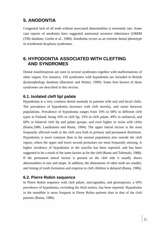
5. ANODONTIA .......................................................................................................... 21
6. HYPODONTIA ASSOCIATED WITH CLEFTING AND SYNDROMES ........... 21
6.1. Isolated cleft lip/palate .................................................................................... 21
6.2. Pierre Robin sequence ..................................................................................... 21
6.3. Van der Woude syndrome ................................................................................ 22
6.4. MSX1 mutation ................................................................................................ 22
6.5. Ectodermal dysplasias (EDs) .......................................................................... 22
6.5.1. Hypohidrotic ectodermal dysplasia (EDA or HED) .............................. 22
6.5.2. Ectrodactyly-ectodermal dysplasia-clefting syndrome (EEC) .............. 23
6.5.3. Cleft lip/palate-ectodermal dysplasia syndrome (CLPED1) ................. 24
6.5.4. Incontinentia pigmenti (IP, Bloch-Sulzberger syndrome) ..................... 24
6.5.5. Hypohidrotic ectodermal dysplasia and immune deficiency (HED-ID) 25
6.5.6. Oral-facial-digital syndrome type1 (OFD1) .......................................... 25
6.5.7. Witkop tooth-nail syndrome ................................................................... 25
6.5.8. Fried syndrome ....................................................................................... 26
6.5.9. Böök syndrome (PHC) ........................................................................... 26
6.5.10. Hair-nail-skin-teeth dysplasias ............................................................. 26
6.6. Rieger syndrome.............................................................................................. 26
6.7. Holoprosencephaly .......................................................................................... 27
1b216251taitto28.9 28.9.2001, 17:495

6
6.8. Down syndrome (trisomy 21) ......................................................................... 27
6.9. Wolf-Hirschhorn syndrome (deletion 4p) ....................................................... 27
6.10. Kabuki syndrome .......................................................................................... 28
6.11. Diastrophic dysplasia (DTD) ........................................................................ 28
6.12. Hemifacial microsomia ................................................................................. 28
6.13. Recessive incisor hypodontia (RIH) ............................................................. 29
7. ASSOCIATED DENTAL ANOMALIES ................................................................ 29
7.1. Delayed formation and eruption of teeth ........................................................ 30
7.2. Reduction in tooth size and form .................................................................... 30
7.3. Malposition of teeth ........................................................................................ 31
7.3.1. Ectopic maxillary canines ...................................................................... 31
7.3.2. Ectopic eruption of other teeth ............................................................... 31
7.4. Infraposition of primary molar(s) ................................................................... 32
7.5. Short roots of teeth ......................................................................................... 32
7.6. Taurodontism ......................................................................................... 32
7.7. Rotation of premolars and/or maxillary lateral incisors ................................. 32
7.8. Enamel hypoplasia, hypocalcification ............................................................ 33
8. TOOTH DEVELOPMENT ...................................................................................... 33
8.1. Initiation and morphogenesis .......................................................................... 33
8.2. Tooth families and the development of dentition ........................................... 34
8.2.1. Evolution ......................................................................................... 34
8.2.2. Tooth families ......................................................................................... 35
8.2.3. Chronology of the development of human dentition ............................. 36
8.3. Signaling networks in tooth development ....................................................... 36
8.3.1. Signals ......................................................................................... 37
8.3.2. Transcription factors ............................................................................... 37
9. TRANSGENIC MICE WITH TOOTH AGENESIS ............................................... 39
10. METHODS FOR IDENTIFYING GENES BEHIND HUMAN DISEASES ...... 40
10.1. Mapping of a disease gene ............................................................................ 40
10.1.1. General principles ................................................................................. 40
10.1.2. Linkage analysis ................................................................................... 41
10.1.3. Genetic markers and the polymerase chain reaction (PCR) ................ 42
AIMS OF THE PRESENT STUDY ......................................................................... 43
SUBJECTS AND METHODS .................................................................................. 44
1. SUBJECTS ......................................................................................... 44
1.1. Families with hypodontia ................................................................................ 44
1b216251taitto28.9 28.9.2001, 17:496
7
1.2. Families with oligodontia ................................................................................ 46
2. METHODS............................................................................................................... 46
2.1. Diagnosis of congenitally missing teeth ............................................................... 46
2.1.1. Studies I, II, and III ................................................................................ 46
2.1.2. Study IV .................................................................................................. 46
2.2. Penetrance ........................................................................................................ 46
2.3. Dental age ........................................................................................................ 47
2.4. Invaginations .................................................................................................... 47
2.5. Taurodontism ................................................................................................... 47
2.6. Reliability of measurements ............................................................................ 48
2.7. Controls ........................................................................................................... 48
2.8. DNA analysis ................................................................................................... 49
2.8.1. DNA extraction ....................................................................................... 49
2.8.2. Genotyping ............................................................................................. 49
2.8.3. Genetic maps .......................................................................................... 49
2.8.4. Sequencing of PAX9 gene ...................................................................... 50
2.9. Statistics ........................................................................................................... 50
RESULTS AND DISCUSSION ........................................................................... 52
1. HYPODONTIA IN FAMILIES ............................................................................... 52
1.1. Characteristics of hypodontia in families ....................................................... 52
1.2. Other dental features in probands ................................................................... 55
1.3. Other dental features in first- and second-degree relatives ............................ 57
1.4. Other dental features in first cousins and more remotely related
individuals ....................................................................................................... 57
1.5. Dentition in obligate carriers .......................................................................... 58
2. OLIGODONTIA IN FAMILIES ............................................................................. 58
3. LINKAGE STUDIES ............................................................................................... 60
4. IDENTIFICATION OF PAX9 MUTATION IN MOLAR OLIGODONTIA .......... 63
GENERAL DISCUSSION ......................................................................................... 65
1. SUBJECTS ............................................................................................................... 65
2. PHENOTYPIC ANALYSIS OF HYPODONTIA AND OLIGODONTIA............. 66
3. GENETIC ANALYSIS............................................................................................. 68
CONCLUDING REMARKS .................................................................................... 69
ACKNOWLEDGEMENTS ....................................................................................... 70
REFERENCES ........................................................................................................... 72
1b216251taitto28.9 28.9.2001, 17:497

8
1b216251taitto28.9 28.9.2001, 17:498
9
LIST OF ORIGINAL PUBLICATIONS
This thesis is based on the following original publications, which are referred to in
the text by their Roman numerals. In addition, some unpublished data are presented.
I Arte S, Nieminen P, Apajalahti S, Haavikko K, Thesleff I, Pirinen S.
Characteristics of incisor-premolar hypodontia in families. J Dent Res
80:1445-1450, 2001.
II Nieminen P, Arte S, Pirinen S, Peltonen L, Thesleff I. Gene defect in
hypodontia: exclusion of MSX1 and MSX2 as candidate genes. Hum Genet
96:305-308, 1995.
III Arte S, Nieminen P, Pirinen S, Thesleff I, Peltonen L. Gene defect in
hypodontia: exclusion of EGF, EGFR, and FGF-3 as candidate genes.
J Dent Res 75:1346-1352, 1996.
IV Nieminen P, Arte S, Tanner D, Paulin L, Alaluusua S, Thesleff I, Pirinen S.
Identification of a nonsense mutation in the PAX9 gene in molar
oligodontia. Eur J Hum Genet 9:743-746, 2001.
1b216251taitto28.9 28.9.2001, 17:499
10
ABBREVIATIONS
BMP bone morphogenetic protein
DLX homeobox transcription factor, homolog of
Drosophila distal-less gene
cM centimorgan
DNA deoxyribonucleic acid
ED ectodermal dysplasia
EDA hypohidrotic ectodermal dysplasia
EGF epidermal growth factor
EGFR epidermal growth factor receptor
FGF fibroblast growth factor
FGFR fibroblast growth factor receptor
GLI transcription factor, homolog of
Drosophila segment polarity gene
INHBA inhibinβ-A
IPH incisor-premolar hypodontia
kb kilobase
LEF lymphoid enhancer-binding factor
Lod logarithm of odds
MSX homeobox transcription factor, homolog of
Drosophila muscle segment gene
mRNA messenger ribonucleic acid
OMIM Online Mendelian Inheritance in Man
PAX paired-box transcription factor, homolog of
Drosophila paired box gene
PCR polymerase chain reaction
p short arm of chromosome
TGF transforming growth factor
TNF tumor necrosis factor
TNFR tumor necrosis factor receptor
q long arm of chromosome
θ theta, recombination fraction
1b216251taitto28.9 28.9.2001, 17:4910
11
ABSTRACT
The congenital lack of teeth has interested dentists for a long time, but genetic studies
using modern DNA techniques have not been published before the last decade.
Studies of odontogenesis at the molecular level, mostly using mouse teeth as models,
have indicated that the development of teeth is under strict genetic control, which
determines the position, number, size, and shape of teeth. More than 200 genes have so
far been identified which are expressed during tooth development, and mutations in
several of these genes cause arrested tooth development in mice.
In this work the segregation and phenotype of hypodontia and associated dental
anomalies were analyzed in 214 family members in three generations of 11 families.
These families were also participating in the genetic linkage study on incisor-premolar
hypodontia (IPH). The analysis confirmed the autosomal dominant transmission with
reduced penetrance of IPH. The prevalence of hypodontia and/or peg-shaped teeth was
over 40% in first- and second-degree relatives and 18% in first cousins of the probands.
The results supported the findings that ectopic canines, rotation of premolars, and
taurodontism are related to hypodontia. Four of nine noted obligate carriers of a
hypodontia gene – with no missing teeth themselves - had minor dental anomalies.
These anomalies were observed at higher than normal frequency also in relatives of the
probands affected with hypodontia.
Linkage analysis is the first step in the localization of human disease genes. In this
study linkage analysis was used to search for the gene locus causing incisor-premolar
hypodontia. First, candidate genes including MSX1, MSX2, EGF, EGFR, and FGF-3
were studied, but no evidence of linkage or association to hypodontia could be found.
Instead, in many families recombinations were found with respect to these genes. In the
second stage, a genome-wide scan was carried out, revealing several regions with
positive lod scores, but no significant evidence for confirming linkage in these families.
In a family with oligodontia involving permanent molars together with upper lateral
incisors and premolars, a nonsense mutation was identified in the PAX9 gene. The
A340T transversion creates a stop codon at lysine 114, and truncates the coded PAX9
protein at the end of the DNA-binding paired-box. All affected members were
heterozygous for the mutation. In another family with molar tooth agenesis this
mutation in the PAX9 gene was absent.
It is evident that both hypodontia and oligodontia are genetically heterogenous
traits. Combining clinical and molecular genetic studies, classification of the differing
forms of these traits will become more exact.
1b216251taitto28.9 28.9.2001, 17:4911
12
INTRODUCTION
Congenital lack of one or more teeth is a common anomaly in man. Lack of one or a
few permanent teeth, hypodontia, without any systemic disorders is the mildest and
most common phenotype. Second premolars and upper lateral incisors are the teeth
most frequently affected. The same teeth are also most often lacking in the more
severe phenotype, oligodontia.
Both environmental and genetic factors can cause failure of tooth development.
Children treated for malignant diseases at tooth-developing ages show a high
frequency of missing teeth. Irradiation produces more severe effects than
chemotherapeutic agents (Maguire et al.,1987; Näsman et al.,1997).
Numerous different genes have been implicated in tooth development by gene
expression and experimental studies in the mouse, and in theory, any of these genes
may cause tooth agenesis (for review, Thesleff, 2000). Family studies show that, as
an isolated form, both hypodontia and oligodontia are inherited as an autosomal
dominant trait with incomplete penetrance and variable expression (Grahnen, 1956;
Burzynski and Escobar, 1983). Sex-linked and polygenic or multifactorial models of
inheritance have also been suggested (Suarez and Spence, 1974; Chosack et al.,
1975; Brook, 1984; Peck et al., 1993). An autosomal recessive model was shown in
one family (Ahmad et al., 1998). Variability in expression includes the number and
region of missing teeth, and various other dental features associated with the trait.
The obscure mechanisms underlying congenital lack of teeth and the differing
results of genetic studies have drawn attention to the phenotypic and genotypic
variation in this phenomenon. Thus, the original purpose of this study was to further
define the phenotype and to map the gene locus responsible for incisor-premolar
hypodontia. During the course of this study, mutations in two transcription factors,
MSX1 and PAX9, have been identified in three families with oligodontia (Vastardis et
al., 1996; Stockton et al., 2000; van den Boogaard et al., 2000). A mutation screening
of the PAX9 gene, was therefore performed in two Finnish families with oligodontia.
1b216251taitto28.9 28.9.2001, 17:4912
13
REVIEW OF THE LITERATURE
1. CONGENITALLY MISSING TEETH
1.1. Definition, diagnosis, and terminologyCongenital lack of a tooth results from disturbances during the early stages of tooth
development. A tooth is defined to be congenitally missing if it has not erupted in the
oral cavity and is not visible in a radiograph. All primary teeth have erupted by the
age of 3 and all permanent teeth except the third molars between the ages of 12 and
14. Therefore, 3- to 4-year-old children are suitable for diagnosis of congenitally
missing primary teeth by clinical examination, and 12- to 14-year-old children, for
diagnosis of permanent teeth, excluding the third molars. Radiographic diagnosis can
be made at younger age depending on tooth group. The use of panoramic
radiography is recommended, together with clinical examination in detecting or
confirming dental development (Pirinen and Thesleff, 1995).
All primary teeth and the crypts of first permanent molars are visible by
radiograph at birth. The crowns of first premolars, second premolars, and second
permanent molars start to mineralize near the second birthday, and all permanent
tooth crowns except the third molars have begun their mineralization by the age of
six. The formation of third molars shows very large variation. Usually at the age of 8
to 10 years, the first signs of the third molars appear on a radiograph but
occasionally, very late appearance (age 14 to 18) occurs (Pirinen and Thesleff, 1995).
The formation of dentition continues many years, and differences exist in
mineralization stages among children depending on race, on gender, and even on
family and on the individual. Especially second premolars may show late onset of
mineralization, and give a false-positive diagnosis of hypodontia in radiographs.
Therefore, diagnosis of tooth agenesis in the permanent dentition should be made
after the age of 6 (Pirinen and Thesleff, 1995) excluding third molars, and after 10
years of age if third molars are also studied.
Hypodontia is the term most frequently used when describing the phenomenon of
congenitally missing teeth in general. Many other terms appear in the literature to
describe a reduction in number of teeth: oligodontia, anodontia, aplasia of teeth,
congenitally missing teeth, absence of teeth, agenesis of teeth, and lack of teeth.
Hypodontia and oligodontia are classified as isolated or nonsyndromic
hypodontia/oligodontia and syndromic hypodontia/oligodontia or hypodontia/
oligodontia associated with syndromes.
1b216251taitto28.9 28.9.2001, 17:4913
14
The term hypodontia is used in a narrow sense when the number of missing teeth is
one or a few. Oligodontia is defined as missing a large number of teeth.
Anodontia is an extreme case, denoting complete absence of teeth. There is no clear
definition in the literature concerning the limits of these classes. In the recent years,
however, the following definitions have been used:
Hypodontia:1 to 6 teeth missing (excluding the third molars)
Oligodontia: more than six teeth missing (excluding the third molars)
Anodontia: complete absence of teeth.
Incisors and premolars are the most frequently missing teeth. Therefore incisor-
premolar hypodontia (IPH) is the term that we have used to describe this form of
the anomaly.
1.2. EtiologyMany theories of the etiology of tooth agenesis have been suggested in the literature,
especially before the intense genetic studies of the present day, and obviously both
genetic and environmental factors may contribute (Grahnen, 1956; Schalk-van der
Weide, 1992; for review, Jorgenson, 1980; Vastardis, 2000).
1.2.1. Environmental factorsIn principle, many environmental factors may cause arrested tooth development.
Different kinds of trauma in the dental region such as fractures, surgical procedures
on the jaws, and extraction of the preceding primary tooth are mentioned in the
literature (for review, Grahnen, 1956; Schalk-van der Weide, 1992).
Developing teeth are irreversibly affected by multiagent chemotherapy and
radiation therapy, and effects depend on age of patient and dosage (Näsman et al.,
1997). Children after treatment for malignant disease at an early age show arrested root
development with short V-shaped roots, roots with premature apical closure, enamel
hypoplasia, microdontia, and hypodontia. Irradiation produces more severe effects than
those caused by chemotherapeutic agents (Maguire et al.,1987; Näsman et al.,1997).
Congenitally missing teeth have been reported in children whose mothers had used
ThalidomideR (N-phthaloylglutamimide) during pregnancy (Axrup et al., 1966). No
definite etiologic relationship has been found between hypodontia and systemic diseas-
es or endocrine disturbances (for review, Grahnen, 1956; Schalk- van der Weide, 1992).
A relationship has been proposed between the function of peripheral nerves and
tooth agenesis (Kjaer et al., 1994). This report focused on an etiological explanation
of hypodontia based on disturbances in nerve tissue, oral mucosa, and supporting
tissues, all interacting in tooth development.
1b216251taitto28.9 28.9.2001, 17:4914
15
1.2.2. Genetic factorsAlthough tooth agenesis is occasionally caused by environmental factors, in the
majority of cases hypodontia has a genetic basis. In hypodontia, the criteria for a
genetic disease are fulfilled: It is more common among individuals related to
hypodontia patients than in population in general (Burzynsky and Escobar, 1983;
Brook, 1984). The classic family study of Grahnen in Sweden of a total of 685
family members of 171 probands affected with hypodontia (Grahnen, 1956) showed
that hypodontia in the permanent dentition is primarily determined by genetic factors.
Differences in frequency of hypodontia exist between races, no environmental etiology
is apparent in individuals with the disease, and greater concordance of hypodontia
appears in identical than in non-identical twins (Markovic, 1982; Kotsomitis et al.,
1996).
In familial hypodontia, the type of inheritance in the majority of families seems
to be autosomal dominant with incomplete penetrance and variable expressivity. In
addition, peg-shaped upper lateral incisors are considered to be a modified
manifestation of the same genotypes as hypodontia (Grahnen, 1956; Alvesalo and
Portin, 1969). Sex-linked inheritance patterns and a polygenic or multifactorial model
of inheritance have also been suggested (Suarez and Spence, 1974; Chosack et al.,
1975; Brook, 1984; Peck et al., 1993), and an autosomal recessive model in one family
(Ahmad et al., 1998). Female predominance has been reported (Bergström, 1977;
Wisth et al., 1974; Stamatiou and Symons, 1991; Kotsomitis et al., 1996), but in most
studies the difference does not reach statistical significance (Grahnen, 1956; Haavikko,
1971; Rolling, 1980).
The most direct evidence for a genetic basis is that during the course of this study,
two mutated genes causing an autosomal dominant form of human nonsyndromic tooth
agenesis were identified (Vastardis et al., 1996; Van den Boogaard et al., 2000). A
missense mutation was found by the Vastardis group in the homeodomain of MSX1
gene in chromosome 4 (4p16) in all affected members of a family with missing
second premolars and third molars as a prominent feature. Some affected individuals
also lacked their maxillary first premolars, mandibular first molars, one or both upper
lateral incisors, or a single lower central incisor. All affected individuals were reported
to have had normal primary dentitions. In a Dutch family, however, a nonsense
mutation in the MSX1 gene was associated with tooth agenesis and various
combinations of cleft lip and/or palate (Van den Boogaard et al., 2000). Another
report has excluded MSX1 as the gene responsible for tooth agenesis (Scarel et al.,
2000). A frameshift mutation in another transcription factor gene, PAX9, in
chromosome 14 (14q21-q13) was identified in a family with autosomal dominant
1b216251taitto28.9 28.9.2001, 17:4915
16
oligodontia (Stockton et al., 2000). The point mutation, insertion of a guanine, causes
a frameshift and premature termination of the protein (Stockton et al., 2000). The
affected individuals lack most permanent molars. Some individuals also were missing
their maxillary and/or mandibular second premolars as well as mandibular central
incisors. The primary dentition had been normal. MSX1 and PAX9 are transcription
factors which, before being associated with human tooth agenesis, had been shown to
regulate early tooth morphogenesis in the mouse. They are expressed in dental
mesenchyme after initiation of tooth development in response to epithelial signals
(for review, Thesleff, 2000). Inactivation of Msx1 and Pax9 genes in the mouse causes
arrested development of teeth at the bud stage and malformations of palate, limb, and
pharyngeal pouch derivatives, whereas heterozygous mice develop normal teeth
(Satokata and Maas, 1994; Peters et al., 1998). In humans, inactivation of one copy
of the gene causes dental defects (Vastardis et al., 1996; Stockton et al., 2000) or
dental defects and clefting in the case of MSX1 nonsense mutation (Van den
Boogaard et al., 2000).
In addition, several gene defects have been identified which cause syndromes
with hypodontia or oligodontia (see section on syndromic hypodontia).
2. CONGENITALLY MISSING TEETH IN PRIMARYDENTITION
Variation in the number of teeth is not as common in the primary as in the permanent
dentition, and no significant difference exists in prevalence of hypodontia by gender
(Grahnen and Granath, 1961; Ravn, 1971; Järvinen and Lehtinen, 1981). The
prevalence varies from 0.4% to 0.9% in the European population (Grahnen and
Granath, 1961; Ravn, 1971; Järvinen and Lehtinen, 1981; Magnusson, 1984; Carvalho,
1998), and is reported to be higher, 2.4%, in Japan (Yonezu et al., 1997) whereas in
New Zealand the prevalence of 0.4% corresponds to that of Europeans (Whittington,
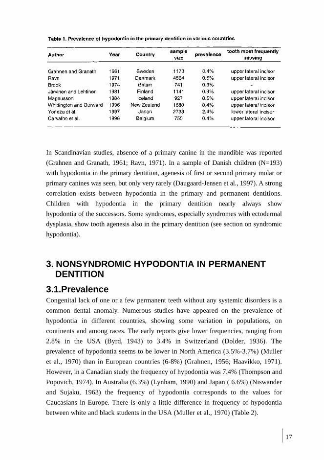
1996). Table 1. Mostly one (55% of the children, according to Daugaard-Jensen et al.,
1997) or two teeth are missing, and the majority of cases represent unilateral hypodontia.
In the primary dentition the incisor region seems to be affected most often
(Grahnen and Granath, 1961; Järvinen and Lehtinen, 1981; Magnusson, 1984;
Daugaard-Jensen et al., 1997; Yonezu et al., 1997). In European studies the upper
primary lateral incisors are the most frequently missing (Järvinen and Lehtinen,
1981; Daugaard-Jensen et al, 1997), whereas the lower lateral incisors are affected
most often in the Japanese (Yonezu et al., 1997). Peg-shaped teeth are seen also in
the primary dentition; this Japanese study reported a frequency of 0.55% .
1b216251taitto28.9 28.9.2001, 17:4916
17
In Scandinavian studies, absence of a primary canine in the mandible was reported
(Grahnen and Granath, 1961; Ravn, 1971). In a sample of Danish children (N=193)
with hypodontia in the primary dentition, agenesis of first or second primary molar or
primary canines was seen, but only very rarely (Daugaard-Jensen et al., 1997). A strong
correlation exists between hypodontia in the primary and permanent dentitions.
Children with hypodontia in the primary dentition nearly always show
hypodontia of the successors. Some syndromes, especially syndromes with ectodermal
dysplasia, show tooth agenesis also in the primary dentition (see section on syndromic
hypodontia).
3. NONSYNDROMIC HYPODONTIA IN PERMANENTDENTITION
3.1.PrevalenceCongenital lack of one or a few permanent teeth without any systemic disorders is a
common dental anomaly. Numerous studies have appeared on the prevalence of
hypodontia in different countries, showing some variation in populations, on
continents and among races. The early reports give lower frequencies, ranging from
2.8% in the USA (Byrd, 1943) to 3.4% in Switzerland (Dolder, 1936). The
prevalence of hypodontia seems to be lower in North America (3.5%-3.7%) (Muller
et al., 1970) than in European countries (6-8%) (Grahnen, 1956; Haavikko, 1971).
However, in a Canadian study the frequency of hypodontia was 7.4% (Thompson and
Popovich, 1974). In Australia (6.3%) (Lynham, 1990) and Japan ( 6.6%) (Niswander
and Sujaku, 1963) the frequency of hypodontia corresponds to the values for
Caucasians in Europe. There is only a little difference in frequency of hypodontia
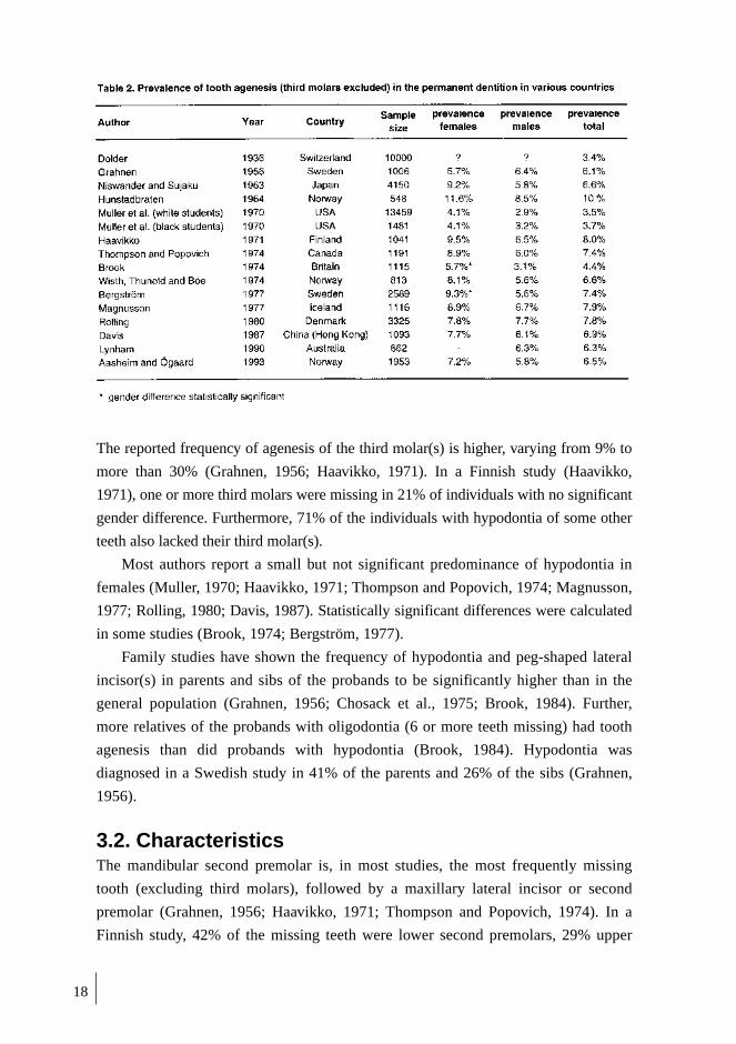
between white and black students in the USA (Muller et al., 1970) (Table 2).
-
1b216251taitto28.9 28.9.2001, 17:4917
18
The reported frequency of agenesis of the third molar(s) is higher, varying from 9% to
more than 30% (Grahnen, 1956; Haavikko, 1971). In a Finnish study (Haavikko,
1971), one or more third molars were missing in 21% of individuals with no significant
gender difference. Furthermore, 71% of the individuals with hypodontia of some other
teeth also lacked their third molar(s).
Most authors report a small but not significant predominance of hypodontia in
females (Muller, 1970; Haavikko, 1971; Thompson and Popovich, 1974; Magnusson,
1977; Rolling, 1980; Davis, 1987). Statistically significant differences were calculated
in some studies (Brook, 1974; Bergström, 1977).
Family studies have shown the frequency of hypodontia and peg-shaped lateral
incisor(s) in parents and sibs of the probands to be significantly higher than in the
general population (Grahnen, 1956; Chosack et al., 1975; Brook, 1984). Further,
more relatives of the probands with oligodontia (6 or more teeth missing) had tooth
agenesis than did probands with hypodontia (Brook, 1984). Hypodontia was
diagnosed in a Swedish study in 41% of the parents and 26% of the sibs (Grahnen,
1956).
3.2. CharacteristicsThe mandibular second premolar is, in most studies, the most frequently missing
tooth (excluding third molars), followed by a maxillary lateral incisor or second
premolar (Grahnen, 1956; Haavikko, 1971; Thompson and Popovich, 1974). In a
Finnish study, 42% of the missing teeth were lower second premolars, 29% upper
1b216251taitto28.9 28.9.2001, 17:4918
19
second premolars, 19% upper lateral incisors, 4% lower first premolars, 3% lower
central incisors, and 1% lower lateral incisors. Hypodontia of second molars and
lower canines were rare, 0.7% of the missing teeth (Haavikko, 1971). In an American
study, an upper lateral incisor was the most frequently missing in the individuals with
agenesis of one or two teeth, while in those who lacked more than two teeth, the second
premolar was most commonly missing (Muller et al., 1970). Absence of maxillary
central incisors, maxillary and mandibular first molars and canines seems to be very
rare. No clear difference in congenitally missing teeth has been found between the
maxilla and the mandible (Grahnen, 1956; Haavikko, 1971; Bergström, 1977).
Unilateral hypodontia is common, with no significant difference between the left and
right sides of the jaws (Magnusson, 1977; Lai and Seow, 1989). Predominance of
hypodontia on the left side has been reported in some Scandinavian studies
(Grahnen, 1956; Haavikko, 1972; Wisth et al., 1974; Bergström, 1977).
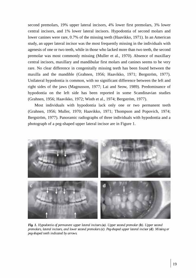
Most individuals with hypodontia lack only one or two permanent teeth
(Grahnen, 1956; Muller, 1970; Haavikko, 1971; Thompson and Popovich, 1974;
Bergström, 1977). Panoramic radiographs of three individuals with hypodontia and a
photograph of a peg-shaped upper lateral incisor are in Figure 1.
� �
� �
Fig. 1. Hypodontia of permanent upper lateral incisors (a). Upper second premolar (b). Upper secondpremolars, lateral incisors, and lower second premolars (c). Peg-shaped upper lateral incisor (d). Missing orpeg-shaped teeth indicated by arrows.
1b216251taitto28.9 28.9.2001, 17:4919
20
4. NONSYNDROMIC OLIGODONTIA IN PERMANENTDENTITION
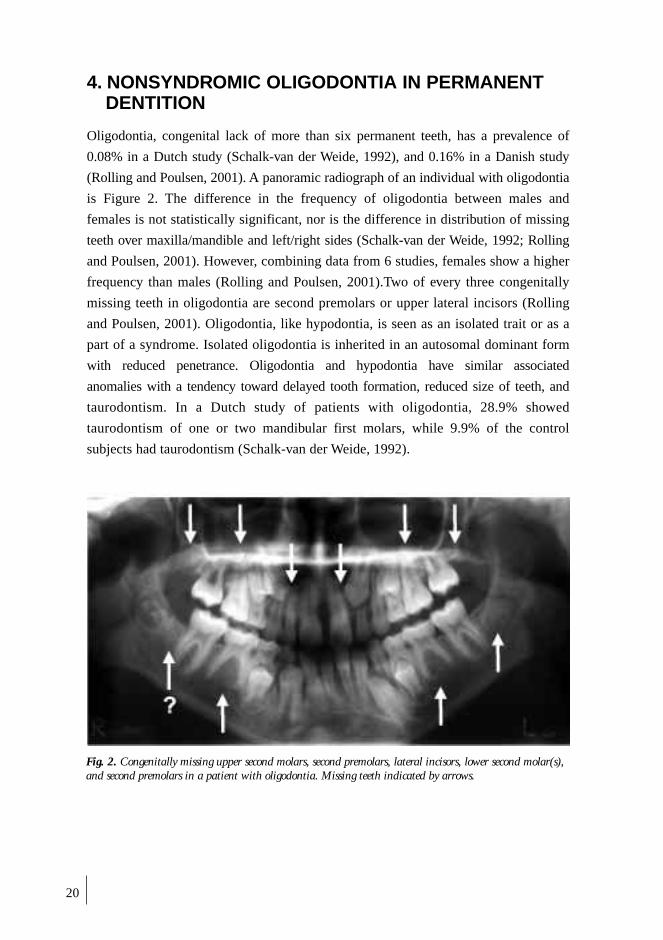
Oligodontia, congenital lack of more than six permanent teeth, has a prevalence of
0.08% in a Dutch study (Schalk-van der Weide, 1992), and 0.16% in a Danish study
(Rolling and Poulsen, 2001). A panoramic radiograph of an individual with oligodontia
is Figure 2. The difference in the frequency of oligodontia between males and
females is not statistically significant, nor is the difference in distribution of missing
teeth over maxilla/mandible and left/right sides (Schalk-van der Weide, 1992; Rolling
and Poulsen, 2001). However, combining data from 6 studies, females show a higher
frequency than males (Rolling and Poulsen, 2001).Two of every three congenitally
missing teeth in oligodontia are second premolars or upper lateral incisors (Rolling
and Poulsen, 2001). Oligodontia, like hypodontia, is seen as an isolated trait or as a
part of a syndrome. Isolated oligodontia is inherited in an autosomal dominant form
with reduced penetrance. Oligodontia and hypodontia have similar associated
anomalies with a tendency toward delayed tooth formation, reduced size of teeth, and
taurodontism. In a Dutch study of patients with oligodontia, 28.9% showed
taurodontism of one or two mandibular first molars, while 9.9% of the control
subjects had taurodontism (Schalk-van der Weide, 1992).
Fig. 2. Congenitally missing upper second molars, second premolars, lateral incisors, lower second molar(s),and second premolars in a patient with oligodontia. Missing teeth indicated by arrows.
1b216251taitto28.9 28.9.2001, 17:4920
21
5. ANODONTIA
Congenital lack of all teeth without associated abnormalities is extremely rare. Some
case reports of anodontia have suggested autosomal recessive inheritance (OMIM
(TM) database; Gorlin et al., 1980). Anodontia occurs as an extreme dental phenotype
in ectodermal dysplasia syndromes.
6. HYPODONTIA ASSOCIATED WITH CLEFTINGAND SYNDROMES
Dental manifestations are seen in several syndromes together with malformations of
other organs. For instance, 150 syndromes with hypodontia are included in British
dysmorphology database (Baraitser and Winter, 1999). Some best known of these
syndromes are described in this section.
6.1. Isolated cleft lip/ palateHypodontia is a very common dental anomaly in patients with oral and facial clefts.
The prevalence of hypodontia increases with cleft severity, and varies between
populations. Prevalence of hypodontia ranges from 10% to 68% in different cleft
types in Finland, being 10% in cleft lip, 33% in cleft palate, 49% in unilateral, and
68% in bilateral cleft lip and palate groups, and even higher in twins with clefts
(Ranta,1986; Laatikainen and Ranta, 1994). The upper lateral incisor is the most
frequently affected tooth in the cleft area both in primary and permanent dentitions.
Hypodontia is more common than in the normal population also outside the cleft
region, where the upper and lower second premolars are most frequently missing. A
higher incidence of hypodontia in the maxilla has been reported, and has been
suggested to be a result of the same factors as for the cleft (Ranta and Tulensalo, 1988).
If the permanent lateral incisor is present on the cleft side it usually shows
abnormalities in size and shape. In addition, the dimensions of other teeth are smaller,
and timing of tooth formation and eruption in cleft children is delayed (Ranta, 1986).
6.2. Pierre Robin sequenceIn Pierre Robin sequence with cleft palate, micrognathia, and glossoptosis, a 50%
prevalence of hypodontia, excluding the third molars, has been reported. Hypodontia
in the mandible is more frequent in Pierre Robin patients than in that of the cleft
patients (Ranta, 1986).
1b216251taitto28.9 28.9.2001, 17:4921
22
6.3. Van der Woude syndromeEven higher prevalence of hypodontia (69%) has been shown in patients with the
autosomal dominant Van der Woude syndrome associated with cleft lip and/or palate
and pits of the lower lip. Genetic heterogeneity in Van der Woude syndrome has been
proven; It has been mapped to 1q32-41 in some families, but the same locus is
excluded in other families (Wong, 2000).
6.4. MSX1 mutationThe MSX1 mutation is associated with clefting and hypodontia. A large Dutch family
with tooth agenesis and various combinations of cleft lip and palate showed a
nonsense mutation in exon 1 of MSX1 in chromosome 4 (Van den Boogaard et al.,
2000). The mutation (Ser104stop) was found to be heterozygous in all affected family
members. Both mandibular and maxillary second premolars were missing in most
affected individuals, but in addition, third molars, upper lateral incisors, lower central
incisors, first premolars, and second molars were missing in some individuals. The
phenotype of the Dutch family corresponds to that of the Msx1-mutant mouse
(Satokata and Maas, 1994), but is more severe than the phenotype of the previously
reported MSX1 missense mutation family (Arg239Pro), which showed oligodontia
without clefts (Vastardis et al., 1996).
6.5. Ectodermal dysplasias (EDs)The term Ectodermal Dysplasia (ED) covers a heterogenous group of conditions
affecting ectodermal organs including hair, teeth, nails, and glands. A condition
characterized by ectodermal signs only is called pure ED. A condition with ectodermal
symptoms associated with other malformations is called ED/malformation syndrome or
ED syndrome. More than 150 EDs have been described and classified into 11 clinical
subgroups (Pinheiro and Freire-Maia, 1994; OMIM (TM) database), with variation in
mode of inheritance as well as genetic heterogeneity demonstrated. EDs include
X-linked, autosomal dominant, and autosomal recessive forms.
6.5.1. Hypohidrotic ectodermal dysplasia (EDA or HED)Hypohidrotic ectodermal dysplasia, EDA, the most common and best-known ED, is
usually inherited as an X-linked semidominant trait, although rarer autosomal
dominant and recessive forms exist. The defective gene behind X-chromosomal EDA
has been identified (Xq12-q13.1) (Kere et al., 1996; Monreal et al., 1998). The
protein product, ectodysplasin, is a novel member of the tumor necrosis factor (TNF)
family and functions as a signaling molecule during epithelial morphogenesis
1b216251taitto28.9 28.9.2001, 17:4922

23
(Mikkola et al., 1999; Thesleff, 2000). Affected males show severe oligodontia or
anodontia, and abnormalities in tooth shapes. Anomalies are seen in both primary
and permanent dentitions. Frontal bossing and nasal and maxillary hypoplasia, and
sparse or absent scalp hair contribute to the typical appearance of EDA patients. Due
to absence of sweat glands these patients suffer from hypohidrosis. Female EDA
carriers have variable, milder phenotypic expressions depending on the consequences
of X-chromosome inactivation (Cambiaghi et al., 2000). They have hypodontia or
oligodontia, and also abnormally shaped teeth. All carrier mothers (N=5) of X-linked
EDA patients in a Finnish study lacked more than four permanent teeth, and one
mother had had hypodontia in her primary dentition. In addition, three of five mothers
had peg-shaped teeth (Söderholm and Kaitila, 1985).
Another gene mutation, which was found to cause both autosomal dominant and
recessive forms of EDA, was identified in chromosome 2 (2q11-q13); it encodes a
TNF receptor (TNFR) called Edar (Monreal et al., 1999). In autosomal recessive
EDA, heterozygous carriers show no features of the disorder, unlike the X-linked
EDA, in which female carriers show mild manifestations (Cambiaghi et al., 2000).
6.5.2. Ectrodactyly-ectodermal dysplasia-clefting syndrome (EEC)The characteristics of EEC syndrome are ectrodactyly of the hands and feet, ectoder-
mal dysplasia, and cleft lip and/or palate (Gorlin et al., 1990). Features of ectodermal
dysplasia in 77% of the patients include sparse hair, dystrophic nails, hypopigmenta-
tion or pigmented nevi of the skin, and abnormal dentition (Roelfsema and Cobben,
1996). Congenitally missing permanent teeth and conical teeth are common. Missing
of maxillary first primary molars has been reported (Gorlin et al.,1990). Cleft lip
and/or palate is seen in 68% of the patients (Roelfsema and Cobben, 1996).
Buss et al. (1995) reported dental features of 24 patients with EEC syndrome: The
permanent dentitions of all patients were affected with oligodontia and microdontia.
The teeth were not as strongly conical as in cases of X-linked EDA, being more often
straight-edged with gaps; taurodontism was also common. The number of teeth was
normal in the primary dentitions, but with abnormal morphology of the tooth crowns.
Microcephaly and mental retardation have been reported in about 10% of the
patients (Gorlin et al., 1990). In addition, anomalies of the lacrimal ducts, urogenital de-
fects, and conductive hearing loss have been reported (Roelfsema and Cobben, 1996).
Autosomal dominant transmission with reduced penetrance and variable expression
is shown in EEC, with proven genetic heterogeneity. EEC1 syndrome is associated
with chromosome 7 (7q11.2-q21.3) (Qumsiyeh, 1992). Linkage of EEC2 to a locus
1b216251taitto28.9 28.9.2001, 17:4923

24
on the chromosome 19 pericentromeric region has been reported in a Dutch kindred
(O´Quinn et al., 1998). The third form, EEC3, is caused by mutations in the
transcription factor gene p63 in chromosome 3q27 where another EEC-like disorders,
limb-mammary syndrome and ADULT syndrome (acro-dermato-ungual-lacrimal-
tooth syndrome) have been mapped (Propping et al., 2000). Several different
mutations of gene p63 have been revealed in EEC families (Celli et al., 1999).
6.5.3. Cleft lip/palate-ectodermal dysplasia syndrome (CLPED1)Cleft lip and/or cleft palate together with ectodermal dysplasia (CLPED1) has been
reported in clinical conditions called Zlotogora-Ogur syndrome (Zlotogora, 1994),
and the Margarita Island form of ectodermal dysplasia (Bustos et al., 1991). The gene
mutated in these autosomal recessive syndromes is identified as PVRL1, located at
the chromosomal region 11q23-q24 (Suzuki et al., 2000). PVRL1 encodes nectin-1, a
cell adhesion molecule. In both syndromes, the patients have scanty eyebrows and
eyelashes, sparse, short and dry scalp hair, syndactyly of the fingers and toes, cleft lip/
palate, nail dysplasia, and hypodontia. Hypodontia affects mainly the upper lateral
incisors, and in addition, changes in size and shape of tooth crowns (Bustos et al.,
1991). Mental retardation is present in Zlotogora-Ogur syndrome but is absent from
Margarita Island ectodermal dysplasia (Zlotogora, 1994).
6.5.4. Incontinentia pigmenti (IP, Bloch-Sulzberger syndrome)Incontinentia Pigmenti (IP) is a rare multisystem disorder classified as an ectodermal
dysplasia with variable abnormalities of the skin, hair, nails, teeth, eyes, and central
nervous system. The skin of IP patients shows vesicular, verrucous, and pigmented
macular lesions. In addition, the patients have dental, ocular, central nervous system,
and structural anomalies (Gorlin et al., 1990). IP is an X-linked dominant disorder
and has been shown to be due to mutations in IKK-gamma (NEMO) gene located in
Xq28 (Smahi et al., 2000). The affected individuals are mostly females (97% of the
patients). It is assumed that males with the mutation usually do not survive through
gestation (Macey-Dare and Goodman, 1999).
In studies on dental anomalies, over 90% of the patients have hypodontia, mostly
classified as severe (6 or more teeth missing), microdontia (generalized microdontia
or peg-shaped teeth), macrodontia (extra cusps in the posterior teeth), delayed
eruption of permanent teeth, or taurodontism (Gorlin et al., 1990; Macey-Dare and
Goodman, 1999). Incidence of congenitally missing teeth has been reported to be as
high as 43%; in addition, 30% of the patients have conical teeth (Macey-Dare and
Goodman, 1999). Tooth anomalies are seen both in primary and permanent
dentitions, but the permanent dentition is usually more severely affected.
1b216251taitto28.9 28.9.2001, 17:4924

25
6.5.5. Hypohidrotic ectodermal dysplasia and immune deficiency (HED-ID)A novel form of EDA, together with immunodeficiency, segregates as an X-linked
recessive trait (Zonana et al., 2000; Döffinger et al., 2001). Clinical findings:
hypohidrosis and abnormal dentition, are similar to those in other forms of EDA.
Tooth agenesis occurs both in primary and in permanent dentitions. Hypodontia,
oligodontia, and conical teeth are the forms of tooth manifestation. Affected males
manifest dysgammaglobulinemia and suffer significant mortality from infections.
Female carriers show no clinical signs of immunodeficiency but have other
manifestations including hypodontia and conical teeth. Mutations in the IKK-gamma
(NEMO) gene have been found in HED-ID patients, and thus HED-ID is allelic to
incontinentia pigmenti. IKKγ is required for activation of NFκB, a transcription
factor transducing TNF signaling. NFκB was recently shown also to transduce signaling
via ectodysplasin/ Edar, which presumably explains the phenotypic similarities
between EDA and HED-ID (Kumar et al., 2001; Koppinen et al., submitted).
6.5.6. Oral-facial-digital syndrome type 1 (OFD1)Oral-facial-digital syndromes are a heterogenous group of developmental disorders
of which at least nine forms have been described (Ferrante et al., 2001). Oral-facial-
digital syndrome type 1 is characterized by malformations of the face, oral cavity,
and digits. Typical characteristics include facial asymmetry, hypertelorism,
micrognathia, broadened nasal bridge, and facial milia (Ferrante et al., 2001). Median
pseudoclefting of the upper lip has been reported in 45%, palatal clefts in over 80%,
clefts of the tongue in 30%. In addition, they have supernumerary frenulae in the oral
cavity and alveolar ridges may be thickened (Gorlin et al., 1990; Ferrante et al., 2001).
Hypodontia typically affects mandibular lateral incisors in about 50% of these patients
(Gorlin et al., 1990). Hypodontia of lower lateral incisors is associated with the fibrous
bands in this region. Oral-facial-digital syndrome type 1 is transmitted as an X-linked
dominant condition affecting females and causing mortality in males. The gene responsible
for this syndrome is CXORF5 in chromosome Xp22.3-22.2; many different mutations
have been found in these patients (Ferrante et al., 2001).
6.5.7. Witkop tooth-nail syndromeDysplasia of nails together with hypodontia was first described by Witkop (1965).
This syndrome is inherited in an autosomal dominant manner. Fingernails and
especially toenails are dysplastic in childhood. Mandibular incisors, second molars,
and maxillary canines are the teeth most often missing or having conical crowns. Some
patients show hypodontia or a conical form of primary teeth. A nonsense mutation in
the homeodomain of MSX1 has been shown to cause Witkop syndrome in a three-
1b216251taitto28.9 28.9.2001, 17:4925

26
generation family (Jumlongras et al., 2001). The predominant missing teeth were
premolars, first molars, and third molars in this family. In a few cases, incisors or
canines were also absent. Permanent teeth showed reduced mesiodistal dimensions
and shorter root lengths than normal teeth. Primary teeth were normal in size, shape,
and number in all patients except one individual with fused mandibular primary
central and lateral incisor (Jumlongras et al., 2001).
6.5.8. Fried syndromeAgenesis of primary incisors or their conical form together with thin hair and nails
has been described by Fried in children born to consanguineus parents (1977), who
suggested autosomal recessive inheritance of this trait.
6.5.9. Böök syndrome (PHC)The features of Böök syndrome include premolar agenesis, hyperhidrosis of the
hands and feet, and early graying of the hair (canities prematura). Early, diffuse
whitening of the hair may appear even in childhood. Hypodontia in the Böök
syndrome affects the premolar region, with one or more premolars missing. The
syndrome has an autosomal dominant inheritance with high or complete penetrance;
the gene defect is unknown (Gorlin et al., 1990; Böök, 1950).
6.5.10. Hair-nail-skin-teeth dysplasiasA large number of rare disorders involving dysplasia of hair, nails, skin, and teeth
have been described (Gorlin et al., 1990). The features of these dysplasias overlap the
ectodermal dysplasias, and both autosomal recessive and autosomal dominant
inheritance patterns have been reported. Oligodontia and/or microdontia, peg-shaped
teeth, and enamel hypoplasias of the teeth are the typical dental manifestations
(Gorlin et al., 1990).
6.6. Rieger syndromeRieger syndrome is an autosomal dominant disorder with malformations of the
anterior chamber of the eye, umbilical anomalies, and hypodontia. The maxillary
primary and permanent incisors and second premolars are the most commonly
missing. Peg-shaped incisors have also been reported. Hypodontia in the anterior
region of the maxilla results in underdevelopment of the premaxillary area (Gorlin et
al., 1990; OMIM (TM)database). Rieger syndome has proven to be genetically
heterogenous, caused by mutations in a homeobox transcription factor gene, PITX2
in 4q25-q26 (Semina et al., 1996). Another locus for Rieger syndrome has been
1b216251taitto28.9 28.9.2001, 17:4926
27
identified on 13q14 by linkage analysis (Phillips et al., 1996), but the gene has not
yet been found.
6.7. HoloprosencephalyHoloprosencephaly is a rare malformation sequence in which the basic feature is
impaired midline cleavage of the embryonic forebrain (Gorlin et al., 1990). It is an
etiologically heterogenous condition; teratogenic and genetic factors may both be
responsible. The phenotype varies widely; the facial dysmorphism includes cyclopia,
hypertelorism, single nostril or flat nose, cleft lip, and hypodontia. A single maxillary
central incisor can be seen as the mildest phenotype of holoprosencephaly. In familial
cases, an autosomal dominant inheritance with reduced penetrance has been observed
(Odent et al., 1998). At least 12 different loci have been associated with holoprosen-
cephaly and several distinct genes identified (Wallis and Muenke, 2000).
6.8. Down syndrome (trisomy 21)Down syndrome, the most common chromosomal abnormality in man, is caused by
trisomy of all or a critical portion of chromosome 21 (21q22.3). The birth prevalence of
trisomy 21 syndrome is 1/650 live births, with the risk of having a child with Down
syndrome increasing with maternal age (Gorlin et al., 1990; OMIM (TM) database).
Down syndrome is characterized by a combination of phenotypic features that includes
typical dysmorphic features and mental retardation. Congenital malformations of the
heart (30-40% of the patients) and gastrointestinal tract are common. Congenital
absence of teeth has been reported in 23 to 47% (Gorlin et al, 1990). Lateral maxillary
incisors, lower incisors, second premolars, and third molars are the most commonly
missing. One or both primary upper lateral incisors are missing in more than 10% of
the patients, and peg-shaped maxillary lateral incisors are seen in 10% (Gorlin et al.,
1990). Shapira et al. (2000) studied hypodontia and other dental anomalies in a sample
of 34 individuals with Down syndrome in Israel. In this group, 74% of the individuals
lacked one or more third molars, and 60% lacked at least one other tooth. In addition,
25% of the individuals had small or peg-shaped upper lateral incisor(s). With third
molars excluded, teeth were missing in 59% of the total sample (Shapira et al., 2000).
6.9. Wolf-Hirschhorn syndrome (deletion 4p)Wolf-Hirschhorn syndrome is a malformation syndrome caused by deletions of the
distal short arm of chromosome 4 (4p16.3), with variations in both the size of the
deletions and position of the breakpoints. It has been suggested that the critical
region is approximately 165kb long (Wright et al., 1997). This region has been
1b216251taitto28.9 28.9.2001, 17:4927

28
sequenced intensively during the search for the genes causing Huntington disease,
achondroplasia, and other skeletal dysplasias, and is found to be a gene-dense region;
the MSX1 gene is located nearby. Wolf-Hirschhorn syndrome is characterized by
severe growth and psychomotor retardation, microcephaly, and striking facial
features, and closure defects: cleft lip or palate, coloboma of the eye, and cardiac
septal defects. About one-third of the patients have an isolated cleft palate, another
third high arched palate with micrognathia, and 10% have cleft lip and palate. Agenesis
of many permanent teeth has been suggested to belong to the oral manifestations of
this syndrome (Burgersdijk and Tan, 1978).
6.10. Kabuki syndromeThe features of Kabuki syndrome (KS) include characteristic facial dysmorphic
features, skeletal abnormalities, dermatoglyphic abnormalities, mild to moderate mental
deficiency, and postnatal growth retardation. The etiology of KS is unknown (Mhanni
et al., 1999). Agenesis of upper and lower permanent incisors or premolars, conical
incisors, and ectopic upper first molars has been reported (Mhanni et al., 1999).
6.11. Diastrophic dysplasia (DTD)Diastrophic dysplasia is a recessively inherited osteochondrodysplasia belonging to
the group of disorders called the ”Finnish Disease Heritage.” Abnormalities in DTD
seem to be restricted mainly to cartilage and bone. The main features include
short-limbed short stature, generalized joint dysplasia, and spinal deformities.
Mutations in the sulphate transporter gene DTDST in the long arm of chromosome 5
result in impaired sulphate uptake of the cells and reduced sulphation of the extracellular
matrix macromolecules, particularly the proteoglycans. One-third of DTD patients
have hypodontia in their permanent dentition; the lower second premolar, upper lateral
incisor, and upper second premolar being the teeth most commonly missing. In
addition, tooth crown sizes may be reduced. Cleft palate or submucous cleft palate is
seen in 30% and 26% of the patients (Karlstedt et al., 1996).
6.12. Hemifacial microsomiaHemifacial microsomia is a condition affecting primarily aural, oral, and mandibular
development. The phenotype varies from mild to severe, it is usually limited to one
side, but bilateral involvement also occurs (Gorlin et al., 1990). The etiology of
hemifacial microsomia is unknown, with both environmental and genetic factors
proposed; 20% of the patients show marked facial asymmetry, but mild asymmetry is
evident in 65% (Gorlin et al., 1990). Asymmetry results from hypoplasia or aplasia
1b216251taitto28.9 28.9.2001, 17:4928
29
of the mandibular condyle and ramus. The maxillary, temporal, and malar bones may
also be reduced in size. Anomalies of the heart, kidney, lung, and eye have been
reported. These patients have unilateral microtia, and preauricular tags of skin and
cartilage are common, and supernumerary ear tags may occur anywhere from the
tragus to the angle of the mouth (Gorlin et al., 1990). The prevalence of hypodontia
in hemifacial microsomia patients has been reported to be 25 to 27% (Farias and
Vargervik, 1988; Maruko et al., 2001) and absence of one tooth is most common.
There is an increase in prevalence of missing teeth with increasing severity of the
mandibular deformity. The tooth most commonly missing is the mandibular second
premolar, followed by the maxillary second molar, mandibular second molar,
mandibular lateral incisor, maxillary second premolar, and maxillary lateral incisor in
the study of Maruko et al. (2001).
6.13. Recessive incisor hypodontia (RIH)A specific form of hypodontia with an autosomal recessive mode of inheritance
characterized by missing primary and permanent incisors and an increased
inclination to eczema and asthma has been found in Finland (Pirinen et al., 2001)
and apparently also in other countries (Fried, 1977; Akyuz and Atasu 1993;
Lyngstadaas et al., 1996). RIH patients lack several lower incisors and upper
permanent lateral incisors. In addition, some other permanent teeth can be missing.
Half the patients have a corresponding primary tooth either missing or peg-shaped.
Taurodontism of the molars is noted in more than half the patients. A large proportion
of the patients (62%) report allergies manifesting as atopic skin (52%), and asthma
(43%). Minor dental anomalies are seen in their parents and siblings in the form of
missing and/or peg-shaped upper lateral incisors and missing third molar(s). The
proportion of atopic diseases, both in the patients and their family members
exceeds reported population prevalences (Anonymous 1998). In a Finnish study,
pedigrees of 31 families have been traced back at least five generations, and in two
families, the parents of the proband had a common ancestor six and seven
generations back, which supports the hypothesis of autosomal recessive inheritance
(Pirinen et al., 2001).
7. ASSOCIATED DENTAL ANOMALIES
In general, two anomalies are considered associated if, in a sample of subjects selected
according to one anomaly, the prevalence of the other anomaly is significantly higher
1b216251taitto28.9 28.9.2001, 17:4929
30
than in the general population or in a control sample. Several dental anomalies have
been reported together with congenitally missing teeth.
7.1. Delayed formation and eruption of teethDelayed formation and eruption of premolars and molars were found in children with
agenesis of the lower third molar(s) or third molar(s) together with some other teeth
(Garn et al., 1961). In children missing 6 to 7 teeth including the third molar(s), a mean
of 1.8 years delay for boys and 2.0 years for girls in relation to chronological age has
been reported by Rune and Sarnäs (1974). No significant pattern of developmental
timing in tooth formation could be ascertained in relation to sex, age, or number and
distribution of missing teeth. However, a tendency to retardation was found in teeth
contralateral to the missing teeth (Rune and Sarnäs, 1974). In oligodontia patients
(more than 6 teeth missing excluding the third molars), great individual variation in
tooth formation has been noticed (Schalk-van der Weide, 1992). Some patients
showed severely delayed tooth formation, whereas others showed normal timing; this
delay was more obvious in males than females (Schalk-van der Weide, 1992).
7.2. Reduction in tooth size and formReduction in the mesiodistal dimensions of tooth crowns has been reported in
individuals with hypodontia (Grahnen,1956; Garn and Lewis, 1970). Tooth-number
reduction was associated with crown-size reduction, so that the more teeth were
missing the greater the possibility of clinically apparent microdontia in the same
individual and the more reduction measured in remaining tooth crowns (Garn and
Lewis, 1970; Brook, 1984).
A relationship between tooth agenesis and molar crown morphology has also
been demonstrated. Third molar agenesis was associated with reductions in the cusp
number of the molars (Garn et al., 1966).
A most striking example of crown-size reduction associated wih hypodontia is a
mesiodistally reduced or peg-shaped upper lateral incisor. Baccetti (1998)
showed significant reciprocal associations between agenesis of second premolars and
reduced upper lateral incisors. The group with agenesis of second premolars showed
a higher prevalence of small maxillary lateral incisors than did the control group and,
conversely, the group with small maxillary laterals showed a higher prevalence of
aplasia of second premolars than did their control group. Peg-shaped upper lateral
incisors were found in 5.5% of the family members of the probands with hypodontia
compared with the frequency of 1.7% in the population (Grahnen, 1956). Alvesalo
and Portin (1969), studying the frequency and inheritance pattern of missing,
1b216251taitto28.9 28.9.2001, 17:4930

31
peg-shaped, and strongly mesio-distally reduced upper lateral incisors in families,
suggested that absence and peg-shaping of upper lateral incisors are different
expressions of one dominant autosomal gene with reduced penetrance.
7.3. Malposition of teeth
7.3.1. Ectopic maxillary caninesEctopic maxillary canines occur in about 2% of the Caucasian population (Thilander
and Jakobsson,1968; Shah et al., 1978; Ericson and Kurol, 1986). Becker et al.
(1981) and Brin et al. (1986) reported that displaced canines and missing or
peg-shaped upper lateral incisors appeared simultaneously. A study of orthodontic
patients with at least one palatal canine showed that, in a high percentage of cases,
the lateral incisors adjacent to these canines were missing (Zilberman et al., 1990). In
this study, 46% of the probands with palatal canines had an anomalous lateral incisor;
5% of the parents and 11% of the siblings also had palatal canines and anomalous
lateral incisors, in 31% and 28%, respectively. Palatal canine displacement showed
significant reciprocal associations with small size of maxillary lateral incisors and
absence of second premolars in a study of a population with no orthodontia (Baccetti,
1998). The frequencies were also significantly higher than for the control group.
Ectopic eruption of maxillary canines occurred at a higher than normal frequency
in children with such dental anomalies as infraocclusion of the primary molars,
ectopic eruption of maxillary first molars, and agenesis of the premolars (Bjerklin et
al., 1992). Svinhufvud et al. (1988) studying tooth malpositions and their association
with hypodontia in four large Finnish kindreds, demonstrated an association of palatal
and labial canine malpositions with hypodontia (Svinhufvud et al., 1988).
Ectopic permanent canines were shown to associate with hypodontia in another
Finnish study (Pirinen et al., 1996). The frequency of hypodontia was analyzed in
106 patients treated for ectopic canines and their family members: 36% of the
patients and 20% of the first-degree relatives were missing some permanent teeth.
Peck et al. (1996, 1998) reported significantly elevated hypodontia frequencies in
individuals with either maxillary canine-first premolar transposition, palatal displacement
of the maxillary canine, or mandibular lateral incisor-canine transposition.
7.3.2. Ectopic eruption of other teethEctopic eruption of the first permanent molar(s) showed a significant association
with agenesis of second premolars and reduced maxillary lateral incisors, the most
common manifestations of hypodontia (Baccetti, 1998). Malpositions of the upper
lateral incisors, lower canines, and second premolars have also been noticed to occur
1b216251taitto28.9 28.9.2001, 17:4931
32
more often than in the general population in a Finnish family study (Svinhufvud et
al., 1988).
7.4. Infraposition of primary molar(s)A reciprocal association exists between infraocclusion of primary molars and aplasia
of premolars (Bjerklin et al., 1992; Baccetti, 1998). In 18% to 22% of the subjects,
aplasia of the second premolars was associated with infraocclusion of the first primary
molars (Baccetti, 1998), whereas the population prevalence of infraocclusion is 10%
(Bjerklin et al., 1992).
7.5. Short roots of teethTooth agenesis has appeared in 46% of individuals with short roots of some permanent
teeth, with maxillary central incisors and premolars the most frequently affected
teeth in this condition, called short root anomaly (Lind, 1972; Apajalahti et al.,
1999). The missing teeth were mostly the same as shown in hypodontia: upper lateral
incisors and second premolars (Apajalahti et al., 1999).
7.6. TaurodontismInvestigations of patients with hypodontia and their siblings have revealed an
association of taurodontism with hypodontia (Stenvik et.al.,1972; Seow and Lai,
1989) as well as with oligodontia (Schalk-van der Weide, 1993). Seow and Lai
(1989) reported taurodontism of the lower molars in 35% of individuals with
hypodontia. Taurodontism of the lower first molar(s) were seen in a Dutch study in
29% of oligodontia patients, compared with 10% of the control group (Schalk-van
der Weide, 1993).
7.7. Rotation of premolars and/or maxillary lateral incisorsThe prevalence of tooth rotation, together with agenesis of nonadjacent teeth, was
studied by Baccetti (1998) in a sample of 1620 subjects and in a control group of
1000 individuals. The occurrence of tooth rotation in association with agenesis of
nonadjacent teeth was significantly higher than in the control group for all the
categories of tooth rotation. This study concluded that rotation of premolars is
significantly associated with congenitally missing upper lateral incisors. Significant
associations also appeared between unilateral agenesis of upper lateral incisors and
rotation of of the lateral incisor on the other side of the dental arch, and between
unilateral agenesis of premolars and rotation of premolars on the other side of the
arch (Baccetti, 1998).
1b216251taitto28.9 28.9.2001, 17:4932
33
7.8. Enamel hypoplasia, hypocalcificationAhmad et al. (1998) reported a recessively inherited hypodontia, in a large fami-
ly, mapped to chromosome 16. Affected individuals had associated dental anoma-
lies such as enamel hypoplasia, hypocalcification, and dentinogenesis imperfecta
(Ahmad et al., 1998). Baccetti (1998) included enamel hypoplasia in seven types
of dental anomalies, the associations of which were investigated in an untreated
orthodontic population. In this study, the group with enamel hypoplasia present-
ed significant associations with agenesis of the second premolars, small size of
the upper lateral incisors, infraocclusion of the primary molars, and palatal
displacement of the upper canines.
8. TOOTH DEVELOPMENT
8.1. Initiation and morphogenesisTeeth develop from the oral ectoderm and from the underlying neural crest-derived
mesenchymal cells, which have migrated from the cranial neural crest to the facial
processes. The first sign of tooth development is thickening of the oral epithelium,
which bud to the underlying neural crest-derived mesenchyme (Koch and Thesleff,
2001). This is accompanied by condensation of the mesenchymal cells around the
bud. The cap stage of development is reached after rapid growth and folding of the
epithelium, allowing the formation of the mesenchymal dental papilla, giving rise to
tooth pulp and the odontoblasts, and to the dental follicle. The follicle gives rise to
the cementoblasts, which deposit dental cementum as well as giving rise to the
periodontal membrane, which connect the roots of the teeth to the alveolar bone. The
dental epithelium forms the enamel knot, which functions as a control center in the
formation of the cusps. During the following bell stage, the cusp pattern is
established, and the morphology of the tooth crown is determined. The mesenchymal
odontoblasts and the epithelial ameloblasts differentiate, and the deposition of dentin
and enamel begins. Mineralization begins at the cusp tips and proceeds in a cervical
direction. Root development follows the crown formation, and Hertwig´s epithelial
root sheath determines the form of the roots. Interactions between epithelial and
underlying mesenchymal tissues regulate the advancing tooth development (Pirinen
and Thesleff, 1995; Koch and Thesleff, 2001). A schematic presentation of tooth
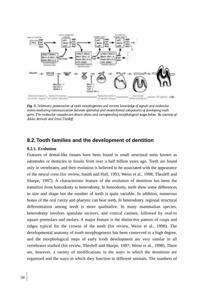
morphogenesis is in Figure 3.
1b216251taitto28.9 28.9.2001, 17:4933
34
8.2. Tooth families and the development of dentition
8.2.1. EvolutionFeatures of dental-like tissues have been found in small structural units known as
odontodes or denticles in fossils from over a half billion years ago. Teeth are found
only in vertebrates, and their evolution is believed to be associated with the appearance
of the neural crest (for review, Smith and Hall, 1993; Weiss et al., 1998; Thesleff and
Sharpe, 1997). A characteristic feature of the evolution of dentition has been the
transition from homodonty to heterodonty. In homodonty, teeth show some differences
in size and shape but the number of teeth is quite variable. In addition, numerous
bones of the oral cavity and pharynx can bear teeth. In heterodonty, regional structural
differentiation among teeth is more qualitative. In many mammalian species,
heterodonty involves spatulate incisors, and conical canines, followed by oval-to
square premolars and molars. A major feature is the distinctive pattern of cusps and
ridges typical for the crowns of the teeth (for review, Weiss et al., 1998). The
developmental anatomy of tooth morphogenesis has been conserved to a high degree,
and the morphological steps of early tooth development are very similar in all
vertebrates studied (for review, Thesleff and Sharpe, 1997; Weiss et al., 1998). There
are, however, a variety of modifications in the ways in which the dentitions are
organized and the ways in which they function in different animals. The numbers of
Fig. 3. Schematic presentation of tooth morphogenesis and current knowledge of signals and molecularevents mediating communication between epithelial and mesenchymal components of developing toothgerm. The molecular cascades are shown above and corresponding morphological stages below. By courtesy ofJukka Jernvall and Irma Thesleff.
1b216251taitto28.9 28.9.2001, 17:5034
35
teeth vary, and the teeth express differing forms with specialized functions (for
review, Thesleff and Sharpe, 1997). The dentition of modern man, 32 permanent
teeth (two incisors, one canine, two premolars, and three molars) is a result of changes
in tooth number during evolution relative to the ancestral dental formula of three
incisors, one canine, four premolars, and three molars (for review, Weiss et al., 1998).
8.2.2. Tooth familiesTeeth are grouped into families according to their specific locations in the jaws. In
mammals, the differences between tooth families correspond to the typical shape
categories, as incisors, canine, premolars, and molars in man. While shape differences
between tooth families are typical, the teeth in the same family resemble each other,
and differences are typically only quantitative. Each tooth group forms from one
epithelial thickening, the dental lamina, and development starts with the most anterior
tooth and proceeds posteriorly (for review, Thesleff and Sharpe, 1997).
While knowledge of the processes in the development of individual teeth has
expanded, much less is known about the control of dental patterning: the location,
number, and differing morphology of the teeth (for review, Weiss et al, 1998; Tucker
and Sharpe, 1999; Jernvall and Thesleff, 2000). The maxilla and mandible develop
differently, but it is suggested that both jaws may be patterned by a consistent process
that occurs before neural crest migration happens (for review, Weiss et al.,1998).
Two different theories have been suggested for the mechanisms of segmental
tooth patterning and regulation of shape differences between the teeth. The field
theory proposes that concentrations of chemical morphogens regulate the different
types of teeth (Butler, 1939; 1995). The clone theory suggests that stem cells giving
rise to different tooth families differ initially from each other (Osborn, 1978), and
that the neural crest cells have their positional identity before their final migration
into the region of tooth development.
Sharpe (1995) proposed that tooth shape and position are specified by combinatorial
activities of different homeobox genes expressed in neural crest-derived jaw
mesenchyme. For instance, several homeobox genes are expressed in specific spatial
patterns in the jaws before initiation of tooth development. These genes are often
expressed also during later stages of tooth development. Some findings in mutant
mice support this suggestion. The Dlx1/Dlx2 double-mutant knockout mouse lacks
maxillary molars, whereas development of mandibular molars and all incisors is
normal (Qiu et al., 1997). The single-mutant mouse has normal teeth. Barx1 is
expressed in the molar region but is almost completely absent from the incisor region
(Tissier-Seta et al., 1995).
1b216251taitto28.9 28.9.2001, 17:5035
36
8.2.3. Chronology of the development of human dentitionThe initiation of tooth development happens in 5- to 6-week-old human embryos.
Calcification begins during 14 to 18 weeks, and the crowns of all 20 primary teeth
are half-way mineralized at birth. Root formation is completed between 1.5 and 3
years (Koch and Thesleff, 2001). Central upper and lower incisors are the first
developing teeth, followed by the first molars, lateral incisors, canines, and second
molars (Pirinen and Thesleff, 1995). The lower incisor is the first tooth erupting, and
mean eruption time is close to 7 months of age. Other primary teeth erupt during the
following 20 to 22 months (Nyström, 1977; Pirinen and Thesleff, 1995).
Development of the permanent teeth also begins prenatally, when the dental
lamina proliferates lingually and distally to the germs of the primary teeth, giving
rise to 32 permanent tooth germs. The cusps of the first permanent molars have started
mineralization at birth. At 2 to 3 months of age, the lower incisors, upper central
incisors, and upper and lower canines start to calcify. Calcification of the upper lateral
incisors begins close to the first birthday. Between the second and third years of life,
the first and second premolars and second molars start to calcify. The crowns of the
permanent teeth (except third molars) are generally completed between 5 and 7 years
of age. Root development takes about 6 to 7 years. The third molars show a very
large variation in development. They start to mineralize between the eight and
eleventh year of life (Koch and Thesleff, 2001). However, sometimes the third molar
appear very late, at 14 to 18 years of age (Pirinen and Thesleff, 1995). A sex-difference
has been observed in tooth development, with girls are average half a year ahead of
boys.
The lower incisors or the first molars are the first permanent teeth to emerge,
with the mean age for their eruption being 6 to 7 years. The second premolars erupt
at the age of 11 to 12, and the second molars at 11.5 to 12.4, as last permanent teeth
excluding third molars. The mean age of eruption for third molars is around 21 years
in males and 23 in females (Pirinen and Thesleff, 1995).
8.3. Signaling networks in tooth developmentDevelopmentally, a tooth is a typical example of an epithelial-mesenchymal organ. A
complex molecular network operates during tooth formation. Studies of odontogenesis
at the molecular level, mostly using mouse teeth as models, have indicated that the
development of teeth is under strict genetic control which determines the position,
number, size, and shape of different teeth (for review, Thesleff and Nieminen, 1996;
Tucker et al., 1998; Tucker and Sharpe, 1999; Jernvall and Thesleff, 2000). More
than 200 genes are included at present in the graphic database illustrating the gene
1b216251taitto28.9 28.9.2001, 17:5036

37
expression patterns during tooth development (Nieminen et al., 2001). Tooth
development is initiated by signals from the epithelial dental lamina to the
mesenchyme, and thereafter the mesenchyme regulates epithelial morphogenesis.
Complex interactions between the epithelium and mesenchyme are responsible for
regulation during all stages of development. In the cap stage the enamel knot is
functioning as an important signaling center regulating tooth shape.
Figure 3. summarizes current knowledge of the signals and molecular events
mediating communication between epithelial and mesenchymal components of the
developing tooth germ.
8.3.1. SignalsSeveral different families of signal molecules, also called growth factors and their
receptors, function reciprocally between the epithelium and mesenchyme during
tooth development. The families most studied are the transforming growth factor β(TGFβ), which includes bone morphogenetic proteins (BMPs), the fibroblast growth
factor (FGF), the epidermal growth factor (EGF), and the hedgehog (Hh) and
wingless (Wnt) families (for review, Thesleff, 2000). One or more members of all of
these five families have been detected in developing teeth and also in other organs.
Enamel knots, transient clusters of dental epithelial cells, act as signaling centers
for cusp development (Jernvall, 1995). It has been proposed that the enamel knot
determines the site of the first cusp of a tooth and regulates the formation of other
cusps in the molars. There is an enamel knot also in the incisors during their cap
stage, and in the molars new enamel knots appear at the sites of new cusps (Jernvall,
1995). The primary enamel knot, at the tip of the epithelial bud, expresses many
signaling molecules, including Fgf4, Fgf3, Fgf9, Bmp2, 4, 7, Shh, Wnt 10a, and 10b,
and it is suggested to regulate the formation of epithelial folding in the cap stage.
The secondary enamel knots at the bell stage of tooth development, at the tip of
developing cusps, are suggested to determine the cusp position and size, resulting in
a functional tooth shape (Jernvall, 1995; Jernvall and Thesleff, 2000). Fgf4 is the
signal specifically located in the secondary enamel knots, but other signals like Shh
and Bmp4 are also expressed.
8.3.2 Transcription factorsTranscription factors are proteins that regulate the transcription of genes in the nucleus,
and are grouped according to those regions in the molecules that mediate their binding
to DNA (for review, Thesleff, 1998). Homeobox-containing transcription factors are
a specific group of transcription factors which have been called master regulatory
1b216251taitto28.9 28.9.2001, 17:5037
38
genes. They contain the homeobox, a 180-base-pair sequence (containing three α-helical
regions) which turns in the peptide chain between these regions (for review, Thesleff
1995; see also Rasko and Downes, 1995). The special feature of these homeobox-
containing genes is that they appear to act as position-specific regulators during
embryogenesis, and their own transcription is regulated according to their order in
the genome. Mutations in these genes cause cells to misunderstand their position and
to form organs normally emerging from other regions. A striking example of the
function of these genes has been shown in the fruit fly, Drosophila; mutation in the
gene Antennapedia causes fruitflies to grow legs on their heads, where antennae
ought to be. Homeotic genes were first detected in Drosophila. They are arranged in
clusters in both the Drosophila and mammalian genome. Antero-posterior patterning
of embryos also in mammals, including man, is regulated by a specific set of
homeobox-containing genes. Four clusters have been identified in the human
genome, each with numerous homeobox genes (Hox-clusters in chromosomes 2, 7,
12, and 17). Hox genes are closely similar in their sequence in different animals,
demonstrating their strong evolutionary conservation (for review, Thesleff, 1998).
No expression of Hox cluster genes has been shown anterior to the second branchial
arch, and thus they do not regulate face and tooth development. However, several other
homeobox-containing genes are important for craniofacial development and that of
different organs. Tooth development is regulated by many homeobox genes including
Msx1, Msx2, Dlx1, Dlx2, and Barx1, in addition to important transcription factors in
other groups. These include the paired box gene Pax9, and Lef1, a HMG-group
transcription factor (for review, Thesleff, 1998; Peters and Balling, 1999).
The homeobox genes MSX1 and MSX2, earlier called HOX7 and HOX8, are related
to the Drosophila muscle-segment homeobox gene (msh). Mouse Msx1 and Msx2 are
expressed in many tissues including teeth and bone during organogenesis and have
been shown to associate with epithelial-mesenchymal interactions, being targets of
Bmp and Fgf signaling (Satokata and Maas, 1994). The expression of Msx1 is very
intense in the dental mesenchyme throughout tooth morphogenesis (for review,
Thesleff, 1995). Msx2 is expressed both in mesenchymal and in epithelial cells
including the enamel knot, which suggest a role in cusp development (for review,
Tucker and Sharpe 1999; Thesleff, 2000). Mouse Msx2 is also present in and
adjacent to the calvarial sutures (Rice, 1999), and mutated MSX2 on chromosome 5
(5q34-35) causes the Boston-type craniosynostosis in man (Jabs et al., 1993).
Loss-of-function deficiency of Msx2 causes defects in skull ossification and persistent
calvarial foramen (Satokata et al., 2000). These mice also have defects in ectodermal
organs, including teeth with enamel defects.
1b216251taitto28.9 28.9.2001, 17:5038

39
PAX9 is a member of the Pax gene family characterized by the presence of the
paired domain which is a DNA-binding motif. Nine different PAX genes have been
isolated in man including PAX9 on chromosome 14 (14q12-q13). Pax genes function
as key regulators of organogenesis at various sites during embryonic development. It
is suggested that these Pax genes determine the time and place of organ initiation or
morphogenesis (for review, Dahl et al., 1997). Interestingly, expression of Pax9 has
been shown to specifically mark the mesenchyme at the prospective sites of all teeth
prior to any morphological manifestations (for review Peters and Balling, 1999).
9. TRANSGENIC MICE WITH TOOTH AGENESIS
Transgenic mice are intensively used for studies of mechanisms that regulate
development and as models of human diseases. In transgenic animals individual
genes can be misexpressed in defined tissues or their function can be disrupted.
Mutations in several different genes lead to arrested tooth development in mice. All
these genes participate in signaling networks in tooth formation (for review, Kettunen
1999; Thesleff, 2000).
Msx1 was the first gene shown to be essential for development of teeth in the
mouse, when Msx1-deficient mice were demonstrated to lack all teeth (Satokata and
Maas, 1994). Later, it was shown that tooth development does not proceed beyond
the bud stage in mice lacking transcription factors Lef1 or Pax9 (van Genderen et al.,
1994; Peters et al., 1998). Msx1 is expressed in the dental mesenchyme, and its
deletion results in inhibition of the expression of Bmp4 and Fgf3, which act as
reciprocal signals to the epithelium (for review, Thesleff, 2000). Recently, it has been
shown that adding BMP4 to cultures of Msx1 mutant tooth germs can restore their
development (Bei et al., 2000).
Arrested tooth development at the lamina stage is seen in mice with double mutants
of Msx1/Msx2, Dlx1/Dlx2 and Gli2/ Gli3 (Maas and Bei, 1997; Thomas et al., 1997;
Hardcastle et al., 1998). Inactivation of Pitx2 and p63 in mice causes total agenesis
of teeth, whereas heterozygous mice are normal (Lin et al., 1999; Mills et al., 1999).
In disruption of the gene encoding activin-binding protein follistatin, development
of incisors is inhibited, whereas molars reach the bell stage, but look abnormal
(Matzuk et al.,1995). Targeted inactivation of activinβA, normally expressed in
dental mesenchyme, causes failure of the development of incisors and mandibular
molars, but maxillary molars remain normal (Ferguson et al., 1998).
Some teeth are missing or their size is abnormal in the spontaneous mouse
mutant Tabby with its ectodysplasin deficiency (Srivastava et al, 1997; Mikkola et al.,
1b216251taitto28.9 28.9.2001, 17:5039
40
1999; Pispa et al., 1999). Ectodysplasin, a novel tumor necrosis factor (TNF), is one
protein required for epithelial morphogenesis. The mutant mouse downless (dl) has
an identical phenotype with the Tabby mouse. Cloning of the gene responsible for the
downless mouse revealed a novel TNF receptor, edar (Headon and Overbeek, 1999).
Both ectodysplasin and edar were recently shown to mediate interactions within the
epithelium (Laurikkala et al., 2001). Thus, identical phenotypes may be caused by
mutations in two genes in the same pathway.
10. METHODS FOR IDENTIFYING GENES BEHINDHUMAN DISEASES
If genetic factors are assumed to cause a human disease, there are, in principle, two
basic approaches - functional or positional cloning - for the identification of the
causative gene. Cloning can be performed by functional cloning, in which no
information as to the chromosomal location of the disease gene is required. In
functional cloning, identification of the gene is based on knowledge of the gene
product (Collins, 1995). This information may include the basic biochemical defect
of the disease, the amino acid sequence of the protein, or the possibility of using
information from an animal model of that disease. If a molecular candidate for the
gene mutated in a disease or the chromosomal location of the disease can be
proposed or an animal model can show possible candidate genes, a method can be
used called the candidate gene approach. In this approach, several polymorphic
markers either within the potential gene or in the flanking region are chosen for
analyses and tested for linkage with the disease in affected families. If only the
phenotype of the disease gene is known, the gene has to be sought on the basis of its
chromosomal location. This method is called positional cloning (Collins,1995). The
first step in positional cloning is assignment of the chromosomal locus, often by
linkage analysis.This analysis is based on the segregation of the markers and the
disease in families. When the chromosomal region has been identified, the genes of
this region are subjected to mutation detection by sequencing or other methods.
10.1. Mapping of a disease gene
10.1.1. General principlesGene mapping refers to the process where a causative gene is localized in the
genome by comparing the inheritance of a disease and genetic marker loci in
families. The first step is to examine a family sample where the disease of interest
1b216251taitto28.9 28.9.2001, 17:5040

41
segregates. The phenotypes of family members must be defined as precisely as possi-
ble and the mode of inheritance determined. The genotypes of family members are
determined with respect to genetic markers covering the human genome, and
segregation of the markers with the disease is analyzed. If two gene loci, a marker
and a disease, are close each other, they tend to segregate together. The closer they
are on the same chromosome, the greater the chance of their staying together at
meiosis, and co-segregation happens more often than expected by chance. Evidence
for linkage is tested by different statistical methods.
10.1.2. Linkage analysisLinkage analysis is the method most often used in mapping studies. In linkage analysis,
the co-segregation of a disease and markers in a family are followed and measured.
Linkage analysis relies on the ability to detect recombination events between the
disease and the marker locus during meiosis. Traditionally, linkage analysis has been
used for studying Mendelian traits, but at present it is used also in complex diseases
(Öhman, 2001). If two loci are close to each other on the same chromosome, the
chance of recombination between them at meiosis is low, and they are said to be linked.
Statistical analysis is necessary to calculate the probability of linkage in a pedigree.
The lod score method is based on calculating the overall likelihood of two alternative
assumptions: that the two loci are linked at the given recombination fraction, L(θ), or
are not linked but are inherited independently, L(1/2). In this case a recombination is
seen in 50% of meiosis. The probability of linkage is expressed as a lod score,
logarithm (to the base 10) of the odds ratio (Morton, 1955; Ott, 1974). The lod score is
estimated at different θ values, usually ranging from 0 to 0.4. A lod score of 3 (odds
ratio of 1000:1) or greater is regarded as significant, and evidence for linkage at a given
recombination fraction is assumed. A lod score less than –2, on the other hand,
indicates exclusion of linkage. A single pedigree does not, however, usually contain
enough information to reach a decisive score. It is therefore necessary to combine data
from several pedigrees. A lod score can be summed from several families to give a final
lod score. More statistical power can be obtained from multipoint analysis, which
utilizes segregation of haplotypes of several markers. In linkage analysis, the
specification of inheritance model, allele frequencies, and penetrance values increases
the power of the analysis and may eliminate false-positive results. Probability methods
and computer programs have been developed to make it possible to collect and count
data from different families and from many marker loci. The most commonly used
program package for linkage analysis has been LINKAGE (Lathrop and Lalouel, 1984;
Lathrop et al., 1984, 1986). Several new algorithms and programs have been developed
1b216251taitto28.9 28.9.2001, 17:5041
42
to overcome limitations of computer power and to provide simulation and non-
parametric methods.
10.1.3. Genetic markers and the polymerase chain reaction (PCR)Second-generation markers, the microsatellites or short tandem repeat markers
(STR), have aided considerably in the mapping of genes by linkage analysis
(Miesfeld et al., 1981). The advantage of microsatellites is their high rate of
polymorphism, as well as their dense distribution throughout the genome.
Microsatellites and the method called the polymerase chain reaction (PCR) have
led to the present technique of detecting length polymorphisms in microsatellite
regions. With PCR, alleles of a microsatellite are amplified a million-fold, and the
products are separated by polyacrylamide gel electrophoresis (Saiki et al., 1988;
Weber and May, 1989). Recently, fluorescence-based semiautomated genotyping of
the marker loci has become more popular in genetic mapping (Reed et. al., 1994).
The Human Genome Project, established in the 1980s, is an international project
established to acquire fundamental information on human genetics and the role of
various genes important in development and diseases (Strachan and Read, 2000).
This system has created numerous microsatellite maps with thousands of markers
from all chromosomes (Weissenbach et al., 1992; Guapay et al., 1994; Murray et al.,
1994; Dib et al., 1996). The final task has been to obtain the ultimate physical map,
the complete sequence of the human genome. This year, 2001, the results of genome
sequencing cover about 94% of the human genome. There appear to be from 30 000
to 40 000 protein-coding genes in the human genome – only about twice as many as
in the fruit fly. However, these genes are more complex, with more alternative splic-
ing generating a large number of protein products (Lander et al., 2001). The project
interacts with research on mapping and identifying human disease genes and
studying genetic variation (Strachan and Read, 2000). Data on mapping and sequencing
are entered into large electronic databases freely accessible through the Internet.
1b216251taitto28.9 28.9.2001, 17:5042

43
AIMS OF THE PRESENT STUDY
The aim of the present study was to investigate the phenotypic and genotypic features
of familial tooth agenesis. Families of eleven probands with the common mild form
of incisor-premolar hypodontia were studied. In addition, mutation analysis of the
PAX9 gene was carried out in two families with oligodontia.
The specific aims were:
- to analyze the inheritance and phenotype of familial hypodontia
- to study the association of dental anomalies with hypodontia
- to search by linkage analysis for the genetic locus causing hypodontia
- to find a putative mutation in the PAX9 gene in two families with oligodontia
1b216251taitto28.9 28.9.2001, 17:5043
44
SUBJECTS AND METHODS
1. SUBJECTS
1.1. Families with hypodontia (I, II, III)The criterion for selecting persons with hypodontia (probands) was the congenital
absence of one to six permanent teeth or the presence of peg-shaped permanent upper
lateral incisors. The probands were nonsyndromic patients at the Department of
Pedodontics and Orthodontics, Institute of Dentistry, University of Helsinki. The
probands and/or their parents were interviewed about occurrence of hypodontia in
their families. Families with more than one individual with hypodontia were first
contacted to learn about the aims of the study. All family members voluntarily
participating in the study were invited for an examination. The study was approved
by the Ethics Committee of the Institute of Dentistry, University of Helsinki.
The study population comprised 11 probands with hypodontia, four males and
seven females (aged 10-36 years), and their family members, totaling 214 individuals
in 11 Finnish families (I). The spouses of the family members were also interviewed
and examined. Information concerning general health was obtained by interview. A
clinical dental examination of the probands and their family members was performed
by three orthodontists (S.A., S.P., I.T.). In addition to hypodontia, other visible dental
abnormalities were recorded. Panoramic radiographs were taken if this was necessary
for the diagnosis of hypodontia. In some cases, information on hypodontia was
confirmed from documents and radiographs in dental files.
Pedigrees of the families were constructed, and individuals with congenitally
missing teeth or peg-shaped/strongly mesio-distally reduced upper lateral incisor(s)
were recorded as affected (Figure 4). The prevalences of different dental anomalies
were calculated separately for family members who were first- and second-degree
relatives, first cousins, and more remotely related relatives of the probands. In
addition, obligate carriers of the hypodontia gene who did not display tooth agenesis
or peg-shaped teeth were identified from the pedigrees and analyzed separately for
dental features.
The clinical study (I) included the whole sample of 11 families. The molecular
genetic exclusion studies (II, III) included 5 (II) and 7 (III) families. In a genome-wide
scan, a sample of 4 families was chosen, including families 1, 2, 6, and 9. Simulation
with the Slink program (Ott, 1989; Weeks et al., 1990) confirmed that, assuming
genetic homogeneity, the families were sufficiently informative to reveal significant
evidence for linkage. Family 10 was excluded from the linkage studies because both
1b216251taitto28.9 28.9.2001, 17:5044
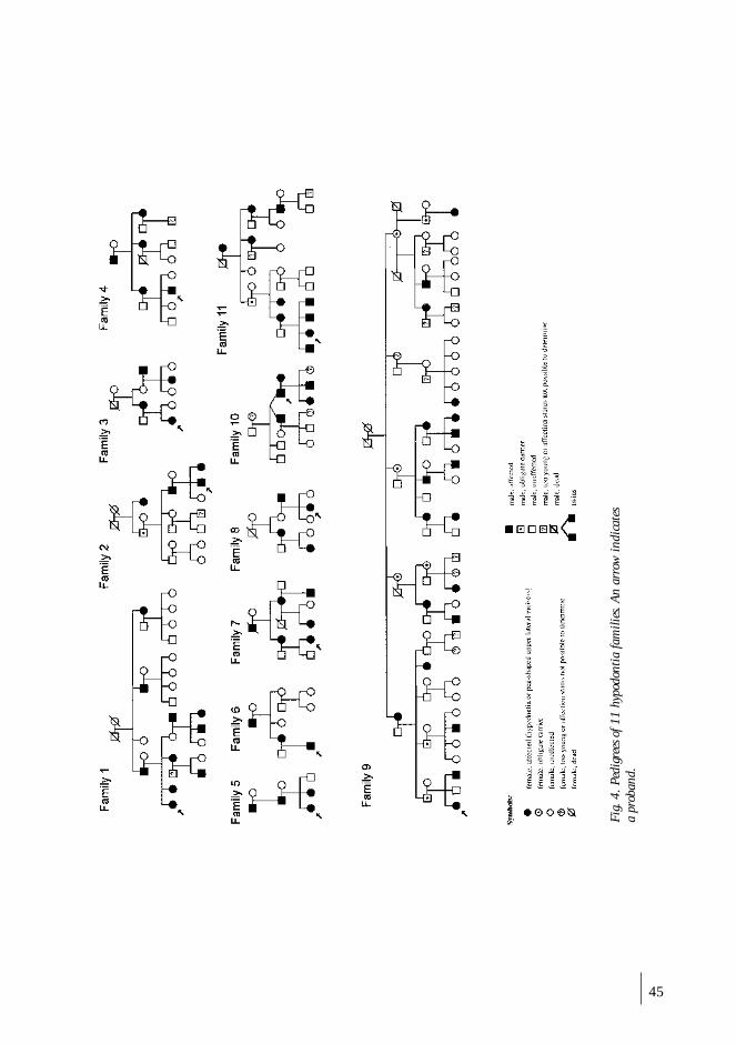
45
Fig.
4. P
edig
rees
of 1
1 hy
podo
ntia
fam
ilies
. An
arro
w in
dica
tes
a pr
oban
d.
1b216251taitto28.9 28.9.2001, 17:5045
46
the mother and the father in the second generation had hypodontia, and family 11
because the proband had a cleft of the soft palate. Families 1 and 2, as well as 3 and
8 overlap, but they have been separated into smaller families in the linkage studies.
1.2. Families with oligodontia (IV)We studied the association of the PAX9 gene mutation with oligodontia in 2 two-
generation families with 3 and 2 affected individuals, respectively (IV). The proband
of family 1 was originally referred for treatment to the Pediatric Dental Department
of the Hospital for Children and Adolescents, Helsinki University Central Hospital.
The proband of family 2 was referred for consultation and treatment to the Department
of Pedodontics and Orthodontics, Institute of Dentistry, University of Helsinki.
The families were informed of the genetic study, and family members consented
to participate. The study was approved by the Ethics Committee of the Institute of
Dentistry, University of Helsinki.
2. METHODS
2.1. Diagnosis of congenitally missing teeth
2.1.1. Studies I, II, and IIIBoth a clinical and a radiographic examination were performed for diagnosing
hypodontia. The third molars were excluded because reliable retrospective data on
them was incomplete. The presence of peg-shaped or strongly mesio-distally reduced
upper lateral incisor(s) was also diagnosed as hypodontia. In addition, retrospective
dental data were collected by questions, and from dental files. Data included the
recording of missing teeth, extractions of teeth, ectopic teeth, orthodontic treatment,
and hypodontia of primary dentition. Children under 6 (N=10) were excluded from
the study because hypodontia cannot be reliably diagnosed at a young age.
2.1.2. Study IVThe diagnosis of missing teeth was based on both clinical and radiographic examinations.
Retrospective dental information was collected from dental files. The youngest child
was 6 years old. Agenesis of third molars was not possible to diagnose in the proband
and in two brothers of the proband in family 1 because of their young ages.
2.2. Penetrance (I)The penetrance of hypodontia in the pedigrees was calculated with the maximum
likelihood method as described by Mather (1957).
1b216251taitto28.9 28.9.2001, 17:5046

47
2.3. Dental age (I)Dental age was assessed from the developing dentition as seen in the panoramic
radiographs of 6 probands (3 females, 3 males) and 32 relatives (17 females, 15
males) by a computer-aided method (Software Publishing Corporation, Mountain
View, CA, USA) with norms corrected for Finnish children (Kataja et al., 1989). If
the index tooth was missing, a contralateral tooth was used. In 3 children, the second
premolar was missing bilaterally, and the value of one stage lower than for the first
premolar was used.
2.4. Invaginations (I)Invaginations in permanent upper lateral incisors were recorded clinically or from
panoramic radiographs. The lingual pit (foramen cecum), fillings at the location for
invagination, and teeth which showed radiographically distinct enamel notching were
recorded as invaginations. The study population was comprised 9 probands and 98
relatives. Individuals with missing upper lateral incisors were excluded. Of the
relatives, 33 were first- and 15 second-degree relatives, 16 were cousins, and 34 were
more remote relatives.
2.5. Taurodontism (I)Taurodontism of the first and second permanent molars was determined from
panoramic radiographs of 11 probands and 96 family members, the latter composed
of 31 first- and 13 second-degree relatives, 24 first cousins, and 28 more remotely
related individuals. Persons with large restorations or extractions of molars were
excluded from the study. The determination was made only on teeth with at least half
of the root developed. Pyramidal or single-rooted molars were excluded. The method
was that described by Shifman and Chanannel (1978), and modified by Tulensalo et
al. (1989). Three vertical measurements were made directly in the panoramic
radiographs with a ruler by one of the authors (S.A.). Measurements were recorded
to the nearest 0.5 mm. Measure 1 was the vertical height of the pulp chamber: the
distance between the lowest point of the roof of the pulp chamber and the highest
point of the floor of the pulp chamber. Measure 2 was the distance between the
lowest point of the roof of the pulp chamber and the apex of the longest root. These
measurements were used to calculate the taurodontic index (TI): TI = (measure1/
measure 2) x 100. Measure 3 was the distance between the baseline connecting the
two cementoenamel junctions and the highest point of the floor of the pulp chamber
(Blumberg et al., 1971).
1b216251taitto28.9 28.9.2001, 17:5047
48
Taurodontism was diagnosed in those molars in which the TI was above 20 or the
variable 3 exceeded 3.5 mm. The value of measure 3 was used in teeth with
incomplete development of the roots. All measurements were performed by the same
author (S.A.) and recorded to the nearest 0.5 mm.
2.6. Reliability of measurementsTo compare assessment reliability for taurodontism, 144 molars in 20 radiographs
were measured twice with a 2-week interval. One molar, 1/144 (0.7%), was classified
differently in the two measurements, leading to a different diagnosis of taurodontism
in 1 individual (1/20) and to an intraexaminer reliability of 95%.
2.7. ControlsWe used the population prevalences of dental anomalies in the literature as controls
(I). In addition, 2 distinct control groups were used (Table 3). The frequency of
invagination(s) in the upper lateral incisor(s) of the study group was compared with
that of an unselected control sample which comprised 170 children with late mixed
or early permanent dentition attending an annual dental examination (91 females, 79
males, age-range 12-14 years). All were clinically examined (S.A.) at the Department
of Pedodontics and Orthodontics.
The control group for taurodontism included 841 panoramic radiographs (389
females, 452 males, age-range 6-21 years), the sample was collected from 1965 to
1970 (Haavikko, 1971). The anomalies were analyzed separately in controls with and
without hypodontia.
1b216251taitto28.9 28.9.2001, 17:5048

49
2.8. DNA analysis
2.8.1. DNA extraction (II, III, IV)Venous blood samples, 10 to 20 ml, were taken for DNA analyses from all available
family members. DNA was extracted from leukocytes according to standard procedures
(Vandenplas et al., 1984).
2.8.2. GenotypingThe polymerase chain reaction (PCR) method with amplifiable microsatellite markers
was used for assessing the genotypes of the individuals (II, III). The second stage of
linkage studies used a genome-wide search with a selection of microsatellites having a
maximum genetic distance of 15cM for genotyping the members of four families.
PCR primers were obtained from Genethon, France, or from the Department of
Clinical Genetics, University Hospital of Uppsala, Sweden, or they were synthesized
in the Laboratory of Human Molecular Genetics of the National Public Health
Institute, Finland. In addition, primers for the MSX2 microsatellite were a gift from
Dr Ethylin Wang Jabs (Johns Hopkins University School of Medicine, MD, USA).
Microsatellite polymorphism was analyzed by radioactive or fluorescence-based
detection methods. When radioactive detection of PCR products was used, one of the
primers was 5‘- end–labeled with 32P-phosphate with T4 kinase (Pharmacia, Sweden)
in a buffer provided by the manufacturer of the enzyme, containing 0.3 µC of µ-32P-
ATP (Amersham, UK). PCR reactions were done in a Perkin-Elmer Cetus Thermal
Cycler 480 (Norwalk, CT, USA) or DNA Engine (MJ Research, MA, USA) in a total
volume of 13µL containing 12ng of chromosomal DNA, 3pmol of each of the
primers, 3 nmol of each of the deoxynucleotides, 0.25 U of enzyme, and 1.5nM
MgCl2 in a buffer described by Weissenbach et al. (1992). Either Taq-polymerase
(Amplitaq, Perkin Elmer Cetus, Ca) or Dynazyme II (Finnzymes, Finland) were
used. PCR products were made 50% of formamide and 10 mM of EDTA, pH 8.0, and
separated in a 5% sequencing gel with 7.5 M urea in an IBI gel apparatus or in an
ABI 377 sequencer (Perkin Elmer Biosystems, CA). 32P-labeled alleles were detected
by autoradiography with Kodak films exposed for 15 to 20 hours and analyzed m
anually. Fluorescent alleles were detected with Genescan software and identified
with Genotyper software (Perkin Elmer Biosystems).
2.8.3. Genetic mapsFor the genome-wide scan of incisor-premolar hypodontia locus, markers were
selected to provide a density of at least one marker per 15cM interval from published
genetic maps of Genethon (Dib et al., 1996; ftp://. Genethon.fr.pub/Gmap/Nature-
1995/), Cooperative Human Linkage Center (CHLC; Murray et al., 1994; http://
1b216251taitto28.9 28.9.2001, 17:5049

50
lpg.nci.nih.gov/CHLC/), and Marshfield Medical Research Foundation (http://
research.marshfieldclinic.org/genetics/). In the multipoint analyses, genetic locations
were based primarily on the Marshfield maps.
2.8.4. Sequencing of the PAX9 geneFour sets of primers were used to amplify the coding region of the PAX9 gene (IV).
Amplification was performed in 100µl with DNA concentrations of 1.5ng/µl, 1.5mM
MgCl2 and 1 mU DynazymeTM EXT polymerase (Finnzymes). The primers and PCR
conditions were as follows, with numbering indicating the position of the first base
of the primer in the coding sequence: exon 2, forward primer 1 AGGCAGCTGTC-
CCAAGCAGCG (exon start–58), reverse primer 1 GGAGGGCACATTGTACTT-
GTCGC (357), annealing T 64°C, 32 cycles; exon 2, forward primer 2 ATCCGAC-
CGTGTGACATCAGCC (109), reverse primer 2 GAGCCCCTACCTTGGTCGGTG
(exon end +10), annealing T 64°C, 30 cycles; exon 3, forward primer GGGAG-
TAAAACTTCACCAGGC (exon start –197), reverse primer CCACCTGGCCTGAC-
CCTC (exon end +28), annealing T 61°C, 32 cycles; exon 4, forward primer GGA-
GAGTAGAGTCAGAGCATTGCTG (exon start –121), reverse primer GAGACCT-
GGGAATTGGGGGA (stop +74), annealing T 61°C, 32 cycles. Primers for exons 2
and 4 were adapted from Stockton et al (2000). PCR products were purified by
agarose gel electrophoresis and the Qiaquick gel extraction kit (Qiagen, Bothell, WA,
USA). Both strands of the PCR products were sequenced with BigDye terminator
chemistry and analyzed on an ABI 377XXL DNA sequencer (Applied Biosystems,
Foster City, CA, USA).
2.9. StatisticsThe significance of differences in prevalence of taurodontism and rotation of
premolar(s) was tested in the study groups and in controls with the Chi-square test (I).
In the studies (II, III), the lod score method served for evaluation of the existence
of linkage (Ott, 1974). The lod score was obtained by calculating the overall
likelihood of the observed data with two alternative assumptions: that the loci are
linked at a given genetic distance (recombination fraction θ) or that they are not
linked. The logarithm to the base 10 of the ratio of these two likelihoods (the odds
ratio) is the lod score. It has been agreed that a lod score exceeding 3 should be taken
as statistically significant proof for linkage, and a lod score less than –2 indicates the
exclusion of linkage.
The hypodontia locus was modeled as an autosomal dominant, two-allele system
with a gene frequency of the hypodontia allele of 8%. A penetrance of hypodontia of
1b216251taitto28.9 28.9.2001, 17:5050

51
86% was used (Burzynski and Escobar, 1983), as this value was in good agreement
with our family data. The informativeness of the family material was evaluated with
the Slink software package, assuming an autosomal dominant model and gene
homogeneity (Ott, 1989; Weeks et al., 1990). Allele frequencies for all markers were
calculated from all known genotypes. The pairwise lod scores for different
recombination fractions were estimated by the Mlink and Ilink options of the
Linkage package, version 5.1 (Lathrop et al., 1984). In affecteds-only analyses, the
phenotypes of the unaffected individuals were coded as unknowns. The Homog
program (Ott, 1991) was used to estimate the lod scores under the assumption of
non-allelic gene heterogeneity.
1b216251taitto28.9 28.9.2001, 17:5051
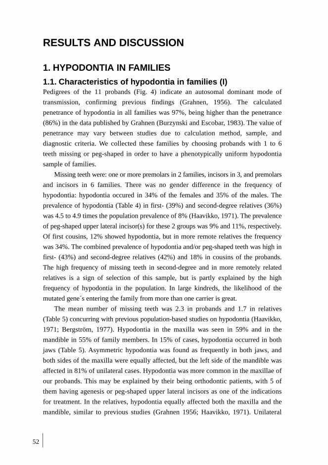
52
RESULTS AND DISCUSSION
1. HYPODONTIA IN FAMILIES
1.1. Characteristics of hypodontia in families (I)Pedigrees of the 11 probands (Fig. 4) indicate an autosomal dominant mode of
transmission, confirming previous findings (Grahnen, 1956). The calculated
penetrance of hypodontia in all families was 97%, being higher than the penetrance
(86%) in the data published by Grahnen (Burzynski and Escobar, 1983). The value of
penetrance may vary between studies due to calculation method, sample, and
diagnostic criteria. We collected these families by choosing probands with 1 to 6
teeth missing or peg-shaped in order to have a phenotypically uniform hypodontia
sample of families.
Missing teeth were: one or more premolars in 2 families, incisors in 3, and premolars
and incisors in 6 families. There was no gender difference in the frequency of
hypodontia: hypodontia occured in 34% of the females and 35% of the males. The
prevalence of hypodontia (Table 4) in first- (39%) and second-degree relatives (36%)
was 4.5 to 4.9 times the population prevalence of 8% (Haavikko, 1971). The prevalence
of peg-shaped upper lateral incisor(s) for these 2 groups was 9% and 11%, respectively.
Of first cousins, 12% showed hypodontia, but in more remote relatives the frequency
was 34%. The combined prevalence of hypodontia and/or peg-shaped teeth was high in
first- (43%) and second-degree relatives (42%) and 18% in cousins of the probands.
The high frequency of missing teeth in second-degree and in more remotely related
relatives is a sign of selection of this sample, but is partly explained by the high
frequency of hypodontia in the population. In large kindreds, the likelihood of the
mutated gene´s entering the family from more than one carrier is great.
The mean number of missing teeth was 2.3 in probands and 1.7 in relatives
(Table 5) concurring with previous population-based studies on hypodontia (Haavikko,
1971; Bergström, 1977). Hypodontia in the maxilla was seen in 59% and in the
mandible in 55% of family members. In 15% of cases, hypodontia occurred in both
jaws (Table 5). Asymmetric hypodontia was found as frequently in both jaws, and
both sides of the maxilla were equally affected, but the left side of the mandible was
affected in 81% of unilateral cases. Hypodontia was more common in the maxillae of
our probands. This may be explained by their being orthodontic patients, with 5 of
them having agenesis or peg-shaped upper lateral incisors as one of the indications
for treatment. In the relatives, hypodontia equally affected both the maxilla and the
mandible, similar to previous studies (Grahnen 1956; Haavikko, 1971). Unilateral
1b216251taitto28.9 28.9.2001, 17:5052

53
1b216251taitto28.9 28.9.2001, 17:5053
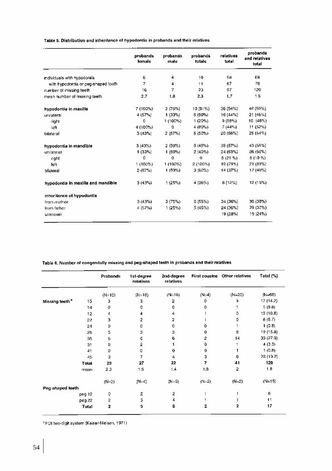
54
1b216251taitto28.9 28.9.2001, 17:5054
55
hypodontia was registered more often in the left side of the mandible, consistent with
results of previous Scandinavian investigations (Grahnen, 1956; Haavikko, 1971; Wisth
et al., 1974; Bergström, 1977), whereas no difference between the right and left
quadrants has been observed in some studies (Magnusson, 1977; Lai and Seow, 1989).
The teeth most frequently missing (N=120) were lower second premolars (47%),
upper second premolars (30%), and upper lateral incisors (17.5%). Further, lower
central incisors and upper first premolars were 4.2% and 1.6% of the missing teeth
(Table 6). The majority of studies have shown that the mandibular second premolar is
the tooth most commonly missing, but some variability is present in the order of the
other teeth. Similar missing teeth and nearly the same frequencies have been reported
in a previous study on Finnish children (Haavikko, 1971), with the exception of a
higher frequency of agenesis of upper first premolars (3.5%) and a lower frequency
of lower central incisors (2.7%) in that study.
Hypodontia of the primary dentition was neither found or reported, but the
youngest persons studied were 7-year-olds. Of 11 probands, 6 had inherited
hypodontia from their mothers and 4 from their fathers. One father without hypodontia
or peg-shaped teeth was identified as an obligate carrier. Moreover, hypodontia had
an equal maternal (36%) and paternal inheritance (36%) among relatives (N=67).
The inheritance pattern remained undetermined for 19 (28%) relatives (Table 5). The
inheritance pattern is in agreement with an autosomal dominant trait.
The mean dental age of the probands and their relatives with developing
dentition (N=38) showed a 0.3-year advance compared with population standards.
The mean dental age of the probands (N=6) was 0.2 years ahead, but the relatives
with hypodontia (N=10) showed a minus 0.2- year delay. The dental age of 2 boys
with hypodontia fell between the –1SD and –2SD percentiles.
1.2. Other dental features in probands (I)Other dental anomalies of the probands are shown in Table 7. Ectopic upper permanent
canines were seen in 2 (18%) probands, one palatally and the other labially
displaced. This prevalence is 9-10 times that of the normal population (Thilander and
Jakobsson, 1968).
The frequency of rotated premolars (18%) exceeds by 3.5 times the population
prevalence reported in an Italian study (Baccetti, 1998). Invaginations in upper lateral
incisors were noted less frequently in the probands (22%) than in the total population
control sample of 170 children (42%). Interestingly, the frequency of invaginations
was also lower among hypodontia patients of the control sample.
1b216251taitto28.9 28.9.2001, 17:5055

56
1b216251taitto28.9 28.9.2001, 17:5056
57
Taurodontism was a common anomaly in the molars of our probands; 7 probands
(64%) had taurodontism of at least one molar. This is nearly 3 times the prevalence
of taurodontism in the total control sample and is statistically significant (p=0.003).
One female proband had been operated on for a cleft of the soft palate. Small
dimensions of the lower first premolars and one upper second premolar were noted in
a male proband with hypodontia of 3 second premolars. The affected mother of this
proband had small lower first premolars, and an affected aunt had a small upper
second molar.
1.3. Other dental features in first- and second-degreerelatives (I)
Dental anomalies are shown in Table 7. Ectopic upper permanent canines were seen
in 3 (7%) first-degree relatives (N=46), all of these individuals with hypodontia. This
is 3.5 times the population prevalence. Two of these ectopic canines were palatally
and one labially displaced. No ectopic canines were noted in second-degree relatives
(N=45).
The frequency of rotated premolars in first- (11%) and second-degree relatives
(7%) exceeded by 2 to 3 times that of the population (3-5%) (Baccetti, 1998). As in
probands, invaginations were seen less frequently in first- and second-degree
relatives with hypodontia than among unselected controls.
The frequency of taurodontism (26%) in first-degree relatives (N=31) was similar
to that in the control sample (22%), but never found in second-degree relatives
(N=13). Taurodontism was assessed more frequently in upper molars, with second
molars affected more often than first molars. Most taurodontic molars showed
hypotaurodontism, with hypertaurodontism not observed.
In addition to the aforementioned family 4 with small premolar dimensions,
small premolars were seen in 4 females: a cousin (7), a relative with hypodontia (9),
an aunt (6), and a relative without hypodontia (11).
An unaffected sister of one proband (8) had fourth molars in the maxilla. A
brother of a female proband with missing second premolars in the left side of the
jaws (11) had had fused primary teeth in the region of the upper lateral incisor.
1.4. Other dental features in first cousins and more remotelyrelated individuals (I)
One (3%) of the cousins without hypodontia had a palatally displaced canine. A
labial canine was noted in another remote relative without hypodontia (2%), which
corresponds to the population prevalence (Table 7).
1b216251taitto28.9 28.9.2001, 17:5057
58
Rotated premolars were noted in 2 first cousins (6%), one with hypodontia and
the other without, which is similar to the population prevalence of 3 to 5%. The
frequency of rotated premolars in remote relatives (12%) was 2-4 times the population
prevalence. Rotated premolars were noted more frequently in individuals with
hypodontia both among first cousins and more remote relatives.
The frequency of invagination(s) in upper lateral incisor(s) was 25% in cousins
(N=16) and 35% in more remote relatives (N=34). No differences were present
between individuals with and without hypodontia.
Taurodontism was assessed in 10 of 24 cousins(42%), which is 1.9 times the
control prevalence. Cousins with hypodontia had a taurodontism frequency of 83%,
which differed significantly from the population control prevalence. The frequency of
taurodontism in the more remote relatives corresponded with that of the controls.
1.5. Dentition in obligate carriers (I)Nine obligate carriers were identified from the pedigrees, with 3 males and 4 females
representing nonpenetrance of familial hypodontia. Three of these showed no dental
anomalies. The dental status of 2 carriers was unavailable due to extraction of most
of their teeth.
Four carriers had anomalies: 1 male had a narrow upper lateral incisor, and 2
males had rotated premolars. One female also had a small upper lateral incisor and
had undergone orthodontic treatment for labially displaced canines.
2. OLIGODONTIA IN FAMILIES (IV)
Clinical evaluation of family members in the oligodontia study revealed no
significant medical problems except for the absence of several teeth in 3 individuals
in family 1 and 2 in family 2 (Table 8 and Fig 5). The rest of the family members had
normal dentition.
The affected individuals in family 1 lacked all second and third permanent
molars as well as both maxillary lateral incisors. The proband and his 2 brothers were
too young for definitive diagnosis of the absence of their third molars. This proband
and his mother also lacked all first permanent molars and several second premolars.
All second molars were missing in the proband‘s primary dentition. The affected
brother had all primary teeth, but the second primary molars were submerged. The
mother and the affected brother had a malposition of the upper permanent canine. In
addition, some permanent teeth appeared smaller than normal in affected patients.
The oligodontia phenotype was even more severe in family 2. The proband
lacked 8 permanent molars, and 16 other permanent teeth. One of her 4 molars
1b216251taitto28.9 28.9.2001, 17:5058
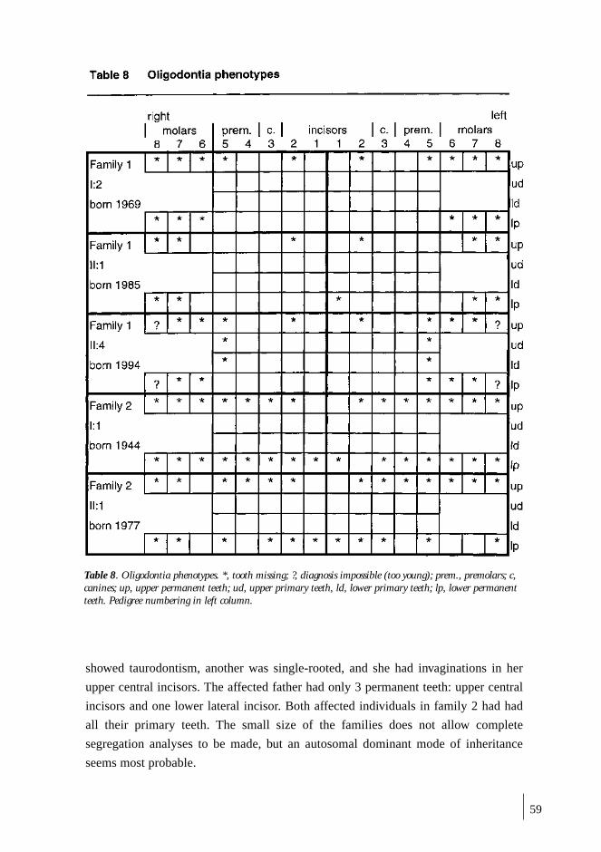
59
showed taurodontism, another was single-rooted, and she had invaginations in her
upper central incisors. The affected father had only 3 permanent teeth: upper central
incisors and one lower lateral incisor. Both affected individuals in family 2 had had
all their primary teeth. The small size of the families does not allow complete
segregation analyses to be made, but an autosomal dominant mode of inheritance
seems most probable.
Table 8. Oligodontia phenotypes. *, tooth missing; ?, diagnosis impossible (too young); prem., premolars; c,canines; up, upper permanent teeth; ud, upper primary teeth, ld, lower primary teeth; lp, lower permanentteeth. Pedigree numbering in left column.
1b216251taitto28.9 28.9.2001, 17:5059

60
3. LINKAGE STUDIES (II, III)
Before starting a genome-wide seach for hypodontia locus we studied linkage of
hypodontia with some selected candidate genes. The finding that tooth development
is inhibited in Msx1-negative transgenic mice directly implicates this gene in tooth
development (Satokata and Maas, 1994). MSX1 and the closely related MSX2 are
homeobox-containing transcription factors, and mutation in the human MSX2 gene
had been identified as the gene causing the Boston-type familial craniosynostosis
(Jabs et al., 1993). At the time of this study, MSX1 had not yet been identified as the
cause of oligodontia.
Epidermal growth factor (EGF) has been implicated in early tooth morphogenesis,
since tooth development is inhibited in vitro with antisense oligonucleotides to EGF
Fig. 5. a. Panoramic radiograph of the proband of Family 1 (II:4) at age 6. Missing teeth depicted witharrows. b. Pedigrees of oligodontia families 1 and 2. Squares, males; circles, females; darkened, affected;arrows, probands.
1b216251taitto28.9 28.9.2001, 17:5060
61
mRNA (Kronmiller et al., 1991). EGF exerts its action on cells via binding to the
specific cell-surface receptor, EGFR. This receptor is also used by other growth
factors in the EGF family, including TGFα. EGFR is expressed in developing teeth
from the bud stage onward, and the pattern is developmentally regulated, suggesting
involvement in the regulation of tooth development.
Roles for FGF-3 in tooth development have been suggested, since it is intensely
expressed in dental mesenchyme during the cap and bell stages (Wilkinson et al.,
1989). Although no defects in dentition are reported in FGF-3 knockout mice
(Mansour et al., 1993), the possibility still exists that a gain-of–function mutation in
the FGF-3 may affect tooth development. It is also possible that this gene defect
causes hypodontia in human dentition without having any effect on mouse dentition.
FGF-4 is expressed in structures called enamel knots in the dental epithelium.
Experimental analysis has indicated that, in dental tissues, FGFs greatly stimulate
cell proliferation and it has been speculated that the function of FGF-4 is to regulate
development of tooth form (Jernvall et al., 1995).
We found evidence against linkage of incisor and premolar hypodontia with MSX1
and MSX2 (II) as well as with EGF, EGFR, and FGF-3 genes (III). The linkage of
MSX1 and MSX2 was studied in 5 families. Both microsatellites were intragenic, i.e.,
they resided in the immediate vicinity of the coding regions. One of the microsatellites
is located in the 3´-flanking region of the MSX1 gene in 4p16 (Padanilam et al.,
1992), with the other lying in the intron of the MSX2 gene in 5q34-35 (Jabs et al.,
1993). Pairwise analyses of the allelic data resulted in negative lod scores of less than
–2 with recombination fractions 0.0 and 0.01. We used the polymorphic microsatellites
in EGF, EGFR, and FGF-3. The tetranucleotide repeat polymorphism within the
gene for EGF in chromosome 4 (4q25-27) resides in the intron 10 of the gene
(Murray et al., 1992) and the dinucleotide repeat polymorphism within the gene for
EGFR (7p12-14) in the first intron (Chi et al., 1992). In the FGF-3 gene in 11q13.3,
there is a dinucleotide repeat polymorphism in the immediate 3´-flanking region
(Polymeropoulos et al., 1990). The FGF-3 and FGF-4 genes are located as a cluster,
and the interval between the genes is less than 35kb (Brookes et al., 1992). The
genotypes of these 3 marker loci in 7 Finnish families with hypodontia were
determined, and the lod scores with hypodontia calculated (III). The lod scores
obtained with recombination fractions 0.00 and 0.01 were less than –2 in the case of
the EGF and FGF-3 microsatellites. According to convention, assuming homogeneity,
this result would exclude linkage of incisor-premolar hypodontia with these genes.
During the genome scan (see below) additional families and also neighboring markers
1b216251taitto28.9 28.9.2001, 17:5061
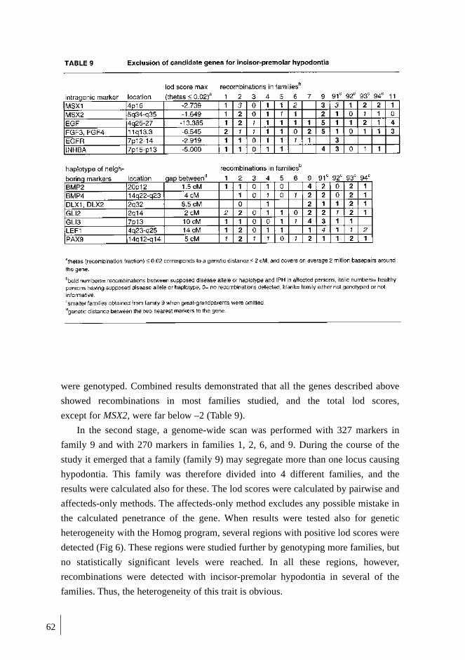
62
were genotyped. Combined results demonstrated that all the genes described above
showed recombinations in most families studied, and the total lod scores,
except for MSX2, were far below –2 (Table 9).
In the second stage, a genome-wide scan was performed with 327 markers in
family 9 and with 270 markers in families 1, 2, 6, and 9. During the course of the
study it emerged that a family (family 9) may segregate more than one locus causing
hypodontia. This family was therefore divided into 4 different families, and the
results were calculated also for these. The lod scores were calculated by pairwise and
affecteds-only methods. The affecteds-only method excludes any possible mistake in
the calculated penetrance of the gene. When results were tested also for genetic
heterogeneity with the Homog program, several regions with positive lod scores were
detected (Fig 6). These regions were studied further by genotyping more families, but
no statistically significant levels were reached. In all these regions, however,
recombinations were detected with incisor-premolar hypodontia in several of the
families. Thus, the heterogeneity of this trait is obvious.
1b216251taitto28.9 28.9.2001, 17:5062
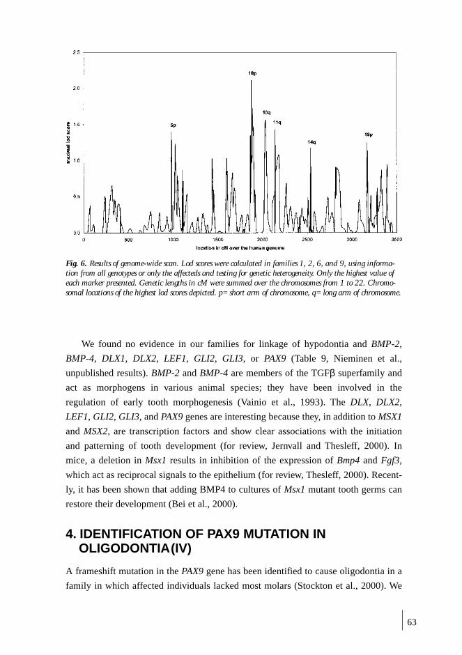
63
We found no evidence in our families for linkage of hypodontia and BMP-2,
BMP-4, DLX1, DLX2, LEF1, GLI2, GLI3, or PAX9 (Table 9, Nieminen et al.,
unpublished results). BMP-2 and BMP-4 are members of the TGFβ superfamily and
act as morphogens in various animal species; they have been involved in the
regulation of early tooth morphogenesis (Vainio et al., 1993). The DLX, DLX2,
LEF1, GLI2, GLI3, and PAX9 genes are interesting because they, in addition to MSX1
and MSX2, are transcription factors and show clear associations with the initiation
and patterning of tooth development (for review, Jernvall and Thesleff, 2000). In
mice, a deletion in Msx1 results in inhibition of the expression of Bmp4 and Fgf3,
which act as reciprocal signals to the epithelium (for review, Thesleff, 2000). Recent-
ly, it has been shown that adding BMP4 to cultures of Msx1 mutant tooth germs can
restore their development (Bei et al., 2000).
4. IDENTIFICATION OF PAX9 MUTATION INOLIGODONTIA(IV)
A frameshift mutation in the PAX9 gene has been identified to cause oligodontia in a
family in which affected individuals lacked most molars (Stockton et al., 2000). We
Fig. 6. Results of genome-wide scan. Lod scores were calculated in families 1, 2, 6, and 9, using informa-tion from all genotypes or only the affecteds and testing for genetic heterogeneity. Only the highest value ofeach marker presented. Genetic lengths in cM were summed over the chromosomes from 1 to 22. Chromo-somal locations of the highest lod scores depicted. p= short arm of chromosome, q= long arm of chromosome.
1b216251taitto28.9 28.9.2001, 17:5063
64
decided to analyze the PAX9 gene in 2 families with oligodontia because most molars
were also affected in our families (IV).
Sequencing of the PAX9 gene of the affected family members (exons 2, 3, and 4
that include the coding region) revealed 3 nucleotide changes as compared to the
published sequences (Hetzer-Egger et al., 2000; Peters et al., 1997). All changes
could be verified from both strands. The nucleotide transversion A340T present in
the heterozygous state of exon 2 in all 3 affected members of family 1 produces a
stop codon at lysine 114 in the end of the DNA-binding paired box of PAX9. This
premature termination of translation creates a truncated protein that lacks the last
alfa-helix (α6) of the paired box and the entire C-terminal region. The insG219 mu-
tation described by Stockton et al. (2000) destroys the paired box 121 nucleotides
earlier, between the N- and C-subdomains (Xu et al., 1999). The lack of the C-termi-
nal region that follows the paired box most probably disrupts the normal function of
both mutant proteins. The mildness of these phenotypes as compared to the PAX9
mouse null mutant suggests that both mutations are loss-of-function and exert their
effect mainly through inactivation of one copy of a protein, leading to haploinsuffi-
ciency. It is possible that the mutant proteins retain some of the DNA-binding capaci-
ty, and especially the slightly more severe phenotype of Family 1 may be attributed
to the dominant negative effect of the more complete paired box of the Lys114Stop
mutant protein. The other nucleotide changes found were in exon 3. The silent
change C717T in the codon of His239 was present in the heterozygous state in the
healthy mother and affected daughter of family 2. A G718C transversion causes the
conservative change Ala240Pro. This change was present in the heterozygous state in
all members of family 2 and in the homozygous or heterozygous state in some of the
members of family 1.
1b216251taitto28.9 28.9.2001, 17:5064
65
GENERAL DISCUSSION
1. SUBJECTS
We collected a sample of families in which hypodontia was segregating in an
autosomal dominant manner in order to define clinical features of hypodontia and to
localize the gene locus behind this anomaly. The frequency of hypodontia was high,
more than 30% in these families. The frequency did not differ significantly between
males and females, with 34% of females and 35% of males having hypodontia. We
therefore combined the sexes but calculated the frequencies of the individual
anomalous features separately in first- and second-degree relatives, first cousins and
more remotely related relatives. The phenotypes confirmed earlier suggestions as to
the hypodontia phenotype as an isolated trait. One individual in these families had a
cleft of the soft palate. However, one cleft patient in a sample of 214 individuals
corresponds with the frequency of cleft patients in Finland. Missing teeth were
premolars in 2 families, incisors in 3 families, and premolars and incisors in 6
families. It probably would have been better to have had families with either incisor
or premolar hypodontia to provide even more uniform phenotypes in the molecular
genetic part of the study. However, affected premolars and upper lateral incisors
seem to appear together very commonly in different combinations, and it may be
impossible to separate families according to this feature.
Simulation with the Slink program (Ott,1989; Weeks et al., 1990) showed that
the families were sufficiently informative to reveal significant evidence for linkage of
hypodontia.We combined data of 5 (II) and 7 (III) families in genetic linkage studies.
In addition, we used 4 families in the second stage of linkage studies in which the
genome-wide search with a selection of microsatellites with a maximum genetic
distance of 15 cM was performed. Large families with several affected individuals
are usually very informative in mapping human diseases. In this case, however, we
had some unexpected results, probably because the hypodontia gene frequency is
high in the population; it is possible that many different genes are involved. In large
kindreds, the possibility to have more than one gene causing tooth agenesis in a
pedigree is high. In statistical analysis, calculations both in whole pedigrees and
pedigrees divided into different families were performed in order to find possible
linkage with different loci.
1b216251taitto28.9 28.9.2001, 17:5065

66
2. PHENOTYPIC ANALYSIS OF HYPODONTIA ANDOLIGODONTIA
Many family studies have shown that isolated hypodontia and oligodontia are inherit-
ed in a dominant manner with reduced penetrance and variable expression. Our study
supports this model. The highest frequency of tooth agenesis in our study was in
first-degree relatives, 39%, followed by second-degree relatives (36%) and first
cousins (12%). The frequency is higher when the peg-shaped upper lateral incisors
are included, being 43%, 42%, and 18% respectively. Most affected individuals with
hypodontia lack only 1 or 2 permanent teeth. Hypodontia of the primary teeth was
not found, confirming the previous studies. Lower second premolars were the most
frequently missing teeth, followed by upper second premolars and upper lateral
incisors. These findings are in agreement with other studies.
In our family with oligodontia, affected individuals with PAX9 mutation lacked
all second and third permanent molars as well as both maxillary lateral incisors. In
addition, 2 affected family members lacked second premolars. While the reason for
these particular teeth to be affected is unknown, it is probably related to the
developmental sequence of tooth formation. It is suggested that those teeth in each
tooth family that develop latest are less stable and do not reach a critical threshold at
an early stage of development.
Even if congenitally missing teeth are an isolated phenomenon, studies have
shown many associated dental features. The same anomalies were seen both in
hypodontia and oligodontia families. The higher frequency of missing teeth and
other dental anomalies has been shown among relatives of the probands than in the
population, and our findings confirm this (Grahnen, 1956; Brook, 1984).
The prevalence of malformed lateral incisors in our families was high (I); being
9% in first- and 11% in second-degree relatives, 6% in cousins, and 3% in other
relatives and exceeding by 2 to 6 times the frequency in the general population
(Grahnen 1956; Alvealo and Portin, 1969). Alvesalo and Portin suggest that missing
and peg-shaped upper lateral incisors are different expressions of a dominant
autosomal gene, and our results confirm this.
An association has been speculated between missing teeth, delayed tooth
development, and tooth size (Grahnen, 1956; Garn and Lewis, 1970; Rune and
Sarnäs, 1974; Brook, 1984). In this study, dental age was within normal limits. This
is understandable because only one or a few teeth were missing in the affected
individuals in our study; they had the mildest form of hypodontia. Based on previous
studies, it seems that the more teeth are missing, the greater delay of development the
remaining teeth may show (Garn et al., 1961; Rune and Sarnäs 1974). However,
1b216251taitto28.9 28.9.2001, 17:5066

67
notable individual variation has been observed, even in oligodontia patients some
affected persons show a severely delayed tooth formation, whereas others show
normal timing. Interestingly, the delay was more obvious in males than females as in
one study of oligodontia (Schalk-van der Weide, 1992).
It was not possible to measure the exact dimensions of teeth. In 5 families,
however, some single small teeth, especially premolars, were evident.
Ectopic permanent canines have been demonstrated in many studies to belong to
the spectrum of dental anomalies related to hypodontia (Peck et al., 1993, 1994;
Pirinen et al., 1996), and our study also confirms this finding. A clear difference
existed between individuals with and without hypodontia; ectopic upper permanent
canines in individuals with hypodontia were observed three times as often than in
individuals without hypodontia or in the population.
The association of rotated premolars with hypodontia has been reported by
Baccetti (1998); our study lends support also to this finding.
Dens invaginatus, an enamel-lined cavity within a tooth, is a dental malformation
mostly affecting maxillary lateral incisors and showing a broad spectrum of
morphological variation. Minor forms of tooth invagination seem to be comparatively
common. Deep palatal-side clefts were present in nearly 40% of British children (Hallet,
1953), and over 7% had definite invaginations showing some degree of dilation beyond
the cervical margin. A frequency of 3% has been reported in the Swedish population
(Koch and Thesleff, 2001). The frequency of invaginations in our families was similar to
that of British children. However, invagination was noticed less frequently in individuals
with hypodontia both in the families studied and in the control group.
The prevalence of taurodontism varies greatly in the literature, mostly because of
different criteria for diagnosing the anomaly and the teeth assessed. The frequency of
taurodontism of lower molars clearly remains below 10% in European populations
(Holt and Brook, 1979; Schalk-van der Weide et al., 1993), with maxillary molars
affected more often (Al-Khateeb and Salako, 1997). Taurodontism has been
described together with hypodontia in many syndromes (Jorgenson, 1982), sex
chromosome anomalies with extra X chromosomes (Varrela and Alvesalo, 1988,
1989), and cleft palate patients (Laatikainen and Ranta, 1996). The association of
taurodontism with hypodontia has been shown in two investigations of patients with
hypodontia and their siblings (Stenvik et al., 1972; Seow and Lai, 1989) as well as in
a study of patients with oligodontia (Schalk-van der Weide, 1993). Seow and Lai
(1989) reported a frequency of taurodontism of 35% in persons with hypodontia, and
Schalk-van der Weide et al. (1993) 29% those with oligodontia, but only lower
molars were assessed. In our study, taurodontism seems to be related to hypodontia,
1b216251taitto28.9 28.9.2001, 17:5067
68
confirming previous studies. However, that the results were not statistically significant
in separate groups of relatives, may relate to the small number of individuals in these
groups. Our findings are in agreement with a Finnish study on cleft patients
(Laatikainen and Ranta, 1996), showing the frequency of taurodontism to be 41% in
39 pairs of twins with cleft lip and/or palate and hypodontia.
A taurodontic tooth is generally believed to result from a disturbance in
Hertwig‘s epithelial root sheath, which is an extension of the dental epithelium after
crown morphogenesis has been completed. Based on clinical observations and this
theory, the association between taurodontism and hypodontia may result from the
same genetic etiology affecting the growth of dental epithelium.
3. GENETIC ANALYSIS
Numerous transcription factors, growth factors, and their receptors as well as
structural molecules of the cell surface and extracellular matrix have been associated
with early tooth morphogenesis (for review, Thesleff and Nieminen, 1996; Jernvall and
Thesleff, 2000). Interestingly, several of these genes have been shown to cause arrested
tooth development in transgenic mice (Satokata and Maas, 1994; Peters et al., 1998). It
is apparent that all these genes are potential candidate genes for hypodontia.
We excluded EGF, EGFR, and FGF-3 as genes behind hypodontia (III;
Nieminen et al., unpublished results). In addition, we excluded MSX1 and MSX2 in
our families (II; Nieminen et al., unpublished results). Later, however, a missense
mutation in the MSX1 gene was identified as the first gene defect causing isolated
tooth agenesis in man (Vastardis et al.,1996). The phenotypes in our families differed
from the phenotype of the family with the MSX1 mutation, where the prominent
feature was bilateral agenesis of second premolars and third molars in both jaws. The
number of missing teeth was lower in our families (hypodontia). Further, agenesis of
molars (except third molars) was absent in our families with incisor-premolar
hypodontia, whereas this was a typical feature of oligodontia both in our family in
study IV and in an American family with mutation of PAX9 (Stockton et al., 2000).
The similarity of the phenotypes in our families and the American family motivated
our PAX9-mutation search. The identification of a new mutation in PAX9 segregating
with oligodontia of molar teeth supports the suggestion that this gene is important
especially for the development of the most distal teeth. However, we did not find the
same mutation in another oligodontia family with missing molars, nor in the families
with incisor-premolar hypodontia. These traits may be caused by mutations in
different gene families or perhaps genes acting in the same signaling pathways with
MSX1 and PAX9. During our genome-wide scan we detected several regions which
may contain mutated genes causing incisor-premolar hypodontia.
1b216251taitto28.9 28.9.2001, 17:5068

69
CONCLUDING REMARKS
Congenital absence of permanent teeth has interested dentists for a long time, but
studies analyzing the molecular pathology with modern DNA techniques have not
been performed before the last decade. When this work was begun, several monogenic
diseases had been successfully mapped, and this encouraged us to search for the gene
behind hypodontia. At that time, genes behind isolated tooth agenesis in man were
unknown, and classification of hypodontia was beginning to be renewed. We excluded
many important genes which had been implicated in tooth development by performing
gene expression and experimental studies in the mouse and causing tooth agenesis in
transgenic mice. Mutations in two of these genes, MSX1 with two different mutations,
and PAX9, were later identified in two big families where dominantly inherited
oligodontia typically affects premolars and molars. We found a new nonsense
mutation in PAX9 in all three affected family members of a family with oligodontia.
We must focus on the fact that while mutations in the MSX1 and PAX9 genes have
been shown to be responsible for tooth agenesis in single families, these genes do not
cause this phenotype in other families.
By performing a genome-wide search for the hypodontia gene, we have found
some loci that can be linked with variable statistical significance to hypodontia in
one or several families. Based on existing evidence, it is obvious that both hypodontia
and oligodontia are heterogenous traits, with several mutated genes responsible. In
the future, it will be possible to study these loci further and identify more genes
behind these traits. Publication of the human genome sequence (Lander et al., 2001;
Venter et al., 2001) is valuable for these studies and accelerates the search for mutated
genes. Combining clinical and genetic results, classification of the different forms of
tooth agenesis will become more exact.
Our results support the findings that ectopic canines, malformed upper lateral
insisors, rotation of premolars, and taurodontism are related to hypodontia and
oligodontia. Interestingly, the same teeth are most often affected, and the similar
associated dental anomalies are seen also in syndromic hypodontia. The last developing
teeth in each tooth family seem to be most frequently affected both in isolated and
syndromic traits. As genes most obviously play the main role in the etiology of
congenitally missing teeth and several dental anomalies, it is important that a family
history of hypodontia/oligodontia as well as dental anomalies is observed in clinical
paediatric dentistry. Screening children of families with segregating tooth agenesis
by clinical and radiographic examinations should be carried out at 6 to 7 years of age
to plan the best possible treatment for the developing dentition.
1b216251taitto28.9 28.9.2001, 17:5069
70
ACKNOWLEDGEMENTS
This study was carried out at the Department of Pedodontics and Orthodontics and at
the Scientific Laboratory, Institute of Dentistry, University of Helsinki. I wish to
thank Professor Jukka H Meurman, the Dean of the Institute of Dentistry, University
of Helsinki, for providing the research facilities.
I express my deepest gratitude to my supervisors Professor Sinikka Pirinen, the
Acting Head of the Department of Pedodontics and Orthodontics, and Professor Irma
Thesleff, Head of the Deparment of Pedodontics and Orthodontics and Director of
the Developmental Biology Programme, University of Helsinki, for placing the
excellent working facilities at my disposal. This work would never have begun without
their enthusiastic and inspiring attitude toward research. I am grateful to Sinikka for
her always wise, kind, and encouraging guidance throughout these years. I thank
Irma for her inspiring supervision, and for her wide knowledge of developmental
biology and molecular genetics.
I am grateful to Professor Leena Peltonen, who always managed to find time for
stimulating guidance and supervision. I also warmly thank her for the opportunity to
utilize the facilities of the laboratory at the Department of Human Molecular Genetics,
National Public Health Institute, Helsinki.
I want to express my special gratitude to Pekka Nieminen for sharing this
research project with me. Without him, this work would not have been possible.
Thank you, Pekka, for organizing and for teaching me everything about working in
the laboratory, and for helping me in many ways during this project.
I sincerely thank my other co-authors Professor Satu Alaluusua, Professor Kaarina
Haavikko, Satu Apajalahti, Lars Paulin, and Dennis Tanner.
I wish to thank the official reviewers of this thesis, Docent Mirja Somer,
Department of Medical Genetics, University of Helsinki, Finland, and Associate
Professor Birgitta Bäckman, Department of Odontology/Pedodontics, Faculty of
Medicine and Odontology, University of Umeå, Sweden, for their constructive criticism
and valuable comments.
I warmly thank Kirsti Kari, Head of the Scientific Laboratory of the Institute of
Dentistry, and Anne Bryk, Maire Holopainen, Riikka Santalahti, and Annikki Siren
for their technical assistance. I also thank Docent Juhani Wolff, the previous Head of
the Department of Dental Radiology, and the whole staff of the department for
collaboration.
I am grateful to the entire staff of the Department of Pedodontics and Orthodontics
for their support: to Docent Marjatta Nyström and colleagues Päivi Hölttä and Eija
1b216251taitto28.9 28.9.2001, 17:5070
71
Kleemola-Kujala for their kind aid, to Anneli Sinkkonen for her assistance, and to
Raila Salminen for helping in practical things.
I owe my heartfelt thanks to my other colleagues and friends Johanna Kotilainen
and Eva Karlstedt, for their empathy and valuable advice during these years, as well
as numerous (more or less) fruitful discussions in the late afternoons.
I warmly thank Helene Javen for checking the references, and Carolyn Norris for
author-editing the language of the manuscript.
I am greatly indebted to all the members of the families for their participation in
this study.
I wish to thank my parents, Elsa and Esko Luoma for their support of and interest
in my work. Finally, I owe my dearest thanks to my husband Pekka, sons Lauri and
Eero, and daughter Elisa for their loyalty and their patience with my work, as well as
for keeping me from losing the rhythm of everyday life.
This study was financially supported by the Finnish Dental Society, Finnish
Women Dentists, and the University of Helsinki.
Helsinki, September 2001
1b216251taitto28.9 28.9.2001, 17:5071
72
REFERENCES
Aasheim B, Ögaard B (1993). Hypodontia in 9-year-old Norwegians related to need of orthodontictreatment. Scand J Dent Res 101:257-260.
Ahmad W, Brancolini V, ul Faiyaz MF, Lam H, ul Haque S, Haider M et al. (1998). A locus forautosomal recessive hypodontia with associated dental anomalies maps to chromosome 16q12.1.Am J Hum Genet 62:987-991.
Akyuz S, Atasu A (1993). Tooth and nail syndrome: genetic, clinical and dermatoglyphicfindings:case report. J Clin Pediatr Dent 17:105-108.
Al-Khateeb T, Salako NO (1997). The incidence of taurodontism in permanent molars in SaudiArabian dental patients. Ped Dent J 7:69-72.
Alvesalo L , Portin P (1969). The inheritance pattern of missing, peg-shaped and strongly mesio-distally reduced upper lateral incisors. Acta Odontol Scand 27:563-575.
Anonymous (1998) Worldwide variation in prevalence of symptoms of asthma, allergic rhinoconjunctivitis,and atopic eczema: ISAAC. The International Study of Asthma and Allergies in Childhood.(ISAAC) Steering Committee. Lancet 351:1225-1232.
Apajalahti S, Arte S, Pirinen S (1999). Short root anomaly in families and its association with otherdental anomalies. Eur J Oral Sci 107:97-101.
Axrup K, D´ Avignon M, Hellgren K, Herikson CO, Juhlin IM, Larsson KS et al. (1966). Childrenwith thalidomide embryopathy: odontological observations and aspects. Acta Odontol Scand24:3-21.
Baccetti T (1998). A controlled study of associated dental anomalies. Angle Orthod 68:267-274.Baccetti T (1998). Tooth rotation associated with aplasia of nonadjacent teeth. Angle Orthod
68:471-474.Baraitser M, Winter RM (1999). The London dysmorphology database (LDDB). Oxford medical
databases, Oxford, Oxford University Press.Becker A, Smith P, Behar R (1981). The incidence of anomalous maxillary lateral incisors in relation
to palatally-displaced cuspids. Angle Orthod 51:24-29.Bei M, Kratochwil K, Maas R (2000). BMP4 rescues a non-cell-autonomous function of Msx1 in
tooth development. Development 127:4711-4718.Bergström K (1977). An orthopantomographic study of hypodontia, supernumeraries and other
anomalies in school children between the ages of 8-9 years. An epidemiological study. Swed DentJ 1:145-157.
Bjerklin K, Kurol J, Valentin J (1992). Ectopic eruption of maxillary first permanent molars andassociation with other tooth and developmental disturbances. Eur J Orthod 14:369-375.
Blumberg JE, Hylander WL, Goepp RA (1971). Taurodontism, a biometric study. Am J Phys An-thropol 34:243-255.
Böök JA (1950). Clinical and genetical studies of hypodontia: I. Premolar aplasia, hyperhidrosis, andcanites prematura: A new hereditary syndrome in man. Am J Hum Genet 2:240-263.
Brin I, Becker A, Shalhav M (1986). Position of the maxillary permanent canine in relation toanomalous or missing lateral incisors: a population study. Eur J Orthod 8:12-16.
Brook AH (1974). Dental anomalies of number, form and size: their prevalence in British schoolchildren. JInt Assoc Dent Child 5:37-53.
Brook AH (1984). A unifying aetiological explanation for anomalies of human tooth number andsize. Arch Oral Biol 29:373-378.
Brookes S, Lammie GA, Schuuring E, Dickson C, Peters G (1992). Linkage map of a region of humanchromosome band 11q13 amplified in breast and squamous cell tumors. Genes ChromosomesCancer 4:290-301.
Burgersdijk R, Tan HL (1978). Oral symptoms of the Wolf syndrome. ASDC J Dent Child 45:488-489.
1b216251taitto28.9 28.9.2001, 17:5072

73
Burzynski NJ, Escobar VH (1983). Classification and genetics of numeric anomalies of dentition.Birth Defects Orig Artic Ser 19:95-106.
Buss PW, Hughes HE, Clarke A (1995). Twenty-four cases of the EEC syndrome: clinical presentationand management. J Med Genet 32:716-723.
Bustos T, Simosa V, Pinto-Cisternas J, Abramovits W, Jolay L, Rodriguez L et al. (1991). Autosomalrecessive ectodermal dysplasia. I. An undescribed dysplasia/malformation syndrome. Am J MedGenet 41:398-404.
Butler P (1939). Studies of the mammalian dentition. Differentiation of the post-canine dentition.Proc Zool Soc Lond 109:1-36.
Butler PM (1995). Ontogenetic aspects of dental evolution. Int J Dev Biol 39:25-34.Byrd ED (1943). Incidence of supernumerary and congenitally missing teeth. J Dent Child 10:84-86.Cambiaghi S, Restano L, Paakkonen K, Caputo R, Kere J (2000). Clinical findings in mosaic carriers
of hypohidrotic ectodermal dysplasia. Acrh Dermatol 136:217-224.Carvalho JC, Vinker F, Declerck D (1998). Malocclusion, dental injuries and dental anomalies in the
primary dentition of Belgian children. Int J Paediatr Dent 8:137-141.Celli J, Duijf P, Hamel BC, Bamshad M, Kramer B, Smits APT et al. (1999). Heterozygous germline
mutations in the p53 homolog p63 are the cause of EEC syndrome. Cell 99:143-153.Chi DD, Hing AV, Helms C, Steinbrueck T, Mishra SK, Donis-Keller H (1992). Two chromosome 7
dinucleotide repeat polymorphisms at gene loci epidermal growth factor receptor (EGFR) andproα2(I)collagen (COL1A2). Hum Mol Genet 1:135.
Chosack A, Eidelman E, Cohen T (1975). Hypodontia: A polygenic trait, a family study among Is-raeli Jews. J Dent Res 54:16-19.
Collins FS (1995). Positional cloning moves from perditional to traditional. Nat Genet 9:347-350.Dahl E, Koseki H, Balling R (1997). Pax genes and organogenesis. Bioessays 19:755-765.Daugaard-Jensen J, Nodal M, Kjaer I (1997). Pattern of agenesis in the primary dentition: a
radiographic study of 193 cases. Int J Paediatr Dent 7:3-7.Davis PJ (1987). Hypodontia and hyperdontia of permanent teeth in Hong Kong schoolchildren.
Community Dent Oral Epiodemiol 15:218-220.Dib C, Faure S, Fizames C, Samson D, Drouot N, Vignal A et al. (1996). A comprehensive genetic
map of the human genome based on 5,264 microsatellites. Nature 380:152-154.Döffinger R, Smahi A, Bessia C, Geissmann F, Feinberg J, Durady A et al. (2001). X-linked anhidrotic
ectodermal dysplasia with immunodeficiency is caused by impaired NF-kappaB signaling.Nature Genet 27:277-284.
Dolder E (1936). Zahn-Unterzahl. Diagnostik, Statistik, Artikulation. Schweiz Monatsschr Zahnmed46:663-701.
Ericson S, Kurol J (1986). Radiographic assessment of maxillary canine eruption in children withclinical signs of eruption disturbance. Eur J Orthod 8:133-140.
Farias M and Vargervik K (1988). Dental development in hemifacial microsomia. I. Eruption andagenesis. Pediatr Dent 10:140-143.
Ferguson CA, Tucker AS, Christensen L, Lau AL, Matzuk MM, Sharpe PT (1998). Activin is anessential early mesenchymal signal in tooth development that is required for patterning of themurine dentition. Genes Dev 12:2636-2649.
Ferrante MI, Giorgio G, Feather SA, Bulfone A, Wright V, Ghiani M et al., 2001). Identification ofthe gene for oral-facial-digital type1 syndrome. Am J Hum Genet 68:569-576.
Fried K (1977). Autosomal recessive hidrotic ectodermal dysplasia. J Med Genet 14:137-139.Garn SM, Lewis AB, Bonne B (1961). Third molar polymorphism and the timing of tooth forma-
tion. Nature 192:989.Garn SM, Lewis AB, Dahlberg AA, Kerewsky RS (1966). Interaction between relative molar size and
relative number of cusps. J Dent Res 45:1240.Garn SM, Lewis AB (1970). The gradient and the pattern of crown-size reduction in simple
hypodontia. Angle Orthod 40:51-58.
1b216251taitto28.9 28.9.2001, 17:5073
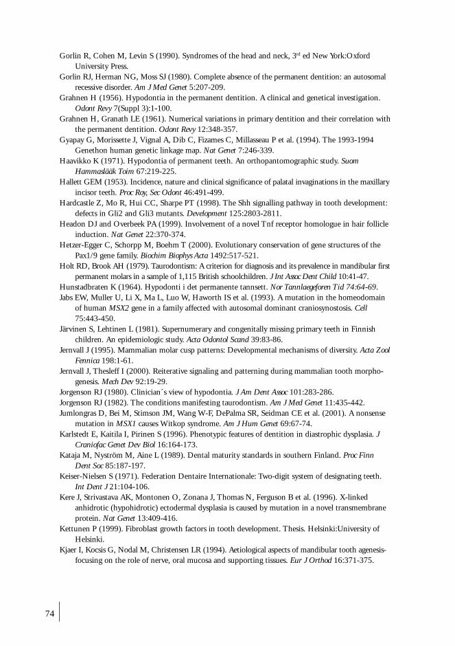
74
Gorlin R, Cohen M, Levin S (1990). Syndromes of the head and neck, 3rd ed New York:OxfordUniversity Press.
Gorlin RJ, Herman NG, Moss SJ (1980). Complete absence of the permanent dentition: an autosomalrecessive disorder. Am J Med Genet 5:207-209.
Grahnen H (1956). Hypodontia in the permanent dentition. A clinical and genetical investigation.Odont Revy 7(Suppl 3):1-100.
Grahnen H, Granath LE (1961). Numerical variations in primary dentition and their correlation withthe permanent dentition. Odont Revy 12:348-357.
Gyapay G, Morissette J, Vignal A, Dib C, Fizames C, Millasseau P et al. (1994). The 1993-1994Genethon human genetic linkage map. Nat Genet 7:246-339.
Haavikko K (1971). Hypodontia of permanent teeth. An orthopantomographic study. SuomHammaslääk Toim 67:219-225.
Hallett GEM (1953). Incidence, nature and clinical significance of palatal invaginations in the maxillaryincisor teeth. Proc Roy, Sec Odont 46:491-499.
Hardcastle Z, Mo R, Hui CC, Sharpe PT (1998). The Shh signalling pathway in tooth development:defects in Gli2 and Gli3 mutants. Development 125:2803-2811.
Headon DJ and Overbeek PA (1999). Involvement of a novel Tnf receptor homologue in hair follicleinduction. Nat Genet 22:370-374.
Hetzer-Egger C, Schorpp M, Boehm T (2000). Evolutionary conservation of gene structures of thePax1/9 gene family. Biochim Biophys Acta 1492:517-521.
Holt RD, Brook AH (1979). Taurodontism: A criterion for diagnosis and its prevalence in mandibular firstpermanent molars in a sample of 1,115 British schoolchildren. J Int Assoc Dent Child 10:41-47.
Hunstadbraten K (1964). Hypodonti i det permanente tannsett. Nor Tannlaegeforen Tid 74:64-69.Jabs EW, Muller U, Li X, Ma L, Luo W, Haworth IS et al. (1993). A mutation in the homeodomain
of human MSX2 gene in a family affected with autosomal dominant craniosynostosis. Cell75:443-450.
Järvinen S, Lehtinen L (1981). Supernumerary and congenitally missing primary teeth in Finnishchildren. An epidemiologic study. Acta Odontol Scand 39:83-86.
Jernvall J (1995). Mammalian molar cusp patterns: Developmental mechanisms of diversity. Acta ZoolFennica 198:1-61.
Jernvall J, Thesleff I (2000). Reiterative signaling and patterning during mammalian tooth morpho-genesis. Mech Dev 92:19-29.
Jorgenson RJ (1980). Clinician´s view of hypodontia. J Am Dent Assoc 101:283-286.Jorgenson RJ (1982). The conditions manifesting taurodontism. Am J Med Genet 11:435-442.Jumlongras D, Bei M, Stimson JM, Wang W-F, DePalma SR, Seidman CE et al. (2001). A nonsense
mutation in MSX1 causes Witkop syndrome. Am J Hum Genet 69:67-74.Karlstedt E, Kaitila I, Pirinen S (1996). Phenotypic features of dentition in diastrophic dysplasia. J
Craniofac Genet Dev Biol 16:164-173.Kataja M, Nyström M, Aine L (1989). Dental maturity standards in southern Finland. Proc Finn
Dent Soc 85:187-197.Keiser-Nielsen S (1971). Federation Dentaire Internationale: Two-digit system of designating teeth.
Int Dent J 21:104-106.Kere J, Strivastava AK, Montonen O, Zonana J, Thomas N, Ferguson B et al. (1996). X-linked
anhidrotic (hypohidrotic) ectodermal dysplasia is caused by mutation in a novel transmembraneprotein. Nat Genet 13:409-416.
Kettunen P (1999). Fibroblast growth factors in tooth development. Thesis. Helsinki:University ofHelsinki.
Kjaer I, Kocsis G, Nodal M, Christensen LR (1994). Aetiological aspects of mandibular tooth agenesis-focusing on the role of nerve, oral mucosa and supporting tissues. Eur J Orthod 16:371-375.
1b216251taitto28.9 28.9.2001, 17:5074
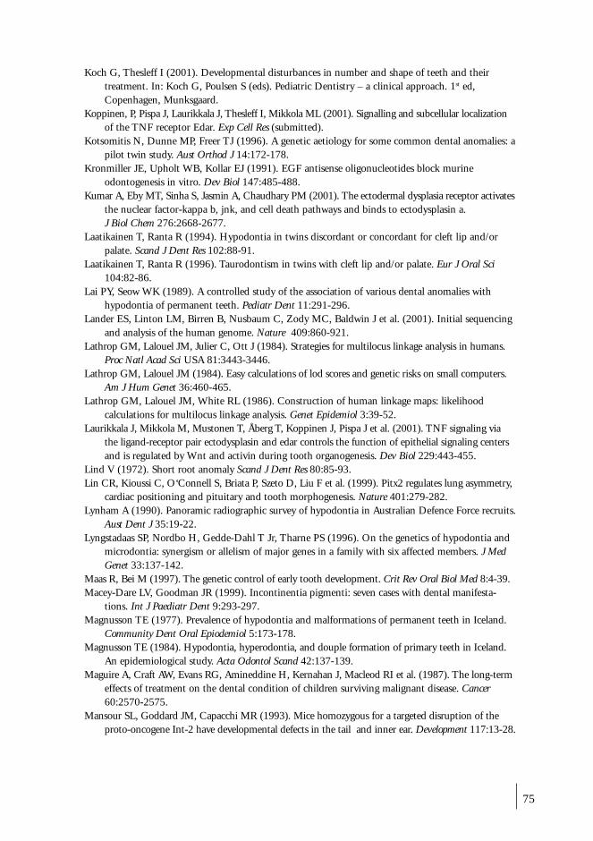
75
Koch G, Thesleff I (2001). Developmental disturbances in number and shape of teeth and theirtreatment. In: Koch G, Poulsen S (eds). Pediatric Dentistry – a clinical approach. 1st ed,Copenhagen, Munksgaard.
Koppinen, P, Pispa J, Laurikkala J, Thesleff I, Mikkola ML (2001). Signalling and subcellular localizationof the TNF receptor Edar. Exp Cell Res (submitted).
Kotsomitis N, Dunne MP, Freer TJ (1996). A genetic aetiology for some common dental anomalies: apilot twin study. Aust Orthod J 14:172-178.
Kronmiller JE, Upholt WB, Kollar EJ (1991). EGF antisense oligonucleotides block murineodontogenesis in vitro. Dev Biol 147:485-488.
Kumar A, Eby MT, Sinha S, Jasmin A, Chaudhary PM (2001). The ectodermal dysplasia receptor activatesthe nuclear factor-kappa b, jnk, and cell death pathways and binds to ectodysplasin a.J Biol Chem 276:2668-2677.
Laatikainen T, Ranta R (1994). Hypodontia in twins discordant or concordant for cleft lip and/orpalate. Scand J Dent Res 102:88-91.
Laatikainen T, Ranta R (1996). Taurodontism in twins with cleft lip and/or palate. Eur J Oral Sci104:82-86.
Lai PY, Seow WK (1989). A controlled study of the association of various dental anomalies withhypodontia of permanent teeth. Pediatr Dent 11:291-296.
Lander ES, Linton LM, Birren B, Nusbaum C, Zody MC, Baldwin J et al. (2001). Initial sequencingand analysis of the human genome. Nature 409:860-921.
Lathrop GM, Lalouel JM, Julier C, Ott J (1984). Strategies for multilocus linkage analysis in humans.Proc Natl Acad Sci USA 81:3443-3446.
Lathrop GM, Lalouel JM (1984). Easy calculations of lod scores and genetic risks on small computers.Am J Hum Genet 36:460-465.
Lathrop GM, Lalouel JM, White RL (1986). Construction of human linkage maps: likelihoodcalculations for multilocus linkage analysis. Genet Epidemiol 3:39-52.
Laurikkala J, Mikkola M, Mustonen T, Åberg T, Koppinen J, Pispa J et al. (2001). TNF signaling viathe ligand-receptor pair ectodysplasin and edar controls the function of epithelial signaling centersand is regulated by Wnt and activin during tooth organogenesis. Dev Biol 229:443-455.
Lind V (1972). Short root anomaly Scand J Dent Res 80:85-93.Lin CR, Kioussi C, O‘Connell S, Briata P, Szeto D, Liu F et al. (1999). Pitx2 regulates lung asymmetry,
cardiac positioning and pituitary and tooth morphogenesis. Nature 401:279-282.Lynham A (1990). Panoramic radiographic survey of hypodontia in Australian Defence Force recruits.
Aust Dent J 35:19-22.Lyngstadaas SP, Nordbo H, Gedde-Dahl T Jr, Tharne PS (1996). On the genetics of hypodontia and
microdontia: synergism or allelism of major genes in a family with six affected members. J MedGenet 33:137-142.
Maas R, Bei M (1997). The genetic control of early tooth development. Crit Rev Oral Biol Med 8:4-39.Macey-Dare LV, Goodman JR (1999). Incontinentia pigmenti: seven cases with dental manifesta-
tions. Int J Paediatr Dent 9:293-297.Magnusson TE (1977). Prevalence of hypodontia and malformations of permanent teeth in Iceland.
Community Dent Oral Epiodemiol 5:173-178.Magnusson TE (1984). Hypodontia, hyperodontia, and douple formation of primary teeth in Iceland.
An epidemiological study. Acta Odontol Scand 42:137-139.Maguire A, Craft AW, Evans RG, Amineddine H, Kernahan J, Macleod RI et al. (1987). The long-term
effects of treatment on the dental condition of children surviving malignant disease. Cancer60:2570-2575.
Mansour SL, Goddard JM, Capacchi MR (1993). Mice homozygous for a targeted disruption of theproto-oncogene Int-2 have developmental defects in the tail and inner ear. Development 117:13-28.
1b216251taitto28.9 28.9.2001, 17:5075
76
Markovic M (1982). Hypodontia in twins. Swed Denl J 15(suppl):153-162.Maruko E, Hayes C, Evans CA, Padwa B, Mulliken JB (2001). Hypodontia in hemifacial microsomia.
Cleft Palate-Craniofac J 38:15-19.Mather K (1957). The measurement of linkage in heredity. Methuen & Co. Ltd, London, New York.Matzuk MM, Lu N, Vogel H, Sellheyer K, Roop DR, Bradley A (1995). Multiple defects and perinatal
death in mice deficient in follistatin. Nature 374:360-363.Mhanni A, Cross H, Chudley AE (1999). Kabuki syndrome: description o f dental findings in 8
patients. Clin Genet 56:154-157.Miesfeld R, Krystal M, Arnheim N (1981). A member of new repeated sequence family which is
conserved throughout eucaryotic evolution is found between the human delta and beta globingenes. Nucleic Acids Res 9:5931-5947.
Mills AA, Zheng B, Wang XJ, Vogel H, Roop DR, Bradley A (1999). p63 is a p53 homologuerequired for limb and epidermal morphogenesis. Nature 398:708-713.
Mikkola ML, Pispa J, Pekkanen M, Paulin L, Nieminen P, Kere J et al. (1999). Ectodysplasin, aprotein required for epithelial morphogenesis, is a novel TNF homologue and promotes cell-matrix adhesion. Mech Dev 88:133-146.
Monreal AW, Zonana J, Ferguson B (1998). Identification of a new splice form of EDA1 gene permitsdetection of nearly all X-linked hypohidrotic ectodermal dysplasia mutations. Am J Hum Genet63:380-389.
Monreal AW, Ferguson BM, Headon DJ, Street SL, Overbeek PA, Zonana J (1999). Mutations in thehuman homologue of mouse dl cause autosomal recessive and dominant hypohidrotic ectodermaldysplasia. Nat Genet 22:366-369.
Morton NE (1955). Sequential tests for the deletion of linkage. Am J Hum Genet 62:690-697.Muller TP, Hill IN, Petersen AC, Blayney JR (1970). A survey of congenitally missing permanent
teeth. J Am Dent Assoc 81:101-107.Murray JC, Buetow KH, Weber JL, Ludwigsen S, Scherpbier-Heddema T, Manion F et al. (1994). A
comprehensive human linkage map with centimorgan density. Cooperative Human LinkageCenter (CHLC). Science 265:2049-2054.
Näsman M, Forsberg C-M, Dahllöf G (1997). Long-term dental development in children aftertreatment for malignant disease. Eur J Orthod 19:151-159.
Nieminen P, Pekkanen M, Åberg T, and Thesleff I. Gene expression in tooth. Developmental Biology Pro-gramme, Institute of Biotechnology, University of Helsinki, Finland, 2001. http://bite-it.helsinki.fi
Niswander JD, Sujaku C (1963). Congenital anomalies of teeth in Japanese Children. Am J PhysAnthropol 21:569-574.
Nyström M (1977). Clinical eruption of deciduous teeth in a series of Finnish children. Proc FinnDent Soc 73:155-161.
Odent S, Le Marec B, Munnich A, Le Merrer M, Bonaiti-Pellie C (1998). Segregation analysis innonsyndromic holoprosencephaly. Am J Med Genet 77:139-143.
Öhman M (2001). The search for genes predisposing to obesity. Thesis. Helsinki: University ofHelsinki.
Online Mendelian Inheritance in Man, OMIM (TM). McKusik-Nathans Institute for Genetic Medicine,Johns Hopkins University (Baltimore, MD) and National Center for Biotechnology Information,National Library of Medicine (Bethesda, MD), 2001. http://www.ncbi.nlm.nih.gov/omim/
O´Quinn JR, Hennekam RCM, Jorde LB, Bamshad M (1998). Syndromic ectrodactyly with severelimb, ectodermal, urogenital, and palatal defects maps to chromosome 19. Am J Hum Genet62:130-135.
Osborn JW (1978). Morphogenetic gradients: fields versus clones. In: Butler, PM and Joysey KA(eds), Development, Function and Evolution of Teeth, London: Academic Press, pp171-201.
Ott J (1974). Estimation of recombination fraction in human pedigrees: efficient computation of thelikelihood for human linkage studies. Am J Hum Genet 26:588-597.
1b216251taitto28.9 28.9.2001, 17:5076
77
Ott J (1989). Computer-simulation methods in human linkage analysis. Proc Natl Acad Sci USA86:4175-4178.
Ott J (1991). Analysis of human genetic linkage. Baltimore, Johns Hopkins University Press.Padanilam BJ, Stadler HS, Mills KA, McLeod LB, Solursh M, Lee B et al. (1992). Characterization of
the human HOX 7 cDNA and identification of polymorphic markers. Hum Mol Genet 1:407-410.Peck L, Peck S, Attia Y (1993). Maxillary canine-first premolar transposition, associated dental
anomalies and genetic bases. Angle Orthod 63:99-109.Peck S, Peck L, Kataja M (1994). The palatally displaced canine as a dental anomaly of genetic origin.
Angle Orthod 64:249-256.Peck S, Peck L, Kataja M (1996). Prevalence of tooth agenesis and peg-shaped maxillary lateral incisor
associated with palatally displaced canine (PDC) anomaly. Am J Orthod Dentofacial Orthop110:441-443.
Peck S, Peck L, Kataja M (1998). Mandibular lateral incisor-canine transposition, concomitant dentalanomalies, and genetic control. Angle Orthod 68:455-466.
Peters H, Schuster G, Neubuser A, Richter T, Hofler H, Balling R (1997). Isolation of the Pax9cDNA from adult human esophagus. Mamm Genome 8:62-64.
Peters H, Neubuser A, Kratochwil K, Balling R (1998). Pax9-deficient mice lack pharyngeal pouchderivatives and teeth and exhibit craniofacial and limb abnormalities. Genes Dev 12:2735-2747.
Peters H and Balling R (1999). Teeth where and how to make them. Trends Genet 15:59-65.Phillips JC, del Bono EA, Haines JL, Pralea AM, Cohen JS, Greff LJ et al. (1996). A second locus
for Rieger syndrome maps to chromosome 13q14. Am J Hum Genet 59:613-619.Pinheiro M, Freire-Maia N (1994). Ectodermal dysplasias: a clinical classification and a causal review.
Am J Med Genet 53:153-162.Pirinen S, Thesleff I (1995). Development of the dentition. In: Thilander B, Rönning O (eds).
Introduction to Orthodontics. Stockholm: Lic Förlag, pp 41-43.Pirinen S, Arte S, Apajalahti S (1996). Palatal displacement of canine is genetic and related to
congenital absence of teeth. J Dent Res 75:1742-1746.Pirinen S, Kentala A, Nieminen P, Varilo T, Thesleff I, Arte S (2001). Recessively inherited lower
incisor hypodontia. J Med Genet 38:551-556.Pispa J, Jung H-S, Jernvall J, Kettunen P, MustonenT, Tabata MJ et al. (1999). Cusp patterning
defect in Tabby mouse teeth and its partial rescue by FGF. Dev Biol 216:521-534.Polymeropoulos MH, Xiao H, Rath DS, Merril CR (1990). Dinucleotide repeat polymorphism at
the int-2 proto-oncogene locus (INT2). Nucleic Acids Res 18:7468.Propping P, Friedl W, Wienker TF, Uhlhaas S, Zerres K (2000). ADULT syndrome allelic to limb
mammary syndrome (LMS). Am J Med Genet 90:179-182.Qiu M, Bulfone A, Ghattas I, Meneses JJ, Christensen L, Sharpe PT et al. (1997). Role of Dlx
homeobox genes in proximodistal patterning of the branchial arches: mutations of Dlx-1, Dlx-2,and Dlx-1 and –2 alter morphogenesis of proximal skeletal and soft tissue structures derivedfrom the first and second arches. Dev Biol 185:165-184.
Qumsiyeh MB (1992). EEC syndrome (ectrodactyly, ectodermal dysplasia and cleft lip/palate) is on7p11.2-q21.3. Clin Genet 42:101.
Ranta R (1986). A Review of tooth formation in children with cleft lip/palate. Am J Orthod DentofacialOrhop 90:11-18.
Ranta R, Tulensalo T (1988). Symmetry and combinations of hypodontia in noncleft and cleft palatechildren. Scand J Dent Res 96:1-8.
Rasko I, Downes CS (1995). Genes in Medicine. Molecular biology and human genetic disorders.London. Chapman & Hall.
Ravn JJ (1971). Aplasia, supernumerary teeth and fused teeth in the primary dentition. Anepidemiologic study. Scand J Dent Res 79:1-6.
1b216251taitto28.9 28.9.2001, 17:5077
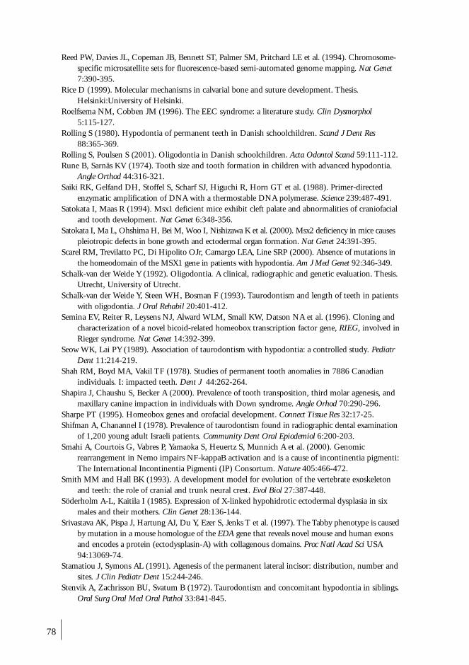
78
Reed PW, Davies JL, Copeman JB, Bennett ST, Palmer SM, Pritchard LE et al. (1994). Chromosome-specific microsatellite sets for fluorescence-based semi-automated genome mapping. Nat Genet7:390-395.
Rice D (1999). Molecular mechanisms in calvarial bone and suture development. Thesis.Helsinki:University of Helsinki.
Roelfsema NM, Cobben JM (1996). The EEC syndrome: a literature study. Clin Dysmorphol5:115-127.
Rolling S (1980). Hypodontia of permanent teeth in Danish schoolchildren. Scand J Dent Res88:365-369.
Rolling S, Poulsen S (2001). Oligodontia in Danish schoolchildren. Acta Odontol Scand 59:111-112.Rune B, Sarnäs KV (1974). Tooth size and tooth formation in children with advanced hypodontia.
Angle Orthod 44:316-321.Saiki RK, Gelfand DH, Stoffel S, Scharf SJ, Higuchi R, Horn GT et al. (1988). Primer-directed
enzymatic amplification of DNA with a thermostable DNA polymerase. Science 239:487-491.Satokata I, Maas R (1994). Msx1 deficient mice exhibit cleft palate and abnormalities of craniofacial
and tooth development. Nat Genet 6:348-356.Satokata I, Ma L, Ohshima H, Bei M, Woo I, Nishizawa K et al. (2000). Msx2 deficiency in mice causes
pleiotropic defects in bone growth and ectodermal organ formation. Nat Genet 24:391-395.Scarel RM, Trevilatto PC, Di Hipolito OJr, Camargo LEA, Line SRP (2000). Absence of mutations in
the homeodomain of the MSX1 gene in patients with hypodontia. Am J Med Genet 92:346-349.Schalk-van der Weide Y (1992). Oligodontia. A clinical, radiographic and genetic evaluation. Thesis.
Utrecht, University of Utrecht.Schalk-van der Weide Y, Steen WH, Bosman F (1993). Taurodontism and length of teeth in patients
with oligodontia. J Oral Rehabil 20:401-412.Semina EV, Reiter R, Leysens NJ, Alward WLM, Small KW, Datson NA et al. (1996). Cloning and
characterization of a novel bicoid-related homeobox transcription factor gene, RIEG, involved inRieger syndrome. Nat Genet 14:392-399.
Seow WK, Lai PY (1989). Association of taurodontism with hypodontia: a controlled study. PediatrDent 11:214-219.
Shah RM, Boyd MA, Vakil TF (1978). Studies of permanent tooth anomalies in 7886 Canadianindividuals. I: impacted teeth. Dent J 44:262-264.
Shapira J, Chaushu S, Becker A (2000). Prevalence of tooth transposition, third molar agenesis, andmaxillary canine impaction in individuals with Down syndrome. Angle Orhod 70:290-296.
Sharpe PT (1995). Homeobox genes and orofacial development. Connect Tissue Res 32:17-25.Shifman A, Chanannel I (1978). Prevalence of taurodontism found in radiographic dental examination
of 1,200 young adult Israeli patients. Community Dent Oral Epiodemiol 6:200-203.Smahi A, Courtois G, Vabres P, Yamaoka S, Heuertz S, Munnich A et al. (2000). Genomic
rearrangement in Nemo impairs NF-kappaB activation and is a cause of incontinentia pigmenti:The International Incontinentia Pigmenti (IP) Consortum. Nature 405:466-472.
Smith MM and Hall BK (1993). A development model for evolution of the vertebrate exoskeletonand teeth: the role of cranial and trunk neural crest. Evol Biol 27:387-448.
Söderholm A-L, Kaitila I (1985). Expression of X-linked hypohidrotic ectodermal dysplasia in sixmales and their mothers. Clin Genet 28:136-144.
Srivastava AK, Pispa J, Hartung AJ, Du Y, Ezer S, Jenks T et al. (1997). The Tabby phenotype is causedby mutation in a mouse homologue of the EDA gene that reveals novel mouse and human exonsand encodes a protein (ectodysplasin-A) with collagenous domains. Proc Natl Acad Sci USA94:13069-74.
Stamatiou J, Symons AL (1991). Agenesis of the permanent lateral incisor: distribution, number andsites. J Clin Pediatr Dent 15:244-246.
Stenvik A, Zachrisson BU, Svatum B (1972). Taurodontism and concomitant hypodontia in siblings.Oral Surg Oral Med Oral Pathol 33:841-845.
1b216251taitto28.9 28.9.2001, 17:5078

79
Stockton DW, Das P, Goldenberg M, D´Souza RN, Patel PI (2000). Mutation of PAX9 is associatedwith oligodontia. Nat Genet 24:18-19.
Strachan T, Read AP (2000). Human molecular genetics. Oxford, UK. BIOS Scientific Publishers Ltd.Suarez BK, Spence MA (1974). The genetics of hypodontia. J Dent Res 53:781-785.Suzuki K, Hu D, Bustos T, Zlotogora J, Richieri-Costa A, Helms JA, Spritz RA (2000). Mutations of
PVRL1, encoding a cell-cell adhesion molecule/herpesvirus receptor, in cleft lip/palate-ectodermaldysplasia. Nat Genet 25:427-430.
Svinhufvud E, Myllärniemi S, Norio R (1988). Dominant inheritance of tooth malpositions and theirassociation to hypodontia. Clin Genet 34:373-381.
Thesleff I (1995). Homeobox genes and growth factors in regulation of craniofacial and tooth mor-phogenesis. Acta Odontol Scand 53:129-134.
Thesleff I, Nieminen P (1996). Tooth morphogenesis and cell differentation. Curr Opin Cell Biol8:844-850.
Thesleff I, Sharpe P (1997). Signalling networks regulating dental development. Mech Dev 67:111-123.Thesleff I (1998). The genetic basis of normal and abnormal craniofacial development. Acta Odontol
Scand 56:321-325.Thesleff I (2000). Genetic basis of tooth development and dental defects. Acta Odontol Scand
58:191-194.Thilander B, Jakobsson SO (1968). Local factors in impaction of maxillary canines. Acta Odontol
Scand 26:145-168.Thomas BL, Tucker AS, Qui M, Ferguson CA, Hardcastle Z, Rubenstein JL et al. (1997). Role of
Dlx-1 and Dlx-2 genes in patterning of the murine dentition. Development 124:4811-4818.Thompson GW, Popovich F (1974). Probability of congenitally missing teeth: results in 1,191children in Burlington Growth Centre in Toronto. Community Dent Oral Epidemiol 2:26-32.Tissier-Seta JP, Mucchielli ML, Mark M, Mattei MG, Goridis C and Brunet JF (1995). Barx1, a new
mouse homeodomain transcription factor expressed in cranio-facial ectomesenchyme and thestomach. Mech Dev 51:3-15.
Tucker AS, Matthews KL, Sharpe PT (1998). Transformation of tooth type induced by inhibition ofBMP signaling. Science 282:1136-1138.
Tucker AS, Sharpe PT (1999). Molecular genetics of tooth morphogenesis and patterning: the rightshape in the right place. J Dent Res 78:826-834.
Tulensalo T, Ranta R, Kataja M (1989). Reliability in estimating taurodontism of permanent molarsfrom orthopantomograms. Community Dent Oral Epiodemiol 17:258-262.
Vainio S, Karavanova I, Jowett A, Thesleff I (1993). Identification of BMP-4 as a signal mediatingsecondary induction between epithelial and mesenchymal tissues during early toothdevelopment. Cell 75:45-58.
van den Boogaard M-JH, Dorland M, Beemer FA, van Amstel HK (2000). MSX1 mutation isassociated with orofacial clefting and tooth agenesis in humans. Nat Genet 24:342-343.
Vandenplas S, Wiid I, Grobler-Rabie A, Brebner K, Ricketts M, Wallis G et al. (1984). Blothybridisation analysis of genomic DNA. J Med Genet 21:164-172.
van Genderen C, Okamura RM, Farinas I, Quo R-G, Parslow TG, Bruhn L et al. (1994). Developmentof several organs that require inductive epithelial- mesenchymal interactions is impaired inLEF-1-deficient mice. Genes Dev 8:2691-2703.
Varrela J, Alvesalo L (1988). Taurodontism in 47 XXY males: An effect of the extra X chromosomeon root development. J Dent Res 67:501-502.
Varrela J, Alvesalo L (1989). Taurodontism in females with extra X chromosomes. J Craniofac GenetDev Biol 9:129-133.
Vastardis H, Karimbux N, Guthua SW, Seidman JG, Seidman CE (1996). A human MSX 1homeodomain missense mutation causes selective tooth agenesis. Nat Genet 13:417-421.
Vastardis H (2000). The genetics of human tooth agenesis: new discoveries for understanding dentalanomalies. Am J Orthod Dentofacial Orthop 117:650-656.
1b216251taitto28.9 28.9.2001, 17:5079
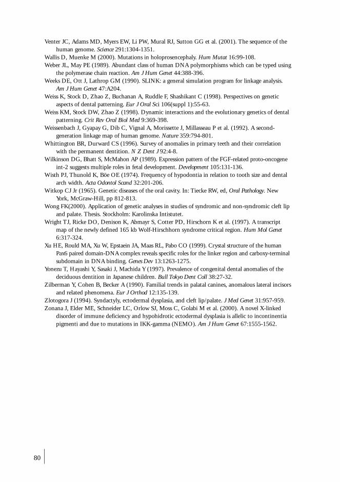
80
Venter JC, Adams MD, Myers EW, Li PW, Mural RJ, Sutton GG et al. (2001). The sequence of thehuman genome. Science 291:1304-1351.
Wallis D, Muenke M (2000). Mutations in holoprosencephaly. Hum Mutat 16:99-108.Weber JL, May PE (1989). Abundant class of human DNA polymorphisms which can be typed using
the polymerase chain reaction. Am J Hum Genet 44:388-396.Weeks DE, Ott J, Lathrop GM (1990). SLINK: a general simulation program for linkage analysis.
Am J Hum Genet 47:A204.Weiss K, Stock D, Zhao Z, Buchanan A, Ruddle F, Shashikant C (1998). Perspectives on genetic
aspects of dental patterning. Eur J Oral Sci 106(suppl 1):55-63.Weiss KM, Stock DW, Zhao Z (1998). Dynamic interactions and the evolutionary genetics of dental
patterning. Crit Rev Oral Biol Med 9:369-398.Weissenbach J, Gyapay G, Dib C, Vignal A, Morissette J, Millasseau P et al. (1992). A second-
generation linkage map of human genome. Nature 359:794-801.Whittington BR, Durward CS (1996). Survey of anomalies in primary teeth and their correlation
with the permanent dentition. N Z Dent J 92:4-8.Wilkinson DG, Bhatt S, McMahon AP (1989). Expression pattern of the FGF-related proto-oncogene
int-2 suggests multiple roles in fetal development. Development 105:131-136.Wisth PJ, Thunold K, Böe OE (1974). Frequency of hypodontia in relation to tooth size and dental
arch width. Acta Odontol Scand 32:201-206.Witkop CJ Jr (1965). Genetic diseases of the oral cavity. In: Tiecke RW, ed, Oral Pathology. New
York, McGraw-Hill, pp 812-813.Wong FK(2000). Application of genetic analyses in studies of syndromic and non-syndromic cleft lip
and palate. Thesis. Stockholm: Karolinska Intistutet.Wright TJ, Ricke DO, Denison K, Abmayr S, Cotter PD, Hirschorn K et al. (1997). A transcript
map of the newly defined 165 kb Wolf-Hirschhorn syndrome critical region. Hum Mol Genet6:317-324.
Xu HE, Rould MA, Xu W, Epstaein JA, Maas RL, Pabo CO (1999). Crystal structure of the humanPax6 paired domain-DNA complex reveals specific roles for the linker region and carboxy-terminalsubdomain in DNA binding. Genes Dev 13:1263-1275.
Yonezu T, Hayashi Y, Sasaki J, Machida Y (1997). Prevalence of congenital dental anomalies of thedeciduous dentition in Japanese children. Bull Tokyo Dent Coll 38:27-32.
Zilberman Y, Cohen B, Becker A (1990). Familial trends in palatal canines, anomalous lateral incisorsand related phenomena. Eur J Orthod 12:135-139.
Zlotogora J (1994). Syndactyly, ectodermal dysplasia, and cleft lip/palate. J Med Genet 31:957-959.Zonana J, Elder ME, Schneider LC, Orlow SJ, Moss C, Golabi M et al. (2000). A novel X-linked
disorder of immune deficiency and hypohidrotic ectodermal dysplasia is allelic to incontinentiapigmenti and due to mutations in IKK-gamma (NEMO). Am J Hum Genet 67:1555-1562.
1b216251taitto28.9 28.9.2001, 17:5080