PhD Thesis - Videncenter For Allergi · Hand eczema, atopic dermatitis, ... This dissertation...
110
UNIVERSITY OF COPENHAGEN FACULTY OF HEALTH AND MEDICAL SCIENCES EPIDEMIOLOGY OF DERMATITIS - A CHARACTERIZATION OF GENETIC PREDISPOSITION AND PERSONAL CONSEQUENCES PhD Thesis Nina Glasser Heede National Allergy Research Centre Department of Dermatology and Allergy Copenhagen University Hospital Herlev-Gentofte 2016
Transcript of PhD Thesis - Videncenter For Allergi · Hand eczema, atopic dermatitis, ... This dissertation...
U N I V E R S I T Y O F C O P E N H A G E N
F A C U L T Y O F H E A L T H A N D M E D I C A L S C I E N C E S
EPIDEMIOLOGY OF DERMATITIS
- A CHARACTERIZATION OF GENETIC PREDISPOSITION
AND PERSONAL CONSEQUENCES
PhD Thesis
Nina Glasser Heede
National Allergy Research Centre
Department of Dermatology and Allergy
Copenhagen University Hospital Herlev-Gentofte
2016

ISBN nr. 978-87-92613-89-9

Epidemiology of dermatitis – a characterization of genetic predisposition and personal consequences
The thesis is a product of a scientific collaboration between
The thesis was submitted to the Graduate School of Health and Medical Sciences, University of Copenhagen, Denmark on 12 September 2016.

i
PhD Student/Author Nina Glasser Heede, MSc
PhD supervisors
Principal supervisor Jeanne Duus Johansen, Professor, MD, PhD National Allergy Research Centre Department of Dermatology and Allergy University Hospital of Copenhagen, Herlev-Gentofte
Co-supervisor Jacob P. Thyssen, Associate Professor, MD, PhD Department of Dermatology and Allergy University Hospital of Copenhagen, Herlev-Gentofte
Co-supervisor Allan Linneberg, Professor, MD, PhD Research Centre for Prevention and Health, The Capital Region of Denmark
Co-supervisor Betina Heinsbæk Thuesen, PhD Research Centre for Prevention and Health, The Capital Region of Denmark
Assessment committee
Chair Lars K. Poulsen, Professor, PhD University of Copenhagen, Denmark
Danish representative Charlotte Gotthard Mørtz, Professor, MD, PhD University of Southern Denmark
International representative Wolfgang Uter, Professor, MD, PhD University of Erlangen-Nürnberg, Germany
ii
This PhD thesis is based on the following four manuscripts:
The manuscripts will be referred to using their roman numerals throughout the thesis.
I Heede NG, Thyssen JP, Thuesen BH, Linneberg A, Johansen JD. Anatomical patterns of dermatitis in adult filaggrin mutation carriers. Journal of the American Academy of Dermatology (2015) 72: 440-8.
II Heede NG, Thyssen JP, Thuesen BH, Linneberg A, Johansen JD. Predictive factors of self-reported hand eczema in adult Danes: a population-based cohort study with 5-year follow-up. British Journal of Dermatology (2016) 175: 287–295.
III Heede NG, Thyssen JP, Thuesen BH, Linneberg A, Szecsi PB, Stender S, Johansen JD. Health- related quality of life in adult dermatitis patients by filaggrin genotype. Submitted for publication in contact dermatitis (2016).
IV Heede NG, Thyssen JP, Thuesen BH, Linneberg A, Szecsi PB, Stender S, Menné T, Johansen JD. Hand eczema, atopic dermatitis, and filaggrin mutations in adult Danes: a registry-based study including risk of disability pension. Submitted for publication in British Journal of Dermatology (2016).

iii
PREFACE
This dissertation received financial funding from the LEO Foundation, funding which is gratefully
acknowledged.
First, I would like to thank the patients who donated data for the patient population.
The scientific work was performed both at the National Allergy Research Centre and the Research Centre
for Prevention and Health. I have been truly privileged by being attached to two outstanding research
environments and I would like to express my gratitude to my four supervisors for constructive feedback and
encouragement — you have all helped me to become a better scientist.
I am grateful to Jeanne Duus Johansen for sharing her enviable knowledge of dermatitis with me and for
giving me the opportunity to write this thesis in a creative and innovative work environment. I would also
like to thank Jacob P. Thyssen for introducing me to the dermatological clinic, discussing new ideas and for
motivational talks. I thank Allan Linneberg for introducing me to the field of epidemiology with his
admirable analytic skills and great biological overview, and I am grateful to Betina Heinsbæk Thuesen for
assisting my transition from biologist to epidemiologist by teaching me SAS (and being willing to look
through many codes), providing statistical advice, and for always having her door open. In addition, I would
like to thank Torkil Menné for great discussions and advice, and Pal B. Szecsi and Steen Stender for a good
collaboration with the FLG genotyping.
A heartfelt thanks to all my colleagues at the National Allergy Research Centre and the Research Centre for
Prevention and Health. It has been inspiring to work with you and I truly appreciate our knowledge sharing
and good discussions. A special thanks to Anne Marie Topp for invaluable assistance in recruiting patients,
data preparation, and for many good talks; Susanne R. Schweitz for secretarial assistance; Pao Lung-Tsai,
Anna B. Olsson, and Anja L. Madsen for IT assistance; Rikke K. Jacobsen for statistical support; Line Tang for
input on SAS-coding; and Kristiane Engebretsen for cheerful discussions on skin barrier function.
I am grateful for the interest my family and friends have in my world of biology, and now epidemiology too,
and I thank my parents, sister and grandfather for their continuous encouragement. Lastly, I thank Rasmus
for always putting a smile on my face when needed, even from a distance.
With appreciation,
Nina Glasser Heede Gentofte, 12 September 2016
iv
ABBREVIATIONS
The following are listed alphabetically; some abbreviations are used only in tables and figures:
AD Atopic dermatitis
CI Confidence interval
DLQI Dermatology life quality index
FLG Filaggrin gene
FLGmut Filaggrin mutation carrier
FLGwt Filaggrin wild type
HE Hand eczema
HRQoL Health-related quality of life
OR Odds ratio

v
SUMMARY
Background and aims
Atopic dermatitis and hand eczema are widespread in the general population with an estimated one-year
prevalence in adults of 2–15% and 10%, respectively. Loss-of function filaggrin gene (FLG) mutations are
also common in Northern European populations and the prevalence in the Danish population is around 8%.
Filaggrin deficiency has been shown to result in impaired skin barrier integrity and the FLG mutations are
further identified as the strongest genetic factor for the development of atopic dermatitis. Additionally, FLG
mutations have been found to be predictive factors of persistent hand eczema in individuals with atopic
dermatitis; the interplay between hand eczema, FLG mutations and atopic dermatitis is, however, still to be
elucidated.
The personal and societal consequences of dermatitis are substantial and include reduced quality of life,
increased healthcare costs and, in the worst case, sick leave, job change, rehabilitation and/or disability
pension. It is currently unknown whether FLG mutation carriers, who often experience severe and
persistent disease, experience worse consequences than do individuals without FLG mutations.
The overall objective of the thesis was to investigate the epidemiology of dermatitis and look into the role
of genetic pre-disposition, defined by FLG mutations, and personal consequences. In detail the aims were:
To investigate the epidemiology of dermatitis in the general population including prevalence,
anatomical localization and association with FLG mutations (Manuscript I).
To investigate incidence and predictive factors of hand eczema in the general adult population
(Manuscript II).
To characterize the adult dermatitis patient with and without FLG mutations focusing on
health-related quality of life (HRQoL), skin characteristics and comorbidity (Manuscript III).
To investigate occupational consequences and previous work in risk occupations among the
adult population with or without dermatitis and FLG mutations (Manuscript IV).
Methods
This thesis builds on data from two populations: I) a population-based cohort study with a 5-year follow-up
called “Health2006” and II) a cross-sectional study of adult dermatitis patients included with atopic
dermatitis and/or hand eczema. Data from participants from both populations were transferred to
Statistics Denmark and linked to central registries for information about socio-economy, occupation, and
social benefits. In addition, all participants completed the same questionnaire about skin symptoms and

vi
dermatitis. Hand eczema, for both populations, was self-reported whereas a history of atopic dermatitis
was defined by the UK criteria in the general populations and was clinician diagnosed in the patients. All
participants were genotyped for three of the most common Northern European loss-of-function FLG
mutations, which together constitute 83% of the total risk alleles associated with atopic dermatitis: R501X,
2282del4, and R2447X. Manuscripts I and II are based on data from the Health2006 population and
Manuscript III is based on data from the patient population. Manuscript IV is based on data from both
populations and from Statistics Denmark.
Results
The overall estimated lifetime prevalence of unspecified dermatitis in the general population was 37.8%.
We also found that FLG mutations were associated with dermatitis on the hands and feet in individuals
with atopic dermatitis (Manuscript I). In our analyses investigating predictive factors of hand eczema in
adult Danes in the general population, we found that a history of atopic dermatitis predicts both incident
and persistent hand eczema (odds ratio (OR) = 9.0; 95% confidence interval (CI) 5.6–14.4 and OR = 3.0; 95%
CI 1.7–5.2, respectively). In contrast, FLG mutations predicted only persistent hand eczema in individuals
with atopic dermatitis and were not associated with incident hand eczema in adults, suggesting that FLG
mutations as a predictive factor for hand eczema decrease with time. Lastly, contact sensitization was also
associated with persistent hand eczema (OR = 2.5; 95% CI 1.2–5.0), independently of a history of atopic
dermatitis (Manuscript II). In relation to HRQoL, we found that patients with atopic dermatitis (± hand
eczema) and FLG mutations reported reduced HRQoL when compared with patients with FLG wild type
suggesting that this subgroup of patients might experience an additional challenge in their everyday life
(Manuscript III). Lastly, we found that self-reported dermatitis, particularly in individuals with FLG
mutations, was significantly associated with receiving disability pension in the general population.
However, the primary diagnosis for awarding disability was unknown (Manuscript IV).
Conclusions
Taken together, our results indicate that FLG mutation carriers with atopic dermatitis are a subgroup of
individuals who stand out on several parameters. The parameters are biologically manifested by increased
prevalence of foot dermatitis and increased persistence of hand eczema, psychologically manifested by
reduced HRQoL, and socially manifested by the finding that self-reported dermatitis was associated with
receiving disability pension, particularly in individuals with FLG mutations. These findings points towards
FLG mutations predisposing to increased severity, highlighting the need for increased skin awareness in this
subgroup.

vii
DANSK RESUMÈ (SUMMARY IN DANISH)
Baggrund og formål
Atopisk eksem og håndeksem er hyppige hudsygdomme i den danske befolkning og har en estimeret 1-års
prævalens blandt voksne på henholdsvis 2-15 % og 10 %. Mutationer i genet, der koder for hudproteinet
filaggrin, er også hyppige. Omkring 8 % af den danske befolkning har mindst én mutation i filaggrin genet
(FLG) hvilket betyder, at de personer har en delvis eller total filaggrinmangel. Filaggrinmangel medfører en
nedsat funktion af hudbarrieren, som er den barriere, der beskytter huden imod påvirkninger fra
omgivelserne. FLG mutationer har yderligere vist sig at være en stærk genetisk risikofaktor for udvikling af
atopisk eksem. Derudover er FLG mutationer blevet identificeret som en risikofaktor for håndeksem, dog
kun blandt personer, der har haft atopisk eksem. Samspillet mellem FLG mutationer, atopisk eksem og
håndeksem er komplekst og er endnu ikke fuldt belyst.
Eksem har både store personlige og samfundsmæssige konsekvenser, da sygdommen er associeret med
reduceret livskvalitet, øget forbrug af sundhedsydelser og i de værste tilfælde, sygefravær, jobskifte,
revalidering og/eller førtidspension. Det er endnu uvist hvorvidt personer med FLG mutationer, der ofte
oplever svær og vedvarende sygdom også oplever større konsekvenser sammenlignet med personer uden
FLG mutationer.
Det overordnede formål med denne Ph.d.-afhandling var, at undersøge epidemiologien af eksem blandt
voksne danskere, både blandt befolkningen og eksempatienter, og se på samspillet med genetisk
disponering, defineret med FLG mutationer, og personlige konsekvenser. De enkelte formål var:
At lave en epidemiologisk undersøgelse af eksem i den generelle befolkning inklusiv prævalens,
anatomisk lokalisation og samspil med FLG mutationer (Manuskript I).
At undersøge incidensen og prædiktive faktorer for håndeksem i den generelle voksne befolkning
(Manuskript II).
At karakterisere voksne eksempatienter, med eller uden FLG mutation, med fokus på
sygdomsrelateret livskvalitet, hudkarakteristika og komorbiditet (Manuskript III).
At undersøge arbejdsrelaterede konsekvenser og historie i risikoerhverv blandt voksne danskere,
med og uden FLG mutationer (Manuskript IV).
Metode
Denne afhandling er baseret på data fra to populationer: I) et populationsbaseret kohortestudie med en 5-
års opfølgning kaldet ”Helbred2006” og II) en tværsnitsundersøgelse af voksne eksempatienter inkluderet
med atopisk eksem og/eller håndeksem. Data fra begge populationer er derudover blevet overført til
Danmarks Statistik, og linket til centrale registre for information omkring socioøkonomi, beskæftigelse og

viii
sociale ydelser. Alle deltagere udfyldte ydermere et spørgeskema omkring hudsymptomer og eksem.
Håndeksem for begge populationer var selvrapporteret, hvorimod diagnosen omkring atopisk eksem var
defineret ved hjælp af UK kriterierne i befolkningen og var lægediagnosticeret blandt patienterne. Alle
deltagerne blev derudover genotypet for tre af de mest almindelige FLG mutationer i Nordeuropa, der
tilsammen repræsenterer 83 % af risikoallelerne for atopisk eksem; R501X, 2282del4 og R2447X.
Manuskript I og II er baseret på data fra ’Helbred2006’ populationen mens Manuskript III er baseret på data
fra patientpopulationen. Manuscript IV er baseret på data fra begge populationer og fra Danmarks Statistik.
Resultater
Den samlede livstidsprævalens for uspecificeret eksem i den danske befolkning var 37,8 %. Derudover fandt
vi, at FLG mutationer disponerede særligt til eksem på hænder og fødder blandt personer med tidligere
atopisk eksem (Manuskript I). I vores analyser omkring prædiktive faktorer for håndeksem fandt vi, at
tidligere atopisk eksem prædikterer både incident og persisterende håndeksem hos voksne (henholdsvist,
OR = 9.0; 95%CI 5.6–14.4 og OR = 3.0; 95%CI 1.7–5.2). Derimod prædikterede FLG mutationer kun
persisterende håndeksem i personer med tidligere atopisk eksem, og var ikke associeret med incident
håndeksem blandt voksne hvilket indikerer, at FLG mutationer udspiller deres rolle tidligt i livet. Ydermere
fandt vi, at allergisk sensibilisering var associeret med persisterende håndeksem uafhængigt af tidligere
atopisk eksem (OR=2.5; 95%CI 1.2–5.0) (Manuskript II). I forhold til sygdomsrelateret livskvalitet fandt vi, at
patienter med atopisk eksem (± håndeksem) og FLG mutationer rapporterede reduceret livskvalitet
sammenlignet med patienter uden FLG mutationer hvilket indikerer, at denne undergruppe af patienter
særligt kan opleve udfordringer i deres dagligdag (Manuskript III). Derudover fandt vi, at selv-rapporteret
eksem, særligt blandt personer med FLG mutationer, var associeret med at være førtidspensionist i den
generelle befolkning. Den primære diagnose for at have fået tildelt førtidspension var dog ukendt i vores
analyse (manuskript IV).
Konklusion
Alt I alt indikerer vores resultater at personer med FLG mutationer og tidligere atopisk eksem er en særlig
undergruppe som adskiller sig på flere parametre; biologisk i form af øget prævalens af eksem på hænder
og fødder og psykisk i form af reduceret hudspecifik livskvalitet. Derudover fandt vi også en social
parameter idet selvrapporteret eksem var særligt associeret med førtidspension blandt deltagere med FLG
mutationer. Disse fund peger imod at FLG mutationer disponerer til øget sværhedsgrad af eksem og øger
behovet for viden omkring hudsymptomer blandt disse personer.

ix
TABLE OF CONTENTS 1. INTRODUCTION .......................................................................................................................................................... 1
1.1 Filaggrin and its role in the skin barrier ............................................................................................................... 1
1.2 Atopic dermatitis .................................................................................................................................................. 3
1.2.1 Comorbidity associated with atopic dermatitis and filaggrin mutations...................................................... 4
1.2.2 Acquired filaggrin deficiency ........................................................................................................................ 4
1.3 Hand eczema – a frequent disease manifestation ............................................................................................... 6
1.3.1 Occupational hand eczema........................................................................................................................... 7
1.4 Consequences of dermatitis ................................................................................................................................ 7
1.4.1 Societal healthcare costs of dermatitis......................................................................................................... 8
1.4.2 Socioeconomic consequences of dermatitis including work-related consequences ................................... 8
1.4.3 Dermatitis and health-related quality of life .............................................................................................. 10
2. METHODS: STUDY POPULATIONS AND REGISTERS .................................................................................................. 11
2.1 The Health2006 cohort ...................................................................................................................................... 11
2.2 The dermatitis patient population ..................................................................................................................... 11
2.3 The Danish registries .......................................................................................................................................... 12
2.4 Ethical statement ............................................................................................................................................... 13
2.5 Statistical analyses ............................................................................................................................................. 13
3. OBJECTIVES OF THE PHD THESIS .............................................................................................................................. 14
4. RESULTS AND MANUSCRIPTS ................................................................................................................................... 15
4.1 Epidemiology of dermatitis in the general population ...................................................................................... 15
Manuscript I ......................................................................................................................................................... 16
4.2 Predictive factors of hand eczema ..................................................................................................................... 25
Manuscript II ........................................................................................................................................................ 26
4.3 Health-related quality of life among adult filaggrin mutation carriers .............................................................. 35
Manuscript III ....................................................................................................................................................... 36
4.4 Occupational consequences of having dermatitis and filaggrin mutations ....................................................... 54
Manuscript IV ...................................................................................................................................................... 55
5. COMMENTS ON METHODOLOGY ............................................................................................................................. 71
5.1 Disease definitions in the general population ................................................................................................... 71
5.2 Considerations about filaggrin genotyping ........................................................................................................ 75
5.3 Establishment of the patient population ........................................................................................................... 75
5.4 Using retrospective registry data ....................................................................................................................... 78
6. DISCUSSION .............................................................................................................................................................. 80
7. CONCLUSIONS .......................................................................................................................................................... 86
8. FUTURE RESEARCH ................................................................................................................................................... 87
9. REFERENCES ............................................................................................................................................................. 88

1
1. INTRODUCTION
Dermatitis is the largest disease group within dermatology and comprises several disease manifestations,
the most prevalent ones being atopic dermatitis and contact dermatitis, which can be present at any
localization, e.g. hand eczema, facial dermatitis or foot dermatitis. The focus on dermatitis has increased
markedly through the last decades. This is reflected in the increasing number of scientific publications
focusing on various aspects of the disease including the underlying biology, diagnosis, treatment, and
personal consequences. The importance of skin barrier integrity and its role in dermatitis has been widely
discussed, and the theories suggesting a genetic predisposition to dermatitis were revolutionized in 2006
with the identification of the FLG mutations.1
This thesis, entitled, “Epidemiology of dermatitis – a characterization of genetic predisposition and personal
consequences” focuses on dermatitis in the general population and in patients and looks into the interplay
between atopic dermatitis, hand eczema, and FLG mutations as well as personal consequences including
HRQoL, socioeconomic measures and history in risk occupations. The background for the four manuscripts
included in the thesis is introduced in the following sections.
1.1 Filaggrin and its role in the skin barrier
The skin is the body’s first line of defence against invading pathogens and external stimuli from the
environment. The epidermis is 0.05–1 mm thick and is the outer layer of the skin, which can be further
divided into the basal layer, spinous layer, granular layer and stratum corneum.2 In short, epidermal cells
(95% keratinocytes) divide in the basal layer and move upwards while differentiating to ultimately end up
in stratum corneum, the outermost layer of the epidermis, as corneocytes without nuclei and cytoplasmic
organells.2 The terminal differentiation is a continuous process and renewal of the epidermis takes
approximately 1 month in humans.3
In 1977, the filaggrin protein was purified for the first time from the stratum corneum of rat epidermis and
was named “stratum corneum basic protein”.4 Four years later, the protein was renamed as filaggrin as a
consequence of its unique biological function of aggregating intermediate filaments (filament aggregating
protein), particularly keratins.5 Filaggrin derives from a large precursor protein called profilaggrin.
Profilaggrin is a major component of the keratohyalin granules, which are organelles found in the granular
layer of the epidermis.6 During the late stages of terminal differentiation, where transition from the
granular to the terminally differentiated cornified cells occurs, profilaggrin is dephosphorylated and
proteolyzed into multiple filaggrin monomers in the epidermis.7,8 The profilaggrin molecule is processed
1
Introduction

2
into 10–12 filaggrin monomers.9 The functional filaggrin monomers hence align keratin filaments to form
the interfilamentous matrix in stratum corneum.10 Subsequently, filaggrin degradation results in the
formation of free amino acids and their derivatives, including, for example 2-pyrrolidone-5-carboxylic
acid and trans-urocanic acid, which are part of the natural moisturizing factors ensuring epidermal
hydration, photoprotection and maintenance of the acid mantle.11
Chromosome 1 is our largest chromosome and contains about 8% of the entire human genomic
information. FLG is located within the epidermal differentiation complex on chromosome 1 region 1q21.12
Mutations in the FLG are among the most prevalent single-gene mutations identified to date.Reviewed in 13 Up
to 10% of the population with Northern European origin are heterozygous carriers of a loss-of-function
mutation within the FLG, resulting in a 50% reduction of expressed protein.14,15 Figure 1 illustrates filaggrin,
and profilaggrin, expression in the different layers of the epidermis for an individual with normal filaggrin
expression (A) and a patient with total filaggrin deficiency (B).
Including the two initially reported mutations (R501X and 2282del4), more than 50 additional mutations
have been identified throughout the profilaggrin molecule, of which many European-specific and Asian-
specific mutations exist.13,16 The three mutations R501X (39%), 2282del4 (41%) and S2447X (3%) together
constitute 83% of the total risk alleles associated with development of atopic dermatitis.13
Figure 1: Filaggrin stain of the epidermis Immunohistochemical staining for profilaggrin and/or filaggrin shows staining in the granular layer and stratum corneum (panel A) in contrast to the staining from a patient homozygous for loss-of-function mutations in the filaggrin gene (panel B). Reproduced with permission from Irvine et al., 2011, N Eng J Med,
13 Copyright Massachusetts Medical Society.
A B
2
Introduction

3
The filaggrin protein has been shown to have numerous functions in epidermis, and mutations in the FLG
have been shown to result in an impaired skin barrier associated with both skin and allergic diseases. This is
presented in the following sections.
1.2 Atopic dermatitis
Atopic dermatitis is a highly pruritic inflammatory skin condition with chronic or recurrent episodes of
dermatitis. The clinical manifestations of the condition are thought to vary with age, but all stages are
characterized by pruritus and xerosis. In infancy, lesions usually emerge on the cheeks and scalp. Later in
childhood, and through adolescence and adulthood, lesions are usually localized to flexures, the neck and
the dorsal aspects of the limbs. Reviewed in 17
An increase in both prevalence and incidence of atopic dermatitis has been reported since World War II.18,19
Today, the lifetime prevalence of atopic dermatitis is estimated to be around 20% in Denmark, in Western
Europe and in the United States.20-24 The proportion of adults experiencing symptoms of atopic dermatitis is
uncertain but persistency of the disease into adulthood and late onset is common.25-27 A meta-analysis from
2016 found that 80% of childhood atopic dermatitis did not persist beyond the age of eight years and that
less than 5% of cases of childhood atopic dermatitis persisted 20 years after diagnosis.28 In contrast, a
prospective Danish cohort study with a 15-year follow-up reported persistence of atopic dermatitis in 50%
of those diagnosed in school age.27 Another Danish study found that the 1-year prevalence of atopic
dermatitis was 14.3% in adults aged 30–89 years and that the prevalence decreased with increasing age.29
In 1996, Herd and colleagues reported that adults over 16 years made up 38% of all patients with atopic
dermatitis and proposed a future cohort effect as a consequence of the rising prevalence.25 In line with this,
a recent cross-sectional cohort study found that it was not until the age of 20 years that 50% of patients
had at least one lifetime episode of a 6-month symptom- and treatment-free period.26 Thus, atopic
dermatitis is not only a childhood disease but can be considered as a life-long phenotype.
A genetic component for the development of atopic dermatitis has been suspected since the 1980s.19 In
2006, loss-of-function variants within the FLG were identified as the strongest risk factors for the
development of atopic dermatitis.1 Today, numerous studies have confirmed this association and meta-
analyses have estimated the overall OR to range from 3.12 to 4.78.30,31 Despite the presence of FLG
mutations being the strongest known risk factor for atopic dermatitis, around half (46%) of the individuals
heterozygous for FLG mutations will not develop signs of dermatitis, underlining that environmental factors
are important.32 A strong association between FLG mutations and both early-onset and persistent disease
has however been shown.33-35 In addition, the rate of children “growing out’’ of their atopic dermatitis has
been reported to be much lower among individuals with FLG mutations.32 Genome-wide association studies
3
Introduction

4
later identified new risk loci for atopic dermatitis, among others, stressing the importance of epidermal
barrier function and immune dysregulation in disease pathogenesis.36,37
1.2.1 Comorbidity associated with atopic dermatitis and filaggrin mutations
Atopic dermatitis is further considered to be the first clinical manifestation of the atopic march, which
describes the phenomenon in persons with early onset of atopic dermatitis of increased risk of developing
allergic rhinitis and/or asthma, and possible food allergy.38 Approximately 70% of patients with severe
atopic dermatitis will develop allergic rhinitis or asthma later in life.39 The relevance of using the term
‘atopic march’ has, however, been questioned recently as a consequence of different time patterns in
developing atopic dermatitis, asthma and allergic sensitization.40,41
Interestingly, FLG mutations have been found to be risk factors for rhinitis,31 asthma,30,42 and food allergy43,
primarily in co-occurrence with atopic dermatitis. In support of this, it has been hypothesized that an
impaired skin barrier can provide entry for environmental allergens and thereby function as a route of
primary sensitization.44-46 Notably, FLG mutations were recently found not to be associated with food and
aeroallergen sensitization in adults without concomitant atopic dermatitis.47 Moreover, results from
population studies have shown that FLG mutation carriers are at increased risk of developing early-onset
and persistent hand eczema, but only in co-occurrence with atopic dermatitis.48
In contrast to the well-known association between FLG mutations and ichthyosis vulgaris,1,49 only few
studies have investigated associations between FLG mutations and skin cancer. It is widely recognised that
UV exposure is an indirect cause of skin cancer—non-melanoma skin cancer (squamous cell carcinoma and
basal cell carcinoma); malignant melanoma; and pre-stages to non-melanoma skin cancer, actinic
keratosis.50 Experimental data have indicated that filaggrin deficiency alone can impair the epidermal
barrier function, resulting in increased UV sensitivity in human skin models, most likely as a consequence of
reduced levels of trans-urocanic acid.51 However, previous studies investigating the association between
filaggrin and skin cancer are ambiguous. A population-based cohort study found no association between
FLG mutations and squamous cell carcinoma or malignant melanoma,52 whereas another study suggested
that complete filaggrin deficiency is associated with squamous cell carcinoma.53 Further, no association
between FLG mutations and basal cell carcinoma was found when comparing the proportion of FLG
mutation carriers in patients with basal cell carcinoma with that of the general population.54
1.2.2 Acquired filaggrin deficiency
When investigating the role of FLG mutations in dermatitis, acquired filaggrin deficiency must be
highlighted as an important modulator. Elevated levels of inflammatory cytokines have been shown to
affect filaggrin expression and can result in an acquired filaggrin deficiency.55-61 As TH2-associated cytokines
4
Introduction

5
are one of the hallmarks of acute atopic dermatitis, the initial studies investigated the effect of IL-4 and IL-
13 on filaggrin expression.55 More recently, a whole panel of cytokines associated with atopic dermatitis,
including IL-17A, IL-22, IL-25 (IL-17E), IL-31, and TNF-alpha, have been associated with acquired filaggrin
deficiency in in vitro studies.55-61
Figure 2 illustrates the complex relationship between impaired barrier function, immunologic hyper-
reactivity and acquired filaggrin deficiency.
Thus research investigating immune dysregulation in patients with atopic dermatitis has shown that
acquired filaggrin deficiency is common, which suggests that disease severity affects filaggrin expression,
irrespective of FLG genotype. Filaggrin expression was also found to be down regulated in nonlesional skin
of adult patients with atopic dermatitis.56 Lastly, prolonged use of topical corticosteroids can reduce
epidermal filaggrin levels.62
Research regarding the immunological profile of hand eczema is limited and studies focusing on acquired
filaggrin deficiency in hand eczema are sparse. Nevertheless, it has been reported that skin barrier
dysfunction also plays a key role in the pathogenesis of chronic hand eczema which, among others proteins,
results on down-regulation of the filaggrin protein.63
Figure 2: Illustration of the interplay between impaired skin barrier function and immunological hyper-reactivity seen in patients with atopic dermatitis and with hand eczema.
5
Introduction

6
1.3 Hand eczema – a frequent disease manifestation
Hand eczema is an inflammatory skin condition with various clinical manifestations including erythema, cell
infiltration, hyperkeratosis, oedema and vesicles. Moreover, secondary signs exist, such as scaling,
hyperkeratotic areas, skin fissures and bacterial infections primarily with Staphylococcus aureus.64
Often, morphology and symptoms vary over time. In general, hand eczema often starts as acute dermatitis
characterized by erythema, oedema and vesicles, which can develop into chronic dermatitis characterized
by hyperkeratosis, infiltrations and fissures.64
Hand eczema is a multifactorial condition that usually develops as a consequence of repeated or prolonged
contact with irritant and/or allergic compounds (contact dermatitis). It particularly affects individuals with a
history of atopic dermatitis and women.64-66 Most cases of contact dermatitis manifest as hand eczema and
factors frequently involved in the aetiology are wet work, detergents, sensitizing chemicals, regular use of
occlusive gloves and exposure to food proteins.64 The pathogenesis of hand eczema is complex, challenging
the traditional clinical distinction between irritant, allergic and atopic phenotypes. Thus atopic dermatitis
can also be manifested on the hands in adults. Frequent sub-diagnoses, or combinations of sub-diagnoses,
have been proposed based on data from 319 patients from 10 European patch test clinics.67 Here, irritant
contact dermatitis (21.5%) was the most frequent subtype, followed by allergic contact dermatitis (15.2%),
irritant contact dermatitis + allergic contact dermatitis (15.2%), vesicular hand eczema (9.3%), atopic hand
eczema + irritant contact dermatitis (7.8%) and atopic hand eczema (5.8%).67
The life-time prevalence of hand eczema in the adult Scandinavian general population has been found to
range between 15% and 21.8%,68,69 and the 1-year prevalence is estimated to be nearly 10%, while the
point prevalence was estimated to be 4%, in a review mainly based on European studies.70 The incidence of
hand eczema varies within age groups and the incidence rate of self-reported hand eczema has been
reported to peak among young woman aged 20–29 years and subsequently decrease with age.71 The fact
that hand eczema has repeatedly been shown to be a female-dominated condition21,69,72 has been
explained by gender differences in domestic and occupational exposures to irritants and allergens, rather
than by susceptibility differences between men and woman.70,73 However, individual susceptibility to hand
eczema has been found to be associated with atopic dermatitis. Rystedt and colleagues showed already in
1985 that patients with persistent and recurrent atopic dermatitis had an increased risk of developing hand
eczema in adulthood.74 Atopic dermatitis has since been confirmed as a risk factor for hand eczema, also in
prospective cohorts of adolescence from the general population.66,75 Other risk factors for hand eczema,
apart from female sex and atopic dermatitis, include contact allergy, wet work, xerosis, and a high use of
tobacco.65,70,76-79 As mentioned, FLG mutation carriers with atopic dermatitis have been found to have an
6
Introduction

7
increased risk of persistent hand eczema;48 however, the interplay between hand eczema, FLG mutations
and atopic dermatitis remains to be elucidated.
1.3.1 Occupational hand eczema
Work-related skin diseases are the most prevalent condition reported to the Danish National Board of
Industrial Injuries. In 2014, 2,889 cases of occupational skin diseases were reported, of which 1,616 were
recognized (55.9%).80 Because most occupational skin diseases manifest as occupational hand eczema,
occupational hand eczema is the most frequently recognized industrial injury and represents a substantial
expense to society.81,82
The epidemiology of occupational contact dermatitis has long been in focus. A review from 1999 found that
the annual incidence rate of the disease was the highest among hairdressers (194 cases per 10,000
employees/year), followed by bakers (64 cases per 10,000 employees/year).83 Moreover, the median
induction period for occupational skin diseases in different professions showed that hairdressers, food
industry workers, health service workers and metal workers belong to high risk professions.83 Apart from
hairdressing and health-care work, other female-dominated occupations involving extensive wet work,
such as cleaning or catering, are characterized as occupations with a high risk of hand eczema.Reviewed in 84 A
recent population-based study investigating water exposure in high-risk occupations showed that more
than 50% of individuals working in service occupations, including kitchen assistants, cleaners, restaurant
workers and hairdressers, report water exposure for more than 2 hours a day.85 Individuals working in
occupations in the health-care sector and the construction sector had equally high proportions of water
exposure.85 As mentioned, atopic dermatitis is the most pronounced risk factor for hand eczema, and skin
atopy has been estimated to double the risk of the disease in occupations where hand eczema is
common.86 In relation to skin barrier, research has shown that FLG mutation carriers with childhood hand
eczema tend to choose occupations with a low risk of exposure to irritants.87 This finding suggests that
individuals with FLG mutations forfeit occupations with a high risk of dermatitis, such as wet work.
Moreover, FLG mutations have been found to be associated with a high prevalence of contact dermatitis in
construction workers.88 However, no studies have investigated the prevalence of FLG mutation carriers
working in a risk occupation, either among patients or in the general population.
1.4 Consequences of dermatitis
The personal consequences of having dermatitis, both atopic dermatitis and hand eczema, are far-reaching
and include financial consequences, work-related consequences, increased use of healthcare services and
decreased quality of life. Thus the overall “cost” of having dermatitis can be considerable.
Introduction
7

8
1.4.1 Societal healthcare costs of dermatitis
The economic burden of atopic dermatitis in the United States is estimated to range from $364 million to $
3.8 billion (€324 million to €3.4 billion)I.89,90 In 1999, a German study also investigated the cost of having
atopic dermatitis and found that the annual cost per patient was €2462II and that the estimated annual
cost to society was €3.57 billionII.91 In addition, a recent systemic review investigating cost-of-illness studies
in hand eczema found that the mean annual total cost per patient ranged from €1712 to €9792 (direct cost
per patient: €521 to €3829 and indirect cost per patient: €100 to €6846) whereof occupational hand
eczema patients had indirect costs up to 70% of the total costs, mainly because of sick days.92 Thus,
dermatitis represents substantial expense to both the individual and society.
An association between the severity of hand eczema and medical consultations has previously been
shown.93 Here, 63.4% of individuals with hand eczema reported medical consultations because of the
disease and 15.6% reported more than five consultations.93 Further, atopic dermatitis has been associated
with increased healthcare costs,94 and the risk of reporting more than one medical consultation due to hand
eczema was increased in individuals with a history of atopic dermatitis (OR = 3.0; 95% CI 1.4–6.4).93 To
date, no studies have investigated whether individuals with a genetically impaired skin barrier have an
increased use of healthcare services. Information about the use of health-care services among FLG
mutation carriers is unknown but might be valuable since they have an increased risk of developing both
atopic disorders and hand eczema and represent a considerable proportion of the population.
1.4.2 Socioeconomic consequences of dermatitis including work-related consequences
Socioeconomic status can be estimated by several variables including income, family status, occupational
status and degree of education. Research has shown that both increased educational level and income are
associated with a better health status.95 For patients with hand eczema, associations between middle-
income households and increased prevalence of hand eczema have been suggested.96 Moreover, reports
have shown that men who live alone tend to report hand eczema more often than those who live with
someone.96 Hand eczema is further known to have widespread work-related consequences including
productivity loss, sick leave, job change, rehabilitation benefits and disability pensions.97-100 In 2005, Meding
and colleagues reported that the proportion of patients with hand eczema, diagnosed in a population-
based study, who experienced far-reaching consequences including extended sick leave, disability pension
and changes of occupation, was about 5%.100 It is currently unknown whether persons with FLG mutation
have different job-related consequences compared with non-mutation carriers.
I The rate 1 USD = 0.89 Euro has been used to calculate the amount (11 September 2016).
II The original study reports an amount in German mark (DM). The rate: 1 DM = 0.51 Euro has been used.
8
Introduction

9
In individuals with atopic dermatitis, associations between high parental education level and increased risk
of atopic diseases in the offspring have been suggested.101,102 Atopic dermatitis has also been associated
with increased work loss when compared with controls.103 However, epidemiological evidence targeting the
relationship between socioeconomic status and dermatitis is limited101 and the potential socioeconomic
consequences of having a genetically impaired skin barrier are unknown. Figure 3 summaries the
widespread consequences of having dermatitis.
Figure 3: Illustration of the widespread consequences associated with dermatitis (eczema). Blue arrows represent known consequences of dermatitis while red dotted arrows represent the unknown consequences of having a genetically disrupted skin barrier.
9
Introduction

10
1.4.3 Dermatitis and health-related quality of life
HRQoL can be estimated using the Dermatology Life Quality Index (DLQI) introduced in 1994 by Finlay and
colleagues as the first skin-specific instrument to assess HRQoL.104 The DLQI has been used to measure
HRQoL in patients with both atopic dermatitis and hand eczema where increased severity has been found
to be associated with greater impairment in DLQI score.105-107 While several studies have found hand
eczema to be associated with reduced HRQoL,107 the association between hand eczema and depression and
anxiety is ambiguous. A Danish study from 2006 including 758 patients with occupational hand eczema
found no association between occupational hand eczema and depression,108 whereas a German study from
2012 found a high prevalence of both anxiety and depression in the study population of patients with
occupational hand eczema.109 Higher anxiety level and increased DLQI scores were associated with female
sex.109 A multicentre study from 2015 also found that depression and anxiety were associated with hand
eczema and atopic dermatitis, suggesting that there is an additional burden of having skin diseases.110 In
line with this, it has repeatedly been shown that individuals with atopic dermatitis report an increased
prevalence of both depression and anxiety.111,112 It is currently unknown whether the psychological impact
of dermatitis differs between FLG mutations carriers and non-mutation carriers.
In light of the extensive literature investigating the biological consequences of having FLG mutations, it is
warranted to investigate the epidemiology of dermatitis with focus on genetic predisposition and personal
consequences.
10
Introduction

11
2. METHODS: STUDY POPULATIONS AND REGISTERS
This PhD thesis builds on data from two populations: I) a population-based cohort study with a 5-year
follow-up called “Health2006” and II) a cross-sectional study of adult dermatitis patients included with
atopic dermatitis and/or hand eczema. Data from participants from both populations (the Health2006
cohort and patients) were transferred to Statistics Denmark and linked to central registries for information
about socio-economy, occupation, and social benefits. In addition, all participants completed
questionnaires about skin symptoms and dermatitis and were genotyped for three of the most common
Northern European loss-of-function mutations in the profilaggrin gene which together constitute 83% of
the total risk alleles associated with atopic dermatitis; R501X, 2282del4, and R2447X.1,13,113
2.1 The Health2006 cohort
The Health2006 population was established between 2006 and 2008 and has previously been described in
detail.114 Participants in the Health2006 study were adults aged 18 to 69 years, with Danish citizenship who
were born in Denmark. Participants were recruited as a random sample of the population. All participants
lived in one of the 11 municipalitiesIII in the south-western part of the Copenhagen County.114 The study
comprised 3471 individuals (participation rate: 44.7%) and was designed to investigate research questions
dealing with lifestyle-related chronic diseases in Denmark, including skin health. All participants completed
detailed questionnaires, including questions on skin health, dermatitis, and co-morbidities, and underwent
a general health examination at the Research Centre for Prevention and Health. In addition, blood samples
were collected for FLG genotyping, and type IV allergy was estimated by patch testing for contact allergens
incorporated in panel 1 and 2 from the standardised ready-to-apply Thin-layer Rapid Use Epicutaneous
(TRUE)-test (Mekos Laboratories, Hillerød, Denmark).114 Finally, type I allergy was estimated by measuring
allergen-specific IgE in serum. All samples were analysed for specific IgE to birch, grass, cat and house dust
mite (ALK-Abello A/S, Hørsholm, Denmark). The 5-year follow-up was completed between 2011 and 2013
and included questions similar to those at baseline. In total, 2308 individuals were included in the follow-up
study (participation rate at follow-up 66.5%).
2.2 The dermatitis patient population
The dermatitis population was established in 2014 as part of this thesis. In total, 1119 adult dermatitis
patients (> 18 years) were invited to participate in the study. Of these individuals, 520 with atopic
dermatitis and/or hand eczema were included (participation rate 46.5%); however, one did not return the
questionnaire. Clinical diagnoses of atopic dermatitis and/or hand eczema were scored by the attending
dermatologist and were registered in the patient’s file along with the internationally recognized MOAHLFA-
III
Albertslund, Ballerup, Brøndby, Glostrup, Herlev, Høje Taastrup, Hvidovre, Ishøj, Ledøje-Smørum, Rødovre and Vallensbæk
11
Methods

12
index (Male, Occupational dermatitis, history of Atopic dermatitis, Hand eczema, Leg dermatitis, Facial
dermatitis, and Age above 40 years)115 which is registered routinely for all patients who are patch tested.
The patch test database at Gentofte Hospital contains clinical data on all patients examined during 1985–
2016. All patients were seen in the clinic between 2006 and 2012. In the clinic, the diagnosis of atopic
dermatitis represents a lifetime prevalence and is based on family history, flexural dermatitis and typically
childhood onset. Hand eczema is a clinical diagnosis made according to the guideline of the Danish
Dermatological Society.116 The diagnoses was made by the attending doctor and was recorded in the
database together with results from patch testing (European baseline series, and relevant additional test
series according to exposure analysis, in accordance with the European patch test guidelines),117 and skin
prick tests testing type I allergies. 97.3% of the population were Danish citizens. Patients collected genomic
DNA by a buccal swap (Isohelix, Harrietsham, United Kingdom) and completed a questionnaire about skin
health and dermatitis. Epidata was used as the data entry module for questionnaire data.118 A random
check was performed on 10% of the questionnaires entered in Epidata and showed an error rate of
0.28%.The project was reported to, and approved by, the Danish Data Protection Agency.
2.3 The Danish registries
To address the personal consequences of having dermatitis, data from participants from both populations
were transferred to Statistics Denmark and linked to central registries for information about socio-
economy, occupation, and social benefits. Statistics Denmark administers an extensive database of register
data that enables various research possibilities. Pseudo anonymised micro data, at individual level, are
available in different registers, which are updated yearly. Participants were linked to the Danish National
Registers using their unique personal identification numbers (CPR numbers). Data in this dissertation
originate from two separate data extractions including 4584 individuals: 3471 individuals from the
Health2006 cohort and 1119 individuals from the patient population, of whom 520 were included in the
study (participation rate 46.5%). The total number of participants only adds up to 4584 because 6 of the
participants in the Health2006 population were also invited to the patient study; 5 chose to participate.
These 6 individuals were deleted as participants in the patient population in Manuscript IV and appear only
once in the analyses.
In the first data extraction, the general population was linked to the Danish registers in 2006 and the
patients were linked in 2013. The following variables were retrieved: family status, occupational status,
highest completed education, and income. In a second data extraction, made by Statistics Denmark, a
retrospective linkage was performed investigating whether participants had a ‘history in a risk-occupation’
during 1994–2013.
12
Methods

13
Yearly variables: Family status, occupational status, education, and income were used to evaluate
socioeconomic position. Exact information regarding all variables was retrieved, for both populations, by
linkage to the National Danish Registers during the inclusion year: 2006 for the Health2006 population and
2013 for the patient population, respectively. The Danish National Registry of Population Statistics includes
information regarding family type and country of origin. The National Danish Registry of Personal Income
contains information regarding gross income and information regarding whether a person is employed,
unemployed or outside the labour force. The Danish Education Register contains information about the
highest completed education (this variable is updated each 1 October) and information about ongoing
education.
Complete tailored data extraction from Statistics Denmark during 1994–2013: Previous work in a risk
occupation was investigated for all individuals in the work active period (16–64 years) using the Danish
Registers on Labour Market Affiliation, which contain information on economic and employment
conditions.119 The registry is an annual labour market statistic based on the individual’s connection to the
labour market estimated on the last working day in November. Statistics Denmark looked into data files,
tracking occupations back for as long as possible, from 1994 to 2013. The occupational classification was
based on DISCO-88120 (1994–2009) and DISCO-08121 (from 2010 and onwards) codes. The changes in DISCO
codes are due to a change in the international standard classification of occupation system.
2.4 Ethical statement
All participants in both populations signed a written informed consent form before inclusion. The ethics
committee of the Capital Region of Denmark approved the Health2006, baseline (KA-20060011) and follow-
up (H-3-2011-081), studies and the patient population (H-1-2013-127). Both studies were further approved
by the Danish Data Protection Agency.
2.5 Statistical analyses
All statistical analyses were performed using SAS software (SAS, Version 9.3 for Windows, SAS Institute Inc.,
Cary, NC, USA). Statistical analyses for dichotomous variables were made using the Chi Square Test, Fishers
Exact Test, or Cochran-Armitage (Exact) Trend Test. Continuous variables were presented as median scores
with 25th and 75th percentiles. Non-parametric variables were tested for group difference using Kruskal-
Wallis Test. Level of significance was set at P < 0.05. Multivariate logistic regression models were used to
adjust for confounders (for example sex, age group and atopic dermatitis) when investigating associations
between a dependent variable and selected explanatory variables. Associations were expressed as OR with
95% CI. Figures were made with SAS, GraphPad Prism version 6.07 for Windows (GraphPad software, La
Jolla, CA, USA) or with NodeXL for Excel.122
13
Methods

14
3. OBJECTIVES OF THE PHD THESIS
This thesis is based on epidemiological data from the adult general population and adult dermatitis
patients.
The overall objective of the thesis was to estimate the prevalence of dermatitis in the general population,
and look into the role of genetic pre-disposition, defined by FLG mutations, and personal consequences of
the conditions.
In more detail, the aims were:
To investigate the epidemiology of dermatitis in the general population including prevalence,
anatomical localization and association with FLG mutations (Manuscript I).
To investigate incidence and predictive factors of hand eczema in the general adult population
(Manuscript II).
To characterize the adult dermatitis patient with and without FLG mutations focusing on HRQoL,
skin characteristics and comorbidity (Manuscript III).
To investigate occupational consequences and previous work in risk occupations among the adult
population with and without dermatitis and FLG mutations (Manuscript IV).
14
Objectives

15
4. RESULTS AND MANUSCRIPTS
This section summarises key findings related to the stated aims. The original manuscripts are included after
each aim. Manuscripts I and II are based on data from the Health2006 cohort, and Manuscript III is based
on data from the patient population. Manuscript IV is based on data from both populations as well as from
Statistics Denmark.
4.1 Epidemiology of dermatitis in the general population
The overall estimated lifetime prevalence of dermatitis in the Health2006 cohort was 37.8% (not presented
in Manuscript I). The fact that more than one third of the general population report a lifetime prevalence of
dermatitis confirms dermatitis as being a prevalent disease affecting individuals from both sexes and all age
groups.
In Manuscript I we evaluated prevalence, anatomical localization and association with FLG mutations. Table
1 is a simplified summary of Tables I and II in Manuscript I and illustrates that hand eczema is the most
frequent localization of dermatitis among participant who both participated in the baseline and follow-up
studies, followed by dermatitis at the combined localization abdomen, chest or back, and third, facial
dermatitis. The prevalence of atopic dermatitis was found to be 9.4% when using of the UK criteria.
Table 1: Lifetime prevalence of dermatitis at different localizations at baseline.
In total, 8.0% were carriers of at least one mutation in the FLG and 70% of the homozygous FLG mutation
carriers had a history of atopic dermatitis. The frequency of foot dermatitis and persistent hand eczema in
the general population was associated with FLG genotype (P = 0.014 and P < 0.001, respectively). However,
when stratifying for FLG genotype and a history of atopic dermatitis, we found that FLG mutations affected
only the lifetime prevalence of foot dermatitis and persistent hand eczema in participants with a history of
atopic dermatitis. Thus the atopic phenotype showed to be a more important factor than FLG genotype in
relation to dermatitis at different localizations.
Lifetime prevalence
Percent (%)
Hand eczema (HE) Lifetime prevalence of HE reported at baseline Lifetime prevalence of HE both at baseline and follow-up (Table II)
20.9 14.2
Abdomen, chest or back 11.8
Facial dermatitis 10.6
Atopic dermatitis 9.4
Axillae 8.0
Foot dermatitis 6.7
15
Results: Manuscript I

Manuscript I
Anatomical patterns of dermatitis in adultfilaggrin mutation carriers
Nina G. Heede, MSc,a Jacob P. Thyssen, MD, PhD,a Betina H. Thuesen, PhD,b
Allan Linneberg, MD, PhD,b,c,d and Jeanne D. Johansen, MD, PhDa
Hellerup, Glostrup, and Copenhagen, Denmark
From
D
G
th
C
an
M
The L
st
Fo
A
Ej
D
La
tio
440
Background: Common filaggrin (FLG) null mutations are associated with severe and early onset of atopicdermatitis (AD). To date, few studies have investigated anatomical patterns of dermatitis and none has beenconducted in the general population.
Objective: We evaluated patterns of dermatitis in an adult general population stratified by FLG genotype.
Methods: Data from a population-based cohort study with a 5-year follow-up were used. This studyincluded 2143 participants aged 18 to 72 years. Information about dermatitis on the hands; feet; face;axillae; and abdomen, chest, or back was obtained by use of questionnaires. Participants were genotypedfor common FLG mutations. A history of AD was defined by the United Kingdom Working Party’sdiagnostic criteria.
Results: The frequency of foot dermatitis in the general population was associated with FLG genotype(P = .014). However, when stratification of FLG genotype and AD was performed, we found that FLGmutations increased the prevalence (odds ratios) of foot dermatitis (odds ratio 10.41; 95% confidenceinterval 5.27-20.60) and persistent hand dermatitis (odds ratio 17.57; 95% confidence interval 8.60-35.89)only in participants with AD.
Limitations: Potential misclassification and recall bias are study limitations.
Conclusion: FLGmutations affected the lifetime prevalence of hand and foot dermatitis in participants witha history of AD. ( J Am Acad Dermatol 2015;72:440-8.)
Key words: atopic dermatitis; epidemiology; filaggrin; foot dermatitis; genotype; hand dermatitis;population study.
Abbreviations used:
AD: atopic dermatitisFLG: filaggrinOR: odds ratio
Loss-of-function mutations within the filaggrin(FLG) gene are associatedwith a dysfunctionalskin barrier and are considered the strongest
genetic risk factors for the development of atopicdermatitis (AD).1-4 The FLG gene is locatedwithin the
the National Allergy Research Center, Department of
ermato-Allergology, Copenhagen University Hospital
entofte, Hellerupa; Research Center for Prevention and Health,
e Capital Region of Denmark, Glostrupb; Department of
linical Experimental Research, Glostrup University Hospitalc;
d Department of Clinical Medicine, Faculty of Health and
edical Sciences, University of Copenhagen.d
EO Foundation is acknowledged for funding. The Health2006
udy was financially supported by grants from the Velux
undation; the Danish Medical Research Council; the Danish
gency for Science, Technology and Innovation; the Aase and
ner Danielsens Foundation; ALK-Abell�o A/S (Hørsholm,
enmark); Timber Merchant Vilhelm Bangs Foundation; MEKOS
boratories (Denmark); and the Research Center for Preven-
n and Health, the Capital Region of Denmark. The Lundbeck
Foundation (grant number: R108-A10225) is also acknowl-
edged for funding.
Conflicts of interest: None declared.
A selection of the results was presented at the 12th Congress of
the European Society of Cutaneous Allergy and Contact
Dermatitis, Barcelona, Spain, June 26, 2014.
Accepted for publication January 5, 2015.
Reprint requests: Nina G. Heede, MSc, National Allergy Research
Center, Department of Dermato-Allergology, Copenhagen
University Hospital Gentofte, Kildeg�ardsvej 28, 2900 Hellerup,
Denmark. E-mail: [email protected].
Published online February 5, 2015.
0190-9622/$36.00
� 2015 by the American Academy of Dermatology, Inc.
http://dx.doi.org/10.1016/j.jaad.2015.01.001
Downloaded from ClinicalKey.com at BS - University of Copenhagen September 12, 2016.For personal use only. No other uses without permission. Copyright ©2016. Elsevier Inc. All rights reserved.
16

J AM ACAD DERMATOL
VOLUME 72, NUMBER 3Heede et al 441Manuscript I
epidermal differentiation complex on chromosome1q21.5 To date, 49 truncating mutations in the pro-FLG molecule have been reported and variationamong European-specific and Asian-specific muta-tions exists.6 Upon normal gene expression, the pro-FLG molecule is dephosphorylated and proteolyzedinto FLG monomers, which help to align keratin
CAPSULE SUMMARY
d Filaggrin mutations are the strongestknown genetic determinants of atopicdermatitis.
d In this general population of Danishadults, filaggrin mutations affected thelifetime prevalence of persistent handdermatitis and foot dermatitis in personswith atopic dermatitis.
d This knowledge might helpdermatologists to identify patients withfilaggrin mutations.
filaments in the stratum cor-neum.7,8 FLG degradationproducts are part of the nat-ural moisturizing factors,which provide epidermalhydration, photoprotection,and maintenance of the acidmantle.9 Hence, FLG muta-tion carriers show signifi-cantly reduced levels ofnatural moisturizing factorsand higher transepidermalwater loss when comparedwith controls.10
About 10% of the popula-tion with Northern Europeanorigin is a heterozygous car-
rier of an FLG mutation.11,12 FLG mutations conveymajor susceptibility to severe and early-onset AD thatpersists into adulthood.13 Results from cross-sectional population studies have further demon-strated that FLG mutations are associated withfissured skin on the hands and that the combinationof AD and FLG mutation is associated with earlyonset and persistent hand dermatitis.14,15 Apart fromthe distinct phenotype of hand dermatitis,16 a strongpositive association between dry skin and FLGmutations has been reported in adults from thegeneral population17 and in adult patients withdermatitis.18 Anatomical localizations of dermatitisstratified by FLGmutation status were investigated ina prospective birth cohort of Danish children duringtheir first 7 years of life.19 FLG mutations wereassociated with a specific endotype of AD primarilycharacterized by predilection to exposed skin areasof the body, in particular the hands and cheeks.19However, associations between FLG mutations anddermatitis on other body parts in the general adultpopulation have been only sparsely investigated. Inthis study, we characterized patterns of self-reporteddermatitis on the hands; feet; face; axillae; orabdomen, chest, or back in the general populationstratified by FLG genotype and AD.
METHODSStudy population
During June 2006 through June 2008, a cross-sectional population study including 3471 persons
Downloaded from ClinicalKey.com at BS - UniveFor personal use only. No other uses without permission. C
17
was conducted in the southwestern part ofCopenhagen. The Health2006 cohort was estab-lished to investigate the epidemiology of chronicdiseases in adult Danes and has been described inmore detail elsewhere.20 The sampling area has beenused for decades and has previously been found tobe representative of the total Danish population in
rsity of Copenhagen September 12opyright ©2016. Elsevier Inc. All
regard to age, sex, andmarital status.21 Participantswere aged 18 to 72 years andwere all Danish citizens bornin Denmark. The cohort wasdrawn as a random sampleof the population obtainedthrough the Danish CentralPersonal Register, Ministry ofInternal Affairs. Participantsattended a general healthexamination and completedquestionnaires. Five-yearfollow-up examinationswere conducted between2011 and 2013. The follow-up examinations included
2308 participants (participation rate 66.5%). Thestudy was approved by the ethics committee ofCopenhagen County (KA-20060011). Writteninformed consent was obtained from all participants.
FLG genotypingGenotyping for the mutations R501X, 2282del4,
and R2447X was performed as previouslydescribed.22 Successful genotyping was obtainedfor 96% of the samples. FLG mutation status wasnoted as wild type, heterozygous, or homozygous/compound heterozygous.
QuestionnaireAll participants completed questionnaires on
health, lifestyle, and socioeconomic factors. Thequestions about dermatitis were introduced by thefollowing description of dermatitis: ‘‘Dermatitis is anitchy skin disorder showing redness, dryness, andpossibly bladders and exudation. Dermatitis remainson the same area of the body for some time.’’ Thefollowing question about hand dermatitis was askedat baseline and follow-up: ‘‘Have you ever had handdermatitis?’’ Participants who gave an affirmativeanswer were further asked ‘‘Have you had handdermatitis within the past 12 months?’’ The baselinequestionnaire further asked the multiple choicequestion: ‘‘Have you ever had dermatitis on otherlocations’’ (feet; face; axillae; abdomen, chest orback; or other locations)?
, 2016. rights reserved.

J AM ACAD DERMATOL
MARCH 2015442 Heede et al Manuscript I
A history of AD was defined by the UnitedKingdom Working Party’s diagnostic criteria as ahistory of an itchy skin condition plus a minimum of2 of 4 minor criteria.23 The major criterion wasan itchy skin condition and the minor criteriawere: (1) a history of involvement of the skin creases,(2) a history of asthma or hay fever, (3) a history ofgeneral dry skin, and (4) onset before the age of2 years.23
Definitions of exposure and outcome variablesParticipants were grouped into a skin barrier
variable according to their history of AD and FLGgenotype: (FLGwt/-AD) participants with no FLGmutations (wild type) and no AD, (FLGmut/-AD)participants with FLG mutations (heterozygous orhomozygous/compound heterozygous) and no AD,(FLGwt/1AD) participants with no FLG mutationsbut with AD, and (FLGmut/1AD) participants withboth FLG mutations and AD. Dermatitis on thefollowing localizations was used as outcomevariables: hands; feet; face; axillae; or abdomen,chest, or back. The follow-up design in this studyenabled differentiation of persistent and occasionalhand dermatitis. Participants who gave affirmativeanswers to the questions about lifetime prevalenceand 1-year prevalence of hand dermatitis, both atbaseline and follow-up, were grouped as persistentcases. Participants who reported hand dermatitis,both at baseline and follow-up, but did not fulfill thecriteria for being grouped as persistent cases weregrouped as participants with occasional handdermatitis.
Statistical analysesWe included 2143 participants (92.9%) in this
analyses; 165 participants were omitted because ofmissing answers in the dermatitis questions, anunsuccessful FLG test, or an undeterminable historyof AD. We included only participants who hadcompleted the follow-up questionnaire. Descriptivestatistics were performed to summarize andcompare self-reported dermatitis among the 4different skin barrier groups. The x2 test or Fisherexact test was used for dichotomous variables. ACochran-Armitage trend test was used to evaluatedifferences across the 3 different genotypes. Logisticregression models adjusted for sex and age group(baseline age: 18-35, 36-55, and 56-72 years) wereused to calculate odds ratios (OR) and 95%confidence intervals. All statistical analyses wereperformed using software (SAS, Version 9.3 forWindows, SAS Institute Inc, Cary, NC). An interactionbetween FLG mutations and AD was found forthe outcome variable hand dermatitis and foot
Downloaded from ClinicalKey.com at BS - UniveFor personal use only. No other uses without permission. C
18
dermatitis. Networks diagrams were constructedwith NodeXL for Excel.24
RESULTSWe included 2143 participants from the general
population to evaluate patterns of dermatitis in adultDanes. Table I shows the differences among partic-ipants entering the follow-up survey and those whowere lost to follow-up. The mean age of theparticipants was significantly higher among partici-pants in the follow-up survey (P \ .001, t test).Moreover, nonparticipants had a higher prevalenceof FLG mutations and AD and they reported signif-icantly more dry skin and hand and foot dermatitis.
Characteristics of the study group and frequenciesof dermatitis stratified by FLG genotype are shown inTable II. The prevalence of AD in wild type,heterozygous, and homozygous or compoundheterozygous carriers was 8.2%, 18.8%, and 70.0%,respectively. Moreover, an FLG genotypeedepend-ent association with foot and hand dermatitis wasobserved (P\ .01, trend test).
In this follow-up study, the lifetime prevalence ofhand dermatitis in wild type, heterozygous, andhomozygous or compound heterozygous carrierswas 13.4%, 20.6%, and 70.0%, respectively (P = .001,trend test). When subdividing participants with handdermatitis (n = 305) into occasional (n = 207) orpersistent (n = 98) hand dermatitis, only persistenthand dermatitis showed a significant trend value withFLG genotype (P\ .001, trend test). Notably, signif-icant differences between heterozygous and homo-zygous or compound heterozygous FLG mutationcarriers were found only for occasional hand derma-titis and foot dermatitis (P = .003, Fisher exact test).
Table III shows the frequencies and ORs ofdermatitis localizations stratified by AD and FLGgenotype. FLG mutations had no effect on dermatitison any of the localizations in participants with nohistory of AD. In contrast, the ORs of all the includedlocalizations were increased more than 3-foldwhen comparing participants with a history of ADbut no FLG mutations (FLGwt/1AD) with thereference group without AD and no FLG mutations(FLGwt/-AD). A difference in OR = 3.03 (95%confidence interval 1.89-4.90) was found whencomparing foot dermatitis as reported by participantswith AD but no FLG mutations (FLGwt/1AD) withthosewithout AD and no FLGmutations (FLGwt/-AD).Furthermore, the OR increased to 10.41 (95%confidence interval 5.27-20.60) when comparingself-reported foot dermatitis in participants with bothAD and FLG mutations (FLGmut/1AD) with partici-pants without AD and no FLGmutations (FLGwt/-AD).Notably, a significant interaction between FLG
rsity of Copenhagen September 12, 2016.opyright ©2016. Elsevier Inc. All rights reserved.

Table II. Characteristics of the study group stratified by filaggrin genotype (N = 2143)
Wild type, n = 1968 HET, n = 165 HOM, n = 10
Cochran-
Armitage
trend test
Fisher
exact test
(HET vs HOM)
Characteristics of the study groupGenotype frequencies, % (n/N)* 91.8 (1968/2143) 7.7 (165/2143) 0.5 (10/2143)Mean age, y (SD)y 50.0 (12.5) 49.6 (12.0) 51.1 (16.0)Male sex, % (n) 46.1 (908) 44.9 (74) 60 (6) P = .892 P = .516
Frequency of dermatitis n = 1968 n = 165 n = 10
Atopic dermatitis % (n)z 8.2 (161) 18.8 (31) 70.0 (7) P\ .001 P = .001Hands (total) % (n) 13.4 (264) 20.6 (34) 70.0 (7) P = .001 P = .002Occasional hand dermatitis % (n) 9.5 (186) 9.7 (16) 50.0 (5) P = .334 P = .003Persistent hand dermatitis % (n) 4.0 (78) 10.9 (18) 20.0 (2) P\ .001 P = .320
Feet % (n) 6.7 (132) 9.7 (16) 50.0 (5) P = .014 P = .003Face % (n) 10.6 (208) 9.7 (16) 30.0 (3) P = .955 P = .080Axillae % (n) 7.6 (149) 12.1 (20) 10.0 (1) P = .037 P = 1.000Abdomen, chest, or back % (n) 11.7 (231) 15.2 (25) 20.0 (2) P = .155 P = .654
The differences in self-reported dermatitis are evaluated across the 3 genotype groups using Cochran-Armitage trend test. Fisher exact test
is used to evaluate differences between HET and HOM.
HET, Heterozygous carriers; HOM, homozygous or compound heterozygous carriers.
*Genotyping for filaggrin mutations (R501X, 2282del4, and R2447X).yAge at baseline.zDefined by 1 major criterion and 2 of 4 minor criteria.23
Table I. Characteristics of participants and nonparticipants in the 5-year follow-up study (N = 3471)
Participants, N = 2308 Nonparticipants, N = 1163 x2 Test for significance
Characteristics of the study groupMean age, y (SD)* 50.0 (12.5) 48.0 (14.0) P\ .001x
Male sex, % (n/N) 45.8 (1058/2308) 42.6 (495/1163) P = .067FLG mutation carriers,y % (n/N) 8.0 (178/2226) 10.8 (121/1120) P = .007R501X (heterozygous), n 57 502282Del4 (heterozygous), n 94 58R2447x (heterozygous), n 16 12R501X (homozygous), n 3 -R2447X (homozygous), n - 12282Del4 (homozygous), n 4 -Compound (heterozygous), n 4 -
Dry skin lifetime prevalence, % (n/N) 13.8 (312/2266) 17.6 (199/1130) P = .003Lifetime prevalence of dermatitisAtopic dermatitis,z % (n/N) 9.4 (212/2253) 12.0 (135/1127) P = .020Hands (total), % (n/N) 20.9 (478/2287) 23.6 (270/1142) P = .067Feet, % (n/N) 6.7 (161/2308) 9.0 (105/1163) P = .032Face, % (n/N) 10.6 (245/2308) 9.2 (107/1163) P = .192Axillae, % (n/N) 8.0 (185/2308) 8.3 (96/1163) P = .808Abdomen, chest or back, % (n/N) 11.8 (273/2308) 11.4 (132/1163) P = .679
At follow-up, 94.5% of the baseline population was invited; 65 participants were either dead or had immigrated.
FLG, Filaggrin.
*Age at baseline.yGenotyping for FLG mutations (R501X, 2282del4, and R2447X).zDefined by 1 major criterion and 2 of 4 minor criteria.23
xt Test is used to evaluate differences in mean age.
J AM ACAD DERMATOL
VOLUME 72, NUMBER 3Heede et al 443Manuscript I
mutations andADwas found for the extremities, handand foot dermatitis, but not for facial dermatitis.
Many of the participants in the Health2006 cohortreported dermatitis on several localizations. The
Downloaded from ClinicalKey.com at BS - UniveFor personal use only. No other uses without permission. C
19
descriptive network diagrams in Fig 1 demonstratethe connection between different anatomical local-izations of dermatitis in participants with AD. Thepercentages in brackets next to the localization refer
rsity of Copenhagen September 12, 2016.opyright ©2016. Elsevier Inc. All rights reserved.

Table III. Frequencies of self-reported dermatitis on the following anatomical localizations: hands; feet; face;abdomen, chest, or back; or axillae OR of self-reported dermatitis adjusted for sex and age group (N = 2143)
Dermatitis localization
Participants without AD Participants with AD Statistics
FLGwt/eAD,
n = 1807
FLGmut/eAD,
n = 137
FLGwt/1AD,
n = 161
FLGmut/1AD,
n = 38
x2 Test for
group difference
Hands; feet; face; axillae; or
abdomen, chest or back
Hands (total) % (n) 11.5 (207) 11.7 (16) 35.4 (57) 65.8 (25) P\ .001Occasional hand dermatitis% (n)
8.1 (147) 7.3 (10) 24.2 (39) 29.0 (11) P\ .001
Persistent hand dermatitis% (n)
3.3 (60) 4.4 (6) 11.2 (18) 36.8 (14) P\ .001
Feet % (n) 5.9 (107) 4.4 (6) 15.5 (25) 39.5 (15) P\ .001Face % (n) 8.1 (146) 5.8 (8) 38.5 (62) 29.0 (11) P\ .001Axillae % (n) 6.3 (114) 7.3 (10) 21.7 (35) 29.0 (11) P\ .001Abdomen, chest, or back % (n) 10.1 (182) 9.5 (13) 30.4 (49) 36.8 (14) P\ .001
Self-reported dermatitis adjusted
for sex and age group
FLGwt/-AD
OR (95% CI)
FLGmut/-AD
OR (95% CI)
FLGwt/1AD
OR (95% CI)
FLGmut/1AD
OR (95% CI)
P value for
interaction
between FLG*AD
Hands (total) 1 1.00 (0.58-1.72) 4.01 (2.80-5.76) 15.23 (7.62-30.42) P = .004Occasional hand dermatitis 1 0.87 (0.44-1.69) 3.24 (2.16-4.87) 4.60 (2.22-9.53) P = .354Persistent hand dermatitis 1 1.32 (0.56-3.11) 3.93 (2.23-6.92) 17.57 (8.60-35.89) P = .038
Feet 1 0.72 (0.31-1.67) 3.03 (1.89-4.90) 10.41 (5.27-20.60) P = .005Face 1 0.69 (0.33-1.44) 6.39 (4.42-9.23) 4.52 (2.18-9.36) P = .960Axillae 1 1.14 (0.58-2.24) 3.69 (2.41-5.67) 5.99 (2.88-12.47) P = .511Abdomen, chest, or back 1 0.93 (0.51-1.68) 3.44 (2.36-5.00) 5.04 (2.54-9.97) P = .345
Each of the localizations was analyzed separately.
Participants with no FLG mutations and no history of AD are references (FLGwt/-AD).
AD, Atopic dermatitis; CI, confidence interval; FLGmut, filaggrin mutation; FLGwt, filaggrin wild type; OR, odds ratio.
J AM ACAD DERMATOL
MARCH 2015444 Heede et al Manuscript I
to the prevalence of dermatitis among the partici-pants in each group. The lines between differentlocalizations refer to the frequency of concomitantdermatitis. Thicker lines indicate higher frequencywhereas the circles show the overall prevalence ofdermatitis on the given location. Separate patterns ofself-reported dermatitis were detected when strati-fying by FLG genotype. For example, only 5.0% ofthe participants without FLG mutations but with ADreported a history of persistent hand and footdermatitis in contrast to 18.4% of the participantswith both FLG mutations and AD.
DISCUSSIONIn this population-based cohort study, with a 5-
year follow-up, investigating anatomical localiza-tions of dermatitis in the adult Danish population,we found that distinct patterns of dermatitis couldbe identified when stratifying by FLG mutations.We found that dermatitis on the hands, on thefeet, and in the axillae was significantly associatedwith FLG genotype (P \ .040, trend test), withhomozygous or compound heterozygous carriersreporting the highest frequencies. Overall, the
Downloaded from ClinicalKey.com at BS - UniveFor personal use only. No other uses without permission. C
20
hands were the most common site of dermatitisin this population. The lifetime prevalence ofhand dermatitis in the Health2006 cohort haspreviously been published.14 Our results regardingFLG mutations and persistent hand dermatitis arein line with the previous literature25,26 but arebased on answers from the 5-year follow-upsurvey, enabling stratification on the persistenceof hand dermatitis.
The associations between FLG mutations anddermatitis on other body parts in the general adultpopulation have only been sparsely investigated. Toour knowledge, this study is the first to report anincreased lifetime prevalence of foot dermatitis inindividuals with FLG mutations. Moreover, 18.4% ofthe participants with FLG mutations and with ADreported a lifetime prevalence of both footand persistent hand dermatitis. A previous study,performed on children of mothers with asthma,showed an association between FLG mutations anddermatitis on exposed areas of the body such as cheeksand hands.19
Notably, FLG mutations did not increase thefrequency of dermatitis on any of the localizations
rsity of Copenhagen September 12, 2016.opyright ©2016. Elsevier Inc. All rights reserved.

Fig 1. Network diagrams showing dermatitis localizations in participants with a history ofatopic dermatitis (N = 199). A, filaggrin (FLG) wild type carriers (n = 161). B, FLG mutationcarriers (n = 38). Circles show the prevalence of dermatitis at the given location. For example,15.5% of the participants without FLG mutations reported foot dermatitis (A) in contrast to39.5% of the participants with FLG mutations (B). Lines show frequencies of concomitantdermatitis. Thicker lines indicate higher frequency and the percentages are reported in clams.In all, 5.0% of the participants without FLG mutations reported persistent hand and footdermatitis (A) in contrast to 18.4% of the participants with FLG mutations (B). Likewise, 6.2%of the participants without FLG mutations reported both a history foot dermatitis anddermatitis on abdomen, chest, or back (A) in contrast to 15.8% of the participants with FLGmutations (B).
J AM ACAD DERMATOL
VOLUME 72, NUMBER 3Heede et al 445Manuscript I
in individuals without previous AD. These results aresimilar to those of Visser et al,27 who suggested thatthe influence of FLGmutations on hand dermatitis inDutch apprentice nurses differed between individ-uals with and without AD. Visser et al27 showed thatthe highest risk of hand dermatitis during
Downloaded from ClinicalKey.com at BS - UniveFor personal use only. No other uses without permission. C
21
traineeships was found among participants withconcomitant FLG mutations and AD (OR 3.6).
The strength of this study is the use of an unse-lected population-based cohort with individual FLGgenotype. We analyzed 3 of the most common FLGmutations in Europe, which, taken together,
rsity of Copenhagen September 12, 2016.opyright ©2016. Elsevier Inc. All rights reserved.

J AM ACAD DERMATOL
MARCH 2015446 Heede et al Manuscript I
constitute 83% of the total risk alleles associated withdevelopment of AD: R501X (39%), 2282del4 (41%),and R2447X (3%).6
As nonparticipants in the follow-up had more ADand reported significantly more hand and footdermatitis at baseline than the included participants,we do not expect that our results are overestimated;however, they may be underestimated.
One of the limitations of this study is missingvariables that were addressed by only analyzingcomplete cases (92.9% of the available participantswere included). Another limitation is the use of self-reported data and the risk of recall bias. Weestimated the lifetime prevalence of AD accordingto the United Kingdom Working Party’s criteria,which have a sensitivity of 69% to 85% and aspecificity of 96%, depending on validation setup.23
The validation has, however, mainly been per-formed in children, which could potentially be asource of misclassification.28 Moreover, Moberget al29 have shown that several factors such asdisease activity in adult life, disease severity, andwho noticed the disease in childhood may influencehow well adults remember childhood AD.
The question about hand dermatitis within thepast 12 months has been validated among carmechanics, dentists, and office workers.30 Medingand Barreg�ard30 reported a sensitivity of 53% to 59%and a specificity of 96% to 99%. Validation of self-reported hand dermatitis has further been demon-strated in Danish hairdressing apprentices with anoverall sensitivity of 70.3% and specificity of 99.8%.31
To increase the sensitivity, we only included partic-ipants who confirmed their lifetime prevalence ofhand dermatitis in the follow-up questionnaire(70.5% of the participants).
The question regarding dermatitis on other local-izations has not been validated, which could lead topotential misclassification. Facial dermatitis could beconfused with other disorders such as seborrheicdermatitis, psoriasis, or acne, whereas foot dermatitiscould be confused with tinea pedis. Moreover, self-reported dermatitis could be a clinical manifestationof contact dermatitis with varying genetic suscepti-bility according to the etiology. An associationbetween FLG mutations and contact sensitization tonickel has been suggested.11,17 However, otherstudies have found that irritant contact dermatitisand contact allergy are associated with FLG muta-tions but only in the presence of atopy.32,33 Hence,we do not suspect that misclassification would bedifferentially related to FLG genotype. Finally, theparticipation rate was higher among the older agegroups34 but because age differences were notdependent on genotype, we do not consider this to
Downloaded from ClinicalKey.com at BS - UniveFor personal use only. No other uses without permission. C
22
affect the outcome of the study. The prevalence ofFLG mutations in general populations have beenshown to be latitude dependent35 and the types ofmutations have been shown to be region specific.36
Participants in the Health2006 cohort were all Danishcitizens born in Denmark, which could reduce thegeneralizability to persons born outside Denmark. Inaddition, the questions about dermatitis were part ofa larger questionnaire, which is why prior powercalculations for the dermatitis questions were notperformed, and the results should therefore beregarded as explorative. Confirmation of the pre-sented results in a cohort of well-characterizeddermatologic patients will be needed to confirmthe clinical relevance of the association between FLGmutations and foot dermatitis. Validation in a cohortof well-characterized patients would also enableinvestigation of both atypical distributions and po-tential differences between the extrinsic and intrinsictype of AD.
FLG genotyping is currently gaining more atten-tion as a diagnostic tool to understand the etiologyof disease when treating patients in dermatologicclinics. Palmar hyperlinearity37 and dry skin38 arecurrently used as indications of FLG haploinsuffi-ciency. In our study the OR of foot dermatitis,among participants with AD, was increased morethan 3-fold when participants also carried FLGmutations.
We suggest that knowledge about previous footdermatitis in individuals with AD could aid derma-tologists in identifying potential FLG mutation car-riers. Knowledge about genetic predisposition todermatitis could also help dermatologists to increasepatients’ disease understanding. In addition, earlyidentification of atopic individuals with concomitantFLG mutations is likely to help optimize patient careand preventive measures because these individualshave an increased risk of developing occupationalirritant contact dermatitis,32 asthma,2,3,39 and foodallergies.40
In conclusion, an association between FLG muta-tions and foot dermatitis was found in participantswith a history of AD and was the strongest inhomozygous or compound heterozygous carriers.FLG mutations did not affect the lifetime prevalenceof dermatitis in participants with no history of ADsuggesting that the atopic phenotype is the mostimportant factor.
REFERENCES
1. Palmer CN, Irvine AD, Terron-Kwiatkowski A, et al. Common
loss-of-function variants of the epidermal barrier protein
filaggrin are a major predisposing factor for atopic dermatitis.
Nat Genet. 2006;38:441-446.
rsity of Copenhagen September 12, 2016.opyright ©2016. Elsevier Inc. All rights reserved.

J AM ACAD DERMATOL
VOLUME 72, NUMBER 3Heede et al 447Manuscript I
2. Weidinger S, O’Sullivan M, Illig T, et al. Filaggrin mutations,
atopic eczema, hay fever, and asthma in children. J Allergy Clin
Immunol. 2008;121:1203-1209.e1.
3. Henderson J, Northstone K, Lee SP, et al. The burden of
disease associated with filaggrin mutations: a
population-based, longitudinal birth cohort study. J Allergy
Clin Immunol. 2008;121:872-877.e9.
4. Baurecht H, Irvine AD, Novak N, et al. Toward a major risk
factor for atopic eczema: meta-analysis of filaggrin polymor-
phism data. J Allergy Clin Immunol. 2007;120:1406-1412.
5. Mischke D, Korge BP, Marenholz I, Volz A, Ziegler A. Genes
encoding structural proteins of epidermal cornification and
S100 calcium-binding proteins form a gene complex
(‘‘epidermal differentiation complex’’) on human chromosome
1q21. J Invest Dermatol. 1996;106:989-992.
6. Irvine AD, McLean WH, Leung DY. Filaggrin mutations
associated with skin and allergic diseases. N Engl J Med.
2011;365:1315-1327.
7. Manabe M, Sanchez M, Sun TT, Dale BA. Interaction of filaggrin
with keratin filaments during advanced stages of normal
human epidermal differentiation and in ichthyosis-vulgaris.
Differentiation. 1991;48:43-50.
8. Sandilands A, Sutherland C, Irvine AD, McLean WH. Filaggrin in
the frontline: role in skin barrier function and disease. J Cell Sci.
2009;122:1285-1294.
9. Rawlings AV, Harding CR. Moisturization and skin barrier
function. Dermatol Ther. 2004;17(Suppl):43-48.
10. Kezic S, Kemperman PM, Koster ES, et al. Loss-of-function
mutations in the filaggrin gene lead to reduced level of
natural moisturizing factor in the stratum corneum. J Invest
Dermatol. 2008;128:2117-2119.
11. Thyssen JP, Johansen JD, Linneberg A, et al. The association
between null mutations in the filaggrin gene and contact
sensitization to nickel and other chemicals in the general
population. Br J Dermatol. 2010;162:1278-1285.
12. Irvine AD, McLean WH. Breaking the (un)sound barrier:
filaggrin is a major gene for atopic dermatitis. J Invest
Dermatol. 2006;126:1200-1202.
13. Barker JN, Palmer CN, Zhao Y, et al. Null mutations in the
filaggrin gene (FLG) determine major susceptibility to
early-onset atopic dermatitis that persists into adulthood. J
Invest Dermatol. 2007;127:564-567.
14. Thyssen JP, Carlsen BC, Menne T, et al. Filaggrin null mutations
increase the risk and persistence of hand eczema in subjects
with atopic dermatitis: results from a general population
study. Br J Dermatol. 2010;163:115-120.
15. Thyssen JP, Ross-Hansen K, Johansen JD, et al. Filaggrin
loss-of-function mutation R501X and 2282del4 carrier status
is associated with fissured skin on the hands: results from a
cross-sectional population study. Br J Dermatol. 2012;166:
46-53.
16. Kaae J, Menne T, Carlsen BC, Zachariae C, Thyssen JP. The
hands in health and disease of individuals with filaggrin
loss-of-function mutations: clinical reflections on the hand
eczema phenotype. Contact Dermatitis. 2012;67:119-124.
17. Novak N, Baurecht H, Schafer T, et al. Loss-of-function
mutations in the filaggrin gene and allergic contact sensitiza-
tion to nickel. J Invest Dermatol. 2008;128:1430-1435.
18. Sergeant A, Campbell LE, Hull PR, et al. Heterozygous null
alleles in filaggrin contribute to clinical dry skin in young
adults and the elderly. J Invest Dermatol. 2009;129:1042-1045.
19. Carson CG, Rasmussen MA, Thyssen JP, Menne T, Bisgaard H.
Clinical presentation of atopic dermatitis by filaggrin gene
mutation status during the first 7 years of life in a prospective
cohort study. PLoS One. 2012;7:e48678.
Downloaded from ClinicalKey.com at BS - UniveFor personal use only. No other uses without permission. C
23
20. Thuesen BH, Cerqueira C, Aadahl M, et al. Cohort profile: The
Health2006 cohort, Research Center for Prevention and
Health. Int J Epidemiol. 2014;43:568-575.
21. Rasmussen BK, Jensen R, Schroll M, Olesen J. Epidemiology of
headache in a general populationea prevalence study. J Clin
Epidemiol. 1991;44:1147-1157.
22. Meldgaard M, Szecsi PB, Carlsen BC, et al. A novel
multiplex analysis of filaggrin polymorphisms: a universally
applicable method for genotyping. Clin Chim Acta. 2012;
413:1488-1492.
23. Williams HC, Burney PG, Pembroke AC, Hay RJ. The UK
Working Party’s diagnostic criteria for atopic dermatitis, III:
independent hospital validation. Br J Dermatol. 1994;131:
406-416.
24. Smith M, Milic-Frayling N, Shneiderman B, Mendes Rodrigues E,
Leskovec J, DunneC.NodeXL: a free and open network overview,
discovery and exploration add-in for Excel 2007/2010 from the
Social Media Research Foundation. Available from: URL: http://
nodexl.codeplex.com/. Accessed May 14, 2014.
25. Thyssen JP, Johansen JD, Linneberg A, Menne T. The
epidemiology of hand eczema in the general populatione
prevalence and main findings. Contact Dermatitis. 2010;62:
75-87.
26. Johannisson A, Ponten A, Svensson A. Prevalence, incidence
and predictive factors for hand eczema in young adultsea
follow-up study. BMC Dermatol. 2013;13:14.
27. Visser MJ, Verberk MM, Campbell LE, et al. Filaggrin
loss-of-function mutations and atopic dermatitis as risk factors
for hand eczema in apprentice nurses: part II of a prospective
cohort study. Contact Dermatitis. 2014;70:139-150.
28. Williams HC, Burney PGJ, Pembroke AC, Hay RJ. Validation of
the UK diagnostic criteria for atopic dermatitis in a population
setting. Br J Dermatol. 1996;135:12-17.
29. Moberg C, Meding B, Stenberg B, Svensson A, Lindberg M.
Remembering childhood atopic dermatitis as an adult:
factors that influence recollection. Br J Dermatol. 2006;155:
557-560.
30. Meding B, Barreg�ard L. Validity of self-reports of hand eczema.
Contact Dermatitis. 2001;45:99-103.
31. Bregnhoj A, Sosted H, Menne T, Johansen JD. Validation of
self-reporting of hand eczema among Danish hairdressing
apprentices. Contact Dermatitis. 2011;65:146-150.
32. Landeck L, Visser M, Skudlik C, Brans R, Kezic S, John SM.
Clinical course of occupational irritant contact dermatitis of
the hands in relation to filaggrin genotype status and atopy.
Br J Dermatol. 2012;167:1302-1309.
33. Carlsen BC, Thyssen JP, Menne T, et al. Association
between filaggrin null mutations and concomitant atopic
dermatitis and contact allergy. Clin Exp Dermatol. 2011;36:
467-472.
34. Thyssen JP, Linneberg A, Menne T, Nielsen NH, Johansen JD.
The prevalence and morbidity of sensitization to fragrance
mix I in the general population. Br J Dermatol. 2009;161:
95-101.
35. Thyssen JP, Bikle DD, Elias PM. Evidence that loss-of-function
filaggrin gene mutations evolved in Northern Europeans to
favor intracutaneous vitamin D3 production. Evol Biol. 2014;41:
388-396.
36. Chen H, Common JE, Haines RL, et al. Wide spectrum of
filaggrin-null mutations in atopic dermatitis highlights differ-
ences between Singaporean Chinese and European popula-
tions. Br J Dermatol. 2011;165:106-114.
37. Brown SJ, Relton CL, Liao H, et al. Filaggrin haploinsufficiency
is highly penetrant and is associated with increased severity of
eczema: further delineation of the skin phenotype in a
rsity of Copenhagen September 12, 2016.opyright ©2016. Elsevier Inc. All rights reserved.

J AM ACAD DERMATOL
MARCH 2015448 Heede et al Manuscript I
prospective epidemiological study of 792 school children. Br J
Dermatol. 2009;161:884-889.
38. Bohme M, Soderhall C, Kull I, Bergstrom A, van Hage M,
Wahlgren CF. Filaggrin mutations increase the risk for
persistent dry skin and eczema independent of sensitization.
J Allergy Clin Immunol. 2012;129:1153-1155.
Downloaded from ClinicalKey.com at BS - UniveFor personal use only. No other uses without permission. C
24
39. Marenholz I, Nickel R, Ruschendorf F, et al. Filaggrin
loss-of-function mutations predispose to phenotypes involved
in the atopic march. J Allergy Clin Immunol. 2006;118:866-871.
40. Brown SJ, Asai Y, Cordell HJ, et al. Loss-of-function variants in
the filaggrin gene are a significant risk factor for peanut
allergy. J Allergy Clin Immunol. 2011;127:661-667.
rsity of Copenhagen September 12, 2016.opyright ©2016. Elsevier Inc. All rights reserved.

25
4.2 Predictive factors of hand eczema
We investigated predictive factors of hand eczema in adult Danes from the general population (Manuscript
II). We used data from 2270 individuals who had answered questions about hand eczema at both baseline
and follow-up (98.4% of the follow-up population).
Table 2 is a simplified summary of Table 3 in Manuscript II. Here, we confirm that a history of atopic
dermatitis is the strongest predictive factor of hand eczema. Even though the CIs overlap in the three
groups, the strongest association was observed with persistent hand eczema showing an OR = 8.95; 95% CI
5.55–14.43. Moreover, we found that a history of atopic dermatitis also predicted incident hand eczema in
adults (OR = 2.97; 95% CI 1.71–5.16).
Table 2: Logistic regression models for the association between hand eczema and baseline variables. Each variable was analysed separately and adjusted for age group and sex.
In contrast, FLG mutations were found to be associated with persistent hand eczema only, suggesting that
the effect of FLG mutations as a predictive factor for hand eczema decreases with time. Other predictive
factors, including contact allergy, were investigated.
We confirmed previous studies showing associations between contact sensitization (23 allergens without
nickel) and persistent hand eczema, but in this cohort the association was also found to be significant in
participants without a history of atopic dermatitis (OR = 2.84; 95% CI 1.24–6.50). Manuscript II thus adds
new knowledge to the interplay between atopic dermatitis, FLG mutations and hand eczema in the general
population.
Lifetime prevalence
Hand eczema group
Odds ratio (95% confidence interval)
Atopic dermatitis Never Incident
Non-persistent Persistent
1 2.97 (1.71 – 5.16) 4.76 (3.38 – 6.70)
8.95 (5.55 – 14.43)
Filaggrin mutation carriers Never Incident
Non-persistent Persistent
1 0.52 (0.21 – 1.29) 1.22 (0.80 – 1.85) 3.07 (1.82 – 5.19)
Contact sensitization (23 allergens without nickel)
Never Incident
Non-persistent Persistent
1 1.58 (0.74 – 3.37) 1.77 (1.10 – 2.85) 2.49 (1.24 – 5.02)
25
Results: Manuscript II

EPIDEMIOLOGYBJD
British Journal of Dermatology
Predictive factors of self-reported hand eczema in adultDanes: a population-based cohort study with 5-year follow-up*N.G. Heede,1 J.P. Thyssen,1 B.H. Thuesen,2 A. Linneberg2,3,4 and J.D. Johansen1
1Department of Dermato-Allergology, The National Allergy Research Centre, Copenhagen University Hospital Herlev-Gentofte, Kildeg�ardsvej 28, 2900, Hellerup, Denmark2Research Centre for Prevention and Health, The Capital Region of Denmark, Copenhagen, Denmark3Department of Clinical Experimental Research, Rigshospitalet, Glostrup, Denmark4Department of Clinical Medicine, Faculty of Health and Medical Sciences, University of Copenhagen, Copenhagen, Denmark
Linked Comment: Wilkinson. Br J Dermatol 2016; 175:244
Correspondence
Nina Glasser Heede.
E-mail: [email protected]
Accepted for publication
29 January 2016
Funding sourcesThe LEO Foundation is acknowledged for funding.
The Health2006 study was financially supported
by grants from the Velux Foundation; the Danish
Medical Research Council; the Danish Agency for
Science, Technology and Innovation; the Aase and
Ejner Danielsens Foundation; ALK-Abell�o A/S
(Hørsholm, Denmark); Timber Merchant Vilhelm
Bangs Foundation; MEKOS Laboratories (Den-
mark); and the Research Centre for Prevention and
Health, the Capital Region of Denmark. The
Health2006 5-year follow-up study was supported
by the Tryg Foundation (7-11-0213). B.H.T.
was supported by the Lundbeck Foundation (grant
number R108-A10225). J.P.T. was supported by
an unrestricted grant from the Lundbeck
Foundation.
Conflicts of interestNone declared.
*Plain language summary available online
DOI 10.1111/bjd.14476
Summary
Background Information about predictive factors of hand eczema is crucial for pri-mary prevention.Objectives To investigate predictive factors of hand eczema in adult Danes from thegeneral population.Methods Participants from a cross-sectional 5-year follow-up study in the generalpopulation, aged 18–72 years (n = 2270), completed questionnaires about skinhealth and were grouped into four hand eczema groups: ‘never’, ‘incident’,‘nonpersistent’ and ‘persistent’. Multiple logistic regression models adjusted forage group and sex were used to evaluate associations with baseline variables. Theparticipation rate for the follow-up study was 66�5% (29�7% of the participantsoriginally invited to the baseline study).Results A history of atopic dermatitis (AD) was associated with both persistentand incident hand eczema [odds ratio (OR) 9�0, 95% confidence interval (95%CI) 5�6–14�4 and OR 3�0, 95% CI 1�7–5�2, respectively]. Thus, even in adult-hood, a history of AD should be considered as a predictor of incident handeczema. While filaggrin gene (FLG) null mutations were not associated with inci-dent hand eczema, a statistically significant association was observed with persis-tent hand eczema (OR 3�1, 95% CI 1�8–5�2). Finally, contact sensitization (23allergens without nickel) was also associated with persistent hand eczema (OR2�5, 95% CI 1�2–5�0), independently of a history of AD.Conclusions This study confirms a history of AD as the strongest predictor of per-sistent hand eczema. We additionally found that a history of AD was associatedwith incident hand eczema in adults, in contrast to FLG mutations, which wereassociated only with persistent hand eczema in individuals with a history of AD,and not with incident hand eczema. Our study adds new knowledge to the inter-play between AD, FLG mutations and hand eczema in the adult general population.
What’s already known about this topic?
• Atopic dermatitis (AD), female sex, contact sensitization and wet work are known
risk factors for hand eczema.
• Knowledge about predictive factors of hand eczema in the adult general population
is limited.
What does this study add?
• A history of AD predicts both incident and persistent hand eczema in adults.
• Filaggrin gene (FLG) null mutations strongly predict persistent hand eczema, but
the association with incident hand eczema in adults is nonsignificant.
© 2016 British Association of Dermatologists British Journal of Dermatology (2016) 175, pp287–295 287
26
Manuscript II

• Contact sensitization is significantly associated with persistent hand eczema, also in
participants without AD.
In Denmark, hand eczema is the most commonly recognized
occupational skin disease, with widespread societal costs.1,2
The lifetime prevalence of hand eczema in the adult general
Scandinavian population ranges between 15�0% and 21�8%,3,4whereas the worldwide 1-year prevalence, based mainly on
European studies, has been estimated at nearly 10%.5
Hand eczema is a multifactorial condition that typically
develops following repeated or prolonged contact with allergic
and/or irritant compounds, and it particularly affects individu-
als with a history of atopic dermatitis (AD).6–8 However, the
pathogenesis of hand eczema is complex, and despite the tra-
ditional clinical distinction between the irritant, allergic and
atopic phenotypes, mixed forms are common.
In 1985, Rystedt showed that patients with persistent or
recurrent AD had a significantly higher risk of having hand
eczema in adulthood.9 This association has later been con-
firmed in various studies, most recently in prospective cohorts
of adolescence from the general population.10,11 Other risk
factors for hand eczema include female sex, contact allergy,
wet work, xerosis and tobacco smoking.5,7,8,12–14 Pertinently,
loss-of-function mutations within the filaggrin gene (FLG)
have been identified as genetic factors that increase the risk
and persistence of hand eczema, in subjects with AD.15,16
However, no studies have investigated whether FLG mutations
are predictors of incident hand eczema in adults.
Recent studies have suggested that cumulative exposure to
irritants impairs skin barrier integrity,17 and that an inherited
skin barrier impairment due to FLG mutations appears to
increase allergen penetration.18 In addition, skin barrier dys-
function has recently been shown to play an essential role in
the pathogenesis of chronic hand eczema, for example by
downregulation of barrier genes including FLG, FLG2 and hor-
nerin.19 However, the interplay between hand eczema and
endogenous factors such as AD and FLG mutations, as well as
exogenous factors including exposure to contact allergens and
irritants, is not completely understood.
Most recent studies investigating predictors of hand eczema
in the general population have focused on adolescents.10,11,20,21
Thus, research focusing on predictors of hand eczema in the
adult general population is warranted. In this 5-year follow-
up study, we aimed to characterize the association between
baseline variables and self-reported hand eczema in the adult
Danish general population, stratified by persistence of hand
eczema.
Materials and methods
The Health2006 cohort
A cross-sectional population-based cohort study including
3471 participants aged 18–72 years was performed in the
south-western part of Copenhagen, between 2006 and 2008
(participation rate 44�7%). The cohort was drawn as a random
sample of the general population using the Danish Central Per-
sonal Register. All participants were Danish citizens born in
Denmark.22 Five-year follow-up examinations including 2308
participants (follow-up participation rate 66�5%) were per-
formed between 2011 and 2012.23 At both time points, par-
ticipants attended general health examinations and completed
questionnaires regarding chronic diseases, including skin
health.
Ethics
All participants signed a written informed consent form prior
to inclusion. The ethics committee of the Capital Region of
Denmark approved the baseline (KA-20060011) and follow-
up (H-3-2011-081) studies.
Defining the hand eczema outcome variable using
baseline and follow-up data
The questions about hand eczema were introduced by the fol-
lowing description of eczema: ‘Eczema is an itchy skin disor-
der showing redness, dryness, and possibly blisters and
exudation. Eczema remains on the same area of the body for
some time’. At baseline and follow-up, participants were
asked, ‘Have you ever had hand eczema?’ Participants who
gave an affirmative answer were further asked, ‘Have you had
hand eczema within the past 12 months?’ Based on their
answers, participants were grouped into a ‘hand eczema vari-
able’ consisting of four groups: (i) participants never report-
ing hand eczema (‘no hand eczema’); (ii) participants
reporting hand eczema (lifetime and/or 12-month prevalence)
only at follow-up but not at baseline (‘incident hand
eczema’); (iii) participants reporting hand eczema within the
past 12 months at both baseline and follow-up (‘persistent
hand eczema’); and (iv) participants reporting hand eczema at
baseline but who did not qualify to be grouped as persistent
or incident cases (‘nonpersistent hand eczema’).
Definition of the questionnaire variables at baseline
Lifetime prevalence of self-reported food allergy was investi-
gated using the question, ‘Has a doctor ever told you that you
have/have had food allergy? (Similarly for self-reported hay
fever and asthma.) Self-reported generalized xerosis was deter-
mined as follows: ‘Have you ever had dry skin all over your
body?’ A question regarding skin symptoms due to use of cos-
metic products was as follows: ‘Have you ever experienced
skin redness, rash and itch caused by exposure to cosmetics,
moisturizers, lotions or such related products?’ A history of
© 2016 British Association of DermatologistsBritish Journal of Dermatology (2016) 175, pp287–295
288 Predictors of hand eczema in adults, N.G. Heede et al.
27
Manuscript II

AD was defined by the U.K. Working Party’s diagnostic crite-
ria with a major criterion of a history of an itchy skin
condition plus a minimum of two of four minor criteria.24
The minor criteria were: (i) a history of involvement of the
skin creases; (ii) a history of asthma or hay fever; (iii) a
history of general dry skin; and (iv) onset before the age of
2 years.24 At follow-up the participants were also asked about
itchy skin within the last 12 months: ‘Have you within the
past 12 months experienced itchy skin where you have
rubbed and scratched a lot?’
Biological measurements at baseline
Filaggrin genotyping
Genotyping for the three most common filaggrin mutations
(R501X, 2282del4 and R2447X) was performed as previously
described.25 A positive filaggrin mutation status was defined as
heterozygote or homozygote/compound heterozygote.
Patch test
Participants were patch tested at baseline with panels 1 and 2
of the ready-to-use TRUE Test� (Mekos Laboratories, Hillerød,
Denmark) as previously described.26 Participants applied the
patch tests to the upper back 2 days prior to the health exami-
nation. All reactions were read by healthcare professionals on
day 2. Readings were in accordance with the guideline from
the International Contact Dermatitis Research Group.27 A
positive patch test was defined as at least one positive reaction
(+, ++ or +++).
Measurements of allergen-specific IgE
Blood samples were analysed for serum-specific IgE against
four of the most common inhalation allergens in Denmark:
birch, grass, cat and house-dust mite (Dermatophagoides pteronyssi-
nus) (ADVIA Centaur� Specific IgE assay; Siemens, Deerfield,
IL, U.S.A.).28 A positive test was defined as a positive reaction
(> 0�35 kU L�1) to at least one of the four allergens tested.
Statistics
We included 2270 participants (98�4%) in the present analy-
ses; 38 participants had been omitted due to missing data on
the hand eczema question. We analysed only complete cases
who participated in the follow-up examination, thereby
enabling stratification on persistence of hand dermatitis. The
incidence rate was calculated as the number of incident cases
(n = 133) divided by the population at risk during the 5 year
follow-up period (participants reporting no hand eczema or
incident hand eczema, n = 1791), thus the incidence
rate = 133/(1791 9 5 years).
Descriptive statistics were used to present and compare vari-
ous baseline determinants among the four different hand
eczema groups. The v2-test or Fisher’s exact test was used for
dichotomous variables. Multiple logistic regression models
adjusted for sex and age group (baseline age 18–35, 36–55and 56–72 years) were used to estimate odds ratios (ORs)
and 95% confidence intervals (95% CIs). Multiple logistic
regression models with stratification for participants without
AD were made with 2019 participants; 208 participants with
AD were omitted together with 43 having an indeterminable
history of AD.
All statistical analyses were performed using SAS version 9.3
for Windows (SAS Institute Inc., Cary, NC, U.S.A.). Statistical
significance was defined as P < 0�05.
Results
In total, 2270 of the 2308 follow-up participants were
included in this analysis (98�4%). Figure 1 illustrates a flow
diagram of the participants in the baseline and follow-up
studies.
The 1-year prevalence of hand eczema at follow-up was
9�5%. The cumulative incidence rate of hand eczema during
the 5-year period was 14�8 per 1000 years, 95% CI 12�6–17(female: 16�1 per 1000 years and male: 13�5 per 1000 years).
The characteristics of the study population are outlined in
Table 1. The participants were divided into four hand eczema
groups. During the 5-year follow-up period 133 participants
reported incident hand eczema and 108 participants reported
persistent hand eczema. Moreover, 371 participants were clas-
sified as having nonpersistent hand eczema and 1658 partici-
pants reported no hand eczema. All three groups with hand
eczema reported significantly more itchy skin within the last
12 months than participants never reporting hand eczema
(P < 0�001, v2-test). The highest prevalence of itchy skin was
found among participants with persistent hand eczema
(75�7%) and the lowest prevalence among participants who
never reported hand eczema (27�9%). Moreover, only 9�0%of the participants in the group reporting nonpersistent hand
eczema reported hand eczema within the past 12 months at
follow-up, in contrast to 57�3% of the participants reporting
incident hand eczema.
Associations between baseline variables and the persis-
tence of hand eczema are illustrated in Table 2. The life-
Fig 1. Participant flow diagram of the Health2006 cohort.
© 2016 British Association of Dermatologists British Journal of Dermatology (2016) 175, pp287–295
Predictors of hand eczema in adults, N.G. Heede et al. 289
28
Manuscript II
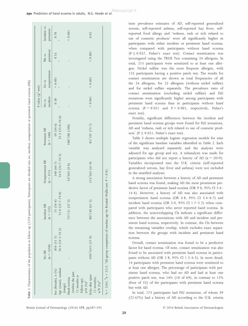
time prevalence estimates of AD, self-reported generalized
xerosis, self-reported asthma, self-reported hay fever, self-
reported food allergy and ‘redness, rash or itch related to
use of cosmetic products’ were all significantly higher in
participants with either incident or persistent hand eczema,
when compared with participants without hand eczema
(P ≤ 0�027, Fisher’s exact test). Contact sensitization was
investigated using the TRUE Test containing 24 allergens. In
total, 215 participants were sensitized to at least one aller-
gen. Nickel sulfate was the most frequent allergen, with
123 participants having a positive patch test. The results for
contact sensitization are shown as total frequencies of all
the 24 allergens, for 23 allergens (without nickel sulfate)
and for nickel sulfate separately. The prevalence rates of
contact sensitization (excluding nickel sulfate) and FLG
mutations were significantly higher among participants with
persistent hand eczema than in participants without hand
eczema (P = 0�021 and P < 0�001, respectively, Fisher’s
exact test).
Notably, significant differences between the incident and
persistent hand eczema groups were found for FLG mutations,
AD and ‘redness, rash or itch related to use of cosmetic prod-
ucts’ (P ≤ 0�021, Fisher’s exact test).Table 3 shows multiple logistic regression models for nine
of the significant baseline variables identified in Table 2. Each
variable was analysed separately and the analyses were
adjusted for age group and sex. A subanalysis was made for
participants who did not report a history of AD (n = 2019).
Variables incorporated into the U.K. criteria (self-reported
generalized xerosis, hay fever and asthma) were not included
in the stratified analyses.
A strong association between a history of AD and persistent
hand eczema was found, making AD the most prominent pre-
dictive factor of persistent hand eczema (OR 9�0, 95% CI 5�6–14�4). However, a history of AD was also associated with
nonpersistent hand eczema (OR 4�8, 95% CI 3�4–6�7) and
incident hand eczema (OR 3�0, 95% CI 1�7–5�2) when com-
pared with participants who never reported hand eczema. In
addition, the nonoverlapping CIs indicate a significant differ-
ence between the associations with AD and incident and per-
sistent hand eczema, respectively. In contrast, the CIs between
the remaining variables overlap, which excludes exact separa-
tion between the groups with incident and persistent hand
eczema.
Overall, contact sensitization was found to be a predictive
factor for hand eczema. Of note, contact sensitization was also
found to be associated with persistent hand eczema in partici-
pants without AD (OR 2�8, 95% CI 1�2–6�5). In more detail,
14 participants with persistent hand eczema were sensitized to
at least one allergen. The percentage of participants with per-
sistent hand eczema, who had no AD and had at least one
positive patch test, was 14% (10 of 69), in contrast to 12%
(four of 33) of the participants with persistent hand eczema
but with AD.
In total, 173 participants had FLG mutations, of whom 39
(22�67%) had a history of AD according to the U.K. criteriaTable
1Characteristics
ofthepo
pulation
atfollo
w-up(227
0participants).Participantsaredividedinto
no,incident,no
npersistentor
persistent
hand
eczema(H
E)
NoHE
(n=16
58)
Incident
HE
(n=13
3)Non
persistent
HE
(n=37
1)Persistent
HE
(n=10
8)
P-value(v
2-test)
Novs.
incident
Novs.
nonp
ersistent
Νοvs.
persistent
Incident
vs.
persistent
Sex(fem
ale),n(%
)85
4(51�5
)75
(56�4
)24
0(64�7
)59
(54�6
)0�2
8<0�0
010�5
30�7
8
Age
(years),
median
(range)
56�4
(24�2
–76�5
)52
�0(25�7
–74�0
)54
�9(24�7
–76�3
)55
�3(25�0
–76�0
)c
cc
c
Handeczema
(withinthepast
12mon
thsat
follo
w-up),
n/N(%
)a
–75
/131
(57�3
)33
/365
(9�0)
108/
108(100
)–
––
<0�0
01
Itchyskin
(past
12mon
ths),
n/N(%
)b
450/
1615
(27�9
)80
/130
(61�5
)15
2/36
3(41�9
)81
/107
(75�7
)<0�0
01<0�0
01<0�0
010�0
2
a n=22
62.bn=22
15.c All-grou
pcomparisonof
medianageby
Kruskal–W
allis
test:P=0�0
1.
© 2016 British Association of DermatologistsBritish Journal of Dermatology (2016) 175, pp287–295
290 Predictors of hand eczema in adults, N.G. Heede et al.
29
Manuscript II

Table
2Frequenciesrepo
rtingtheassociationbetw
eenhand
eczema(H
E)andbaselin
evariables.The
threedifferentP-values
representFisher’sexacttestperformed
on‘never
vs.incident’,‘never
vs.
persistent’and‘incidentvs.persistent’HE.
Totalparticipants=22
70
Never
HE
(n=16
58)
Incident
HE
(n=13
3)
Non
persistent
HE(n
=37
1)
Persistent
HE(n
=10
8)
P-value(Fisher’sexacttest)
Never
vs.
incident
Never
vs.
persistent
Incident
vs.
persistent
Allergen
specificIgE(n
=22
57)a
283(23�2
)38
(29�0
)98
(26�5
)24
(22�9
)0�1
41�0
00�3
0Con
tact
sensitization(all24
allergens)
(n=21
62)b
141(8�9)
14(11�0
)46
(13�0
)14
(13�6
)0�4
20�1
20�6
9
(i)Con
tact
sensitization(23allergens)
64(4�1)
8(6�3)
26(7�3)
10(9�7)
0�25
0�02
0�46
(ii)Nickelsulfate(1
allergen)
85(5�4)
7(5�5)
26(7�3)
5(4�9)
0�84
1�00
1�00
Filaggrinmutationcarriers
(n=21
89)c
117/
1606
(7�3)
5/12
7(3�9)
31/3
54(8�8)
20/1
02(19�6
)0�2
1<0�0
01<0�0
01Atopicderm
atitis(n
=22
27)d
81/1
632(5�0)
18/1
30(13�9
)76
/359
(21�1
)33
/106
(31�1
)<0�0
01<0�0
010�0
02
Self-reportedgeneralized
xerosis(n
=22
40)
249/
1636
(15�2
)32
/132
(24�2
)11
4/36
6(31�2
)35
/106
(33�0
)0�0
09<0�0
010�1
5Self-reportedfood
allergy(n
=22
31)e
58/1
630(3�6)
15/1
31(11�5
)31
/364
(8�5)
16/1
06(15�1
)<0�0
01<0�0
010�4
4
Self-reportedhayfever(n
=22
46)e
259/
1644
(15�8
)31
/132
(23�5
)83
/364
(22�8
)35
/106
(33�0
)0�0
3<0�0
010�1
1Self-reportedasthma(n
=22
48)e
132/
1642
(8�0)
21/1
32(15�9
)49
/367
(13�4
)24
/107
(22�4
)0�0
05<0�0
010�2
4
Redness,rash
oritch
relatedto
useof
cosm
etic
prod
ucts(n
=22
58)
695/
1649
(42�2
)77
/132
(58�3
)23
6/36
9(64�0
)79
/108
(73�2
)<0�0
01<0�0
010�0
2
Valuesaren(%
)or
n/N(%
).a Allergen
specificIgEagainstbirch,
grass,catandho
use-du
stmite.
bAll24
allergensin
theTRUETest(215
participantssensitized
toat
leaston
eallergen).(i)TRUETest
withat
leaston
epo
sitive
reaction
,witho
utnickel
sulfate;
(ii)TRUETestpo
sitive
foron
lynickel
sulfate;
16participantsarein
both
grou
ps,which
iswhy
thetotalnu
mberof
positive
testsis23
1.c Genotypingforfilaggrinmutations
(R50
1X,22
82del4
andR24
47X).
dAtopicderm
atitiswas
definedby
theU.K.Working
Party’scriteria,withon
emajor
criterionandtw
oof
four
minor
criteria.
e ‘Has
ado
ctor
ever
told
youthat
youhave/h
avehad...?’
© 2016 British Association of Dermatologists British Journal of Dermatology (2016) 175, pp287–295
Predictors of hand eczema in adults, N.G. Heede et al. 291
30
Manuscript II

and 133 (77�32%) did not (one participant had an undeter-
minable AD history). A significant association between FLG
mutations and persistent hand eczema was seen (OR 3�1, 95%CI 1�8–5�2). However, the association was nonsignificant in
participants without AD (OR 1�3, 95% CI 0�6–3�1).‘Skin rash related to use of cosmetic products’ and self-
reported food allergy were associated with hand eczema, also
in participants with no history of AD. As xerosis, hay fever
and asthma are criteria incorporated into the definition of AD,
the associations with hand eczema were determined only
when analysing all participants. Figure 2 summarizes the pre-
dictive factors found to influence persistent hand eczema.
Discussion
In this population-based cohort study with 5-year follow-up,
we aimed to evaluate the complex relationship between hand
eczema in adults and predictive factors such as AD, FLG
mutations, asthma, hay fever, food allergy, xerosis, specific
inhalation allergens and contact sensitization. Of these predic-
tive factors, we found that a history of AD was the strongest
predictive factor for adult hand eczema.
Our data confirmed previous literature showing that a his-
tory of AD strongly predicted persistent hand eczema (OR
9�0, 95% CI 5�6–14�4).9,10,20 Moreover, a history of AD was
Table 3 Logistic regression models for the association between hand eczema (HE) and baseline variables. Each variable was analysed separately
and adjusted for age group and sex
Variable HE group
All participants (n = 2270)Stratification: participantswithout AD (n = 2019)
OR adjusted for agegroup and sex (95% CI)
OR adjusted for age groupand sex (95% CI)
AD (n = 2227)a Never 1 –Incident 2�97 (1�71–5�16) –Nonpersistent 4�76 (3�38–6�70) –Persistent 8�95 (5�55–14�43) –
Self-reported food allergy (n = 2231;
non-AD, n = 1994)bNever 1 1
Incident 3�42 (1�87–6�26) 4�05 (2�11–7�77)Nonpersistent 2�39 (1�51–3�77) 1�76 (0�98–3�17)Persistent 5�03 (2�76–9�16) 2�91 (1�20–7�10)
Redness, rash or itch related to use
of cosmetic products (n = 2258;non-AD, n = 2011)
Never 1 1
Incident 1�85 (1�27–2�68) 1�72 (1�15–2�56)Nonpersistent 2�22 (1�75–2�83) 2�04 (1�56–2�67)Persistent 3�91 (2�49–6�13) 3�45 (2�05–5�82)
Self-reported asthma (n = 2248;
non-AD, n = 2007)bNever 1 –Incident 2�07 (1�25–3�42) –Nonpersistent 1�67 (1�17–2�38) –Persistent 3�34 (2�04–5�47) –
Filaggrin mutation carrier (n = 2189;
non-AD, n = 1946)cNever 1 1
Incident 0�52 (0�21–1�29) 0�51 (0�19–1�42)Nonpersistent 1�22 (0�80–1�85) 0�88 (0�52–1�49)Persistent 3�07 (1�82–5�19) 1�29 (0�55–3�06)
Self-reported generalized xerosis
(n = 2240; non-AD, n = 2005)
Never 1 –Incident 1�77 (1�16–2�71) –Nonpersistent 2�38 (1�83–3�09) –Persistent 2�75 (1�79–4�24) –
Self-reported hay fever (n = 2246;
non-AD, n = 2006)bNever 1 –Incident 1�57 (1�03–2�40) –Nonpersistent 1�55 (1�17–2�06) –Persistent 2�59 (1�69–3�98) –
Contact sensitization (all 24 allergens)
(n = 2162; non-AD, n = 1925)dNever 1 1
Incident 1�18 (0�65–2�12) 1�00 (0�51–1�99)Nonpersistent 1�30 (0�91–1�87) 1�20 (0�79–1�82)Persistent 1�49 (0�82–2�73) 1�76 (0�86–3�58)
Contact sensitization (23 allergens)
(n = 2162; non-AD, n = 1925)eNever 1 1
Incident 1�58 (0�74–3�37) 1�21 (0�47–3�09)Nonpersistent 1�77 (1�10–2�85) 1�70 (0�98–2�95)Persistent 2�49 (1�24–5�02) 2�84 (1�24–6�50)
OR, odds ratio; CI, confidence interval; hand eczema (HE). aAD was defined by the U.K. Working Party’s criteria with one major criterion
and two of four minor criteria. b‘Has a doctor ever told you that you have/have had . . .?’. cGenotyping for filaggrin null mutations (R501X,
2282del4 and R2447X). dContact sensitization based on all 24 allergens in the TRUE Test. eContact sensitization based on 23 allergens in the
TRUE Test (without nickel sulfate).
© 2016 British Association of DermatologistsBritish Journal of Dermatology (2016) 175, pp287–295
292 Predictors of hand eczema in adults, N.G. Heede et al.
31
Manuscript II

associated with incident hand eczema in adults from the gen-
eral population (OR 3�0, 95% CI 1�7–5�2), which, to our
knowledge, has not been shown previously. The hypothesis
that a smaller proportion of cases of AD would be detected
among patients with incident hand eczema when compared
with persistent cases has been suggested previously,20 but
Johannisson et al. found no significant difference in the pro-
portion of childhood eczema among their group with incident
hand eczema in a longitudinal cohort study of young adults.
However, they did find a higher proportion of individuals
reporting childhood eczema among participants with hand
eczema at both baseline and follow-up (similar to that in our
persistent group).20
In addition to a history of AD, several other factors were
identified as predictors of incident hand eczema in adults. The
ORs for incident hand eczema were increased in relation to
self-reported generalized xerosis, self-reported asthma, self-
reported hay fever, self-reported food allergy and ‘redness,
rash or itch related to use of cosmetic products’, when com-
pared with participants without hand eczema. However,
because of overlapping CIs, no clear distinction can be made
between the three hand eczema groups. A previous study per-
formed in a general population concluded that female sex, a
history of AD, and asthma/hay fever were associated with
incident hand eczema only in participants aged < 30 years.29
Our regression analyses were adjusted for age group and sex,
but because our data build on measures of lifetime prevalence
we were not able to investigate whether the association was
significant only in participants aged < 30 years. The associa-
tion between asthma and hay fever as a predictor of hand
eczema should therefore be interpreted with caution.
Of note, ‘redness, rash or itch related to use of cosmetic
products’ was associated with all groups of hand eczema. In
addition, the association between incident hand eczema and a
lifetime prevalence of ‘redness, rash or itch related to use of
cosmetic products’ was still significant when analysing only
participants without a history of AD, when compared with
participants who never reported hand eczema (OR 1�7, 95%CI 1�2–2�6). Information about the aetiology of the symptoms
is missing in this set-up, and it is possible that some partici-
pants report symptoms of allergic contact dermatitis and
others of irritant contact dermatitis. However, no statistical
difference was found when comparing the frequency of con-
tact sensitization in the incident group with that in the group
reporting no hand eczema.
In contrast, a significantly higher prevalence of contact sensi-
tization (to 23 allergens without nickel sulfate) was found
among participants with persistent hand eczema when com-
pared with participants without hand eczema (P = 0�021, Fish-er’s exact test). The association between persistent hand
eczema and contact sensitization is known,30 but in this cohort
the association was also found to be significant in participants
without a history of AD (OR 2�8, 95% CI 1�2–6�5). The associ-ation between FLG mutations and persistent hand eczema in
individuals with a history of AD has previously been published
using data from the Health2006 cohort.15, 31 However, the cur-
rent article includes data from a 5-year follow-up examination,
which enables investigation of the association between FLG
mutations and incident hand eczema.
In this study we report that FLG mutations are not associ-
ated with incident hand eczema in adults. The fact that FLG
mutations were associated only with persistent hand eczema
could suggest that the effect of the mutations, as a risk factor
for hand eczema, decreases with time. Despite conflicting
experimental results in the origin of the downregulation of
FLG, two experimental studies have suggested that FLG muta-
Fig 2. Illustration of factors associated with persistent hand eczema in adult Danes from the general population. The odds ratios (ORs) and 95%
confidence intervals are adjusted for age group and sex, and were retrieved by comparing participants reporting persistent hand eczema with
participants who never reported hand eczema. All variables refer to a self-reported lifetime prevalence, except for filaggrin null mutations and
contact sensitization (23 allergens, without nickel sulfate).
© 2016 British Association of Dermatologists British Journal of Dermatology (2016) 175, pp287–295
Predictors of hand eczema in adults, N.G. Heede et al. 293
32
Manuscript II

tions exert an effect early in life, but that the effect might be
neutralized with increasing age.32,33
Advantages of this study include the follow-up design and
the use of an unselected cohort of adult individuals. When
using questionnaire-derived data the most important limita-
tions are the risk of recall bias and misclassification. In addi-
tion, the 2308 participants enrolled in the follow-up
examination constitute only 29�7% of the originally invited
subjects, which could introduce a selection bias. However, the
follow-up participation rate was 66�5% of the participants
included in the baseline study. The differences between
participants and nonparticipants in the 5-year follow-up study
have previously been published, showing that participants in
the follow-up study reported significantly less AD (P = 0�020,v2-test) and slightly less hand eczema (20�9%) than the non-
participants (23�6%) (P = 0�067, v2-test).33 Thus, our results
might be an underestimation.
Moreover, there was no clinical examination by a dermatol-
ogist to confirm the hand eczema diagnoses or to evaluate
potential subtypes of hand eczema. Nevertheless, the question
about hand eczema within the past 12 months has been vali-
dated among car mechanics, dentists and office workers,
resulting in a sensitivity of 53–59% and a specificity of
96–99%.34 Validation has further been conducted in Danish
hairdressing apprentices, with an overall sensitivity of 70�3%and specificity of 99�8%.35 However, no validation has been
conducted in a follow-up design, and our definition of
incident hand eczema builds upon two lifetime prevalence
measures, which might increase the risk of recall bias, and
gives no information about when the symptoms began during
the 5 year follow-up period. Overall, the total 1-year preva-
lence of hand eczema was 9�5% at follow-up, which is in
agreement with the average 1-year prevalence of 10%
reviewed by Thyssen et al.5
The U.K. criteria have previously been validated.24,36 How-
ever, a recent study concluded that recall bias for childhood
AD is a significant problem in retrospective questionnaire stud-
ies evaluating a history of AD.37 A risk of recall bias affecting
the association between AD and hand eczema should therefore
be highlighted. Moreover, Mortz et al. also reports that recall
bias regarding asthma and allergic rhinitis was less than for
AD, possibly as a result of the later debut of these diseases.37 It
is however not possible to make a direct comparison between
the study by Mortz et al.37 and our study because of the differ-
ences in age and study design.
Lastly, our study set-up enabled patch test readings for con-
tact sensitization only on day 2. The European patch test
guideline recommends an occlusion time of 2 days,38 but the
risk of false-negative results is increased when reading only on
day 2.39 The prevalence of contact sensitization to nickel was
shown separately in Table 2 to increase the transparency of
our data, because nickel was the most frequent allergen. In
addition, the risk of allergic contact sensitization to nickel has
been suggested to be increased in FLG mutation carriers,40
possibly due to the nickel-binding properties of the filaggrin
protein.41 However, the lowest frequency of contact sensitiza-
tion to nickel was found in the group with persistent hand
eczema, which had the highest prevalence of FLG mutation
carriers (19�6%).Our data show that contact sensitization (23 allergens with-
out nickel) is associated with persistent hand eczema, also for
participants without a history of AD. This result, together with
the high prevalence of ‘redness, rash or itch related to use of
cosmetic products’, stresses the need for primary prevention
by reducing exposure to common allergens in cosmetic prod-
ucts, such as fragrances and isothiazolinones. In addition, we
suggest that education about skin health and skin protection
in the general adult population is needed, irrespective of age,
and especially in individuals with AD, who have an increased
risk of hand eczema throughout life. Despite our data suggest-
ing that the effect of FLG mutations, as a genetic marker for
hand eczema, is associated with persistent hand eczema only,
a further understanding of skin barrier function is still vital.
This is supported by the fact that the hands of individuals
with FLG mutations have a distinct phenotype.42 Frequent use
of emollients has been suggested to improve the skin barrier43
and to be useful in the therapy of hand eczema.44,45 In line
with this, our data identified the highest prevalence of gener-
alized xerosis among the participants reporting persistent hand
eczema. Notably, individual skin counselling has already pro-
ven effective against hand eczema in healthcare workers.46
In conclusion, we found that a history of AD predicted inci-
dent hand eczema, but that FLG mutations had no effect on
incident hand eczema in the general adult population. Finally,
contact sensitization was associated with persistent disease in
participants with and without a history of AD, which alto-
gether provides new knowledge to the research field investi-
gating the interplay between hand eczema, AD, contact
sensitization and FLG mutations.
References
1 Halkier-Sorensen L. Occupational skin diseases: reliability and util-
ity of the data in the various registers; the course from notificationto compensation and the costs – a case study from Denmark. Con-
tact Dermatitis 1998; 39:71–8.2 Skoet R, Olsen J, Mathiesen B et al. A survey of occupational hand
eczema in Denmark. Contact Dermatitis 2004; 51:159–66.3 Meding B, Liden C, Berglind N. Self-diagnosed dermatitis in adults
– results from a population survey in Stockholm. Contact Dermatitis2001; 45:341–5.
4 Bryld LE, Agner T, Kyvik KO et al. Hand eczema in twins: a ques-
tionnaire investigation. Br J Dermatol 2000; 142:298–305.5 Thyssen JP, Johansen JD, Linneberg A, Menn�e T. The epidemiol-
ogy of hand eczema in the general population – prevalence andmain findings. Contact Dermatitis 2010; 62:75–87.
6 Menn�e T, Johansen JD, Sommerlund M et al. Hand eczema guideli-nes based on the Danish guidelines for the diagnosis and treat-
ment of hand eczema. Contact Dermatitis 2011; 65:3–12.7 Nilsson EJ, Knutsson A. Atopic dermatitis, nickel sensitivity and
xerosis as risk factors for hand eczema in women. Contact Dermatitis1995; 33:401–6.
8 Meding B, Swanbeck G. Predictive factors for hand eczema. ContactDermatitis 1990; 23:154–61.
© 2016 British Association of DermatologistsBritish Journal of Dermatology (2016) 175, pp287–295
294 Predictors of hand eczema in adults, N.G. Heede et al.
33
Manuscript II
9 Rystedt I. Factors influencing the occurrence of hand eczema inadults with a history of atopic-dermatitis in childhood. Contact Der-
matitis 1985; 12:185–91.10 Mortz CG, Bindslev-Jensen C, Andersen KE. Hand eczema in The
Odense Adolescence Cohort Study on Atopic Diseases and Dermati-tis (TOACS): prevalence, incidence and risk factors from adoles-
cence to adulthood. Br J Dermatol 2014; 171:313–23.11 Grohagen C, Liden C, Wahlgren CF et al. Hand eczema in adoles-
cents and atopic dermatitis: a prospective cohort study from theBAMSE project. Br J Dermatol 2015; 173:1175–82.
12 Berglind IA, Alderling M, Meding B. Life-style factors and hand
eczema. Br J Dermatol 2011; 165:568–75.13 Visser MJ, Verberk MM, van Dijk FJH et al. Wet work and hand
eczema in apprentice nurses; part I of a prospective cohort study.Contact Dermatitis 2014; 70:44–55.
14 Uter W. Risk factors for occupational hand dermatitis in high-riskoccupations. Allergologie 2003; 26:377–86.
15 Thyssen JP, Carlsen BC, Menn�e T et al. Filaggrin null mutationsincrease the risk and persistence of hand eczema in subjects with
atopic dermatitis: results from a general population study. Br J Der-matol 2010; 163:115–20.
16 Visser MJ, Verberk MM, Campbell LE et al. Filaggrin loss-of-func-tion mutations and atopic dermatitis as risk factors for hand
eczema in apprentice nurses: part II of a prospective cohort study.Contact Dermatitis 2014; 70:139–50.
17 Angelova-Fischer I, Dapic I, Hoek AK et al. Skin barrier integrityand natural moisturising factor levels after cumulative dermal
exposure to alkaline agents in atopic dermatitis. Acta Derm Venereol2014; 94:640–4.
18 Fallon PG, Sasaki T, Sandilands A et al. A homozygous frameshiftmutation in the mouse Flg gene facilitates enhanced percutaneous
allergen priming. Nat Genet 2009; 41:602–8.19 Molin S, Merl J, Dietrich KA et al. The hand eczema proteome:
imbalance of epidermal barrier proteins. Br J Dermatol 2015;172:994–1001.
20 Johannisson A, Ponten A, Svensson A. Prevalence, incidence andpredictive factors for hand eczema in young adults – a follow-up
study. BMC Dermatol 2013; 13:14.21 Gr€onhagen CM, Lid�en C, Bergstr€om A et al. Prevalence and
incidence of hand eczema in adolescence: report from BAMSE –a population-based birth cohort. Br J Dermatol 2014; 171:609–14.
22 Thuesen BH, Cerqueira C, Aadahl M et al. Cohort profile: the
Health2006 cohort, research centre for prevention and health. Int J
Epidemiol 2014; 43:568–75.23 Horwitz A, Skaaby T, K�arhus LL et al. Screening for celiac disease
in Danish adults. Scand J Gastroenterol 2015; 50:824–31.24 Williams HC, Burney PG, Pembroke AC, Hay RJ. The U.K. Work-
ing Party’s Diagnostic Criteria for Atopic Dermatitis. III. Indepen-dent hospital validation. Br J Dermatol 1994; 131:406–16.
25 Meldgaard M, Szecsi PB, Carlsen BC et al. A novel multiplex analy-sis of filaggrin polymorphisms: a universally applicable method for
genotyping. Clin Chim Acta 2012; 413:1488–92.26 Thyssen JP, Linneberg A, Menn�e T et al. Contact allergy to aller-
gens of the TRUE-test (panels 1 and 2) has decreased modestly inthe general population. Br J Dermatol 2009; 161:1124–9.
27 Wilkinson DS, Fregert S, Magnusson B et al. Terminology of con-tact dermatitis. Acta Derm Venereol 1970; 50:287–92.
28 Petersen AB, Gudmann P, Milvang-Grønager P et al. Performanceevaluation of a specific IgE assay developed for the ADVIA centaur
immunoassay system. Clin Biochem 2004; 37:882–92.
29 Meding B, Jarvholm B. Incidence of hand eczema – a population-based retrospective study. J Invest Dermatol 2004; 122:873–7.
30 Meding B, Wrangsjo K, Jarvholm B. Fifteen-year follow-up ofhand eczema: predictive factors. J Invest Dermatol 2005; 124:893–7.
31 Heede NG, Thyssen JP, Thuesen BH et al. Anatomical patterns ofdermatitis in adult filaggrin mutation carriers. J Am Acad Dermatol
2015; 72:440–8.32 Rinnerthaler M, Duschl J, Steinbacher P et al. Age-related changes
in the composition of the cornified envelope in human skin. ExpDermatol 2013; 22:329–35.
33 Takahashi M, Tezuka T. The content of free amino acids in the
stratum corneum is increased in senile xerosis. Arch Dermatol Res2004; 295:448–52.
34 Meding B, Barregard L. Validity of self-reports of hand eczema.Contact Dermatitis 2001; 45:99–103.
35 Bregnhøj A, Søsted H, Menn�e T, Johansen JD. Validation of self-reporting of hand eczema among Danish hairdressing apprentices.
Contact Dermatitis 2011; 65:146–50.36 Williams HC, Burney PG, Pembroke AC, Hay RJ. Validation of the
U.K. diagnostic criteria for atopic dermatitis in a population set-ting. U.K. Diagnostic Criteria for Atopic Dermatitis Working Party.
Br J Dermatol 1996; 135:12–7.37 Mortz CG, Andersen KE, Bindslev-Jensen C. Recall bias in child-
hood atopic diseases among adults in The Odense AdolescenceCohort Study. Acta Derm Venereol 2015; 95:968–72.
38 Johansen JD, Aalto-Korte K, Agner T et al. European Society ofContact Dermatitis guideline for diagnostic patch testing – recom-
mendations on best practice. Contact Dermatitis 2015; 73:195–221.39 Uter WJ, Geier J, Schnuch A. Good clinical practice in patch test-
ing: readings beyond day 2 are necessary: a confirmatory analysis.Members of the Information Network of Departments of Derma-
tology. Am J Contact Dermat 1996; 7:231–7.40 Novak N, Baurecht H, Sch€afer T et al. Loss-of-function mutations
in the filaggrin gene and allergic contact sensitization to nickel. JInvest Dermatol 2008; 128:1430–5.
41 Ross-Hansen K, Østergaard O, Tanassi JT et al. Filaggrin is apredominant member of the denaturation-resistant nickel-
binding proteome of human epidermis. J Invest Dermatol 2014; 134:1164–6.
42 Kaae J, Menn�e T, Carlsen BC et al. The hands in health and diseaseof individuals with filaggrin loss-of-function mutations: clinical
reflections on the hand eczema phenotype. Contact Dermatitis 2012;67:119–24.
43 Simpson EL, Chalmers JR, Hanifin JM et al. Emollient enhancement
of the skin barrier from birth offers effective atopic dermatitis pre-vention. J Allergy Clin Immunol 2014; 134:818–23.
44 Kucharekova M, van de Kerkhof PCM, van der Valk PGM. A ran-domized comparison of an emollient containing skin-related lipids
with a petrolatum-based emollient as adjunct in the treatment ofchronic hand dermatitis. Contact Dermatitis 2003; 48:293–9.
45 Loden M, Wiren K, Smerud KT et al. The effect of a corticosteroidcream and a barrier-strengthening moisturizer in hand eczema. A
double-blind, randomized, prospective, parallel group clinical trial.J Eur Acad Dermatol 2012; 26:597–601.
46 Ibler KS, Jemec GBE, Diepgen TL et al. Skin care education andindividual counselling versus treatment as usual in healthcare work-
ers with hand eczema: randomised clinical trial. BMJ 2012; 345:e7822.
© 2016 British Association of Dermatologists British Journal of Dermatology (2016) 175, pp287–295
Predictors of hand eczema in adults, N.G. Heede et al. 295
34
Manuscript II
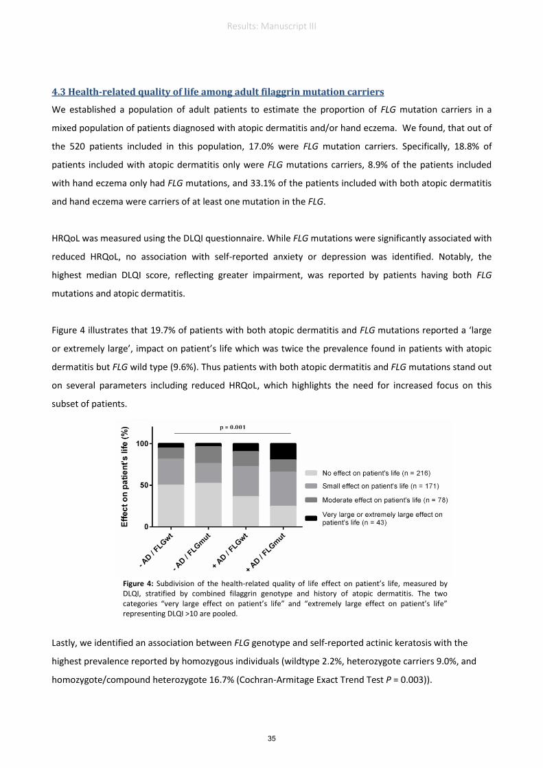
35
4.3 Health-related quality of life among adult filaggrin mutation carriers
We established a population of adult patients to estimate the proportion of FLG mutation carriers in a
mixed population of patients diagnosed with atopic dermatitis and/or hand eczema. We found, that out of
the 520 patients included in this population, 17.0% were FLG mutation carriers. Specifically, 18.8% of
patients included with atopic dermatitis only were FLG mutations carriers, 8.9% of the patients included
with hand eczema only had FLG mutations, and 33.1% of the patients included with both atopic dermatitis
and hand eczema were carriers of at least one mutation in the FLG.
HRQoL was measured using the DLQI questionnaire. While FLG mutations were significantly associated with
reduced HRQoL, no association with self-reported anxiety or depression was identified. Notably, the
highest median DLQI score, reflecting greater impairment, was reported by patients having both FLG
mutations and atopic dermatitis.
Figure 4 illustrates that 19.7% of patients with both atopic dermatitis and FLG mutations reported a ‘large
or extremely large’, impact on patient’s life which was twice the prevalence found in patients with atopic
dermatitis but FLG wild type (9.6%). Thus patients with both atopic dermatitis and FLG mutations stand out
on several parameters including reduced HRQoL, which highlights the need for increased focus on this
subset of patients.
Lastly, we identified an association between FLG genotype and self-reported actinic keratosis with the
highest prevalence reported by homozygous individuals (wildtype 2.2%, heterozygote carriers 9.0%, and
homozygote/compound heterozygote 16.7% (Cochran-Armitage Exact Trend Test P = 0.003)).
Figure 4: Subdivision of the health-related quality of life effect on patient’s life, measured by DLQI, stratified by combined filaggrin genotype and history of atopic dermatitis. The two categories “very large effect on patient’s life” and “extremely large effect on patient’s life” representing DLQI >10 are pooled.
35
Results: Manuscript III

Health-related quality of life in adult dermatitis patients
by filaggrin genotype
Running head: Quality of life by filaggrin genotype
Word count and illustrations in the manuscript:
Abstract: 200 words. Manuscript: 3421 words.
Illustrations: 2 figures, 3 tables and 1 supplementary table
Authors and affiliations:
Nina G. Heede,1 Jacob P. Thyssen,
1 Betina H. Thuesen,
2 Allan Linneberg,
2,3,4 Pal B.
Szecsi,5 Steen Stender,
5 and Jeanne D. Johansen
1
1The National Allergy Research Centre, Department of Dermato-Allergology, Copenhagen University Hospital
Herlev-Gentofte, Denmark. 2Research Centre for Prevention and Health, the Capital Region of Denmark,
Copenhagen, Denmark. 3Department of Clinical Experimental Research, Rigshospitalet, Glostrup, Denmark.
4Department of Clinical Medicine, Faculty of Health and Medical Sciences, University of Copenhagen,
Copenhagen, Denmark. 5Department of Clinical Biochemistry, Copenhagen University Hospital Herlev-
Gentofte, Denmark.
Author contributions: The 7 authors are listed in the same order as above
NGH: Planned the study, generated data, performed data analyses and drafted the manuscript.
JPT: Planned the study, reviewed data analysis and reviewed the manuscript
BHT: Planned the study, reviewed data analyses and reviewed the manuscript
ALL: Planned the study, reviewed data analysis and reviewed the manuscript
PBS: Generated data and reviewed the manuscript
STS: Generated data and reviewed the manuscript
JDH: Planned the study, reviewed data analysis and reviewed the manuscript
Conflict of interest: None declared
Funding: This study was supported by a grant from the LEO Foundation. Jacob P. Thyssen is
funded by an unrestricted grant from the Lundbeck Foundation. Betina H. Thuesen is funded
by the Lundbeck Foundation (grant no. R108-A10225).
Corresponding author
Nina Glasser Heede
E-mail: [email protected]
Direct phone: +45 38 67 73 08 Fax number: +45 39 77 71 18
The National Allergy Research Centre, Department of Dermato-Allergology, Copenhagen University
Hospital Gentofte, Kildegårdsvej 28, Opgang 20A, 1. Sal, 2900 Hellerup, Denmark
36
Manuscript III

ABSTRACT
Background: Information concerning health-related quality of life (HRQoL) and comorbidities of
adult dermatitis patients stratified by loss-of-function mutations in the filaggrin gene (FLG) is limited.
Method: This cross-sectional study included patients diagnosed with atopic dermatitis and/or hand
eczema (N=520). Patients completed questionnaires about dermatitis, skin symptoms, HRQoL, and
comorbidities including actinic keratosis, as well as atopic and mental disorders.
Results: FLG mutations (R501X, 2282del4, and R2447X) were identified in 16.9% and were
significantly associated not only with atopic dermatitis, but also independently with skin fissures on
the fingers and heels, as well as self-reported actinic keratosis. While FLG mutations were
significantly associated with reduced HRQoL, measured by the Dermatological Life Quality Index
(DLQI), no association with self-reported anxiety or depression was identified. Notably, the highest
median DLQI score, reflecting greater impairment, was reported by patients having both FLG
mutation and atopic dermatitis. Overall, 19.7% of patients with both atopic dermatitis and FLG
mutations reported a ‘large or extremely large’, impact on patient’s life; twice the prevalence in
patients with atopic dermatitis and FLG wild type (9.6%).
Conclusion: The prominent proportion of atopic dermatitis patients with common FLG mutations
stands out on reduced HRQoL, stressing the need for increased focus on this subset of patients.
Keywords: Filaggrin mutations, atopic dermatitis, hand eczema, health-related quality of life, actinic
keratosis.
37
Manuscript III

INTRODUCTION
Research addressing the etiological role of skin barrier impairment in dermatological disease has
intensified over the last decade following the discovery of loss-of-function mutations in the filaggrin
gene (FLG) and its influence on atopic dermatitis (1). Atopic dermatitis is a chronic pruritic
inflammatory skin condition that is estimated to affect 10-20% of school children in Western Europe
and in the United States (2). While many children outgrow their atopic dermatitis, persistence into
adulthood is surprisingly common (3,4). Independent of age, patients with atopic dermatitis may
experience repercussions including increased need of healthcare (5), anxiety (6), depression (7), and
decreased health-related quality of life (HRQoL). HRQoL has frequently been measured by the
Dermatological Life Quality Index (DLQI) (8), where increasing disease severity is associated with
greater impairment in HRQoL (9,10). Moreover, associations between FLG mutations and atopic
disorders such as asthma (11,12), allergic rhinitis (13), and food allergy (14) respectively, primarily in
co-occurrence with atopic dermatitis, have been shown.
Hand eczema is an equivalent common skin condition with a lifetime prevalence ranging between
15% and 21.8% in the adult general population (15,16). Hand eczema severity correlates with a
reduced HRQoL, measured by DLQI (17), and increased frequency of sick-leave and job change are
known consequences (18). Hand eczema can be a clinical manifestation of atopic dermatitis, but is in
many cases an independent disease provoked by environmental exposure to allergenic and/or irritant
compounds. While a recent European multicenter study found no increased risk of contact allergy in
individuals with atopic dermatitis from the general population (19), patients with atopic dermatitis
have repeatedly been shown to have a compromised skin barrier function resulting in an increased
reactivity to skin irritants (20-22). The biological interplay between contact dermatitis, atopic
dermatitis and FLG mutations remain to be fully elucidated, but it has been shown that FLG mutations
increase the risk of developing hand eczema (irritant contact dermatitis (23) and combined irritant and
allergic contact dermatitis (24)), often with a chronic course, and in particular in individuals with a
history of atopic dermatitis (25). A direct association, independent of atopic dermatitis, between FLG
mutations and hand eczema has only been identified in one study (26). Last, FLG mutations have
recently been found to be associated with contact dermatitis in construction workers, most strongly in
individuals with an atopic predisposition (27).
Despite the dramatic increase in our understanding of the pathogenic role of FLG mutations in
dermatitis patients, only few studies have made thorough clinical characterizations of adult dermatitis
patients investigating both FLG mutation status, skin symptoms, and dermatological and non-
dermatological comorbidities. The objective of this study was to analyse dermatitis, skin symptoms,
HRQoL, and comorbidities in adult FLG mutation carriers, to evaluate the incentive for extra care
among this group of patients.
38
Manuscript III

MATERIALS AND METHODS
Study population
The study included patients who had been examined at our tertiary Dermatology Department at
Gentofte Hospital between 2006 and 2012. Patients were diagnosed with atopic dermatitis and/or hand
dermatitis and were all ≥ 18 years. Patients received a written invitation and contacted the Department
if they were interested to get more information about the study. Interested patients were verbally
informed about the study and completed the questionnaire and collected genomic DNA by a buccal
swap (Isohelix, Harrietsham, United Kingdom). The participation rate was 46.5 % (520/1119). Of the
520 included patients; one patient did not return the questionnaire. Written informed consent was
obtained for all participants. The study was approved by the Ethics committee of Copenhagen County
(H-1-2013-127).
Clinical diagnoses and patch test
Clinical diagnoses of atopic dermatitis and/or hand eczema were scored by the attending dermatologist
and were registered in the patient’ file along with the internationally recognized MOAHLFA-index
(Male, Occupational dermatitis, history of Atopic dermatitis, Hand eczema, Leg dermatitis, Facial
dermatitis, and Age above 40 years) (28) which is registered routinely for all patients who are patch
tested. Patch tests (European baseline series and relevant additional test series according to exposure
analysis) were applied on the upper back and occluded for two days in accordance with The European
patch test guidelines (29). Readings were carried out recommending readings at day 2, day 3 or day 4
and day 7. Results were registered in The National Database of Contact Allergy from where the
dataset was extracted.
Genotyping
FLG genotyping was performed at the Department of Clinical Biochemistry at Gentofte Hospital (30).
A positive test was defined for heterozygote, and homozygote or compound heterozygote mutation
carriers. Genotyping was conducted for three of the most common Northern European loss-of-function
mutations in the profilaggrin gene which together constitute 83% of the total risk alleles associated
with atopic dermatitis; R501X, 2282del4, and R2447X (1,31,32).
Extraction of genomic DNA from buccal swaps
Buccal swaps were stored at -80oC until DNA extraction was performed with JetQuick kit (Genomed,
Löhne, Germany) according to manufactures’ instructions. Purified genomic DNA was stored at -20oC
before allele-specific PCR was performed.
Manuscript III
39

Allele-specific PCR, microbead-specific hybridization and detection
We used a slightly modified version of the method which has previously been described in detail (30).
Briefly, regions covering the FLG mutations were amplified by multiplexed, allele-specific
asymmetric PCR using tagged primers. The single stranded PCR products hence underwent tag-
specific hybridization to microbeads which were analyzed on Bio-Plex 200 (Bio-Rad) flow cytometer
using Bio-Plex Manager software, version 5.0 (30).
Questionnaire
The following definition of dermatitis was listed prior to the questions about dermatitis: ‘‘dermatitis is
an itchy skin disorder showing redness, dryness, and possibly bladders and exudation. Dermatitis
remains on the same area of the body for some time.’’ Questions used in the questionnaire are listed in
Table S1. The Dermatological Life Quality Index by Finley et al., 1994 was used to address skin
specific quality of life (8).
Statistics
Statistical analyses for dichotomous variables were made by use of the Chi Square Test, Fishers Exact
Test, or Cochran-Armitage Exact Trend Test. DLQI median scores and 25th and 75
th percentiles were
presented based on responses from 515 participants. Non-parametric Kruskal-Wallis Test for group
difference was performed. Level of significance was set at P < 0.05. We made sub-analyses of the
DLQI scores using relative percentages obtained by adding up the total scores in each of the six
questions and divide the result by the number of patients with the likely maximum score of 6 (3 in the
work/school and treatment category) ((nvery_much x 3 + na_lot x 2 + na_little x1) / (ntotal x 6)) * 100%.
Epidata was used as data entry module (33) and all statistical analyses were performed using SAS
software (SAS, Version 9.3 for Windows, SAS Institute Inc., Cary, NC, USA). Figures were made in
SAS or GraphPad Prism version 6.07 for Windows (GraphPad software, La Jolla, CA, USA).
RESULTS
Patient characteristics
Characteristics of the patients’ dermatitis were registered by use of the MOAHLFA-index (28). The
MOAHLFA-index and patch test results for the 1,119 invited patients are shown in Table 1. Table 1a
shows, that included participants (n=520) were significantly older, and had a higher prevalence of
atopic dermatitis, than non-participants (n=599) (P < 0.001, Chi square Test). In contrast, participants
had a significant lower point prevalence of hand eczema (80.4%) and fewer cases had occupational
relevance of dermatitis (43.9%) when compared with non-participants (86.7% and 55.5%, (P ≤ 0.005,
Chi square Test), respectively). Stratification for the MOAHLFA-index, among the 520 included
patients, with the three inclusion diagnoses (atopic dermatitis, hand eczema, or atopic dermatitis and
Manuscript III
40

hand eczema) is presented in Table 1b and shows that there was no sex difference between the groups,
but that participants in the group with ‘hand eczema only’ were significantly older (P < 0.001,
Kruskal-Wallis Test).
Filaggrin gene mutation carriers reported an increased prevalence of skin symptoms, including
skin fissures in acral areas
To examine skin symptoms of FLG mutation carriers, all participants were genotyped for three
common FLG mutations. A successful genotype was determined for 98.6% of the included patients.
The prevalence of FLG mutations carriers was significantly higher in the group of patients diagnosed
with both atopic dermatitis and hand eczema (33.1%), followed by the group with atopic dermatitis
only (18.8%) and finally the hand eczema group (8.9%) (P< 0.001, Chi Square Test, Table 2a).
Because the MOAHLFA-index only specifies hand eczema at the time of patch testing, a
questionnaire regarding skin symptoms and hand eczema was distributed. The question regarding self-
reported lifetime prevalence of hand eczema demonstrated an overall sensitivity of 96.7% (three
patients had missing values and 11 patients included with hand eczema reported “no hand eczema”).
Table 2b shows stratification by combined genotype and history of atopic dermatitis. Patients with
both FLG mutations and atopic dermatitis reported an increased point prevalence of hand eczema
(71.0%) compared with atopic dermatitis patients who were FLG wild type carriers (53.6%)(P =
0.018, Chi square Test). Moreover, skin fissures on the hands and heels were significantly more
prevalent among FLG mutation carriers, independently of atopic dermatitis status (P ≤ 0.031, Chi
square Test). The highest prevalence was found among patients with hand eczema and FLG mutations
whereof 76.0% reported fissures on the hands and 68.0% reported fissures on the heels. Last, atopic
dermatitis patients with FLG mutations reported significantly more xerosis than atopic dermatitis
patients with wild type FLG status, 80.7% and 59.6% (P = 0.003, Chi square Test), respectively.
Self-reported estimates of severity and current disease showed no significant difference among the
four combinations of FLG genotype and atopic dermatitis phenotype in Table 2b. Among patients with
both FLG mutation and atopic dermatitis, 5.0% reported that they no longer suffered from dermatitis,
63.3% reported an improvement, and 31.7% reported no effect or a worsening since they last visited
our dermatological clinic.
Patients with both filaggrin gene mutations and atopic dermatitis report decreased HRQoL
To investigate whether FLG mutation carriers differed from non-mutation carriers in relation to
HRQoL, we asked patients to complete the DLQI questionnaire, as well as, answering questions
regarding lifetime prevalence of anxiety or depression. In total, 6.6% of the patients reported a lifetime
prevalence of anxiety and 17.1% reported a lifetime prevalence of depression. Notably, no
associations between FLG mutations and, respectively, anxiety or depression were found (Table 3).
Manuscript III
41

DLQI scores stratified by inclusion diagnoses, age group, sex, FLG genotype, and combined genotype
and atopic dermatitis phenotype are presented in Figure 1. Overall, the total DLQI score ranges
between 0 and 30 where increased DLQI scores reflect reduced HRQoL. The following references
were used for total DLQI sum: 0-1 ‘no effect on patient’s life’, ≥2 ‘small impact on patient’s life’, 5-
10 ‘moderate effect on patients life’, and > 10 ‘large or extreme effect on patient’s life’ (8). In total,
515 patients (99.2%) completed the DLQI questionnaire correctly. Patients with atopic dermatitis only
(median (M) score (25th and 75
th percentiles)) (M 3 (1 - 7)), or combined atopic dermatitis and hand
eczema (M 3 (1 - 6)) reported higher DLQI score than patients with hand eczema only (M 1 (0 -
4)(Figure 1a)). No significant sex difference was detected (Figure 1b), but the younger age group
reported a lower DLQI score (Figure 1c) possibly influenced by the fact that patients with atopic
dermatitis were, on average 10 years, younger than the patients with hand eczema (Table 2b).
Moreover, FLG mutation carriers reported significantly higher DLQI scores than non-mutation
carriers (P = 0.033, Kruskal-Wallis Test, Figure 1d). The highest median score was found for 61
patients with both FLG mutations and atopic dermatitis (M 5 (2-7)) (Figure 1e). In total, 59 patients no
longer suffered from dermatitis (Table 2a). The association between FLG mutations and decreased
HRQoL remained highly significant if the 59 participants who did not report dermatitis any longer
were excluded.
Subdivision of the DLQI is shown in Figure 2. Most participants reported that “symptoms and
feelings” affected their HRQoL. Participants with both FLG mutation and atopic dermatitis reported
the highest scores in all categories, especially “symptoms and feelings” (35.8%), “daily activities”
(18.8%), and “treatment” (18.6%) (Figure 2a). The overall association between DLQI and HRQoL, in
this study population, shows that more participants with atopic dermatitis reported a ‘very large’ or
‘extremely large’ effect on patient’s life (DLQI score >10) (Figure 2b). In total, 19.7% of the
participants with both FLG mutation and atopic dermatitis reported an extremely large effect on
patient’s life.
The highest prevalence of atopic comorbidities in adults is reported by patients with both atopic
dermatitis and filaggrin gene mutations
To fully characterize our study population, we investigated the prevalence of atopic diseases which, in
addition, have both been associated with FLG mutations and reduced HRQoL. The overall prevalence
of atopic dermatitis in this population consisting of patients with atopic dermatitis and/or hand eczema
was 44.8%. Twenty-eight percent reported a lifetime prevalence of asthma, 42.7% reported rhinitis
and 30.5% reported food allergy. The highest prevalence of all of the self-reported atopic diseases was
reported by patients with both atopic dermatitis and FLG mutations (asthma 45.2%, rhinitis 62.9% and
food allergy 41.0%). No FLG genotype dependent differences were however found for asthma, rhinitis
or food allergy, irrespective of atopic dermatitis (Table 3). If we, instead of descriptive statistics, use a
Manuscript III
42

logistic regression model we do, however, see a significant association between FLG mutations and
both asthma (odds ratio (OR) = 1.81; 95% confidence interval (CI): 1.10 – 2.96) and rhinitis (OR =
1.70; 95%CI: 1.06 – 2.72). The significance is however lost for both associations when adjusting for
age group, sex and atopic dermatitis.
Association between filaggrin gene mutations and actinic keratosis
We asked participants whether a doctor had ever told them that they have, or have had, actinic
keratosis, non-melanoma skin cancer (squamous cell cancer or basal cell cancer), or malignant
melanoma. The percentages of participants reporting a lifetime prevalence of non-melanoma skin
cancer or melanoma skin cancer were, 1.8% and 0.8%, respectively. In total, 20 (4.0%) patients
reported a lifetime prevalence of actinic keratosis. Patients reporting actinic keratosis were
significantly older (mean age 60.4 (standard deviation (SD) 13.0)) than patient who did not report
actinic keratosis (mean age 46.5 (SD 14.7)) (P < 0.001, Kruskal-Wallis Test). Of the 20 participants
reporting actinic keratosis; three had a missing FLG genotype. However, a significant association
between FLG mutations and actinic keratosis was found in patients with atopic dermatitis (P = 0.022)
(Table 3). In total, 13 of the 20 patients (65.0%) reporting a history of actinic keratosis had a clinical
atopic dermatitis diagnoses. When stratifying for heterozygote and homozygote/compound
heterozygote carriers, the prevalence of actinic keratosis was: wild type 2.2 % (n = 9), heterozygote
9.0% (n = 7) and homozygote/compound heterozygote 16.7% (n =1) (Cochran-Armitage Exact Trend
Test P = 0.003). If we assume that the three patients with missing FLG mutations status were all wild
type carriers the association remained significant (data not shown).
DISCUSSION
In this cross-sectional study, we showed that the prevalence of FLG mutations was 8.9% among adult
patients with hand eczema but without atopic dermatitis; an estimate that is equivalent to estimates
from the general Danish population (34). However, the prevalence of FLG mutations among patients
with atopic dermatitis only was 18.8%, which is similar to the prevalence of 17.5% originally reported
by Palmar and colleagues (1). Moreover, 33.1% of adult patients with both atopic dermatitis and hand
eczema had at least one FLG mutation. Collectively, the results support the notion that FLG mutations
primarily constitute a risk factor for hand eczema in individuals with atopic dermatitis (25,35)
Adult dermatitis patients with both FLG mutations and atopic dermatitis reported decreased HRQoL
and differed in terms of daily impairment, when compared with atopic dermatitis patients that were
FLG wild type. Hence, nearly 20% of patients with concomitant FLG mutation and atopic dermatitis
reported that their disease affected their lives in a ‘large’ or ‘extremely large’ way and, furthermore,
that the category with ‘symptoms and feelings’ had the greatest negative impact. It is well-known that
Manuscript III
43

multiple skin disorders have detrimental effects upon HRQoL (8,36). Both asthma and rhinitis reduce
HRQoL (37,38) which could potentially influence HRQoL, since both disorders were prevalent among
patients with both atopic dermatitis and FLG mutations. The DLQI questionnaire is, however, skin
specific and the strong association between reduced HRQoL and FLG mutations is strengthen by the
higher DLQI score among homozygous and compound heterozygous individuals in the population
(Figure 1d). Our result indicates that patient-centered management is important in atopic dermatitis
patients with concomitant FLG mutations. Notably, neither depression nor anxiety was associated with
FLG genotype or atopic dermatitis (Table 3), indicating that the difference is skin specific. In line with
this, we observed no association between FLG genotype and generic HRQoL measured by the EQ-5D
(39) (data not shown).
Patients in this study population were not included based on disease severity. In fact, 11.7% of the
population reported that they no longer suffered from dermatitis, which we believe makes the results
more comparable to patients with all severities of dermatitis. The decreased disease severity may also
be reflected in the average DLQI scores which were lower than results from other studies investigating
HRQoL in patients with atopic dermatitis (8,9) or hand eczema (17). We did however not exclude the
108 patients reporting a total DLQI score of zero in the analyses which lowers the average DLQI
score.
We examined the putative association between selected dermatological manifestations and FLG
mutations. We found significant associations between FLG mutations and a life time prevalence of
skin fissures on the hand and heels (Table 2b). While fissures in the heels have never been linked to
FLG genotype, fissured skin on the hands has previously been reported as a clinical marker used to
identify FLG mutation carriers (26,40), together with palmar hyperlinearity (41) and xerosis (42). In
line, a significantly higher prevalence of xerosis was reported by patients with both FLG mutations
and atopic dermatitis (71.0%) when compared with atopic dermatitis patients with FLG wild type
(53.6%). No association between xerosis and FLG mutations among patients without atopic dermatitis
was found, a finding that is in line with a cohort study of children from the general population (43).
We found no association between FLG mutations and non-melanoma skin cancer or malignant
melanoma. We did however show that self-reported actinic keratosis was more prevalent among FLG
mutation carriers when compared with non-carriers (Table 3). Degradation of the filaggrin protein in
the stratum corneum results in trans-urocanic acid (44); an absorber of UV radiation in the stratum
corneum (45,46). Thus, the association between self-reported actinic keratosis and FLG genotype
could possibly be a result of the reduced photoprotective capacity of filaggrin deficient skin, but likely
also explained by repeated UV therapy to treat patients’ dermatitis. The primary risk factor for actinic
keratosis is UV radiation (linked to mutations in the p53 gene) (47), increasing age, male sex, and fair
Manuscript III
44

skin (48). We found that the prevalence increased with age but found no sex difference. Because of the
small number of cases, the association between FLG mutations and actinic keratosis should be
regarded as explorative and needs to be confirmed. In addition, we wish to highlight that we did not
make prior power calculations for each of the included comorbidities. However, if confirmed, it might
be valuable to know patients’ FLG genotype prior to light treatment of atopic dermatitis, and to
increase awareness against extensive sun exposure, especially for the homozygous / compound
heterozygous FLG mutation carriers. Thus, patients with atopic dermatitis and FLG mutations had
significantly more dermatological manifestations when compared with patients with atopic dermatitis
and FLG wild type.
The strengths of this study are the combination of clinical data, self-reported data, and FLG genotype.
We did however only genotype the study population for three of the most common mutations,
covering 83% of the identified loss-of-function mutations (32). When using self-reported data, recall
bias, misclassification, and selection bias are potential limitations. The DLQI score however reflects
how skin symptoms affected patients’ life during the past week, thereby minimizing recall bias. In
addition, we had no clinical severity score for atopic dermatitis or hand eczema, and no data on
treatment in relation to systemic medicine and potential UV treatment. Last, we had 25 patients in the
group with no atopic dermatitis and FLG wild type (patients with hand eczema) which only represents
4.8% of the study population.
In conclusion, FLG mutation carriers reported decreased HRQoL. Patients with both FLG mutation
and a history of atopic dermatitis were mostly affected and nearly 20% of this subset of patients
reported that their skin disease influenced their lives in a ‘large’ or ‘extremely large’ way, confirming
that these patients need extra care on several parameters.
CONFLICT OF INTEREST
None declared
ACKNOWLEDGEMENTS
We thank research nurse Anne Marie Topp for help with patient recruitment and data preparation. This
study was supported by a grant from the LEO Foundation. Jacob P. Thyssen is funded by an
unrestricted grant from the Lundbeck Foundation. Betina H. Thuesen is funded by the Lundbeck
Foundation (grant no. R108-A10225).
Manuscript III
45

REFERENCE LIST
1 Palmer CN, Irvine AD, Terron-Kwiatkowski A et al. Common loss-of-function variants of the
epidermal barrier protein filaggrin are a major predisposing factor for atopic dermatitis. Nat Genet
2006; 38: 441-446.
2 Larsen FS, Hanifin JM. Epidemiology of atopic dermatitis. Immunol. Allerg. Clin. North Am. 2002; 22:
1.
3 Herd RM, Tidman MJ, Prescott RJ et al. Prevalence of atopic eczema in the community: the Lothian
Atopic Dermatitis study. Br J Dermatol 1996; 135: 18-19.
4 Margolis JS, Abuabara K, Bilker W et al. Persistence of mild to moderate atopic dermatitis. JAMA
Dermatol 2014; 150: 593-600.
5 Silverberg JI. Health Care Utilization, Patient Costs, and Access to Care in US Adults With Eczema: A
Population-Based Study. JAMA Dermatol 2015; 151: 743-752.
6 Slattery MJ, Essex MJ, Paletz EM et al. Depression, anxiety, and dermatologic quality of life in
adolescents with atopic dermatitis. J Allergy Clin Immunol 2011; 128: 668-671.
7 Yu SH, Silverberg JI. Association between Atopic Dermatitis and Depression in US Adults. J Invest
Dermatol 2015; 135: 3183-3186.
8 Finlay AY, Khan GK. Dermatology Life Quality Index (DLQI)--a simple practical measure for routine
clinical use. Clin Exp Dermatol 1994; 19: 210-216.
9 Maksimovic N, Jankovic S, Marinkovic J et al. Health-related quality of life in patients with atopic
dermatitis. J Dermatol 2012; 39: 42-47.
10 Holm EA, Wulf HC, Stegmann H et al. Life quality assessment among patients with atopic eczema. Br
J Dermatol 2006; 154: 719-725.
11 Rodriguez E, Baurecht H, Herberich E et al. Meta-analysis of filaggrin polymorphisms in eczema and
asthma: robust risk factors in atopic disease. J Allergy Clin Immunol 2009; 123: 1361-1370 e1367.
12 Weidinger S, O'Sullivan M, Illig T et al. Filaggrin mutations, atopic eczema, hay fever, and asthma in
children. J Allergy Clin Immunol 2008; 121: 1203-1209 e1201.
13 van den Oord RA, Sheikh A. Filaggrin gene defects and risk of developing allergic sensitisation and
allergic disorders: systematic review and meta-analysis. BMJ 2009; 339: b2433.
14 Brown SJ, Asai Y, Cordell HJ et al. Loss-of-function variants in the filaggrin gene are a significant risk
factor for peanut allergy. J Allergy Clin Immunol 2011; 127: 661-667.
15 Meding B, Liden C, Berglind N. Self-diagnosed dermatitis in adults - Results from a population survey
in Stockholm. Contact Dermatitis 2001; 45: 341-345.
16 Bryld LE, Agner T, Kyvik KO et al. Hand eczema in twins: a questionnaire investigation. Br J
Dermatol 2000; 142: 298-305.
17 Agner T, Andersen KE, Brandao FM et al. Hand eczema severity and quality of life: a cross-sectional,
multicentre study of hand eczema patients. Contact Dermatitis 2008; 59: 43-47.
18 Lerbaek A, Kyvik KO, Ravn H et al. Clinical characteristics and consequences of hand eczema - an 8-
year follow-up study of a population-based twin cohort. Contact Dermatitis 2008; 58: 210-216.
19 Diepgen TL, Ofenloch RF, Bruze M et al. Prevalence of contact allergy in the general population in
different European regions. Br J Dermatol 2016; 174: 319-329.
20 Agner T. Susceptibility of atopic dermatitis patients to irritant dermatitis caused by sodium lauryl
sulphate. Acta Derm Venereol 1991; 71: 296-300.
21 Bandier J, Carlsen BC, Rasmussen MA et al. Skin reaction and regeneration after single sodium lauryl
sulfate exposure stratified by filaggrin genotype and atopic dermatitis phenotype. Br J Dermatol 2015;
172: 1519-1529.
22 Angelova-Fischer I, Hoek AK, Dapic I et al. Barrier function and natural moisturizing factor levels
after cumulative exposure to a fruit-derived organic acid and a detergent: different outcomes in atopic
and healthy skin and relevance for occupational contact dermatitis in the food industry. Contact
Dermatitis 2015; 73: 358-363.
23 Visser MJ, Landeck L, Campbell LE et al. Impact of atopic dermatitis and loss-of-function mutations in
the filaggrin gene on the development of occupational irritant contact dermatitis. Br J Dermatol 2013;
168: 326-332.
24 Molin S, Vollmer S, Weiss EH et al. Filaggrin mutations may confer susceptibility to chronic hand
eczema characterized by combined allergic and irritant contact dermatitis. Br J Dermatol 2009; 161:
801-807.
25 Thyssen JP, Carlsen BC, Menne T et al. Filaggrin null mutations increase the risk and persistence of
hand eczema in subjects with atopic dermatitis: results from a general population study. Br J Dermatol
2010; 163: 115-120.
46
Manuscript III

26 Visser MJ, Verberk MM, Campbell LE et al. Filaggrin loss-of-function mutations and atopic dermatitis
as risk factors for hand eczema in apprentice nurses: part II of a prospective cohort study. Contact
Dermatitis 2014; 70: 139-150.
27 Timmerman JG, Heederik D, Spee T et al. Contact dermatitis in the construction industry: the role of
filaggrin loss-of-function mutations. Br J Dermatol 2016; 174: 348-355.
28 Schnuch A, Geier J, Uter W et al. National rates and regional differences in sensitization to allergens of
the standard series. Population-adjusted frequencies of sensitization (PAFS) in 40,000 patients from a
multicenter study (IVDK). Contact Dermatitis 1997; 37: 200-209.
29 Johansen JD, Aalto-Korte K, Agner T et al. European Society of Contact Dermatitis guideline for
diagnostic patch testing - recommendations on best practice. Contact Dermatitis 2015; 73: 195-221.
30 Meldgaard M, Szecsi PB, Carlsen BC et al. A novel multiplex analysis of filaggrin polymorphisms: a
universally applicable method for genotyping. Clinica chimica acta; international journal of clinical
chemistry 2012; 413: 1488-1492.
31 Sandilands A, Terron-Kwiatkowski A, Hull PR et al. Comprehensive analysis of the gene encoding
filaggrin uncovers prevalent and rare mutations in ichthyosis vulgaris and atopic eczema. Nat Genet
2007; 39: 650-654.
32 Irvine AD, McLean WH, Leung DY. Filaggrin mutations associated with skin and allergic diseases. N
Engl J Med 2011; 365: 1315-1327.
33 Lauritsen JM, Bruus M. EpiData Entry (version 1.4.4.4). A comprehensive tool for validated entry and
documentation of data. The EpiData Association. Odense, Denmark. In. 2015.
34 Thyssen JP, Johansen JD, Linneberg A et al. The association between null mutations in the filaggrin
gene and contact sensitization to nickel and other chemicals in the general population. Br J Dermatol
2010; 162: 1278-1285.
35 Heede NG, Thyssen JP, Thuesen BH et al. Predictive factors of self-reported hand eczema in adult
Danes - a population based cohort study with 5 year follow-up. Br J Dermatol 2016.
DOI 10.1111/bjd.14476
36 Jowett S, Ryan T. Skin disease and handicap: an analysis of the impact of skin conditions. Soc Sci Med
1985; 20: 425-429.
37 Juniper EF, Guyatt GH, Epstein RS et al. Evaluation of impairment of health related quality of life in
asthma: development of a questionnaire for use in clinical trials. Thorax 1992; 47: 76-83.
38 Meltzer EO, Nathan RA, Selner JC et al. Quality of life and rhinitic symptoms: Results of a nationwide
survey with the SF-36 and ROLO questionnaires. J Allergy Clin Immun 1997; 99: S815-S819.
39 EuroQol G. EuroQol--a new facility for the measurement of health-related quality of life. Health Policy
1990; 16: 199-208.
40 Thyssen JP, Ross-Hansen K, Johansen JD et al. Filaggrin loss-of-function mutation R501X and
2282del4 carrier status is associated with fissured skin on the hands: results from a cross-sectional
population study. Br J Dermatol 2012; 166: 46-53.
41 Brown SJ, Relton CL, Liao H et al. Filaggrin haploinsufficiency is highly penetrant and is associated
with increased severity of eczema: further delineation of the skin phenotype in a prospective
epidemiological study of 792 school children. Br J Dermatol 2009; 161: 884-889.
42 Sergeant A, Campbell LE, Hull PR et al. Heterozygous null alleles in filaggrin contribute to clinical dry
skin in young adults and the elderly. J Invest Dermatol 2009; 129: 1042-1045.
43 Brown SJ, Relton CL, Liao H et al. Filaggrin null mutations and childhood atopic eczema: a
population-based case-control study. J Allergy Clin Immunol 2008; 121: 940-946 e943.
44 Scott IR, Harding CR, Barrett JG. Histidine-rich protein of the keratohyalin granules. Source of the free
amino acids, urocanic acid and pyrrolidone carboxylic acid in the stratum corneum. Biochim Biophys
Acta 1982; 719: 110-117.
45 de fine Olivarius F, Wulf HC, Crosby J et al. The sunscreening effect of urocanic acid. Photodermatol
Photoimmunol Photomed 1996; 12: 95-99.
46 Barresi C, Stremnitzer C, Mlitz V et al. Increased sensitivity of histidinemic mice to UVB radiation
suggests a crucial role of endogenous urocanic acid in photoprotection. J Invest Dermatol 2011; 131:
188-194.
47 Ziegler A, Jonason AS, Leffell DJ et al. Sunburn and p53 in the onset of skin cancer. Nature 1994; 372:
773-776.
48 Memon AA, Tomenson JA, Bothwell J et al. Prevalence of solar damage and actinic keratosis in a
Merseyside population. Br J Dermatol 2000; 142: 1154-1159.
47
Manuscript III

Tab
le 1
: C
linic
al c
har
acte
rist
ics
by t
he
MO
AH
LF
A i
ndex
1 s
trat
ifie
d b
y p
arti
cipan
ts a
nd n
on
-par
tici
pan
ts (
Tab
le 1
a) a
nd
str
atif
ied
by i
ncl
usi
on
dia
gn
ose
s fo
r
the
incl
uded
pat
ien
ts (
Tab
le 1
b).
a
A
ll i
nvit
ed p
atie
nts
(n =
11
19
)
B
Incl
ud
ed p
atie
nts
(n =
52
0)
P
arti
cip
an
ts
(n =
520
) N
on
-
pa
rti
cip
an
ts
(n =
599
)
P v
alu
e
(Ch
i sq
uar
e)
AD
(n =
102
) H
E
( n
= 2
87
) A
D/H
E
(n =
131
) P
va
lue
(Ch
i sq
uar
e)
Mal
e, %
(n
) 2
6.2
(13
6)
31.2
(18
7)
= 0
.062
21.6
(22
) 3
0.0
(86
) 2
1.4
(28
) =
0.0
90
Occ
up
atio
nal
rel
evan
ce o
f d
erm
atit
is, %
(n
) 4
3.9
(22
8)
55.9
(33
5)
< 0
.00
1
2.0
(2
) 6
1.0
(17
5)
38.9
(51
) <
0.0
01
A h
isto
ry o
f at
op
ic d
erm
atit
is, %
(n
) 4
4.8
(23
3)
35.6
(21
3)
= 0
.00
2
100
(1
02
) -
100
(1
31
) .
Han
d e
czem
a, %
(n
) 8
0.4
(41
8)
86.6
(51
9)
= 0
.00
5
- 1
00
(2
87
) 1
00
(1
31
) .
Leg
der
mat
itis
, %
(n
) 0
.8 (
4)
0.3
(2
) =
0.4
25
2
- 1
.4 (
4)
- .
Fac
ial
der
mat
itis
, %
(n
) 2
8.1
(14
6)
18.2
(10
9)
< 0
.00
1
64.7
(66
) 1
2.2
(35
) 3
4.4
(45
) <
0.0
01
Ag
e ab
ove
40
yea
rs,
% (
n)
62.5
(32
5)
42.4
(25
4)
< 0
.00
1
50.0
(51
) 7
3.5
(21
1)
48.1
(63
) <
0.0
01
Ag
e (m
edia
n i
n y
ears
, m
in –
max
) 4
9 (
18
-83
) 4
0 (
18
-86
) <
0.0
01
3
42 (
18
- 8
3)
52 (
20
- 8
2)
42 (
19
-8
1)
< 0
.00
13
≥ 1
posi
tive
pat
ch t
est,
4 %
(n
) 4
4.8
(23
3)
35.6
(21
3)
= 0
.00
2
34.3
(35
) 5
0.5
(14
5)
40.5
(53
) =
0.0
09
AD
; at
opic
der
mat
itis
, H
E;
han
d e
czem
a;
1M
OA
HL
FA
-in
dex
: M
ale,
Occ
up
atio
nal
rel
evan
ce,
Ato
pic
der
mat
itis
(li
feti
me
pre
val
ence
), H
and e
czem
a (p
oin
t p
reval
ence
), L
eg d
erm
atit
is (
poin
t p
reval
ence
), F
acia
l d
erm
atit
is (
poin
t p
reval
ence
) an
d A
ge
above
40
yea
rs.
2F
ish
er E
xac
t T
est.
3K
rusk
al-W
alli
s T
est.
4 P
atie
nts
wer
e te
sted
wit
h t
he
Eu
rop
ean
bas
elin
e se
ries
an
d r
elev
ant
supp
lem
ents
.
48
Manuscript III

Tab
le 2
: O
ver
vie
w o
f fi
lag
gri
n g
eno
type,
sel
f-re
port
ed d
erm
atit
is,
dis
ease
sev
erit
y,
and c
urr
ent
stat
e o
f d
erm
atit
is s
trat
ifie
d b
y i
ncl
usi
on
dia
gn
ose
s
(Tab
le 2
a), an
d c
om
bin
ed f
ilag
gri
n g
enoty
pe
and h
isto
ry o
f at
opic
der
mat
itis
(T
able
2b).
AD
; at
opic
der
mat
itis
, H
E;
han
d e
czem
a, F
LG
wt;
Fil
aggri
n w
ild
typ
e, F
LG
mu
t; F
ilag
gri
n n
ull
muta
tion
car
rier
1 C
lin
ical
ly d
iagn
ose
d l
ifet
ime
pre
val
ence
of
atop
ic d
erm
atit
is.
2 T
he
clin
ical
han
d e
czem
a dia
gn
osi
s w
as m
ade
at t
he
tim
e of
pat
ch-t
esti
ng a
nd
rep
rese
nts
a p
oin
t p
reval
ence
. W
e th
eref
ore
ask
ed a
bou
t li
feti
me
pre
val
ence
of
han
d e
czem
a in
th
e q
ues
tionn
aire
. 3 T
he
spec
ific
ity o
f se
lf-r
eport
ed h
and e
czem
a in
th
is p
op
ula
tion w
as 9
6.7
%.
4P
atie
nts
wer
e gen
oty
ped
for
the
thre
e F
LG
muta
tion
s: R
501
X;
22
82d
el4
; an
d
R244
7X
; 7
par
tici
pan
ts h
ad a
mis
sin
g F
LG
gen
oty
pe,
5 “
Do y
ou
som
etim
es e
xp
erie
nce
cra
cks
on
you
r fi
nger
tip
s?”,
6“D
o y
ou
som
etim
es e
xp
erie
nce
dee
p c
rack
s in
you
r h
eels
or
the
tip
of
you
r to
es”?
, 7
“H
ow
wil
l you
rat
e you
r d
erm
atit
is o
n a
sca
le f
rom
0 -
10
? (t
oday
an
d w
hen
it
is w
ors
t)”,
8M
edia
n a
nd 2
5th –
75
th p
erce
nti
les,
9“H
ow
is
you
r d
erm
atit
is t
od
ay,
com
par
ed t
o w
hen
you
wer
e in
con
tact
wit
h G
ento
fte
Hosp
ital
?”,
10 F
ish
er’s
Exac
t T
est,
11K
rusk
al-W
alli
s T
est.
a
(n
= 5
20
) b
(n
= 5
13
)
A
D1
(n =
10
2)
HE
2
(n =
28
7)
AD
/HE
(n =
13
1)
- A
D /
FL
Gw
t
n =
257
(0)
- A
D /
FL
Gm
ut
n =
25
(1)
+ A
D /
FL
Gw
t
n =
169
(2)
+ A
D /
FL
Gm
ut
n =
62
(3)
P v
alu
e
(Gro
up
0 v
s 1
)
(Ch
i sq
ua
re)
P v
alu
e
( G
rou
p 2
vs
3)
(Ch
i sq
ua
re)
Fil
agg
rin
mu
tati
on
car
rier
s4
(n=
51
3)
│(n
= 5
13
) (%
(n
))
18.8
(19
) 8
.9 (
25
) 3
3.1
(43
) 0
100
(2
5)
0
100
(6
2)
- -
Lif
e ti
me
pre
val
ence
of
han
d e
czem
a
(n=
51
7)
│(n
= 5
10
) (%
(n
))
68.3
(69
)2
96.8
(27
6)3
9
7.7
(12
8)3
9
6.5
(24
6)
100
(2
5)
83.3
(14
0)
90.3
(56
) =
1.0
00
10
= 0
.185
Poin
t p
reval
ence
of
han
d e
czem
a
(n =
51
2)
│(n
= 5
05
) (%
(n))
4
1.0
(41
) 5
5.3
(15
6)
71.5
(93
) 5
5.6
(14
0)
56.0
0 (
14
) 5
3.6
(89
) 7
1.0
(44
) =
0.9
66
= 0
.01
8
Lif
e ti
me
pre
val
ence
of
fiss
ure
s on
the
fin
ger
s5
(n =
49
3)
│(n
= 4
87
) (%
(n))
3
5.8
(34
) 5
4.0
(14
7)
53.2
(67
) 5
1.4
4 (
125
) 7
6.0
(19
) 4
1.1
(65
) 5
7.4
(35
) =
0.0
19
= 0
.03
1
Lif
e ti
me
pre
val
ence
of
skin
fis
sure
s on
th
e h
eels
6
(n =
49
1)
│ (
n =
48
5)
(% (
n))
3
3.0
(31
) 3
8.0
(10
3)
40.5
(51
) 3
5.5
(86
) 6
8.0
(17
) 2
9.9
(47
) 5
5.7
(34
) =
0.0
02
< 0
.00
1
Lif
e ti
me
pre
val
ence
of
xer
osi
s (n
= 5
12
) │
(n =
50
5)
(% (
n))
6
9.0
(69
) 2
8.7
(81
) 6
3.1
(82
) 2
7.8
(70
) 3
6.0
(9
) 5
9.6
(99
) 8
0.7
(50
) =
0.3
85
= 0
.00
3
Severit
y o
f d
erm
ati
tis7
(n
=51
2)
│ (
n =
50
5)
Sev
erit
y o
f you
r d
erm
atit
is t
od
ay
(Sco
re
1-1
0)
(n =
505
) (m
edia
n (
25
th –
75
th p
8))
2.5
(1 –
5)
2.0
(1 –
4)
3.0
(2 –
5)
2.0
(1 –
4)
1.5
(0 –
4)
3.0
(1 –
5)
3.0
(2 –
5)
= 0
.619
11
= 0
.536
11
Sev
erit
y o
f you
r d
erm
atit
is w
hen
its
wors
t
(Sco
re 1
-10
) (
n =
505
) (m
edia
n (
25
th –
75
th p
8))
9.0
(7 –
10
)
8.0
(6 –
10
)
9.0
(8 –
10
)
8.0
(6 –
10
)
8.0
(5.5
– 9
)
9.0
(7–
10
)
9.0
(8–
10
)
= 0
.291
11
= 0
.193
11
Cu
rren
t o
vera
ll s
tate
of
der
ma
titi
s9
(n =
50
3)
│ (
n =
49
6)
(%
(n))
No l
on
ger
der
mat
itis
1
4.6
(14
) 1
4.7
(41
) 3
,1 (
4)
13.6
(34
) 2
5.0
(6
) 8
.6 (
14
) 5
.0 (
3)
=
0.3
13
=
0.5
52
A l
ot
or
a li
ttle
bet
ter
55.2
(53
) 6
0.2
(16
8)
60.2
(77
) 6
0.4
(15
1)
54.2
(13
) 5
6.8
(92
) 6
3.3
(38
)
Th
e sa
me,
wors
e or
a lo
t w
ors
e
30.2
(29
) 2
5.1
(70
) 3
6.7
(47
) 2
6.0
(65
) 2
0.8
(5
) 3
4.6
(56
) 3
1.7
(19
)
49
Manuscript III

Tab
le 3
: P
reval
ence
of
der
mat
olo
gic
al a
nd n
on
-der
mato
logic
al c
om
orb
idit
ies:
ato
pic
der
mat
itis
, as
thm
a, r
hin
itis
, fo
od
all
ergy,
acti
nic
ker
atosi
s, n
on
-
mel
anom
a sk
in c
ance
r, m
alig
nan
t m
elan
om
a, a
nxie
ty, an
d d
epre
ssio
n s
trat
ifie
d b
y f
ilag
gri
n g
enoty
pe
and
his
tory
of
ato
pic
der
mat
itis
.
AD
; at
op
ic d
erm
atit
is,
FL
Gw
t; F
ilag
gri
n w
ild
typ
e, F
LG
mu
t; F
ilag
gri
n n
ull
mu
tati
on
car
rier
, S
CC
; sq
uam
ou
s ce
ll c
ance
r, B
CC
; B
asal
cel
l ca
nce
r
1 P
atie
nts
wer
e gen
oty
ped
for
the
thre
e F
LG
mu
tati
on
s: R
50
1X
; 2
282
del
4;
and
R244
7X
. 7 p
atie
nts
had
a m
issi
ng F
LG
gen
oty
pe
wh
ich
can
cau
se d
iffe
ren
ce c
om
par
ed w
ith t
he
tota
l p
reval
ence
. 2 C
lin
ical
ly d
iagn
ose
d l
ifet
ime
pre
val
ence
of
atop
ic d
erm
atit
is.
3 ”H
ave
a d
oct
or
ever
to
ld y
ou
th
at y
ou
hav
e/h
ave
had
act
inic
ker
atosi
s (p
recu
rsor
to s
kin
can
cer/
sola
r dam
ages
)?”
4 C
hi
Squ
are
Tes
t. 5
Fis
her
’s E
xac
t T
est
T
ota
l
prevale
nce
1
- A
D /
FL
Gw
t
n =
257
(0)
- A
D /
FL
Gm
ut
n =
25
(1)
+ A
D /
FL
Gw
t
n =
169
(2)
+ A
D /
FL
Gm
ut
n =
62
(3)
P v
alu
e
(Gro
up
0 v
s 1
)
(Fis
her’s
Exact
Test
)
P v
alu
e
( G
rou
p 2
vs
3)
(Ch
i S
qu
are
Test
)
Ato
pic
der
mat
itis
2
(% (
n/n
tota
l))
44.8
(2
33
/52
0)
N/A
N
/A
100
(1
69
) 1
00
(6
2)
N/A
N
/A
Sel
f-re
port
ed a
sth
ma
(%
(n/n
tota
l))
28.0
(139
/49
7)
18.2
(44
) 1
9.1
(4
) 3
7.0
(61
) 4
5.2
(28
) =
1.0
00
= 0
.260
Sel
f-re
port
ed r
hin
itis
(%
(n/n
tota
l))
42.7
(214
/50
1)
29.8
(73
) 2
7.3
(6
) 5
6.4
(93
) 6
2.9
(39
) =
0.8
04
4
= 0
.374
Sel
f-re
port
ed f
ood
all
erg
y
(% (
n/n
tota
l))
30.5
(151
/49
5)
22.0
(53
) 2
2.7
(5
) 4
0.9
(67
) 4
1.0
(25
) =
1.0
00
= 0
.986
Sel
f-re
port
ed a
ctin
ic k
erat
osi
s3
(% (
n/n
tota
l))
4.0
3
(20/4
96
) 1
.7 (
4)
4.6
(1
) 3
.1 (
5)
11.3
(7
) =
0.3
54
= 0
.02
25
Sel
f-re
port
ed
non
-mel
anom
a sk
in c
ance
r
(SC
C/B
CC
) (%
(n/n
tota
l))
1.8
(9/4
98
)
1.6
(4
) 4
.6 (
1)
0.6
(1
) 3
.2 (
2)
= 0
.353
= 0
.185
5
Sel
f-re
port
ed
mel
anom
a sk
in c
ance
r
(% (
n/n
tota
l))
0.8
(4
/49
5)
0.8
(2
) N
/A
0.6
(1
) 1
.6 (
1)
= 1
.000
= 0
.478
5
Sel
f-re
port
ed a
nxie
ty
(%
(n/n
tota
l))
6.6
(
33/4
98
) 6
.9 (
17
) 4
.8 (
1)
6.2
(10
) 6
.5 (
4)
= 1
.000
= 1
.000
5
Sel
f-re
port
ed d
epre
ssio
n
(% (
n/n
tota
l))
17.0
(85/5
01
)
17.1
(42
) 9
.1 (
2)
17.7
(29
) 1
9.4
(12
) =
0.5
47
= 0
.771
50
Manuscript III

FIGURES
Figure 1: DLQI scores stratified by inclusion diagnoses, sex, age group, FLG genotype, and combined FLG
genotype and atopic dermatitis phenotype. Total DLQI scores stratified by inclusion diagnoses (a), sex (b), age group
(c), FLG genotype (d), and combined FLG genotype and atopic dermatitis phenotype (e). The DLQI values refer to
HRQoL within the past week and ranges from 0 - 30. Higher DLQI values indicate lower HRQoL. Mean (diamond) and
median (line) DLQI scores are shown within the box. The box represents the 25th
and 75th
percentile and the whiskers
show the minimum observation and the maximum observation below the upper fence (1.5 interquartile range).
Maximum observations are illustrated as circles. The horisontal line, DLQI = 6, represents a moderate effect on
patient’s life. A non-parametric Kruskal-Wallis Test shows overall statistical significance between groups in each
figure. FLGwt; Filaggrin wild type, FLGmut; Filaggrin null mutation carrier; HET = heterozygous FLG mutation
carriers; HOM = homozygous or compound heterozygous FLG mutations carriers;
AD = atopic dermatitis; HE = hand
eczema.
51
Manuscript III

Figure 2: Subdivision of the DLQI questionnaire
The DLQI questionnaire can be subdivided into six categories defined by Finley and Kahn(8) (Figure 2a). The scores
are relative percentages and can be compared with one another. Figure 2b show subdivision of the health-related effect
on patient’s life, stratified by combined filaggrin genotype and history of atopic dermatitis. The two categories “very
large effect on patient’s life” and “extremely large effect on patient’s life” representing DLQI >10 is pooled in figure
2b.
52
Manuscript III

SUPPLEMENTARY MATERIAL
Table S1: Questionnaire specification
Variable: Question:
Lifetime prevalence of hand eczema
”Have you ever had hand eczema?” (yes /no)
Point prevalence of hand eczema*
The point prevalence was calculated
based on the pooled answers in the
first column: ”I have it now”.
On which location of your hands have you last had eczema? (Set one mark, in
the table below, in the rows that are relevant to your eczema):
I have it
now
3-12 months
ago
More than 12 month
ago
Fingers
The finger tips
Space between fingers
Back of the hand
Palm
Wrist
Forearm
Fissures on the fingers “Do you sometimes experience cracks on your fingertips”?
Fissures on the heels “Do you sometimes experience deep cracks in your heels or the tip of your
toes”?
Xerosis ”Have you ever had dry skin all over your body?”
Self-reported asthma ”Have a doctor ever told you that you have/have had asthma?”
Self-reported rhinitis ”Have a doctor ever told you that you have/have had hay fever?”
Self-reported food allergy ”Have a doctor ever told you that you have/have had food allergy?”
Self-reported actinic keratosis ”Have a doctor ever told you that you have/have had actinic keratosis
(precursor to skin cancer/ solar damages)?”
Self-reported non-melanoma skin
cancer (BSS/SCC)
”Have a doctor ever told you that you have/have had skin cancer (basal cell
cancer/ or spinocellulare cancer)?”
Self-reported melanoma skin
cancer
”Have a doctor ever told you that you have/have had birthmark cancer
(malignant melanoma)?”
Self-reported anxiety ”Have a doctor ever told you that you have/have had anxiety?”
Self-reported depression ”Have a doctor ever told you that you have/have had depression?”
Current overall state of dermatitis ”How will you score your dermatitis today compared to when you were in
contact with the Dermatological Department at Gentofte Hospital?”
(No longer dermatitis, a lot better, a little better, the same, a little worse, a lot
worse, don’t-know)
Self-rated severity of dermatitis
(VAS scale)*
“How will you rate your dermatitis on a scale from 0 - 10?”
(set one mark in each of the figures)
Today When it is worst
10 (Severe dermatitis) 10 (Severe dermatitis)
9 9
8 8
7 7
6 6
5 5
4 4
3 3
2 2
1 1
0 No dermatitis 0 No dermatitis
*Question adapted from the Nordic Occupational Skin Questionnaire (NOSQ-2002)
53
Manuscript III

54
4.4 Occupational consequences of having dermatitis and filaggrin mutations
When investigating occupational status in the general population, stratified by dermatitis, we found that
the proportion of disability pension was increased among participants with both hand eczema and atopic
dermatitis. Table 3 shows the association between disability pension and different explanatory variables
and is a simplified illustration of Table 2 in Manuscript IV.
Table 3: Logistic regression analyses with ‘disability pension’ as dependent variable and FLG genotype or combined dermatitis and
FLG genotype as explanatory variables are shown. OR = Odds ratio; 95 CI = 95% confidence interval; FLGwt = filaggrin wild type;
FLGmut = filaggrin mutation carrier; HE = hand eczema; AD = atopic dermatitis. Age group (18–40, 41–55, and > 56 years).
N = 2772
*Adjusted for age group, sex, and atopic dermatitis ** adjusted for age group and sex
By linkage to occupational status obtained from Statistics Denmark, we found that the proportion of
individuals receiving disability pension was increased among FLG mutation carriers (6.4%) when compared
with wild type individuals (3.45%) in the general population. When stratifying by self-reported dermatitis
and FLG genotype together, we found that FLG genotype increased the association between disability
pension and both hand eczema and atopic dermatitis in the general population (OR = 4.02; 95% CI 1.15–
14.11 and OR = 6.01; 95% CI 2.37–15.24, respectively). The grounds for awarding disability pension were
unknown in this setup, which means that the primary diagnosis can be either of physical, psychological or
social character, or a combination.
Among participants reporting hand eczema symptoms, around 60% of the FLG mutation carriers with
concomitant atopic dermatitis reported symptoms of hand eczema before adulthood in both populations in
contrast to 16–30% of the FLG mutation carriers reporting hand eczema but without atopic dermatitis.
Lastly, half of the patients had a history of work in a risk occupation in contrast to one third of participants
from the general population.
Explanatory variables Percent on disability
pension (n=103)
% (n)
Crude
OR (95% CI)
Adjusted
OR (95% CI)
Filaggrin genotype
Wild type
Mutation carrier
3.45 (87)
6.40 (16)
1 (reference)
1.91 (1.10 – 3.32)
1 (reference)
1.79 ( 1.02 – 3.15)*
Dermatitis and filaggrin status
No dermatitis (FLGwt)
No dermatitis (FLGmut) Hand eczema (no AD) (FLGwt)
Hand eczema (no AD) (FLGmut)
Atopic dermatitis (±HE) (FLGwt) Atopic dermatitis (±HE) (FLGmut)
2.60 (48)
4.32 (7) 5.39 (23)
9.38 (3)
6.43 (16) 10.71 (6)
1 (reference)
1.69 (0.75 – 3.80) 2.13 (1.28 – 3.55)
3.88 (1.14 – 13.16)
2.57 (1.44 – 4.60) 4.50 (1.84 – 11.00)
1 (reference)**
1.60 (0.71 – 3.64) 2.02 (1.21 – 3.83)
4.02 (1.15 – 14.11)
2.74 (1.51 – 4.99) 6.01 (2.37 – 15.24)
54
Results: Manuscript IV

Title:
Hand eczema, atopic dermatitis, and filaggrin mutations in adult Danes: a registry-based study including risk
of disability pension
Running title:
Hand eczema, atopic dermatitis, and filaggrin mutations in adult Danes
Word count and illustrations in the manuscript:
Abstract: 246 words. Manuscript: 2972 words.
Illustrations: 1 figure, 3 tables
Authors and affiliations:
N. G. Heede,1 B. H. Thuesen,
2 J. P. Thyssen,
1 A. Linneberg,
2,3,4 P. B. Szecsi,
5 S. Stender,
5 T. Menné
1 and J.D.
Johansen1
1The National Allergy Research Centre, Department of Dermato-Allergology, Copenhagen University
Hospital Herlev-Gentofte, Denmark. 2Research Centre for Prevention and Health, the Capital Region of
Denmark, Copenhagen, Denmark. 3Department of Clinical Experimental Research, Rigshospitalet, Glostrup,
Denmark. 4Department of Clinical Medicine, Faculty of Health and Medical Sciences, University of
Copenhagen, Copenhagen, Denmark. 5Department of Clinical Biochemistry, Copenhagen University
Hospital Herlev-Gentofte, Denmark.
Conflict of interest: None declared
Corresponding author
Nina Glasser Heede
E-mail: [email protected]
Direct phone: +45 38 67 73 08 Fax number: +45 39 77 71 18
The National Allergy Research Centre, Department of Dermato-Allergology, Copenhagen University
Hospital Herlev-Gentofte, Kildegårdsvej 28, Opgang 20A, 1. Sal, 2900 Hellerup, Denmark
Funding: This study was supported by a grant from the LEO Foundation. Jacob P. Thyssen is funded by an
unrestricted grant from the Lundbeck Foundation. Betina H. Thuesen is funded by the Lundbeck Foundation
(grant no. R108-A10225). The Health2006 study was financially supported by grants from the Velux
Foundation; the Danish Medical Research Council; the Danish Agency for Science, Technology and
Innovation; the Aase and Ejner Danielsens Foundation; ALK-Abellò A/S (Hørsholm, Denmark); Timber
Merchant Vilhelm Bangs Foundation; MEKOS Laboratories (Denmark); and the Research Centre for
Prevention and Health, the Capital Region of Denmark.
55
Manuscript IV

What is already known?
Severe AD and hand eczema are associated with sick leave and/or disability pension.
FLG mutations increase the risk of AD and hand eczema in individuals with AD.
Knowledge about occupational consequences among FLG mutation carriers is limited.
What does this study add?
Self-reported dermatitis was associated with receiving disability pension in the general
population, particularly among FLG mutation carriers.
Among individuals with AD and FLG mutations who develop hand eczema, 60% report
symptoms before adulthood.
56
Manuscript IV

ABSTRACT
Background: Atopic dermatitis (AD) and hand eczema increase the risk of disability pension. It is currently
unknown whether individuals with loss-of-function filaggrin gene (FLG) mutations, who often have severe
and early onset of dermatitis, experience occupational consequences.
Method: Adult Danes from the general population (n=3,247) and patients with AD and/or hand eczema
(n=496) were genotyped for three common FLG mutations and completed a questionnaire about skin
symptoms and hand eczema. In the general population, AD was estimated using the UK criteria.
Socioeconomic variables and information about previous work in risk occupations were retrieved from
national registries, thus the primary diagnoses for granting disability pension in this setup was unknown.
Descriptive statistics and logistic regression models were used to evaluate significance.
Results: Disability pension was associated with both AD and hand eczema in the general population.
Moreover, the proportion of individuals receiving disability pension was increased among FLG mutation
carriers (6.40%) when compared with non-mutation carriers (3.45%)(P=0.019). Notably, self-reported hand
eczema and AD were associated with particularly high risk of disability pension among FLG mutation
carriers (odds ratio (OR) = 4.02; 95% confidence interval (CI) 1.15–14.11 and OR= 6.01; 95%CI 2.37–
15.34, respectively), although these interactions did not reach statistical significance.
Conclusion: Self-reported dermatitis, particularly in persons with a genetically determined impaired skin
barrier, was significantly associated with disability pension in the general population suggesting that
individuals with FLG mutations and dermatitis could benefit from early attention with respect to choice of
occupation and optimized treatment.
57
Manuscript IV

INTRODUCTION
Dermatitis is the most common skin disease and covers several disease entries, where AD and hand eczema
are among the most prevalent in the general population with an estimated one-year prevalence in adults of 2–
15% and 10%, respectively.1-3
Hand eczema is further the most recognized occupational skin disease in
Denmark.4 Because individuals with AD are at particular risk of developing hand eczema later in life, early
occupational counselling and preventive strategies are encouraged.5-8
The personal and societal consequences of dermatitis are substantial and include reduced quality of life,9-11
increased healthcare costs,12-14
increased sick leave, and even reduced work productivity, job change,
rehabilitation, and/or disability pension.8,15-20
Changes in the average total number of disability pensions due
to skin diseases in Denmark have been reported in the last decades: 79 per year during 1970–1976, 115.7 per
year during 1999–2002, and 95.8 per year during 2003–2008, the changes might reflect societal changes in
terms of granting disability pensions, changes in environmental exposure, and advances in treatment.18,19,21
FLG mutations are found in approximately 8% of the general population in Northern European countries 22,23
and are associated with an increased risk of, often severe, AD.24-26
Notably, FLG mutations have also been
found to increase the risk of early onset and persistent hand eczema in individuals with AD.27
It is currently
unknown whether FLG mutation carriers experience increased occupational consequences, when compared
to non-mutation carriers.
In this study we investigated the personal consequences of having both FLG mutations and dermatitis. We
investigated socioeconomic measures, including disability pension, and history in risk-occupations to
evaluate whether occupational counselling and preventive strategies should be focused not only on
individuals with AD, but also on individuals with FLG mutations.
METHOD
Study populations
In this study we used data from two populations; (i) a cross-sectional study including adult dermatological
patients and (ii) a cross-sectional study of the general Danish population.
The patient population
We included 515 patients (≥18 years) diagnosed with AD and/or hand eczema at the Dermatology
Department at Gentofte Hospital during 2006–2012 (participation rate 46.5%). All patients received a written
invitation and interested patients got oral information about the study and completed a questionnaire.
58
Manuscript IV

Genomic DNA was collected by a buccal swap (Isohelix, Harrietsham, United Kingdom) and stored at -80oC
until extraction was performed with a JetQuick kit (Genomed, Löhne, Germany) according to manufactures’
instructions. FLG genotyping was performed as previously described 28
for three of the most common FLG
mutations: R501X, 2282del4, and R2447X, which together constitute 83% of the total risk alleles associated
with AD; 24,29,30
. A positive test was defined for heterozygote, homozygote, and compound heterozygote
mutation carriers.
The general population
We included 3,471 participants (≥18 years) from a cross-sectional study performed in the south-western part
of Copenhagen (participation rate: 44.7%). The population was selected as a random sample of the general
population by use of the Danish Central Personal Registry and the inclusion period started in 2006. Detailed
information about the study has previously been described.31
Participants were subjected to a general health
examination, which did not include a skin examination, and completed a questionnaire regarding lifestyle
factors and chronic diseases, including skin disorders and symptoms. A history of AD was estimated using
the U.K. Working Party’s diagnostic criteria as a history of an itchy skin condition plus a minimum of two of
four minor criteria.32
The major criterion was an itchy skin condition and the minor criteria were: (i) a history
of involvement of the skin creases, (ii) a history of asthma or hay fever, (iii) a history of generally dry skin
and (iv) onset before the age of 2 years.32
FLG genotyping was based on blood samples and performed as
described above.28
Questions about history of self-reported hand eczema
The questions about hand eczema in both populations were introduced by the following description of
eczema: ‘‘Eczema is an itchy skin disorder showing redness, dryness, and possibly vesicles and exudation.
Eczema remains on the same area of the body for some time.’’ Participants were asked: ‘‘Have you ever had
hand eczema?’’ Participants who gave an affirmative answer were further asked ‘At what age did you have
hand eczema on the first occurrence?’(<6 years, 6–14 years, 15–18 years or >18 years).
Linkage to registry data
Data from both populations were transferred to Statistics Denmark and linked to information from different
nationwide registries on an individual level. Socioeconomic data were retrieved from the general population
in 2006 and from the patient population in 2013. Logistic regression analyses were used to investigate the
associations with disability pension. Occupational status, including disability pension, was obtained from the
National Registry of Personal Income. The grounds for granting disability were unknown. A dichotomous
variable for disability pension was created. Participants receiving disability pension formed one group, while
individuals who were either ‘working’, ‘unemployed’ or ‘on leave or sick leave’ formed another group ‘not
59
Manuscript IV
receiving disability pension’. Retired participants (n=429) were set to be ‘missing’ because they represent a
group outside the work force and cannot be considered as candidates for disability pension. Because of the
low number of individuals with more than one FLG mutation and the privacy rules of Statistics Denmark, it
was not possible to investigate whether the proportion of individuals receiving disability pension differed
between homozygous/compound heterozygote FLG mutation carriers and heterozygous FLG mutation
carriers. A retrospective linkage of both populations during 1994–2013 investigating ‘work in a risk
occupation’ was performed for all individuals in the work active period (16–64 years) using the Danish
Registers on Labour Market Affiliation, which record occupational classification based on DISCO codes.33
We use published literature to define risk occupations for dermatitis.34-36
Ethics
Written informed consent was obtained for all participants, in both populations, prior to inclusion. The
patient study was approved by the Ethics committee of Copenhagen County (H-1-2013-127) and so was the
general population study (KA-20060011). The study was further approved by the Danish Data Protection
Agency.
Statistics
We included 3,247 (93.5%) of the participants from the general population and 496 (96.3%) of the patients
from the patient population in the present analyses; the remaining participants were omitted due to missing
data on FLG genotype, AD (only in the general population) or the hand eczema questions. We analysed only
complete cases to enable stratification on dermatitis and FLG genotype. Few people were registered as
‘missing cases’ in the different registries which causes small variations in the total n presented in the tables.
Statistical analyses for dichotomous variables were made by use of the Chi Square Test or Fishers Exact
Test. Non-parametric variables were tested for group difference using Kruskal-Wallis Test. Level of
significance was set at P<0.05. Continuous variables were presented as median scores with 25th and 75
th
percentiles. Multiple logistic regression models were used to estimate OR and 95% CIs. All statistical
analyses were performed using SAS software (SAS, Version 9.3 for Windows, SAS Institute Inc., Cary, NC,
USA) and the figure was made using GraphPad Prism version 6.07 for Windows (GraphPad software, La
Jolla, CA, USA).
RESULTS
Dermatitis is reflected in occupational status in the general population
Table 1 shows characteristics of the two populations stratified by dermatitis and FLG genotype. In the
general population, significant differences were found for age, sex, occupational status and income (Table
1A). Participants with AD (± hand eczema) were younger and dominated by female sex (P<0.001, Table
60
Manuscript IV

1A). In relation to occupational status, the proportion of individuals receiving disability pension was
increased among participants with a history of dermatitis; AD ± hand eczema (6.6%) and hand eczema only
(5.0%) when compared with participants without dermatitis (2.3%) (P<0.001, Table 1A). In addition, a
significant difference between occupational status and FLG mutations was observed. Income was also found
to be lower among individuals with both dermatitis and FLG mutations, possibly as a result of occupational
status.
In contrast, no sex difference was found among the patients with or without AD. However, the patients with
AD were significantly younger and the proportion of patients under education was significantly increased
when compared to patients without AD (P<0.001, Table 1B). Of note, the proportion of patients with a short
education was highest among patients with hand eczema and no history of AD, irrespective of FLG genotype
(hand eczema + FLGwt = 66.8%, hand eczema + FLGmut = 79.2%, AD + FLGwt = 48.8%, and AD +
FLGmut = 50.9%) (P < 0.001). In addition to the data presented in Table 1B, 18% of the patients were ≤ 30
years. Of these individuals, a significantly higher proportion of young patients were found among FLG
mutation carriers when compared to non-mutation carriers which may influence the educational level (25.6%
and 16.6%, respectively, P=0.048).
Self-reported dermatitis in the general population was associated with receiving disability pension,
particularly among filaggrin mutation carriers
To further investigate the difference in occupational status among participants with and without dermatitis in
the general population, we made logistic regression analyses with ‘disability pension’ as the dependent
variable (Table 2). Self-reported hand eczema was found to be associated with receiving disability pension,
irrespective of AD (adjusted OR=2.30; 95% CI 1.50–3.54). In addition, a history of AD was also associated
with receiving disability pension, although the association became weaker when adjusting for hand eczema,
sex and age group (crude OR=2.29; 95% CI 1.41–3.73 and adjusted OR=1.89; 95% CI 1.11–3.21,
respectively). The proportion of individuals receiving disability pension was increased among FLG mutation
carriers (6.40%) when compared with non-mutation carriers (3.45%) (crude OR=1.91; 95% CI (1.10–3.32).
As FLG mutations themselves cannot predispose to disability pension but may result in symptoms leading to
disability pension, we made a regression analysis taking both dermatitis and FLG genotype into account.
Individuals without dermatitis and FLG wild type were used as references. Notably, self-reported hand
eczema and/or AD were associated with particularly high risk of receiving disability pension among FLG
mutation carriers, (OR= 4.02; 95%CI 1.15–14.11 and OR= 6.01; 95%CI 2.37–15.34, respectively) although
these interactions did not reach statistical significance (P = 0.449 and P = 0.953, respectively).
61
Manuscript IV

The patterns of onset of hand eczema in the general population and among patients are very similar
Fig. 1 illustrates onset of hand eczema symptoms by AD and FLG genotype for the general population (Fig.
1A) and patients (Fig. 1B). The trend was very similar for the two populations and confirms that FLG
mutations increase the risk of early onset of hand eczema symptoms in individuals with AD. In detail, 40%
of the FLG mutations carriers with AD developed symptoms of hand eczema before the age of six and 60%
before adulthood (18 years). Notably, FLG mutations did not predispose for early symptoms of hand eczema
in individuals without a history of AD. The majority of the participants who developing hand eczema, but
without a history of AD, developed hand eczema in adulthood presumably as a consequence of
environmental exposure to allergens and/or irritants.
History of work in a high-risk occupation
We investigated employment in high-risk dermatitis occupations by retrospective linkage to Statistics
Denmark during 1994–2013 (Table 3). Almost one third of the individuals in the general population had
been hired in a risk occupation at some time during the period. In contrast, almost half of the patients had
been occupied in a risk occupation. For both populations, the most prevalent occupations were cleaners
followed by nursing assistants and nurses which together represented more than half of the participants
working in a risk occupation (Table 3A).
When stratified by dermatitis and FLG genotype, the prevalence of having worked in a risk occupation was
increased among participants who reported hand eczema and/or AD in the general population (P<0.001)
(Table 3B). In addition to the data in table 3B, no difference in the proportion of FLG mutation carriers were
found between risk occupations and non-risk occupations among individuals in the general population or
among patients (P=0.972 and P=0.121, respectively).
DISCUSSION
We showed that the proportion of individuals receiving disability pension in the general population was
increased among participant reporting hand eczema and/or had a history of AD. Moreover, we showed that
self-reported dermatitis in the general population was significantly associated with receiving disability
pension, particularly in individuals with a genetically determined impaired skin barrier, in this context
manifested by presence of FLG mutations. FLG mutations themselves cannot predispose to disability
pension but may result in skin symptoms leading to disability pension.
The grounds for granting disability pension was unknown in this study. However, the association between
disability pension and self-reported dermatitis suggests that individuals receiving disability pension stand out
in relation to reporting skin symptoms.
62
Manuscript IV

Albeit the number of disability pension granted due to skin diseases is low, previous publications have
repeatedly shown associations between disability pension and both hand eczema and AD. 8,17,18,37
An
investigation of disability pensions granted due to skin diseases in Denmark identified contact dermatitis as
the dominant diagnosis in the 1970’s.18
During 2003–2008 disability pension granted due to contact
dermatitis was halved, which might be an effect of regulatory interventions aiming to reduce environmental
exposure to nickel.19,38
Today, the biological interplay between contact dermatitis, FLG mutations and AD
remains to be elucidated. Recently, FLG mutations were associated with contact dermatitis in construction
workers, most strongly in individuals with an atopic predisposition 39
. Moreover, a Swedish study showed
that 36% of disability pensions granted due to skin diseases were granted because of eczema. Of those
granted disability pension, painters, cement workers, and plumbers formed the major group.20
In our setup it
was not possibly to link disability pension and occupation. In future studies, it could, however, be relevant to
investigate whether FLG mutation carriers working in risk occupations, such as construction workers, have
an increased prevalence of receiving disability pension, or rehabilitation benefits, when compared to wild
type carriers.
Although the incidence of AD in Scandinavian children is now stable,40
it is well-described that the
incidence of AD has increased over the past decades.41-43
In line with this, the number of disability pensions
granted per year with AD as primary diagnosis has increased from the 1970’s to the 2000’s.18,19
In our study,
the prevalence of AD in the general population was found to decrease with increasing age: 13.7% (18–40
years), 10.9% (41–55 years), and 7.5% (>56 years). It could therefore be hypothesized whether more
individuals are likely to apply for disability pension as a consequence of AD when the younger generations
with more AD ages, which encourages effective preventive measures and early detection of skin symptoms.
In addition, more individuals vulnerable to develop hand eczema will hence enter the work force in the years
to come, as a history of AD is the most pronounced predictive factor for hand eczema.6,44,45
The finding that self-reported dermatitis in the general population was associated with receiving disability
pension, primarily among FLG mutation carriers, was not reproduced in patients which might have several
explanations. First, fewer FLG mutation carriers were found among patients included with hand eczema
only. In total, 8.9% of the patients with hand eczema but without AD were FLG mutation carriers in contrast
to 26.8% of the patients with AD (P<0.001). Moreover, disability pension is mainly granted to elder
individuals. In the patient population, patients with AD (representing 71.3% of the FLG mutation carriers)
were significantly younger that the patients with hand eczema but without a history of AD.
Furthermore, we found that the pattern of onset of symptoms of hand eczema in the general population and
among patients was very similar when stratified by FLG genotype and AD. The trend in the patient
population can thereby be interpreted as control for the trend seen in the general population. It could be
63
Manuscript IV

hypothesized, that because 60% of the individuals with FLG mutations and concomitant AD, and 40% of the
non-mutation carriers with AD, who develop hand eczema, will have onset of symptoms before adulthood,
individuals with AD might have ‘an advantage’ in terms of choosing a future career with low risk of
exposure to irritants and allergens or in terms of using preventive measures. In line with this, it has been
suggested that FLG mutation carriers, with onset of hand eczema in childhood, avoid professional exposure
to irritants.46
However, previous publications have shown that a history of AD does not influence the choice
of occupation.47-50
Nevertheless, Wei et al., (2016) report, that having received occupational counselling was
associated with higher use of preventive measures which encourages continued occupational counselling.47
In contrast, patients with hand eczema but without AD, was characterized by onset of hand eczema in
adulthood, short education, and an increased prevalence of history in a risk occupation which suggest a
different pattern in the development of hand eczema symptoms when compared with patients with AD.
The strengths of this study are the combination of cross-sectional data from two populations combined with
registry data. However, there is always risk of misclassification and/or recall bias when using questionnaire
data which can influence the sensitivity and specificity of both self-reported hand eczema and AD (defined
by the UK criteria). Nevertheless, the greatest limitation of this study is that we had no information about the
primary diagnoses leading to the local authority granting disability pension. The grounds for disability
pension can therefore be of physical, psychological, or social character, or a combination.
In conclusion, we showed that self-reported dermatitis, particularly in persons with a genetically determined
impaired skin barrier, was significantly associated with receiving disability pension in the general
population. Although the grounds of receiving disability pension were unknown, focus on early detection of
skin symptoms is suggested. Moreover, our data indicate that an economical burden on society might be
expected, as the younger generations with a higher rate of AD ages, unless appropriate actions are taken to
prevent this such as medical attention with respect to decisions on early change of occupation and optimized
treatment.
ACKNOWLEDGEMENT
This study was supported by a grant from the LEO Foundation. Jacob P. Thyssen is funded by an
unrestricted grant from the Lundbeck Foundation. Betina H. Thuesen is funded by the Lundbeck Foundation
(grant no. R108-A10225). The Health2006 study was financially supported by grants from the Velux
Foundation; the Danish Medical Research Council; the Danish Agency for Science, Technology and
Innovation; the Aase and Ejner Danielsens Foundation; ALK-Abellò A/S (Hørsholm, Denmark); Timber
Merchant Vilhelm Bangs Foundation; MEKOS Laboratories (Denmark); and the Research Centre for
Prevention and Health, the Capital Region of Denmark.
64
Manuscript IV

REFERENCES
1 Herd RM, Tidman MJ, Prescott RJ et al. Prevalence of atopic eczema in the community: the Lothian Atopic
Dermatitis study. Br J Dermatol 1996; 135: 18-9.
2 Vartiainen E, Petays T, Haahtela T et al. Allergic diseases, skin prick test responses, and IgE levels in North
Karelia, Finland, and the Republic of Karelia, Russia. J Allergy Clin Immunol 2002; 109: 643-8.
3 Thyssen JP, Johansen JD, Linneberg A et al. The epidemiology of hand eczema in the general population--
prevalence and main findings. Contact Dermatitis 2010; 62: 75-87.
4 Skoet R, Olsen J, Mathiesen B et al. A survey of occupational hand eczema in Denmark. Contact Dermatitis
2004; 51: 159-66.
5 Meding B, Swanbeck G. Predictive factors for hand eczema. Contact Dermatitis 1990; 23: 154-61.
6 Rystedt I. Factors influencing the occurrence of hand eczema in adults with a history of atopic dermatitis in
childhood. Contact Dermatitis 1985; 12: 185-91.
7 Johannisson A, Ponten A, Svensson A. Prevalence, incidence and predictive factors for hand eczema in young
adults - a follow-up study. BMC Dermatol 2013; 13: 14.
8 Mortz CG, Bindslev-Jensen C, Andersen KE. Hand eczema in The Odense Adolescence Cohort Study on
Atopic Diseases and Dermatitis (TOACS): prevalence, incidence and risk factors from adolescence to
adulthood. Br J Dermatol 2014; 171: 313-23.
9 Maksimovic N, Jankovic S, Marinkovic J et al. Health-related quality of life in patients with atopic dermatitis.
J Dermatol 2012; 39: 42-7.
10 Holm EA, Wulf HC, Stegmann H et al. Life quality assessment among patients with atopic eczema. Br J
Dermatol 2006; 154: 719-25.
11 Agner T, Andersen KE, Brandao FM et al. Hand eczema severity and quality of life: a cross-sectional,
multicentre study of hand eczema patients. Contact Dermatitis 2008; 59: 43-7.
12 Ellis CN, Drake LA, Prendergast MM et al. Cost of atopic dermatitis and eczema in the United States. J Am
Acad Dermatol 2002; 46: 361-70.
13 Lerbaek A, Kyvik KO, Ravn H et al. Clinical characteristics and consequences of hand eczema - an 8-year
follow-up study of a population-based twin cohort. Contact Dermatitis 2008; 58: 210-6.
14 Silverberg JI. Health Care Utilization, Patient Costs, and Access to Care in US Adults With Eczema: A
Population-Based Study. JAMA Dermatol 2015; 151: 743-52.
15 Meding B, Swanbeck G. Consequences of having hand eczema. Contact Dermatitis 1990; 23: 6-14.
16 Fowler JF, Ghosh A, Sung J et al. Impact of chronic hand dermatitis on quality of life, work productivity,
activity impairment, and medical costs. J Am Acad Dermatol 2006; 54: 448-57.
17 Meding B, Wrangsjo K, Jarvholm B. Fifteen-year follow-up of hand eczema: persistence and consequences. Br
J Dermatol 2005; 152: 975-80.
18 Menne T, Bachmann E. Permanent disability from skin diseases. A study of 564 patients registered over a six
year period. Derm Beruf Umwelt 1979; 27: 37-42.
19 Ibler KS, Jemec GB. Permanent disability pension due to skin diseases in Denmark 2003-2008. Acta
Dermatovenerol Croat 2011; 19: 161-4.
20 Meding B, Wrangsjo K, Burdorf A et al. Disability Pensions due to Skin Diseases: A Cohort Study in Swedish
Construction Workers. Acta Derm Venereol 2016; 96: 232-6.
21 Holm EA, Esmann S, Jemec GB. The handicap caused by atopic dermatitis--sick leave and job avoidance. J
Eur Acad Dermatol Venereol 2006; 20: 255-9.
22 Thyssen JP, Johansen JD, Linneberg A et al. The association between null mutations in the filaggrin gene and
contact sensitization to nickel and other chemicals in the general population. Br J Dermatol 2010; 162: 1278-
85.
23 Irvine AD, McLean WH. Breaking the (un)sound barrier: filaggrin is a major gene for atopic dermatitis. J
Invest Dermatol 2006; 126: 1200-2.
24 Palmer CN, Irvine AD, Terron-Kwiatkowski A et al. Common loss-of-function variants of the epidermal
barrier protein filaggrin are a major predisposing factor for atopic dermatitis. Nat Genet 2006; 38: 441-6.
25 Rodriguez E, Baurecht H, Herberich E et al. Meta-analysis of filaggrin polymorphisms in eczema and asthma:
robust risk factors in atopic disease. J Allergy Clin Immunol 2009; 123: 1361-70 e7.
26 Brown SJ, Sandilands A, Zhao Y et al. Prevalent and low-frequency null mutations in the filaggrin gene are
associated with early-onset and persistent atopic eczema. J Invest Dermatol 2008; 128: 1591-4.
27 Thyssen JP, Carlsen BC, Menne T et al. Filaggrin null mutations increase the risk and persistence of hand
eczema in subjects with atopic dermatitis: results from a general population study. Br J Dermatol 2010; 163:
115-20.
65
Manuscript IV
28 Meldgaard M, Szecsi PB, Carlsen BC et al. A novel multiplex analysis of filaggrin polymorphisms: a
universally applicable method for genotyping. Clinica chimica acta; international journal of clinical chemistry
2012; 413: 1488-92.
29 Sandilands A, Terron-Kwiatkowski A, Hull PR et al. Comprehensive analysis of the gene encoding filaggrin
uncovers prevalent and rare mutations in ichthyosis vulgaris and atopic eczema. Nat Genet 2007; 39: 650-4.
30 Irvine AD, McLean WH, Leung DY. Filaggrin mutations associated with skin and allergic diseases. N Engl J
Med 2011; 365: 1315-27.
31 Thuesen BH, Cerqueira C, Aadahl M et al. Cohort Profile: The Health2006 cohort, Research Centre for
Prevention and Health. International journal of epidemiology 2013.
32 Williams HC, Burney PG, Pembroke AC et al. The U.K. Working Party's Diagnostic Criteria for Atopic
Dermatitis. III. Independent hospital validation. Br J Dermatol 1994; 131: 406-16.
33 Petersson F, Baadsgaard M, Thygesen LC. Danish registers on personal labour market affiliation. Scand J
Public Health 2011; 39: 95-8.
34 Diepgen TL. Occupational skin-disease data in Europe. Int Arch Occup Environ Health 2003; 76: 331-8.
35 Anveden Berglind I, Alderling M, Jarvholm B et al. Occupational skin exposure to water: a population-based
study. Br J Dermatol 2009; 160: 616-21.
36 Schwensen JF, Friis UF, Menne T et al. One thousand cases of severe occupational contact dermatitis. Contact
Dermatitis 2013; 68: 259-68.
37 Meding B, Lantto R, Lindahl G et al. Occupational skin disease in Sweden--a 12-year follow-up. Contact
Dermatitis 2005; 53: 308-13.
38 Thyssen JP, Ross-Hansen K, Menne T et al. Patch test reactivity to metal allergens following regulatory
interventions: a 33-year retrospective study. Contact Dermatitis 2010; 63: 102-6.
39 Timmerman JG, Heederik D, Spee T et al. Contact dermatitis in the construction industry: the role of filaggrin
loss-of-function mutations. Br J Dermatol 2016; 174: 348-55.
40 Henriksen L, Simonsen J, Haerskjold A et al. Incidence rates of atopic dermatitis, asthma, and allergic
rhinoconjunctivitis in Danish and Swedish children. J Allergy Clin Immunol 2015; 136: 360-6 e2.
41 Wadonda-Kabondo N, Sterne JA, Golding J et al. A prospective study of the prevalence and incidence of
atopic dermatitis in children aged 0-42 months. Br J Dermatol 2003; 149: 1023-8.
42 Taylor B, Wadsworth J, Wadsworth M et al. Changes in the reported prevalence of childhood eczema since the
1939-45 war. Lancet 1984; 2: 1255-7.
43 Larsen FS, Holm NV, Henningsen K. Atopic dermatitis. A genetic-epidemiologic study in a population-based
twin sample. J Am Acad Dermatol 1986; 15: 487-94.
44 Gronhagen C, Liden C, Wahlgren CF et al. Hand eczema and atopic dermatitis in adolescents: a prospective
cohort study from the BAMSE project. Br J Dermatol 2015; 173: 1175-82.
45 Heede NG, Thyssen JP, Thuesen BH et al. Predictive factors of self-reported hand eczema in adult Danes - a
population based cohort study with 5 year follow-up. Br J Dermatol 2016.
46 Bandier J, Ross-Hansen K, Carlsen BC et al. Carriers of filaggrin gene (FLG) mutations avoid professional
exposure to irritants in adulthood. Contact Dermatitis 2013; 69: 355-62.
47 Wei J, Gerlich J, Vogelberg C et al. Do young adults with atopic dermatitis avoid harmful workplace exposure
at their first job? A prospective cohort study. Int Arch Occup Environ Health 2016; 89: 397-406.
48 Nyren M, Lindberg M, Stenberg B et al. Influence of childhood atopic dermatitis on future worklife. Scand J
Work Environ Health 2005; 31: 474-8.
49 Visser MJ, Verberk MM, van Dijk FJ et al. Wet work and hand eczema in apprentice nurses; part I of a
prospective cohort study. Contact Dermatitis 2014; 70: 44-55.
50 Bregnhoj A, Sosted H, Menne T et al. Healthy worker effect in hairdressing apprentices. Contact Dermatitis
2011; 64: 80-4.
66
Manuscript IV
Tables 1: Demographics of the populations including: age, sex, education, family status, occupational status, and income. The
general population was linked to the registries in 2006 while patients were linked in 2013. P value1 = all group difference using Chi
Square Test (Kruskal-Wallis Test for age and income). P value2 = difference between the variable and filaggrin genotype: wild type
vs. mutation carriers using Chi Square Test (Kruskal-Wallis Test for age and income). Education (highest Education Attained) was
available from the Attainment Registry, family status was available from the National Danish Registry of Population Statistics, and
occupational status and income were available from the National Registry of Personal Income.
a Highest obtained education. b Not shown because of privacy restrictions. c Fishers Exact Test
A The general adult population
Total
N= 3247 No dermatitis Hand eczema
(no atopic
dermatitis)
Atopic dermatitis
± Hand eczema
P
value1 P
value2
FLGwt
(n=2187)
FLGmut
(n=195)
FLGwt
(n=493)
FLGmut
(n=36)
FLGwt
(n=276)
FLGmut
(n=60)
Age (n=3247) (median (25-75 percentiles))
50 (40– 60)
51 (41–61)
52 (42–61)
50 (41–60)
47 (39–55)
47 (37–57)
46 (35–54)
<0.001 0.744
Sex (n=3247) (%, female) 55.0 51.2 50.3 61.1 66.7 74.3 65.0 <0.001 0.829
Education (n=3223)(%)a
Under education Individuals with a short education
5.4 71.3
5.3 71.1
3.1 75.3
4.9 73.8
5.6 72.2
8.8 67.3
3.3 66.7
0.501
Family status (n=3246)
(%, living alone w/wo children)
22.3
21.6
25.1
21.3
27.8
25.5
30.0
0.306
0.077
Occupational status (n=3201) (%)
Working
Unemployed (receiving social benefit Leave, maternity leave or sick leave
Disability pension
Retired
80.9
1.9 0.6
3.2
13.4
81.2
1.5 0.5
2.3
14.4
78.4
3.3 1.2
5.0
12.2
Not shownb
2.4 Not shownb
6.6
8.1
<0.001
< 0.001
Income (gross) (n=3247) (mean DKK)
316,719 324,522 298,596 307,955 296,617 291,096 293,141 = 0.001 0.044
B Adult dermatitis patients
Total Hand eczema
(no atopic dermatitis)
Atopic dermatitis
± Hand eczema
P
value1 P
value2
N = 496 FLGwt
(n=245)
FLGmut
(n=25)
FLGwt
(n=165)
FLGmut
(n=61)
Age (n=496)
(median (25-75 percentiles))
48.5
(36-58)
50
(42 – 59)
52
(47 – 57)
42
(30 – 53)
42
(27 – 55) <0.001 0.265
Sex (n=496) (%, female) 74.6 70.2 80.0 80.6 73.8 = 0.109 0.741
Education (n=487)(%)a
Under education
Individuals with a short education
11.5
59.6
5.7
66.8
12.5
79.2
15.2
48.8
25.5
50.9
< 0.001
= 0.008
Family status (n=496)
(%, living alone w/wo children)
28.4
24.5
28.0
30.9
37.7
0.176
0.144
Occupational status (n=487) (%)
Working
Unemployed (receiving social benefit Leave, maternity leave or sick leave
Disability pension
Retired
79.1
4.5 2.1
3.7
10.7
76.2
4.5 1.9
4.9
12.5
82.4
4.5 2.2
2.3
8.6
0.321
0.683c
Income gross (n=496)
(mean DKK)
353,449 341,439 312,529 389,841 320,019 0.387 0.177
67
Manuscript IV

Table 2: Logistic regression analyses with ‘disability pension’ as dependent variable and filaggrin genotype, hand
eczema or atopic dermatitis as explanatory variables are shown for participants in the general population. A
dichotomous variable for disability pension was created. Participants who were working, unemployed or on’ leave or
sick leave’ was considered as ‘not on disability pension’ while retired participants (n=429) were set to be ‘missing’
because they were outside the work force. Students were also considered as working. The reason for granting disability
pension to the 103 individuals is unknown. The analyses for each explanatory variable were performed separately. n =
2,772
OR = Odds ratio; 95 CI = 95% confidence interval; FLGwt = filaggrin wild type; FLGmut = filaggrin mutation carrier; HE = hand
eczema; AD = atopic dermatitis. a Hand eczema is primarily granted to individuals between aged 50–59 years. b adjusted for age
group (18–40, 41–55, and >56 years). c Adjusted for age group, sex and atopic dermatitis. dadjusted for age group, sex and hand
eczema, e adjusted for age group and sex.
Explanatory variables (ngroup)
Percent on
disability
pension
(n=103)
% (n)
Crude
OR (95% CI)
Adjusted
OR (95% CI)
Age group (n=809, n=1,221 and n=742)
18 – 40 years
41 – 55 years
>56 years
0.74 (6)
3.52 (43)
7.28 (54)
Not calculateda
Not calculateda
Sex (n=1,245 and n=1,527)
Male
Female
2.73 (34)
4.52 (69)
1 (reference)
1.69 (1.11 – 2.56)
1 (reference)
1.87 (1.23 – 2.86)b
Filaggrin genotype (n=2,522 and n=250)
Wild type
Mutation carrier
3.45 (87)
6.40 (16)
1 (reference)
1.91 (1.10 – 3.32)
1 (reference)
1.79 ( 1.02 – 3.15)c
Hand eczema (n=2,157 and n=613)
Never
Lifetime prevalence
2.74 (59)
7.18 (44)
1 (reference)
2.75 (1.84 – 4.11)
1 (reference)
2.30 (1.50 – 3.54)c
Atopic dermatitis (n=2,467 and n=305)
No
Lifetime prevalence
3.28 (81)
7.21 (22)
1 (reference)
2.29 (1.41 – 3.73)
1 (reference)
1.89 (1.11 – 3.21)d
Dermatitis and filaggrin status (n=1,846, n=162, n=427, n=32, n=249, n=56)
No dermatitis (FLGwt)
No dermatitis (FLGmut)
Hand eczema (no AD) (FLGwt)
Hand eczema (no AD) (FLGmut)
Atopic dermatitis (±HE) (FLGwt)
Atopic dermatitis (±HE) (FLGmut)
2.60 (48)
4.32 (7)
5.39 (23)
9.38 (3)
6.43 (16)
10.71 (6)
1 (reference)
1.69 (0.75 – 3.80)
2.13 (1.28 – 3.55)
3.88 (1.14 – 13.16)
2.57 (1.44 – 4.60)
4.50 (1.84 – 11.00)
1 (reference)e
1.60 (0.71 – 3.64)
2.02 (1.21 – 3.83)
4.02 (1.15 – 14.11)
2.74 (1.51 – 4.99)
6.01 (2.37 – 15.24)
68
Manuscript IV

Table 3: Prevalence of employment in risk occupations (A). Prevalence of employment in a risk occupation
stratified by dermatitis and filaggrin genotype (B). Data is obtained by linkage to the Danish Registry on Labor
Market Affiliation in the period 1994-2013. Only complete cases with a filaggrin genotype, history of atopic
dermatitis and hand eczema are included. Cells, with a number of participants smaller than three, are deleted
because of discretion policies, causing the difference in total n. Risk occupations were selected based on the
literature and represented occupations with high exposure to allergens and irritants, such as wet work.34-36
FLGwt = filaggrin wild type; FLGmut = filaggrin mutation carrier; HE = hand eczema; AD = atopic dermatitis.
a P<0.001 (overall group difference, chi square test), b P = 0.002 (overall group difference, chi square test).
A
Risk occupations, %(n)
The general adult
population
(ntotal=3245)
Adult dermatitis patients
(ntotal = 490)
Hair dressers and beauticians 0.68 (22) 4.49 (22)
Bakers 0.34 (11) 1.02 (5)
Cooks 1.85 (60) 4.49 (22)
Butcher 0.15 (5) -
Painters 0.77 (25) 1.02 (5)
Mechanics 1.97 (64) 2.45 (12)
Machine workers 1.05 (34) -
Cleaners 6.56 (213) 8.78 (43)
Electricians 1.73 (56) 1.02 (5)
Plumbers 0.92 (30) -
Black Smith / Lock Smith 1.23 (40) 0.82 (4)
Nurses 2.50 (81) 6.94 (34)
Nursing assistants 7.61 (247) 12.65 (62)
Physicians 0.31 (10) 2.24 (11)
Dentists and dental surgery assistants 0.12 (4) 1.02 (5)
Laboratory technicians 1.33 (43) 2.04 (10)
History of work in a risk occupation %(n) 29.12 (945) 49.18 (241)
No history of work in a risk occupation %(n) 70.88 (2300) 50.82 (249)
B
Stratification by dermatitis and filaggrin genotype, %(n)
Prevalence in risk
occupation
Prevalence in risk
occupation
No dermatitis (FLGwt) 26.84 (587) -
No dermatitis (FLGmut) 28.72 (56) -
Hand eczema (no AD) (FLGwt) 35.50 (175) 57.72 (142)
Hand eczema (no AD) (FLGmut) 30.56 (11) 44.00 (11)
Atopic dermatitis (±HE) (FLGwt) 35.77 (98) 40.24 (66)
Atopic dermatitis (±HE) (FLGmut) 30.00 (18)a 40.00 (22)b
69
Manuscript IV

FIGURE LEGEND
Figure 1: Age at debut of self-reported hand eczema symptoms stratified by history of atopic dermatitis and
filaggrin gene (FLG) mutations in the general population (A) and in patients (B). ‘FLGwt = filaggrin wild type;
FLGmut = filaggrin mutation carrier; AD = atopic dermatitis.
70
Manuscript IV

71
5. COMMENTS ON METHODOLOGY
In the following section the strengths and weaknesses of the thesis are considered. General comments on
disease definitions used in the Health2006 cohort in Manuscript I and II are presented first, followed by
general comments on genotyping, considerations regarding the patient population, and use of registry
data.
5.1 Disease definitions in the general population
Atopic dermatitis: When reviewing the definition of atopic dermatitis in population-based studies with no
clinical confirmation, several disease definitions exist. In 1980, Hanifin and Rajka formulated a guideline for
the clinical diagnoses of atopic dermatitis based on a set of three or more basic features plus three or more
minor features.123 However, the guidelines were intended to be used in hospital settings and in clinical
trials, so in the 1990s the UK working party (consisting of 13 dermatologists) refined the Hanifin and Rajka
criteria and developed a new guideline for use in epidemiological studies. The UK diagnostic criteria were
published in 1994 and consist of one major criterion of a pruritic skin condition plus three or more of the
following minor criteria: history of flexural involvement, personal history of asthma or hay fever, history of
a generally dry skin, onset of symptoms under the age of 2 years, and visible flexural dermatitis.124 The UK
criteria have since been widely used to define atopic dermatitis in epidemiological studies with a sensitivity
of 69–85% and a specificity of 96%, depending on the validation design.125 A limitation is that the validation
of the criteria in population settings has been mainly in children126 and their usefulness in adults is
questionable, leading to several other diagnostic criteria for atopic dermatitis being suggested. In Denmark,
other definitions include the Schultz Larsen criteria, which have been validated in epidemiological studies,24
and the Nordic Occupational Skin Questionnaire including the question “Have you ever had eczema on the
front of the elbows or behind your knees?” has also been used to estimate atopic dermatitis in adult work-
place populations.127,128 Jepsen and Flyvholm (2007) conclude that the UK question ‘Have you ever had an
itchy rash that has been coming and going for at least 6 months, and at some time has affected skin
creases? (by skin creases we mean folds of elbows, behind the knees, fronts of ankles, under buttocks,
around the neck, ears, or eyes)’ led to an over-reporting of atopic dermatitis when compared with the
question from the Nordic Occupational Skin Questionnaire.127 However, they included only the major
criterion of the UK criteria in their analyses,127 which is why an overrepresentation must be expected, and
stress the importance of using both the major and minor criteria.
We used a slightly modified version of the UK criteria to estimate the prevalence of atopic dermatitis in the
Health2006 population. Our version include a major criterion ‘history of an itchy skin condition’ plus a
minimum of 2 of 4 minor criteria: (1) a history of involvement of the skin creases, (2) a history of asthma or
71
Comments on Methodology

72
hay fever, (3) a history of generally dry skin, and (4) onset before the age of 2 years. We have no possibility
to validate the UK criteria in the Health2006 cohort, but we can look into the criteria in the patient
population because this population was asked the same questions. In the patient population we have both
a clinically diagnosed lifetime prevalence of atopic dermatitis reported in the MOAHLFA-index and the UK
criteria. Table 4 illustrates the frequencies of patients diagnosed with atopic dermatitis based on the
MOAHLFA-index and the self-reported UK criteria.
Table 4: Comparison of atopic dermatitis diagnoses by a dermatologist and by using the UK criteria for questionnaire studies.
Frequency
Col. Per cent.
Clinical diagnoses of atopic dermatitis from the
MOAHLFA-index
Total
NO YES
UK diagnostic criteria of atopic dermatitis
NO 176
65.9
39
17.0
215
YES 91
34.1
190
83.0
281
Total 267 229 496
Of the patients, 83% diagnosed with atopic dermatitis at Gentofte Hospital also had a history of atopic
dermatitis according to the UK criteria. This percentage could be compared to a “measure of sensitivity” if
we consider the clinical diagnoses as the true estimate. This “sensitivity measure” is comparable to the
previous report on sensitivity of the UK criteria.129 However, the “specificity measure” of 65.9% is very low
because 91 patients fulfil the UK criteria for atopic dermatitis but have no clinical diagnoses. Nevertheless,
it would be wrong to view the proportion of 65.9% as a “specificity measure”. First, because all patients
included in this population had been in contact with Gentofte Hospital between 2006 and 2012 and they
completed the questionnaire throughout 2014, which means that patients could have developed atopic
dermatitis in the meantime. Moreover, even though a clinical diagnosis is considered as the gold standard,
all clinical databases rely on the validity of the data entered, which could introduce misclassification. If it
was the “sensitivity measure” that was in question, we would have had the possibility to retrieve the
patient’s file and confirm the atopic dermatitis diagnosis; however, this is not possible with the “specificity
measure”. Last, we removed the minor criterion regarding visible eczema because we were interested in
estimating the lifetime prevalence of atopic dermatitis, which might compromise the validity of the UK
criteria. Nonetheless, there is no evidence that the abovementioned should vary between subgroups e.g.
FLG mutation carriers and non-carriers. Table 4 is an attempt to address the challenges of using the UK
criteria to estimate atopic dermatitis in the general population and we do not use the UK criteria to access
72
Comments on Methodology

73
atopic dermatitis in the patient population (in Manuscript III and IV). Lastly, a systemic review from 2008
concluded that no diagnostics criteria for atopic dermatitis have 100% validity, but the UK criteria are the
most extensive validated which is why they were chosen when the Health2006 population was
established.129
Hand eczema: Several epidemiological studies have aimed to validate self-reported hand eczema in adults
from various populations. Meding and colleagues (1990) were among the first to investigate the 1-year
prevalence of hand eczema in the general Swedish population,130 and many later studies have also focused
on reporting 1-year prevalence.70 Meding and Barregård (2001) validated the following question regarding
1-year prevalence: “Have you had hand eczema on any occasion during the past 12 months” in a population
of car mechanics, dentists and office workers, and found the sensitivity to be 53–59% and the specificity to
be 96–99% in the three groups.131 Results from questionnaire studies depend on the definitions in the
analyses, which is why questionnaires should be as reliable and valid as possible. Moreover, prevalence
estimates may differ because of methodological decisions such as using a “symptom-based diagnosis” or a
“self-reported diagnosis”.128,132 In line with this, reports have shown that it can be difficult for the
individuals to identify skin symptoms compatible with the clinical diagnoses and this is why self-reported
diagnoses asking “Do you have hand eczema?” are preferable.133
Many studies have used the question “Have you ever had hand eczema?” to estimate lifetime prevalence of
hand eczema in the population in question. In 2011, Bregnhøj and colleagues validated hand eczema
questions from the Nordic Occupational Skin Questionnaire designed to provide standardized data that
could be compared between countries;128 good agreement was found between self-reported point
prevalence of hand eczema and clinical examination with a sensitivity of 70.3% and a specificity of 99.8%.134
Self-reported data on hand eczema were used throughout this thesis including lifetime-, 1-year- and point
prevalence. Because of the follow-up design in the Health2006 cohort, participants answered questions
about hand eczema twice, separated by a 5-year interval.
Figure 5 illustrates self-reported hand eczema at baseline and follow-up in the Health2006 population and
presents the hand eczema groups used in Manuscripts I, II, and IV. The lifetime prevalence at baseline and
follow-up was very similar being 21.8% and 20.7%, respectively. When looking more closely into the data,
the lifetime prevalence at follow-up is not a confirmation of the lifetime prevalence reported by the
participants at baseline. At follow-up, 138 participants (29.0%) did not confirm their lifetime prevalence five
years later. Thus, the lifetime prevalence of hand eczema at follow-up is close to that at baseline because of
the 133 incident cases.
73
Comments on Methodology
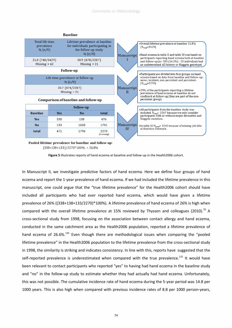
74
Figure 5 Illustrates reports of hand eczema at baseline and follow-up in the Health2006 cohort.
In Manuscript II, we investigate predictive factors of hand eczema. Here we define four groups of hand
eczema and report the 1-year prevalence of hand eczema. If we had included the lifetime prevalence in this
manuscript, one could argue that the “true lifetime prevalence” for the Health2006 cohort should have
included all participants who had ever reported hand eczema, which would have given a lifetime
prevalence of 26% ((338+138+133/2270)*100%). A lifetime prevalence of hand eczema of 26% is high when
compared with the overall lifetime prevalence at 15% reviewed by Thyssen and colleagues (2010).70 A
cross-sectional study from 1998, focusing on the association between contact allergy and hand eczema,
conducted in the same catchment area as the Health2006 population, reported a lifetime prevalence of
hand eczema of 26.6%.135 Even though there are methodological issues when comparing the “pooled
lifetime prevalence” in the Health2006 population to the lifetime prevalence from the cross-sectional study
in 1998, the similarity is striking and indicates consistency. In line with this, reports have suggested that the
self-reported prevalence is underestimated when compared with the true prevalence.131 It would have
been relevant to contact participants who reported “yes” to having had hand eczema in the baseline study
and “no” in the follow-up study to estimate whether they had actually had hand eczema. Unfortunately,
this was not possible. The cumulative incidence rate of hand eczema during the 5-year period was 14.8 per
1000 years. This is also high when compared with previous incidence rates of 8.8 per 1000 person-years,
74
Comments on Methodology

75
which have been found in two studies with Danes from the general population.66,136 However, the incident
cases in Manuscript II were defined by comparing two lifetime prevalence. As discussed in the manuscript,
this method has not been validated and might increase recall bias. Last, the following definition of
dermatitis: ”Dermatitis is an itchy skin disorder showing redness, dryness, and possibly bladders and
exudation. Dermatitis remains on the same area of the body for some time”, was introduced prior to the
questions on dermatitis. It was intended to help participants define dermatitis and thereby reduce
misclassification. It is unknown whether the definition of dermatitis works as intended.
5.2 Considerations about filaggrin genotyping
We aimed to investigate the patterns of dermatitis and the personal consequences of a genetically
impaired skin barrier, which we defined by the presence of at least one mutation in the gene coding for the
filaggrin protein. To date, more than 50 mutations have been identified throughout the profilaggrin
molecule, of which 23 are variants found in European populations and 8 are believed to be recurrent.13,113
In our analyses we genotyped all participants for three of the most common loss-of-function mutations
constituting 83% of the total risk alleles associated with atopic dermatitis; R501X, 2282del4, and
R2447X.1,13,113 If we had examined two additional mutations S3247X113 and 3702delA137 we could have
increased the coverage to include 96% of the identified risk alleles, 10% and 3%, respectively. The FLG tests
for both populations were performed at the Department of Clinical Biochemistry, Copenhagen University
Hospital Herlev-Gentofte. In this project we use a slightly modified version of the method which has
previously been described in detail.138 At the Department of Clinical Biochemistry, efforts have been made
to include additional mutations in the routine analyses for FLG mutations. The allele-specific PCR prior to
the microbead-specific hybridization in the FLG genotype is complex and primers for the specific mutations
have different optima regarding temperature and salt concentrations. Thus inclusion of an additional
primer, representing another mutation, is complex and was not possible at the time of genotyping. Other
strategies for full sequencing have been reported113 but were not chosen because of time and financial
aspects. Nevertheless, we believe that the three mutations included are representative of the Northern
European population, but we cannot determine whether some individuals might be carriers of some of the
rarer FLG mutations.
5.3 Establishment of the patient population
Defining atopic dermatitis and hand eczema in the patient population: Clinical diagnoses of atopic
dermatitis and hand eczema were scored by the attending dermatologist and were registered in the
patient’s file using the internationally recognized MOAHLFA-index115 (Male, Occupational dermatitis,
history of Atopic dermatitis, Hand eczema, Leg dermatitis, Facial dermatitis, and Age above 40 years), which
75
Comments on Methodology

76
is registered routinely for all patients who are patch tested. It should be highlighted that while the
diagnosis of atopic dermatitis reflects a lifetime prevalence, the hand eczema diagnosis reflects a point
prevalence at the time of patch testing. Thus many patients in the group with atopic dermatitis may have
had hand eczema at some point in life, especially since atopic dermatitis is known to be a major risk factor
of hand eczema.66,74,75 This was confirmed in the questions about hand eczema included in the
questionnaire completed by the patients showing that 68.3% of the 102 patients included with atopic
dermatitis only reported a lifetime prevalence of hand eczema (Table II, Manuscript III). To address this, we
divided patients into a “skin barrier variable” (Manuscripts III and IV) based on history of atopic dermatitis
and on results from FLG genotyping. By doing this, the group with atopic dermatitis only and the group with
both atopic dermatitis and hand eczema were pooled. Notably, one third of the participants with atopic
dermatitis and hand eczema in this cohort were FLG mutation carriers, which highlights the importance of
the interplay between FLG mutations, hand eczema and atopic dermatitis.
Expected prevalence of filaggrin mutation carriers: When we made power calculations for the patient
population prior to project initiation, we estimated that the prevalence of FLG mutations in hand eczema
patients would be around 20%.139 Moreover, reports showed that the prevalence of FLG mutations in
patients with atopic dermatitis is around 25%.140 In adult patients with severe and persistent atopic
dermatitis, the prevalence of FLG mutations was found to be around 40%.34 Because we did not consider
severity when including patients with atopic dermatitis, we expected a prevalence of FLG mutations of 25%.
When considering the results from Manuscript III, the following prevalence of FLG mutations was found:
patients included with atopic dermatitis (18.8%), patients included with hand eczema (8.9%) and patients
included with both atopic dermatitis and hand eczema (33.1%). In retrospect, we overestimated the
prevalence of FLG mutation carriers among patients with hand eczema and no history of atopic dermatitis,
which showed to be similar to the prevalence found in the general population.15 Because of this, we had
only 25 patients in the group with no atopic dermatitis and FLG wild type (patients with hand eczema),
which represented 4.8% of the study population.
Participation rate: The low number of participants in this group is also a result of the 46.5% participation
rate. It is generally considered that participation rates are a measure of representativeness and should be
as high as possible to minimize the risk of selection bias.141 Nevertheless, it is also known that there is a
declining response rate for participation in health surveys and that change in the non-respondents group
exists.142 Table 5 presents socioeconomic variables for the 1119 invited patients and reveals that there are
several significant differences between the two groups. Non-participants differed significantly from
76
Comments on Methodology

77
participants with respect to sociodemographic characteristics. Non-participants were younger and a
significantly larger proportion were receiving social benefits, which may have bearing on the lower income
observed in the non-participant group when compared with the participant group (P≤0.025). Because the
non-participants were significantly younger, it could be speculated whether the lower income is a result of
a larger proportion of students in group. In total, 4.9% of the patients included were students, and although
a higher prevalence of students was found among non-participants (5.5%) when compared with
participants (4.2%), the difference was non-significant.
Table 5: Socio-demographic characteristics of participants and non-participants in the patient population. *Kruskal-Wallis test.
In 2013, Thuesen and colleagues published a cohort profile of the Health2006 cohort showing the same
tendency. Here, non-respondents to the baseline examination were younger, had lower educational level
and a lower personal income than did respondents.114 In the patient cohort, it was however not only a
questionnaire that should be completed and send back. Because of the FLG genotyping, all interested
patient should actively take contact to the National Allergy Research Centre where they got oral
information about the study prior to inclusion. The results from Table 5 could point to participants having
larger resources than non-participants. However, we have no reason to believe than the non-participants
should differ from participants in relation to prevalence of FLG mutation carriers. Concluding, Rothman and
colleagues (2013) state that “It is not representativeness of the study subjects that enhances the
generalization, it is knowledge of specific conditions and an understanding of mechanism that makes for a
proper generalization”.143 Parts of the work with the patient population however have a descriptive aim
which is why one could argue that a high participation rate would, of cause, had been preferable.143
Usefulness of the DLQI questionnaire: In Manuscript III, we evaluated HRQoL using the DLQI questionnaire.
The DLQI was developed to assess skin-specific HRQoL.104 In general, HRQoL can be defined as the
Participants (n = 520)
Non-participants (n = 599)
P value Chi square test
Sex (Male, %)
26.2
31.2
= 0.062
Age (median in years, min – max)
49 (18-83)
40 (18-86)
< 0.001*
Family status (%, living alone w/wo children)
28.3%
32.1%
0.098
Occupational status Working (%) Unemployed (%, receiving social benefits) Leave, sick leave or early retirement (%) Retired (%)
79.1 4.3 5.7
11.0
76.0 8.7 6.3 9.1
0.025
Income (gross) (mean in dkk, (n))
352,619
268,444 <0.001*
77
Comments on Methodology

78
subjective perception of the impact of health status, including disease, treatment, and both physical and
psychological wellbeing.144 The DLQI has been widely used since its introduction; however, the clinical
validity of the instruments has been debated. Twiss and colleagues (2012) questioned whether the DLQI
could be used to measure the impact of psoriasis and atopic dermatitis in patients,145 and later Nijsten
(2012) concluded that the DLQI had limitations and that instruments assessing quality of life had evolved
since 1994.146 Despite the critique, the DLQI instrument has repeatedly been found to be reliable, valid, and
easy to use.147 Disease-specific questionnaires to measure HRQoL continue to be developed. One of the
newer hand eczema questionnaires ‘The Quality of Life in Hand Eczema Questionnaire’ was introduced in
2014 by Ofenloch and colleagues and the authors concluded that it showed strong properties in terms of
validity, reliability and responsiveness to change.148 In general, disease-specific questionnaire are
preferable to catch the relevant symptoms, but because we had different diagnoses in the patient
population, hand eczema and/or atopic dermatitis, we needed a skin-specific questionnaire rather than a
disease-specific questionnaire, which is why we believe that the DLQI questionnaire was the best choice for
this study population.
5.4 Using retrospective registry data
In Manuscript IV we investigated the personal consequences of having a genetically impaired skin barrier
using self-reported questionnaire data and registry data from Statistics Denmark. We chose to present data
from both patients and the general population in the same manuscript but did not compare the two
populations directly because there are differences in time of inclusion (2014 vs. 2006), sex and age.
Because of the high number of participants in the Health2006 population, we could have chosen to match
participants in the Health2006 population, on sex and age, with the patients. However, if we had chosen
this method, we would have discarded data from many FLG mutation carriers and would then have
compromised power in the analyses. Therefore, we chose to include all participants from both populations
and made descriptive analyses, well-knowing that the populations were representative of the adult general
population and adult dermatitis patients but could not be considered as “controls and cases”, respectively.
When we investigated the association between disability pension and dermatitis/FLG genotype in Table 2,
Manuscript IV, we made a dichotomous variable for disability pension to use in the regression analyses. In
this ‘disability pension variable’ participants receiving disability pension formed one group, while
individuals who were either ‘working’, ‘unemployed’ or ‘on leave or sick leave’ formed another group ‘not
receiving disability pension’. Retired participants (n=429) were set to be ‘missing’ because they represent a
group outside the work force and cannot be considered as candidates for disability pension. The grounds
for granting disability pension were unknown in this setup, but we can conclude that the proportion of
78
Comments on Methodology

79
individuals receiving disability pension in the general population was increased among individuals reporting
dermatitis. To account for time differences, we repeated the association between FLG mutations and
disability pension in three additional years, 2008, 2010 and 2013, and found significant associations (P
≤0.025). The relevance of this control can be discussed since FLG genotype is permanent and most cases of
disability pensions are granted as lifelong benefits. However, societal changes in social benefits, including
retirement age and terms for granting social benefits, are introduced on a regular basis, which could
influence the terms of granting disability pension in the period and introduce bias.
In Manuscript IV we also investigated the differences in former and present work in a risk occupation.
Statistics Denmark made a retrospective analysis where they investigated whether the participants in
question had a history in risk occupations during 1994–2013 (as long as possible using the Danish Register
on Labour Market Affiliation). In Table 3, Manuscript IV, we defined 18 risk occupations based on the
existing literature.83,149-151 By using this approach we were able to retrieve prevalence estimates about
work in a risk occupation and years in the most frequent risk occupation. Because of the form in which the
tables were delivered, if was not possible to account for risk time in the occupations.
Moreover, Statistics Denmark has some strict rules regarding privacy to ensure data protection. This means
that if one cell contains less than three observations, another cell must be left blank to ensure that it is
impossible to trace the individual. Because of this we were not able to make separate analyses on
homozygous and heterozygous FLG mutation carriers but had to focus our analyses on participants with or
without a FLG mutation. Lastly, not all the registers are 100% complete, which is why there are small
variations in Ntotal in the different tables. That being said, the Danish registers represent a unique data
source that is very accurate because of the linkages using the personal identification numbers.
79
Comments on Methodology

80
6. DISCUSSION
This section includes a condensed discussion additional to the discussion sections presented in the
manuscripts.
Epidemiology of dermatitis in the general population:
One third of the adult Danish population reported dermatitis at some point in their lives on some point of
their bodies. Hand eczema was the most prevalent site of dermatitis with a lifetime prevalence at baseline
of 20.9%; of this percentage, 14.2% of the individuals confirmed having had hand eczema at any point in
their lives at follow-up. In relation to genetic predisposition of dermatitis, we showed that patients with
atopic dermatitis had a distinct pattern of dermatitis and that FLG mutations increase the lifetime
prevalence of persistent hand eczema and dermatitis on the feet.Manuscript I
Our finding that FLG mutations increased the prevalence of self-reported dermatitis only on the extremities
is possibly influenced by the daily environmental and/or physical challenges, such as irritants, allergens,
sweat and/or pressure, to which the hands and feet are subjected. Accordingly, a clinical report evaluating
contact allergy in children with atopic dermatitis concludes that children with dermatitis on the hands
and/or feet should always be patch-tested.152 In line with our findings, Isaksson and colleagues (2015)
indicate that patients with atopic dermatitis and hand or foot dermatitis stand out, but no genotyping for
FLG mutations was performed in this study.152 Notably, body regional variations in epidermal thickness
exist.153,154 Further, regional variations for drug penetration have been shown with decreased absorption in
the regions of the skin having a thickened stratum corneum, such as the feet.155 These notions could be
contradictive with a hypothesis of increased penetration in the foot and hand region, where the epidermis
is thick: sole (636.5 µm), dorsum of foot (177.5 µm), palm (602.4 µm) and dorsum of hand (189.5 µm) and
where fewer hair follicles are present.153 Nevertheless, we also showed that FLG mutations were
significantly associated with skin fissures on the hand and the heels, which may increase penetration of
exogenous agents. Manuscript III
In agreement with this, research in filaggrin-deficient mice (often flaky tail (ft/ft) mice)156 has shown that
filaggrin deficiency predisposes to enhanced epicutaneous sensitization with less resistance to mechanical
stress in the deeper layers of stratum corneum.157-159 This supports a hypothesis of increased penetration in
the foot and hand region, regions which are exposed to mechanical stress. However, it must be highlighted
that the majority of the FLG mutation carriers in the Health2006 population were heterozygous carriers and
that atopic skin itself is known to affect the skin barrier, leading to, for example, defects in lipid
metabolism, altered protease activity and tight junction defects. Reviewed in 46,160
80
Discussion

81
Several factors predispose to hand eczema:
Several factors predispose to hand eczema. In Manuscript II we confirmed previous studies showing a
relationship between persistent hand eczema and contact sensitization.161 Notably, we also found the
association between contact allergy and persistent hand eczema to be significant in individuals without
atopic dermatitis (OR = 2.84; 95% CI 1.24–5.60).Manuscript II
While positive associations between FLG mutations and intolerance to nickel have previously been
reported,15,162,163 other studies in patients have rejected an association between FLG mutations and contact
sensitization.164 A direct association, independent of atopic dermatitis, between FLG mutations and hand
eczema has been identified in few studies.165,166 An increased risk of developing hand eczema, both irritant
contact dermatitis167 and combined irritant and allergic contact dermatitis,166 has been found for FLG
mutations carriers, particularly in individuals with a history of atopic dermatitis.48 Recently, FLG mutations
were found to be associated with contact dermatitis in construction workers, particularly in individuals with
an atopic predisposition, stressing the need for prevention among individuals with an impaired skin
barrier.88
Of the 108 individuals with persistent hand eczema in Manuscript II, 19.6 % (n=20) were FLG mutation
carriers compared with 7.5% of those with incident and non-persistent hand eczema (P<0.001). FLG
mutations were found to predict persistent hand eczema, but no difference was found in the prevalence of
contact sensitization between FLG mutation carriers and non-carriers (data not shown). Other predictive
factors for hand eczema were also found including self-reported food allergy and ‘redness, rash or itch
related to use of cosmetic products’, which confirms that environmental factors in daily life have a major
impact on the disease.Manuscript II As patients with atopic dermatitis have repeatedly been shown to have a
compromised skin barrier function, resulting in an increased reactivity to skin irritants168-170 and food
allergy,43 we stratified for atopic dermatitis and found both food allergy and ‘redness, rash or itch related to
use of cosmetic products’ to be predictors of hand eczema independently of atopic dermatitis. Again, the
finding stresses the need for continuous skin care and knowledge about irritants and allergens in the
population. Notably, a recent European multicentre study found no increased overall risk of contact
sensitization in individuals with atopic dermatitis from the general population.171
Thus the interplay between contact dermatitis, FLG mutations and dermatitis remains to be elucidated to
identify whether FLG mutations are contributing factors or causal components for contact sensitization.
A clear tendency towards debut of hand eczema depending on history of atopic dermatitis and FLG
genotype was illustrated in Manuscript IV, and the patterns were very similar for patients and individuals
81
Discussion

82
from the general population. We report that almost 40% of participants with both atopic dermatitis and
FLG mutations report a debut of hand eczema before the age of 6 years, and around 60% will have
developed symptoms of hand eczema before reaching adulthood (> 18 years).Manuscript IV The fact that the
majority of individuals with FLG mutations and atopic dermatitis will develop signs of hand eczema before
adulthood shows the importance of early detection of skin symptoms and frequent use of emollients,
which improves the skin barrier172 and has been found to be useful in the therapy of hand eczema.173,174
Do filaggrin mutations exert their function early in life?
In Manuscript II we showed that FLG mutations are not associated with incident hand eczema in adults and
introduced the hypothesis that FLG mutations exert their function early in life. This notion is supported by a
few experimental studies.175;176
Today, we know that a delicate balance between genetic and acquired filaggrin deficiency exists.55-61
However, we do not know whether this balance changes in relation to time. In this thesis we did not look
into the underlying biology but aimed to investigate the complexity using epidemiological analyses. In both
Manuscripts I and II, we stratified for atopic dermatitis and found that without co-existence with atopic
dermatitis, the effect of having impaired skin barrier was neutralized. In other words, there was a tendency
to regard FLG mutations as an “add on effect” in individuals with a history of atopic dermatitis.
The role of filaggrin on comorbidity:
Research investigating cutaneous and non-cutaneous comorbidity associated with FLG mutations has
advanced in recent years. In our cohort of patients, we investigated self-reported comorbidity using the
question: ‘has a doctor ever told you that you have/have had…’ Here we found, that the highest prevalence
of atopic comorbidities (asthma, rhinitis and food allergy) was reported by patients with both atopic
dermatitis and FLG mutations.Manuscript III However, the association between FLG mutations and both asthma
and rhinitis was lost when adjusting for age group, sex and a history of atopic dermatitis, which is in line
with the existing literature.30,31,42
We also identified an association between FLG mutations and self-reported actinic keratosis, with the
highest prevalence reported by homozygous carriers, followed by heterozygous carriers and wild type
individuals (P = 0.003).Manuscript III Degradation of the filaggrin protein in the stratum corneum results in trans-
urocanic acid,177 an important absorber of UV radiation in the stratum corneum.178,179 Accordingly, it could
be suggested that the association between self-reported actinic keratosis and FLG genotype could be a
result of the reduced photoprotective capacity of filaggrin deficient skin. However, it is also possible that
the association is an artefact of UV therapy to treat patients’ dermatitis. Because of the small number of
cases and lack of prior power calculations, the association between FLG mutations and actinic keratosis
82
Discussion

83
needs to be confirmed. A consensus report from 2016 calls for action in recognizing non-melanoma skin
cancer, including actinic keratosis, as an occupational disease180 because outdoor workers were found to
have a higher risk of actinic keratosis when compared with indoor workers (OR=1.55; 95% CI 1.55–2.54). If
the association between FLG mutations and actinic keratosis is confirmed, use of UV therapy in FLG
mutation carriers should be reconsidered. Finally, FLG mutation carriers, especially homozygous individuals,
should be advised to avoid excessive sun exposure, and workers in an outdoor occupation should have
extensive information about primary protection.
Personal consequences of dermatitis:
Research addressing the personal consequences of having FLG mutations is limited. This was the reasoning
behind looking into the personal consequences of having FLG mutations in adult dermatitis patients.
In Manuscript III, we showed that patients with concomitant atopic dermatitis and FLG mutations also
stood out in relation to HRQoL. We investigated quality of life using the DLQI questionnaire and found that
FLG mutations significantly reduced HRQoL. When stratifying for a history of atopic dermatitis, FLG
mutations reduced HRQoL only in patients with a history of atopic dermatitis and not in patients with only
hand eczema. This finding is in line with our previous hypothesis, based on results from the general
population, indicating that FLG mutations mainly have a function in coexistence with atopic dermatitis.
HRQoL is an important validated estimation of patients’ wellbeing and our data suggest that FLG mutation
carriers with atopic dermatitis can experience an additional challenge in their everyday lives that can have
clinical implications.
We also investigated the personal consequences of having a genetically impaired skin barrier by linkage to
socioeconomic variables from Statistics Denmark in Manuscript IV. In general, we found that among
participants receiving disability pension, 21% had a lifetime prevalence of atopic dermatitis, 40% reported a
lifetime prevalence of hand eczema, and 15.7% were filaggrin mutation carriers. Using a logistic regression
model we found that disability pension was associated with both atopic dermatitis and hand eczema in the
general population. Notably, self-reported hand eczema and atopic dermatitis were associated with a
particularly high risk of disability in FLG mutation carriers, although these interactions did not reach
statistical significance. From a causal point to view, FLG mutations themselves cannot predispose to
disability pension but may result in symptoms leading to disability pension.
According to the Social Appeals Board in Denmark, 8198 individuals applied for disability pension in 2014,
of these applications 75% were recognized.181 The majority of cases were due to psychological diagnoses,
and the second most prevalent diagnosis was related to diseases in the musculoskeletal system. Notably,
dermatitis was not included as a separate diagnosis but is assumed to be incorporated in the third most
83
Discussion

84
prevalent category ‘other or missing diagnoses’.181 Moreover, disability pension was reported to be most
frequently recognized among individuals aged 50–59 years.181
In our setup we had no information about the diagnoses leading to the local authority granting disability
pension. The grounds for disability pension can therefore be of physical, psychological, or social character,
or a combination. In general, Ibler and Jemec (2011) concluded that the number of permanent disability
pensions granted due to skin diseases in Denmark was low when compared with the high prevalence of
dermatitis and suggested that skin diseases might have less impact on the individual or are not considered
as debilitating as other prevalent diseases by the authorities.182 Thus several diagnoses can be grounds for
granting disability pension, but only one disease will be registered.
In Denmark, the number of disability pensions granted because of atopic dermatitis increased from 3.6 per
year in the 1970s to 14.2 per year in the 2000s.182,183 Although the incidence rate of atopic dermatitis in
children has stabilized,184 it is well-described that the incidence of atopic dermatitis has increased over the
past decades, which may affect the observed trend.185 The prevalence of atopic dermatitis in the
Health2006 population was found to decrease with increasing age: 13.7% (18–40 years), 10.9% (41–55
years), and 7.5% (> 56 years). It can therefore be speculated whether the number of disability pensions
granted because of atopic dermatitis will increase when the younger generations ages. If this is the case,
this also increases the incentive to promote awareness of skin diseases from a societal point of view.
In contrast, contact dermatitis was found to be the dominant diagnosis of disability pensions due to skin
diseases in Denmark in the 1970s.183 During 2003–2008 disability pensions granted due to contact
dermatitis were halved, which has been proposed to be a consequence of the National and European nickel
legislation, aimed at reducing environmental exposure to nickel.182,186
A recent paper from Sweden found eczema to be the cause of 36% of all disability pensions granted due to
skin diseases. Of those granted disability pension, painters, cement workers and plumbers formed the
major group.187 Moreover, a Danish study from 2009 suggests potential for prevention of work-related
disabilities, especially among job groups exposed to cleaning agents.188 In our setup we could not link
disability pension and job title. We did, however, see that 6.6% of the general population had been working
in a cleaning occupation at some point. Moreover, the majority of patients included with hand eczema (and
no atopic dermatitis) had a short education, irrespective of filaggrin genotype (hand eczema + FLGwt 66.8%
and hand eczema + FLGmut = 79.2%).Manuscript IV In addition to having a short education, patients with hand
eczema but without atopic dermatitis were characterized by onset of hand eczema in adulthood and an
increased prevalence of history in a risk occupation which indicate a different disease pattern when
compared with individuals with hand eczema but with a history of atopic dermatitis.Manuscript IV
84
Discussion

85
Although several studies have found that having childhood atopic dermatitis does not prevent future work
in risk occupations,77,189,190 Wei and colleagues (2016) found that having received job counselling was
associated with higher use of preventive measures,190 which encourages occupational guidance from an
early time and knowledge about preventive measures in risk occupations.
In addition, a healthy-worker effect has previously been suggested based on data from the Health2006
cohort where Bandier and colleagues (2013) reported that FLG mutation carriers with early onset of hand
eczema in childhood avoid occupational exposure to irritants later in life.87 In our analyses we found that
individuals with a history in a risk occupation reported more dermatitis, but we did not see any self-
exclusion from risk occupations among individuals with atopic dermatitis or FLG mutations. Manuscript IV
However, our data build on retrospective linkage, thereby showing a lifetime prevalence and we cannot
know whether the dermatitis preceded the work in a risk occupation or was a consequence. Thus new
studies, preferably prospective cohort studies, are needed to address the relationship between risk
occupations, dermatitis and FLG mutations and they should include duration of the work. When
considering risk occupations, new studies must also consider that the use of job title as a proxy for water
exposure has been shown to underestimate skin exposure to water150 and that high water exposure outside
work is frequent.191
Taken together, our results indicate that FLG mutation carriers with atopic dermatitis are a subgroup of
patients who stand out on several parameters. The parameters are biologically manifested by increased
prevalence of foot dermatitis and increased persistence of hand eczema; psychologically manifested by
reduced HRQoL; and socially manifested by the finding that self-reported dermatitis was associated with
disability pension, particularly in individuals with FLG mutations. This points towards FLG mutations
predisposing to increased severity, stressing the need to further promote skin awareness in this subgroup.
85
Discussion

86
7. CONCLUSIONS
This thesis concerns the epidemiology of dermatitis and focuses on genetic predisposition, defined by FLG
mutations, and personal consequences of dermatitis. The main findings from the four manuscripts included
are listed below:
More than 1/3 of the Danish population reports dermatitis
FLG mutations are associated with dermatitis on the hands and feet in individuals with atopic
dermatitis
A history of atopic dermatitis predicts both incident and persistent hand eczema in adults
FLG mutations strongly predict persistent hand eczema, but the association with incident hand
eczema in adults is nonsignificant
Contact sensitization is significantly associated with persistent hand eczema, also in participants
without atopic dermatitis
Patients with atopic dermatitis and FLG mutations report reduced HRQoL of life. However, no
association between HRQoL and FLG mutations was found in patients without atopic dermatitis
An association between self-reported actinic keratosis and FLG mutations is suggested
Self-reported dermatitis is associated with receiving disability pension in the general population,
particularly in individuals with FLG mutations
Of the individuals with atopic dermatitis and FLG mutations who develop hand eczema, more than
50% are likely to develop symptoms before adulthood
86
Conclusions

87
8. FUTURE RESEARCH
Although research within skin diseases and skin barrier function is increasing, many aspects need further
investigation. In this thesis, estimated genetic predisposition to dermatitis was defined by measuring the
genotype of a single gene. However, the effects of FLG mutations, and potentially other susceptibility
genes, are difficult to assess quantitatively because of the complex interplay between FLG genotype,
dermatitis and, specially, occupational exposure.
It has previously been suggested that surveillance programmes in high-risk occupations can contribute to
better identification of susceptible individuals and to personalized prevention.77 This is supported by the
findings of this thesis. New studies are needed to build upon the results from this thesis and to investigate
whether FLG mutation carriers are overrepresented among individuals receiving rehabilitation benefits
and/or disability pension granted as a consequence of dermatitis. If this is the case, the need for medical
attention with respect to decisions on early change of occupation is further highlighted.
Moreover, future research should aim to elucidate the biological mechanisms controlling filaggrin
expression in the skin and investigate changes in expression according to different body sites, difference in
age groups, and the effect of environmental down-regulation.192
It remains a mystery why only some FLG mutation carriers develop dermatitis and some do not, assuming
that all heterozygote carriers have the same risk of sensitization. It is evident that environmental exposure
and consumer habits can play a key role. However, inter-individual variations in the level of compensatory
mechanisms also exist. One of the other genes encoded within the epidermal differentiation complex on
human chromosome 1q21.3 is the filaggrin-2 gene.193 Filaggrin-2 has been suggested to share many
properties with filaggrin including maintenance of skin barrier integrity. In line with this, studies have
shown that filaggrin-2 levels are down-regulated in skin affected by atopic dermatitis and hand eczema.56,63
It has also been proposed that filaggrin-2 might be able to compensate in diseases characterized by an
abnormal or lost function of filaggrin.Reviewed in 194
Thus research investigating gene-environment interactions and individual susceptibility driving different
forms of dermatitis is needed. New studies should focus on identifying disease patterns and should use
bioinformatics tools to combine differences in transcriptome activity, immune profile and even epigenetics
to understand the molecular pathophysiology of atopic dermatitis and irritant and/or allergic contact
dermatitis.
87
Future Research

88
9. REFERENCES
1 Palmer CN, Irvine AD, Terron-Kwiatkowski A et al. Common loss-of-function variants of the epidermal barrier protein filaggrin are a major predisposing factor for atopic dermatitis. Nat Genet 2006; 38: 441-6.
2 McGrath JA, Uitto J. Anatomy and Organization of Human Skin. In: Rook's Textbook of Dermatology, Vol. 1, Eighth Edition: Wiley-Blackwell. 2010; 1-53.
3 Houben E, De Paepe K, Rogiers V. A keratinocyte's course of life. Skin Pharmacol Physiol 2007; 20: 122-32. 4 Dale BA. Purification and characterization of a basic protein from the stratum corneum of mammalian
epidermis. Biochim Biophys Acta 1977; 491: 193-204. 5 Steinert PM, Cantieri JS, Teller DC et al. Characterization of a class of cationic proteins that specifically
interact with intermediate filaments. Proc Natl Acad Sci U S A 1981; 78: 4097-101. 6 Manabe M, Sanchez M, Sun TT et al. Interaction of filaggrin with keratin filaments during advanced stages of
normal human epidermal differentiation and in ichthyosis vulgaris. Differentiation 1991; 48: 43-50. 7 Sandilands A, Sutherland C, Irvine AD et al. Filaggrin in the frontline: role in skin barrier function and disease.
J Cell Sci 2009; 122: 1285-94. 8 Harding CR, Scott IR. Histidine-rich proteins (filaggrins): structural and functional heterogeneity during
epidermal differentiation. J Mol Biol 1983; 170: 651-73. 9 Gan SQ, McBride OW, Idler WW et al. Organization, structure, and polymorphisms of the human profilaggrin
gene. Biochemistry 1990; 29: 9432-40. 10 Lonsdale-Eccles JD, Teller DC, Dale BA. Characterization of a phosphorylated form of the intermediate
filament-aggregating protein filaggrin. Biochemistry 1982; 21: 5940-8. 11 Rawlings AV, Harding CR. Moisturization and skin barrier function. Dermatol Ther 2004; 17 Suppl 1: 43-8. 12 Mischke D, Korge BP, Marenholz I et al. Genes encoding structural proteins of epidermal cornification and
S100 calcium-binding proteins form a gene complex ("epidermal differentiation complex") on human chromosome 1q21. J Invest Dermatol 1996; 106: 989-92.
13 Irvine AD, McLean WH, Leung DY. Filaggrin mutations associated with skin and allergic diseases. The New England journal of medicine 2011; 365: 1315-27.
14 Irvine AD, McLean WH. Breaking the (un)sound barrier: filaggrin is a major gene for atopic dermatitis. J Invest Dermatol 2006; 126: 1200-2.
15 Thyssen JP, Johansen JD, Linneberg A et al. The association between null mutations in the filaggrin gene and contact sensitization to nickel and other chemicals in the general population. Br J Dermatol 2010; 162: 1278-85.
16 Li M, Liu Q, Liu J et al. Mutations analysis in filaggrin gene in northern China patients with atopic dermatitis. J Eur Acad Dermatol Venereol 2013; 27: 169-74.
17 Bieber T. Atopic dermatitis. N Engl J Med 2008; 358: 1483-94. 18 Taylor B, Wadsworth J, Wadsworth M et al. Changes in the reported prevalence of childhood eczema since
the 1939-45 war. Lancet 1984; 2: 1255-7. 19 Larsen FS, Holm NV, Henningsen K. Atopic dermatitis. A genetic-epidemiologic study in a population-based
twin sample. J Am Acad Dermatol 1986; 15: 487-94. 20 Larsen FS, Hanifin JM. Epidemiology of atopic dermatitis. Immunol. Allerg. Clin. North Am. 2002; 22: 1-+. 21 Mortz CG, Lauritsen JM, Bindslev-Jensen C et al. Prevalence of atopic dermatitis, asthma, allergic rhinitis, and
hand and contact dermatitis in adolescents. The Odense Adolescence Cohort Study on Atopic Diseases and Dermatitis. Br J Dermatol 2001; 144: 523-32.
22 Laughter D, Istvan JA, Tofte SJ et al. The prevalence of atopic dermatitis in Oregon schoolchildren. J Am Acad Dermatol 2000; 43: 649-55.
23 Kay J, Gawkrodger DJ, Mortimer MJ et al. The prevalence of childhood atopic eczema in a general population. J Am Acad Dermatol 1994; 30: 35-9.
24 Schultz Larsen F, Diepgen T, Svensson A. The occurrence of atopic dermatitis in north Europe: an international questionnaire study. J Am Acad Dermatol 1996; 34: 760-4.
25 Herd RM, Tidman MJ, Prescott RJ et al. Prevalence of atopic eczema in the community: the Lothian Atopic Dermatitis study. Br J Dermatol 1996; 135: 18-9.
26 Margolis JS, Abuabara K, Bilker W et al. Persistence of mild to moderate atopic dermatitis. JAMA Dermatol 2014; 150: 593-600.
27 Mortz CG, Andersen KE, Dellgren C et al. Atopic dermatitis from adolescence to adulthood in the TOACS cohort: prevalence, persistence and comorbidities. Allergy 2015; 70: 836-45.
88
References

89
28 Kim JP, Chao LX, Simpson EL et al. Persistence of atopic dermatitis (AD): A systematic review and meta-analysis. J Am Acad Dermatol 2016.
29 Vinding GR, Zarchi K, Ibler KS et al. Is adult atopic eczema more common than we think? - A population-based study in Danish adults. Acta Derm Venereol 2014; 94: 480-2.
30 Rodriguez E, Baurecht H, Herberich E et al. Meta-analysis of filaggrin polymorphisms in eczema and asthma: robust risk factors in atopic disease. J Allergy Clin Immunol 2009; 123: 1361-70 e7.
31 van den Oord RA, Sheikh A. Filaggrin gene defects and risk of developing allergic sensitisation and allergic disorders: systematic review and meta-analysis. BMJ 2009; 339: b2433.
32 Henderson J, Northstone K, Lee SP et al. The burden of disease associated with filaggrin mutations: a population-based, longitudinal birth cohort study. J Allergy Clin Immunol 2008; 121: 872-7 e9.
33 Flohr C, England K, Radulovic S et al. Filaggrin loss-of-function mutations are associated with early-onset eczema, eczema severity and transepidermal water loss at 3 months of age. Br J Dermatol 2010; 163: 1333-6.
34 Barker JN, Palmer CN, Zhao Y et al. Null mutations in the filaggrin gene (FLG) determine major susceptibility to early-onset atopic dermatitis that persists into adulthood. J Invest Dermatol 2007; 127: 564-7.
35 Brown SJ, Sandilands A, Zhao Y et al. Prevalent and low-frequency null mutations in the filaggrin gene are associated with early-onset and persistent atopic eczema. J Invest Dermatol 2008; 128: 1591-4.
36 Paternoster L, Standl M, Chen CM et al. Meta-analysis of genome-wide association studies identifies three new risk loci for atopic dermatitis. Nat Genet 2012; 44: 187-92.
37 Hirota T, Takahashi A, Kubo M et al. Genome-wide association study identifies eight new susceptibility loci for atopic dermatitis in the Japanese population. Nat Genet 2012; 44: 1222-6.
38 Spergel JM. From atopic dermatitis to asthma: the atopic march. Ann Allergy Asthma Immunol 2010; 105: 99-106; quiz 07-9, 17.
39 Zheng T, Yu J, Oh MH et al. The atopic march: progression from atopic dermatitis to allergic rhinitis and asthma. Allergy Asthma Immunol Res 2011; 3: 67-73.
40 Schoos AM, Chawes BL, Rasmussen MA et al. Atopic endotype in childhood. J Allergy Clin Immunol 2016; 137: 844-51 e4.
41 Belgrave DC, Granell R, Simpson A et al. Developmental profiles of eczema, wheeze, and rhinitis: two population-based birth cohort studies. PLoS Med 2014; 11: e1001748.
42 Weidinger S, O'Sullivan M, Illig T et al. Filaggrin mutations, atopic eczema, hay fever, and asthma in children. J Allergy Clin Immunol 2008; 121: 1203-09 e1.
43 Brown SJ, Asai Y, Cordell HJ et al. Loss-of-function variants in the filaggrin gene are a significant risk factor for peanut allergy. J Allergy Clin Immunol 2011; 127: 661-7.
44 Bisgaard H, Simpson A, Palmer CN et al. Gene-environment interaction in the onset of eczema in infancy: filaggrin loss-of-function mutations enhanced by neonatal cat exposure. PLoS Med 2008; 5: e131.
45 Linneberg A, Fenger RV, Husemoen LL et al. Association between loss-of-function mutations in the filaggrin gene and self-reported food allergy and alcohol sensitivity. Int Arch Allergy Immunol 2013; 161: 234-42.
46 De Benedetto A, Kubo A, Beck LA. Skin barrier disruption: a requirement for allergen sensitization? J Invest Dermatol 2012; 132: 949-63.
47 Thyssen JP, Tang L, Husemoen LL et al. Filaggrin gene mutations are not associated with food and aeroallergen sensitization without concomitant atopic dermatitis in adults. J Allergy Clin Immunol 2015; 135: 1375-8 e1.
48 Thyssen JP, Carlsen BC, Menne T et al. Filaggrin null mutations increase the risk and persistence of hand eczema in subjects with atopic dermatitis: results from a general population study. Br J Dermatol 2010; 163: 115-20.
49 Smith FJ, Irvine AD, Terron-Kwiatkowski A et al. Loss-of-function mutations in the gene encoding filaggrin cause ichthyosis vulgaris. Nat Genet 2006; 38: 337-42.
50 Armstrong BK, Kricker A. The epidemiology of UV induced skin cancer. J Photochem Photobiol B 2001; 63: 8-18.
51 Mildner M, Jin J, Eckhart L et al. Knockdown of filaggrin impairs diffusion barrier function and increases UV sensitivity in a human skin model. J Invest Dermatol 2010; 130: 2286-94.
52 Skaaby T, Husemoen LL, Thyssen JP et al. Filaggrin loss-of-function mutations and incident cancer: a population-based study. Br J Dermatol 2014; 171: 1407-14.
53 Kaae J, Szecsi PB, Meldgaard M et al. Individuals with complete filaggrin deficiency may have an increased risk of squamous cell carcinoma. Br J Dermatol 2014; 170: 1380-1.
89
References

90
54 Kaae J, Thyssen JP, Johansen JD et al. Filaggrin gene mutations and risk of basal cell carcinoma. Br J Dermatol 2013; 169: 1162-4.
55 Howell MD, Kim BE, Gao P et al. Cytokine modulation of atopic dermatitis filaggrin skin expression. J Allergy Clin Immunol 2007; 120: 150-5.
56 Pellerin L, Henry J, Hsu CY et al. Defects of filaggrin-like proteins in both lesional and nonlesional atopic skin. J Allergy Clin Immunol 2013; 131: 1094-102.
57 Gutowska-Owsiak D, Schaupp AL, Salimi M et al. Interleukin-22 downregulates filaggrin expression and affects expression of profilaggrin processing enzymes. Br J Dermatol 2011; 165: 492-8.
58 Gutowska-Owsiak D, Schaupp AL, Salimi M et al. IL-17 downregulates filaggrin and affects keratinocyte expression of genes associated with cellular adhesion. Exp Dermatol 2012; 21: 104-10.
59 Hvid M, Vestergaard C, Kemp K et al. IL-25 in atopic dermatitis: a possible link between inflammation and skin barrier dysfunction? J Invest Dermatol 2011; 131: 150-7.
60 Kim BE, Howell MD, Guttman-Yassky E et al. TNF-alpha downregulates filaggrin and loricrin through c-Jun N-terminal kinase: role for TNF-alpha antagonists to improve skin barrier. J Invest Dermatol 2011; 131: 1272-9.
61 Cornelissen C, Marquardt Y, Czaja K et al. IL-31 regulates differentiation and filaggrin expression in human organotypic skin models. J Allergy Clin Immunol 2012; 129: 426-33, 33 e1-8.
62 Sheu HM, Tai CL, Kuo KW et al. Modulation of epidermal terminal differentiation in patients after long-term topical corticosteroids. J Dermatol 1991; 18: 454-64.
63 Molin S, Merl J, Dietrich KA et al. The hand eczema proteome: imbalance of epidermal barrier proteins. Br J Dermatol 2015; 172: 994-1001.
64 Menne T, Johansen JD, Sommerlund M et al. Hand eczema guidelines based on the Danish guidelines for the diagnosis and treatment of hand eczema. Contact Dermatitis 2011; 65: 3-12.
65 Meding B, Swanbeck G. Predictive factors for hand eczema. Contact Dermatitis 1990; 23: 154-61. 66 Mortz CG, Bindslev-Jensen C, Andersen KE. Hand eczema in The Odense Adolescence Cohort Study on Atopic
Diseases and Dermatitis (TOACS): prevalence, incidence and risk factors from adolescence to adulthood. Br J Dermatol 2014; 171: 313-23.
67 Diepgen TL, Andersen KE, Brandao FM et al. Hand eczema classification: a cross-sectional, multicentre study of the aetiology and morphology of hand eczema. Br J Dermatol 2009; 160: 353-8.
68 Meding B, Liden C, Berglind N. Self-diagnosed dermatitis in adults - Results from a population survey in Stockholm. Contact Dermatitis 2001; 45: 341-45.
69 Bryld LE, Agner T, Kyvik KO et al. Hand eczema in twins: a questionnaire investigation. Br J Dermatol 2000; 142: 298-305.
70 Thyssen JP, Johansen JD, Linneberg A et al. The epidemiology of hand eczema in the general population--prevalence and main findings. Contact Dermatitis 2010; 62: 75-87.
71 Meding B, Jarvholm B. Incidence of hand eczema-a population-based retrospective study. J Invest Dermatol 2004; 122: 873-7.
72 Johannisson A, Ponten A, Svensson A. Prevalence, incidence and predictive factors for hand eczema in young adults - a follow-up study. BMC Dermatol 2013; 13: 14.
73 Bjornberg A. Skin reactions to primary irritants in men and women. Acta Derm Venereol 1975; 55: 191-4. 74 Rystedt I. Factors influencing the occurrence of hand eczema in adults with a history of atopic dermatitis in
childhood. Contact Dermatitis 1985; 12: 185-91. 75 Gronhagen C, Liden C, Wahlgren CF et al. Hand eczema and atopic dermatitis in adolescents: a prospective
cohort study from the BAMSE project. Br J Dermatol 2015; 173: 1175-82. 76 Nilsson EJ, Knutsson A. Atopic dermatitis, nickel sensitivity and xerosis as risk factors for hand eczema in
women. Contact Dermatitis 1995; 33: 401-6. 77 Visser MJ, Verberk MM, van Dijk FJ et al. Wet work and hand eczema in apprentice nurses; part I of a
prospective cohort study. Contact Dermatitis 2014; 70: 44-55. 78 Anveden Berglind I, Alderling M, Meding B. Life-style factors and hand eczema. Br J Dermatol 2011; 165: 568-
75. 79 Meding B, Alderling M, Wrangsjo K. Tobacco smoking and hand eczema: a population-based study. Br J
Dermatol 2010; 163: 752-6. 80 Arbejdsskadestyrelsen. Arbejdsskadestatistik 2015 - bilag.
http://www.ask.dk/~/media/ASK/pdf/statistik/Arbejdsskadestatistik_2015/Bilag%20til%20arbejdsskadestatistik%202015.ashx Date accessed 07.07.2016. 2015.
90
References

91
81 Halkier-Sorensen L. Occupational skin diseases: reliability and utility of the data in the various registers; the course from notification to compensation and the costs - A case study from Denmark. Contact Dermatitis 1998; 39: 71-78.
82 Skoet R, Olsen J, Mathiesen B et al. A survey of occupational hand eczema in Denmark. Contact Dermatitis 2004; 51: 159-66.
83 Diepgen TL, Coenraads PJ. The epidemiology of occupational contact dermatitis. Int Arch Occup Environ Health 1999; 72: 496-506.
84 Meding B. Differences between the sexes with regard to work-related skin disease. Contact Dermatitis 2000; 43: 65-71.
85 Meding B, Anveden Berglind I, Alderling M et al. Water exposure--challenging differences between occupations. Contact Dermatitis 2016; 74: 22-8.
86 Coenraads PJ, Diepgen TL. Risk for hand eczema in employees with past or present atopic dermatitis. Int Arch Occup Environ Health 1998; 71: 7-13.
87 Bandier J, Ross-Hansen K, Carlsen BC et al. Carriers of filaggrin gene (FLG) mutations avoid professional exposure to irritants in adulthood. Contact Dermatitis 2013; 69: 355-62.
88 Timmerman JG, Heederik D, Spee T et al. Contact dermatitis in the construction industry: the role of filaggrin loss-of-function mutations. Br J Dermatol 2016; 174: 348-55.
89 Mancini AJ, Kaulback K, Chamlin SL. The socioeconomic impact of atopic dermatitis in the United States: a systematic review. Pediatr Dermatol 2008; 25: 1-6.
90 Ellis CN, Drake LA, Prendergast MM et al. Cost of atopic dermatitis and eczema in the United States. J Am Acad Dermatol 2002; 46: 361-70.
91 Gieler U, Hohmann M, Niemeier V et al. Cost evaluation in atopic eczema. J. Dermatol. Treat. 1999; 10: S15-S20.
92 Politiek K, Oosterhaven JA, Vermeulen KM et al. Systematic review of cost-of-illness studies in hand eczema. Contact Dermatitis 2016; 75: 67-76.
93 Lerbaek A, Kyvik KO, Ravn H et al. Clinical characteristics and consequences of hand eczema - an 8-year follow-up study of a population-based twin cohort. Contact dermatitis 2008; 58: 210-16.
94 Silverberg JI. Health Care Utilization, Patient Costs, and Access to Care in US Adults With Eczema: A Population-Based Study. JAMA Dermatol 2015; 151: 743-52.
95 Kawachi I, Adler NE, Dow WH. Money, schooling, and health: Mechanisms and causal evidence. Annals of the New York Academy of Sciences 2010; 1186: 56-68.
96 Dalgard F, Svensson A, Holm JO et al. Self-reported skin morbidity in Oslo. Associations with sociodemographic factors among adults in a cross-sectional study. The British journal of dermatology 2004; 151: 452-7.
97 Adisesh A, Meyer JD, Cherry NM. Prognosis and work absence due to occupational contact dermatitis. Contact dermatitis 2002; 46: 273-9.
98 Meding B, Swanbeck G. Consequences of having hand eczema. Contact Dermatitis 1990; 23: 6-14. 99 Fowler JF, Ghosh A, Sung J et al. Impact of chronic hand dermatitis on quality of life, work productivity,
activity impairment, and medical costs. J Am Acad Dermatol 2006; 54: 448-57. 100 Meding B, Wrangsjo K, Jarvholm B. Fifteen-year follow-up of hand eczema: persistence and consequences. Br
J Dermatol 2005; 152: 975-80. 101 Miyake Y, Tanaka K, Sasaki S et al. Parental employment, income, education and allergic disorders in children:
a prebirth cohort study in Japan. The international journal of tuberculosis and lung disease : the official journal of the International Union against Tuberculosis and Lung Disease 2012; 16: 756-61.
102 Wang IJ, Wen HJ, Chiang TL et al. Maternal employment and atopic dermatitis in children: a prospective cohort study. Br. J. Dermatol. 2013; 168: 794-801.
103 Fowler J, Duh MS, Lobo F et al. The health care and work loss costs associated with atopic dermatitis. J. Am. Acad. Dermatol. 2007; 56: AB68-AB68.
104 Finlay AY, Khan GK. Dermatology Life Quality Index (DLQI)--a simple practical measure for routine clinical use. Clin Exp Dermatol 1994; 19: 210-6.
105 Maksimovic N, Jankovic S, Marinkovic J et al. Health-related quality of life in patients with atopic dermatitis. J Dermatol 2012; 39: 42-7.
106 Holm EA, Wulf HC, Stegmann H et al. Life quality assessment among patients with atopic eczema. Br J Dermatol 2006; 154: 719-25.
91
References
92
107 Agner T, Andersen KE, Brandao FM et al. Hand eczema severity and quality of life: a cross-sectional, multicentre study of hand eczema patients. Contact Dermatitis 2008; 59: 43-7.
108 Cvetkovski RS, Zachariae R, Jensen H et al. Quality of life and depression in a population of occupational hand eczema patients. Contact Dermatitis 2006; 54: 106-11.
109 Boehm D, Schmid-Ott G, Finkeldey F et al. Anxiety, depression and impaired health-related quality of life in patients with occupational hand eczema. Contact Dermatitis 2012; 67: 184-92.
110 Dalgard FJ, Gieler U, Tomas-Aragones L et al. The psychological burden of skin diseases: a cross-sectional multicenter study among dermatological out-patients in 13 European countries. J Invest Dermatol 2015; 135: 984-91.
111 Slattery MJ, Essex MJ, Paletz EM et al. Depression, anxiety, and dermatologic quality of life in adolescents with atopic dermatitis. J Allergy Clin Immunol 2011; 128: 668-71.
112 Yu SH, Silverberg JI. Association between Atopic Dermatitis and Depression in US Adults. J Invest Dermatol 2015; 135: 3183-86.
113 Sandilands A, Terron-Kwiatkowski A, Hull PR et al. Comprehensive analysis of the gene encoding filaggrin uncovers prevalent and rare mutations in ichthyosis vulgaris and atopic eczema. Nat Genet 2007; 39: 650-4.
114 Thuesen BH, Cerqueira C, Aadahl M et al. Cohort Profile: the Health2006 cohort, research centre for prevention and health. Int J Epidemiol 2014; 43: 568-75.
115 Schnuch A, Geier J, Uter W et al. National rates and regional differences in sensitization to allergens of the standard series. Population-adjusted frequencies of sensitization (PAFS) in 40,000 patients from a multicenter study (IVDK). Contact Dermatitis 1997; 37: 200-9.
116 Menne T, Johansen JD, Sommerlund M et al. Hand eczema guidelines based on the Danish guidelines for the diagnosis and treatment of hand eczema. Contact dermatitis 2011; 65: 3-12.
117 Johansen JD, Aalto-Korte K, Agner T et al. European Society of Contact Dermatitis guideline for diagnostic patch testing - recommendations on best practice. Contact Dermatitis 2015; 73: 195-221.
118 Lauritsen JM, Bruus M. EpiData Entry (version 1.4.4.4). A comprehensive tool for validated entry and documentation of data. The EpiData Association. Odense, Denmark. In. 2015.
119 Petersson F, Baadsgaard M, Thygesen LC. Danish registers on personal labour market affiliation. Scand J Public Health 2011; 39: 95-8.
120 DST. Danmarks Statistik. Stillingsbeskrivelse i DISCO-88. Last accessed: 12.07.2016. http://www.dst.dk/da/Statistik/dokumentation/Nomenklaturer/DISCO-88/Stillingsbeskrivelser.
121 DST. Danmarks Statistik. DISCO-08. Danmarks Statistiks fagklassifikation. Last accessed: 12.07.2016. http://www.dst.dk/pukora/epub/upload/16711/disco.pdf.
122 Smith M, Milic-Frayling N, Shneiderman B et al. NodeXL: a free and open network overview, discovery and exploration add-in for Excel 2007/2010 from the Social Media Research Foundation. Available from: http://nodexl.codeplex.com/. Accessed May, 2014.
123 Hanifin JM, Rajka G. DIAGNOSTIC FEATURES OF ATOPIC-DERMATITIS. Acta Derm-Venereol 1980: 44-47. 124 Williams HC, Burney PGJ, Hay RJ et al. THE UK WORKING PARTY DIAGNOSTIC-CRITERIA FOR ATOPIC-
DERMATITIS .1. DERIVATION OF A MINIMUM SET OF DISCRIMINATORS FOR ATOPIC-DERMATITIS. Br J Dermatol 1994; 131: 383-96.
125 Williams HC, Burney PG, Pembroke AC et al. The U.K. Working Party's Diagnostic Criteria for Atopic Dermatitis. III. Independent hospital validation. Br J Dermatol 1994; 131: 406-16.
126 Williams HC, Burney PG, Pembroke AC et al. Validation of the U.K. diagnostic criteria for atopic dermatitis in a population setting. U.K. Diagnostic Criteria for Atopic Dermatitis Working Party. Br J Dermatol 1996; 135: 12-7.
127 Jepsen KF, Flyvholm MA. Identification of subjects with atopic dermatitis in questionnaire studies. Contact Dermatitis 2007; 56: 218-23.
128 Susitaival P, Flyvholm MA, Meding B et al. Nordic Occupational Skin Questionnaire (NOSQ-2002): a new tool for surveying occupational skin diseases and exposure. Contact Dermatitis 2003; 49: 70-6.
129 Brenninkmeijer EE, Schram ME, Leeflang MM et al. Diagnostic criteria for atopic dermatitis: a systematic review. Br J Dermatol 2008; 158: 754-65.
130 Meding B. Epidemiology of hand eczema in an industrial city. Acta Derm Venereol Suppl (Stockh) 1990; 153: 1-43.
131 Meding B, Barregard L. Validity of self-reports of hand eczema. Contact Dermatitis 2001; 45: 99-103. 132 Smit HA, Coenraads PJ, Lavrijsen AP et al. Evaluation of a self-administered questionnaire on hand dermatitis.
Contact Dermatitis 1992; 26: 11-6.
92
References

93
133 Svensson A, Lindberg M, Meding B et al. Self-reported hand eczema: symptom-based reports do not increase the validity of diagnosis. Br J Dermatol 2002; 147: 281-4.
134 Bregnhoj A, Sosted H, Menne T et al. Validation of self-reporting of hand eczema among Danish hairdressing apprentices. Contact Dermatitis 2011; 65: 146-50.
135 Nielsen NH, Linneberg A, Menne T et al. The association between contact allergy and hand eczema in 2 cross-sectional surveys 8 years apart. Contact Dermatitis 2002; 47: 71-7.
136 Lerbaek A, Kyvik KO, Ravn H et al. Incidence of hand eczema in a population-based twin cohort: genetic and environmental risk factors. Br J Dermatol 2007; 157: 552-7.
137 Sandilands A, O'Regan GM, Liao H et al. Prevalent and rare mutations in the gene encoding filaggrin cause ichthyosis vulgaris and predispose individuals to atopic dermatitis. J Invest Dermatol 2006; 126: 1770-5.
138 Meldgaard M, Szecsi PB, Carlsen BC et al. A novel multiplex analysis of filaggrin polymorphisms: a universally applicable method for genotyping. Clinica chimica acta; international journal of clinical chemistry 2012; 413: 1488-92.
139 Thyssen JP, Carlsen BC, Johansen JD et al. Filaggrin haploinsufficiency among patients with dermatitis from a tertiary referral centre: early findings and possible phenotype. Contact Dermatitis 2010; 62: 182-3.
140 Greisenegger EK, Novak N, Maintz L et al. Analysis of four prevalent filaggrin mutations (R501X, 2282del4, R2447X and S3247X) in Austrian and German patients with atopic dermatitis. J Eur Acad Dermatol 2010; 24: 607-10.
141 Mindell J, Aresu M, Becares L et al. Representativeness of participants in a cross-sectional health survey by time of day and day of week of data collection. Eur J Public Health 2012; 22: 364-9.
142 Tolonen H, Helakorpi S, Talala K et al. 25-year trends and socio-demographic differences in response rates: Finnish adult health behaviour survey. Eur J Epidemiol 2006; 21: 409-15.
143 Rothman KJ, Gallacher JE, Hatch EE. Why representativeness should be avoided. Int J Epidemiol 2013; 42: 1012-4.
144 Hongbo Y, Thomas CL, Harrison MA et al. Translating the science of quality of life into practice: What do dermatology life quality index scores mean? J Invest Dermatol 2005; 125: 659-64.
145 Twiss J, Meads DM, Preston EP et al. Can we rely on the Dermatology Life Quality Index as a measure of the impact of psoriasis or atopic dermatitis? J Invest Dermatol 2012; 132: 76-84.
146 Nijsten T. Dermatology life quality index: time to move forward. J Invest Dermatol 2012; 132: 11-3. 147 Finlay AY, Basra MK, Piguet V et al. Dermatology life quality index (DLQI): a paradigm shift to patient-centered
outcomes. J Invest Dermatol 2012; 132: 2464-5. 148 Ofenloch RF, Weisshaar E, Dumke AK et al. The Quality of Life in Hand Eczema Questionnaire (QOLHEQ):
validation of the German version of a new disease-specific measure of quality of life for patients with hand eczema. Br J Dermatol 2014; 171: 304-12.
149 Diepgen TL. Occupational skin-disease data in Europe. Int Arch Occup Environ Health 2003; 76: 331-8. 150 Anveden Berglind I, Alderling M, Jarvholm B et al. Occupational skin exposure to water: a population-based
study. Br J Dermatol 2009; 160: 616-21. 151 Schwensen JF, Friis UF, Menne T et al. One thousand cases of severe occupational contact dermatitis. Contact
Dermatitis 2013; 68: 259-68. 152 Isaksson M, Olhardt S, Radehed J et al. Children with Atopic Dermatitis Should Always be Patch-tested if They
Have Hand or Foot Dermatitis. Acta Derm Venereol 2015; 95: 583-6. 153 Lee Y, Hwang K. Skin thickness of Korean adults. Surg Radiol Anat 2002; 24: 183-9. 154 Holbrook KA, Odland GF. Regional differences in the thickness (cell layers) of the human stratum corneum: an
ultrastructural analysis. J Invest Dermatol 1974; 62: 415-22. 155 Feldmann RJ, Maibach HI. Regional variation in percutaneous penetration of 14C cortisol in man. J Invest
Dermatol 1967; 48: 181-3. 156 Presland RB, Boggess D, Lewis SP et al. Loss of normal profilaggrin and filaggrin in flaky tail (ft/ft) mice: an
animal model for the filaggrin-deficient skin disease ichthyosis vulgaris. J Invest Dermatol 2000; 115: 1072-81. 157 Fallon PG, Sasaki T, Sandilands A et al. A homozygous frameshift mutation in the mouse Flg gene facilitates
enhanced percutaneous allergen priming. Nat Genet 2009; 41: 602-8. 158 Oyoshi MK, Murphy GF, Geha RS. Filaggrin-deficient mice exhibit TH17-dominated skin inflammation and
permissiveness to epicutaneous sensitization with protein antigen. J Allergy Clin Immunol 2009; 124: 485-93, 93 e1.
159 Kawasaki H, Nagao K, Kubo A et al. Altered stratum corneum barrier and enhanced percutaneous immune responses in filaggrin-null mice. J Allergy Clin Immunol 2012; 129: 1538-46 e6.
93
References

94
160 Boguniewicz M, Leung DY. Atopic dermatitis: a disease of altered skin barrier and immune dysregulation. Immunol Rev 2011; 242: 233-46.
161 Agner T, Andersen KE, Brandao FM et al. Contact sensitisation in hand eczema patients-relation to subdiagnosis, severity and quality of life: a multi-centre study. Contact Dermatitis 2009; 61: 291-6.
162 Novak N, Baurecht H, Schafer T et al. Loss-of-function mutations in the filaggrin gene and allergic contact sensitization to nickel. J Invest Dermatol 2008; 128: 1430-5.
163 Ross-Hansen K, Menne T, Johansen JD et al. Nickel reactivity and filaggrin null mutations--evaluation of the filaggrin bypass theory in a general population. Contact Dermatitis 2011; 64: 24-31.
164 Carlsen BC, Johansen JD, Menne T et al. Filaggrin null mutations and association with contact allergy and allergic contact dermatitis: results from a tertiary dermatology clinic. Contact Dermatitis 2010; 63: 89-95.
165 Visser MJ, Verberk MM, Campbell LE et al. Filaggrin loss-of-function mutations and atopic dermatitis as risk factors for hand eczema in apprentice nurses: part II of a prospective cohort study. Contact Dermatitis 2014; 70: 139-50.
166 Molin S, Vollmer S, Weiss EH et al. Filaggrin mutations may confer susceptibility to chronic hand eczema characterized by combined allergic and irritant contact dermatitis. Br J Dermatol 2009; 161: 801-7.
167 Visser MJ, Landeck L, Campbell LE et al. Impact of atopic dermatitis and loss-of-function mutations in the filaggrin gene on the development of occupational irritant contact dermatitis. Br J Dermatol 2013; 168: 326-32.
168 Agner T. Susceptibility of atopic dermatitis patients to irritant dermatitis caused by sodium lauryl sulphate. Acta Derm Venereol 1991; 71: 296-300.
169 Bandier J, Carlsen BC, Rasmussen MA et al. Skin reaction and regeneration after single sodium lauryl sulfate exposure stratified by filaggrin genotype and atopic dermatitis phenotype. Br J Dermatol 2015; 172: 1519-29.
170 Angelova-Fischer I, Hoek AK, Dapic I et al. Barrier function and natural moisturizing factor levels after cumulative exposure to a fruit-derived organic acid and a detergent: different outcomes in atopic and healthy skin and relevance for occupational contact dermatitis in the food industry. Contact Dermatitis 2015; 73: 358-63.
171 Diepgen TL, Ofenloch RF, Bruze M et al. Prevalence of contact allergy in the general population in different European regions. Br J Dermatol 2016; 174: 319-29.
172 Simpson EL, Chalmers JR, Hanifin JM et al. Emollient enhancement of the skin barrier from birth offers effective atopic dermatitis prevention. J Allergy Clin Immunol 2014; 134: 818-23.
173 Kucharekova M, Van De Kerkhof PC, Van Der Valk PG. A randomized comparison of an emollient containing skin-related lipids with a petrolatum-based emollient as adjunct in the treatment of chronic hand dermatitis. Contact Dermatitis 2003; 48: 293-9.
174 Loden M, Wiren K, Smerud KT et al. The effect of a corticosteroid cream and a barrier-strengthening moisturizer in hand eczema. A double-blind, randomized, prospective, parallel group clinical trial. J Eur Acad Dermatol Venereol 2012; 26: 597-601.
175 Rinnerthaler M, Duschl J, Steinbacher P et al. Age-related changes in the composition of the cornified envelope in human skin. Experimental dermatology 2013; 22: 329-35.
176 Takahashi M, Tezuka T. The content of free amino acids in the stratum corneum is increased in senile xerosis. Archives of dermatological research 2004; 295: 448-52.
177 Scott IR, Harding CR, Barrett JG. Histidine-rich protein of the keratohyalin granules. Source of the free amino acids, urocanic acid and pyrrolidone carboxylic acid in the stratum corneum. Biochim Biophys Acta 1982; 719: 110-7.
178 de fine Olivarius F, Wulf HC, Crosby J et al. The sunscreening effect of urocanic acid. Photodermatol Photoimmunol Photomed 1996; 12: 95-9.
179 Barresi C, Stremnitzer C, Mlitz V et al. Increased sensitivity of histidinemic mice to UVB radiation suggests a crucial role of endogenous urocanic acid in photoprotection. J Invest Dermatol 2011; 131: 188-94.
180 John SM, Trakatelli M, Gehring R et al. CONSENSUS REPORT: Recognizing non-melanoma skin cancer, including actinic keratosis, as an occupational disease - A Call to Action. J Eur Acad Dermatol Venereol 2016; 30 Suppl 3: 38-45.
181 Ankestyrelsen. Ankestyrelsen statistikker. Førtidspension. Årsstatistik 2014. Ankestyrelsen, juni 2015. https://www.google.dk/url?sa=t&rct=j&q=&esrc=s&source=web&cd=3&cad=rja&uact=8&ved=0ahUKEwia4bfM3dLOAhUHWywKHQSADy0QFggoMAI&url=https%3A%2F%2Fast.dk%2Fpublikationer%2Flidt-flere-nye-fortidspensionister-i-
94
References

95
2014%2F%40%40download%2Fpublication&usg=AFQjCNGkaCF14JCqkfMwZEu8E8Rx5Ax6Fg&bvm=bv.129759880,d.bGg. Last accessed: 21.08.2016.
182 Ibler KS, Jemec GB. Permanent disability pension due to skin diseases in Denmark 2003-2008. Acta Dermatovenerol Croat 2011; 19: 161-4.
183 Menne T, Bachmann E. Permanent disability from skin diseases. A study of 564 patients registered over a six year period. Derm Beruf Umwelt 1979; 27: 37-42.
184 Henriksen L, Simonsen J, Haerskjold A et al. Incidence rates of atopic dermatitis, asthma, and allergic rhinoconjunctivitis in Danish and Swedish children. J Allergy Clin Immunol 2015; 136: 360-6 e2.
185 Wadonda-Kabondo N, Sterne JA, Golding J et al. A prospective study of the prevalence and incidence of atopic dermatitis in children aged 0-42 months. Br J Dermatol 2003; 149: 1023-8.
186 Thyssen JP, Ross-Hansen K, Menne T et al. Patch test reactivity to metal allergens following regulatory interventions: a 33-year retrospective study. Contact Dermatitis 2010; 63: 102-6.
187 Meding B, Wrangsjo K, Burdorf A et al. Disability Pensions due to Skin Diseases: A Cohort Study in Swedish Construction Workers. Acta Derm Venereol 2016; 96: 232-6.
188 Feveile H, Christensen KB, Flyvholm MA. Self-reported occupational skin contact with cleaning agents and the risk of disability pension. Contact Dermatitis 2009; 60: 131-5.
189 Nyren M, Lindberg M, Stenberg B et al. Influence of childhood atopic dermatitis on future worklife. Scand J Work Environ Health 2005; 31: 474-8.
190 Wei J, Gerlich J, Vogelberg C et al. Do young adults with atopic dermatitis avoid harmful workplace exposure at their first job? A prospective cohort study. Int Arch Occup Environ Health 2016; 89: 397-406.
191 Meding B, Lindahl G, Alderling M et al. Is skin exposure to water mainly occupational or nonoccupational? A population-based study. Br J Dermatol 2013; 168: 1281-6.
192 Katagiri C, Sato J, Nomura J et al. Changes in environmental humidity affect the water-holding property of the stratum corneum and its free amino acid content, and the expression of filaggrin in the epidermis of hairless mice. J Dermatol Sci 2003; 31: 29-35.
193 Wu Z, Hansmann B, Meyer-Hoffert U et al. Molecular identification and expression analysis of filaggrin-2, a member of the S100 fused-type protein family. PloS one 2009; 4: e5227.
194 Thyssen JP, Maibach HI. Filaggrin. Basic science, Epidemiology, Clinical Aspects and Management. Schröder and Hansmann: Filaggrin-2, 49-62: Springer, Berlin Heidelberg. 2014.
Regarding Figure 1 (page 2) from Irvine et al., 2011.13
Statement from website allowing reuse of content within a thesis or dissertation: http://www.nejm.org/page/about-nejm/permissions. Last accessed 11.09.2016.
95
References

1
REVISION SHEET
Regarding manuscripts
The publication status of two of the included manuscripts has changed since submission of the thesis:
Manuscript III has been accepted for publication in Contact Dermatitis.
Manuscript IV has been submitted for publication in Contact Dermatitis.
Public defence
Defence of the thesis: 5 December 2016
Gentofte
22 November 2016
96


PhD Thesis 2016
ISBN nr. 978-87-92613-89-9