Chapter 13. Nose Pharynx Larynx Trachea Bronchi Lungs—alveoli.

PHARYNXLARYNX
Khaleel Alyahya, PhD, MEd
www.khaleelalyahya.net

Resources
Essential of Human Anatomy & Physiology
By Elaine Marieb and Suzanne Keller
Atlas of Human Anatomy Gray’s Anatomy KENHUB
By Frank Netter By Richard Drake, Wayne Vogl & Adam Mitchell
www.kenhub.com
PHARYNX

“The human pharynx is the part of
the throat situated immediately inferior
to the oral and nasal cavities, and
superior to the esophagus and larynx.
(Wikipedia)
Introduction
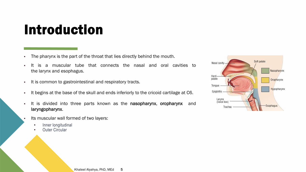
▪ The pharynx is the part of the throat that lies directly behind the mouth.
▪ It is a muscular tube that connects the nasal and oral cavities to
the larynx and esophagus.
▪ It is common to gastrointestinal and respiratory tracts.
▪ It begins at the base of the skull and ends inferiorly to the cricoid cartilage at C6.
▪ It is divided into three parts known as the nasopharynx, oropharynx and
laryngopharynx.
▪ Its muscular wall formed of two layers:
• Inner longitudinal• Outer Circular
5Khaleel Alyahya, PhD, MEd

Nasopharynx
▪ It extends from the base of the skull to the upper surface of the soft
palate.
▪ The anterior aspect of the nasopharynx communicates with the
nasal cavities through the choanae.
▪ This part of the pharynx is lined with respiratory epithelium: ciliated
pseudo-stratified columnar epithelium with goblet cells.
▪ It performs a respiratory function by conditioning inspired air and
propagating it to the larynx.
6Khaleel Alyahya, PhD, MEd

Oropharynx
▪ It is the middle part of the pharynx, located between the soft
palate and the superior border of the epiglottis.
▪ It lies behind the oral cavity, extending from the uvula to the level of
the hyoid bone.
▪ It contains the following structures:
• Posterior 1/3 of the tongue.• The lingual tonsils – Located inferiorly to the tongue.• The palatine tonsils• Superior constrictor muscle
▪ It is involved in the voluntary and involuntary phases of swallowing.
▪ Because both food and air pass through the pharynx, a flap of
connective tissue called the epiglottis closes over the glottis when
food is swallowed to prevent aspiration.
7Khaleel Alyahya, PhD, MEd

Laryngopharynx
▪ The most distal part of the pharynx, located between the superior border of
the epiglottis and inferior border of the cricoid cartilage (C6).
▪ It is found posterior to the larynx and communicates with it via the laryngeal
inlet.
▪ It is the part of the throat that is connected to the esophagus.
▪ It lies inferior to the epiglottis and extends to the location where this common
pathway diverges into the respiratory (larynx) and digestive (esophagus)
pathways.
▪ At that point, the laryngopharynx is continuous with the esophagus
posteriorly where it conducts food and fluids to the stomach.
▪ The laryngopharynx contains the middle and inferior pharyngeal constrictors.
8Khaleel Alyahya, PhD, MEd
Muscles
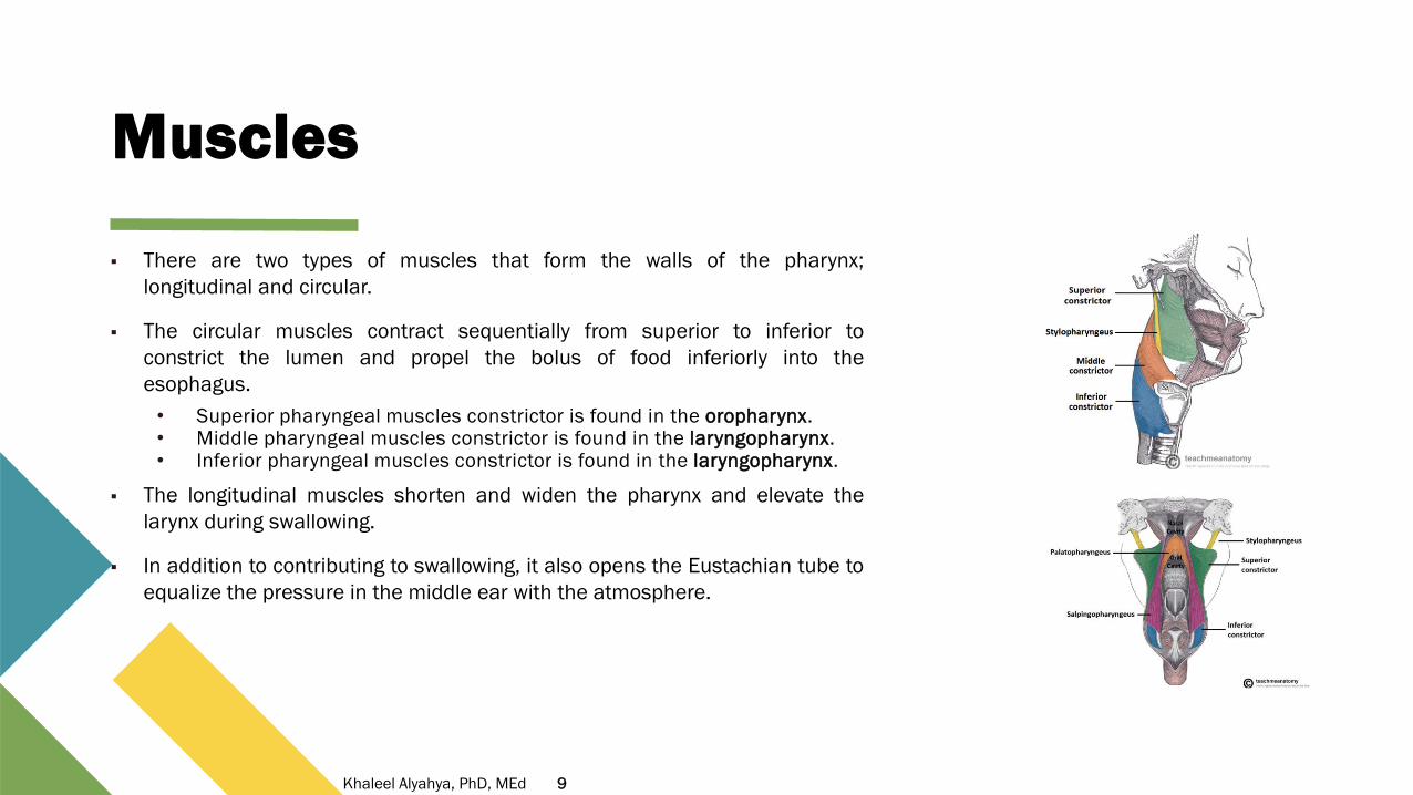
▪ There are two types of muscles that form the walls of the pharynx;
longitudinal and circular.
▪ The circular muscles contract sequentially from superior to inferior to
constrict the lumen and propel the bolus of food inferiorly into the
esophagus.
• Superior pharyngeal muscles constrictor is found in the oropharynx.• Middle pharyngeal muscles constrictor is found in the laryngopharynx.• Inferior pharyngeal muscles constrictor is found in the laryngopharynx.
▪ The longitudinal muscles shorten and widen the pharynx and elevate the
larynx during swallowing.
▪ In addition to contributing to swallowing, it also opens the Eustachian tube to
equalize the pressure in the middle ear with the atmosphere.
9Khaleel Alyahya, PhD, MEd
Blood Supply
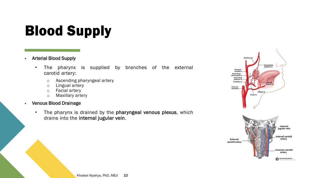
▪ Arterial Blood Supply
• The pharynx is supplied by branches of the externalcarotid artery:
o Ascending pharyngeal arteryo Lingual arteryo Facial arteryo Maxillary artery
▪ Venous Blood Drainage
• The pharynx is drained by the pharyngeal venous plexus, whichdrains into the internal jugular vein.
10Khaleel Alyahya, PhD, MEd
Innervation
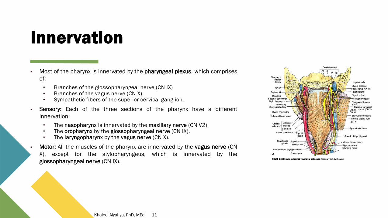
▪ Most of the pharynx is innervated by the pharyngeal plexus, which comprises
of:
• Branches of the glossopharyngeal nerve (CN IX)• Branches of the vagus nerve (CN X)• Sympathetic fibers of the superior cervical ganglion.
▪ Sensory: Each of the three sections of the pharynx have a different
innervation:
• The nasopharynx is innervated by the maxillary nerve (CN V2).• The oropharynx by the glossopharyngeal nerve (CN IX).• The laryngopharynx by the vagus nerve (CN X).
▪ Motor: All the muscles of the pharynx are innervated by the vagus nerve (CN
X), except for the stylopharyngeus, which is innervated by the
glossopharyngeal nerve (CN IX).
11Khaleel Alyahya, PhD, MEd
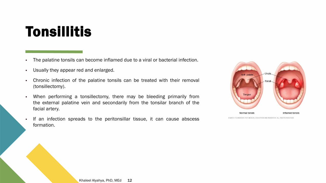
Tonsillitis
▪ The palatine tonsils can become inflamed due to a viral or bacterial infection.
▪ Usually they appear red and enlarged.
▪ Chronic infection of the palatine tonsils can be treated with their removal
(tonsillectomy).
▪ When performing a tonsillectomy, there may be bleeding primarily from
the external palatine vein and secondarily from the tonsilar branch of the
facial artery.
▪ If an infection spreads to the peritonsillar tissue, it can cause abscess
formation.
12Khaleel Alyahya, PhD, MEd
LARYNX

“The larynx is an organ in human neck that is
involved in breathing, sound production, and
trachea protection against food aspiration.
(Wikipedia)

Introduction
▪ Specialized organ at the inlet of air passage.
▪ It is an organ located in the anterior neck.
▪ The structure of the larynx is primarily cartilaginous
and is held together by a series of ligaments and
membranes.
▪ Internally, the laryngeal muscles move components
of the larynx for phonation and breathing.
▪ Superiorly, attached to hyoid bone.
▪ Inferiorly, continues with trachea.
15Khaleel Alyahya, PhD, MEd
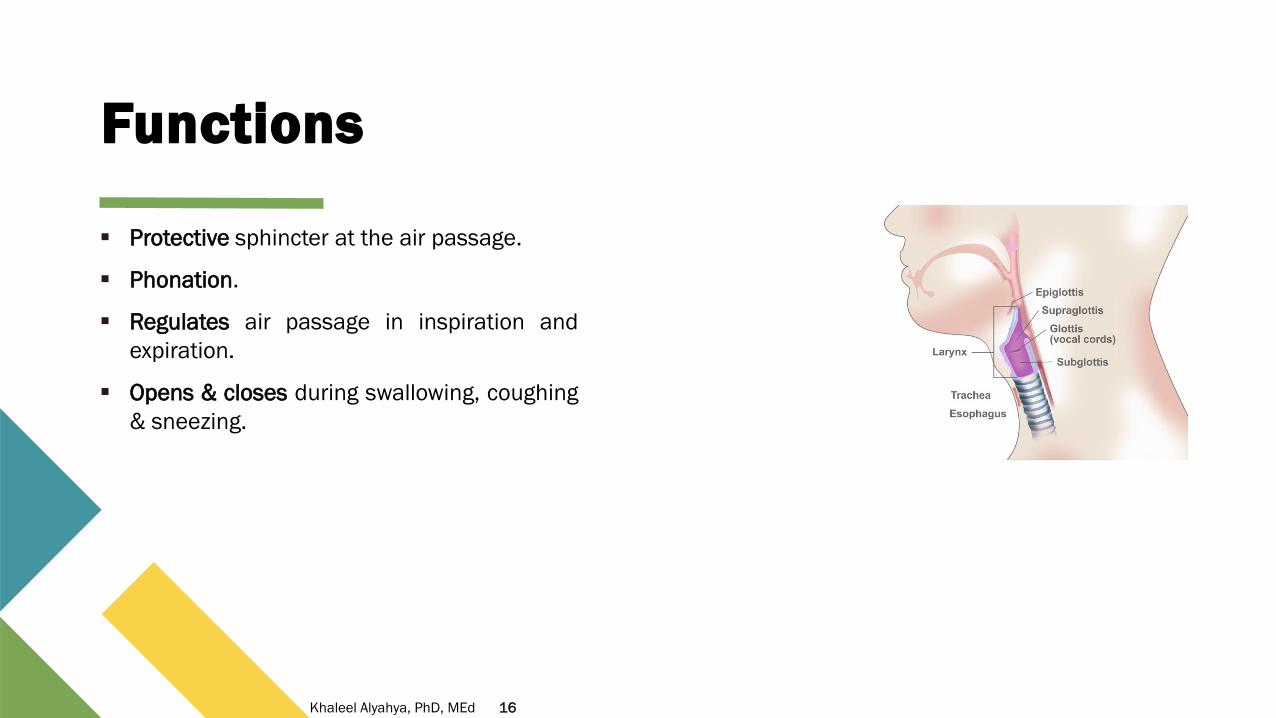
Functions
▪ Protective sphincter at the air passage.
▪ Phonation.
▪ Regulates air passage in inspiration and
expiration.
▪ Opens & closes during swallowing, coughing
& sneezing.
16Khaleel Alyahya, PhD, MEd
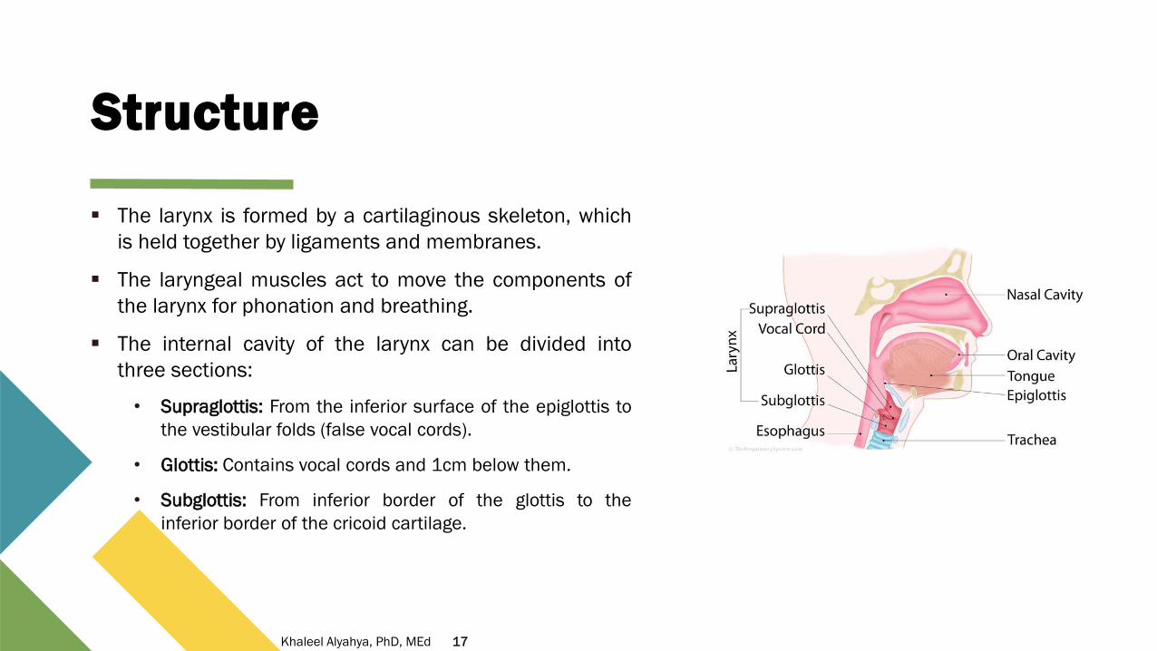
Structure
▪ The larynx is formed by a cartilaginous skeleton, which
is held together by ligaments and membranes.
▪ The laryngeal muscles act to move the components of
the larynx for phonation and breathing.
▪ The internal cavity of the larynx can be divided into
three sections:
• Supraglottis: From the inferior surface of the epiglottis to
the vestibular folds (false vocal cords).
• Glottis: Contains vocal cords and 1cm below them.
• Subglottis: From inferior border of the glottis to the
inferior border of the cricoid cartilage.
17Khaleel Alyahya, PhD, MEd

Position & Relation
▪ The larynx is located in the anterior compartment of the
neck, suspended from the hyoid bone and spanning
between C3 and C6.
▪ It is continuous inferiorly with the trachea and opens
superiorly into the laryngeal part of the pharynx.
▪ It is covered anteriorly by the infrahyoid muscles
(sternohyoid, sternothyroid, thyrohyoid and omohyoid
muscles), and laterally by the lobes of the thyroid gland.
Posteriorly, the oesophagus is located.
▪ The larynx is also closely related to the major blood vessels
of neck, which laterally as they ascend up to the head.
18Khaleel Alyahya, PhD, MEd

Cartilages
▪ Unpaired (Single) Cartilages
• Epiglottis
• Thyroid cartilage
• Cricoid
▪ Paired Cartilages
• Arytenoid
• Corniculate
• Cuneiform
▪ All cartilages are hyaline
• Except the epiglottis
o elastic cartilage
19Khaleel Alyahya, PhD, MEd

Framework
▪ The framework of the larynx is made up of cartilages.
▪ These cartilages are connected by joints, membranes &
ligaments.
▪ Moved by muscles.
▪ Lined by mucous membranes.
20Khaleel Alyahya, PhD, MEd

Thyroid cartilage
▪ It is the largest of the laryngeal cartilages.
▪ Formed of two laminae, each has superior & inferior horns.
▪ The angle between two laminae is 90 in male & 120 in female.
▪ It has two notches superior & inferior at the meeting of the two
laminae.
▪ Connections:
• Superior: To hyoid bone by thyrohyoid membrane.
• Inferior: To cricoid cartilage by the cricothyroid joint &
cricothyroid membrane
21Khaleel Alyahya, PhD, MEd

Cricoid cartilage
▪ Hyaline cartilage.
▪ Ring shaped, having a narrow anterior arch and wide posterior lamina.
▪ It is the only complete ring of cartilage around the trachea.
▪ Connected superiorly to thyroid cartilage by cricothyroid joint and
cricothyroid membrane.
▪ Inferiorly is connected to the 1st ring of cartilages around the trachea by
the coracobrachial ligament.
▪ The function of the cricoid cartilage is to provide attachments for
muscles, cartilages, and ligaments.
▪ Also it is involved in opening and closing the airway and in speech
production.
22Khaleel Alyahya, PhD, MEd

Epiglottis
▪ Leaf-shaped elastic cartilage.
▪ It projects obliquely upwards behind the tongue and the hyoid
bone.
▪ Stands open during breathing allowing air pass into the larynx.
▪ It closes during swallowing to prevent aspiration, forcing the
swallowed liquids or food to the esophagus.
▪ Connected by its stalk to the back of the thyroid cartilage.
▪ Its sides are connected to the arytenoid cartilage by
aryepiglottic fold.
▪ Its upper end is free.
23Khaleel Alyahya, PhD, MEd

Arytenoid cartilage
▪ Paired hyaline cartilage.
▪ Shaped like a 3-sided pyramid.
▪ Its base sits on the superior surface of the cricoid lamina.
▪ Apex: directed superiorly, supports the corniculate cartilage.
▪ Muscular process directed laterally, gives attachment to three
muscles:
• Posterior & lateral cricoarytenoid.
• Thyroarytenoid.
▪ Vocal process: directed forward and gives attachment to the
vocal ligament.
24Khaleel Alyahya, PhD, MEd
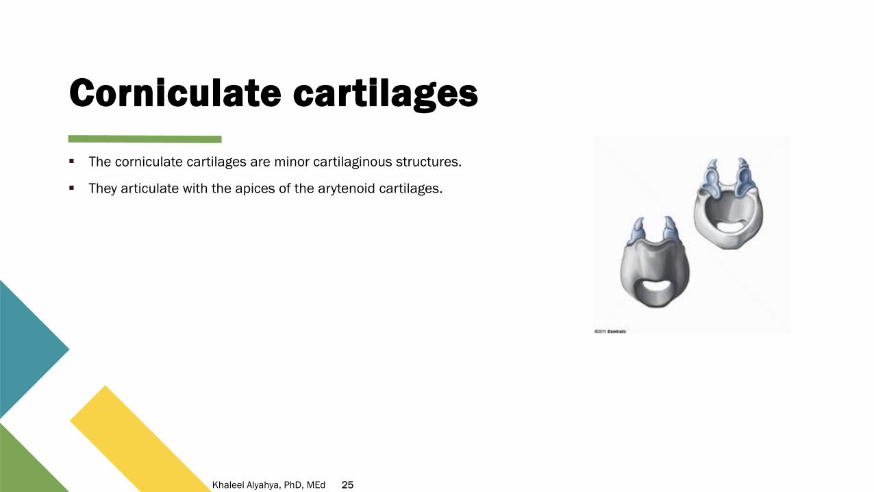
Corniculate cartilages
▪ The corniculate cartilages are minor cartilaginous structures.
▪ They articulate with the apices of the arytenoid cartilages.
25Khaleel Alyahya, PhD, MEd

Cuneiform cartilages
▪ The cuneiform cartilages are located within the ary-epiglottic
folds.
▪ They have no direct attachment, but act to strengthen the folds.
26Khaleel Alyahya, PhD, MEd

Muscles
▪ Two Major Groups of Muscles:
• Extrinsic Muscles – movement of the whole larynx
o Elevators:
▪ Digastric, stylohyoid, mylohyoid, geniohyoid, stylopharyngeus, salpingopharyngeus & palatopharyngeus.
o Depressors:
▪ Sternothyroid, sternohyoid & omohyoid.
• Intrinsic Muscles - movement within larynx
o Control of laryngeal inlet
o Control of rima glottidis
o Control of length & tension of vocal cords
o All intrinsic muscles lie inside the larynx cricothyroid
27Khaleel Alyahya, PhD, MEd
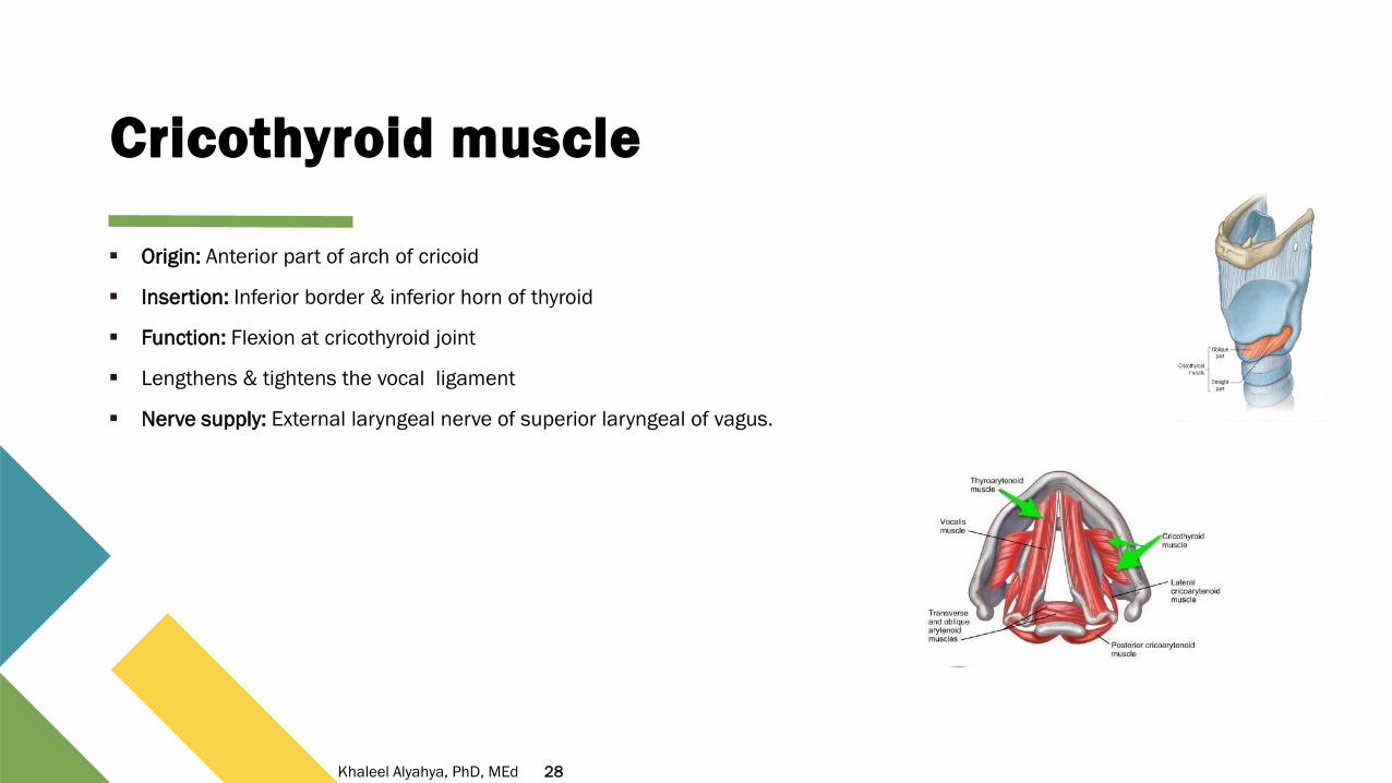
Cricothyroid muscle
▪ Origin: Anterior part of arch of cricoid
▪ Insertion: Inferior border & inferior horn of thyroid
▪ Function: Flexion at cricothyroid joint
▪ Lengthens & tightens the vocal ligament
▪ Nerve supply: External laryngeal nerve of superior laryngeal of vagus.
28Khaleel Alyahya, PhD, MEd

Thyroarytenoid muscle
▪ Origin: Front the lower half of the angle of the thyroid cartilage, and
from the middle cricothyroid ligament.
▪ Insertion: Into the base and anterior surface of the arytenoid cartilage.
▪ Function: Draw the arytenoid cartilages forward toward the thyroid to
relax and shorten the vocal folds.
▪ Nerve supply: Recurrent laryngeal nerve.
29Khaleel Alyahya, PhD, MEd

Posterior cricoarytenoid muscle
▪ Origin: The posterior surface of the cricoid cartilage.
▪ Insertion: The muscular process of the arytenoid cartilage.
▪ Function: Abducts vocal folds.
▪ Nerve supply: Inferior laryngeal nerve (branch of recurrent laryngeal).
30Khaleel Alyahya, PhD, MEd

Lateral cricoarytenoid muscle
▪ Origin: The arch of the cricoid cartilage.
▪ Insertion: The muscular process of the arytenoid cartilage.
▪ Function: Adducts vocal folds.
▪ Nerve supply: Inferior laryngeal nerve (branch of recurrent laryngeal).
31Khaleel Alyahya, PhD, MEd

Transverse & Oblique cricoarytenoid muscle
▪ Origin: Spans from one arytenoid cartilage to the opposite arytenoid.
▪ Function: Adducts the arytenoid cartilages.
▪ Nerve supply: Inferior laryngeal nerve (branch of recurrent laryngeal).
32Khaleel Alyahya, PhD, MEd
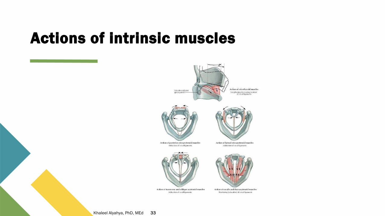
Actions of intrinsic muscles
33Khaleel Alyahya, PhD, MEd

Interior of larynx
▪ It is divided into three parts:
• Vestibule: between laryngeal inlet & vestibular fold.
• Ventricle: a depression extending laterally between vestibular & vocal folds.
• Infraglottic cavity lies between the vocal fold & lower border of cricoid cartilage.
▪ It is continuous with the trachea inferiorly.
34Khaleel Alyahya, PhD, MEd

Vocal cords
▪ The vocal cords (known also as vocal folds) are located within the
larynx (also colloquially
▪ Known as the voice box at the top of the trachea.
▪ Vocal cords are open during inhalation and come together to close
during swallowing and phonation.
▪ When cords are closed, the vocal folds may vibrate and modulate
the expelled airflow from the lungs to produce speech and singing..
35Khaleel Alyahya, PhD, MEd

Innervation
▪ MOTOR
• All muscles of the larynx are supplied by recurrent laryngeal nerveexcept Cricothyroid
o Supplied by external laryngeal branch of superior laryngeal nerve
▪ SENSORY & SECRETOMOTOR
• Above Vocal Cords: internal laryngeal
• Below Vocal Cords: recurrent laryngeal
▪ RECURRENT LARYNGEAL NERVE
▪ A branch of the vagus nerve that supplies all the intrinsic musclesof the larynx, with the exception of the cricothyroid muscles.
▪ The nerves emerge from the vagus nerve at the level of the arch ofaorta, and then travel up the side of the trachea to the larynx.
36Khaleel Alyahya, PhD, MEd
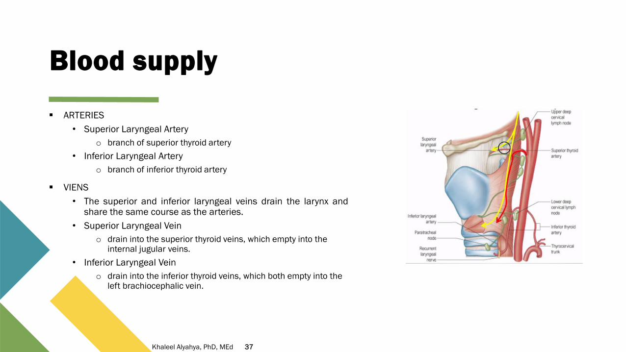
Blood supply
▪ ARTERIES
• Superior Laryngeal Artery
o branch of superior thyroid artery
• Inferior Laryngeal Artery
o branch of inferior thyroid artery
▪ VIENS
• The superior and inferior laryngeal veins drain the larynx andshare the same course as the arteries.
• Superior Laryngeal Vein
o drain into the superior thyroid veins, which empty into the internal jugular veins.
• Inferior Laryngeal Vein
o drain into the inferior thyroid veins, which both empty into the left brachiocephalic vein.
37Khaleel Alyahya, PhD, MEd
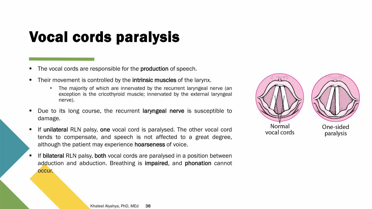
Vocal cords paralysis
▪ The vocal cords are responsible for the production of speech.
▪ Their movement is controlled by the intrinsic muscles of the larynx.
• The majority of which are innervated by the recurrent laryngeal nerve (anexception is the cricothyroid muscle; innervated by the external laryngealnerve).
▪ Due to its long course, the recurrent laryngeal nerve is susceptible to
damage.
▪ If unilateral RLN palsy, one vocal cord is paralysed. The other vocal cord
tends to compensate, and speech is not affected to a great degree,
although the patient may experience hoarseness of voice.
▪ If bilateral RLN palsy, both vocal cords are paralysed in a position between
adduction and abduction. Breathing is impaired, and phonation cannot
occur.
38Khaleel Alyahya, PhD, MEd