Pests and Diseases of Honeybees and their Management
30
Published By: , Department of Agriculture & Cooperation, Ministry of Agriculture, Government of India. B Wing, 2 nd Floor, Janpath Bhawan, Janpath, New Delhi -110001. Pests and Diseases of Honeybees and their Management
Transcript of Pests and Diseases of Honeybees and their Management

Published By:
, Department of Agriculture & Cooperation,
Ministry of Agriculture, Government of India.
B Wing, 2nd Floor, Janpath Bhawan, Janpath, New Delhi -110001.
Pests and Diseases of Honeybees and their Management

{(--CONTENTS--)}
Sr.No. Titles Page No. Part 1st : Insect of Honeybee
1. 1.1) The Greater wax moth (Galleria mellonella) 1-3 1.2) The lesser wax moth (Achroia grisella) 3
2. Ants Wasps and Hornets- 2.1) Ants 4
2.2) Wasps & Hornets. 4-6
3. Honey bee Mites-
3.1) Varroa mite 7-11
3.2) Topilaelaps mite 11-13 3.3) Tracheal mite 13-14
4 Bee Scorpion. 14
5. Reptiles. 14-15
6. Honey Bee Eater Birds- 16-17
7. Mammals:
7.1)Pine Martens
7.2)Raccoons
7.3) Bears
7.4) Skunks
7.5) Rodents
17-18
Part 2nd : Disease of Honeybee 1. BACTERIAL DISEASES- 1. American foulbrood disease(AFB) 19-21
2. European foulbrood disease(EFB) 21-22
3. Chalk brood disease (Ascosphaerosis) 22-23
2. VIRAL DISEASE. 1. Sac-brood disease 23-25
3. FUNGAL DISEASE. 1. Nosema disease (Nosemosis) 26-27
4. SOURCES 28

The greater wax moth is often reported to cause damage both to honey bee colonies and to bee products in tropical and sub-tropical Asia. It is observed throughout the year but its occurrence is severe during July to October and November to December. Empty combs, rendered wax, comb foundation and bee collected pollen, if not properly stored and left unattended, almost always suffer considerable damage from wax-moth infestation.
Fig: Greater Wax Moth (GWM)
According to many reports, the wax moth is a major pest of Apis. cerana, often causing colonies to abscond. In wax-moth attacks on colonies, the adult female enters the hive at night, through the entrance or cracks in the walls, deposits her eggs directly onto the combs or in narrow crevices that permit ovipositor and offers protection against removal by worker bees. From 50 to 150 eggs are laid in each batch; they are glued together and adhere firmly to the surface on which they are laid.
Fig: 1-7 Life stage of Greater Wax Moth (GWM)
(i) Egg. (ii) Larva (iii) Cocoon (IV) Pupa (VII) Adult on Comb.
Fig: Male & Female of Greater Wax Moth (GWM)
(i) Male (ii) Female
The newly-hatched Galleria larvae feed on honey and pollen, and then burrow into pollen storage cells or the outer edge of cell walls, later extending their
1.1 The Greater wax moth (Galleria mellonella)
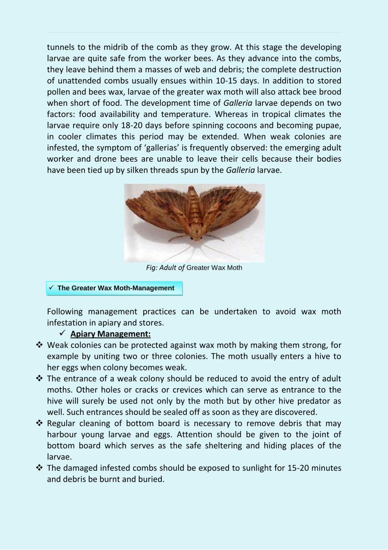
tunnels to the midrib of the comb as they grow. At this stage the developing larvae are quite safe from the worker bees. As they advance into the combs, they leave behind them a masses of web and debris; the complete destruction of unattended combs usually ensues within 10-15 days. In addition to stored pollen and bees wax, larvae of the greater wax moth will also attack bee brood when short of food. The development time of Galleria larvae depends on two factors: food availability and temperature. Whereas in tropical climates the larvae require only 18-20 days before spinning cocoons and becoming pupae, in cooler climates this period may be extended. When weak colonies are infested, the symptom of ‘gallerias’ is frequently observed: the emerging adult worker and drone bees are unable to leave their cells because their bodies have been tied up by silken threads spun by the Galleria larvae.
Fig: Adult of Greater Wax Moth
Following management practices can be undertaken to avoid wax moth infestation in apiary and stores. Apiary Management:
Weak colonies can be protected against wax moth by making them strong, for example by uniting two or three colonies. The moth usually enters a hive to her eggs when colony becomes weak.
The entrance of a weak colony should be reduced to avoid the entry of adult moths. Other holes or cracks or crevices which can serve as entrance to the hive will surely be used not only by the moth but by other hive predator as well. Such entrances should be sealed off as soon as they are discovered.
Regular cleaning of bottom board is necessary to remove debris that may harbour young larvae and eggs. Attention should be given to the joint of bottom board which serves as the safe sheltering and hiding places of the larvae.
The damaged infested combs should be exposed to sunlight for 15-20 minutes and debris be burnt and buried.
The Greater Wax Moth-Management

Storage Management: A water soluble concentrate of spores of Bacillus thuringiensis Serotype 7
provides an excellent protection of stored combs without affecting the organoleptic properties of the honey.
Stack the empty combs in supers (up to 8-9 super) leaving some empty space in lower most super. Make it airtight by using mixture of mud and dung .
Avoid fumigation with naphthalene, ethylene die-bromide and PDB as they lead to their residues in honey.
Fumigate the empty combs with sulphur powder @ 230g/m3 and after that seal them properly. Store the empty combs at low temperature (o to -10°C) either permanently or for 5 hours. All the stages of wax moth are destroyed at low temperature.
The lesser wax moth is generally smaller than the greater wax moth, except when the latter is dwarfed owing to poor diet during its larval stage. Adult Achroia grisella are silver-grey in colour, with a distinct yellow head. The insect is quite small, with a slender body: normal body lengths of adult female and male are about 13 and 10 mm, respectively. The life-span of the adult female is about seven days, during which she can lay 250 to 300 eggs.
Infestation by the lesser wax moth usually occurs in weak honey bee colonies. The larvae prefer to feed on dark comb, with pollen or brood cells. They are often found on the bottom board among the wax debris. As larvae prefer to form small canals between the bottoms of the brood cells the brood is lifted. The bees continue constructing cells heading upward leading to the typical scratched comb surface .
Fig: Life stages of Lesser Wax Moth
(i) Egg. (ii) Larva (iii) Adult (iv) Adult on Comb.
Fig: Damage done by lesser wax moth
1.2 The lesser wax moth (Achroia grisella)

The methods employed in controlling Galleria mellonella are equally effective for the control of Achroia grisella.
All type of ants are among the most common predators of honey bees in tropical and subtropical areas. They are highly social insects and will attack the hives en masse, taking virtually everything in them: dead or alive adult bees, the brood and honey. In addition to this destruction, they can also be a nuisance to beekeepers and may sometimes cause pain from their bites. Apiaries of Apis mellifera under ant attack become aggressive and difficult to manage; weak colonies will sometimes abscond, which is also the defense of A. cerana against frequent ant invasions. Many ant genera and species are reported to cause problems to traditional beekeeping.
Fig: Ants of Monomorium sp. attacking Apis mellifera colonies
Control:
The following management measure may be taken by beekeepers to control ants attack in beekeeping.
Keep the apiary clean and place the hive stand post in bowls of tin or plastic or earthen pots filled with water. Clean the bowls regularly to avoid the formation of bridges or earth that can be crossed by ants. Replenish the liquid frequently.
These insects are dangerous against the honey bees in all Asian countries including India. Among the most frequently reported are social wasps of the genus Vespa, which are widely distributed throughout the world. Colonies of both A. cerana and A. mellifera are frequently attacked. Hornet invasion of A. cerana colonies generally causes the bees to abscond, and similar behavior is reported of weak colonies of A. mellifera. In addition to hornets of the genus Vespa, other wasp species have occasionally been reported to cause damage to apiaries. Table 2 lists wasps and hornets that have been reported as major predators of the two honey bee species in Asia. Predation by Vespa spp. on commercial apiaries is generally a rainy season problem. Hornet’s attack on apiaries reach their peak of intensity during September-October, whereas in tropical countries the most serious
Management
2. Ants Wasps and Hornets- Enemies of Honeybee
2.1 Ants.
2.2 Wasps and Hornets.

wasp invasions take place during the monsoon season, particularly from late June to August. Apiaries situated near the foothills and tropical forests suffer more acutely than those on the plains.
Fig: Different species of Wasps & Hornet (Vespa spp.)
(i)Vespa orientalis (ii)Nest of Vespa orientalis (iii) Vespa tropica (ii)Nest of Vespa tropica
Fig: Hornet (Vespa mandarina) attacking Apis mellifera colonies
Fig: Vespa velutina fig: Nest of Vespa velutina Fig: Vespa affinis
Fig: Nest of Vespa affinis Fig:
Vespa crabro Fig: Vespa binghami
To manage the wasps and hornets problem in beekeeping, beekeepers are advised to adopt methods such as bait-trapping, trapping at the hive entrance and using
Management of Wasps and Hornets.

protective screens. Locating hornet nests by following flight passes of individual wasps back to their nests and then destroying the nests may be very time consuming and, if too many of these nests are in the area, not very efficient. Where labor costs are not prohibitive, beekeepers have resorted to capturing and killing individual hornets foraging in the vicinity of their apiaries. In some areas, this approach has proved to be quite effective, largely because the period of most intense hornet attacks is only two to three months. It has been seen that the real damage inflicted by hornet attacks on honey bee colonies occurs during the slaughter and occupation phases. Killing hornets in the early stage of predation has the effect of disrupting the hunting phase and preventing the predation process from reaching the more destructive phases. Mass destruction of the colonies is thus prevented or, at the least, minimized. TABLE 2 Wasps and hornets that attack bees in Asia.
Scientific Name Recorded distribution Vespa orientalis India, Pakistan
Vespa mandarina India, Burma, Thailand, Lao, Viet Nam, Democratic Kampuchea,China, Republic of Korea, Japan
Vespa tropica Tropical Asia
Vespa velutina Tropical Asia
Vespa cincta Tropical Asia
Vespa affinis Tropical and sub-tropical Asia
Vespa crabro Japan, and perhaps all temperate Asia
Vespa mongolica Japan, and perhaps all temperate Asia
Vespula lewisii Japan
Vespula vulgaris Republic of Korea
Preventive measure
Keep bee colonies strong in the apiary and ensure proper food availability in the colonies. Provide artificial feeding if required. Reduce the hive entrance to give passage to only one or two bees at a time.
Killing the wasp or hornets by flapping at early stage of predation is more effective in disrupting the hunting phase and preventing the predation process from reaching the more destructive phase. Thus losses can be minimized.
Beekeepers are also advised to migrate the bee colonies to safer place or search out the wasp nests in vicinity of the apiary and destroy them.
Parasitic mites are among the most serious enemies of honey bees with which beekeepers have to cope. The success or failure of beekeeping operations with Apis
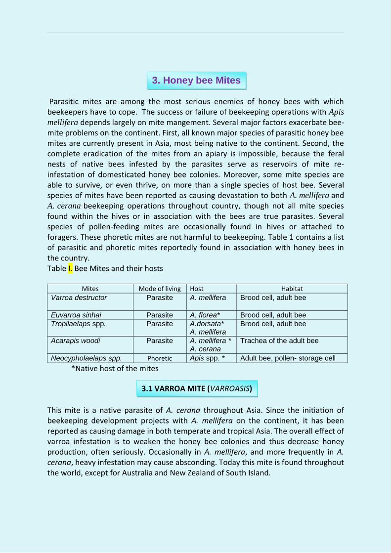
mellifera depends largely on mite mangement. Several major factors exacerbate bee-mite problems on the continent. First, all known major species of parasitic honey bee mites are currently present in Asia, most being native to the continent. Second, the complete eradication of the mites from an apiary is impossible, because the feral nests of native bees infested by the parasites serve as reservoirs of mite re-infestation of domesticated honey bee colonies. Moreover, some mite species are able to survive, or even thrive, on more than a single species of host bee. Several species of mites have been reported as causing devastation to both A. mellifera and A. cerana beekeeping operations throughout country, though not all mite species found within the hives or in association with the bees are true parasites. Several species of pollen-feeding mites are occasionally found in hives or attached to foragers. These phoretic mites are not harmful to beekeeping. Table 1 contains a list of parasitic and phoretic mites reportedly found in association with honey bees in the country. Table I. Bee Mites and their hosts
Mites Mode of living Host Habitat Varroa destructor Parasite A. mellifera Brood cell, adult bee
Euvarroa sinhai Parasite A. florea* Brood cell, adult bee Tropilaelaps spp. Parasite A.dorsata*
A. mellifera Brood cell, adult bee
Acarapis woodi Parasite A. mellifera * A. cerana
Trachea of the adult bee
Neocypholaelaps spp. Phoretic Apis spp. * Adult bee, pollen- storage cell
*Native host of the mites This mite is a native parasite of A. cerana throughout Asia. Since the initiation of beekeeping development projects with A. mellifera on the continent, it has been reported as causing damage in both temperate and tropical Asia. The overall effect of varroa infestation is to weaken the honey bee colonies and thus decrease honey production, often seriously. Occasionally in A. mellifera, and more frequently in A. cerana, heavy infestation may cause absconding. Today this mite is found throughout the world, except for Australia and New Zealand of South Island.
3. Honey bee Mites
3.1 VARROA MITE (VARROASIS)

Fig: Life cycle of Varroa Mite.
Varroa destructor is quite large, as compared with other mite species, and can be seen with the unaided eye. The shape of the adult female is distinctive: observed from above, the width of the body is clearly seen to be greater than the length, i.e. about 1.6 x 1.1 mm. The mite is reddish brown in colour and shiny and the body is dorsoventrally flattened covered with short hairs (setae). Adult females of V. destructor are found inside brood cells or walking rapidly on comb surfaces. Individual mites are often seen clinging tightly to the body
of adult bees, mostly on the abdomen, where the segments overlap, between the thorax and
the abdomen and at the ventral entry. Adult males, and the immature stages of both sexes
(egg, protonymph and deuteronymph), are not commonly seen outside the brood cells (see
Plate 7). All immature stages of the parasite live inside the brood cells. They can be observed
when infested cells are opened and the brood is carefully removed. The immature mites are
bright white and the adult females are brown, while male mites are smaller than females and
are rarely seen since they are only found inside brood cells.
Fig: Varroa mite showing Nymph stages and male.
Symptoms
Causes:
Varroa causes injuries to honey bees by direct feeding. The adult female pierces the
bees’ soft intersegmental membrane with their pointed chelicerae and sucks the bees`
haemolymph (‘blood’). The adult bee, however, is only damaged if the infestation is
severe. The condition of a honeybee colony being infested with Varroa mites is called
varroasis. If more than one parasitic female mite infests the brood cell the brood
decays or deformations occur including shortened abdomen or deformed wings. If
only one mite infests a cell symptoms may not be visible, although the bees’ life-span
is considerably shortened. Moreover, the bee’s behavior may be disturbed, e.g. in
orientation or gathering food. Infested bees often have a reduced fat body that
hampers the functioning of their glands or increases their susceptibility to pesticides.
The semen production of drones may be considerably reduced. Varroosis is a multi-
factorial disease. Virus diseases that may have caused little damage before infestation
by varroa mites often accompany it. Normally, the exoskeleton protects the bees from
many virus infections. However, the mite penetrates this natural barrier transferring
viruses or stimulating the multiplication of viruses with its saliva. In turn viruses seem
to speed the development of varroasis enhancing the parasite’s virulence. Other
diseases such as nosema and sac brood have similar effects.
Moreover, unfavorable climatic conditions or insufficient stocks of pollen and nectar
can increase the process of disintegration. Without treatment the colonies normally die
after two to three years, management errors may also cause the collapse of colonies.
Colonies destroyed by the varroa mite are often left with only a handful of bees and
the queen, the other bees having died during foraging or having drifted to neighboring
colonies, where the mite population can increase before killing these colonies also. In
this way mites may cause colonies to die, as in some kind of domino effect, over wide
areas.
The presence of adult bees with deformed wings, crawling on comb surfaces or near
the hive entrance, usually indicates a late stage of heavy mite infestation. Several
other methods may be used to detect mites. The most reliable, perhaps the most time-
consuming, is direct sampling by the random opening of brood cells, particularly
drone cells. The older the larvae/pupae the easier this procedure becomes. The brood
is removed from the cell with a fine forceps and the cell is inspected for the presence
of the mites. Between 100 and 200 cells must be opened before an assessment of the
level of mite infestation can be made. To inspect adult bees, the bees are captured
from the brood combs and placed in jars, into which chloroform, ether or alcohol is
introduced on a piece of cotton wool. The bees are intoxicated and the mites crawl on
the glass wall. Returning foragers may also be captured by hand at the hive entrance
and held up against the sunlight; any mites attached to the bees’ abdomens may be
seen. Another method is to use specially constructed zinc, plastic or wood trays, built
to the size of the bottom board, with a white or light-colored floor. The trays, equipped with a screen of a mesh less than 2 mm fixed at about 1 cm above the tray floor, are placed on the bottom boards of the hives and are inspected one to three days later for the presence of dead mites. The screen prevents the bees from removing the dead parasites from the hive. The control of V. destructor is one of the most difficult tasks facing apiculturists and beekeepers throughout the world. The mite is a highly successful parasite, whose life history is well synchronized with that of its host. Two principal approaches to its control are currently available:

1. Control methods and hive manipulation techniques. Chemical control is by far not a common method of varroa control. The organic acids: formic acid, the ethereal oil thymol may be chosen to treat colonies with brood.
Fig: Bee larvae with varroa mites.
i. Formic acid
Formic acid can kill some of the mites in the sealed brood cells. It is recommended that the formic acid be allowed to evaporate in colonies with sealed brood for at least two to three weeks. In this way, mites emerging from the brood will also be killed. The external temperature should not be less than 12°C (54°F) and not more than 25°C (77°F). The formic acid should be introduced into the colony only in the late afternoon to avoid damage to bees and brood. In addition, physiological tolerance is improved if the entrance hole is wide open. An easier way to introduce formic acid is to use a sponge or a similarly absorbent material. A solution of 50 ml of 60 percent formic acid is applied onto the sponge tissue per comb (Langstroh size). The quantity must be reduced accordingly for smaller comb sizes. A grid fixed above the tissues on the bottom of the hive, will prevent the bees from burning themselves with the acid. The grid should be as far away from the brood as possible. The application can be repeated two times at intervals of ten days. iii. Oxalic acid Contrary to formic acid oxalic acid does not act via evaporation but through contact with the bees. Thirty two grams of crystal oxalic acid (dehydrate) is thinned in one litre of sugar water (1:1). When handling crystal acid special precautions must be taken because of the health risks. Protective spectacles and acid-proof gloves must be work together with an adequate mouth protector. Depending on the size of the colony 20 to 30 ml of the suspension per chamber are dropped into the bee-ways. A repetition of the treatment can lead to damage to the bees. Applicators are available by which the acid can be evaporated. iv. Lactic acid Lactic acid is clearly better tolerated by bees and does not cause problems in warmer climatic zones. The disadvantage is that every single comb must be extracted to spray the bees with the acid. The dosage applied per comb side is 8 ml of 15 percent acid. This treatment can be repeated two times at intervals of seven days. v. Etheric oils
The only etheric oil that is sufficiently affective against varroa mites is thymol. Thymol can be applied as a commercially available ready-made preparation or in crystal form. For this purpose, put 5g of thymol crystal into a gauze bag and kep on the top bars for two weeks. In this way mites emerging from the brood will be covered.
viii. Control by hive manipulation: The varroa mite depends on bee brood to complete its development cycle. Since the mite prefers drone brood to worker brood, empty frames are given to the colonies, which will rear drone brood in them. When the cells are sealed, the frames, containing the mites trapped inside the cells, can be removed and destroyed. Modern beekeeping with Apis mellifera in tropical and sub-tropical part of the Country frequently encounters problems caused by infestation with Tropilaelaps spp. This mite is a native parasite of the giant honey bee A. dorsata, widely distributed throughout tropical part of the country, and whenever A. mellifera is kept within the range of distribution of A. dorsata, mite infestation of the colonies cannot be avoided. Thus, beekeepers consider Tropilaelaps to be a more serious pest than varroa-mites, even though it may be easy to control. Dual parasitism of A. mellifera colonies by both parasites sometimes occurs, the population of Tropilaelaps often being greater than that of varroa, as the Tropilaelaps mite can almost completely prevent multiplication of the varroa mite. Cause Tropilaelaps mites are much smaller than varroa mites, although the trained unaided eye can still see them. The adult female mite is light reddish-brown in colour, with an oval-shaped body about 0.96 mm in length and 0.55 mm in width. The mite’s entire body is covered with short setae. A red streak running longitudinally on the ventral surface of the adult female may be perceived through a strong magnifying glass. When the mites are present in a honey bee colony in large numbers, they can be observed walking rapidly on the surface of the comb. They are rarely found on adult bees. In all its immature stages, the mite lives within the brood cells of the bees, feeding on the brood’s haemolymph. Fertilized adult females enter the cells before they are capped to lay their eggs. The stages of development of the mite are as follows: egg, six-legged larva, protonymph, deutonymph, adult. Adult males of Tropilaelaps do not feed, their chelicerae (the organs originally used for piercing the bees’ integument) having been modified to transfer sperm as with the varroa mite. The life cycle of the mite is well synchronized with that of the host bee. Symptoms
3.2 TROPILAELAPS MITE

The damage caused to colonies by Tropilaelaps infestation is similar to that brought about by varroa and the injuries inflicted on bee brood are same. The abdomen of bees surviving mite attacks is reduced in size, and they have a shorter life-span than healthy bees In heavily infested colonies, bees with deformed wings can be observed crawling about the vicinity of the hive entrance and on the comb surfaces, while piece of dead bee brood evacuated from the hive by the house bees can be seen in front of the entrance. Inspection of hives severely infested by Tropilaelaps reveals an irregular pattern of sealed and unsealed brood as found with all brood diseases. Since this symptom can be taken as a sign of a poor-laying queen, the position must be verified. The best means is to open sealed cells gently and inspect them for the presence of the mite. If mites are present, adult females will be seen walking rapidly out of the cells. To obtain a reasonably accurate estimate of the level of infestation, 100-200 cells should be opened and the brood removed with forceps for close inspection.
Fig: Parasitism by Varroa jacobsonii or Tropilaelaps clareae
usually results in deformation of the bees’ wings.
Control: The chemotherapeutic measures described above for the control of varroa are also effective in the control of Tropilaelaps. Formic acid is effective in its treatment. Colony manipulation techniques: Many beekeepers prefer not to use chemicals to control Tropilaelaps, but to manipulate the brood rearing cycle of their infested colonies in such a way that the mites are deprived of sealed and unsealed brood (broodlessness) , their food, for at least two weeks. During this period, a large proportion of the mite population will starve to death. There are several means of creating this brood less situation in infested colonies. In smaller apiaries, the beekeeper can simply remove the brood-comb frames -- both sealed and unsealed-- from the infested colonies and put them in new hives. Before the new larvae hatch, the hives manipulated in this way will be short of brood for two to three days, time enough to starve most of the mites. The new hives with the removed brood frames are given mated queens, which are caged for 14 days, a period that

allows most of the brood to emerge, while no new brood has been reared because the queen has been confined. The best time of year to carry out these colony-manipulation techniques is during a heavy pollen-flow season, enabling the colonies to rear brood after the period of brood deprivation. In some Asian regions, this season coincides with the monsoon months, when there is no nectar flow but when pollen is abundant. This is also the season in which beekeepers feed sugar syrup to their bees, rear new queens and propagate colonies. While colony manipulation to control Tropilaelaps is time-consuming, it causes no noticeable harm to the colonies, nor does it affect productivity. The availability of pollen, coupled with the feeding of sugar, enables both the treated and the newly-formed colonies to regain their full strength before the nectar flow begins.
This mite, Acarapis woodi, infests the tracheal system of adult bees, queens, workers and drones, which are all equally susceptible to its attack. Since it was first reported in Apis mellifera colonies in Europe in 1921, opinions regarding the extent of the damage it can cause to honey bee colonies have varied. Reports from India and Pakistan indicate that the tracheal mite caused severe loss of Apis Cerana colonies.
Fig: Tracheal mite, Acarapis woodi..
Cause A. woodi is a very small mite (0.1 mm) species that lives and breeds within the thoracic tracheae of adult bees . The mite penetrates through the spiracles into the first tracheal pair of the thorax of 10-day old honey bees. There it lays eggs at intervals of a few days. After the deutonymph stage, male offspring emerge after around 12 days and females after 13 to 16 days. Symptoms Ttypical visible symptoms of infestation are presence of crawlers bees around the hive and ‘K’ type wing condition. Indeed, it has been demonstrated that bees severely infested with the mite can forage normally. Nevertheless, some differences exist with regard to the over-wintering capability of infested and
3.3 TRACHEAL MITE (ACARINE DISEASE)

healthy colonies. Infestation shortens the lifespan of the individual bees, so that severe infestation of colonies causes them to loose strength and thus increases a colony’s susceptibility to winter losses. The most reliable diagnostic method is laboratory dissection. Samples of 20 or more bees found crawling near the hive and unable to fly are killed, their heads and legs removed and their thoraxes dissected for microscopic examination. If present, the mites are usually found at the end of the first pair of trachea in the thorax. Control: Chemotherapeutic measures are widely adopted for mite control. Best results could be achieved with evaporating substances such as formic acid and ethereal oils. Formic acid Formic acid produces good results by applying by the method as described in varroa control This insect usually clings to the legs of the bees and accompanies them to the nest. It is usually observed in the comb of Indian honey bee A. cerana.
Fig: (i.) Braula coeca
Most commonly reptiles are found in tropical forests, woods, grasslands and urban areas. Among the reptile species that are regularly recorded as present in commercial apiaries are, Calotes spp., Acanthosaura spp., Sphenomorphus spp. Arboreal reptiles such as many geckos and skinks can attack bees either near the hive entrance or on the limbs of flowering trees visited by forager bees. Smaller lizards, such as the gecko Hemidactylus frenatus, often hide in the empty space between the outer and inner covers of the hive.
4. Bee Scorpion.
5. REPTILES

Fig: Predation on honey bees by a lizard
Management:
The beekeeper can do little to prevent the loss of foragers to the highly mobile arboreal reptiles, usually well hidden in the trees. Hives placed on stands that are about 40-60 cm high are reasonably safe from reptiles attacking from the ground, coating the legs of the stands with used engine oil or grease may deter the reptiles from climbing up to the hive entrance. A well-kept bee yard that is frequently mowed, without dense bushes, shrubs and tall grass, that provide safe hiding places to the predators, has less chance of suffering losses from reptiles than an untended one.

In agriculture sector various species of birds may be useful because they are reducing insect pest population from cropping field. Many Birds prey upon many insect species and honey bees are no exception. Once airborne, the bees are virtually defenseless against birds, several species of which can tolerate their venomous stinging defense. The heavy traffic of bees flying in and out of the hives of commercial apiaries provides an exceptional opportunity for insectivorous birds, large numbers of which may be attracted by this situation. The level of damage caused by honey bee eater birds varies. An attack by a single bird or by a few together rarely constitutes a serious problem, but when a large flock descends upon a few colonies or an apiary, a substantial decline in the worker population in some or all the hives may be observed. Whereas the degree of damage to commercial apiaries caused by predatory birds depends largely on the number of the predators and the intensity of the attack, the mere presence of a few predators in apiaries engaged in queen rearing can inflict serious losses. Fig: Different species of birds:
(i.)Blue-bearded (ii) Blue-cheeked (iii) Green bee eater (iv) Blue tailed (v) Chestnut-headed
(vi) Ashy Drongo (vii) Spangled Drongo (viii) Red-blacked shrike (ix)Brown shrike
(x) Lesser Yellownape (xi) Greater Yellownape(xii)Streak-throated (xiii) Yellow Rumped (xiv) Common Swift
Management
While beekeepers regard insectivorous birds as pests, sometimes serious, other branches of agriculture generally do not consider them as problematic. In fact, birds that prey on insects are mostly considered to be beneficial to farming, in that they help in the control of insect pests. Where heavy predation by birds on apiary bees tends to occur at fixed periods (e.g. during the migration season of swifts), the most practical means of solving
6. Honey Bee Eater Birds
the problem is usually to avoid the birds, through careful site selection and by temporary relocation of the apiaries, at least until the migration period is over. The following management measurement are recommended to beekeepers/farmers for controlling birds problems in beekeeping.
Making noise by beating drum and scaring away the predatory birds by slingshots.
By using red reflective ribbons around the apiary. Management should also be taken to choose the apiary site away from the bird
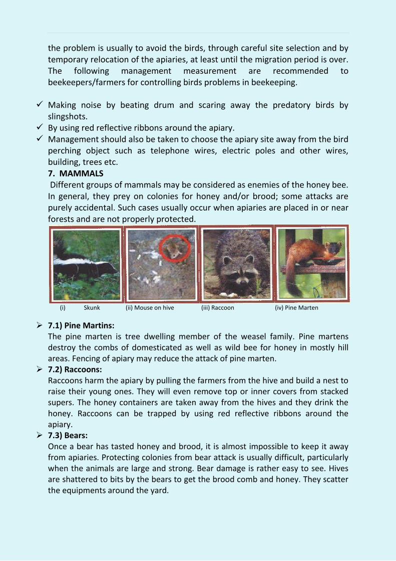
perching object such as telephone wires, electric poles and other wires, building, trees etc. 7. MAMMALS Different groups of mammals may be considered as enemies of the honey bee. In general, they prey on colonies for honey and/or brood; some attacks are purely accidental. Such cases usually occur when apiaries are placed in or near forests and are not properly protected.
(i) Skunk (ii) Mouse on hive (iii) Raccoon (iv) Pine Marten
7.1) Pine Martins: The pine marten is tree dwelling member of the weasel family. Pine martens destroy the combs of domesticated as well as wild bee for honey in mostly hill areas. Fencing of apiary may reduce the attack of pine marten.
7.2) Raccoons: Raccoons harm the apiary by pulling the farmers from the hive and build a nest to raise their young ones. They will even remove top or inner covers from stacked supers. The honey containers are taken away from the hives and they drink the honey. Raccoons can be trapped by using red reflective ribbons around the apiary.
7.3) Bears: Once a bear has tasted honey and brood, it is almost impossible to keep it away from apiaries. Protecting colonies from bear attack is usually difficult, particularly when the animals are large and strong. Bear damage is rather easy to see. Hives are shattered to bits by the bears to get the brood comb and honey. They scatter the equipments around the yard.
Placing the apiary in location out of the bear’s path reduces its attack. Electrified barbed wire fences are often used where bears represent a common problem. Moving hives closer to human habitation is also effective. 6.5) Skunks: Skunks scratch the bottom board or the front of hive body to get the bees coming out of the hive and eat the bees. Skunks visit the apiary in the evening time and dark hours. Raising the hive 15 to 18 inches above the ground and use of wire netting around the hive is effective to prevent the skunks.
6.6) Rodents: Rodents such as mice and rats are common pest to the beekeeping. They build nest in hive boxes, destroy comb in the frames, and make hole in equipments. In addition, they leave dropping all over the place. Rats can be serious problems in storage areas where bee equipments are kept. To keep mice out of hives, a mouse trap can be placed on the entrance of the hive. Reducing the entrance of the hive to ¼ of an inch, bees will be able to come and go, but mice will not be able to enter. Bait traps can be used for both rats and mice.
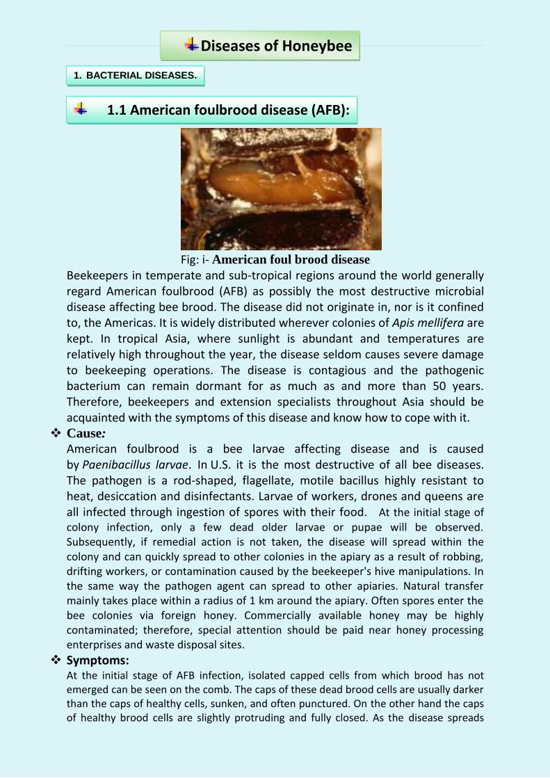
Fig: i- American foul brood disease
Beekeepers in temperate and sub-tropical regions around the world generally regard American foulbrood (AFB) as possibly the most destructive microbial disease affecting bee brood. The disease did not originate in, nor is it confined to, the Americas. It is widely distributed wherever colonies of Apis mellifera are kept. In tropical Asia, where sunlight is abundant and temperatures are relatively high throughout the year, the disease seldom causes severe damage to beekeeping operations. The disease is contagious and the pathogenic bacterium can remain dormant for as much as and more than 50 years. Therefore, beekeepers and extension specialists throughout Asia should be acquainted with the symptoms of this disease and know how to cope with it.
Cause:
American foulbrood is a bee larvae affecting disease and is caused by Paenibacillus larvae. In U.S. it is the most destructive of all bee diseases. The pathogen is a rod-shaped, flagellate, motile bacillus highly resistant to heat, desiccation and disinfectants. Larvae of workers, drones and queens are all infected through ingestion of spores with their food. At the initial stage of colony infection, only a few dead older larvae or pupae will be observed. Subsequently, if remedial action is not taken, the disease will spread within the colony and can quickly spread to other colonies in the apiary as a result of robbing, drifting workers, or contamination caused by the beekeeper's hive manipulations. In the same way the pathogen agent can spread to other apiaries. Natural transfer mainly takes place within a radius of 1 km around the apiary. Often spores enter the bee colonies via foreign honey. Commercially available honey may be highly contaminated; therefore, special attention should be paid near honey processing enterprises and waste disposal sites.
Symptoms: At the initial stage of AFB infection, isolated capped cells from which brood has not emerged can be seen on the comb. The caps of these dead brood cells are usually darker than the caps of healthy cells, sunken, and often punctured. On the other hand the caps of healthy brood cells are slightly protruding and fully closed. As the disease spreads
Diseases of Honeybee
1. BACTERIAL DISEASES.
1.1 American foulbrood disease (AFB):

within the colony, a scattered, irregular pattern of sealed and unsealed brood cells (see Fig.) can be easily distinguished from the normal, compact pattern of healthy brood cells observed in healthy colonies. The bee brood affected by AFB is usually at the stage of older sealed larvae or young pupae, upright in the cells. Often therefore, a protruding tongue can be found with the rest of the body already decayed. At first the dead brood is dull white in colour, but it gradually changes to light brown, coffee brown, and finally dark brown or almost black. The consistency of the decaying brood is soft. Once the dead brood has dried into scales, the test cannot be used. The dry brood lies flat on the lower side of the cell wall, adhering closely to it – in contrast to sac brood. This scale is usually black or dark brown and brittle. Often, a fine, threadlike proboscis or tongue of the dead pupa can be seen protruding from the scale, angling toward the upper cell wall.
Fig: Irregular pattern of sealed brood with sunken and punctured
caps, typifying American Foul Brood infection.
The pathogen bacteria may be identified using a microscopic preparation or, more frequently, by cultivation on selective culture media.
Stretch test A simple way of determining whether AFB caused the death of the brood is the ‘stretch test’ (See Fig…). A small stick, match or toothpick is inserted into the body of the decayed larva and then gently and slowly, withdrawn. If the disease is present, the dead larva will adhere to the tip of the stick, stretching for up to 2.5 cm before breaking and snapping back in a somewhat elastic way. This symptom called ‘ropiness’, confirms American foulbrood disease, but it can be observed in decaying brood only.
Fig: Stretch test for American foulbrood disease.

Control: In several countries, where apiculture includes large commercial operations, frequent, efficient inspection services are particularly advanced and a ‘search and destroy’ strategy is adopted in an attempt to minimize damage to apiaries caused by this serious honey bee disease.
This disease was first reported in 1885 from U.K. in Apis mellifera and India in 1970 from
Maharashtra. The range of distribution of European foulbrood disease is not confined to Europe alone and the disease is found in all continents where Apis mellifera colonies are kept. A. cerana colonies are also subject to EFB infection. The damage inflicted on honey bee colonies by the disease is variable. The disease is caused by non-spore-forming bacterium, Melissococcus plutonius. The disease affects larvae of all castes.
Fig:1 & 2. European foul brood disease. Symptoms: Honey bee larvae killed by EFB are younger than those killed by AFB. The diseased larvae die when they are four to five days old, or in the coiled stage. The colour of the larva changes as it decays from shiny white to pale yellow and then to brown. When dry, the scales of larvae killed by EFB, in contrast to AFB scales, do not adhere to the cell walls and can be removed with ease. The texture of the scales is rubbery rather than brittle, as with AFB. A sour odour can be detected from the decayed larvae. The clinical picture and the odour can vary depending on the kind of other bacteria involved (Bacillus alvei, Streptococcus faecalis, Achromobacter eurydice). Another symptom that is characteristic of EFB is that most of the affected larvae die before their cells are capped. The sick larvae appear somewhat displaced in the cells.
1.2 European foulbrood disease (EFB)

Fig: Larvae in coiled stage, killed by European foulbrood disease.
When a scattered pattern of sealed and unsealed brood is observed in a diseased colony, this is normally an indication that the colony has reached a serious stage of infection and may be significantly weakened. However, this is the case with all brood diseases. EFB is transferred in the same way as AFB. Control: The choice of an EFB control method depends on the strength of the infection, i.e. how many brood cells and combs are infested. If the infection is low, it is often sufficient to stimulate the hygiene behavior of the bees. Either they are placed at a good foraging site or they are fed with honey or syrup. In case of severe infection, removing the most infected brood combs is effective. Replace infected combs with empty fresh/ sterilized combs. Since the bees’ hygiene behavior is also genetically determined, replacement of the queen is also effective. Requeening can strengthen the colony by giving it a better egg-laying queen, thus increasing its resistance to the disease and interrupting the ongoing brood cycle giving the house bees enough time to remove infected larvae from the hive. Isolation of diseased colonies is also recommended for its control. In Asia including India, chalk brood is rarely considered to be a serious honeybee disease.
Fig: Chalk brood disease
Cause: Chalk brood is a disease caused by the fungus Ascosphaera apis. As its name implies, it affects honey bee brood. This fungus only forms spores during
1.3 Chalk brood disease (Ascosphaerosis)

sexual reproduction. Infection by spores of the fungus is usually observed in larvae that is three to four days old. The spores are absorbed either via food or the body surface. They cause mummification of the diseased larvae. Symptoms: Initially, the dead larvae swell to the size of the cell and are covered with the whitish mycelia of the fungus. Subsequently, the dead larvae mummify, harden, shrink and appear chalklike. The colour of the dead larvae varies with the stage of growth of the mycelia: first white, then grey and finally, when the fruiting bodies are formed, black when infestation is heavy, much of the sealed brood dies and dries out within their cells. When such combs are shaken the mummified larvae make a rattling sound. In the laboratory the fungus can be identified by its morphology.
Fig: Brood killed by Chalk-brood: white and black mummies.
Control: As with other brood diseases, the bees remove the infected brood with their hygiene behavior, which is especially effective for white mummies. Though as soon as the fruit bodies of A. apis have developed, cleaning honey bees spread the spores within the colony by this behaviour. During the white mummy stage the fungus continues to develop at the hive bottom. If the mummies are not removed quickly, the spores may enter the brood cells carried there by circulating air. In most cases, the method of stimulating hygiene behaviour, already described is sufficient for chalkbrood control. The beekeeper should ensure that the colony has a strong worker population, and that the hive is well ventilated and free from accumulated moisture. At early stages of chalkbrood infection, adding young adult workers and hatching brood, combined with sugar-syrup feeding, often proves to be helpful.
Many virus types and strains have been recorded as disease pathogens of adult bees and bee brood in past, nearly all are RNA viruses. The damage caused to colonies by viral infection varies considerably according to a number of factors, which include the type and strain of virus involved, the strength of the colony, weather conditions, the season and food availability. Basically, bees are well-
2. VIRAL DISEASES

protected against infection with their chitin body shell and gut coating. Parasitic mites sucking the blood of the bees, however, can penetrate this protection. Therefore, increased infestation by parasites is often accompanied by increased virus infection.
Sac brood is a virus disease attacking Apis mellifera brood. The diseased larvae appear sac like and hence the name. Sac-brood disease is perhaps the most common viral disease of honey bees. In Asia including India , at least two major types have been recorded. Sac-brood disease that affects the common honey bee Apis mellifera and Thai sac-brood disease of the Asian hive bee A. cerana. A new type of sac-brood virus has recently been reported in Asian colonies of A. cerana. It is highly probable that the virus is native to the continent and that it has been with the Asian hive bees over the long period of its evolution. Since its first discovery in Thailand in 1976, it has been found in association with A. cerana in India and known as Thai Sac brood virus disease (TSBV).
Fig: Sac brood disease Several reports indicate that nurse bees are the vectors of the disease. Larvae are infected via brood-food gland secretions of worker bees. Symptoms:- Field inspection to determine whether the pathogenic virus has infected a colony can be easily carried out following symptoms.
Diseased larvae fail to pupate after four days; they remain stretched out on their backs within their cells (distinct from the mostly twisted position of larvae affected by European foulbrood).
The anterior section of the larva, consisting of its head and thorax, is the first part of its body to change colour, changing from white to pale yellow and finally to dark brown and black. On removing the larvae from their cell the inspector can easily observe that their skin is quite tough and that its contents are watery; the infected larva thus has the appearance of a small, watery sac.
Dead larvae remaining within their cells eventually dry out to flat scales that adhere loosely to the cell floor.
2.1 Sac-brood disease
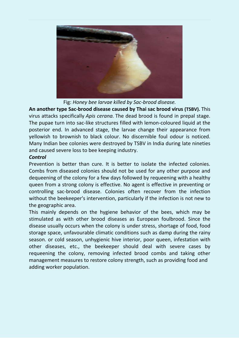
Fig: Honey bee larvae killed by Sac-brood disease.
An another type Sac-brood disease caused by Thai sac brood virus (TSBV). This virus attacks specifically Apis cerana. The dead brood is found in prepal stage. The pupae turn into sac-like structures filled with lemon-coloured liquid at the posterior end. In advanced stage, the larvae change their appearance from yellowish to brownish to black colour. No discernible foul odour is noticed. Many Indian bee colonies were destroyed by TSBV in India during late nineties and caused severe loss to bee keeping industry. Control Prevention is better than cure. It is better to isolate the infected colonies. Combs from diseased colonies should not be used for any other purpose and dequeening of the colony for a few days followed by requeening with a healthy queen from a strong colony is effective. No agent is effective in preventing or controlling sac-brood disease. Colonies often recover from the infection without the beekeeper's intervention, particularly if the infection is not new to the geographic area. This mainly depends on the hygiene behavior of the bees, which may be stimulated as with other brood diseases as European foulbrood. Since the disease usually occurs when the colony is under stress, shortage of food, food storage space, unfavourable climatic conditions such as damp during the rainy season. or cold season, unhygienic hive interior, poor queen, infestation with other diseases, etc., the beekeeper should deal with severe cases by requeening the colony, removing infected brood combs and taking other management measures to restore colony strength, such as providing food and adding worker population.

This disease is caused by a Microsporadium, Nosema apis/ N. ceranae. It
affects adult bees during cold/foggy and rainy weather. Nosema disease is
generally regarded as one of the most destructive diseases of adult bees,
affecting workers, queens and drones alike. Seriously affected worker bees are
unable to fly and may crawl about at the hive entrance or stand trembling on
top of the frames resulting into heavy mortality. The bees appear to age
physiologically: their life-span is much shortened and their hypopharyngeal
glands deteriorate, the result is a rapid dwindling of colony strength. Other
important effects are abnormally high rates of winter losses and queen
supersedures. In climates with pronounced long periods of flight restrictions,
i.e. no flight opportunities even for a day, the infection easily reaches a severe
stage that visibly affects the strength of the colony. Less obvious infection
levels in other climates often go undetected. The damage caused by Nosema
disease should not be judged by its effect on individual colonies alone as
collectively it can cause great losses in apiary productivity.
Fig: Nosema apis spores.
Cause: The disease is caused by the Microsporadium, Nosema apis, whose 5 .0 mm sized spores infests the bees, is absorbed with the food and germinates in the midgut. After penetration into the gut wall the cells multiply forming new spores that infect new gut cells or can be defeacated. The nutrition of the bees is impaired, particularly protein metabolism. Symptoms: Unfortunately, there is no reliable field diagnostic symptom enabling a diseased worker bee to be identified without killing it, nor can the beekeeper recognize an infected queen. However, in severe cases of infection, it is sometimes possible to separate healthy from diseased bees, the abdomen of an infected worker often being swollen and shiny in appearance. On dissection,
3. FUNGAL DISEASES
3.1 Nosema disease:

the individual circular constrictions in the alimentary canals of uninfected bees are clearly visible, while the constrictions cannot be seen clearly in diseased bees. Easy separation, after killing, of first abdominal segments with intestines attached, which shows white if strongly infected, versus a normal transparent, darker grey colour if there is no or only a low infection. In productive beekeeping, a healthy queen with a good egg-laying capability is always required, and Nosema disease in queens is therefore critical. The queen’s egg laying ability can be reduced possibly inducing her supersedures. She may also become the major cause of spreading the disease within the colony. On the other hand, beekeepers are naturally reluctant to destroy queens in the uncertain possibility that they are infected. The microscopic inspection of her feaces makes it possible to verify the presence or absence of the disease in the queen. Placed alone in a Petri dish, the queen will defecate in about an hour, the faces appearing as colourless drops of clear liquid. This liquid can be examined under the microscope for the presence of spores, without further preparation.
Control: Nosema disease can best be controlled by keeping colonies as strong as possible and removing possible causes of stress. Colonies and apiaries should receive adequate ventilation and protection from the cold and from humidity. The bees should have the possibility of for aging regularly in order to defecate. This prevents spreading of the spores within the colony. Beekeepers should also ensure that their colonies and queens come from disease-free stock. Hive equipment that is suspected of being contaminated by Nosema apis spores should be thoroughly decontaminated, preferably by heat treatment and fumigation. The best prevention is to change the combs once every two years. During normal wax processing the Nosema spores are killed.
1. Heat treatment: Infected equipment is maintained at 49°C (120°F) for 24-hours, ensuring that hot air passes through all stacked combs during the entire period of treatment. The temperature must however be carefully regulated, because heat at levels higher than that specified will melt wax. 2. Fumigation: A pad of cotton or other absorbent material, soaked with 80 percent acetic acid, is placed over the top-bars of the comb in each hive. The hive bodies are stacked together, the entrance is closed, all cracks are sealed, and the stacks are placed in an open shed for about a week. After this period, the hives are opened and the pads of acetic acid are removed. The combs must then be allowed to air for 48 hours to rid them of acetic acid residue so that they can be used again. The spores in the food cannot be killed. Therefore, the food combs have to be centrifuged before decontamination. The food should not be used anymore for bees.
Heat treatment and fumigation for Nosema Control.

SOURCES:
For preparation of this bulletin some are views and some other contents taken from various sources including the internet. The related materials are taken from following booklet/patrika/internet sources/web site etc. 1. The book namely “Honey Bee Enemies and their Management”
Written by M.S. Khan, Poonam Srivastava-AICRP on Honey Bees and Pollinators CCSHAU-Hisar(HR)
2. The book namely “Primary information of Beekeeping- Arya Gramoudhyog Sansthan -Kandara, Bagpat (U.P.)
3. Abstract of research publication- R.K. Thakur,H D Kaushik and Sunita Yadav, AICRP on Honey Bees and Pollinators, CCSHAU-Hisar(HR) 4. The Food & Agriculture Organization’s web site-www.fao.org/icatalog
/search /dett.asp?aries_id=107959 5. The List of diseases of the honey bee - Wikipedia, the free
encyclopedia, web site- en.wikipedia.org/wiki/List_of_diseases_of_the_honey_bee.
6. Honey Bee Parasites, Pests, Predators and Diseases - View, Website- https://agdev.anr.udel.edu/.../honey-bee.../honey-bee-parasites-pests-pred.
7. Honey Bee Pests & Diseases - Beebase - Beekeeping information, Website- secure.fera.defra.gov.uk › Public Pages › Bee Pests, Diseases & Maps...
8. [PDF]Honey bee diseases and pests: a practical guide - FAO.org, Website-ftp: //ftp.fao.org/docrep/fao/012/a0849e/a0849e00.pdf
9. Honey Bee Pests and Diseases Ontario Beekeepers' Association, Website- Www. ontariobee.com/outreach/honey-bee-pests-and-diseases.
10. Diseases and Pests of Honey Bees – Bee source Beekeeping, Website-www. beesource.com › Resources › USDA
11. Self reliance by beekeeping, Natural Resources Development Multi State Cooperative Society Limited(NARCO), Sec.12,Noida (U.P.)