Perturbation of Maize Phenylpropanoid Metabolism by … · Perturbation of Maize Phenylpropanoid...
19
Perturbation of Maize Phenylpropanoid Metabolism by an AvrE Family Type III Effector from Pantoea stewartii 1[OPEN] Jo Ann E. Asselin 2 , Jinshan Lin, Alvaro L. Perez-Quintero 3 , Irene Gentzel, Doris Majerczak, Stephen O. Opiyo, Wanying Zhao, Seung-Mann Paek, Min Gab Kim, David L. Coplin, Joshua J. Blakeslee, and David Mackey* Department of Horticulture and Crop Science (J.E.A., J.L., A.L.P.-Q., Do.M., W.Z., J.J.B., Da.M.), Molecular and Cellular Imaging Center-Columbus, Ohio Agricultural Research and Development Center (J.L., S.O.O., J.J.B.), Translational Plant Sciences Graduate Program (I.G.), Center for Applied Plant Sciences (I.G., Da.M.), Department of Plant Pathology (D.L.C.), and Department of Molecular Genetics (Da.M.), Ohio State University, Columbus, Ohio 43210; and College of Pharmacy, Research Institute of Pharmaceutical Science, Plant Molecular Biology and Biotechnology Research Center, Gyeongsang National University, Jinju 660–751, Republic of Korea (S.-M.P., M.G.K.) AvrE family type III effector proteins share the ability to suppress host defenses, induce disease-associated cell death, and promote bacterial growth. However, despite widespread contributions to numerous bacterial diseases in agriculturally important plants, the mode of action of these effectors remains largely unknown. WtsE is an AvrE family member required for the ability of Pantoea stewartii ssp. stewartii (Pnss) to proliferate efficiently and cause wilt and leaf blight symptoms in maize (Zea mays) plants. Notably, when WtsE is delivered by a heterologous system into the leaf cells of susceptible maize seedlings, it alone produces water-soaked disease symptoms reminiscent of those produced by Pnss. Thus, WtsE is a pathogenicity and virulence factor in maize, and an Escherichia coli heterologous delivery system can be used to study the activity of WtsE in isolation from other factors produced by Pnss. Transcriptional profiling of maize revealed the effects of WtsE, including induction of genes involved in secondary metabolism and suppression of genes involved in photosynthesis. Targeted metabolite quanti fication revealed that WtsE perturbs maize metabolism, including the induction of coumaroyl tyramine. The ability of mutant WtsE derivatives to elicit transcriptional and metabolic changes in susceptible maize seedlings correlated with their ability to promote disease. Furthermore, chemical inhibitors that block metabolic flux into the phenylpropanoid pathways targeted by WtsE also disrupted the pathogenicity and virulence activity of WtsE. While numerous metabolites produced downstream of the shikimate pathway are known to promote plant defense, our results indicate that misregulated induction of phenylpropanoid metabolism also can be used to promote pathogen virulence. Pantoea stewartii ssp. stewartii (Pnss) is a bacterial pathogen that causes Stewart’s wilt and leaf blight in maize (Zea mays). Wilt results when Pnss bacteria infect xylem vessels of seedlings and block water transport through the production of exopolysaccharide. In older plants, leaf blight develops as streaks along leaf veins and can result in the loss of leaves. Currently, the eco- nomic significance of Pnss-associated disease is limited to sweet maize production in the central and northeastern United States (White, 1999). While the economic impact of Pnss on maize has been reduced through the use of resistant cultivars, the tools available for both host and pathogen make the Pnss-maize interaction highly suit- able for laboratory study. Maize is of major importance as a source of food, secondary products, and energy. As such, substantial tools have been developed for its study, including genome sequences for inbred lines B73 and Mo17 and the Mexican landrace Palomero Toluqueño as well as curated online tools and databases available to the maize research community (Brunner et al., 2005; Schnable et al., 2009; Vielle-Calzada et al., 2009; Schaeffer et al., 2011). For Pnss, targeted mutagenesis and trans- formation are relatively easy, and a large collection of genetically characterized mutants is available. Addition- ally, Pnss infection assays are quick and uniform. Maize seedlings are ready for inoculation as soon as 6 d after sowing, leaf cells can be uniformly targeted following vacuum infiltration, the appearance of water-soaking symptoms occurs in less than 1 d, and in planta bacterial 1 This work was supported by the National Science Foundation (grant no. MCB–0718882), the U.S. Department of Agriculture (grant no. National Institute of Food and Agriculture 2008–35319–04506), the Korean Rural Development Administration Next-Generation BioGreen Program (System and Synthetic Agro-Biotech Center and grant nos. PJ009088 and PJ011091), and the Ohio Agricultural Research and De- velopment Center of Ohio State University. 2 Present address: School of Integrative Plant Science, Section of Plant Pathology and Plant-Microbe Biology, Cornell University, Ithaca, NY 14853. 3 Present address: Unité Mixte de Recherche Résistance des Plantes aux Bioagresseurs, Institut de Recherche pour le Développement, Centre de Coopération Internationale en Recherche Agronomique pour le Développement-Université de Montpellier II, 34000 Montpellier cedex 5, France. * Address correspondence to [email protected]. The author responsible for distribution of materials integral to the findings presented in this article in accordance with the policy de- scribed in the Instructions for Authors (www.plantphysiol.org) is: David Mackey ([email protected]). [OPEN] Articles can be viewed without a subscription. www.plantphysiol.org/cgi/doi/10.1104/pp.114.253120 Plant Physiology Ò , March 2015, Vol. 167, pp. 1117–1135, www.plantphysiol.org Ó 2014 American Society of Plant Biologists. All Rights Reserved. 1117 www.plantphysiol.org on June 27, 2018 - Published by Downloaded from Copyright © 2015 American Society of Plant Biologists. All rights reserved.
Transcript of Perturbation of Maize Phenylpropanoid Metabolism by … · Perturbation of Maize Phenylpropanoid...

Perturbation of Maize Phenylpropanoid Metabolism by anAvrE Family Type III Effector from Pantoea stewartii1[OPEN]
Jo Ann E. Asselin2, Jinshan Lin, Alvaro L. Perez-Quintero3, Irene Gentzel, Doris Majerczak,Stephen O. Opiyo, Wanying Zhao, Seung-Mann Paek, Min Gab Kim, David L. Coplin,Joshua J. Blakeslee, and David Mackey*
Department of Horticulture and Crop Science (J.E.A., J.L., A.L.P.-Q., Do.M., W.Z., J.J.B., Da.M.), Molecular andCellular Imaging Center-Columbus, Ohio Agricultural Research and Development Center (J.L., S.O.O., J.J.B.),Translational Plant Sciences Graduate Program (I.G.), Center for Applied Plant Sciences (I.G., Da.M.),Department of Plant Pathology (D.L.C.), and Department of Molecular Genetics (Da.M.), Ohio StateUniversity, Columbus, Ohio 43210; and College of Pharmacy, Research Institute of Pharmaceutical Science,Plant Molecular Biology and Biotechnology Research Center, Gyeongsang National University, Jinju 660–751,Republic of Korea (S.-M.P., M.G.K.)
AvrE family type III effector proteins share the ability to suppress host defenses, induce disease-associated cell death, and promotebacterial growth. However, despite widespread contributions to numerous bacterial diseases in agriculturally important plants, themode of action of these effectors remains largely unknown. WtsE is an AvrE family member required for the ability of Pantoea stewartiissp. stewartii (Pnss) to proliferate efficiently and cause wilt and leaf blight symptoms in maize (Zea mays) plants. Notably, whenWtsE is delivered by a heterologous system into the leaf cells of susceptible maize seedlings, it alone produces water-soaked diseasesymptoms reminiscent of those produced by Pnss. Thus, WtsE is a pathogenicity and virulence factor in maize, and an Escherichiacoli heterologous delivery system can be used to study the activity of WtsE in isolation from other factors produced by Pnss.Transcriptional profiling of maize revealed the effects of WtsE, including induction of genes involved in secondary metabolism andsuppression of genes involved in photosynthesis. Targeted metabolite quantification revealed that WtsE perturbs maize metabolism,including the induction of coumaroyl tyramine. The ability of mutant WtsE derivatives to elicit transcriptional and metabolic changesin susceptible maize seedlings correlated with their ability to promote disease. Furthermore, chemical inhibitors that block metabolicflux into the phenylpropanoid pathways targeted by WtsE also disrupted the pathogenicity and virulence activity of WtsE. Whilenumerous metabolites produced downstream of the shikimate pathway are known to promote plant defense, our results indicate thatmisregulated induction of phenylpropanoid metabolism also can be used to promote pathogen virulence.
Pantoea stewartii ssp. stewartii (Pnss) is a bacterialpathogen that causes Stewart’s wilt and leaf blight inmaize (Zea mays). Wilt results when Pnss bacteria infectxylem vessels of seedlings and block water transport
through the production of exopolysaccharide. In olderplants, leaf blight develops as streaks along leaf veinsand can result in the loss of leaves. Currently, the eco-nomic significance of Pnss-associated disease is limited tosweet maize production in the central and northeasternUnited States (White, 1999). While the economic impactof Pnss on maize has been reduced through the use ofresistant cultivars, the tools available for both host andpathogen make the Pnss-maize interaction highly suit-able for laboratory study. Maize is of major importanceas a source of food, secondary products, and energy. Assuch, substantial tools have been developed for its study,including genome sequences for inbred lines B73 andMo17 and the Mexican landrace Palomero Toluqueño aswell as curated online tools and databases available tothe maize research community (Brunner et al., 2005;Schnable et al., 2009; Vielle-Calzada et al., 2009; Schaefferet al., 2011). For Pnss, targeted mutagenesis and trans-formation are relatively easy, and a large collection ofgenetically characterized mutants is available. Addition-ally, Pnss infection assays are quick and uniform. Maizeseedlings are ready for inoculation as soon as 6 d aftersowing, leaf cells can be uniformly targeted followingvacuum infiltration, the appearance of water-soakingsymptoms occurs in less than 1 d, and in planta bacterial
1 This work was supported by the National Science Foundation(grant no. MCB–0718882), the U.S. Department of Agriculture (grantno. National Institute of Food and Agriculture 2008–35319–04506), theKorean Rural Development Administration Next-Generation BioGreenProgram (System and Synthetic Agro-Biotech Center and grant nos.PJ009088 and PJ011091), and the Ohio Agricultural Research and De-velopment Center of Ohio State University.
2 Present address: School of Integrative Plant Science, Section of PlantPathology andPlant-Microbe Biology,Cornell University, Ithaca,NY14853.
3 Present address: UnitéMixte de Recherche Résistance des Plantes auxBioagresseurs, Institut de Recherche pour le Développement, Centre deCoopération Internationale en Recherche Agronomique pour leDéveloppement-Université de Montpellier II, 34000 Montpelliercedex 5, France.
* Address correspondence to [email protected] author responsible for distribution of materials integral to the
findings presented in this article in accordance with the policy de-scribed in the Instructions for Authors (www.plantphysiol.org) is:David Mackey ([email protected]).
[OPEN] Articles can be viewed without a subscription.www.plantphysiol.org/cgi/doi/10.1104/pp.114.253120
Plant Physiology�, March 2015, Vol. 167, pp. 1117–1135, www.plantphysiol.org � 2014 American Society of Plant Biologists. All Rights Reserved. 1117 www.plantphysiol.orgon June 27, 2018 - Published by Downloaded from
Copyright © 2015 American Society of Plant Biologists. All rights reserved.

growth can be measured over the course of several days.Thus, Pnss-maize is a highly tractable system for study ofthe interaction of a crop with a pathogenic bacterium.
Many bacterial pathogens of both plants and ani-mals utilize type III secretion systems (T3SSs) to transporteffector proteins (called type III effectors [T3Es]) directlyinto the cytoplasm of host cells. Remarkably, Pnss en-codes two distinct T3SSs, one that is active in corn fleabeetles, which harbor Pnss over the winter and transmit itto maize plants in the spring and late summer via feed-ing, and a second, hypersensitive response and patho-genicity (Hrp) T3SS that is active in maize tissues (Correaet al., 2012). The study of T3Es from plant pathogenicbacteria has been limited by the fact that deletion of anindividual T3E often has little or no effect on virulence,due in part to functional redundancy within the suite ofT3Es encoded by a pathogen. To study the action of T3Esin isolation, numerous studies have used transgenic ex-pression of T3Es in planta, often under the control ofchemically inducible promoters. Although this approachhas been very useful, it includes uncertainties about thetiming and level of T3Es inside plant cells compared withwhen the T3Es are naturally delivered. Thus, anotheradvantage to studying Pnss is the key role of a single typeIII effector protein, WtsE, the deletion of which com-pletely compromises the ability of Pnss to cause diseasein maize (Frederick et al., 2001). Extensive mutagenesisrevealed no other T3E that is needed for pathogenicity inmaize (Frederick et al., 2001), although it cannot be ruledout that other, yet unknown, T3Es may have minor orredundant roles. Thus, due to the detectable contributionof WtsE to bacterial disease, the Pnss-maize pathosystemis attractive for the study of this T3E in particular.
WtsE is a member of the AvrE/HopR superfamily ofT3Es that are widely distributed among plant-associated,gram-negative bacteria, including strains of Pseudomonas,Ralstonia, Erwinia, and Xanthomonas spp. (Kvitko et al.,2009). Members of this superfamily often have markedeffects on pathogenicity. WtsE from Pnss is most closelyrelated to AvrE family T3Es and in particular to DspA/Eproteins from Erwinia spp. Just as a functional WtsE isrequired for Pnss pathogenicity (Frederick et al., 2001),DspA/E is required for the ability of Erwinia amylovora toelicit fire blight symptoms in apple (Malus domestica), Co-toneaster spp., and pear (Pyrus communis; Gaudriault et al.,1997; Bogdanove et al., 1998). Additionally, AvrE, alongwith the sequence-unrelated but functionally redundantT3E, HopM1, and the AvrE superfamily member HopR1(Badel et al., 2006; Kvitko et al., 2009), is required for thevirulence of Pseudomonas syringae pv tomato on tomato(Solanum lycopersicum), Arabidopsis (Arabidopsis thaliana),and Nicotiana benthamiana. Thus, members of this impor-tant superfamily of T3Es make key contributions to thepathogenicity of several genera of bacteria on plantsranging from grasses to fruit trees. Given the importanceand widespread distribution of this superfamily, insightsobtained from the study of WtsE are likely to be broadlyrelevant to plant-pathogen interactions.
Although the mechanisms by which AvrE/HopRT3Es promote virulence are not well understood, the
consequences of their actions in plant cells have beenexamined. Many AvrE family T3Es, including WtsE,have been shown to elicit water-soaked diseasesymptoms in susceptible host plants and to promote thegrowth and survival of the bacteria in planta (Bogdanoveet al., 1998; Badel et al., 2006; Boureau et al., 2006; Hamet al., 2009). Basal defense, as assessed by callose de-position, was suppressed by DspA/E of E. amylovora inapple and AvrE of P. syringae and WtsE of Pnss inArabidopsis (DebRoy et al., 2004; Ham et al., 2008). Innonhost plants, both DspA/E of E. amylovora and WtsEof Pnss have been shown to increase bacterial growthbefore bacterial population decline begins, presumablydue to the onset of host defenses (Oh et al., 2007; Hamet al., 2008). Our previous work indicates that there areat least two main functions for WtsE (Ham et al., 2006,2008, 2009). First, it induces a water-soaking response inmaize that may release water and nutrients to supportinitial and continued bacterial growth. Second, it sup-presses plant defenses in Arabidopsis and likely doesthe same in maize.
A clue to the mechanism of action of AvrE familyT3Es came from the identification of important proteinmotifs conserved within the family. Most AvrE familymembers contain a putative endoplasmic reticulummembrane retention signal (ERMRS) and one or twoWxxxE motifs (Ham et al., 2009). The WxxxE motifswere first discovered in T3Es of animal pathogens (Altoet al., 2006). Derivatives of WtsE with either the putativeERMRS or both WxxxE motifs mutated (DFEMK andw12 mutants, respectively) failed to elicit diseasesymptoms in maize or to suppress defense responses inArabidopsis (Ham et al., 2009). Interestingly, the growthof these site-directed wtsE mutants of Pnss in maizewas only modestly reduced. However, given that manyT3Es have multiple targets, and given the large size ofAvrE/HopR T3Es, it seems likely that WtsE might havemultiple functions and targets within plant cells. TheWxxxE motifs of AvrE family members are hypothe-sized to form part of the fold that mimics the active siteof a guanine-exchange factor (GEF) protein and thus tomodulate the activity of host GTPase proteins, as hasbeen demonstrated for Map/IpgB/Sif family membersfrom bacterial pathogens of animals (Huang et al., 2009).Confirmation of this biochemical prediction, along withlocalization in plant cells, have proven difficult due to thecell toxicity of the AvrE family T3Es (Ham et al., 2006).Intriguingly, HopM1, which is functionally redundant toAvrE for promoting the full virulence of P. syringae pvtomato strain DC3000, targets for degradation anADP ribosylation factor-GEF called Arabidopsis HopM-Interactor7 (AtMIN7; Nomura et al., 2006). AtMIN7 isrequired for pathogen-associated molecular pattern(PAMP)-triggered immunity and salicylic acid (SA)-regulated responses to pathogens, including callosedeposition (Nomura et al., 2006). Because ADP ribo-sylation factor-GEFs are regulators of the plant cyto-skeleton and vesicle traffic, it has been hypothesizedthat HopM1 and AvrE family effectors manipulatethese pathways (Nomura et al., 2006; Ham et al., 2009).
1118 Plant Physiol. Vol. 167, 2015
Asselin et al.
www.plantphysiol.orgon June 27, 2018 - Published by Downloaded from Copyright © 2015 American Society of Plant Biologists. All rights reserved.
The molecular targets of T3Es from plant pathogensare numerous and varied, including proteins involvedin the recognition of PAMPs at the plant plasmamembrane, defense-associated mitogen-activated pro-tein kinase signaling cascades, the plant cytoskeleton,components of the chloroplast, isoflavanone biosynthe-sis, and proteins involved in vesicle trafficking (for re-view, see Block and Alfano, 2011; Deslandes and Rivas,2012). While many, perhaps most, T3Es suppress plantdefense responses, others may be involved in promot-ing bacterial growth by providing bacterial access toplant nutrients (Chen et al., 2010). In addition to theirintended defense-suppressing function(s), T3Es also canelicit plant defenses via direct or indirect interactionwith specific host resistance proteins. While the specificmechanisms by which WtsE and other AvrE familyT3Es function remain largely a mystery, the data pre-sented here indicate secondary metabolism, particularlythe phenylpropanoid pathway, as a potential target.Products of plant metabolism play important roles
in plant-microbe interactions, including plant de-fense. For example, phytoalexin terpenoids, such askauralexins and zealexins, accumulate in response to
fungal attack (Ahuja et al., 2012). The shikimatepathway also gives rise to numerous products im-portant to plant defense. Chorismate, the precursor tothe synthesis of aromatic amino acids, is utilized forthe production of the important pathogen defense-related plant hormone SA (Wildermuth et al., 2001).Trp and its indole precursor are converted to glucosi-nolates and benzoxazinoids in Arabidopsis and maize,respectively, with roles in defense signaling (Bednareket al., 2009; Ahmad et al., 2011). Phe-derived com-pounds are a major class of defense-associated metab-olites (Naoumkina et al., 2010). Preformed phenolics areinvolved in the lignification of cell walls associated withthe hypersensitive response (Beckman, 2000). Hydrox-ycinnamic acid amides fortify the plant cell wall byreducing its digestibility to pathogens (Facchini et al.,2002). In addition to abiotic stress, flavonoids alsocontribute to plant defense against biotic stresses(Treutter, 2005). On the microbe side of the interactions,secondary metabolites derived from numerous plant-associated bacteria also can affect plant physiologyand block host responses. For example, the coronatinetoxin from P. syringae can reopen stomata and
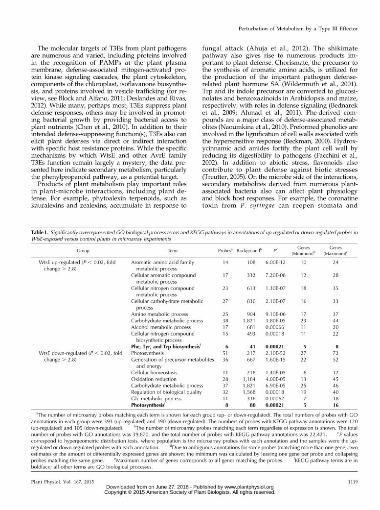
Table I. Significantly overrepresented GO biological process terms and KEGG pathways in annotations of up-regulated or down-regulated probes inWtsE-exposed versus control plants in microarray experiments
Group Term Probesa Backgroundb P c Genes
(Minimum)dGenes
(Maximum)e
WtsE up-regulated (P , 0.02, foldchange . 2.8)
Aromatic amino acid familymetabolic process
14 108 6.00E-12 10 24
Cellular aromatic compoundmetabolic process
17 332 7.20E-08 12 28
Cellular nitrogen compoundmetabolic process
23 613 1.30E-07 18 35
Cellular carbohydrate metabolicprocess
27 830 2.10E-07 16 33
Amine metabolic process 25 904 9.10E-06 17 37Carbohydrate metabolic process 38 1,821 3.80E-05 23 44Alcohol metabolic process 17 681 0.00066 11 20Cellular nitrogen compoundbiosynthetic process
15 493 0.00018 11 22
Phe, Tyr, and Trp biosynthesisf 6 41 0.00021 5 8WtsE down-regulated (P , 0.02, fold
change . 2.8)Photosynthesis 51 217 2.10E-52 27 72Generation of precursor metabolitesand energy
36 667 1.60E-15 22 52
Cellular homeostasis 11 218 1.40E-05 6 12Oxidation reduction 28 1,184 4.00E-05 13 45Carbohydrate metabolic process 37 1,821 6.90E-05 25 46Regulation of biological quality 32 1,568 0.00018 19 40Glc metabolic process 11 336 0.00062 7 18Photosynthesisf 8 80 0.00021 5 16
aThe number of microarray probes matching each term is shown for each group (up- or down-regulated). The total numbers of probes with GOannotations in each group were 393 (up-regulated) and 390 (down-regulated). The numbers of probes with KEGG pathway annotations were 120(up-regulated) and 105 (down-regulated). bThe number of microarray probes matching each term regardless of expression is shown. The totalnumber of probes with GO annotations was 39,870, and the total number of probes with KEGG pathway annotations was 22,421. cP valuescorrespond to hypergeometric distribution tests, where population is the microarray probes with each annotation and the samples were the up-regulated or down-regulated probes with each annotation. dDue to ambiguous annotations for some probes (matching more than one gene), twoestimates of the amount of differentially expressed genes are shown; the minimum was calculated by leaving one gene per probe and collapsingprobes matching the same gene. eMaximum number of genes corresponds to all genes matching the probes. fKEGG pathway terms are inboldface; all other terms are GO biological processes.
Plant Physiol. Vol. 167, 2015 1119
Perturbation of Metabolism by a Type III Effector
www.plantphysiol.orgon June 27, 2018 - Published by Downloaded from Copyright © 2015 American Society of Plant Biologists. All rights reserved.

antagonize SA-dependent and SA-independent defensesignaling (Melotto et al., 2006; Geng et al., 2012; Zhenget al., 2012).
Here, we show that WtsE modulates maize metabo-lism. The effect of this single effector is mediated, at leastin part, through alteration of the expression of enzymesinvolved in phenylpropanoid metabolism. Introductionof WtsE into the cells of maize seedling leaves through anEscherichia coli delivery system (EcDS) elicited numerousplant responses, including genome-wide changes intranscription. Induced expression of genes involved in thephenylpropanoid pathway by WtsE was confirmed byquantitative reverse transcription (qRT)-PCR. WtsE wasfound to elicit the accumulation of coumaroyl tyramine(CouTyr), a hydroxycinammic acid amide typically as-sociated with lignin in plant cell walls. The ability ofderivatives of WtsE to elicit transcriptional and metabolicchanges in susceptible maize seedlings was correlatedwith their ability to induce disease symptoms. Also,chemical inhibitors of the shikimate pathway or Pheammonia lyase limited the ability of WtsE to induceCouTyr and to promote disease. These results indicatethat the virulence activity of WtsE depends on the per-turbation of phenylpropanoid metabolism.
RESULTS
Genome-Wide Transcriptional Response of MaizeSeedlings to WtsE
To investigate the effect of WtsE on transcription inmaize, we delivered WtsE into cells of maize seedling
leaves using a heterologous EcDS in order to eliminateother Pnss-associated factors that might also affectmaize transcription. This system utilized a strain ofE. coli MC4100 carrying the entire Dickeya dadantii hrpgene cluster in plasmid pCPP2156 (Ham et al., 1998),which becomes a WtsE delivery system (EcWtsE)when it carries a second plasmid expressing wtsEF orderivatives thereof. Previous work has indicated thatWtsE, when delivered by EcWtsE, induces disease-likewater-soaking symptoms in leaves of susceptiblemaize seedlings (Ham et al., 2006, 2009). While vali-dating the use of the EcDS to study the mechanism ofaction of WtsE, this study also highlighted the im-portance of selecting a time point that precedes po-tential secondary transcriptional changes associatedwith widespread cell death. Macroscopic investigationrevealed that WtsE-induced symptoms first becameapparent approximately 8 h after infiltration (hai;Supplemental Fig. S1). Using electrolyte leakage tomeasure the loss of cell integrity provided a moresensitive assay to assess WtsE-induced cell death(Supplemental Fig. S1). The conductivity of water-containing maize leaves remained consistently lowuntil approximately 6 hai, at which time plants thatreceived WtsE showed increasing signal relative tocontrol plants. Thus, 6 hai was chosen as the time pointfor transcriptome analysis.
We used a microarray approach to examine the effectof WtsE on transcription in maize cells (http://www.maizegdb.org/microarray.php). Of the more than43,000 maize oligonucleotides on the array, 1,047 ol-igonucleotides showed highly significant differences
Figure 1. WtsE induces CouTyraccumulation in sweet maize‘Seneca Horizon’ seedlings. A, Liq-uid chromatography-tandem massspectrometry (LC-MS/MS) chromato-grams showing overlaid total ioncounts (TIC) of precursor mass ionscans (positive ion mode) of extractsof cv Seneca Horizon seedlings from20 h after infiltration with wild-typePnss (DC283; black trace) and thewtsE null mutant (DM5101; graytrace). WtsE-dependent accumula-tion of CouTyr is apparent at an ac-quisition time of approximately 7.1min (arrow). B and C, Extracted ioncounts (EIC) of putative CouTyr spe-cies (m/z = 284) in extracts fromplants infiltrated with DC283 (B) orDM5101 (C) as in A. D, Quantifica-tion of CouTyr in extracts of plantstreated as in A. Shown are combineddata and SD from six biological rep-licates (n = 12). FW, Fresh weight.
1120 Plant Physiol. Vol. 167, 2015
Asselin et al.
www.plantphysiol.orgon June 27, 2018 - Published by Downloaded from Copyright © 2015 American Society of Plant Biologists. All rights reserved.

between treatment (EcWtsE) and control (EcDS) basedon a minimum fold change of 2.83 and a P value of lessthan 0.02 (Supplemental Fig. S2). The oligonucleotideswere annotated to genes from the maize B73 genomebased on the work of Seifert et al. (2012). We found that586 of the 1,047 oligonucleotides were annotated tosingle genes. Of these, 314 and 272 were up- or down-regulated by WtsE, corresponding to 290 and 249 uniquegenes, respectively (Supplemental Table S1). Another 285of the 1,047 oligonucleotides remained unannotated, and176 oligonucleotides could not be unambiguously an-notated; they may have annealed to products frommultiple genes during hybridization. Our results indicatethat WtsE caused large-scale transcriptional reprogram-ming within maize seedling leaves within 6 hai ofEcWtsE infiltration.Differentially expressed probes were assigned Gene
Ontology (GO) terms using the AgriGO platform (Duet al., 2010) and Kyoto Encyclopedia of Genes and Ge-nomes (KEGG) pathways using Kobas 2.0 (Xie et al.,2011). We assessed whether certain GO terms or KEGGpathways might be significantly enriched in our datasets (Table I). Photosynthesis was significantly repre-sented among probes that were down-regulated inplants exposed to WtsE. Secondary metabolism, par-ticularly the phenylpropanoid pathway, was highly
represented in WtsE-induced probes. Additional probesup-regulated in response to WtsE included several thatare associated with genes involved in the biosynthesisof aromatic amino acids that are precursors to phenyl-propanoid metabolism (see below).
CouTyr Induction by WtsE in Maize
Given the significant induction of genes involved inthe synthesis of phenolic and phenylpropanoid com-pounds, we sought to examine the effect of WtsE on thelevels of these metabolites in maize seedlings. Seedlingleaves infiltrated with either a wild-type Pnss strain(DC283) or a Pnss wtsE::miniTn5 mutant strain(DM5101) were harvested at 20 hai, which is approxi-mately 2 h after the first macroscopic water-soakedsymptoms produced by DC283 became apparent.Samples were extracted with 80% (v/v) methanol,diluted 100-fold in liquid chromatography-mass spec-troscopy (LC-MS)-grade methanol and analyzed via LC-MS/MS (Fig. 1). Compounds were separated using anAgilent C18 Poroshell column, subjected to electrosprayionization, and monitored in positive ion mode. Initialprecursor ion scans (which show the mass of the intact,nonfragmented parent molecule) showed several peaks
Figure 2. Generalized pathway of secondary metabolism from chorismate to aromatic amino acids and their secondary pro-ducts. Enzyme abbreviations are shown over the step(s) they catalyze; numbers next to enzyme names indicate the maximumobserved fold increases in expression in WtsE-exposed versus control plants in microarray experiments. Arrow thickness relatesto this fold-change value. Numbers in parentheses indicate how many genes were found up-regulated with P , 0.05. Dashedarrows indicate steps not shown. CAD, Cinnamoyl alcohol dehydrogenase; CCoAMT, caffeoyl-CoA O-methyltransferase;CcoAR, cinnamoyl-CoA reductase; CHI, chalcone isomerase; CHS, chalcone synthase; CM, chorismate mutase; COMT, caffeicacid O-methyltransferase; HCT, hydroxycinnamoyl transferase; THT, tyramine N-hydroxycinnamoyltransferase.
Plant Physiol. Vol. 167, 2015 1121
Perturbation of Metabolism by a Type III Effector
www.plantphysiol.orgon June 27, 2018 - Published by Downloaded from Copyright © 2015 American Society of Plant Biologists. All rights reserved.

eluting in the DC283-infiltrated leaves that were close tothe limit of detection or absent in DM5101-infiltratedleaves (Fig. 1, A–C). The most prominent of thesepeaks exhibited a retention time of approximately 7.1min and a mass-to-charge ratio (m/z) of 284 (Fig. 1A).Subsequent product ion scans indicated that this peakwas likely a derivative of coumaric acid, and its mass(m/z = 284), combined with the function of genes in-duced by WtsE, led us to speculate that the peak at re-tention time = 7.1 min was CouTyr. Genuine CouTyr(para-N-transcoumaroyl tyramine) was synthesized(Pedersen et al., 2010) and used as an authentic standardto investigate the identity of the m/z = 284 peak. Com-parison of retention time, precursor ion mass, and fourprecursor-product ion mass transitions (284→146.9,284→118.8, 284→91.2, and 284→77.1; SupplementalTable S2) confirmed that the peak induced in DC283-induced leaves was CouTyr. Quantification based onstandard curves generated using genuine CouTyr(Supplemental Fig. S3) revealed that infiltration withDC283 induced the accumulation of 160 mg g21 freshweight of CouTyr (Fig. 1D). Interestingly, while quan-tifying CouTyr, a peak with an identical precursor ionmass (m/z = 284) was observed to elute from the columnapproximately 0.5 min prior to CouTyr (retention time =6.6 min). Further investigation revealed that this peakexhibited a fragmentation pattern identical to that ob-served with CouTyr, including the four mass transitionsused to positively identify and quantify CouTyr bycomparison with the genuine standard. Because of this,we hypothesize that the m/z = 284 peak eluting at re-tention time = 6.6 min is either a stereoisomer of CouTyrgenerated during the synthesis of this compoundor an oxidative breakdown/catalysis product withan open-ring structure. While the structure of thisputative stereoisomer and its potential role in WtsEvirulence are currently unclear, since the CouTyrstructural isomer most likely formed during thesynthesis of this molecule (ortho-N-transcoumaroyltyramine) exhibits a significant shift in both reten-tion time and mass fragmentation patterns (data notshown), our current hypothesis favors the presenceof an oxidized CouTyr isomer.
WtsE-Induced Expression of Genes EncodingPhenylpropanoid Biosynthetic Enzymes
The observation that WtsE induces a high level ofCouTyr accumulation led us to look more closely at theexpression of genes encoding biosynthetic enzymes in-volved in the biosynthesis of coumaric acid and tyra-mine. Indeed, the microarray data showed that WtsEinduces numerous genes that could account for the ac-cumulation of these compounds as well as additionalcompounds generated from coumaroyl-CoA (Fig. 2).
Next, we used qRT-PCR to further investigate theexpression of a subset of WtsE-induced genes. Theresults of these experiments served three purposes.The first was to validate the microarray data by
independently confirming that these genes were WtsEinduced. Second, the experiments provided additionalinformation on the effects of the EcDS on the expres-sion of these genes. The microarray only compared
Figure 3. Expression of five genes associated with phenolic metab-olism in maize seedlings vacuum infiltrated with buffer, EcDS, orEcWtsE. Tissue was recovered at 2, 4, and 6 hai, and expression wasdetermined for TD (A), 4CL (B), C4H (C), P/AD (D), and PAL (E)transcripts. Plants exposed to WtsE had greater mRNA accumulationof each of these genes than controls. Shown are average values and SE
of six qRT-PCRs (three reactions each for the complementary DNAfrom two biological replicates). Each biological replicate consisted ofthe first true leaves from two seedlings. Significantly different ex-pression levels for a given gene and time point are indicated withletters (ANOVA and Fisher’s LSD, P , 0.05). No values at 2 hai, northe 4-hai time point for P/AD, were found to significantly differ fromcontrols.
1122 Plant Physiol. Vol. 167, 2015
Asselin et al.
www.plantphysiol.orgon June 27, 2018 - Published by Downloaded from Copyright © 2015 American Society of Plant Biologists. All rights reserved.

two treatments (EcWtsE versus the EcDS control) andthus only revealed changes in gene expression with orwithout WtsE over a possible background of responsesto PAMPs from the EcDS. By including a buffertreatment, the qRT-PCR provided information aboutthe effect of PAMPs on the expression of the WtsE-regulated genes. Third, the experiments provided in-formation about how the expression of WtsE-inducedgenes changes over time in response to the differenttreatments.Maize seedlings were infiltrated with buffer, EcDS, or
EcWtsE. The expression of WtsE-induced genes was
analyzed by qRT-PCR at 2, 4, and 6 hai (Fig. 3). By 4 hai,plants infiltrated with EcWtsE contained signifi-cantly more transcripts for tyrosine decarboxylase(TD), 4-coumarate-coenzyme A ligase (4CL), cinnamate4-hydroxylase (C4H), and phenylalanine ammonia ly-ase (PAL) than plants infiltrated with EcDS or buffer(Fig. 3, A–C and E, respectively). By 6 hai, prephanate/arogenate dehydratase (P/AD) also was more highlyexpressed in EcWtsE-infiltrated plants than thoseinfiltrated with EcDS or buffer (Fig. 3D). These resultsconfirm that the induction of these genes is depen-dent on WtsE. Furthermore, because buffer- and
Figure 4. Pnss strains harboring mutated forms of wtsE were reduced in growth and virulence in maize seedlings. A, Map ofWtsE showing the locations of the internal deletions in the listed plasmids and derivatives used for further analyses in boldface.Each tick represents 100 bp. B, Production and secretion of WtsE-FLAG and derivatives. Plasmid pJA017 encodes C terminallyFLAG-tagged WtsE and the chaperone WtsF in vector pRK415. Pnss strains DM5101 (wtsE mutant) and DM711 (hrpJ type IIIsecretion mutant) were transformed with pRK415, pJA017, or variants of pJA017 expressing wtsE derivatives. Proteins werevisualized by anti-FLAG and anti-b-galactosidase (b-gal) immunoblotting from lysed cells (left) or from culture medium (right).Positions of WtsE derivatives and b-galactosidase, a cytoplasmic protein that serves as a loading control for cell lysis and anegative control for leakage of cell contents into the culture supernatant, are indicated by arrows. DM5101 (lanes 1–6) andDM711 (lanes 7–12) contained the following plasmids: lanes 1 and 7, pRK415; lanes 2 and 8, pJA017; lanes 3 and 9, pJA052(wtsE w12 mutant, W694A, and W840A); lanes 4 and 10, pDM5117; lanes 5 and 11, pDM5153; and lanes 6 and 12,pDM5155. C and D, Average values and SD from three biological replicates measuring bacterial growth (C) and virulence (D) ofDM5101 carrying the same plasmids as for lanes 1 to 6 in B. Both measurements were made 3 d after whorl inoculation withvirulence rated on a scale from 0 to 3, with 3 being the most severe and 0 being symptomless. Different letters in C indicatesignificant differences in bacterial growth (ANOVA and Tukey’s honestly significant difference, P , 0.05). Different letters in Dindicate significant differences in virulence (Kruskal-Wallis and Tukey’s mean rank tests, P , 0.05).
Plant Physiol. Vol. 167, 2015 1123
Perturbation of Metabolism by a Type III Effector
www.plantphysiol.orgon June 27, 2018 - Published by Downloaded from Copyright © 2015 American Society of Plant Biologists. All rights reserved.

EcDS-infiltrated plants did not differ significantly forany gene at any of the times assessed, PAMPs associatedwith the EcDS did not induce the expression of any ofthese genes.
The Functionality of WtsE Derivatives in Biological AssaysCorrelates with Transcriptional andMetabolic Perturbations
Fifteen partial deletions (approximately 300 bp each)distributed across the C terminally FLAG-tagged wtsEreading frame were constructed (Fig. 4A). Based onour previous finding that deletion of the C-terminalERMRS abolished the virulence activity of WtsE, wesuspected that placing a tag at the C terminus of WtsEmight similarly disrupt the function of WtsE. However,to our surprise, wild-type WtsE with a C-terminal FLAGtag remained active; thus, this tagging strategy wasemployed for the other derivatives. One goal was to
investigate which regions of the gene were dispensablefor the plant cell death-inducing or bacterial growth-promoting functions of WtsE. A second goal was touse the derivatives to determine if the virulence activityof WtsE correlates with its ability to modulate gene ex-pression and induce CouTyr synthesis.
Each wtsE derivative was cloned into a broad-host-range plasmid and introduced into the wtsE null mutantstrain DM5101. Western blotting of extracts from bac-teria grown on an hrp-inducing medium demonstratedthat 13 of the 15 WtsE derivatives were detectablyproduced inside the bacteria. However, of the13 expressed deletion derivatives, only three (thoseproduced from pDM5117 [wtsED709-1017], pDM5153[wtsED2935-3234], and pDM5155 [wtsED3235-3534]) wereshown to be secreted in a type III-dependent manner asefficiently as wild-type WtsE (Fig. 4B). Each of thesethree expressed and secreted WtsE derivatives, alongwith the w12 derivative mutated in both WxxxE motifs,was significantly compromised, relative to wild-type
Figure 5. Effects of wild-type and mutated forms of WtsE on the expression of five genes associated with phenolic metabolismin maize seedlings. Six-day-old cv Seneca Horizon seedlings were vacuum infiltrated with EcDS (pRK415; 1), EcWtsE-FLAG(pJA017; 2), or the EcDS-expressing mutated forms of FLAG-tagged wtsE: w12 mutant (pJA052; 3), wtsED709-1017 (pDM5109;4), wtsED2935-3234 (pDM5153; 5), or wtsED3235-3534 (pDM5155; 6). Samples were collected at 4 hai, and the expression ofTD (A), 4CL (B), C4H (C), P/AD (D), and PAL (E) was assessed. Shown are average values and SE of qRT-PCRs done in triplicatefor each of three biological replicates. Each biological replicate is two pooled first true leaves. Significantly different expressionlevels for a given gene and time point are indicated with letters (ANOVA and Fisher’s LSD, P , 0.05). For PAL, gene expressiondid not differ significantly among these treatments.
1124 Plant Physiol. Vol. 167, 2015
Asselin et al.
www.plantphysiol.orgon June 27, 2018 - Published by Downloaded from Copyright © 2015 American Society of Plant Biologists. All rights reserved.
WtsE, in its ability to complement either the growth orvirulence defects of DM5101 (Fig. 4, C and D).Next we used these four functionally compromised
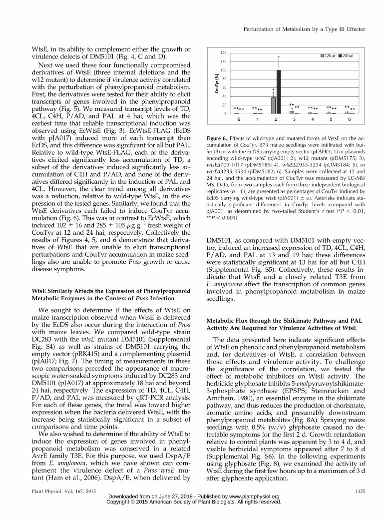
derivatives of WtsE (three internal deletions and thew12 mutant) to determine if virulence activity correlatedwith the perturbation of phenylpropanoid metabolism.First, the derivatives were tested for their ability to elicittranscripts of genes involved in the phenylpropanoidpathway (Fig. 5). We measured transcript levels of TD,4CL, C4H, P/AD, and PAL at 4 hai, which was theearliest time that reliable transcriptional induction wasobserved using EcWtsE (Fig. 3). EcWtsE-FLAG (EcDSwith pJA017) induced more of each transcript thanEcDS, and this difference was significant for all but PAL.Relative to wild-type WtsE-FLAG, each of the deriva-tives elicited significantly less accumulation of TD, asubset of the derivatives induced significantly less ac-cumulation of C4H and P/AD, and none of the deriv-atives differed significantly in the induction of PAL and4CL. However, the clear trend among all derivativeswas a reduction, relative to wild-type WtsE, in the ex-pression of the tested genes. Similarly, we found that theWtsE derivatives each failed to induce CouTyr accu-mulation (Fig. 6). This was in contrast to EcWtsE, whichinduced 102 6 16 and 285 6 105 mg g21 fresh weight ofCouTyr at 12 and 24 hai, respectively. Collectively theresults of Figures 4, 5, and 6 demonstrate that deriva-tives of WtsE that are unable to elicit transcriptionalperturbations and CouTyr accumulation in maize seed-lings also are unable to promote Pnss growth or causedisease symptoms.
WtsE Similarly Affects the Expression of PhenylpropanoidMetabolic Enzymes in the Context of Pnss Infection
We sought to determine if the effects of WtsE onmaize transcription observed when WtsE is deliveredby the EcDS also occur during the interaction of Pnsswith maize leaves. We compared wild-type strainDC283 with the wtsE mutant DM5101 (SupplementalFig. S4) as well as strains of DM5101 carrying theempty vector (pRK415) and a complementing plasmid(pJA017; Fig. 7). The timing of measurements in thesetwo comparisons preceded the appearance of macro-scopic water-soaked symptoms induced by DC283 andDM5101 (pJA017) at approximately 18 hai and beyond24 hai, respectively. The expression of TD, 4CL, C4H,P/AD, and PAL was measured by qRT-PCR analysis.For each of these genes, the trend was toward higherexpression when the bacteria delivered WtsE, with theincrease being statistically significant in a subset ofcomparisons and time points.We also wished to determine if the ability of WtsE to
induce the expression of genes involved in phenyl-propanoid metabolism was conserved in a relatedAvrE family T3E. For this purpose, we used DspA/Efrom E. amylovora, which we have shown can com-plement the virulence defect of a Pnss wtsE mu-tant (Ham et al., 2006). DspA/E, when delivered by
DM5101, as compared with DM5101 with empty vec-tor, induced an increased expression of TD, 4CL, C4H,P/AD, and PAL at 13 and 19 hai; these differenceswere statistically significant at 13 hai for all but C4H(Supplemental Fig. S5). Collectively, these results in-dicate that WtsE and a closely related T3E fromE. amylovora affect the transcription of common genesinvolved in phenylpropanoid metabolism in maizeseedlings.
Metabolic Flux through the Shikimate Pathway and PALActivity Are Required for Virulence Activities of WtsE
The data presented here indicate significant effectsof WtsE on phenolic and phenylpropanoid metabolismand, for derivatives of WtsE, a correlation betweenthese effects and virulence activity. To challengethe significance of the correlation, we tested theeffect of metabolic inhibitors on WtsE activity. Theherbicide glyphosate inhibits 5-enolpyruvoylshikimate-3-phosphate synthase (EPSPS; Steinrücken andAmrhein, 1980), an essential enzyme in the shikimatepathway, and thus reduces the production of chorismate,aromatic amino acids, and presumably downstreamphenylpropanoid metabolites (Fig. 8A). Spraying maizeseedlings with 0.5% (w/v) glyphosate caused no de-tectable symptoms for the first 2 d. Growth retardationrelative to control plants was apparent by 3 to 4 d, andvisible herbicidal symptoms appeared after 7 to 8 d(Supplemental Fig. S6). In the following experimentsusing glyphosate (Fig. 8), we examined the activity ofWtsE during the first few hours up to a maximum of 3 dafter glyphosate application.
Figure 6. Effects of wild-type and mutated forms of WtsE on the ac-cumulation of CouTyr. B73 maize seedlings were infiltrated with buf-fer (B) or with the EcDS carrying empty vector (pLAFR3; 1) or plasmidsencoding wild-type wtsE (pJA001; 2), w12 mutant (pDM5175; 3),wtsED709-1017 (pDM5189; 4), wtsED2935-3234 (pDM5184; 5), orwtsED3235-3534 (pDM5182; 6). Samples were collected at 12 and24 hai, and the accumulation of CouTyr was measured by LC-MS/MS. Data, from two samples each from three independent biologicalreplicates (n = 6), are presented as percentages of CouTyr induced byEcDS carrying wild-type wtsE (pJA001) 6 SD. Asterisks indicate sta-tistically significant differences in CouTyr levels compared withpJA001, as determined by two-tailed Student’s t test (*P , 0.01,**P , 0.001).
Plant Physiol. Vol. 167, 2015 1125
Perturbation of Metabolism by a Type III Effector
www.plantphysiol.orgon June 27, 2018 - Published by Downloaded from Copyright © 2015 American Society of Plant Biologists. All rights reserved.

First, we determined that, as predicted, glyphosateinhibited the WtsE-induced accumulation of CouTyr(Fig. 8B). We next sought to determine whether glyph-osate, by suppressing the production of CouTyr andother metabolites downstream from the shikimatepathway, could disrupt the virulence activity of WtsE.Indeed, the WtsE-dependent ability of wild-type Pnssstrain DC283 to induce disease symptoms and ionleakage and to grow to high levels in maize seed-lings was significantly impaired by glyphosate (Fig. 8,C–E).
Our results with glyphosate are consistent with met-abolic flux through the shikimate pathway being re-quired for the virulence activity of WtsE. However, weconsidered two alternative hypotheses. The first was thatthe inhibition of WtsE activity resulted from an adverseeffect of glyphosate on the bacteria. For example, theobserved ability of glyphosate to modestly compromisethe in planta growth of the wtsEmutant strain (DM5101)in this experiment might have resulted from an adverseeffect on the bacteria (Fig. 8E). To distinguish the effectsof glyphosate on the bacteria from its effects on the plant,we utilized a Pnss-susceptible maize line (PioneerP1615XR) that is resistant to glyphosate. Unlike someglyphosate-resistant plants, Pioneer P1615XR is not re-sistant due to altered uptake and subcellular partitioningof glyphosate, which could alter the exposure of thebacteria. Rather, these plants express a bacterial EPSPSenzyme that is glyphosate resistant and thus will moreefficiently produce chorismate in the presence ofglyphosate. Glyphosate did not significantly affect theability of DC283 to induce CouTyr production inPioneer P1615XR (Fig. 8B). Also, the effect of glyphosateon the ability of DC283 to cause disease symptoms andion leakage in Pioneer P1615XR was less than that in cvSeneca Horizon (Fig. 8, C and D). Thus, while glyph-osate may have a slight detrimental effect on the bac-teria, this effect is insufficient to account for theobserved inhibition of WtsE virulence activity insideplant cells.
The second hypothesis we considered was thatglyphosate inhibits WtsE activity by inhibiting aminoacid production and thus protein synthesis. Our previ-ous studies with cycloheximide (Ham et al., 2006, 2008)showed that inhibition of protein synthesis blocks theability of WtsE to elicit hypersensitive response-like celldeath in nonhost plants but has no effect on the abilityof WtsE to induce disease-like symptoms in susceptiblemaize seedlings. These results argue against glyphosateinhibiting WtsE activity by inhibiting protein synthesis.As another approach to alleviate this concern, we tested
Figure 7. Expression of five genes associated with phenolic metabo-lism in maize seedlings following infiltration with a Pnss wtsE nullmutant and a complemented strain. Tissue from cv Seneca Horizonseedlings was harvested at 13 and 19 hai with a wtsE null mutant ofPnss (DM5101) carrying empty vector (pRK415; white bars) or plasmid-borne wtsE (pJA017; gray bars), and the accumulation of TD (A), 4CL
(B), C4H (C), P/AD (D), and PAL (E) transcripts was assessed byquantitative PCR. Shown are average values and SE of qRT-PCRs donein triplicate for each of three biological replicates. Asterisks indicatesignificantly different expression levels relative to the infiltration ofDM5101 for a given gene at a single time point (two-tailed Student’st test, P , 0.05).
1126 Plant Physiol. Vol. 167, 2015
Asselin et al.
www.plantphysiol.orgon June 27, 2018 - Published by Downloaded from Copyright © 2015 American Society of Plant Biologists. All rights reserved.

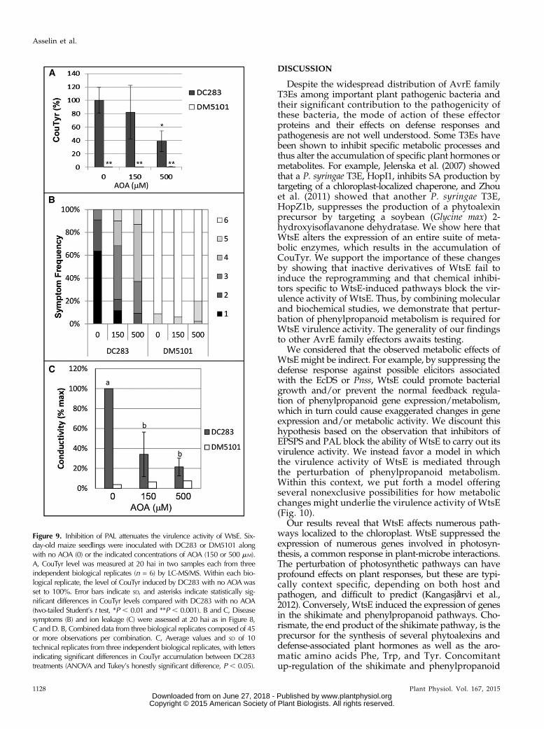
the effect of (aminooxy)acetic acid (AOA), an inhibitorof PAL (Fig. 8A; Carver et al., 1991; Peiser et al., 1998).Our results indicate that, when coinfiltrated into maizeseedling leaves along with Pnss strains, AOA couldinhibit the ability of DC283 to induce the productionof CouTyr (Fig. 9A). Furthermore, AOA attenuatedWtsE-induced disease symptoms and ion leakage (Fig.9, B and C). Thus, two independent inhibitors that blockWtsE-induced CouTyr production each also blocks thevirulence activity of WtsE.Recent work with Ustilago maydis showed that the
fungal effector Cmu1 is a chorismate mutase that at-tenuates SA production in infected maize (Djamei et al.,2011). We hypothesized that WtsE, through its
perturbation of phenolic metabolism, might similarlysuppress SA accumulation. To our surprise, Pnss, in aWtsE-dependent manner, induced strong accumulationof SA in maize seedling leaves (Supplemental Fig. S7).DC283-infiltrated leaves accumulated SA levels exceed-ing 2 ng g21 fresh weight at 12 hai, prior to the onset ofmacroscopic disease symptoms, and more than 10 ng g21
fresh weight at 24 hai, after the onset of water-soakedlesions. Relative to DC283, the wtsE mutant strainDM5101 induced significantly lower levels of SA at 12and 24 h. Infiltration of maize leaves with buffer orDM711 (a type III secretion-deficient strain of Pnss) in-duced no detectable increase in SA above the backgroundobserved in untouched seedlings.
Figure 8. Inhibition of the shikimate pathway attenuates the virulence activity of WtsE. A, Schematic showing the point ofaction of glyphosate (and AOA). Boxes contain multiple enzymatic steps and/or processes. B to D, Glyphosate-susceptiblecv Seneca Horizon (GS) or glyphosate-resistant Pioneer P1615XR (GR) maize seedlings were sprayed with buffer (2) or 0.5%(w/v) glyphosate (+). Six hours later, plants were infiltrated with wild-type Pnss strain DC283 or wtsE mutant strain DM5101.At 20 hai, assessments were made of CouTyr accumulation (B), symptom severity (C), and ion leakage (D). B, CouTyr levelwas measured in two samples each from three independent biological replicates (n = 6) by LC-MS/MS. Data are presented aspercentages of the appropriate varietal treatment with DC283 in the absence of glyphosate 6 SD. Asterisks indicate statis-tically significant differences in CouTyr levels compared with the DC283 treatment in the same variety and in the absence ofglyphosate, as determined by two-tailed Student’s t tests (*P , 0.05 and **P , 0.001). C, Symptom development of in-oculated plants was assessed on a scale from 1 to 6, with 1 representing plants with very severe symptoms, and 6 repre-senting nonsymptomatic plants. Shown are combined data from three biological replicates composed of 30 or moreobservations per combination. D, Average values and SD from three biological replicates for cv Seneca Horizon and twobiological replicates for Pioneer P1615XR. Asterisks indicate statistically significant differences compared with DC283without glyphosate as determined by two-tailed Student’s t tests (*P , 0.05 and **P , 0.001). E, Plants of cv Seneca Horizonsprayed 6 h earlier with buffer (2) or 0.5% (w/v) glyphosate (+) were infiltrated with a low titer of DC283 or DM5101, andbacterial growth was measured over the following 66 h. Shown are average values and SD of pooled data from three in-dependent biological replicates.
Plant Physiol. Vol. 167, 2015 1127
Perturbation of Metabolism by a Type III Effector
www.plantphysiol.orgon June 27, 2018 - Published by Downloaded from Copyright © 2015 American Society of Plant Biologists. All rights reserved.
DISCUSSION
Despite the widespread distribution of AvrE familyT3Es among important plant pathogenic bacteria andtheir significant contribution to the pathogenicity ofthese bacteria, the mode of action of these effectorproteins and their effects on defense responses andpathogenesis are not well understood. Some T3Es havebeen shown to inhibit specific metabolic processes andthus alter the accumulation of specific plant hormones ormetabolites. For example, Jelenska et al. (2007) showedthat a P. syringae T3E, HopI1, inhibits SA production bytargeting of a chloroplast-localized chaperone, and Zhouet al. (2011) showed that another P. syringae T3E,HopZ1b, suppresses the production of a phytoalexinprecursor by targeting a soybean (Glycine max) 2-hydroxyisoflavanone dehydratase. We show here thatWtsE alters the expression of an entire suite of meta-bolic enzymes, which results in the accumulation ofCouTyr. We support the importance of these changesby showing that inactive derivatives of WtsE fail toinduce the reprogramming and that chemical inhibi-tors specific to WtsE-induced pathways block the vir-ulence activity of WtsE. Thus, by combining molecularand biochemical studies, we demonstrate that pertur-bation of phenylpropanoid metabolism is required forWtsE virulence activity. The generality of our findingsto other AvrE family effectors awaits testing.
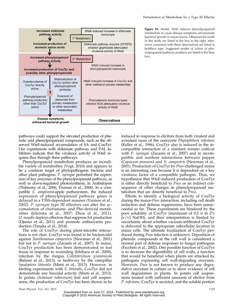
We considered that the observed metabolic effects ofWtsE might be indirect. For example, by suppressing thedefense response against possible elicitors associatedwith the EcDS or Pnss, WtsE could promote bacterialgrowth and/or prevent the normal feedback regula-tion of phenylpropanoid gene expression/metabolism,which in turn could cause exaggerated changes in geneexpression and/or metabolic activity. We discount thishypothesis based on the observation that inhibitors ofEPSPS and PAL block the ability of WtsE to carry out itsvirulence activity. We instead favor a model in whichthe virulence activity of WtsE is mediated throughthe perturbation of phenylpropanoid metabolism.Within this context, we put forth a model offeringseveral nonexclusive possibilities for how metabolicchanges might underlie the virulence activity of WtsE(Fig. 10).
Our results reveal that WtsE affects numerous path-ways localized to the chloroplast. WtsE suppressed theexpression of numerous genes involved in photosyn-thesis, a common response in plant-microbe interactions.The perturbation of photosynthetic pathways can haveprofound effects on plant responses, but these are typi-cally context specific, depending on both host andpathogen, and difficult to predict (Kangasjȁrvi et al.,2012). Conversely, WtsE induced the expression of genesin the shikimate and phenylpropanoid pathways. Cho-rismate, the end product of the shikimate pathway, is theprecursor for the synthesis of several phytoalexins anddefense-associated plant hormones as well as the aro-matic amino acids Phe, Trp, and Tyr. Concomitantup-regulation of the shikimate and phenylpropanoid
Figure 9. Inhibition of PAL attenuates the virulence activity of WtsE. Six-day-old maize seedlings were inoculated with DC283 or DM5101 alongwith no AOA (0) or the indicated concentrations of AOA (150 or 500 mM).A, CouTyr level was measured at 20 hai in two samples each from threeindependent biological replicates (n = 6) by LC-MS/MS. Within each bio-logical replicate, the level of CouTyr induced by DC283 with no AOA wasset to 100%. Error bars indicate SD, and asterisks indicate statistically sig-nificant differences in CouTyr levels compared with DC283 with no AOA(two-tailed Student’s t test, *P , 0.01 and **P , 0.001). B and C, Diseasesymptoms (B) and ion leakage (C) were assessed at 20 hai as in Figure 8,C and D. B, Combined data from three biological replicates composed of 45or more observations per combination. C, Average values and SD of 10technical replicates from three independent biological replicates, with lettersindicating significant differences in CouTyr accumulation between DC283treatments (ANOVA and Tukey’s honestly significant difference, P , 0.05).
1128 Plant Physiol. Vol. 167, 2015
Asselin et al.
www.plantphysiol.orgon June 27, 2018 - Published by Downloaded from Copyright © 2015 American Society of Plant Biologists. All rights reserved.
pathways could support the elevated production of phe-nolic and phenylpropanoid compounds, such as the ob-served WtsE-induced accumulation of SA and CouTyr.Our experiments with shikimate pathway and PAL in-hibitors indicate that the virulence activity of WtsE re-quires flux through these pathways.Phenylpropanoid metabolism produces an incredi-
ble variety of metabolites (Vogt, 2010) and appears tobe a common target of phytopathogenic bacteria andother plant pathogens. P. syringae perturbed the expres-sion of key enzymes of the phenylpropanoid pathway, aswell as down-regulated photosynthesis, in Arabidopsis(Thilmony et al., 2006; Truman et al., 2006). In a com-patible E. amylovora-apple pathosystem, the inducedexpression of phenylpropanoid pathway genes isdelayed in a T3SS-dependent manner (Venisse et al.,2002). P. syringae type III effectors can alter the ac-cumulation of chorismate- and Phe-derived metab-olites (Jelenska et al., 2007; Zhou et al., 2011).U. maydis deploys effectors that suppress SA production(Djamei et al., 2011) and promote anthocyanin pro-duction (Tanaka et al., 2014).The role of CouTyr during plant-microbe interac-
tions is not clear. CouTyr was found to be bactericidalagainst Xanthomonas campestris (Newman et al., 2001)but not to P. syringae (Zacarés et al., 2007). In maize,CouTyr production has been demonstrated in leaftissue in response to wounding (Ishihara et al., 2000),infection by the fungus Colletotrichum graminicola(Balmer et al., 2013), or herbivory by the caterpillarSpodoptera littoralis (Marti et al., 2013). However, infeeding experiments with S. littoralis, CouTyr did notdemonstrate any biocidal activity (Marti et al., 2013).In potato (Solanum tuberosum) leaf and cell suspen-sions, the production of CouTyr has been shown to be
induced in response to elicitors from both virulent andavirulent races of the oomycete Phytophthera infestans(Keller et al., 1996). CouTyr also is induced in the in-compatible interaction of a resistant tomato cultivarwith P. syringae (Zacarés et al., 2007) and in incom-patible and nonhost interactions between pepper(Capsicum annuum) and X. campestris (Newman et al.,2001). Production of CouTyr by Pnss-challenged maizeis an interesting case because it is dependent on a keyvirulence factor of a compatible pathogen. Thus, wehypothesize that WtsE-induced production of CouTyris either directly beneficial to Pnss or an indirect con-sequence of other changes in phenylpropanoid me-tabolism that are directly beneficial to Pnss.
Efforts to identify a biological activity of CouTyrduring the maize-Pnss interaction, including cell deathinduction and defense suppression, have been unsuc-cessful so far. These experiments are hindered by thepoor solubility of CouTyr (maximum of 0.2 M in 2%[v/v] NaOH), and their interpretation is limited byuncertainty about whether vacuum-infiltrated CouTyris delivered to the appropriate subcellular location inmaize cells. The ultimate localization of CouTyr pro-duced during Pnss infection is unknown. Deposition ofphenolic compounds at the cell wall is considered anormal part of defense responses to fungal pathogens(Facchini et al., 2002). One possible function of CouTyris to decrease the digestibility of cell walls, a functionthat would be beneficial when plants are attacked bypathogens expressing cell wall-degrading enzymes.However, Pnss is not known to produce any degra-dative enzymes in culture or to show evidence of cellwall degradation in planta. In potato cell suspen-sions treated with sufficient amounts of elicitor fromP. infestans, CouTyr is secreted, and the soluble portion
Figure 10. Model. WtsE induces phenylpropanoidmetabolism to cause disease symptoms and promotebacterial growth in maize leaves. Observations madein this study are listed in the box to the right. Infer-ences consistent with these observations are listed inboldface type. Suggested modes of action of phe-nylpropanoid pathway products are listed in the bluebox.
Plant Physiol. Vol. 167, 2015 1129
Perturbation of Metabolism by a Type III Effector
www.plantphysiol.orgon June 27, 2018 - Published by Downloaded from Copyright © 2015 American Society of Plant Biologists. All rights reserved.
is almost 50 times greater than that in the cell wall orcytoplasm. An observation that is possibly related tothe cell death induced by WtsE is that secretion ofCouTyr by elicitor-treated cell suspensions is accom-panied by their browning (Keller et al., 1996). Themechanism by which CouTyr moves out of the plantcell may involve transport carrier vesicles or transportcarrier proteins. Given the potential for WtsE to affectvesicle traffic through the mimicry of GEFs, the ma-nipulation of CouTyr localization may affect its accu-mulation and alter its fate in Pnss-infected maize andits role in Pnss pathogenesis.
Rather than possessing biological activity, CouTyrmay indirectly affect the maize-Pnss interaction. Theselective misregulation of genes involved in phenolicand phenylpropanoid metabolism could be a virulencestrategy to promote or divert carbon flow into productsbeneficial or detrimental, respectively, to bacterial sur-vival (Yao et al., 1995). For example, Tin2 fromU. maydisis hypothesized to promote anthocyanin production todivert metabolites away from cell wall lignification thatimpedes the fungal infection (Tanaka et al., 2014). Sim-ilarly, WtsE-induced up-regulation of enzymes in thephenylpropanoid pathway, including those involved in
the production of CouTyr and perhaps other hydrox-ycinnamic acid amides, could have the effect of directingcarbon away from the biosynthesis of defense-associatedmetabolites.
The results of this work have revealed several pre-viously unknown effects of WtsE on maize physiology.WtsE elicits major disruptions in several pathways,each focused in the chloroplast. These changes includethe down-regulation of photosynthesis and the up-regulation of the shikimate and phenylpropanoidpathways. The latter causes a substantial increase inthe production of the hydroxycinnamic acid amideCouTyr. Up-regulation of the shikimate pathway andphenylpropanoid pathways is directly linked by car-bon flow. Although the proximity of photosynthesisenzymes to those involved in the shikimate and phe-nylpropanoid pathways is intriguing, it is unclearwhether down-regulating photosynthesis has a directimpact on these or other pathways. These findingspoint to future study of WtsE, and perhaps other AvrEfamily T3Es, to examine how the accumulation andlocalization of CouTyr and other chorismate-derivedmetabolites affect maize and Pnss cells during theirinteraction.
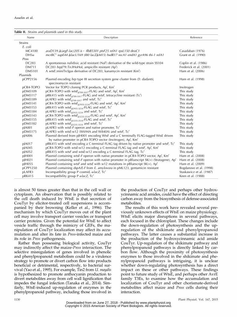
Table II. Strains and plasmids used in this study.
Name Description Reference
StrainsE. coliMC4100 araD139 D(argF-lac)205 l2 flbB5301 ptsF25 relA1 rpsL150 deoC1 Casadaban (1976)DH5a mcrBC+ supE44 DlacU169 (f80 lacZDM15) hsdR17 recA1 endA1 gyrA96 thi-1 relA1 Grant et al. (1990)
PnssDC283 A spontaneous nalidixic acid resistant (Nalr) derivative of the wild-type strain SS104 Coplin et al. (1986)DM711 DC283 hrpJ79::Tn3HoHoI, ampicilln resistant (Apr) Frederick et al. (2001)DM5101 A wtsE::miniTn5gus derivative of DC283, kanamycin resistant (Kmr) Ham et al. (2006)
PlasmidspCPP2156 Plasmid encoding hrp type III secretion system gene cluster from D. dadantii,
specinomycin resistantHam et al. (1998)
pCR4-TOPO Vector for TOPO cloning PCR products, Apr, Kmr InvitrogenpDM5109 pCR4-TOPO with wtsED709-1017-FLAG and wtsF, Apr, Kmr This studypDM5117 pRK415 with wtsED709-1017-FLAG and wtsF, tetracycline resistant (Tcr) This studypDM5189 pLAFR3 with wtsED709-1017 and wtsF, Tcr This studypDM5141 pCR4-TOPO with wtsED2935-3234-FLAG and wtsF, Apr, Kmr This studypDM5153 pRK415 with wtsED2935-3234-FLAG and wtsF, Tcr This studypDM5184 pLAFR3 with wtsED2935-3234 and wtsF, Tcr This studypDM5143 pCR4-TOPO with wtsED3235-3534-FLAG and wtsF, Apr, Kmr This studypDM5155 pRK415 with wtsED3235-3534-FLAG and wtsF, Tcr This studypDM5182 pLAFR3 with wtsED3235-3534 and wtsF, Tcr This studypJA001 pLAFR3 with wtsE-F operon and native promoter, Tcr This studypDM5175 pLAFR3 with wtsE-w12 (W694A and W840A) and wtsF, Tcr This studypJA006 Plasmid derived from pJH001 encoding WtsF and a C terminally FLAG-tagged WtsE driven
by native promoter in pCR4-TOPO vector (Invitrogen), Apr, KmrThis study
pJA017 pRK415 with wtsE encoding a C-terminal FLAG tag driven by native promoter and wtsF, Tcr This studypJA045 pCR4-TOPO with wtsE-w12 encoding a C-terminal FLAG tag and wtsF, Apr, Kmr This studypJA052 pRK415 with wtsF and wtsE-w12 encoding a C-terminal FLAG tag, Tcr This studypJH001 Plasmid containing wtsE-F operon with native promoter in pCR4-TOPO vector, Apr, Kmr Ham et al. (2008)pJH021 Plasmid containing wtsE-F operon with native promoter in pBluescript SK(+) (Stratagene), Apr Ham et al. (2008)pJH055 Plasmid containing wtsF and wtsE with w12 mutations in pBluescript SK(+), Apr Ham et al. (2009)pCPP1250 Plasmid containing dspA/E-F from E. amylovora in pML123, gentamicin resistant Bogdanove et al. (1998)pLAFR3 Incompatibility group P cosmid, alacZ, Tcr Staskawicz et al. (1987)pRK415 Incompatibility group P alacZ, Tcr Keen et al. (1988)
1130 Plant Physiol. Vol. 167, 2015
Asselin et al.
www.plantphysiol.orgon June 27, 2018 - Published by Downloaded from Copyright © 2015 American Society of Plant Biologists. All rights reserved.

MATERIALS AND METHODS
Bacterial Strains, Plasmids, and Growth Conditions
Strains and plasmids used in this study are listed in Table II. Bacteria wereroutinely grown in Luria-Bertani (LB) broth or agar (Sambrook and Russell,2001) supplemented with the appropriate antibiotics and grown at 28°C forPantoea stewartii ssp. stewartii or 37°C for Escherichia coli.
For vacuum infiltrations and inoculations of maize (Zea mays) seedlings,overnight cultures of E. coli or Pnss were centrifuged at 2,500g for 10 min, andbacteria were resuspended in 10 mM potassium phosphate buffer, pH 7.2,containing 0.2% (v/v) Tween 40. For E. coli, the optical density at 540 nm wasadjusted to 1. For Pnss, the optical density at 540 nm was adjusted to 0.57(equivalent to 1 3 109 colony-forming units [cfu] mL21). This 1 3 109 cfu mL21
suspension was used for all vacuum infiltration-based experiments, with theexception of the growth curve in Figure 9E, for which the bacteria were dilutedto 1 3 106 cfu mL21 prior to infiltration. For whorl inoculations and associatedgrowth and virulence measurements (Fig 4, C and D), the bacteria were dilutedto 1 3 108 cfu mL21 prior to inoculation.
Construction of Plasmids
pJA001 was created by digesting pJH021 DNA (Ham et al., 2009) with re-striction enzymes EcoRI and BamHI (Invitrogen) and ligating the resulting 6.4-kbfragment (containing the wtsE-F operon and its native promoter from Pnss) intothe vector pLAFR3 (Staskawicz et al., 1987), also cut with EcoRI and BamHI.
Plasmid pJA017 encodes WtsF and C terminally FLAG-tagged, but otherwiseunmutated, WtsE (WtsE-FLAG) in the vector pRK415. In making pJA017, pJH021was used as template in separate PCRs with primers 59-ACAGCCTCAATGC-TAAACTCACAGAAG-39 (wtsE F1386A-F) with 59-TCATTTGTCGTCATCG-TCTTTGTAGTCGCTTTTCATTTCAAACCCTTC-39 (wtsE-FLAG N R) and59-GACTACAAAGACGATGACGACAAATGAGGTTATGATTAATTCATC-39(wtsE-FLAG C F) with the M13 Reverse universal primer 59-AGCGGATAA-CAATTTCACACAGG-39. The products were used together as template for PCRwith external primers wtsE F1386A-F and M13 Reverse. The newly createdfragment was then digested with BamHI and SstI and ligated into pJH001, whichharbors the same insert as pJH021 (Ham et al., 2008) cut with the same enzymes,thus creating the intermediate plasmid JA006 in the high copy number back-ground of pCR4-TOPO (Invitrogen). The entire wtsE-F region was then excisedfrom pJA006 using EcoRI and BamHI and ligated into pRK415 to create pJA017.
Plasmid pDM5153 contains wtsED2935-3234-FLAG and wtsF. Two separate PCRswere performed using pJH021 as template and primers 59-GCACTTCACAA-CCTTTCCGAT-39 (wtsE outer 3-F) paired with 59-GTTAAAGCTTTGTATTT-GAAGATTGTATGCAATATTTTTCAG-39 (wtsE d8-R) and 59-GCATACAATC-TTCAAATACAAAGCTTTAACGTTAACCG-39 (wtsE d8-F) paired with59-ACGACCAAACGTGAGATTTATAAGC-39 (wtsE outer 3-R). The resulting pro-ducts were then used together as template in a PCR using wtsE outer 3-F andwtsE outer 3-R as primers. The PCR product was cut with PstI and KpnI andligated into pJA006 cut with the same enzymes, creating the intermediateplasmid pDM5141. The insert from pDM5141 was excised using EcoRI andBamHI and cloned into pRK415, creating pDM5153.
Plasmid pDM5155 contains wtsED3235-3534-FLAG and wtsF. Two separatePCR products were created using wtsE outer 3-F paired with 59-ATG-TAGCTGGGCTATCTGTGCCGCTTTACTGATCGG-39 (wtsE d9-R) and59-ATCAGTAAAGCGGCACAGATAGCCCAGCTACAT-39 (wtsE d9-F) withwtsE outer 3-R. The resulting products were then used together as template inPCR using wtsE outer 3-F and wtsE outer 3-R as primers. The resulting PCRproduct was cut with PstI and KpnI and ligated into pJA006 cut with the sameenzymes, creating the intermediate plasmid pDM5143. The insert frompDM5143 was excised using EcoRI and BamHI and cloned into pRK415, cre-ating pDM5155.
Plasmid pDM5117 contains wtsED709-1017-FLAG and wtsF. To construct thepartial deletion of wtsE, we took advantage of two naturally occurring re-striction sites: ClaI (bases 502–507 of the wtsE-coding DNA sequence) and NdeI(bases 1,018–1,023). A fragment representing bases 315 to 708 of wtsE wasamplified via PCR using the primers 59-CAGTCGGATTCGGAGACCCAC-39(wtsE outer 1-F) and 59-GCGCCATATGATCAAGATGCCGCTGCGGAAG-39(wtsE d1-R NdeI). This fragment was then digested with the enzymes ClaI andNdeI, creating a 206-bp fragment that was ligated into pJA006 cut with thesame enzymes. This created the intermediate plasmid pDM5109. The insertfrom pDM5109 was then cut with EcoRI and BamHI and ligated into pRK415to create pDM5117.
Plasmid pJA052 encodes wtsE-FLAG with the w12 mutations (i.e. W694Aand W840A, mutations of both WxxxE motifs) and wtsF. In making pJA052, a1,836-bp KpnI-to-PstI fragment containing wtsE w12 was cut from pJH055(Ham et al., 2009), gel purified, and ligated into pJA006 cut with the sameenzymes, creating the intermediate plasmid pJA045. Plasmid pJA045 wasdigested with EcoRI and BamHI and ligated into pRK415 cut with the sameenzymes, creating pJA052.
To construct plasmid pDM5189 containing wtsED709-1017 and wtsF, afragment of WtsE (155–708 bp) was amplified from pJH021 using primers59-CCAGTATGCCCGCTATTCGTCAGC-39 (wtsE outer F) and wtsE d1-R NdeI.The PCR product was digested with BspE and NdeI and ligated to pJH021digested with the same enzymes to create an intermediate plasmid, which wasthen digested with EcoRI and BamHI and cloned into pLAFR3 to createpDM5189.
To construct plasmid pDM5184 containing wtsED2935-3234 and wtsF, thesame PCRs were performed as to construct plasmid pDM5153. The resultingPCR product was digested with PstI and KpnI, cloned into pJH021 digested bythe same enzymes to create an intermediate plasmid, and the fragment createdby digestion with EcoRI and BamHI was cloned into pLAFR3.
To construct plasmid pDM5182 containing wtsED3235-3534 and wtsF, thesame PCRs were performed as to construct plasmid pDM5155. The resultingPCR product was digested with PstI and KpnI, cloned into pJH021 to create anintermediate plasmid, and the fragment created by digestion with EcoRI andBamHI was cloned into pLAFR3.
To construct plasmid pDM5157, a fragment containing wtsE with the w12mutations and wtsF was released from pJH055 by EcoRI and BamHI and li-gated into pLAFR3.
PCR products to be used as template in further PCRs were resolved byagarose gel electrophoresis and excised from the gel prior to purification. PCRproducts were cleaned before digestion, and digested DNAwas cleaned beforeligation. DNA was cleaned using the QIAquick PCR purification kit (Qiagen).Digested vectors were routinely treated with calf intestinal alkaline phos-phatase and cleaned prior to ligation. The sequences of DNA fragments clonedusing PCRwere confirmed by DNA sequencing at the Plant-Microbe GenomicsFacility at Ohio State University. Manipulation of DNA was performedaccording to standard techniques (Ausubel et al., 1997).
Secretion Assays
Overnight cultures of Pnss in LB broth were pelleted by centrifugation,washed twice, and suspended in hrp-inducing medium (Ahmad et al., 2001)supplemented with 1 mM isopropylthio-b-galactoside and appropriate anti-biotics. hrp-inducing medium cultures were grown overnight at 22°C andadjusted to equal optical densities before harvesting. Pellet and supernatantfractions were separated by centrifugation at 20,200g. To the supernatantfraction, phenylmethylsulfonyl fluoride was used at a concentration of 1 to2 mM to prevent protein degradation. TCA was then added to a concentrationof 10% (v/v), and the fraction was incubated on ice overnight to precipitateproteins. Proteins were pelleted by centrifugation at 20,200g for 30 min at 4°Cand washed in acetone. Bacterial cell and TCA-precipitated protein pelletswere resuspended in 1/50th and 1/350th of the original culture or supernatantvolume, respectively, in 20 mM Tris, pH 7.5, supplemented with 1 mM phe-nylmethylsulfonyl fluoride.
Proteins were separated by SDS-PAGE using a 10% (w/v) resolving gel.Proteins were blotted onto Immobilon-P polyvinylidene difluoride (Millipore)using the Mini-Trans Blot Cell (Bio-Rad) according to the manufacturer’s in-structions. Blots were developed with a-FLAG M2 and a-b-galactosidaseprimary antibodies and anti-mouse (Jackson ImmunoResearch) and anti-rabbit (Amersham, GE Healthcare) secondary antibodies, respectively.
Plant Growth, Infiltration, and Inoculation
Seeds of sweet maize (variety rugosa) ‘Seneca Horizon’ or maize lines Pi-oneer P1615XR (a glyphosate-resistant hybrid) and B73 were sown in 4.5-inch-diameter pots in Metro-Mix 360 (Sun Gro Horticulture), eight to 10 seeds perpot, and were watered daily for 5 d. Plants were grown in a growth chambermaintained at 30°C with a daily cycle of 18 h of daylight (85 mE m22 s21) and6 h of darkness.
Six-day-old maize seedlings were vacuum infiltrated with bacteria as de-scribed above. A humidifier was placed in the growth chamber to increase therelative humidity to greater than 65%. For most infiltrations, the humidifier wasplaced in the chamber 2 d prior to infiltration. For plants sprayed with
Plant Physiol. Vol. 167, 2015 1131
Perturbation of Metabolism by a Type III Effector
www.plantphysiol.orgon June 27, 2018 - Published by Downloaded from Copyright © 2015 American Society of Plant Biologists. All rights reserved.

glyphosate, humidifiers were placed in the chamber the morning of infiltration.Plants lacking a visible second true leaf or that appeared unhealthywere culled.Soil was covered, and the pots were inverted over beakers containing inoculumor buffer so that the seedlings were submerged. Seedlings were then vacuuminfiltrated three times, 5 min each time, at a minimum vacuum pressure of 200mmHg. First true leaves from infiltrated plants were examined against the lightimmediately following infiltration, and those appearing well infiltrated werelabeled for harvesting and/or scoring at later times. WtsE-induced diseasesymptomswere scored on a scale from 1 to 6 (with 1 indicating most severe and6 indicating no symptoms; Supplemental Fig. S8). At harvest, 3.5- to 5-cmlengths (including the tips) of the first true leaves were tested for ion leakageor immediately frozen in liquid nitrogen and stored at 280°C. The remaining,unharvested leaves were observed at 24 to 36 hai, and the experiment wascarried forward only if WtsE-induced symptoms were robust.
For whorl inoculation of seedlingswith Pnss strains (Fig. 4, C andD), plantswerenot watered for 2 d prior to inoculation. To inoculate the plants, 50 mL of inoculumwas pipetted directly into the whorl of 6-d-old maize seedlings and allowed to dry.After drying, plants were watered and maintained in the growth chamber untilgrowth measurement or symptom assessment was done 3 d following inoculation.Pnss-induced disease symptoms following whorl inoculation were rated on a scalefrom 0 to 3 (with 3 indicating most severe and 0 indicating no symptoms) as de-scribed previously (Ahmad et al., 2001; Ham et al., 2008).
Ion Leakage
Following bacterial infiltration, the tips of first true leaves were removed atthe indicated times. Tissue alongside a ruler was optically scanned, and thentwo leaf tips per vial were placed in 30 mL of milli-Q purified water (Millipore)in glass screw-cap tubes. Because leaf pieces tended to float out of the water andcling to the sides of the tubes, plungers from 10-mL syringes were added to thetubes to raise thewater level and gently force leaves to remain in the liquid. Leaftissue was allowed to sit in water for approximately 30 min, vials were shakento homogenize liquid, and then electrical conductivity was measured using aCond 330i (WTW). Leaf area was determined using ImageJ (Schneider et al.,2012), and conductivity per cm2 of leaf area was calculated for each sample.
Chemical Inhibitors
For glyphosate treatment, 6-d-old cv Seneca Horizon maize seedlings weresprayed with 0% or 0.5% (w/v) glyphosate (Syngenta) in 0.2% (v/v) Tween 40.After 5 h, plants were inverted in water for 10 min to wash away glyphosateremaining on leaves. Plants then were infiltrated with Pnss strains, and tissue wascollected, as described above.
For AOA treatment, 6-d-old cv Seneca Horizon maize seedlings werevacuum infiltrated with inoculum containing Pnss strains, as described above,and a final concentration of 0, 150, or 500 mM AOA (Sigma-Aldrich). A 25 mM
stock solution of AOA in water was produced fresh on the day of the ex-periment and added to the inoculum.
Recovery and Enumeration of Bacteria from Leaf Tissue
To assess bacterial titer in plant tissues, maize seedlings were cut off at thesoil line (Fig. 4C) and placed in 5 mL of 10 mM potassium phosphate buffer,pH 7.2, or leaf discs were harvested (Fig. 8E) and placed in 200 mL of the samebuffer in a microcentrifuge tube. Maize tissue was thoroughly macerated us-ing a Power Gen 700 homogenizer (Fisher Scientific) or microfuge grindingtip. Supernatant from the grinding was serially diluted, and the dilutions werespotted onto LB agar plates supplemented with appropriate antibiotics. Thecfu per milliliter of supernatants was calculated and compared with the initialplated inoculum (Fig. 4C), or a measurement was made shortly after infil-tration (Fig. 8E) to assess bacterial multiplication in planta.
Microarray Tissue Collection and Preparation of RNA
Seedlings of cv Seneca Horizon were vacuum infiltrated with EcWtsE (E. coliMC4100[pCPP2156, pJA001]) or EcDS with empty vector (E. coli MC4100[pCPP2156, pLAFR3]), and tissue was collected at 6 hai. Tissue for microarrayexperiments represented four biological replicates. Plants for one biologicalreplicate were grown, infiltrated, and harvested before the seeds for the fol-lowing biological replicate were sown. Tissue from the first true leaves of fourseparate plants that had been infiltrated and harvested on the same day was
pooled to form each biological replicate. Tissue was ground with a mortar andpestle under liquid nitrogen, and RNA was isolated using the RNAqueous kit(Ambion) according to kit instructions. RNA yield and purity were estimatedusing a NanoDrop (Thermo Fisher Scientific) and visualized by agarose gelelectrophoresis. RNA from the four separate biological replicates was used forhybridization of four separate two-channel microarrays, with RNA fromtreatment plants (those infiltrated with EcWtsE) being compared with thatfrom control plants (those infiltrated with EcDS) within each array. RNA wassent to the Maize Oligonucleotide Array Project at the University of Arizonafor RNA amplification, RNA labeling, hybridization, and reading of thearrays. Array protocols were done according to Galbraith et al. (2011). Arrayswere scanned using a GenePix 4000AL microarray scanner, and raw data wereextracted with the GenePix 6.0 program and analyzed according to Jandovaet al. (2012).
Statistical analysis of microarrays also was performed at the Maize Oli-gonucleotide Array Project. Statistical analysis was performed in R (http://www.r-project.org/) using the Limma statistical package. Loess normalizationwas used to obtain the average intensity of each gene from the four arrays.Significant differences between treatment and control groups were assessedusing Student’s t test (a = 0.02). Significant differences in dye effect wereassessed using Student’s t test (a = 0.05).
Annotation of Microarray Results and Analysis of GOTerms and KEGG Pathways
GO terms were assigned to the differentially expressed probes using theAgriGO platform (Du et al., 2010); AgriGO was then used to do a hyper-geometric distribution test with Yekutieli correction (false discovery rate un-der dependency) to identify overrepresented terms in the groups of repressedor induced probes when compared with GO terms assigned to all microarrayprobes. Probes also were assigned to maize KEGG genes and KEGG pathwayterms using Kobas 2.0 (mapping e , 0.01; Xie et al., 2011). A hypergeometricdistribution test with Yekutieli correction also was applied to identify over-represented KEGG pathways.
Quantitative PCR
Genes involved in the phenylpropanoid pathway were chosen for exami-nation by qRT-PCR. The genes chosen were TD (MaizeGDB gene identifierGRMZM2G093125), P/AD (GRMZM2G437912), 4CL (GRMZM2G174732),PAL (GRMZM2G170692), and cinnamic acid to 4-coumaric acid(GRMZM2G147245). Primers for actin (GRMZM2G126010) were used as acontrol for normalizing results. Because it was unclear from microarray resultswhich possible splice variants were altered in expression between treatmentand control plants, PCR primers were not designed to amplify productsspanning introns (Supplemental Table S3).
First true leaves from bacteria-infiltrated plants were collected at 2, 4, 6, 13,or 19 hai. Two leaves were pooled for each biological replicate. RNA wasisolated using the RNAqueous kit, and RNA quality was checked viaNanodrop. RNA preparations were treated with DNase I AmplificationGrade (Invitrogen) according to the manufacturer’s instructions. Productionof complementary DNA was done according to the Reverse TranscriptionSystem (Promega). Quantitative PCR assays were performed on two bio-logical replicates, each in triplicate. Reactions were 25 mL in volume and runon 96-well plates in the iQ5 Multicolor Real-Time PCR Detection System (Bio-Rad) using the following program: 95°C for 3 min; 40 cycles of 95°C for 30 s,55°C for 30 s, and 72°C for 30 s; followed by melting-curve analysis from 55°C to 95°C in 30-s cycles. Data were analyzed using the iQ5 Optical SystemSoftware.
Metabolite Extraction from Maize Seedling Leaves
Leaf tip samples were collected in 1.5-mL centrifuge tubes (DOT Scientific;part 609) and ground in liquid nitrogen using a plastic pestle. Approximately 22mg of each sample was transferred to a second 1.5-mL centrifuge tube andground a second time. One milliliter of 80% (v/v) methanol (LC-MS/MS grade;Fisher Scientific; part A456-1) was added into each sample, along with 10 mLof 50 ng mL21 formononetin internal standard (Sigma-Aldrich) dissolved in100% LC-MS/MS-grade methanol. Samples were vortexed, extracted at 4°Cfor 20 min on a nutator, and finally centrifuged at 12,000g for 15 min at 4°C.The resulting supernatants were then filtered through 0.2-mm nylon filters anddiluted 100-fold with 100% methanol.
1132 Plant Physiol. Vol. 167, 2015
Asselin et al.
www.plantphysiol.orgon June 27, 2018 - Published by Downloaded from Copyright © 2015 American Society of Plant Biologists. All rights reserved.

LC-MS/MS Analyses
During LC-MS/MS analyses, samples were stored at a temperature of 4°Cin a chilled autosampler chamber. For quantification of CouTyr, 1 mL of eachsample was injected into an Agilent 1260 Infinity LC system, and compoundswere separated using an Agilent Poroshell 120 EC-C18 (3.5 3 50 mm, 2.7 mm)column at 30°C and an acidified water:methanol buffer system (buffer A,0.1% [v/v] acetate and 5% [v/v] methanol in water; buffer B, 0.1% [v/v]acetate in water). Gradient conditions were as follows: 1-min hold at 20% B, 3 minat 20% to 80% B, 5 min at 80% to 100% B, 2-min hold at 100% B, and back to 20%B in 1.5 min. Eluted compounds were further separated and quantified using anAgilent G6460 triple quadrupole dual mass spectrometer equipped with anelectrospray ionization source. LC-MS/MS settings were as follows: gas temper-ature of 250°C, gas flow rate of 10 L min21, nebulizer pressure of 60 p.s.i., sheathgas flow of 12 Lmin21, sheath gas temperature of 400°C, capillary voltage of 3,000 V,and nozzle voltage of 700 V. Compounds were quantified using positive ionmode, with four mass transitions (one quantification transition and three con-firmatory qualification transitions) monitored for each compound of interest(Supplemental Table S2), and verified through comparison with authenticstandards. The limit of quantitation for CouTyr for this methodwas calculated tobe 51 ng g21 tissue harvested.
Ultra High-Performance Liquid ChromatographyMeasurement of SA
Leaf tissues were weighed, frozen on liquid nitrogen, and ground with two3.2-mm stainless steel beads in each tube using a Qiagen TissueLyser. SA wasextracted twice with 500 mL per 100 mg fresh weight of 100% methanolcontaining 1 mM butylated hydroxyanisole (Sigma-Aldrich). Each extractionwas for 24 h in the dark at 4°C, and the clear supernatant was removed aftercentrifugation at 13,000 rpm for 15 min in a tabletop microcentrifuge. The twopooled extractions were stored in darkness at 220°C. Ultra high-performanceliquid chromatography analysis was done within 1 week of extraction. Ex-tracts were run through a 0.2-mm, 2.1-mm guard filter (Waters) prior tochromatographic separation on a 2.1- 3 100-mm, 1.7-mm ACQUITY UPLCRP18 BEH shield column (Waters) using the Acquity UPLC system (Waters).Column temperature was set to 30°C, and the sample manager was main-tained at 4°C. HPLC-grade water with 2% (v/v) acetic acid (A) and HPLC-grade methanol with 2% (v/v) acetic acid (B) were used as solvents. An 11.33-min gradient program with a flow rate of 0.3 mL min21 was used: 95% A and5% B at 0 min, 90% A and 10% B at 0.67 min, 50% A and 50% B at 6.67 min,20% A and 80% B at 10 min, 100% B at 10.67 min, and 95% A and 5% B at 11.33min. SA was detected using fluorescence at emission wavelength of 400 nm.Peak areas of SA in standards and tissue samples were normalized to thebutylated hydroxyanisole internal standard. Calibration curves were plottedby peak areas of a serial diluted standard of commercial SA (Sigma-Aldrich)with a correlation coefficient higher than 0.999.
Supplemental Data
The following supplemental materials are available.
Supplemental Figure S1. Symptoms and ion leakage in maize leaves infil-trated with EcWtsE.
Supplemental Figure S2. Statistical significance versus fold change ofmaize transcripts with EcWtsE or EcDS.
Supplemental Figure S3. Standard curves for quantification of CouTyr.
Supplemental Figure S4. WtsE-induced phenolic metabolism gene expres-sion in maize seedlings.
Supplemental Figure S5. DspA/E-induced phenolic metabolism gene ex-pression in maize seedlings.
Supplemental Figure S6.Macroscopic effects of glyphosate onmaize seedlings.
Supplemental Figure S7. Quantification of salicylic acid induction follow-ing infiltration of maize seedlings with Pnss.
Supplemental Figure S8. Leaves displaying virulence phenotypes asscored in Figures 8C and 9B.
Supplemental Table S1. WtsE elicited significantly altered expression ofnumerous genes from maize seedlings in microarray experiments.
Supplemental Table S2. Mass spectrometry settings for CouTyr and for-mononetin quantitations.
Supplemental Table S3. Primers used in quantitative PCR experiments.
ACKNOWLEDGMENTS
We thank Terry Graham and Mike Kelly for help with LC-MS/MS, EnricoBonello for help with ultra high-performance liquid chromatography, KatieWilkins for help with data sorting, and Jason Parrish for assistance withglyphosate application and glyphosate-resistant maize.
Received November 3, 2014; accepted January 24, 2015; published January 29,2015.
LITERATURE CITED
Ahmad M, Majerczak DR, Pike S, Hoyos ME, Novacky A, Coplin DL(2001) Biological activity of harpin produced by Pantoea stewartii subsp.stewartii. Mol Plant Microbe Interact 14: 1223–1234
Ahmad S, Veyrat N, Gordon-Weeks R, Zhang Y, Martin J, Smart L,Glauser G, Erb M, Flors V, Frey M, et al (2011) Benzoxazinoid me-tabolites regulate innate immunity against aphids and fungi in maize.Plant Physiol 157: 317–327
Ahuja I, Kissen R, Bones AM (2012) Phytoalexins in defense againstpathogens. Trends Plant Sci 17: 73–90
Alto NM, Shao F, Lazar CS, Brost RL, Chua G, Mattoo S, McMahon SA,Ghosh P, Hughes TR, Boone C, et al (2006) Identification of a bacterial typeIII effector family with G protein mimicry functions. Cell 124: 133–145
Ausubel FM, Brent R, Kingston RE,More DD, Seidman JG, Smith JA, Struhl K(1997) Short Protocols in Molecular Biology: A Compendium of Methods fromCurrent Protocols in Molecular Biology, Ed 3. John Wiley & Sons, New York
Badel JL, Shimizu R, Oh HS, Collmer A (2006) A Pseudomonas syringae pv.tomato avrE1/hopM1 mutant is severely reduced in growth and lesionformation in tomato. Mol Plant Microbe Interact 19: 99–111
Balmer D, de Papajewski DV, Planchamp C, Glauser G, Mauch-Mani B(2013) Induced resistance in maize is based on organ-specific defenceresponses. Plant J 74: 213–225
Beckman CH (2000) Phenolic-storing cells: keys to programmed cell deathand periderm formation in wilt disease resistance and in general defenceresponses in plants? Physiol Mol Plant Pathol 57: 101–110
Bednarek P, Pislewska-Bednarek M, Svatos A, Schneider B, Doubsky J,Mansurova M, Humphry M, Consonni C, Panstruga R, Sanchez-ValletA, et al (2009) A glucosinolate metabolism pathway in living plant cellsmediates broad-spectrum antifungal defense. Science 323: 101–106
Block A, Alfano JR (2011) Plant targets for Pseudomonas syringae type III effec-tors: virulence targets or guarded decoys? Curr Opin Microbiol 14: 39–46
Bogdanove AJ, Kim JF, Wei Z, Kolchinsky P, Charkowski AO, ConlinAK, Collmer A, Beer SV (1998) Homology and functional similarity ofan hrp-linked pathogenicity locus, dspEF, of Erwinia amylovora and theavirulence locus avrE of Pseudomonas syringae pathovar tomato. Proc NatlAcad Sci USA 95: 1325–1330
Boureau T, ElMaarouf-Bouteau H, Garnier A, Brisset MN, Perino C, Pucheu I,Barny MA (2006) DspA/E, a type III effector essential for Erwinia amylovorapathogenicity and growth in planta, induces cell death in host apple andnonhost tobacco plants. Mol Plant Microbe Interact 19: 16–24
Brunner S, Fengler K, Morgante M, Tingey S, Rafalski A (2005) Evolution ofDNA sequence nonhomologies among maize inbreds. Plant Cell 17: 343–360
Carver TLW, RobbinsMP, Zeyen RJ (1991) Effects of two PAL inhibitors on thesusceptibility and localized autofluorescent host cell responses of oat leavesattacked by Erysiphe graminis DC. Physiol Mol Plant Pathol 39: 269–287
Casadaban MJ (1976) Transposition and fusion of the lac genes to selectedpromoters in Escherichia coli using bacteriophage lambda and Mu.J Mol Biol 104: 541–555
Chen LQ, Hou BH, Lalonde S, Takanaga H, Hartung ML, Qu XQ, GuoWJ, Kim JG, Underwood W, Chaudhuri B, et al (2010) Sugar trans-porters for intercellular exchange and nutrition of pathogens. Nature468: 527–532
Coplin DL, Frederick RD, Majerczak DR, Haas ES (1986) Molecular cloningof virulence genes from Erwinia stewartii. J Bacteriol 168: 619–623
Correa VR, Majerczak DR, Ammar D, Merighi M, Pratt RC, HogenhoutSA, Coplin DL, Redinbaugh MG (2012) The bacterium Pantoea stewartii
Plant Physiol. Vol. 167, 2015 1133
Perturbation of Metabolism by a Type III Effector
www.plantphysiol.orgon June 27, 2018 - Published by Downloaded from Copyright © 2015 American Society of Plant Biologists. All rights reserved.

uses two different type III secretion systems to colonize its plant hostand insect vector. Appl Environ Microbiol 78: 6327–6336
DebRoy S, Thilmony R, Kwack YB, Nomura K, He SY (2004) A family of con-served bacterial effectors inhibits salicylic acid-mediated basal immunity andpromotes disease necrosis in plants. Proc Natl Acad Sci USA 101: 9927–9932
Deslandes L, Rivas S (2012) Catch me if you can: bacterial effectors andplant targets. Trends Plant Sci 17: 644–655
Djamei A, Schipper K, Rabe F, Ghosh A, Vincon V, Kahnt J, Osorio S,Tohge T, Fernie AR, Feussner I, et al (2011) Metabolic priming by asecreted fungal effector. Nature 478: 395–398
Du Z, Zhou X, Ling Y, Zhang Z, Su Z (2010) agriGO: a GO analysistoolkit for the agricultural community. Nucleic Acids Res 38: W64–W70
Facchini PJ, Hagel J, Zulak KG (2002) Hydroxycinnamic acid amide me-tabolism: physiology and biochemistry. Can J Bot 80: 577–589
Frederick RD, Ahmad M, Majerczak DR, Arroyo-Rodríguez AS,Manulis S, Coplin DL (2001) Genetic organization of the Pantoeastewartii subsp. stewartii hrp gene cluster and sequence analysis of thehrpA, hrpC, hrpN, and wtsE operons. Mol Plant Microbe Interact 14: 1213–1222
Galbraith DW, Janda J, Lambert GM (2011) Multiparametric analysis,sorting, and transcriptional profiling of plant protoplasts and nucleiaccording to cell type. Methods Mol Biol 699: 407–429
Gaudriault S, Malandrin L, Paulin JP, Barny MA (1997) DspA, an essentialpathogenicity factor of Erwinia amylovora showing homology with AvrEof Pseudomonas syringae, is secreted via the Hrp secretion pathway in aDspB-dependent way. Mol Microbiol 26: 1057–1069
Geng X, Cheng J, Gangadharan A, Mackey D (2012) The coronatine toxinof Pseudomonas syringae is a multifunctional suppressor of Arabidopsisdefense. Plant Cell 24: 4763–4774
Grant SG, Jessee J, Bloom FR, Hanahan D (1990) Differential plasmidrescue from transgenic mouse DNAs into Escherichia coli methylation-restriction mutants. Proc Natl Acad Sci USA 87: 4645–4649
Ham JH, Bauer DW, Fouts DE, Collmer A (1998) A cloned Erwinia chrys-anthemi Hrp (type III protein secretion) system functions in Escherichiacoli to deliver Pseudomonas syringae Avr signals to plant cells and tosecrete Avr proteins in culture. Proc Natl Acad Sci USA 95: 10206–10211
Ham JH, Majerczak D, Ewert S, Sreerekha MV, Mackey D, Coplin D(2008) WtsE, an AvrE-family type III effector protein of Pantoea stewartiisubsp. stewartii, causes cell death in non-host plants. Mol Plant Pathol 9:633–643
Ham JH, Majerczak DR, Arroyo-Rodriguez AS, Mackey DM, Coplin DL(2006) WtsE, an AvrE-family effector protein from Pantoea stewartii subsp.stewartii, causes disease-associated cell death in corn and requires a chape-rone protein for stability. Mol Plant Microbe Interact 19: 1092–1102
Ham JH, Majerczak DR, Nomura K, Mecey C, Uribe F, He SY, Mackey D,Coplin DL (2009) Multiple activities of the plant pathogen type III ef-fector proteins WtsE and AvrE require WxxxE motifs. Mol Plant MicrobeInteract 22: 703–712
Huang Z, Sutton SE, Wallenfang AJ, Orchard RC, Wu X, Feng Y, Chai J,Alto NM (2009) Structural insights into host GTPase isoform selectionby a family of bacterial GEF mimics. Nat Struct Mol Biol 16: 853–860
Ishihara A, Kawata N, Matsukawa T, Iwamura H (2000) Induction of N-hydroxycinnamoyltyramine synthesis and tyramine N-hydroxycinnamoyl-transferase (THT) activity by wounding in maize leaves. Biosci BiotechnolBiochem 64: 1025–1031
Jandova J, Janda J, Sligh JE (2012) Changes in mitochondrial DNA alterexpression of nuclear encoded genes associated with tumorigenesis. ExpCell Res 318: 2215–2225
Jelenska J, Yao N, Vinatzer BA, Wright CM, Brodsky JL, Greenberg JT(2007) A J domain virulence effector of Pseudomonas syringae remodelshost chloroplasts and suppresses defenses. Curr Biol 17: 499–508
Kangasjȁrvi S, Neukermans J, Li S, Aro EM, Noctor G (2012) Photosyn-thesis, photorespiration, and light signalling in defence responses. J ExpBot 63: 1619–1636
Keen NT, Tamaki S, Kobayashi D, Trollinger D (1988) Improved broad-host-range plasmids for DNA cloning in gram-negative bacteria. Gene 70: 191–197
Keller H, Hohlfeld H, Wray V, Hahlbrock K, Scheel D, Strack D (1996)Changes in the accumulation of soluble and cell wall-bound phenolics inelicitor-treated cell suspension cultures and fungus-infected leaves ofSolanum tuberosum. Phytochemistry 42: 389–396
Kvitko BH, Park DH, Velásquez AC, Wei CF, Russell AB, Martin GB,Schneider DJ, Collmer A (2009) Deletions in the repertoire of
Pseudomonas syringae pv. tomato DC3000 type III secretion effector genesreveal functional overlap among effectors. PLoS Pathog 5: e1000388
Marti G, Erb M, Boccard J, Glauser G, Doyen GR, Villard N, Robert CA,Turlings TC, Rudaz S, Wolfender JL (2013) Metabolomics revealsherbivore-induced metabolites of resistance and susceptibility in maizeleaves and roots. Plant Cell Environ 36: 621–639
Melotto M, Underwood W, Koczan J, Nomura K, He SY (2006) Plantstomata function in innate immunity against bacterial invasion. Cell 126:969–980
Naoumkina MA, Zhao Q, Gallego-Giraldo L, Dai X, Zhao PX, Dixon RA(2010) Genome-wide analysis of phenylpropanoid defence pathways.Mol Plant Pathol 11: 829–846
Newman MA, von Roepenack-Lahaye E, Parr A, Daniels MJ, Dow JM(2001) Induction of hydroxycinnamoyl-tyramine conjugates in pepperby Xanthomonas campestris, a plant defense response activated by hrpgene-dependent and hrp gene-independent mechanisms. Mol Plant Mi-crobe Interact 14: 785–792
Nomura K, Debroy S, Lee YH, Pumplin N, Jones J, He SY (2006) A bac-terial virulence protein suppresses host innate immunity to cause plantdisease. Science 313: 220–223
Oh CS, Martin GB, Beer SV (2007) DspA/E, a type III effector of Erwiniaamylovora, is required for early rapid growth in Nicotiana benthamianaand causes NbSGT1-dependent cell death. Mol Plant Pathol 8: 255–265
Pedersen HA, Steffensen SK, Christophersen C, Mortensen AG,Jørgensen LN, Niveyro S, de Troiani RM, Rodríguez-Enríquez RJ,Barba-de la Rosa AP, Fomsgaard IS (2010) Synthesis and quantitationof six phenolic amides in Amaranthus spp. J Agric Food Chem 58: 6306–6311
Peiser G, López-Gálvez G, Cantwell M, Saltveit ME (1998) Phenylalanineammonia lyase inhibitors control browning of cut lettuce. PostharvestBiol Technol 14: 171–177
Sambrook J, Russell DW (2001) Molecular Cloning: A Laboratory Manual,Ed 3. Cold Spring Harbor Laboratory Press, Cold Spring Harbor, NY
Schaeffer ML, Harper LC, Gardiner JM, Andorf CM, Campbell DA,Cannon EK, Sen TZ, Lawrence CJ (2011) MaizeGDB: curation andoutreach go hand-in-hand. Database (Oxford) 2011: bar022
Schnable PS, Ware D, Fulton RS, Stein JC, Wei F, Pasternak S, Liang C,Zhang J, Fulton L, Graves TA, et al (2009) The B73 maize genome:complexity, diversity, and dynamics. Science 326: 1112–1115
Schneider CA, Rasband WS, Eliceiri KW (2012) NIH Image to ImageJ: 25years of image analysis. Nat Methods 9: 671–675
Seifert F, Thiemann A, Pospisil H, Scholten S (2012) Re-annotation of themaize oligonucleotide array. Maydica 57: 49–55
Staskawicz B, Dahlbeck D, Keen N, Napoli C (1987) Molecular charac-terization of cloned avirulence genes from race 0 and race 1 of Pseudo-monas syringae pv. glycinea. J Bacteriol 169: 5789–5794
Steinrücken HC, Amrhein N (1980) The herbicide glyphosate is a potentinhibitor of 5-enolpyruvyl-shikimic acid-3-phosphate synthase. BiochemBiophys Res Commun 94: 1207–1212
Tanaka S, Brefort T, Neidig N, Djamei A, Kahnt J, Vermerris W, KoenigS, Feussner K, Feussner I, Kahmann R (2014) A secreted Ustilago maydiseffector promotes virulence by targeting anthocyanin biosynthesis inmaize. eLife 3: e01355
Thilmony R, Underwood W, He SY (2006) Genome-wide transcriptionalanalysis of the Arabidopsis thaliana interaction with the plant pathogenPseudomonas syringae pv. tomato DC3000 and the human pathogenEscherichia coli O157:H7. Plant J 46: 34–53
Treutter D (2005) Significance of flavonoids in plant resistance and en-hancement of their biosynthesis. Plant Biol (Stuttg) 7: 581–591
Truman W, de Zabala MT, Grant M (2006) Type III effectors orchestrate acomplex interplay between transcriptional networks to modify basaldefence responses during pathogenesis and resistance. Plant J 46: 14–33
Venisse JS, Malnoy M, Faize M, Paulin JP, Brisset MN (2002) Modulationof defense responses of Malus spp. during compatible and incompatibleinteractions with Erwinia amylovora. Mol Plant Microbe Interact 15:1204–1212
Vielle-Calzada JP, Martínez de la Vega O, Hernández-Guzmán G, Ibarra-Laclette E, Alvarez-Mejía C, Vega-Arreguín JC, Jiménez-Moraila B,Fernández-Cortés A, Corona-Armenta G, Herrera-Estrella L, et al(2009) The Palomero genome suggests metal effects on domestication.Science 326: 1078
Vogt T (2010) Phenylpropanoid biosynthesis. Mol Plant 3: 2–20White DG (1999) Compendium of Corn Diseases, Ed 3. APS Press, St. Paul
1134 Plant Physiol. Vol. 167, 2015
Asselin et al.
www.plantphysiol.orgon June 27, 2018 - Published by Downloaded from Copyright © 2015 American Society of Plant Biologists. All rights reserved.

Wildermuth MC, Dewdney J, Wu G, Ausubel FM (2001) Isochorismate synthaseis required to synthesize salicylic acid for plant defence. Nature 414: 562–565
Xie C, Mao X, Huang J, Ding Y, Wu J, Dong S, Kong L, Gao G, Li CY, WeiL (2011) KOBAS 2.0: a Web server for annotation and identification ofenriched pathways and diseases. Nucleic Acids Res 39: W316–W322
Yao K, De Luca V, Brisson N (1995) Creation of a metabolic sink fortryptophan alters the phenylpropanoid pathway and the susceptibilityof potato to Phytophthora infestans. Plant Cell 7: 1787–1799
Zacarés L, López-Gresa MP, Fayos J, Primo J, Bellés JM, Conejero V(2007) Induction of p-coumaroyldopamine and feruloyldopamine, two
novel metabolites, in tomato by the bacterial pathogen Pseudomonassyringae. Mol Plant Microbe Interact 20: 1439–1448
Zheng XY, Spivey NW, Zeng W, Liu PP, Fu ZQ, Klessig DF, He SY, DongX (2012) Coronatine promotes Pseudomonas syringae virulence in plantsby activating a signaling cascade that inhibits salicylic acid accumula-tion. Cell Host Microbe 11: 587–596
Zhou H, Lin J, Johnson A, Morgan RL, Zhong W, Ma W (2011) Pseudo-monas syringae type III effector HopZ1 targets a host enzyme to suppressisoflavone biosynthesis and promote infection in soybean. Cell HostMicrobe 9: 177–186
Plant Physiol. Vol. 167, 2015 1135
Perturbation of Metabolism by a Type III Effector
www.plantphysiol.orgon June 27, 2018 - Published by Downloaded from Copyright © 2015 American Society of Plant Biologists. All rights reserved.