Persistent Infection of Macaques with Simian-Human Immunodeficiency Viruses
11
JOURNAL OF VIROLOGY, Nov. 1995, p. 7061–7071 Vol. 69, No. 11 0022-538X/95/$04.0010 Copyright q 1995, American Society for Microbiology Persistent Infection of Macaques with Simian-Human Immunodeficiency Viruses JOHN T. LI, 1 MATILDA HALLORAN, 3 CAROL I. LORD, 3 ANDREW WATSON, 4 JANE RANCHALIS, 4 MICHAEL FUNG, 5 NORMAN L. LETVIN, 3 AND JOSEPH G. SODROSKI 1,2 * Division of Human Retrovirology, Dana-Farber Cancer Institute, Department of Pathology, 1 and Division of Viral Pathogenesis, Beth Israel Hospital, 3 Harvard Medical School, and Department of Cancer Biology, Harvard School of Public Health, 2 Boston, Massachusetts; Bristol-Myers Squibb, Pharmaceutical Research Institute, Seattle, Washington 4 ; and Tanox Biosystems, Inc., Houston, Texas 5 Received 7 June 1995/Accepted 15 August 1995 Chimeric simian-human immunodeficiency viruses (SHIV) containing the human immunodeficiency virus type 1 (HIV-1) tat, rev, env, and, in some cases, vpu genes were inoculated into eight cynomolgus monkeys. Viruses could be consistently recovered from the CD8-depleted peripheral blood lymphocytes of all eight animals for at least 2 months. After this time, virus isolation varied among the animals, with viruses continuing to be isolated from some animals beyond 600 days after inoculation. The level of viral RNA in plasma during acute infection and the frequency of virus isolation after the initial 2-month period were higher for the Vpu-positive viruses. All of the animals remained clinically healthy, and the absolute numbers of CD4-positive lymphocytes were stable. Antibodies capable of neutralizing HIV-1 were generated at high titers in animals exhibiting the greatest consistency of virus isolation. Strain-specific HIV-1-neutralizing antibodies were ini- tially elicited, and then more broadly neutralizing antibodies were elicited. env sequences from two viruses isolated more than a year after infection were analyzed. In the Vpu-negative SHIV, for which virus loads were lower, a small amount of env variation, which did not correspond to that found in natural HIV-1 variants, was observed. By contrast, in the Vpu-positive virus, which was consistently isolated from the host animal, extensive variation of the envelope glycoproteins in the defined variable gp120 regions was observed. Escape from neutralization by CD4 binding site monoclonal antibodies was observed for the viruses with the latter envelope glycoproteins, and the mechanism of escape appears to involve decreased binding of the antibody to the monomeric gp120 glycoproteins. The consistency with which SHIV infection of cynomolgus monkeys is initiated and the similarities in the neutralizing antibody response to SHIV and HIV-1 support the utility of this model system for the study of HIV-1 prophylaxis. Human immunodeficiency virus types 1 and 2 (HIV-1 and -2) are etiologic agents of AIDS in humans (3, 7, 12, 15). These viruses are related to the simian immunodeficiency viruses (SIV), which infect a number of monkey species and can in- duce an AIDS-like disease in macaques (10, 11). The infection of chimpanzees by HIV-1 and the infection of monkeys by SIV have been useful animal models for the study of prophylaxis against the primate immunodeficiency viruses (10, 14). The expense and limited availability of chimpanzees and the differ- ences between HIV-1 and SIV (23) have motivated searches for animal models in which HIV-1-like viruses infect readily available monkey species. HIV-1 has been reported to infect pig-tailed macaques, but the great inoculum size required for these infections may limit the general utility of this model for prophylactic studies (1, 13). Another approach involves chi- meric viruses containing HIV-1 and SIV mac genetic informa- tion. These chimeric viruses, called simian-human immunode- ficiency viruses (SHIV), have been shown to infect monkey peripheral blood mononuclear cells (PBMC) and to initiate the infection of cynomolgus monkeys (29, 35, 37). In addition to the gag, pol, and env genes typical of retrovi- ruses, the primate immunodeficiency viruses encode several regulatory proteins (21). While functional Tat and Rev pro- teins are absolutely required for viral replication, the in vitro phenotypic effects associated with the deletion of the vif, vpx, vpr, vpu, and nef genes are dependent upon the host cell type. The study of SIV variants containing deletions in the vpr or nef genes from monkeys has revealed in vivo functions in virus replication and pathogenicity, some of which are not evident in vitro (25, 27). Unlike the vif, vpx, vpr, and nef genes, the vpu gene is unique in HIV-1 and SIV cpz . The Vpu protein down-regulates the steady-state levels of the CD4 glycoprotein (28, 43, 44), the receptor for the primate immunodeficiency viruses (9, 26). Vpu also facilitates the assembly and release of virion proteins in a cell-type-dependent manner (16, 17, 40, 46). The HIV-1 Vpu protein has been shown to enhance the release of virions from a wide range of retroviruses, including HIV-2, Maedi-Visna virus, and Moloney murine leukemia virus (17). Here we de- scribe the construction of a SHIV capable of encoding the HIV-1 Vpu protein and compare the course of infection and elicitation of humoral immune responses in cynomolgus mon- keys with those of the Vpu-negative counterpart. MATERIALS AND METHODS Plasmid construction, cells, transfections, and reverse transcriptase assays. The 59 and 39 halves of the SHIV proviral clone were contained on two separate plasmids, designated p59SHIV and p39SHIV, respectively. The Vpu 2 SHIV used in this study corresponds to SHIV-4 in reference 29. The vpu sequence in p39SHIV was modified by site-directed mutagenesis to create the p39u1SHIV plasmid. The introduced changes were confirmed by DNA sequencing (see Fig. 1). Ligation of SphI-digested p59SHIV with either SphI-digested p39u1SHIV or p39SHIV resulted in the formation of complete proviral DNA for the Vpu 1 or Vpu 2 SHIV, respectively. The ligated DNA was transfected into CEMx174 cells as described previously (29). CEMx174 cells were grown in RPMI 1640 supple- mented with 10% fetal bovine serum. Virus production in the supernatants of * Corresponding author. 7061 Downloaded from https://journals.asm.org/journal/jvi on 16 January 2022 by 185.7.180.135.
Transcript of Persistent Infection of Macaques with Simian-Human Immunodeficiency Viruses
JOURNAL OF VIROLOGY, Nov. 1995, p. 7061–7071 Vol. 69, No. 110022-538X/95/$04.0010Copyright q 1995, American Society for Microbiology
Persistent Infection of Macaques with Simian-HumanImmunodeficiency Viruses
JOHN T. LI,1 MATILDA HALLORAN,3 CAROL I. LORD,3 ANDREW WATSON,4 JANE RANCHALIS,4
MICHAEL FUNG,5 NORMAN L. LETVIN,3 AND JOSEPH G. SODROSKI1,2*
Division of Human Retrovirology, Dana-Farber Cancer Institute, Department of Pathology,1 and Division of ViralPathogenesis, Beth Israel Hospital,3 Harvard Medical School, and Department of Cancer Biology, Harvard
School of Public Health,2 Boston, Massachusetts; Bristol-Myers Squibb, Pharmaceutical ResearchInstitute, Seattle, Washington4; and Tanox Biosystems, Inc., Houston, Texas5
Received 7 June 1995/Accepted 15 August 1995
Chimeric simian-human immunodeficiency viruses (SHIV) containing the human immunodeficiency virustype 1 (HIV-1) tat, rev, env, and, in some cases, vpu genes were inoculated into eight cynomolgus monkeys.Viruses could be consistently recovered from the CD8-depleted peripheral blood lymphocytes of all eightanimals for at least 2 months. After this time, virus isolation varied among the animals, with viruses continuingto be isolated from some animals beyond 600 days after inoculation. The level of viral RNA in plasma duringacute infection and the frequency of virus isolation after the initial 2-month period were higher for theVpu-positive viruses. All of the animals remained clinically healthy, and the absolute numbers of CD4-positivelymphocytes were stable. Antibodies capable of neutralizing HIV-1 were generated at high titers in animalsexhibiting the greatest consistency of virus isolation. Strain-specific HIV-1-neutralizing antibodies were ini-tially elicited, and then more broadly neutralizing antibodies were elicited. env sequences from two virusesisolated more than a year after infection were analyzed. In the Vpu-negative SHIV, for which virus loads werelower, a small amount of env variation, which did not correspond to that found in natural HIV-1 variants, wasobserved. By contrast, in the Vpu-positive virus, which was consistently isolated from the host animal, extensivevariation of the envelope glycoproteins in the defined variable gp120 regions was observed. Escape fromneutralization by CD4 binding site monoclonal antibodies was observed for the viruses with the latter envelopeglycoproteins, and the mechanism of escape appears to involve decreased binding of the antibody to themonomeric gp120 glycoproteins. The consistency with which SHIV infection of cynomolgus monkeys is initiatedand the similarities in the neutralizing antibody response to SHIV and HIV-1 support the utility of this modelsystem for the study of HIV-1 prophylaxis.
Human immunodeficiency virus types 1 and 2 (HIV-1 and-2) are etiologic agents of AIDS in humans (3, 7, 12, 15). Theseviruses are related to the simian immunodeficiency viruses(SIV), which infect a number of monkey species and can in-duce an AIDS-like disease in macaques (10, 11). The infectionof chimpanzees by HIV-1 and the infection of monkeys by SIVhave been useful animal models for the study of prophylaxisagainst the primate immunodeficiency viruses (10, 14). Theexpense and limited availability of chimpanzees and the differ-ences between HIV-1 and SIV (23) have motivated searchesfor animal models in which HIV-1-like viruses infect readilyavailable monkey species. HIV-1 has been reported to infectpig-tailed macaques, but the great inoculum size required forthese infections may limit the general utility of this model forprophylactic studies (1, 13). Another approach involves chi-meric viruses containing HIV-1 and SIVmac genetic informa-tion. These chimeric viruses, called simian-human immunode-ficiency viruses (SHIV), have been shown to infect monkeyperipheral blood mononuclear cells (PBMC) and to initiate theinfection of cynomolgus monkeys (29, 35, 37).In addition to the gag, pol, and env genes typical of retrovi-
ruses, the primate immunodeficiency viruses encode severalregulatory proteins (21). While functional Tat and Rev pro-teins are absolutely required for viral replication, the in vitrophenotypic effects associated with the deletion of the vif, vpx,vpr, vpu, and nef genes are dependent upon the host cell type.
The study of SIV variants containing deletions in the vpr or nefgenes from monkeys has revealed in vivo functions in virusreplication and pathogenicity, some of which are not evident invitro (25, 27).Unlike the vif, vpx, vpr, and nef genes, the vpu gene is unique
in HIV-1 and SIVcpz. The Vpu protein down-regulates thesteady-state levels of the CD4 glycoprotein (28, 43, 44), thereceptor for the primate immunodeficiency viruses (9, 26). Vpualso facilitates the assembly and release of virion proteins in acell-type-dependent manner (16, 17, 40, 46). The HIV-1 Vpuprotein has been shown to enhance the release of virions froma wide range of retroviruses, including HIV-2, Maedi-Visnavirus, and Moloney murine leukemia virus (17). Here we de-scribe the construction of a SHIV capable of encoding theHIV-1 Vpu protein and compare the course of infection andelicitation of humoral immune responses in cynomolgus mon-keys with those of the Vpu-negative counterpart.
MATERIALS AND METHODSPlasmid construction, cells, transfections, and reverse transcriptase assays.
The 59 and 39 halves of the SHIV proviral clone were contained on two separateplasmids, designated p59SHIV and p39SHIV, respectively. The Vpu2 SHIV usedin this study corresponds to SHIV-4 in reference 29. The vpu sequence inp39SHIV was modified by site-directed mutagenesis to create the p39u1SHIVplasmid. The introduced changes were confirmed by DNA sequencing (see Fig.1).Ligation of SphI-digested p59SHIV with either SphI-digested p39u1SHIV or
p39SHIV resulted in the formation of complete proviral DNA for the Vpu1 orVpu2 SHIV, respectively. The ligated DNA was transfected into CEMx174 cellsas described previously (29). CEMx174 cells were grown in RPMI 1640 supple-mented with 10% fetal bovine serum. Virus production in the supernatants of* Corresponding author.
7061
Dow
nloa
ded
from
http
s://j
ourn
als.
asm
.org
/jour
nal/j
vi o
n 16
Jan
uary
202
2 by
185
.7.1
80.1
35.
transfected cells was monitored by measurement of reverse transcriptase activity(33). The KpnI-BamHI fragment encompassing env from viruses recovered frommonkey 357-91 at 461 days post-inoculation (dpi) was cloned into p39u1SHIV togenerate clones A through F. These plasmids were ligated to p59SHIV andprepared for CEMx174 transfections as described above.Macaque PBMC were isolated from fresh blood as described previously (29).
PBMC were propagated in RPMI 1640 supplemented with 10% fetal bovineserum and 20 U of recombinant human interleukin 2 (Collaborative Research)per ml.Virus production in transfected or infected cultures was monitored every 3 to
4 days by reverse transcriptase assays as described previously, with 1.5 ml ofcell-free supernatant (29). After supernatants for reverse transcriptase assayswere removed, cells were resuspended in an amount of fresh medium sufficientto maintain the cell density between 105 and 106 cells/ml.Preparation of virus stocks, determination of tissue culture infective doses,
and infection of monkeys. The Vpu1 SHIV was propagated in cynomolgusmonkey PBMC and titrated on CEMx174 cells, as was previously described forthe Vpu2 SHIV (29). Four cynomolgus monkeys were inoculated intravenouslywith 3 ml of cell-free supernatant from cynomolgus monkey PBMC containing atotal of 7,000 50% tissue culture infective doses of Vpu1 SHIV.Virus isolations. Heparinized blood was obtained from the inoculated mon-
keys on the noted days postinoculation. PBMC were isolated by Ficoll-diatrizo-ate density gradient centrifugation and stimulated overnight with 6.25 mg ofconcanavalin A per ml in RPMI 1640 supplemented with 10% fetal calf serum,1% penicillin-streptomycin, and 0.02% gentamicin. The cells were washed andCD81 lymphocytes were removed with the anti-CD8 monoclonal antibody7PT3F9 (S. Schlossman, Dana-Farber Cancer Institute, Boston, Mass.) and goatanti-mouse immunoglobulin (Ig)-coated magnetic beads (Dynal, Oslo, Norway).The resulting cells were cultivated in interleukin 2-supplemented medium (20U/ml) at 106 cells/ml, and culture supernatants obtained every 3 to 4 days for 18days were assessed for SIVmac Gag antigen by a commercially available enzyme-linked immunoassay (Coulter). Cultures in which at least a single supernatanthad detectable SIVmac Gag antigen are considered positive.A logistic regression analysis of the virus isolation data was performed with a
subroutine for generalized estimating equations (47). The probability of a pos-itive virus isolation was modeled as a decay curve represented as ea 1 b(vpu) 1
g(1/time)/[1 1 ea 1 b(vpu) 1 g(1/time)], where vpu is either 1 (for Vpu-positiveviruses) or 0 (for Vpu-negative viruses) and the time refers to days followinginoculation. The coefficients a and g were 22.49 and 187.4, respectively, withboth coefficients having P values of less than 0.0004. a and g define the param-eters of the decay curve, and their significance indicates that the mathematicalmodel we have chosen is appropriate for the data set. The coefficient b repre-sents the contribution of Vpu status to a positive virus isolation, with a b of 0indicating no effect of Vpu on virus isolation. The estimate of the coefficient b,based on the virus isolation data, was 1.74, with a P value of 0.028.Quantitative-competitive PCR for measurement of plasma viral RNA. Plasma
samples were centrifuged (10,000 3 g, 15 min, 48C) to clarify the plasma andremove the platelets. Then, 250 ml of clarified plasma was diluted 1:8 withphosphate-buffered saline (PBS), and virus was pelleted at 40,000 rpm for 70 minin a Beckman type 50.3 rotor (115,200 3 g). The virus pellet was resuspended in300 ml of lysis buffer (20 mM Tris-HCl [pH 7.5], 150 mM NaCl, 2 mM EDTA,0.1% sodium dodecyl sulfate [SDS], and 1 mg of proteinase K per ml) andincubated at 378C for 1.5 to 2 h. Samples were extracted three times withphenol-chloroform and once with chloroform. The aqueous phase was adjustedto contain glycogen (40 mg/ml) and a 7.5-kb synthetic RNA (10 ng/ml) (BethesdaResearch Laboratories), and RNA was precipitated with ethanol. The RNA wasthen pelleted and resuspended in 25 ml of water.The SIV gag primers used for quantitative-competitive PCR were 59-AAAGC
CTGTTGGAGAACAAAGAAG-39 and 59-AATTTTACCCAGGCATTTA-39,corresponding to nucleotides 143 to 167 (positive strand) and 479 to 460 (neg-ative strand), respectively, of the gag gene of the SIVmne sequence in the LosAlamos database. For cDNA production, 3 ml of the viral RNA preparation wasserially diluted (1:4) into buffer containing 500 copies of internal competitorRNA, placental RNase inhibitor (20 U; Boehringer), Moloney murine leukemiavirus reverse transcriptase (50 U; Bethesda Research Laboratories), negative-strand primer to a 0.5 mM final concentration, and MgCl2 to a 6 mM finalconcentration, all in a final volume of 30 ml. Reverse transcription was performedat 428C for 15 min, and then inactivation was performed at 998C for 5 min. PCRwas performed by adding to each reaction mixture 70 ml of a mix containing 103PCR buffer (13 after addition to the PCR mixture; Boehringer), Taq polymerase(2.5 U; Boehringer) mixed 1:1 with Taq Start antibody (Clontech), positive-strand primer to a 0.15 mM final concentration, and MgCl2 to a 3.5 mM finalconcentration. Thermocycling conditions were 958C for 1 min and 45 cycles of958C for 10 s, 608C for 30 s, and 728C for 30 s, followed by extension at 728C for9 min.Amplified products were separated on 30-well 3% agarose HT gels (ISS Corp.)
containing 0.25 mg of ethidium bromide per ml. The internal control PCRproduct contained an 83-bp deletion which enables discrimination between viral(336-bp) and internal control (253-bp) amplified products. Viral RNA levelswere calculated from the dilution of sample, which gives a visual signal equiva-lent to that of the internal control. For calibration of the assay to absolutenumbers of viral particles, SIV tissue culture stocks were counted by transmission
electron microscopy and these results were compared with those obtained byquantitative-competitive PCR.Analysis of DNA of isolated viruses. PBMC culture supernatants containing
virus from SHIV-infected animals were passed through a 0.45-mm-pore-size filterand then centrifuged at 120,000 3 g to pellet the virus. Virus pellets wereresuspended in 0.5 ml of lysis buffer (100 mM Tris-HCl, pH 8.5, 5 mM EDTA,0.2% SDS, 200 mM NaCl) and extracted three times with an equal volume of 1:1phenol-chloroform. Genomic RNA was ethanol precipitated and resuspended inwater.Primer P16777 (59-CATCTTCCACCTCTCCTAAGAGTCTCCC-39), a nega-
tive-strand primer downstream of the RsrII site in the SHIV proviral sequence(30), and Moloney murine leukemia virus reverse transcriptase (GIBCO-BRL)were used to initiate cDNA synthesis reaction with 1 mg of genomic RNA. The20-ml synthesis reaction mixture was diluted to 100 ml with water, and 1 ml wasused for first-round PCR. First-round PCR primers were P11744 (59-CAACCTGGGGGAGGAAATCCTCTCTC-39), a positive-strand primer upstream of theSphI site in SIVmac239 vpr, and P16775 (59-GCCCGCAAGAGTCTCTGTCGCAGATC-39), a negative-strand primer downstream of the RsrII site in SHIV.The second-round primers were P11745 (59-GCTATACCGCCCTCTAGAAGCATGC-39) (SphI) and P16774 (59-CAGACGGCCTGGACCGCCTCATGG-39) (RsrII). The resulting PCR product was 2.9 kb in length.PCR products were agarose-gel purified with Gene Clean II (Bio 101) and
sequenced with the fmol DNA sequencing system (Promega) and [g-32P]ATP.Sequencing reactions were run on 6% urea–polyacrylamide gels and visualizedby autoradiography. Alternatively, PCR products were digested with KpnI (site inC1 region of gp120) and BamHI (site in the cytoplasmic tail of gp41) and clonedinto the p39u1SHIV vector. Sequencing of clones was conducted with dye-labeled dideoxy terminators, and samples were analyzed on an Applied Biosys-tems model 373A automated DNA sequencer as described previously (38).Radioimmunoprecipitation assays. Approximately 107 CEMx174 cells chron-
ically infected with SHIV were labeled overnight with [35S]Cys or [35S]Met, werelysed, and were immunoprecipitated with pooled sera from patients with AIDSor a mouse polyclonal anti-Vpu serum (a gift from Eric Cohen, University ofMontreal) as described previously (41).Plasma anti-gp120 and anti-gp160 antibody titers. For anti-gp120 enzyme-
linked immunosorbent assay (ELISA) titers, 96-well round-bottom microtiterplates (Costar) were incubated with 100 ml of a solution of HIV-1 IIIB gp120(Agmed) (1 mg of 100 mM Na2HCO3-Na3CO3, pH 9.5, per ml) per well over-night at 48C. The next day, the liquid was removed from the wells and 200 ml ofblocking buffer (2% milk powder–5% fetal bovine serum in PBS) was added for2 h at room temperature. After three washes with 0.1% Tween 20 in PBS, variousdilutions of monkey plasma in blocking buffer were added for 1 h at roomtemperature. After three washes with 0.1% Tween 20 in PBS, peroxidase-con-jugated goat anti-monkey IgG serum (1:10,000 dilution in blocking buffer; SigmaImmunochemicals) was added for 30 min at room temperature. After beingwashed three times, the plates were developed with 3,39,5,59-tetramethybenzi-dine peroxidase enzyme immunoassay substrate (BioRad) and the reaction wasstopped with 0.2 N sulfuric acid. The A450 was measured. Wells were consideredpositive if both wells of the duplicate at a given plasma dilution exhibited anoptical density greater than 0.6 (for the 1/500 plasma dilution) or greater than0.275 (for all higher dilutions). These values are three times the mean opticaldensity of the preinfection plasma samples used as negative controls.For anti-gp160 antibody titers, 5 ml of animal plasma was used to immuno-
precipitate [35S]Cys-labeled HXBc2 envelope glycoproteins in lysates of SHIV-infected CEMx174 cells as described previously (41). Immunoprecipitates weresubjected to SDS-polyacrylamide gel electrophoresis (PAGE) and visualized byautoradiography. The gp160 bands were quantitated by densitometry.Neutralization of recombinant HIV-1 viruses. Recombinant HIV-1 viruses
expressing the bacterial chloramphenicol acetyltransferase gene and containingthe HXBc2, MN, 89.6, or clone C and clone E (from animal 357-91) envelopeglycoproteins were prepared in COS-1 cells as described previously (22). TheMN env gene was a gift from Phillip Berman (Genentech). The KpnI-BsmIfragment of the MN env gene (20) was cloned into the pSVIIIenv expressorplasmid, resulting in a plasmid expressing an envelope glycoprotein with almostthe complete gp120 and part of the gp41 ectodomain derived from the MN virus.The 89.6 strain of HIV-1 has been described previously (8). The KpnI-BamHIfragment of the 89.6 env gene was cloned into the pSVIIIenv expressor plasmid,resulting in DNA capable of expressing almost the complete gp120 and the wholeectodomain of gp41 from the 89.6 virus. The KpnI-BamHI fragments of envclones C and E, derived from animal 357-91 at 461 dpi, were similarly cloned intopSVIIIenv. For most neutralization assays, 100 ml of the recombinant virusproduced from transfected COS-1 cells was incubated with 2 ml of monkeyplasma for 1 h at 378C before the addition of Jurkat lymphocytes. In some assays,greater dilutions of monkey plasma were utilized. Chloramphenicol acetyltrans-ferase activities in the target cells were measured 3 days later as describedpreviously (41).Monomeric gp120 binding assays. The pSVIIIenv plasmids containing the
wild-type HXBc2 env gene or the clone C and clone E env genes were transfectedinto COS-1 cells to produce soluble monomeric gp120 glycoproteins in thesupernatants. Equal volumes of radiolabeled supernatant were precipitated with1 mg of monoclonal antibodies or an excess of pooled sera from patients withAIDS as described previously (41). The immunoprecipitated gp120 glycoprotein
7062 LI ET AL. J. VIROL.
Dow
nloa
ded
from
http
s://j
ourn
als.
asm
.org
/jour
nal/j
vi o
n 16
Jan
uary
202
2 by
185
.7.1
80.1
35.
was subjected to SDS-PAGE and quantitated by densitometry. Envelope expres-sion levels in different COS-1 transfections were controlled for by normalizing allvalues to the level of gp120 glycoprotein precipitated by serum from patients withAIDS.
RESULTS
Infection of cynomolgus monkeys with Vpu1 and Vpu2
SHIV. To examine the contribution of the HIV-1 Vpu proteinto the replication of the SHIV chimera, a Vpu1 SHIV con-taining a functional vpu gene was constructed (Fig. 1). Thesequence of the vpu gene in the Vpu1 SHIV is identical to thatof the BH10 strain of HIV-1, which has been shown to encodea functional Vpu protein (40).The Vpu1 and Vpu2 SHIV were produced by transfection
of proviral clones into CEMx174 cells as previously described(29). Figure 2A shows the production of reverse transcriptasein the supernatants of these cultures at various times aftertransfection. No significant difference in the replication kinet-ics of Vpu1 and Vpu2 SHIV in CEMx174 cells was observed.Cell-free infection of cynomolgus monkey PBMC with SHIVderived from the CEMx174 cultures also indicated that thereplication rates of the Vpu1 and Vpu2 SHIV viruses werecomparable (Fig. 2B).Four cynomolgus monkeys were intravenously inoculated
with 7,000 50% tissue culture infective doses of the Vpu2
SHIV, and another four animals were inoculated with the samedose of the Vpu1 SHIV. All of the cynomolgus monkeyswere 1 to 2 years old at the time of inoculation. The animalinfections were monitored by the isolation of virus fromCD8-positive lymphocyte-depleted, concanavalin A-stimulatedPBMC of the monkeys at various times after inoculation. Thedata in Fig. 3 indicate that all of the monkeys inoculated witheither the Vpu1 or Vpu2 SHIV became infected. For theanimals infected with the Vpu2 SHIV, virus isolation at .60dpi was infrequent and intermittent. Nonetheless, virusescould be isolated from two of these animals at .500 dpi. Afterthe initial 2 months of infection, virus isolation from the Vpu1
SHIV-infected monkeys was more frequent than virus isolationfrom the Vpu2 SHIV-infected animals (P 5 0.028; see Mate-rials and Methods). For two of the animals inoculated with theVpu1 SHIV, virus isolation was consistently successful for upto 300 dpi. Even at .850 dpi, Vpu1 SHIV isolation fromanimal 357-91 was consistently positive (data not shown). Allof the animals remained clinically healthy during the course ofthe study, and no consistent changes in the absolute numbersof CD4- or CD8-positive lymphocytes were observed (data notshown).Although the frequency of virus isolation is an approximate
indicator of virus loads, we sought to directly measure the levelof viral RNA in the plasma of SHIV-infected animals. Dur-ing the acute phase of infection, the level of plasma viremiaachieved in Vpu1 SHIV infection was greater than thatachieved in Vpu2 SHIV infection, during which viral loadswere below the level of detection (Table 1). When laterplasma samples were examined, the viral loads from all eightanimals were below the level of detection (data not shown). Inthe Vpu1 SHIV-infected animals, the level of early plasmavirus RNA was higher in the animals with the greater fre-quency of successful virus isolations.Antibody responses in SHIV-infected animals. To analyze
the humoral immune response in SHIV-infected monkeys,plasma samples obtained at different times after infection wereused to precipitate 35S-labeled HIV-1 envelope glycoproteinsfrom lysates of SHIV-infected CEMx174 cells. The amount ofgp160 glycoprotein precipitated was quantitated by densito-metric analysis of autoradiograms of SDS-PAGE gels of theprecipitates. By this assay, antibodies capable of specificallyprecipitating the HIV-1 gp160 glycoprotein were detected in
FIG. 1. Structure of the SHIV chimeras and construction of the Vpu1 SHIV.The upper portion of the figure shows the composition of the chimeric SHIVused in this study, with the components derived from SIVmac239 in black squaresand the HIV-1-specific components in white squares. The 39 end of the vpr genewas reconstituted as a result of the ligation at the SphI (S) site and is stippled inthe diagram. The 39 HIV-1–SIV junction is at the RsrII (R) site. The vpu gene isdesignated with an asterisk. The lower portion of the figure shows the nucleotideand predicted amino acid sequences of the 59 end of the vpu gene. The sequencesin the Vpu2 SHIV are similar to those of the HXBc2 HIV-1 variant, while thesequences in the Vpu1 SHIV are similar to those of the BH10 HIV-1 variant.
FIG. 2. Replication of Vpu1 and Vpu2 SHIV in vitro. (A) The rates ofproduction of reverse transcriptase in the supernatants of CEMx174 lymphocytestransfected with proviral DNA for the Vpu1 and Vpu2 SHIV are shown. (B)Cynomolgus monkey PBMC were infected with equivalent reverse transcriptaseunits of cell-free Vpu1 and Vpu2 SHIV, and the reverse transcriptase activitieswere monitored in the cell supernatants.
VOL. 69, 1995 PERSISTENT INFECTION OF MACAQUES WITH SHIV 7063
Dow
nloa
ded
from
http
s://j
ourn
als.
asm
.org
/jour
nal/j
vi o
n 16
Jan
uary
202
2 by
185
.7.1
80.1
35.
all infected animals within 2 months of inoculation (Fig. 4 anddata not shown). Variability in the anti-gp160 immune re-sponse among all eight animals, regardless of the Vpu status ofthe infecting virus, was seen (Fig. 4). Animals 13355 and 357-91exhibited the highest anti-gp160 responses.The immunoprecipitation analysis described above also in-
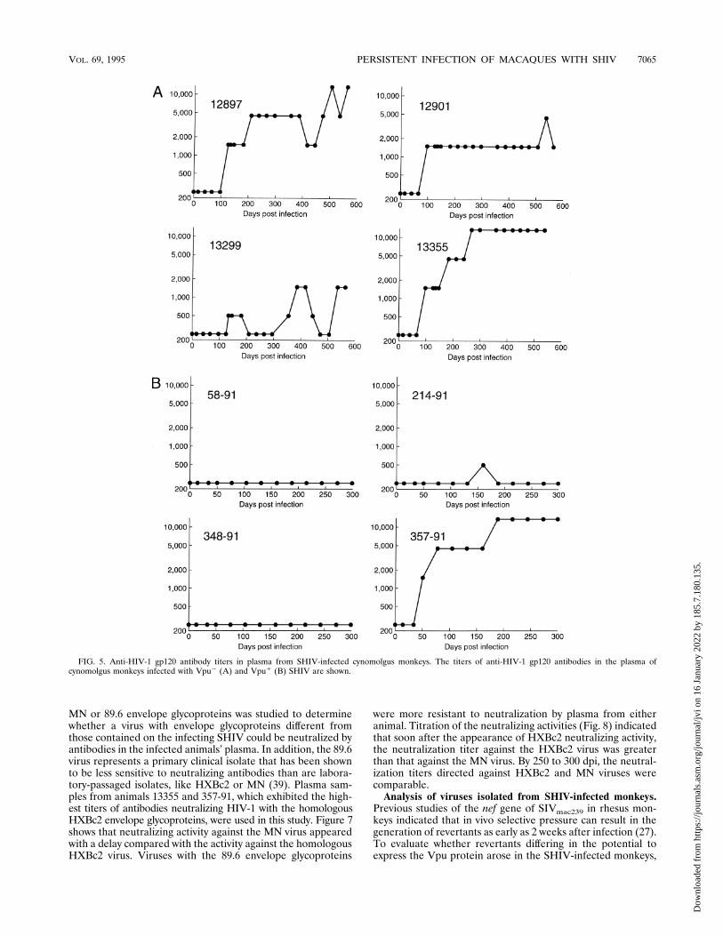
dicated that all of the inoculated monkeys generated serumantibodies reactive with the HIV-1 gp120 glycoprotein (datanot shown). The titers of anti-gp120 antibodies at differenttimes following infection with the Vpu1 or Vpu2 SHIV weredetermined by ELISA (Fig. 5). Within each group of animals,a wide range of anti-gp120 antibody titers was observed. Aswas observed for the anti-gp160 immune response, animals13355 and 357-91 generated the highest titers of antibodiesreactive with the HIV-1 gp120 glycoprotein. With the excep-tion of monkey 357-91, the Vpu1 SHIV-infected animals ex-hibited anti-gp120 antibody titers lower than those seen in theVpu2 SHIV-infected animals (Fig. 5). This may relate to theeffect of the vpu initiation codon on the level of expression ofenvelope glycoproteins (36).SHIV were neutralized in tissue culture assays by monoclo-
nal antibodies directed against the V3 loop or CD4 binding siteof the HIV-1 gp120 glycoprotein (data not shown). To deter-mine if SHIV infection results in the elicitation of HIV-1-specific neutralizing antibodies, we used a single-step envcomplementation assay. In this assay, recombinant HIV-1 viri-ons containing the envelope glycoproteins of the HXBc2, MN,or 89.6 HIV-1 strains and encoding the bacterial chloramphen-icol acetyltransferase protein were produced in transfectedCOS-1 cells as described previously (22). Recombinant virionswere incubated with plasma derived from the monkeys either
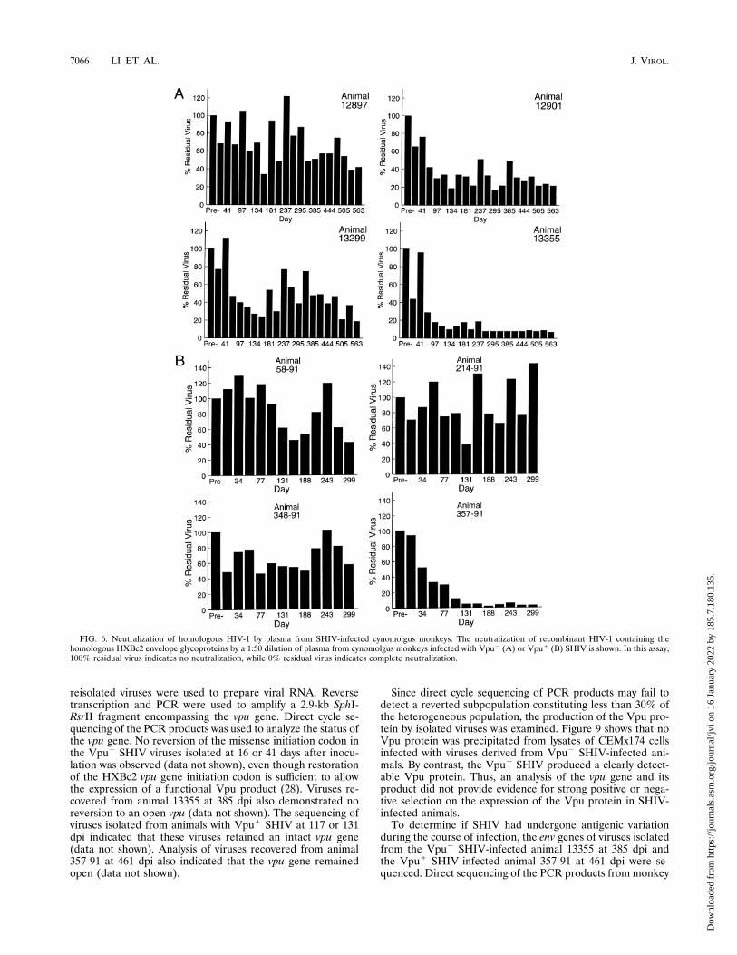
prior to infection or at various times following infection. Figure6 illustrates the neutralizing activity for the homologousHXBc2 virus in infected monkey plasma. The neutralizing ac-tivity roughly correlated with the anti-gp120 and anti-gp160antibody titers. Monkeys 13355 and 357-91 exhibited the high-est anti-gp120 titers and the greatest potencies of neutraliza-tion against the HXBc2 virus.Neutralization of the recombinant HIV-1 virions containing
FIG. 3. Isolation of SHIV from the CD8-depleted, ConA-stimulated PBMC of cynomolgus monkeys at various times following inoculation. The first four monkeyslisted were infected with Vpu2 SHIV, while the other four animals listed were inoculated with Vpu1 SHIV.
FIG. 4. Anti-HIV-1 gp160 antibody titers in plasma from Vpu2 (A) andVpu1 (B) SHIV-infected monkeys. Monkey numbers are shown on the right.
TABLE 1. Plasma viral RNA levels 2 weeks after inoculationwith SHIV
vpu status of SHIV Animal no. Particles/ml of plasmaa
Negative 12897 ,10,00012901 ,10,00013299 ,10,00013355 ,10,000
Positive 58-91 66,000214-91 352,000348-91 704,000357-91 2,820,000
a The limit of detection for this assay is 10,000 particle equivalents per ml.
7064 LI ET AL. J. VIROL.
Dow
nloa
ded
from
http
s://j
ourn
als.
asm
.org
/jour
nal/j
vi o
n 16
Jan
uary
202
2 by
185
.7.1
80.1
35.
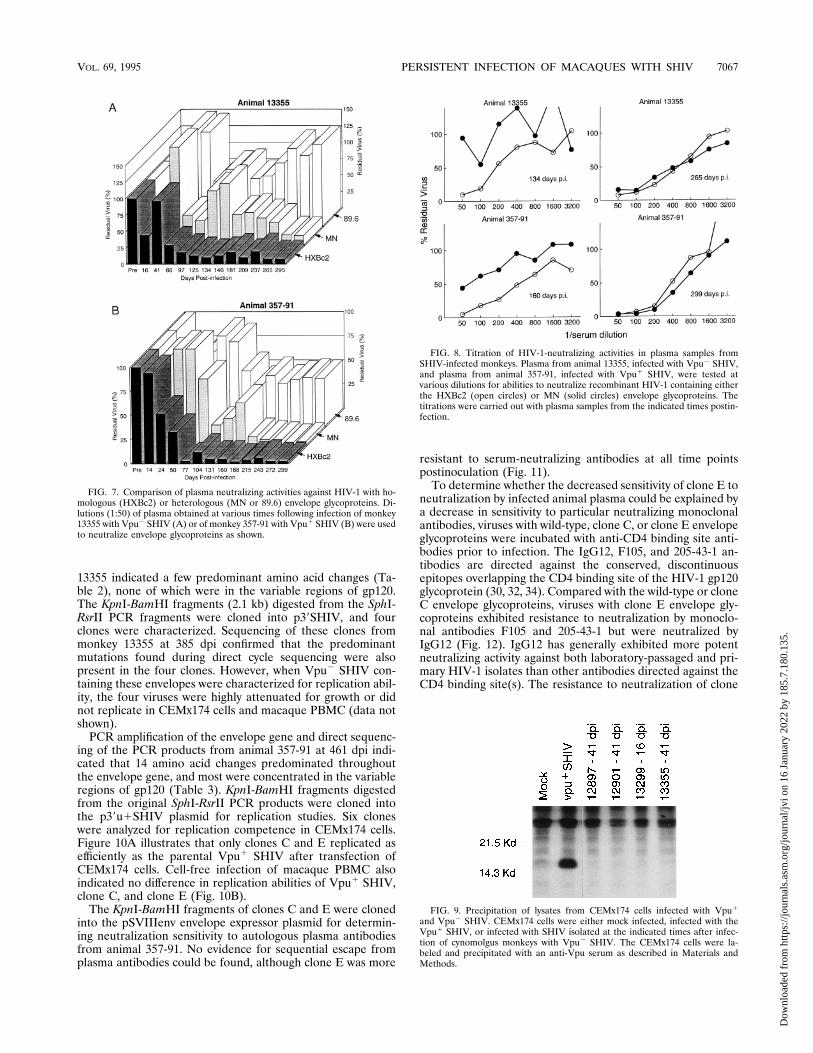
MN or 89.6 envelope glycoproteins was studied to determinewhether a virus with envelope glycoproteins different fromthose contained on the infecting SHIV could be neutralized byantibodies in the infected animals’ plasma. In addition, the 89.6virus represents a primary clinical isolate that has been shownto be less sensitive to neutralizing antibodies than are labora-tory-passaged isolates, like HXBc2 or MN (39). Plasma sam-ples from animals 13355 and 357-91, which exhibited the high-est titers of antibodies neutralizing HIV-1 with the homologousHXBc2 envelope glycoproteins, were used in this study. Figure 7shows that neutralizing activity against the MN virus appearedwith a delay compared with the activity against the homologousHXBc2 virus. Viruses with the 89.6 envelope glycoproteins
were more resistant to neutralization by plasma from eitheranimal. Titration of the neutralizing activities (Fig. 8) indicatedthat soon after the appearance of HXBc2 neutralizing activity,the neutralization titer against the HXBc2 virus was greaterthan that against the MN virus. By 250 to 300 dpi, the neutral-ization titers directed against HXBc2 and MN viruses werecomparable.Analysis of viruses isolated from SHIV-infected monkeys.
Previous studies of the nef gene of SIVmac239 in rhesus mon-keys indicated that in vivo selective pressure can result in thegeneration of revertants as early as 2 weeks after infection (27).To evaluate whether revertants differing in the potential toexpress the Vpu protein arose in the SHIV-infected monkeys,
FIG. 5. Anti-HIV-1 gp120 antibody titers in plasma from SHIV-infected cynomolgus monkeys. The titers of anti-HIV-1 gp120 antibodies in the plasma ofcynomolgus monkeys infected with Vpu2 (A) and Vpu1 (B) SHIV are shown.
VOL. 69, 1995 PERSISTENT INFECTION OF MACAQUES WITH SHIV 7065
Dow
nloa
ded
from
http
s://j
ourn
als.
asm
.org
/jour
nal/j
vi o
n 16
Jan
uary
202
2 by
185
.7.1
80.1
35.
reisolated viruses were used to prepare viral RNA. Reversetranscription and PCR were used to amplify a 2.9-kb SphI-RsrII fragment encompassing the vpu gene. Direct cycle se-quencing of the PCR products was used to analyze the status ofthe vpu gene. No reversion of the missense initiation codon inthe Vpu2 SHIV viruses isolated at 16 or 41 days after inocu-lation was observed (data not shown), even though restorationof the HXBc2 vpu gene initiation codon is sufficient to allowthe expression of a functional Vpu product (28). Viruses re-covered from animal 13355 at 385 dpi also demonstrated noreversion to an open vpu (data not shown). The sequencing ofviruses isolated from animals with Vpu1 SHIV at 117 or 131dpi indicated that these viruses retained an intact vpu gene(data not shown). Analysis of viruses recovered from animal357-91 at 461 dpi also indicated that the vpu gene remainedopen (data not shown).
Since direct cycle sequencing of PCR products may fail todetect a reverted subpopulation constituting less than 30% ofthe heterogeneous population, the production of the Vpu pro-tein by isolated viruses was examined. Figure 9 shows that noVpu protein was precipitated from lysates of CEMx174 cellsinfected with viruses derived from Vpu2 SHIV-infected ani-mals. By contrast, the Vpu1 SHIV produced a clearly detect-able Vpu protein. Thus, an analysis of the vpu gene and itsproduct did not provide evidence for strong positive or nega-tive selection on the expression of the Vpu protein in SHIV-infected animals.To determine if SHIV had undergone antigenic variation
during the course of infection, the env genes of viruses isolatedfrom the Vpu2 SHIV-infected animal 13355 at 385 dpi andthe Vpu1 SHIV-infected animal 357-91 at 461 dpi were se-quenced. Direct sequencing of the PCR products from monkey
FIG. 6. Neutralization of homologous HIV-1 by plasma from SHIV-infected cynomolgus monkeys. The neutralization of recombinant HIV-1 containing thehomologous HXBc2 envelope glycoproteins by a 1:50 dilution of plasma from cynomolgus monkeys infected with Vpu2 (A) or Vpu1 (B) SHIV is shown. In this assay,100% residual virus indicates no neutralization, while 0% residual virus indicates complete neutralization.
7066 LI ET AL. J. VIROL.
Dow
nloa
ded
from
http
s://j
ourn
als.
asm
.org
/jour
nal/j
vi o
n 16
Jan
uary
202
2 by
185
.7.1
80.1
35.
13355 indicated a few predominant amino acid changes (Ta-ble 2), none of which were in the variable regions of gp120.The KpnI-BamHI fragments (2.1 kb) digested from the SphI-RsrII PCR fragments were cloned into p39SHIV, and fourclones were characterized. Sequencing of these clones frommonkey 13355 at 385 dpi confirmed that the predominantmutations found during direct cycle sequencing were alsopresent in the four clones. However, when Vpu2 SHIV con-taining these envelopes were characterized for replication abil-ity, the four viruses were highly attenuated for growth or didnot replicate in CEMx174 cells and macaque PBMC (data notshown).PCR amplification of the envelope gene and direct sequenc-
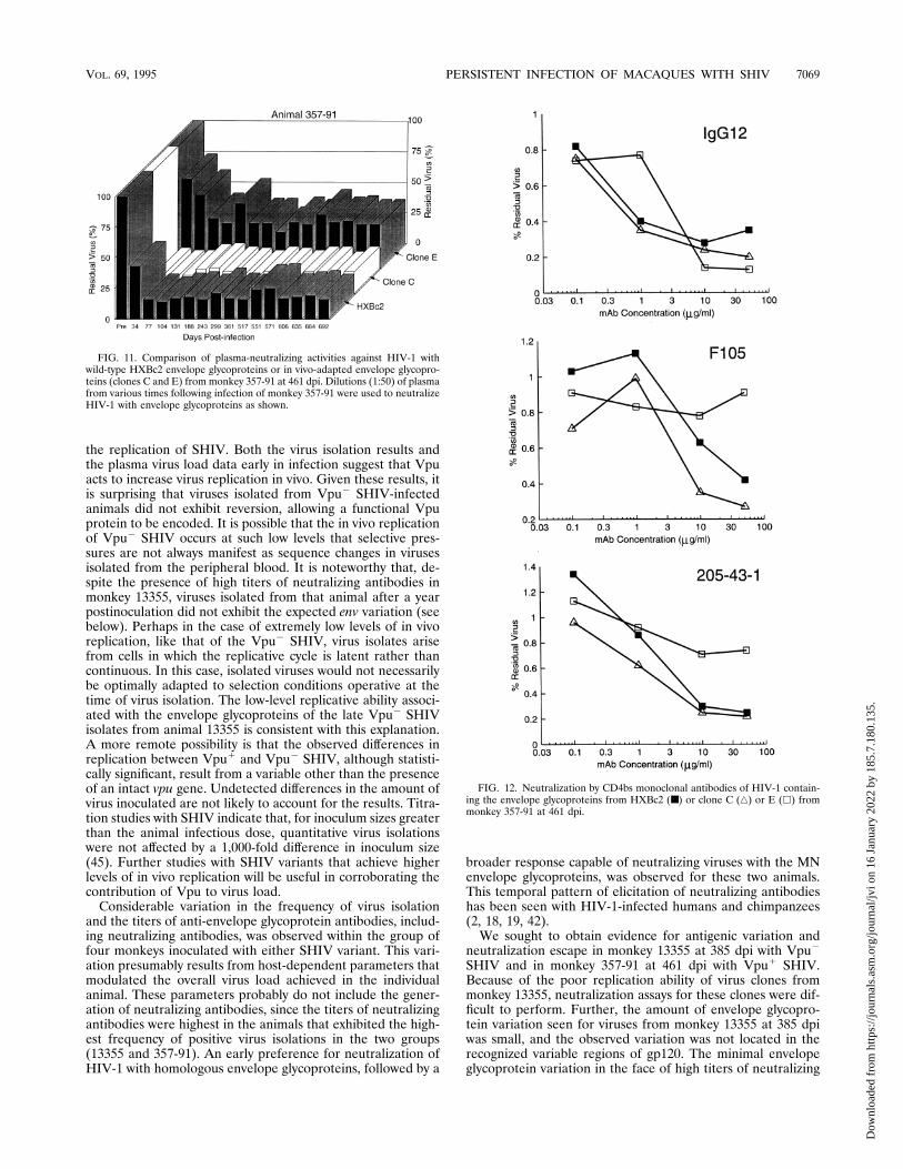
ing of the PCR products from animal 357-91 at 461 dpi indi-cated that 14 amino acid changes predominated throughoutthe envelope gene, and most were concentrated in the variableregions of gp120 (Table 3). KpnI-BamHI fragments digestedfrom the original SphI-RsrII PCR products were cloned intothe p39u1SHIV plasmid for replication studies. Six cloneswere analyzed for replication competence in CEMx174 cells.Figure 10A illustrates that only clones C and E replicated asefficiently as the parental Vpu1 SHIV after transfection ofCEMx174 cells. Cell-free infection of macaque PBMC alsoindicated no difference in replication abilities of Vpu1 SHIV,clone C, and clone E (Fig. 10B).The KpnI-BamHI fragments of clones C and E were cloned
into the pSVIIIenv envelope expressor plasmid for determin-ing neutralization sensitivity to autologous plasma antibodiesfrom animal 357-91. No evidence for sequential escape fromplasma antibodies could be found, although clone E was more
resistant to serum-neutralizing antibodies at all time pointspostinoculation (Fig. 11).To determine whether the decreased sensitivity of clone E to
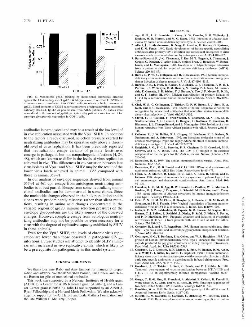
neutralization by infected animal plasma could be explained bya decrease in sensitivity to particular neutralizing monoclonalantibodies, viruses with wild-type, clone C, or clone E envelopeglycoproteins were incubated with anti-CD4 binding site anti-bodies prior to infection. The IgG12, F105, and 205-43-1 an-tibodies are directed against the conserved, discontinuousepitopes overlapping the CD4 binding site of the HIV-1 gp120glycoprotein (30, 32, 34). Compared with the wild-type or cloneC envelope glycoproteins, viruses with clone E envelope gly-coproteins exhibited resistance to neutralization by monoclo-nal antibodies F105 and 205-43-1 but were neutralized byIgG12 (Fig. 12). IgG12 has generally exhibited more potentneutralizing activity against both laboratory-passaged and pri-mary HIV-1 isolates than other antibodies directed against theCD4 binding site(s). The resistance to neutralization of clone
FIG. 7. Comparison of plasma neutralizing activities against HIV-1 with ho-mologous (HXBc2) or heterologous (MN or 89.6) envelope glycoproteins. Di-lutions (1:50) of plasma obtained at various times following infection of monkey13355 with Vpu2 SHIV (A) or of monkey 357-91 with Vpu1 SHIV (B) were usedto neutralize envelope glycoproteins as shown.
FIG. 8. Titration of HIV-1-neutralizing activities in plasma samples fromSHIV-infected monkeys. Plasma from animal 13355, infected with Vpu2 SHIV,and plasma from animal 357-91, infected with Vpu1 SHIV, were tested atvarious dilutions for abilities to neutralize recombinant HIV-1 containing eitherthe HXBc2 (open circles) or MN (solid circles) envelope glycoproteins. Thetitrations were carried out with plasma samples from the indicated times postin-fection.
FIG. 9. Precipitation of lysates from CEMx174 cells infected with Vpu1
and Vpu2 SHIV. CEMx174 cells were either mock infected, infected with theVpu1 SHIV, or infected with SHIV isolated at the indicated times after infec-tion of cynomolgus monkeys with Vpu2 SHIV. The CEMx174 cells were la-beled and precipitated with an anti-Vpu serum as described in Materials andMethods.
VOL. 69, 1995 PERSISTENT INFECTION OF MACAQUES WITH SHIV 7067
Dow
nloa
ded
from
http
s://j
ourn
als.
asm
.org
/jour
nal/j
vi o
n 16
Jan
uary
202
2 by
185
.7.1
80.1
35.
E is at least partially explained by decreased binding of themonoclonal antibodies to the monomeric gp120 glycoproteinsof clone E (Fig. 13). Interestingly, the loss of monomeric en-velope recognition by neutralizing monoclonal antibodies is
one mechanism by which natural variants of SIVmac escapeneutralization by monoclonal antibodies (6).
DISCUSSION
This study demonstrates that SHIV chimeras can consis-tently initiate infection of cynomolgus monkeys. In all of theinfected animals, virus could be recovered for at least 2 monthsfollowing inoculation. Consistency in initiating infection is oneprerequisite for an animal model useful for the study of pro-phylaxis. It has been reported that virus could be recoveredfrom only one of two cynomolgus monkeys inoculated with aSHIV lacking the vpr and nef genes (35). The integrity of theSIVmac nef gene has been shown to be important for the main-tenance of high virus loads in monkeys (25, 27). The abilities ofthe SHIV chimeras used in this study to encode functional Nefproteins probably contribute to the observed consistency ininitiating in vivo infection.We also studied the course of infection of cynomolgus mon-
keys by SHIV chimeras differing in the ability to express theHIV-1 Vpu protein. In tissue culture systems, expression of theHIV-1 Vpu protein leads to specific degradation of CD4 (28,43, 44), the receptor for the primate immunodeficiency viruses(9, 26). Since the HIV-1 Vpu protein can also facilitate thebudding of a wide range of retroviruses (17), both defined invitro functions of the Vpu proteins could potentially modulate
FIG. 10. Replication of clones from monkey 357-91 at 461 dpi. (A) ProviralDNA was transfected into CEMx174 cells, and the level of supernatant reversetranscriptase activity after each transfection is shown. (B) A total of 5,000 reversetranscriptase units of CEMx174-grown Vpu1 SHIV, clone C, or clone E wasused to initiate infection of rhesus macaque PBMC. The level of reverse tran-scriptase activity in the culture supernatant after each infection is shown.
TABLE 2. Changes in envelope glycoprotein sequences derivedfrom animal 13355 at 385 dpia
Nucleotide no. (change) Amino acid no. (change) Region
6396 G/A 58 A/T C16402 G/A 60 A/T C17528 A/C or A/T 435 Y/S or Y/F C48043 G/A 607 A/T gp41b
8101 C/T 626 T/M gp41b
a Each nucleotide change is reported as the proviral sequence number, ac-cording to the sequence of Muesing et al. (31), with the original base on the leftof the shill and the base found in the late virus on the right. The predicted changein amino acid is also shown, with the original residue on the left of the shill andthe altered residue on the right. Changes for this animal were observed in thebulk population and were confirmed in all four env clones examined.b The altered amino acid was found in the exterior domain of this glycoprotein
region.
TABLE 3. Changes in envelope glycoprotein sequences derivedfrom animal 357-91 at 461 dpia
Nucleotide no.(change)
Amino acid no.(change) Region
Change observed inb:
Bulk Clone C Clone E
5877 G/A Silent X NS NS5922 T/C Silent X NS NS6238 A/G 146 R/G V1 X X X6249 G/A 149 M/I V1 X X X6253 A/C 151 K/Q V1 X6284 T/C 161 I/T V2 X X X6313 A/G 171 K/E V2 X X X6634 A/G 278 T/A C2 X X X6700-01 A/G-A/G 300 N/G V3 X6701 A/G 300 N/S V3 X X6748 G/A 316 A/T V3 X X X6809 C/T 336 A/V C3 X X X6853 G/A 351 E/K C3 X X X7033 A/G 411 S/G V4 X X7068 A/G Silent X7088 A/G 429 K/R C4 X X7104 G/A 434 M/I C4 X7112 C/A 437 P/H C4 X7158 G/A Silent X X X7183 A/G 461 S/G V5 X X X7284 A/G Silent X X X7323 G/A Silent X X7401 C/G Silent X X7428 A/G Silent X7479 T/A or T/C Silent X7679 C/T 626 T/M gp41 (ext.) X X X7743 A/C 647 E/D gp41 (ext.) X X7785 G/A Silent X X7888 A/G 696 R/G gp41 (TM) X X7917 G/A Silent X X X7923 G/A Silent X X8020 A/G 740 R/G gp41 (cyt.) X8326-27 C/T-A/T 842 H/I gp41 (cyt.) X NS NS
a Each nucleotide change is reported as the proviral sequence number, ac-cording to the sequence of Muesing et al. (31), with the original base on the leftof the shill and the base found in the late virus on the right. The predicted changein amino acid is also shown, with the original residue on the left of the shill andthe altered residue on the right. The region of the gp120 or gp41 glycoprotein inwhich the altered amino acid is found is also noted: ext., exterior domain; TM,transmembrane region; cyt., cytoplasmic tail.b X indicates a change that was observed in direct cycle sequencing of a
PCR-derived fragment spanning env (Bulk) or observed in sequencing a plasmidcontaining the clone C or clone E env fragment. NS, not sequenced.
7068 LI ET AL. J. VIROL.
Dow
nloa
ded
from
http
s://j
ourn
als.
asm
.org
/jour
nal/j
vi o
n 16
Jan
uary
202
2 by
185
.7.1
80.1
35.
the replication of SHIV. Both the virus isolation results andthe plasma virus load data early in infection suggest that Vpuacts to increase virus replication in vivo. Given these results, itis surprising that viruses isolated from Vpu2 SHIV-infectedanimals did not exhibit reversion, allowing a functional Vpuprotein to be encoded. It is possible that the in vivo replicationof Vpu2 SHIV occurs at such low levels that selective pres-sures are not always manifest as sequence changes in virusesisolated from the peripheral blood. It is noteworthy that, de-spite the presence of high titers of neutralizing antibodies inmonkey 13355, viruses isolated from that animal after a yearpostinoculation did not exhibit the expected env variation (seebelow). Perhaps in the case of extremely low levels of in vivoreplication, like that of the Vpu2 SHIV, virus isolates arisefrom cells in which the replicative cycle is latent rather thancontinuous. In this case, isolated viruses would not necessarilybe optimally adapted to selection conditions operative at thetime of virus isolation. The low-level replicative ability associ-ated with the envelope glycoproteins of the late Vpu2 SHIVisolates from animal 13355 is consistent with this explanation.A more remote possibility is that the observed differences inreplication between Vpu1 and Vpu2 SHIV, although statisti-cally significant, result from a variable other than the presenceof an intact vpu gene. Undetected differences in the amount ofvirus inoculated are not likely to account for the results. Titra-tion studies with SHIV indicate that, for inoculum sizes greaterthan the animal infectious dose, quantitative virus isolationswere not affected by a 1,000-fold difference in inoculum size(45). Further studies with SHIV variants that achieve higherlevels of in vivo replication will be useful in corroborating thecontribution of Vpu to virus load.Considerable variation in the frequency of virus isolation
and the titers of anti-envelope glycoprotein antibodies, includ-ing neutralizing antibodies, was observed within the group offour monkeys inoculated with either SHIV variant. This vari-ation presumably results from host-dependent parameters thatmodulated the overall virus load achieved in the individualanimal. These parameters probably do not include the gener-ation of neutralizing antibodies, since the titers of neutralizingantibodies were highest in the animals that exhibited the high-est frequency of positive virus isolations in the two groups(13355 and 357-91). An early preference for neutralization ofHIV-1 with homologous envelope glycoproteins, followed by a
broader response capable of neutralizing viruses with the MNenvelope glycoproteins, was observed for these two animals.This temporal pattern of elicitation of neutralizing antibodieshas been seen with HIV-1-infected humans and chimpanzees(2, 18, 19, 42).We sought to obtain evidence for antigenic variation and
neutralization escape in monkey 13355 at 385 dpi with Vpu2
SHIV and in monkey 357-91 at 461 dpi with Vpu1 SHIV.Because of the poor replication ability of virus clones frommonkey 13355, neutralization assays for these clones were dif-ficult to perform. Further, the amount of envelope glycopro-tein variation seen for viruses from monkey 13355 at 385 dpiwas small, and the observed variation was not located in therecognized variable regions of gp120. The minimal envelopeglycoprotein variation in the face of high titers of neutralizing
FIG. 11. Comparison of plasma-neutralizing activities against HIV-1 withwild-type HXBc2 envelope glycoproteins or in vivo-adapted envelope glycopro-teins (clones C and E) from monkey 357-91 at 461 dpi. Dilutions (1:50) of plasmafrom various times following infection of monkey 357-91 were used to neutralizeHIV-1 with envelope glycoproteins as shown.
FIG. 12. Neutralization by CD4bs monoclonal antibodies of HIV-1 contain-ing the envelope glycoproteins from HXBc2 (■) or clone C (Ç) or E (h) frommonkey 357-91 at 461 dpi.
VOL. 69, 1995 PERSISTENT INFECTION OF MACAQUES WITH SHIV 7069
Dow
nloa
ded
from
http
s://j
ourn
als.
asm
.org
/jour
nal/j
vi o
n 16
Jan
uary
202
2 by
185
.7.1
80.1
35.
antibodies is paradoxical and may be a result of the low level ofin vivo replication associated with the Vpu2 SHIV. In additionto the factors already discussed, selection pressure exerted byneutralizing antibodies may be operative only above a thresh-old level of virus replication. It has been previously reportedthat neutralization escape variants of primate lentivirusesemerge in pathogenic but not nonpathogenic infections (4, 24,48), which are known to differ in the levels of virus replicationachieved in vivo. The differences in env variation between latevirus isolates of Vpu2 and Vpu1 SHIV may be the result of thelower virus loads achieved in animal 13355 compared withthose in animal 357-91.In our analysis of envelope sequences derived from animal
357-91 at 461 dpi, escape from autologous neutralizing anti-bodies is at best partial. Escape from some neutralizing mono-clonal antibodies can be demonstrated in some clones. Sincethe nucleotide changes observed in the bulk population and inclones were predominantly missense rather than silent muta-tions, resulting in amino acid changes concentrated in thevariable regions of gp120, immune selection pressures on theenvelope glycoproteins are the likely sources of the observedchanges. However, complete escape from autologous neutral-izing antibodies may not be possible or even necessary for avirus with the degree of replicative capacity exhibited by SHIVin these animals.Even for the Vpu1 SHIV, the levels of chronic virus repli-
cation are lower than those observed in pathogenic SIVmacinfections. Future studies will attempt to identify SHIV chime-ras with increased in vivo replicative ability, which is likely tobe a prerequisite for pathogenicity in this model system.
ACKNOWLEDGMENTS
We thank Lorraine Rabb and Amy Emmert for manuscript prepa-ration and artwork. We thank Marshall Posner, Eric Cohen, and Den-nis Burton for gifts of monoclonal antibodies.This work was supported by a National Institutes of Health grant
(AI33832), a Center for AIDS Research grant (AI28691), and a Can-cer Center grant (CA06516). John Li was supported by an Albert J.Ryan Fellowship and a Harvard Merit Fellowship. We also acknowl-edge the support of the G. Harold and Leila Mathers Foundation andthe late William F. McCarty-Cooper.
REFERENCES
1. Agy, M. B., L. R. Frumkin, L. Corey, R. W. Coombs, S. M. Wolinsky, J.Koehler, W. R. Morton, and M. G. Katze. 1992. Infection of Macaca nem-estrina by human immunodeficiency virus type-1. Science 257:103–106.
2. Albert, J., B. Abrahamsson, K. Nagy, E. Aurelius, H. Gaines, G. Nystrom,and E. M. Fenyo. 1990. Rapid development of isolate-specific neutralizingantibodies after primary HIV-1 infection and consequent emergence of virusvariants which resist neutralization by autologous sera. AIDS 4:107–112.
3. Barre-Sinoussi, F., J.-C. Chermann, F. Rey, M. T. Nugeyre, S. Chamaret, J.Gruest, C. Dauguet, C. Axler-Blin, F. Vezinet-Brun, C. Rouzioux, W. Rozen-baum, and L. Montagnier. 1983. Isolation of a T-lymphotropic retrovirusfrom a patient at risk for acquired immune deficiency syndrome (AIDS).Science 220:868–871.
4. Burns, D. P. W., C. Collignon, and R. C. Desrosiers. 1993. Simian immuno-deficiency virus mutants resistant to serum neutralization arise during per-sistent infection of rhesus monkeys. J. Virol. 67:4104–4113.
5. Burton, D. R., J. Pyati, R. Koduri, S. J. Sharp, G. B. Thornton, P. W. H. I.Parren, L. S. W. Sawyer, R. M. Hendry, N. Dunlop, P. L. Nara, M. Lamac-chia, E. Garratty, E. R. Stiehm, Y. J. Bryson, Y. Cao, J. P. Moore, D. D. Ho,and C. F. Barbas III. 1994. Efficient neutralization of primary isolates ofHIV-1 by a recombinant human monoclonal antibody. Science 266:1024–1027.
6. Choi, W. S., C. Collingnon, C. Thiriart, D. P. W. Burns, E. J. Stott, K. A.Kent, and R. C. Desrosiers. 1994. Effects of natural sequence variation onrecognition by monoclonal antibodies that neutralize simian immunodefi-ciency virus infectivity. J. Virol. 68:5395–5402.
7. Clavel, F., D. Guetard, F. Brun-Vezinet, S. Chamaret, M.-A. Rey, M. O.Santos-Ferreira, A. G. Laurent, C. Dauguet, C. Katlama, C. Rouzioux, D.Klatzman, J. L. Champalimaad, and L. Montagnier. 1986. Isolation of a newhuman retrovirus from West African patients with AIDS. Science 233:343–346.
8. Collman, R., J. W. Balliet, S. A. Gregory, H. Friedman, K. L. Kolson, N.Nathanson, and A. Srinivasan. 1992. An infectious molecular clone of anunusual macrophage-tropic and highly cytopathic strain of human immuno-deficiency virus type 1. J. Virol. 66:7517–7521.
9. Dalgleish, A. G., P. C. L. Beverley, P. R. Clapham, D. H. Crawford, M. F.Greaves, and R. A. Weiss. 1984. The CD4 (T4) antigen is an essentialcomponent of the receptor for the Aids retrovirus. Nature (London) 312:763–767.
10. Desrosiers, R. C. 1989. The simian immunodeficiency viruses. Annu. Rev.Immunol. 8:557–558.
11. Desrosiers, R. C., M. D. Daniel, and Y. Li. 1989. HIV-related lentiviruses ofnonhuman primates. AIDS Res. Hum. Retroviruses 5:465–473.
12. Fauci, A., A. Macher, D. Longo, H. C. Lane, A. Rook, H. Masur, and E.Gelman. 1984. Acquired immunodeficiency syndrome: epidemiologic, clini-cal, immunologic, and therapeutic considerations. Ann. Intern. Med. 100:92–106.
13. Frumkin, L. R., M. B. Agy, R. W. Coombs, L. Panther, W. R. Morton, J.Koehler, M. J. Florey, J. Dragavon, A. Schmidt, M. G. Katze, and L. Corey.1993. Acute infection of Macaca nemestrina by human immunodeficiencyvirus type 1. Virology 195:422–431.
14. Fultz, P. N., H. M. McClure, H. Daugharty, A. Brodie, C. R. McGrath, B.Swenson, and D. P. Francis. 1986. Vaginal transmission of human immuno-deficiency virus (HIV) in a chimpanzee. J. Infect. Dis. 154:896–900.
15. Gallo, R. C., S. Z. Salahuddin, M. Papovic, G. M. Shearer, M. Kaplan, B. F.Haynes, T. J. Palker, R. Redfield, J. Oleske, B. Safai, G. White, P. Foster,and P. D. Markham. 1984. Frequent detection and isolation of cytopathicretroviruses (HTLV-III) from patients with AIDS and at risk for AIDS.Science 224:500–503.
16. Geraghty, R. J., and A. T. Paganiban. 1993. Human immunodeficiency virustype 1 Vpu has a CD4- and an envelope glycoprotein-independent function.J. Virol. 67:4190–4194.
17. Gottlinger, H. G., T. Dorfman, E. A. Cohen, and W. A. Haseltine. 1993. Vpuprotein of human immunodeficiency virus type 1 enhances the release ofcapsids produced by gag gene constructs of widely divergent retroviruses.Proc. Natl. Acad. Sci. USA 90:7381–7385.
18. Goudsmit, J., C. Debouck, R. H. Meloen, L. Smit, M. Bakker, D. M. Asher,A. V. Wolff, C. J. Gibbs, Jr., and D. C. Gajdusek. 1988. Human immunode-ficiency virus type 1 neutralization epitope with conserved architecture elicitsearly type-specific antibodies in experimentally infected chimpanzees. Proc.Natl. Acad. Sci. USA 85:4478–4482.
19. Goudsmit, J., C. Thiriart, L. Smit, C. Bruck, and C. J. Gibbs, Jr. 1988.Temporal development of cross-neutralization between HTLV-IIIB andHTLV-III RF in experimentally infected chimpanzees. Vaccine 6:229–232.
20. Gurgo, C., H.-G. Guo, G. Franchini, A. Aldovini, E. Collalti, K. Farrell, F.Wong-Staal, R. C. Gallo, and M. S. Reits, Jr. 1988. Envelope sequences oftwo new United States HIV-1 isolates. Virology 164:531–536.
21. Haseltine, W. A. 1988. Replication and pathogenicity of the AIDS virus. J.Acquired Immune Defic. Syndr. 1:217–240.
22. Helseth, E., M. Kowalski, D. Gabuzda, U. Olshevsky, W. Haseltine, and J.Sodroski. 1990. Rapid complementation assays measuring replicative poten-
FIG. 13. Monomeric gp120 binding by monoclonal antibodies directedagainst the CD4 binding site of gp120. Wild-type, clone C, or clone E pSVIIIenvexpressors were transfected into COS-1 cells to obtain soluble, monomericgp120. Equal amounts of COS-1 supernatants were precipitated with monoclonalantibody 205-43-1, IgG12, or pooled sera from AIDS patients. All values werenormalized to the amount of gp120 precipitated by patient serum to control forenvelope glycoprotein expression in COS-1 cells.
7070 LI ET AL. J. VIROL.
Dow
nloa
ded
from
http
s://j
ourn
als.
asm
.org
/jour
nal/j
vi o
n 16
Jan
uary
202
2 by
185
.7.1
80.1
35.
tial of human immunodeficiency virus type 1 envelope glycoprotein mutants.J. Virol. 64:2416–2420.
23. Javaherian, K., A. J. Langlois, S. Schmidt, M. Kaufmann, N. Cates, J. P. M.Langeduk, R. H. Meloen, R. C. Desrosiers, D. P. W. Burns, D. P. Bolognesi,G. J. LaRosa, and S. D. Putney. 1992. The principal neutralization determi-nant of simian immunodeficiency virus differs from that of human immuno-deficiency virus type 1. Proc. Natl. Acad. Sci. USA 89:1418–1422.
24. Joag, S. V., M. G. Anderson, J. E. Clements, M. F. McEntee, D. P. Sharma,R. J. Adams, and O. Narayan. 1993. Antigenic variation of molecularlycloned SIVmac239 during persistent infection in a rhesus macaque. Virology198:406–412.
25. Kestler, H. W., D. J. Ringler, K. Mori, D. L. Panicali, P. K. Sehgal, M. D.Daniel, and R. C. Desrosiers. 1991. Importance of the nef gene for mainte-nance of high virus loads and for development of AIDS. Cell 65:651–662.
26. Klatzman, D., E. Champagne, S. Chamaret, J. Gruest, D. Guetard, T. Her-cend, J.-C. Gluckman, and L. Montagnier. 1984. T-lymphocyte T4 moleculebehaves as the receptor for human retrovirus LAV. Nature (London) 312:767–768.
27. Lang, S. M., M. Weeger, C. Stahl-Hennig, C. Coulibaly, G. Hunsmann, J.Muller, H. Muller-Hermelink, D. Fuchs, H. Wachter, M. M. Daniel, R. C.Desrosiers, and B. Fleckenstein. 1993. Importance of vpr for infection ofrhesus monkeys with simian immunodeficiency virus. J. Virol. 67:902–912.
28. Lenburg, M. E., and N. R. Landau. 1993. Vpu-induced degradation of CD4:requirement for specific amino acid residues in the cytoplasmic domain ofCD4. J. Virol. 67:7238–7245.
29. Li, J., C. I. Lord, W. Haseltine, N. L. Letvin, and J. Sodroski. 1992. Infectionof cynomolgus monkeys with a chimeric HIV-1/SIVmac virus that expressesthe HIV-1 envelope glycoproteins. J. Acquired Immune Defic. Syndr. 5:639–646.
30. Moore, J. P., F. E. McCutchan, S.-W. Poon, J. Mascola, J. Liu, Y. Cao, andD. D. Ho. 1994. Exploration of antigenic variation in gp120 from clades Athrough F of human immunodeficiency virus type 1 by using monoclonalantibodies. J. Virol. 68:8350–8364.
31. Muesing, M. A., D. H. Smith, C. D. Cabradilla, C. V. Benton, L. A. Lasky,and D. J. Capon. 1985. Nucleic acid structure and expression of the humanAIDS/lymphadenopathy retrovirus. Nature (London) 313:450–458.
32. Posner, M. R., T. Hideshima, T. Cannon, M. Mukherjee, K. H. Mayer, andR. A. Byrn. 1991. An IgG human monoclonal antibody that reacts withHIV-1/GP120, inhibits virus binding to cells, and neutralizes infection. J.Immunol. 146:4325–4329.
33. Rho, H., B. Poiesz, F. Ruscetti, and R. C. Gallo. 1981. Characterization of thereverse transcriptase from a new retrovirus (HTLV) produced by a humancutaneous T-cell lymphoma cell line. Virology 112:355–360.
34. Roben, P., J. P. Moore, M. Thali, J. Sodroski, C. F. Barbas III, and D. R.Burton. 1994. Recognition properties of a panel of human recombinant Fab
fragments to the CD4 binding site of gp120 that show differing abilities toneutralize human immunodeficiency virus type 1. J. Virol. 68:4821–4828.
35. Sakuragi, S., R. Shibata, R. Mukai, T. Komatsu, M. Fukasawa, H. Sakai,J.-I. Sakuragi, M. Kawamura, K. Ibuki, M. Hayami, and A. Adachi. 1992.Infection of macaque monkeys with a chimeric human and simian immuno-deficiency virus. J. Gen. Virol. 73:2983–2987.
36. Schwartz, S., B. K. Felber, E.-M. Fenyo, and G. N. Pavlakis. 1990. Env andVpu proteins of human immunodeficiency virus type 1 are produced frommultiple bicistronic mRNAs. J. Virol. 64:5448–5456.
37. Shibata, R., M. Kawamura, H. Sakai, M. Hayami, A. Ishimoto, and A.Adachi. 1991. Generation of a chimeric human and simian immunodefi-ciency virus infectious to monkey peripheral blood mononuclear cells. J.Virol. 65:3514–3520.
38. Smith, L. M., J. Z. Sanders, R. J. Kaiser, P. Hughes, C. Dodd, C. R. Connell,C. Heiner, S. B. H. Kent, and L. E. Hood. 1986. Fluorescence detection inautomated DNA sequence analysis. Nature (London) 321:674–679.
39. Sullivan, N., Y. Sun, J. Li, W. Hoffmann, and J. Sodroski. 1995. Replicativefunction and neutralization sensitivity of envelope glycoproteins from pri-mary and T-cell line-passaged human immunodeficiency virus type 1 isolates.J. Virol. 69:4413–4422.
40. Terwilliger, E. F., E. A. Cohen, Y. Lu, J. G. Sodroski, and W. A. Haseltine.1989. Functional role of human immunodeficiency virus type 1 Vpu. Proc.Natl. Acad. Sci. USA 86:5163–5167.
41. Thali, M., U. Olshevsky, C. Furman, D. Gabuzda, M. Posner, and J. So-droski. 1991. Characterization of a discontinuous human immunodeficiencyvirus type 1 gp120 epitope recognized by a broadly reactive neutralizinghuman monoclonal antibody. J. Virol. 65:6188–6193.
42. von Gegerfelt, A., J. Albert, L. Morfeldt-Manson, K. Broliden, and E. M.Fenyo. 1991. Isolate-specific neutralizing antibodies in patients with progres-sive HIV-1-related disease. Virology 185:162–168.
43. Willey, R. L., F. Maldarelli, M. A. Martin, and K. Strebel. 1992. Humanimmunodeficiency virus type 1 Vpu protein induces rapid degradation ofCD4. J. Virol. 66:7193–7200.
44. Willey, R. L., F. Maldarelli, M. A. Martin, and K. Strebel. 1992. Humanimmunodeficiency virus type 1 Vpu protein regulates the formation of in-tracellular gp160-CD4 complexes. J. Virol. 66:226–234.
45. Wyand, M., J. Li, J. Sodroski, and Y. Lu. Unpublished observations.46. Yao, X. J., H. Gottlinger, W. A. Haseltine, and E. A. Cohen. 1992. Envelope
glycoprotein and CD4 independence of vpu-facilitated human immunodefi-ciency virus type 1 capsid export. J. Virol. 66:5119–5126.
47. Zeger, S., and K.-Y. Liang. 1986. Longitudinal data analysis for discrete andcontinuous outcomes. Biometrics 42:121–130.
48. Zhang, Y. J., P. Putkonen, J. Albert, P. Ohman, G. Biberfeld, and E. M.Fenyo. 1994. Stable biological and antigenic characteristics of HIV-2(SBL6669) in nonpathogenic infection of macaques. Virology 200:583–589.
VOL. 69, 1995 PERSISTENT INFECTION OF MACAQUES WITH SHIV 7071
Dow
nloa
ded
from
http
s://j
ourn
als.
asm
.org
/jour
nal/j
vi o
n 16
Jan
uary
202
2 by
185
.7.1
80.1
35.


![Paulina black macaques [recovered]](https://static.fdocuments.in/doc/165x107/5559dee9d8b42a39498b4992/paulina-black-macaques-recovered.jpg)