Perivascular M2 Macrophages Stimulate Tumor Relapse after ... · Microenvironment and Immunology...
14
Microenvironment and Immunology Perivascular M2 Macrophages Stimulate Tumor Relapse after Chemotherapy Russell Hughes 1 , Bin-Zhi Qian 2 , Charlotte Rowan 1 , Munitta Muthana 1 , Ioanna Keklikoglou 3 , Oakley C. Olson 4 , Simon Tazzyman 1 , Sarah Danson 1 , Christina Addison 5 , Mark Clemons 5 , Ana Maria Gonzalez-Angulo 6 , Johanna A. Joyce 4 , Michele De Palma 3 , Jeffrey W. Pollard 2,7 , and Claire E. Lewis 1 Abstract Tumor relapse after chemotherapy-induced regression is a major clinical problem, because it often involves inoperable metastatic disease. Tumor-associated macrophages (TAM) are known to limit the cytotoxic effects of chemotherapy in preclinical models of cancer. Here, we report that an alternatively activated (M2) subpopulation of TAMs (MRC1 þ TIE2 Hi CXCR4 Hi ) accumu- late around blood vessels in tumors after chemotherapy, where they promote tumor revascularization and relapse, in part, via VEGF-A release. A similar perivascular, M2-related TAM subset was present in human breast carcinomas and bone metastases after chemotherapy. Although a small proportion of M2 TAMs were also present in hypoxic tumor areas, when we genetically ablated their ability to respond to hypoxia via hypoxia-inducible factors 1 and 2, tumor relapse was unaffected. TAMs were the predominant cells expressing immunoreactive CXCR4 in chemo- therapy-treated mouse tumors, with the highest levels expressed by MRC1 þ TAMs clustering around the tumor vasculature. Fur- thermore, the primary CXCR4 ligand, CXCL12, was upregulated in these perivascular sites after chemotherapy, where it was selectively chemotactic for MRC1 þ TAMs. Interestingly, HMOX- 1, a marker of oxidative stress, was also upregulated in perivas- cular areas after chemotherapy. This enzyme generates carbon monoxide from the breakdown of heme, a gas known to upre- gulate CXCL12. Finally, pharmacologic blockade of CXCR4 selec- tively reduced M2-related TAMs after chemotherapy, especially those in direct contact with blood vessels, thereby reducing tumor revascularization and regrowth. Our studies rationalize a strategy to leverage chemotherapeutic efficacy by selectively targeting this perivascular, relapse-promoting M2-related TAM cell population. Cancer Res; 75(17); 3479–91. Ó2015 AACR. Introduction The regrowth of tumors after treatment with cytotoxic agents poses a major threat to survival in cancer patients, particularly those with inoperable primary and/or metastatic tumors as they often rely heavily on chemotherapy to slow tumor growth and reduce its burden. The development of resistance results in poor survival rates for patients with, for example, inoperable pancreatic or lung cancer, who often survive for less than 12 months after diagnosis (1, 2). New therapeutic strategies are, therefore, urgently needed to delay or prevent tumor regrowth after early cycles of chemotherapy as these would extend life. Malignant tumors contain various CD11b þ myeloid cells, including granulocytes, myeloid-derived suppressor cells (MDSC), and tumor-associated macrophages (TAM; ref. 3). The latter are recruited as monocytes from the peripheral blood, which are themselves derived from progenitor cells in the bone marrow (4, 5). After entry into tumors, monocytes differentiate into macrophages (6) and promote tumor progression by stimulating tumor invasion, neovascularization and metastasis, and suppres- sing antitumor immunity (5, 7). TAMs express a broad spectrum of activation states between the two extreme forms of "classical" (M1) and "alternative"' (M2) activation, with a tendency toward the latter (8). M2-skewed TAMs are characterized by their upre- gulation of various receptors, including the mannose receptor C-type lectin (MRC1/CD206) and the angiopoietin receptor, TIE2 (9). Indeed, TAMs expressing high levels of these two receptors have been shown to play an essential role in promoting angio- genesis in untreated mouse tumors (10). A variety of anticancer therapies have been shown to stimulate the recruitment of CD11b þ myeloid cells by mouse tumors (11, 12). For example, TAMs accumulate in mouse tumors after chemotherapy (13–15), ionizing radiation (16–18), and the vascular disrupting agent combretastatin-A4-P (CA-4-P; ref. 19). 1 Department of Oncology, University of Sheffield Medical School, Sheffield, United Kingdom. 2 MRC Centre for Reproductive Health, Queen's Medical Research Institute, University of Edinburgh, Edin- burgh, Scotland, United Kingdom. 3 Swiss Institute for Experimental Cancer Research (ISREC), School of Life Sciences, Ecole Polytechni- que F ed erale de Lausanne (EPFL), Lausanne, Switzerland. 4 Cancer Biology and Genetics Program, Memorial Sloan Kettering Cancer Center, New York, New York. 5 Cancer Therapeutics Program, Ottawa Hospital Research Institute, and Department of Medicine, University of Ottawa, Ontario, Canada. 6 Breast Medical Oncology,The University of Texas MD Anderson Cancer Center, Houston,Texas. 7 Department of Developmental and Molecular Biology, Albert Einstein College of Medicine, New York, New York. Note: Supplementary data for this article are available at Cancer Research Online (http://cancerres.aacrjournals.org/). R. Hughes and B.-Z. Qian share first authorship of this article. J.W. Pollard and C.E. Lewis share senior authorship of this article. Corresponding Author: Claire E. Lewis, University of Sheffield Medical School, Beech Hill Road, Sheffield, Yorkshire S10 2RX, United Kingdom. Phone/Fax: 44- 114-271-2903; E-mail: claire.lewis@sheffield.ac.uk doi: 10.1158/0008-5472.CAN-14-3587 Ó2015 American Association for Cancer Research. Cancer Research www.aacrjournals.org 3479 on April 8, 2020. © 2015 American Association for Cancer Research. cancerres.aacrjournals.org Downloaded from Published OnlineFirst August 12, 2015; DOI: 10.1158/0008-5472.CAN-14-3587
Transcript of Perivascular M2 Macrophages Stimulate Tumor Relapse after ... · Microenvironment and Immunology...
Microenvironment and Immunology
Perivascular M2 Macrophages Stimulate TumorRelapse after ChemotherapyRussell Hughes1, Bin-Zhi Qian2, Charlotte Rowan1, Munitta Muthana1,Ioanna Keklikoglou3, Oakley C. Olson4, Simon Tazzyman1, Sarah Danson1,Christina Addison5, Mark Clemons5, Ana Maria Gonzalez-Angulo6,Johanna A. Joyce4, Michele De Palma3, Jeffrey W. Pollard2,7, andClaire E. Lewis1
Abstract
Tumor relapse after chemotherapy-induced regression is amajor clinical problem, because it often involves inoperablemetastatic disease. Tumor-associated macrophages (TAM) areknown to limit the cytotoxic effects of chemotherapy inpreclinicalmodels of cancer. Here, we report that an alternatively activated(M2) subpopulation of TAMs (MRC1þTIE2HiCXCR4Hi) accumu-late around blood vessels in tumors after chemotherapy, wherethey promote tumor revascularization and relapse, in part, viaVEGF-A release. A similar perivascular, M2-related TAM subsetwas present in human breast carcinomas and bone metastasesafter chemotherapy. Although a small proportion of M2 TAMswere also present in hypoxic tumor areas, when we geneticallyablated their ability to respond to hypoxia via hypoxia-induciblefactors 1 and 2, tumor relapse was unaffected. TAMs were thepredominant cells expressing immunoreactive CXCR4 in chemo-
therapy-treated mouse tumors, with the highest levels expressedby MRC1þ TAMs clustering around the tumor vasculature. Fur-thermore, the primary CXCR4 ligand, CXCL12, was upregulatedin these perivascular sites after chemotherapy, where it wasselectively chemotactic for MRC1þ TAMs. Interestingly, HMOX-1, a marker of oxidative stress, was also upregulated in perivas-cular areas after chemotherapy. This enzyme generates carbonmonoxide from the breakdown of heme, a gas known to upre-gulate CXCL12. Finally, pharmacologic blockade of CXCR4 selec-tively reduced M2-related TAMs after chemotherapy, especiallythose in direct contact with blood vessels, thereby reducing tumorrevascularization and regrowth. Our studies rationalize a strategyto leverage chemotherapeutic efficacy by selectively targeting thisperivascular, relapse-promoting M2-related TAM cell population.Cancer Res; 75(17); 3479–91. �2015 AACR.
IntroductionThe regrowth of tumors after treatment with cytotoxic agents
poses a major threat to survival in cancer patients, particularlythose with inoperable primary and/or metastatic tumors as theyoften rely heavily on chemotherapy to slow tumor growth and
reduce its burden. The development of resistance results in poorsurvival rates for patientswith, for example, inoperable pancreaticor lung cancer, who often survive for less than 12 months afterdiagnosis (1, 2).New therapeutic strategies are, therefore, urgentlyneeded to delay or prevent tumor regrowth after early cycles ofchemotherapy as these would extend life.
Malignant tumors contain various CD11bþ myeloid cells,including granulocytes, myeloid-derived suppressor cells(MDSC), and tumor-associated macrophages (TAM; ref. 3). Thelatter are recruited asmonocytes from theperipheral blood,whichare themselves derived from progenitor cells in the bone marrow(4, 5). After entry into tumors, monocytes differentiate intomacrophages (6) and promote tumor progression by stimulatingtumor invasion, neovascularization and metastasis, and suppres-sing antitumor immunity (5, 7). TAMs express a broad spectrumof activation states between the two extreme forms of "classical"(M1) and "alternative"' (M2) activation, with a tendency towardthe latter (8). M2-skewed TAMs are characterized by their upre-gulation of various receptors, including the mannose receptorC-type lectin (MRC1/CD206) and the angiopoietin receptor, TIE2(9). Indeed, TAMs expressing high levels of these two receptorshave been shown to play an essential role in promoting angio-genesis in untreated mouse tumors (10).
A variety of anticancer therapies have been shown to stimulatethe recruitment of CD11bþ myeloid cells by mouse tumors(11, 12). For example, TAMs accumulate in mouse tumors afterchemotherapy (13–15), ionizing radiation (16–18), and thevascular disrupting agent combretastatin-A4-P (CA-4-P; ref. 19).
1Department of Oncology, University of Sheffield Medical School,Sheffield, United Kingdom. 2MRC Centre for Reproductive Health,Queen's Medical Research Institute, University of Edinburgh, Edin-burgh, Scotland, United Kingdom. 3Swiss Institute for Experimental
Cancer Research (ISREC), School of Life Sciences, �Ecole Polytechni-que F�ed�erale de Lausanne (EPFL), Lausanne, Switzerland. 4CancerBiology and Genetics Program, Memorial Sloan Kettering CancerCenter, New York, New York. 5Cancer Therapeutics Program, OttawaHospitalResearch Institute, andDepartmentofMedicine,UniversityofOttawa,Ontario, Canada. 6Breast Medical Oncology,The University ofTexas MD Anderson Cancer Center, Houston, Texas. 7Department ofDevelopmental and Molecular Biology, Albert Einstein College ofMedicine, New York, New York.
Note: Supplementary data for this article are available at Cancer ResearchOnline (http://cancerres.aacrjournals.org/).
R. Hughes and B.-Z. Qian share first authorship of this article.
J.W. Pollard and C.E. Lewis share senior authorship of this article.
Corresponding Author: Claire E. Lewis, University of Sheffield Medical School,Beech Hill Road, Sheffield, Yorkshire S10 2RX, United Kingdom. Phone/Fax: 44-114-271-2903; E-mail: [email protected]
doi: 10.1158/0008-5472.CAN-14-3587
�2015 American Association for Cancer Research.
CancerResearch
www.aacrjournals.org 3479
on April 8, 2020. © 2015 American Association for Cancer Research. cancerres.aacrjournals.org Downloaded from
Published OnlineFirst August 12, 2015; DOI: 10.1158/0008-5472.CAN-14-3587
Importantly, the mononuclear phagocyte growth factor CSF1(13) and chemokines CCL2 (14) and CXCL12 (16, 19) areincreased in tumors after such anticancer therapies, and can triggermonocyte recruitment (4, 5). These cells then reduce the efficacyof chemotherapy by limiting vascular permeability via theirexpression of MMP9 (14), promoting resistance to therapy-induced death via their expression of cathepsin serine proteases(15) and by suppressing the recruitment/activation of cytotoxicT cells (12, 13).
Considerable evidence has emerged recently for M2-activat-ed macrophages playing an important role in repair andremodeling after tissue injury. For example, they are promi-nent in diseased tissues in spinal cord injury, myocardialinfarction, and various forms of renal disease, (20). Further-more, TAMs with similar phenotypes have been implicated intumor relapse after therapies like irradiation and CA-4-P(17, 19). Although MRC1þ TAMs are increased in MMTV-PyMT mammary tumors after doxorubicin treatment (14), therole of M2 TAMs in tumor relapse after chemotherapy has notbeen defined.
Our studies show that MRC1þ TAMs are elevated in mousetumors after treatment with various chemotherapeutic agents.Moreover, this TAM subset was further defined as MRC1Hi
TIE2HiCXCR4HiVEGFAþ and shown to accumulate preferentiallyin vascularized, CXCL12-rich regions of tumors after chemother-apy. Blockade ofCXCR4 signaling prevented this close associationwith the tumor vasculature after chemotherapy, resulting in amarked delay in subsequent tumor revascularization and relapse.These findings suggest that selective targeting of vessel-associated,M2-skewed TAMs after chemotherapy could increase the relapse-free survival of cancer patients.
Materials and MethodsMouse studies
To investigate the mechanisms regulating tumor relapseafter chemotherapy, we primarily used the Lewis lung carci-noma model (21) and the MMTV-PyMT model of breast cancer(15). These syngeneic tumor models respond to chemotherapywith an initial phase of tumor growth inhibition, followed bya distinct regrowth phase. Transgenic tumor models werenot considered suitable as their responses to some cytotoxicagents can be so minimal that a relapse phase is not evident(13, 22). Furthermore, tumors in these models are oftenmultifocal making the kinetics of tumor relapse difficult toassess accurately.
Our mouse studies were conducted in accordance with eitherUK Home Office regulations (C.E. Lewis/M. Muthana/R.Hughes), the Veterinary Authorities of the Canton Vaud (M. DePalma), the institutional standards of the Research AnimalsResource Centre at Memorial Sloan Kettering Cancer Centre(J.A. Joyce; New York, NY), and the Albert Einstein College ofMedicine Animal Use Committee (J.W. Pollard). LLC1/cyclo-phosphamide studies using 8-week-old C57BL/6 mice were per-formed asdescribed previously (21). For LLC1/AMD3100 studies,AMD3100 was given concurrently (10 mg/kg) with cyclophos-phamide, i.p. every day for 7 days. Tumors and organs wereharvested at the indicated times. Mice were treated i.p. withpimonidazole (60 mg/kg) and bromodeoxyuridine (BrdUrd;100 mg/kg) 2 hours before sacrifice to label hypoxia and prolif-erating cells, respectively.
4T1/paclitaxel studiesFemale Balb/c mice (>8 weeks) were orthotopically implanted
with 1 � 106 4T1 murine mammary adenocarcinoma cells.Tumors were grown for 10 days before mice were injected i.p.with either cremaphore or cremophore þ paclitaxel (10 mg/kg)every 5 days for a total of three doses, themice were sacrificed andtissues harvested 4 days after the last treatment. OrthotopicMMTV-PyMT/doxorubicin studies were performed as previouslydescribed (15). Mice bearing implanted tumors were injected i.v.with a single dose of doxorubicin (5 mg/kg) when at 250 mm3
and sacrificed 7 days after therapy.
Monocyte/macrophage-specific ablation of Vegfa studiesFemale Tg(Csf1r-Mer-iCre-Mer)1jwp;Vegfa
fl/fl) mice (>8 weeks),shown to specifically lack the expression of VEGFA in the mono-cyte/macrophage lineage after treatment with tamoxifen (23), orCre-negative litter mates (ages 8 weeksþ), were orthotopicallyimplantedwith the syngeneicMMTV-PyMT cell line F246-6. Vegfafloxedmicewere the kind gift ofDr.Naploeone Ferrar,Genentech.Mice with established tumors were treated with a single i.v.injection of either vehicle (PBS) or doxorubicin (5 mg/kg) and24 hours later with tamoxifen via their food (3 mg/20 g bodyweight/day). Treatment with tamoxifen was then maintainedthroughout tumor regrowth. Tumor growth was monitored bycalipermeasurements. In the LLC1/TAMadoptive transfer studies,C57BL/6 mice implanted with LLCs and treated with cyclophos-phamide as outline above, 48 hours after the last dose of cyclo-phosphamide, donormice were sacrificed, their tumors removed,enzymatically dissociated, and MRC1Hi and MRC1Lo TAMs werepurified by flow cytometry. Recipient mice were randomized intothree groups and received either 50�103MRC1Hi TAMs, 50�103
MRC1Lo TAMs, or saline via a 25 mL intratumoral injection. Afterinjection of these cells or saline, tumor regrowth was thenmonitored.
Human breast carcinomas and metastatic bone lesionsFour patients with newly diagnosed breast cancer underwent
neoadjuvant chemotherapy as previously described (24). Resid-ual tumor tissues were collected at the time of surgery (which tookplace 21–28days after the last dose of paclitaxel). Biopsies of bonemetastatic lesions were also obtained from five advanced breastcancer patients. All biopsies were obtained under informed con-sent following procedures approved by the Ottawa HealthSciences Research Ethics Board. Samples were collected as previ-ously described (25). All biopsies were taken within 6 to 15 daysof treatment with either chemotherapy alone (paclitaxel, doce-taxel, or 5-fluoruracil/doxorubicin/cyclophosphamide) or with-out pamidronate.
Immunofluorescence and IHC studiesFrozen tumor sections were blocked with 1% BSA and 5% goat
serum for 30 minutes and incubated with various primary anti-bodies (Supplementary Table S1), for 1 hour at room tempera-ture. Alexa fluor–conjugated anti-rabbit or anti-rat secondaryantibodies were used with unconjugated primary antibodies. ForBrdUrd staining, DNA in frozen tissue sections was denaturedwith 2NHCl for 15minutes before immunostaining, as describedabove. Nuclei in all tumor sections were counterstained withDAPI. Formalin-fixed, paraffin-embedded tissues were rehy-drated, peroxidase blocked, then antigen retrieved, serumblocked, and incubated with primary antibodies for 1 to 2 hours.
Hughes et al.
Cancer Res; 75(17) September 1, 2015 Cancer Research3480
on April 8, 2020. © 2015 American Association for Cancer Research. cancerres.aacrjournals.org Downloaded from
Published OnlineFirst August 12, 2015; DOI: 10.1158/0008-5472.CAN-14-3587
Primary antibodies were detected with appropriate ABC or Poly-mer detection kits followed by chromagen staining with DAB.
DistributionofMRC1þTAMs relative tobloodvessels inhumanprimary breast tumors
Thenumber ofMRC1þTAMspresentwithin a250-mmradius ofa given blood vessel was counted in six randomly selected areasper tumor section. Then,within this 250mmradius, thedistance ofeach MRC1þ TAM was measured to the nearest vessel and thenumber of MRC1þ TAMs within or beyond 150 mm from thatvessel was expressed as a%MRC1þ TAMs within the region beinganalyzed. These were defined as "perivascular" or "avascular"MRC1þ TAM subsets, respectively.
Tumor dissociation/flow-cytometric studiesTumors were enzymatically dissociated and labeled with anti-
bodies as described previously (9). All antibody incubations wereperformed for 1hour at 4�C.Cell staining analyses and cell sortingexperiments were performed using BD LSRII and BD FACSAria,respectively.
VEGFA ELISA studiesCD45þCD11bþLy6G�F4/80þ TAMs were isolated from three
pooled, dissociated control, and cyclophosphamide-treated LLC1tumors.Onehundred thousand sorted cellswere seeded in100mLof medium and cultured overnight. Conditioned medium wasexamined for VEFGA release using a mouse VEGFA QuantikineSandwich ELISA (R&D Systems).
Chemotaxis assayMRC1þ/Hi (CD11bþLy6G_F4/80þMRC1Hi) and MRC1�/Lo
(CD11bþLy6G_F4/80þMRC1Hi) TAMs were FACSorted from dis-sociated cyclophosphamide-treated LLC1 tumors and seeded intoTranswell inserts (4mmpores, VWR International), 50� 103 cell perwell. Lower chambers contained either medium alone, mediumsupplemented with 10 nmol/L BSA or 10 nmol/L recombinantmurine CXCL12 (100 ng/mL). After an incubation of 6 hours at37�C, the chambersweredissassembledand themembranes stainedwith crystal violet. The upper surface of each Transwell insert wasscraped to remove non-migrated cells before quantification.
Quantification of spontaneous LLC1 lung metastasesSections (4 mm thick) were cut from formalin-fixed, paraffin-
embedded lungs and stainedwith haemotoxylin and eosin. Thesestained sectionswere imaged using the Aperio slide scanner (LeicaBiosystems), and the number of metastases per section and totalmetastatic area quantified using ImageScope analysis software(Leica Biosystems).
Statistical analysisAll data representmean values� SEM.P values of less than 0.05
were considered to be significant. All statistical comparisons weremade using the nonparametric Mann–Whitney U test (paired orunpaired as appropriate).
ResultsM2-skewed (MRC1þTIE2HIVEGFAþ) TAMs are abundant inmouse tumors after chemotherapy and promote their relapse
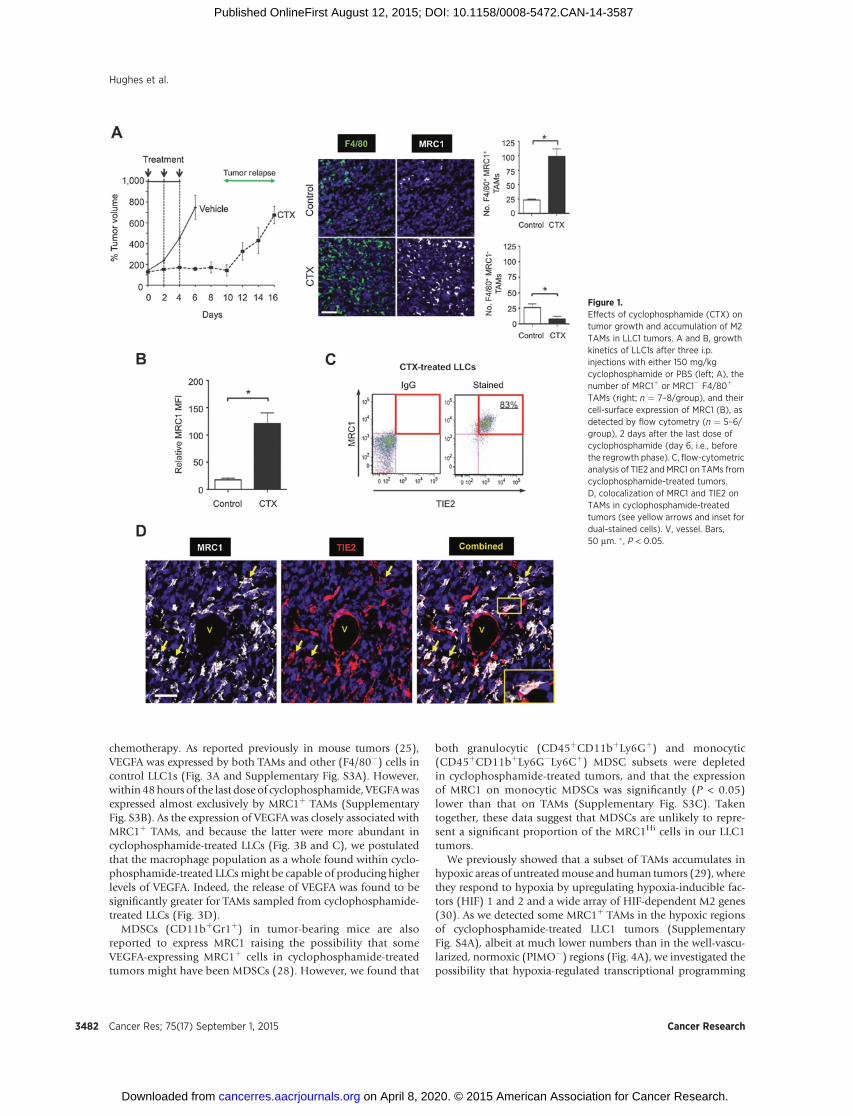
Three i.p. injections of the cytotoxic agent, cyclophosphamide,resulted in the complete cessation of LLC1 tumor growth, fol-
lowedby regrowthbeginning 7days after treatment stops (Fig. 1A,left). Forty-eight hours after the last cyclophosphamide injection(day 6), LLC1s contained significantly (P < 0.05) shorter bloodvessels and more hypoxia than size-matched controls (Supple-mentary Fig. S1A). Consistent with earlier observations forMMTV-PyMT implants treated with paclitaxel (13), TAMs wereenriched among those leukocytes present in LLC1s 48 hours aftercyclophosphamide (Supplementary Fig. S1B). In addition,there was a significant (P < 0.05) increase in the overall numberof F4/80þ TAMs in cyclophosphamide-treated LLCs comparedwith size-matched controls. This was also seen in paclitaxel-treated, orthotopic 4T1 tumors, and doxorubicin-treated ortho-topic MMTV-PyMT implants (Supplementary Fig. S1C).
Cyclophosphamide treatment of LLC1s resulted in a significant(P < 0.05) increase in the number of F4/80þ TAMs expressing theM2-marker, MRC1, relative to size-matched controls. In contrast,there was a significant (P < 0.05) drop in F4/80þ/MRC1� TAMsafter cyclophosphamide (Fig. 1A, right). Also, TAMs from cyclo-phosphamide-treated LLC1s expressed higher surface MRC1 thanthose from size-matched controls (Fig. 1B). Consistent withprevious findings in untreated mouse tumors (9, 10), MRC1Hi
TAMs in cyclophosphamide-treated LLC1s coexpressed elevatedTIE2 (Fig. 1C and D). A similar increase inMRC1þ TAMs was alsoseen in orthotopic 4T1 andMMTV-PyMT implants after paclitaxeland doxorubicin, respectively (Supplementary Fig. S1D). In allthreemodels, the vastmajority ofMRC1þ cells F4/80þ TAMs, andthe small number of F4/80�MRC1þ (presumably dendritic) cellswere not significantly increased after treatment with cyclophos-phamide, paclitaxel, or doxorubicin (Supplementary Fig. S2A).
BrdUrd uptake was negligible in MRC1þ TAMs in both size-matched control and cyclophosphamide-treated LLC1 tumors,indicating their nonproliferative status (Supplementary Fig. S2B).As chemotherapy-induced changes in circulating hematopoieticstem andprogenitor cells (HS/PCs) could, in theory, contribute tothe increase of TAM numbers after therapy, we also performeddual immunofluorescence labeling for HS/PC markers, c-Kit andSca1. Although c-KitþSca1þ cells were detected in the spleens ofLLC1 tumor-bearing mice (positive control for the staining), nosuch cells were detected in either control or cyclophosphamide-treated LLC1s (Supplementary Fig. S2C). These data indicatethat the chemotherapy-induced increase in TAMs is most likelyto be due to increased monocyte recruitment rather than theproliferation of existing TAMs or their differentiation fromrecruited HS/PCs.
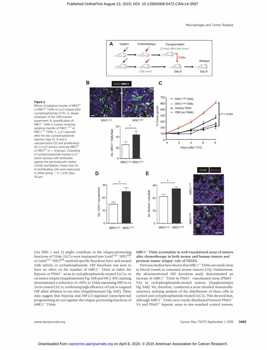
We then isolated F4/80þMRC1þ/Hi and F4/80þMRC1�/Lo
TAMs from cyclophosphamide-treated LLC1 tumors by FACS andinjected them into cyclophosphamide-treated LLC1 tumors inlittermates (Fig. 2A). MRC1þ/Hi TAMs, but not MRC1�/Lo TAMsfrom treated tumors or TAMs from vehicle-treated tumors, accel-erated tumor regrowth after cyclophosphamide. This was accom-panied by a significant (P < 0.05) increase in the number ofMRC1þ TAMs, CD31þ blood vessels, and a moderate increase inBrdUrdþ (proliferating) cells in cyclophosphamide-treated LLC1sreceiving MRC1þ/Hi TAMs. Very few (< 1%) of CD45þ leukocytescontained immunodetectable BrdUrd (Fig. 2B–E).
MRC1þ TAMs are proangiogenic in untreated tumors andexpress the important proangiogenic mediator VEGFA (9, 26).Moreover, myeloid-specific deletion of VEGFA is known to haveprofound effects on the vascularization and progression ofuntreated mouse tumors (27). We, therefore, investigated therole of VEGFA derived from MRC1þ TAMs in tumor-relapse after
Macrophages and Tumor Relapse
www.aacrjournals.org Cancer Res; 75(17) September 1, 2015 3481
on April 8, 2020. © 2015 American Association for Cancer Research. cancerres.aacrjournals.org Downloaded from
Published OnlineFirst August 12, 2015; DOI: 10.1158/0008-5472.CAN-14-3587
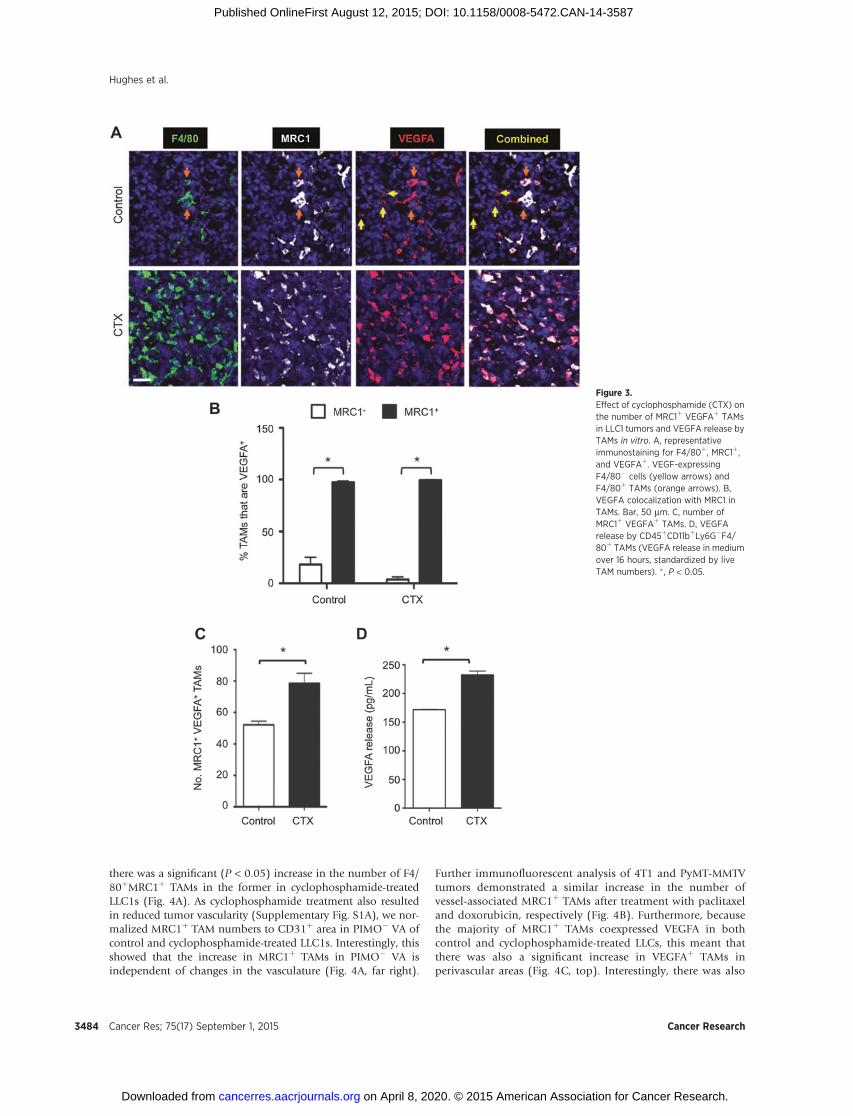
chemotherapy. As reported previously in mouse tumors (25),VEGFA was expressed by both TAMs and other (F4/80�) cells incontrol LLC1s (Fig. 3A and Supplementary Fig. S3A). However,within 48hours of the last dose of cyclophosphamide, VEGFAwasexpressed almost exclusively by MRC1þ TAMs (SupplementaryFig. S3B). As the expression of VEGFA was closely associated withMRC1þ TAMs, and because the latter were more abundant incyclophosphamide-treated LLCs (Fig. 3B and C), we postulatedthat the macrophage population as a whole found within cyclo-phosphamide-treated LLCsmight be capable of producing higherlevels of VEGFA. Indeed, the release of VEGFA was found to besignificantly greater for TAMs sampled from cyclophosphamide-treated LLCs (Fig. 3D).
MDSCs (CD11bþGr1þ) in tumor-bearing mice are alsoreported to express MRC1 raising the possibility that someVEGFA-expressing MRC1þ cells in cyclophosphamide-treatedtumors might have been MDSCs (28). However, we found that
both granulocytic (CD45þCD11bþLy6Gþ) and monocytic(CD45þCD11bþLy6G�Ly6Cþ) MDSC subsets were depletedin cyclophosphamide-treated tumors, and that the expressionof MRC1 on monocytic MDSCs was significantly (P < 0.05)lower than that on TAMs (Supplementary Fig. S3C). Takentogether, these data suggest that MDSCs are unlikely to repre-sent a significant proportion of the MRC1Hi cells in our LLC1tumors.
We previously showed that a subset of TAMs accumulates inhypoxic areas of untreatedmouse and human tumors (29), wherethey respond to hypoxia by upregulating hypoxia-inducible fac-tors (HIF) 1 and 2 and a wide array of HIF-dependent M2 genes(30). As we detected some MRC1þ TAMs in the hypoxic regionsof cyclophosphamide-treated LLC1 tumors (SupplementaryFig. S4A), albeit at much lower numbers than in the well-vascu-larized, normoxic (PIMO�) regions (Fig. 4A), we investigated thepossibility that hypoxia-regulated transcriptional programming
Figure 1.Effects of cyclophosphamide (CTX) ontumor growth and accumulation of M2TAMs in LLC1 tumors. A and B, growthkinetics of LLC1s after three i.p.injections with either 150 mg/kgcyclophosphamide or PBS (left; A), thenumber of MRC1þ or MRC1� F4/80þ
TAMs (right; n ¼ 7–8/group), and theircell-surface expression of MRC1 (B), asdetected by flow cytometry (n ¼ 5–6/group), 2 days after the last dose ofcyclophosphamide (day 6, i.e., beforethe regrowth phase). C, flow-cytometricanalysis of TIE2 and MRC1 on TAMs fromcyclophosphamide-treated tumors.D, colocalization of MRC1 and TIE2 onTAMs in cyclophosphamide-treatedtumors (see yellow arrows and inset fordual-stained cells). V, vessel. Bars,50 mm. � , P < 0.05.
Hughes et al.
Cancer Res; 75(17) September 1, 2015 Cancer Research3482
on April 8, 2020. © 2015 American Association for Cancer Research. cancerres.aacrjournals.org Downloaded from
Published OnlineFirst August 12, 2015; DOI: 10.1158/0008-5472.CAN-14-3587
(via HIFs 1 and 2) might contribute to the relapse-promotingfunctions of TAMs. LLC1s were implanted into LysMCre/þHIF1fl/fl
or LysMcre/þHIF2fl/fl myeloid-specific knockout mice and treatedwith vehicle or cyclophosphamide. HIF knockout was seen tohave no effect on the number of MRC1þ TAMs in either thehypoxic or PIMO� areas in cyclophosphamide-treated LLC1s, oron tumor relapse (Supplementary Fig. S4B and S4C). IHC stainingdemonstrated a reduction of >90% in TAMs expressing HIF1a or2a in control LLC1s, confirming high efficiency of LysCre-targetedHIF allele ablation in our mice (Supplementary Fig. S4D). Thesedata suggest that hypoxia and HIF1/2-regulated transcriptionalprogramming do not regulate the relapse-promoting functions ofMRC1þ TAMs.
MRC1þ TAMs accumulate in well-vascularized areas of tumorsafter chemotherapy in both mouse and human tumors andpromote tumor relapse: role of VEGFA
Previous studies have shown thatMRC1þTAMs can reside closeto blood vessels in untreated mouse tumors (10). Furthermore,the aforementioned HIF knockout study demonstrated anincrease in MRC1þ TAMs in PIMO� vascularized areas (PIMO�
VA) in cyclophosphamide-treated tumors (SupplementaryFig. S4B). We, therefore, conducted a more detailed immunoflu-orescence staining analysis of the distribution of these cells incontrol and cyclophosphamide-treated LLC1s. This showed that,although MRC1þ TAMs were evenly distributed between PIMO�
VA and PIMOþ hypoxic areas in size-matched control tumors,
Figure 2.Effects of adoptive transfer of MRC1Hi
vs MRC1Lo TAMs on LLC1 relapse aftercyclophosphamide (CTX). A, designschematic of the TAM transferexperiment. B, quantification ofMRC1þ TAMs in tumors receivingadoptive transfer of MRC1�/Lo orMRC1þ/Hi TAMs. C, LLC1 regrowthafter the last cyclophosphamideinjection (day 0). D and E,vascularization (D) and proliferation(E) in LLC1 tumors receiving MRC1Hi
or MRC1Lo (n ¼ 5/group). Costainingof cyclophosphamide-treated LLC1tumor sections with antibodiesagainst the pan-leukocyte marker(CD45) and BrdUrd. Fewer than 1%of proliferating cells were leukocytesin either group. � , P < 0.05. Bars,50 mm.
Macrophages and Tumor Relapse
www.aacrjournals.org Cancer Res; 75(17) September 1, 2015 3483
on April 8, 2020. © 2015 American Association for Cancer Research. cancerres.aacrjournals.org Downloaded from
Published OnlineFirst August 12, 2015; DOI: 10.1158/0008-5472.CAN-14-3587
there was a significant (P < 0.05) increase in the number of F4/80þMRC1þ TAMs in the former in cyclophosphamide-treatedLLC1s (Fig. 4A). As cyclophosphamide treatment also resultedin reduced tumor vascularity (Supplementary Fig. S1A), we nor-malized MRC1þ TAM numbers to CD31þ area in PIMO� VA ofcontrol and cyclophosphamide-treated LLC1s. Interestingly, thisshowed that the increase in MRC1þ TAMs in PIMO� VA isindependent of changes in the vasculature (Fig. 4A, far right).
Further immunofluorescent analysis of 4T1 and PyMT-MMTVtumors demonstrated a similar increase in the number ofvessel-associated MRC1þ TAMs after treatment with paclitaxeland doxorubicin, respectively (Fig. 4B). Furthermore, becausethe majority of MRC1þ TAMs coexpressed VEGFA in bothcontrol and cyclophosphamide-treated LLCs, this meant thatthere was also a significant increase in VEGFAþ TAMs inperivascular areas (Fig. 4C, top). Interestingly, there was also
Figure 3.Effect of cyclophosphamide (CTX) onthe number of MRC1þ VEGFAþ TAMsin LLC1 tumors and VEGFA release byTAMs in vitro. A, representativeimmunostaining for F4/80þ, MRC1þ,and VEGFAþ. VEGF-expressingF4/80� cells (yellow arrows) andF4/80þ TAMs (orange arrows). B,VEGFA colocalization with MRC1 inTAMs. Bar, 50 mm. C, number ofMRC1þ VEGFAþ TAMs. D, VEGFArelease by CD45þCD11bþLy6G�F4/80þ TAMs (VEGFA release in mediumover 16 hours, standardized by liveTAM numbers). � , P < 0.05.
Hughes et al.
Cancer Res; 75(17) September 1, 2015 Cancer Research3484
on April 8, 2020. © 2015 American Association for Cancer Research. cancerres.aacrjournals.org Downloaded from
Published OnlineFirst August 12, 2015; DOI: 10.1158/0008-5472.CAN-14-3587
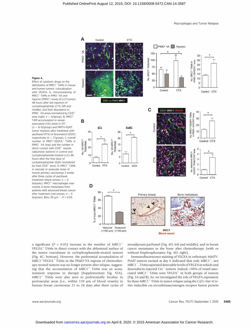
a significant (P < 0.05) increase in the number of MRC1þ
VEGFAþ TAMs in direct contact with the abluminal surface ofthe tumor vasculature in cyclophosphamide-treated tumors(Fig. 4C, bottom). However, the preferential accumulation ofMRC1þVEGFAþ TAMs in the PIMO_VA regions of chemother-apy-treated tumors was no longer present after relapse, suggest-ing that the accumulation of MRC1þ TAMs was an acute,transient response to therapy (Supplementary Fig. S5A).MRC1þ TAMs were also seen to preferentially localize inperivascular areas (i.e., within 150 mm of blood vessels) inhuman breast carcinomas 21 to 28 days after three cycles of
neoadjuvant paclitaxel (Fig. 4D, left and middle), and in breastcancer metastases in the bone after chemotherapy (with orwithout bisphosphonates; Fig. 4D, right).
Immunofluorescence staining of VEGFA in orthotopic MMTV-PyMT tumors excised at day 6 indicated that only MRC1þ, notMRC1�, TAMs expresseddetectable levels of VEGFA in vehicle anddoxorubicin-injected Cre� tumors. Indeed, >98% of vessel-asso-ciated MRC1þ TAMs were VEGFAþ in both groups of tumors(Fig. 5A and B). So, we investigated the role of VEGFA expressionby these MRC1þ TAMs in tumor relapse using the Csf1r-Mer-iCre-Mer inducible cre-recombinase/estrogen receptor fusion protein
Figure 4.Effect of cytotoxic drugs on thedistribution of MRC1þ TAMs in mouseand human tumors: colocalizationwith VEGFA. A, immunostaining ofMRC1þ TAMs in PIMO�VA andhypoxic (PIMOþ) areas of LLC1 tumors48 hours after last injection ofcyclophosphamide (CTX; left andmiddle), and their abundance inPIMO� VA areas normalized by CD31þ
area (right; n ¼ 4/group). B, MRC1þ
TAM accumulation in vessel-associated (VA) areas in 4T1(n ¼ 8–9/group) and MMTV-PyMTtumor implants after treatment withpaclitaxel (PTX) or doxorubicin (DOX)respectively (n ¼ 7/group). C, overallnumber of MRC1þVEGFAþ TAMs inPIMO� VA (top) and the number indirect contact with CD31þ vessels(abluminal; bottom) in control andcyclophosphamide-treated LLCs 48hours after the final dose ofcyclophosphamide (both normalizedby total CD31þ area). D, MRC1þ TAMsin vascular or avascular areas ofhuman primary carcinomas 3 weeksafter three cycles of paclitaxeltreatment (black arrows; n ¼ 4biopsies). MRC1þ macrophages nearvessels in bone metastases frompatients with advanced breast cancerafter treatment (red arrows; n ¼ 4biopsies). Bars, 50 mm. � , P < 0.05.
Macrophages and Tumor Relapse
www.aacrjournals.org Cancer Res; 75(17) September 1, 2015 3485
on April 8, 2020. © 2015 American Association for Cancer Research. cancerres.aacrjournals.org Downloaded from
Published OnlineFirst August 12, 2015; DOI: 10.1158/0008-5472.CAN-14-3587
model for the tamoxifen-induced ablation of VEGFA selectively inmonocytes/macrophages (23).
Female Csf1r-Mer-iCre-Mer FVB/n mice were orthotopicallyimplantedwith syngeneicMMTV-PyMT tumors and administeredtamoxifen 24 hours after a single injection of either vehicle ordoxorubicin. In addition, tamoxifen was given continuouslythereafter to ensure VEGFA knockout until mice were sacrificedat the end of the experiment (day 14), with the same treatment
given to control Cre-recombinase negative mice (Fig. 5C). Asthe implanted PyMT cells in these tumors did not carry theCsf1r-driven Cre recombinase and expression of Csf1r is largelyconfined to TAMs in such tumors, the knockdown of VEGFA wasrestricted to the macrophage lineage, a significant source ofVEGFA in the PyMT model (as in cyclophosphamide-treatedLLC1s; ref. 31). Tamoxifen-induced ablation of this TAM-derivedVEGFA (Fig. 5C) caused a significant (P<0.05)delay in the growth
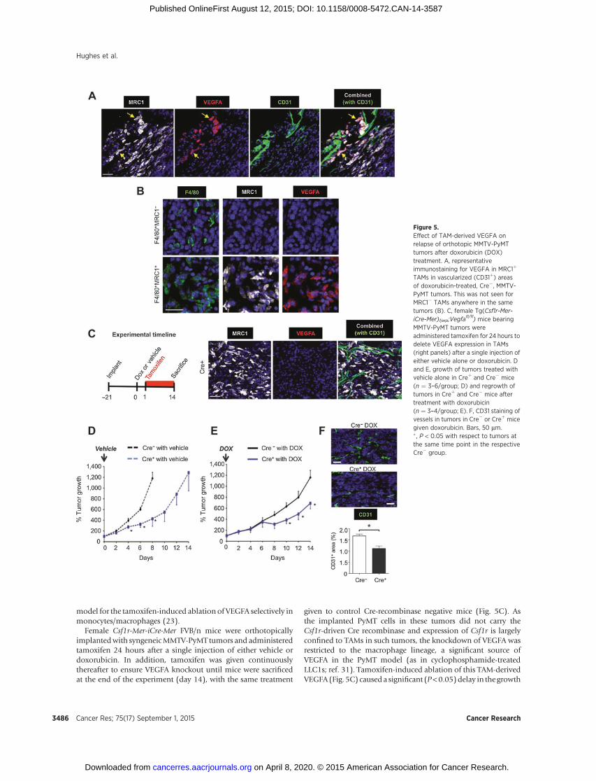
Figure 5.Effect of TAM-derived VEGFA onrelapse of orthotopic MMTV-PyMTtumors after doxorubicin (DOX)treatment. A, representativeimmunostaining for VEGFA in MRC1þ
TAMs in vascularized (CD31þ) areasof doxorubicin-treated, Cre�, MMTV-PyMT tumors. This was not seen forMRC1� TAMs anywhere in the sametumors (B). C, female Tg(Csf1r-Mer-iCre-Mer)1jwp;Vegfa
fl/fl) mice bearingMMTV-PyMT tumors wereadministered tamoxifen for 24 hours todelete VEGFA expression in TAMs(right panels) after a single injection ofeither vehicle alone or doxorubicin. Dand E, growth of tumors treated withvehicle alone in Creþ and Cre� mice(n ¼ 3–6/group; D) and regrowth oftumors in Creþ and Cre� mice aftertreatment with doxorubicin(n ¼ 3–4/group; E). F, CD31 staining ofvessels in tumors in Cre� or Creþ micegiven doxorubicin. Bars, 50 mm.� , P < 0.05 with respect to tumors atthe same time point in the respectiveCre� group.
Hughes et al.
Cancer Res; 75(17) September 1, 2015 Cancer Research3486
on April 8, 2020. © 2015 American Association for Cancer Research. cancerres.aacrjournals.org Downloaded from
Published OnlineFirst August 12, 2015; DOI: 10.1158/0008-5472.CAN-14-3587
of vehicle-treated MMTV-PyMT tumors (Fig. 5D). Furthermore,relapse of doxorubicin-treated tumors lacking VEGFAþ TAMs(Creþ) was significantly (P < 0.05) slower than that of doxoru-bicin-treated tumors in VEGFA-expressing (Cre�) mice (Fig. 5E).Thiswas accompaniedby a significant (P<0.05) decrease in vesselarea in these tumors (Fig. 5F).
Pharmacologic blockade of CXCR4 prevents perivascularaccumulationofMRC1þVEGFAþTAMsafter chemotherapy anddelays tumor relapse.
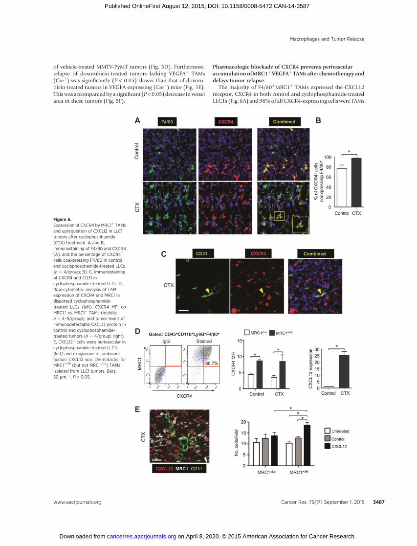
The majority of F4/80þMRC1þ TAMs expressed the CXCL12receptor, CXCR4 in both control and cyclophosphamide-treatedLLC1s (Fig. 6A) and 98%of all CXCR4-expressing cells were TAMs
Figure 6.Expression of CXCR4 by MRC1þ TAMsand upregulation of CXCLl2 in LLC1tumors after cyclophosphamide(CTX) treatment. A and B,immunostaining of F4/80 and CXCR4(A), and the percentage of CXCR4þ
cells coexpressing F4/80 in controland cyclophosphamide-treated LLCs(n ¼ 4/group; B). C, immunostainingof CXCR4 and CD31 incyclophosphamide-treated LLCs. D,flow-cytometric analysis of TAMexpression of CXCR4 and MRC1 indispersed cyclophosphamide-treated LLCs (left), CXCR4 MFI onMRC1þ vs. MRC1� TAMs (middle;n ¼ 4–5/group), and tumor levels ofimmunodetectable CXCL12 protein incontrol and cyclophosphamide-treated tumors (n ¼ 4/group; right).E, CXCL12þ cells were perivascular incyclophosphamide-treated LLC1s(left) and exogenous recombinanthuman CXCL12 was chemotactic forMRC1þ/Hi (but not MRC�/Lo) TAMsisolated from LLC1 tumors. Bars,50 mm. �, P < 0.05.
Macrophages and Tumor Relapse
www.aacrjournals.org Cancer Res; 75(17) September 1, 2015 3487
on April 8, 2020. © 2015 American Association for Cancer Research. cancerres.aacrjournals.org Downloaded from
Published OnlineFirst August 12, 2015; DOI: 10.1158/0008-5472.CAN-14-3587
in cyclophosphamide-treated LLC1 tumors, compared with 78%in control LLCs (where other cell types were also CXCR4þ
; Fig. 6B). As reported previously (32, 33), LLC1 cells do notexpress CXCR4 (Fig. 6A and B).Moreover, less than 1%of CD31þ
blood vessels expressed CXCR4 in cyclophosphamide-treatedtumors (Fig. 6C). MRC1Hi TAMs in both control and cyclophos-phamide-treated tumors expressed elevated levels of CXCR4compared with the MRC1Lo TAMs (Fig. 6D).
These changes in CXCR4þ cells after cyclophosphamide wereaccompanied by a marked increase in tumor levels of its ligand,CXCL12, on day 6 (Fig. 6D, far right). These CXCL12þ cells wereCD31� (Fig. 6E, left) and most likely tumor cells and/or fibro-blasts (34). As a hypoxia-inducible gene (35), itwas not surprisingto find CXCL12 more highly expressed in hypoxic than normoxicareas of control LLC1 tumors. However, after cyclophosphamide,tumor levels of HIF1 and -2 were markedly reduced in all areas oftumors (Supplementary Fig. S6A, i and ii), but CXCL12 wasabundant in both hypoxic and normoxic, vascularized areas(Supplementary Fig. S6B). This suggested CXCL12 upregulationby factors other than hypoxia in such tumors.
Cyclophosphamide is known to induce oxidative stress (36), acellular response known to regulate the expression of CXCL12(37), so we investigated the expression of a well-defined markerfor oxidative stress, heme oxygenase-1 (HMOX-1) in controlversus cyclophosphamide-treated tumors. Interestingly, this wasfound to be upregulated in perivascular, PIMO�, CXCL12-richareas of tumors after cyclophosphamide treatment but not incontrol tumors (Supplementary Fig. S6C, i and ii). Both MRC1þ
TAMs and MRC1� cells expressed HMOX-1 in these vascularizedareas (Supplementary Fig. S6C, iii).
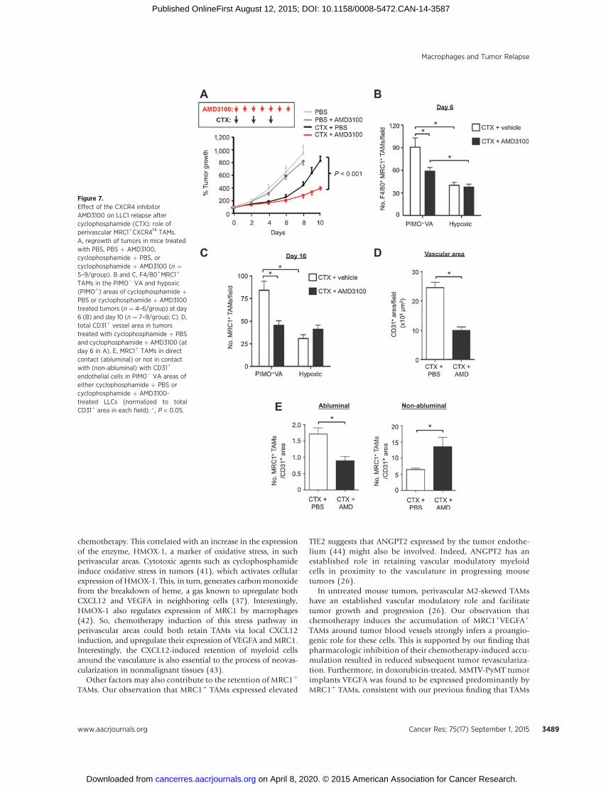
We then investigated whether CXCL12 might recruit and/orretain CXCR4þMRC1þ TAMs in LLC1 tumors. First, we showed inan in vitro chemotaxis assay that CXCL12 is selectively chemotacticfor MRC1þ/Hi TAMs isolated from LLC1 tumors (Fig. 6E, right).Then we administered cyclophosphamide to LLC1-bearing micealone or in combination with the CXCR4 antagonist, plerixafor(AMD3100; Fig 7A). This was feasible as TAMs were the predom-inant cell type expressing CXCR4 after cyclophosphamide(Fig. 6B). CXCR4 blockade significantly inhibited cyclophospha-mide-induced accumulation of F4/80þ MRC1þ TAMs in thePIMO� VA (while the numbers in PIMOþ areas of LLC1 tumorswere unchanged, as was F4/80þ MRC1� TAMs in either of theseareas) and delayed tumor relapse (Fig. 7A and B; SupplementaryS5B). At day 10 (when tumors had relapsed in the cyclophos-phamide alone group), a similar distribution of MRC1þ TAMswas seen compared with day 6 (the start of the relapse period;Fig. 7B and C). In addition, tumors administered cyclophos-phamide þ AMD3100 contained significantly (P < 0.05)shorter CD31þ blood vessels, higher levels of hypoxia, andfewer BrdUrdþ (proliferating) cells than tumors exposed tocyclophosphamide with the vehicle for AMD3100 (PBS; Fig.7D; Supplementary Fig. S5C).
We then examined the effect of CXCR4 blockade on thedistribution of MRC1þ TAMs across PIMO� VAs in cyclophos-phamide-treated tumors. Interestingly, this was found to signif-icantly (P < 0.05) reduce the number of MRC1þ TAMs in directcontact with the abluminal surface of CD31þ blood vessels(standardized by CD31þ vessel area as this differed betweencyclophosphamide þ PBS and cyclophosphamide þ AMD3100groups), and increase them elsewhere in the PIMO�VAs (Fig. 7E).These data suggest that CXCR4 regulates the direct association of
alternatively activated TAMs with blood vessels in cyclophospha-mide-treated LLC1s.
A recent study demonstrated a marked rebound in the growthof pulmonary metastases after treatment with an antibody toCCL2 (38). This was the result of increased mobilization ofmonocytes from the bone marrow after CCL2 inhibition, andincreased blood vessel formation and cancer cell proliferation inthe lungs. We, therefore, investigated the possibility of a similarrebound effect after CXCR4 inhibition using AMD3100 as thiscould disrupt the bone marrow niche. When we extended thelength of the AMD3100 experiment, primary LLC1 tumorsrelapsed eventually, but not at an accelerated rate, and micereceiving cyclophosphamide þ AMD3100 showed increased sur-vival compared with those in the cyclophosphamide alone group(or controls). Importantly, there was also no rebound in pulmo-nary metastases (Supplementary Fig. S8).
DiscussionOur studies show that M2-skewed TAMs (MRC1þTIE2þ-
CXCR4HiVEGFAþ) selectively accumulate in vascularized, well-oxygenated areas of LLC1 tumors after treatment with cyclophos-phamide. A similar increase in such vessel-associated, M2-skewedTAMswas also seen in orthotopic 4T1 andMMTV-PyMT implantsafter treatment with paclitaxel and doxorubicin, respectively, andin human breast carcinomas after neoadjuvant treatment withpaclitaxel. This perivascular accumulation was found to beCXCR4-dependent, especially the increased, direct contact of thisTAM subset with the abluminal surface of blood vessels inchemotherapy-treated tumors. When this was disrupted using aCXCR4 inhibitor, tumor revascularization and regrowth afterchemotherapy were markedly impaired. We also show, using aninducible, macrophage-specific, gene knockdown model thatVEGFA expressed by such MRC1þ TAMs mediates, in part, theirability to promote tumor relapse after therapy. Consistent withthis, genetic deletion of HIF signaling in TAMs in hypoxic tumorareas had no effect on this rescue.
The mobilization and accumulation in tumors, of suchBMDCs as myelomonocytic cells, MDSCs, endothelial progen-itor cells (EPC) and macrophages after various forms of anti-cancer therapy are now well established (13–19, 39, 40).However, our data show that M2-skewed TAMs represent asignificant proportion of such BMDCs in tumors after chemo-therapy, and is not accompanied by TAM proliferation orincreased numbers of stem/progenitor cells. These data suggestthat increased recruitment of circulating monocytes precedesthe accumulation of such M2 TAMs in tumors after chemo-therapy. It remains to be seen whether monocytes are alreadyM2-skewed upon arrival in treated tumors and/or activated byfactors produced in the perivascular niche. Of note, the recruit-ment, activation, and perivascular retention of these cells doesnot appear to be just an acute response to chemotherapy-induced tumor damage, as increased numbers of perivascularM2 TAMs persisted throughout the relapse phase.
Our observations suggest that the accumulation of MRC1þ
VEGFAþ TAMs on and around the tumor vasculature plays animportant part in tumor revascularization and relapse after che-motherapy. Furthermore, this was accompanied by a markedchange in the pattern of CXCL12 expression in tumors. AlthoughCXCL12 was mainly confined to hypoxic areas of control tumors,it was upregulated in vascularized, well-oxygenated areas after
Hughes et al.
Cancer Res; 75(17) September 1, 2015 Cancer Research3488
on April 8, 2020. © 2015 American Association for Cancer Research. cancerres.aacrjournals.org Downloaded from
Published OnlineFirst August 12, 2015; DOI: 10.1158/0008-5472.CAN-14-3587
chemotherapy. This correlated with an increase in the expressionof the enzyme, HMOX-1, a marker of oxidative stress, in suchperivascular areas. Cytotoxic agents such as cyclophosphamideinduce oxidative stress in tumors (41), which activates cellularexpression of HMOX-1. This, in turn, generates carbonmonoxidefrom the breakdown of heme, a gas known to upregulate bothCXCL12 and VEGFA in neighboring cells (37). Interestingly,HMOX-1 also regulates expression of MRC1 by macrophages(42). So, chemotherapy induction of this stress pathway inperivascular areas could both retain TAMs via local CXCL12induction, and upregulate their expression of VEGFA and MRC1.Interestingly, the CXCL12-induced retention of myeloid cellsaround the vasculature is also essential to the process of neovas-cularization in nonmalignant tissues (43).
Other factors may also contribute to the retention of MRC1þ
TAMs. Our observation that MRC1þ TAMs expressed elevated
TIE2 suggests that ANGPT2 expressed by the tumor endothe-lium (44) might also be involved. Indeed, ANGPT2 has anestablished role in retaining vascular modulatory myeloidcells in proximity to the vasculature in progressing mousetumors (26).
In untreated mouse tumors, perivascular M2-skewed TAMshave an established vascular modulatory role and facilitatetumor growth and progression (26). Our observation thatchemotherapy induces the accumulation of MRC1þVEGFAþ
TAMs around tumor blood vessels strongly infers a proangio-genic role for these cells. This is supported by our finding thatpharmacologic inhibition of their chemotherapy-induced accu-mulation resulted in reduced subsequent tumor revasculariza-tion. Furthermore, in doxorubicin-treated, MMTV-PyMT tumorimplants VEGFA was found to be expressed predominantly byMRC1þ TAMs, consistent with our previous finding that TAMs
Figure 7.Effect of the CXCR4 inhibitorAMD3100 on LLC1 relapse aftercyclophosphamide (CTX): role ofperivascular MRC1þCXCR4Hi TAMs.A, regrowth of tumors in mice treatedwith PBS, PBS þ AMD3100,cyclophosphamide þ PBS, orcyclophosphamide þ AMD3100 (n ¼5–9/group). B and C, F4/80þMRC1þ
TAMs in the PIMO� VA and hypoxic(PIMOþ) areas of cyclophosphamide þPBS or cyclophosphamide þ AMD3100treated tumors (n ¼ 4–6/group) at day6 (B) and day 10 (n ¼ 7–9/group; C). D,total CD31þ vessel area in tumorstreated with cyclophosphamide þ PBSand cyclophosphamideþ AMD3100 (atday 6 in A). E, MRC1þ TAMs in directcontact (abluminal) or not in contactwith (non-abluminal) with CD31þ
endothelial cells in PIMO� VA areas ofeither cyclophosphamide þ PBS orcyclophosphamide þ AMD3100-treated LLCs (normalized to totalCD31þ area in each field). � , P < 0.05.
Macrophages and Tumor Relapse
www.aacrjournals.org Cancer Res; 75(17) September 1, 2015 3489
on April 8, 2020. © 2015 American Association for Cancer Research. cancerres.aacrjournals.org Downloaded from
Published OnlineFirst August 12, 2015; DOI: 10.1158/0008-5472.CAN-14-3587
are a major source of VEGFA in MMTV-PyMT tumors (31). Theselective ablation of VEGFA in MRC1þ TAMs resulted indelayed tumor relapse, post-therapy. However, PyMT tumorsstill relapsed, albeit at a reduced rate, in the absence of TAM-derived VEGFA, indicating that other TAM-derived factors mayalso contribute to tumor relapse and/or the possible inductionof resistance mechanisms in tumors to VEGFA knockout. Thepresence of the latter has been demonstrated in tumors afteranti-VEGFA therapy and shown to include increased expressionof such alternative proangiogenic mediators as FGF2 (45),ANGPT2 (46), or PlGF (47) and the recruitment of tumor-promoting, CD11bþ Gr1þ myeloid cells (48).
Recently, the Condeelis group has used intravital imaging tocharacterize a distinct subset of perivascular TAMs in untreatedmouse tumors thatmake contact with endothelial cells and tumorcells expressing high levels of Mena (a protein that enhances theirmotility), and directly stimulate tumor cell intravasation (49).These cell trios have been termed "tumor microenvironments ofmetastasis" (TMEM). It is possible that some of the perivascularMRC1þ M2-skewed TAMs accumulating around tumor bloodvessels after chemotherapy form TMEMs and promote metastasisas well as relapse, a dangerous combination in patients withinoperable tumors.
The effect of CXCR4 blockade on tumor relapse after chemo-therapy in our study suggests that CXCR4 inhibitors might besuccessfully combined with chemotherapy. This combinationcould extend relapse-free survival in patients with inoperabletumors, although our data suggests that multiple rounds of theinhibitor would have to be administered to maintain a suppres-sive effect on relapse. Such sustained use of a CXCR4 antagonistafter chemotherapy could conceivably disrupt the marrow niche.However, this is unlikely to lead to clinical problems as a recent,first-in-human clinical trial has shown that repeated daily injec-tions of the CXCR4 antagonist, LY2510924 over a number ofconsecutive, 28-day cycles was well tolerated in advanced cancerpatients (50).
Taken together, our data suggest that the selective targeting ofrelapse-promoting, perivascular TAMs could delay the relapse ofboth primary and metastatic tumors in patients after chemother-apy, thereby extending their relapse-free survival.
Disclosure of Potential Conflicts of InterestNo potential conflicts of interest were disclosed.
Authors' ContributionsConception and design: R. Hughes, B.-Z. Qian, M. Muthana, J.W. Pollard,C.E. LewisDevelopment ofmethodology:R.Hughes, B.-Z.Qian, C. Addison, J.W. Pollard,C.E. LewisAcquisition of data (provided animals, acquired and managed patients,provided facilities, etc.): R. Hughes, B.-Z. Qian, C. Rowan, I. Keklikoglou,O.C. Olson, S. Tazzyman, C. Addison, M. Clemons, A.M. Gonzalez-Angulo,J.A Joyce, M. De Palma, J.W. Pollard, C.E. LewisAnalysis and interpretation of data (e.g., statistical analysis, biostatistics,computational analysis): R. Hughes, B.-Z. Qian, C. Rowan, S. Tazzyman,S. Danson, J.W. Pollard, C.E. LewisWriting, review, and/or revision of the manuscript: R. Hughes, B.-Z. Qian,S. Danson, C. Addison, A.M. Gonzalez-Angulo, J.A Joyce, M. De Palma,J.W. Pollard, C.E. LewisAdministrative, technical, or material support (i.e., reporting or organizingdata, constructing databases): R. Hughes, M. MuthanaStudy supervision: R. Hughes, M. Muthana, J.W. Pollard, C.E. Lewis
Grant SupportThis work was supportedmainly by a Cancer Research UK grant (C.E. Lewis),
the grant support of the Swiss Cancer League (M. De Palma), Breast CancerResearch Foundation (O.C. Olson and J.A Joyce), a Chancellor's Fellowshipfrom the University of Edinburgh (B.-Z. Qian), and TheWellcome Trust (SeniorInvestigator Award; J.W. Pollard).
The costs of publication of this articlewere defrayed inpart by the payment ofpage charges. This article must therefore be hereby marked advertisement inaccordance with 18 U.S.C. Section 1734 solely to indicate this fact.
Received December 10, 2014; revised July 3, 2015; accepted July 10, 2015;published OnlineFirst August 12, 2015.
References1. Savir G, Huber KE, Saif MW. Locally advanced pancreatic cancer. Looking
beyond traditional chemotherapy and radiation. JOP 2013;14:337–9.2. Strom HH, Bremnes RM, Sundstrom SH, Helbekkmo N, Aasebo U. Poor
prognosis patients with inoperable locally advanced NSCLC and largetumors benefit frompalliative chemoradiotherapy: a subset analysis fromarandomized clinical phase III trial. J Thorac Oncol 2014;9:825–33.
3. Coffelt SB, Lewis CE, Naldini L, Brown JM, Ferrara N, De Palma M. Elusiveidentities and overlapping phenotypes of proangiogenic myeloid cells intumors. Am J Pathol 2010;176:1564–76.
4. Noy R, Pollard JW. Tumor-Associated Macrophages: From Mechanisms toTherapy. Immunity 2014;41:49–61.
5. Qian BZ, Pollard JW. Macrophage diversity enhances tumor progressionand metastasis. Cell 2010;141:39–51.
6. Movahedi K, Laoui D, Gysemans C, BaetenM, Stange G, Van den Bossche J,et al. Different tumor microenvironments contain functionally distinctsubsets of macrophages derived from Ly6C(high) monocytes. Cancer Res2010;70:5728–39.
7. Coffelt SB, Hughes R, Lewis CE. Tumor-associated macrophages: effectorsof angiogenesis and tumor progression. Biochimica et biophysica acta2009;1796:11–8.
8. Biswas SK, Sica A, Lewis CE. Plasticity of macrophage function duringtumor progression: regulation by distinct molecular mechanisms. J Immu-nol 2008;180:2011–7.
9. Pucci F, Venneri MA, Biziato D, Nonis A, Moi D, Sica A, et al. A distinguish-ing gene signature shared by tumor-infiltrating Tie2-expressingmonocytes,
blood "resident" monocytes, and embryonic macrophages suggestscommon functions and developmental relationships. Blood 2009;114:901–14.
10. De Palma M, Venneri MA, Galli R, Sergi Sergi L, Politi LS, Sampaolesi M,et al. Tie2 identifies a hematopoietic lineage of proangiogenic monocytesrequired for tumor vessel formation and a mesenchymal population ofpericyte progenitors. Cancer Cell 2005;8:211–26.
11. Daenen LG, Houthuijzen JM, Cirkel GA, Roodhart JM, Shaked Y, Voest EE.Treatment-induced host-mediated mechanisms reducing the efficacy ofantitumor therapies. Oncogene 2014;33:1341–7.
12. De Palma M, Lewis CE. Macrophage regulation of tumor responses toanticancer therapies. Cancer Cell 2013;23:277–86.
13. DeNardoDG, BrennanDJ, Rexhepaj E, Ruffell B, Shiao SL,Madden SF, et al.Leukocyte complexity predicts breast cancer survival and functionallyregulates response to chemotherapy. Cancer Discov 2011;1:54–67.
14. Nakasone ES, Askautrud HA, Kees T, Park JH, Plaks V, Ewald AJ, et al.Imaging tumor-stroma interactions during chemotherapy reveals contri-butions of the microenvironment to resistance. Cancer Cell 2012;21:488–503.
15. Shree T, Olson OC, Elie BT, Kester JC, Garfall AL, Simpson K, et al.Macrophages and cathepsin proteases blunt chemotherapeutic responsein breast cancer. Genes Dev 2011;25:2465–79.
16. AhnGO, TsengD, Liao CH,DorieMJ, Czechowicz A, Brown JM. Inhibitionof Mac-1 (CD11b/CD18) enhances tumor response to radiation by reduc-ing myeloid cell recruitment. Proc Natl Acad Sci U S A 2010;107:8363–8.
Hughes et al.
Cancer Res; 75(17) September 1, 2015 Cancer Research3490
on April 8, 2020. © 2015 American Association for Cancer Research. cancerres.aacrjournals.org Downloaded from
Published OnlineFirst August 12, 2015; DOI: 10.1158/0008-5472.CAN-14-3587
17. Kioi M, Vogel H, Schultz G, Hoffman RM, Harsh GR, Brown JM.Inhibition of vasculogenesis, but not angiogenesis, prevents the recur-rence of glioblastoma after irradiation in mice. J Clin Invest 2010;120:694–705.
18. Kozin SV, Kamoun WS, Huang Y, Dawson MR, Jain RK, Duda DG.Recruitment of myeloid but not endothelial precursor cells facilitatestumor regrowth after local irradiation. Cancer Res 2010;70:5679–85.
19. Welford AF, Biziato D, Coffelt SB, Nucera S, Fisher M, Pucci F, et al. TIE2-expressing macrophages limit the therapeutic efficacy of the vascular-disrupting agent combretastatin A4 phosphate in mice. J Clin Invest 2011;121:1969–73.
20. Mantovani A, Biswas SK, Galdiero MR, Sica A, Locati M. Macrophageplasticity and polarization in tissue repair and remodelling. J Pathol2013;229:176–85.
21. Browder T, Butterfield CE, Kraling BM, Shi B, Marshall B, O'Reilly MS, et al.Antiangiogenic scheduling of chemotherapy improves efficacy againstexperimental drug-resistant cancer. Cancer Res 2000;60:1878–86.
22. Affara NI, Ruffell B, Medler TR, Gunderson AJ, Johansson M, Bornstein S,et al. B cells regulate macrophage phenotype and response to chemother-apy in squamous carcinomas. Cancer Cell 2014;25:809–21.
23. Qian BZ, Li J, Zhang H, Kitamura T, Zhang J, Campion LR, et al. CCL2recruits inflammatory monocytes to facilitate breast-tumour metastasis.Nature 2011;475:222–5.
24. Wyckoff JB, Wang Y, Lin EY, Li JF, Goswami S, Stanley ER, et al. Directvisualization of macrophage-assisted tumor cell intravasation in mamma-ry tumors. Cancer Res 2007;67:2649–56.
25. Hilton JF, Amir E, Hopkins S, Nabavi M, DiPrimio G, Sheikh A, et al.Acquisition of metastatic tissue from patients with bone metastases frombreast cancer. Breast Cancer Res Treat 2011;129:761–5.
26. Mazzieri R, Pucci F,MoiD, Zonari E, Ranghetti A, Berti A, et al. Targeting theANG2/TIE2 axis inhibits tumor growth and metastasis by impairingangiogenesis and disabling rebounds of proangiogenic myeloid cells.Cancer Cell 2011;19:512–26.
27. Stockmann C, Doedens A, Weidemann A, Zhang N, Takeda N, GreenbergJI, et al. Deletion of vascular endothelial growth factor in myeloid cellsaccelerates tumorigenesis. Nature 2008;456:814–8.
28. Kodumudi KN, Woan K, Gilvary DL, Sahakian E, Wei S, Djeu JY. A novelchemoimmunomodulating property of docetaxel: suppression of mye-loid-derived suppressor cells in tumor bearers. Clin Cancer Res 2010;16:4583–94.
29. Murdoch C, Giannoudis A, Lewis CE. Mechanisms regulating the recruit-ment of macrophages into hypoxic areas of tumors and other ischemictissues. Blood 2004;104:2224–34.
30. Fang HY, Hughes R, Murdoch C, Coffelt SB, Biswas SK, Harris AL, et al.Hypoxia-inducible factors 1 and 2 are important transcriptional effec-tors in primary macrophages experiencing hypoxia. Blood 2009;114:844–59.
31. Lin EY, Li JF, Gnatovskiy L, Deng Y, Zhu L, Grzesik DA, et al. Macrophagesregulate the angiogenic switch in a mouse model of breast cancer. CancerRes 2006;66:11238–46.
32. Miao Z, Luker KE, Summers BC, Berahovich R, Bhojani MS, Rehemtulla A,et al. CXCR7 (RDC1) promotes breast and lung tumor growth in vivo and isexpressed on tumor-associated vasculature. Proc Natl Acad Sci U S A2007;104:15735–40.
33. Weiss ID, JacobsonO, Kiesewetter DO, Jacobus JP, Szajek LP, Chen X, et al.Positron emission tomography imaging of tumors expressing the humanchemokine receptor CXCR4 in mice with the use of 64Cu-AMD3100. MolImaging Biol 2012;14:106–14.
34. Feig C, Jones JO, Kraman M, Wells RJ, Deonarine A, Chan DS, et al.Targeting CXCL12 from FAP-expressing carcinoma-associated fibroblastssynergizes with anti-PD-L1 immunotherapy in pancreatic cancer. Proc NatlAcad Sci U S A 2013;110:20212–7.
35. Hitchon C, Wong K, Ma G, Reed J, Lyttle D, El-Gabalawy H. Hypoxia-induced production of stromal cell-derived factor 1 (CXCL12) and vascularendothelial growth factor by synovial fibroblasts. Arthritis Rheum 2002;46:2587–97.
36. Li L, Jiang L, Geng C, Cao J, Zhong L. The role of oxidative stress inacrolein-induced DNA damage in HepG2 cells. Free Radic Res 2008;42:354–61.
37. Lin HH, Chen YH, Chang PF, Lee YT, Yet SF, Chau LY. Heme oxygenase-1promotes neovascularization in ischemic heart by coinduction of VEGFand SDF-1. J Mol Cell Cardiol 2008;45:44–55.
38. Bonapace L, Coissieux MM, Wyckoff J, Mertz KD, Varga Z, Junt T, et al.Cessation of CCL2 inhibition accelerates breast cancer metastasis bypromoting angiogenesis. Nature 2014;515:130–3.
39. Diaz-Montero CM, Salem ML, Nishimura MI, Garrett-Mayer E, Cole DJ,Montero AJ. Increased circulating myeloid-derived suppressor cells corre-late with clinical cancer stage, metastatic tumor burden, and doxorubicin-cyclophosphamide chemotherapy. Cancer Immunol Immunother 2009;58:49–59.
40. Shaked Y,Henke E, Roodhart JM,Mancuso P, LangenbergMH,ColleoniM,et al. Rapid chemotherapy-induced acute endothelial progenitor cellmobilization: implications for antiangiogenic drugs as chemosensitizingagents. Cancer Cell 2008;14:263–73.
41. Chen Y, Jungsuwadee P, Vore M, Butterfield DA, StClair DK. Collateraldamage in cancer chemotherapy: oxidative stress in nontargeted tissues.Mol Interv 2007;7:147–56.
42. Tu TH, Joe Y, Choi HS, Chung HT, Yu R. Induction of heme oxygenase-1with hemin reduces obesity-induced adipose tissue inflammation viaadipose macrophage phenotype switching. Mediators Inflamm 2014;2014:290708.
43. GrunewaldM, Avraham I,Dor Y, Bachar-Lustig E, Itin A, Jung S, et al. VEGF-induced adult neovascularization: recruitment, retention, and role ofaccessory cells. Cell 2006;124:175–89.
44. Stratmann A, Risau W, Plate KH. Cell type-specific expression of angio-poietin-1 and angiopoietin-2 suggests a role in glioblastoma angiogenesis.Am J Pathol 1998;153:1459–66.
45. Casanovas O, Hicklin DJ, Bergers G, Hanahan D. Drug resistance byevasion of antiangiogenic targeting of VEGF signaling in late-stage pan-creatic islet tumors. Cancer Cell 2005;8:299–309.
46. Rigamonti N, Kadioglu E, Keklikoglou I, Rmili CW, LeowCC,De PalmaM.Role of angiopoietin-2 in adaptive tumor resistance to VEGF signallingblockade. Cell Rep 2014;8:696–706
47. FischerC, JonckxB,MazzoneM,Zacchigna S, Loges S, Pattarini L, et al. Anti-PlGF inhibits growthof VEGF(R)-inhibitor-resistant tumorswithout affect-ing healthy vessels. Cell 2007;131:463–75.
48. Shojaei F, Wu X, Malik AK, Zhong C, Baldwin ME, Schanz S, et al. Tumorrefractoriness to anti-VEGF treatment is mediated by CD11bþGr1þ mye-loid cells. Nat Biotechnol 2007;25:911–20.
49. Roussos ET, Goswami S, Balsamo M, Wang Y, Stobezki R, Adler E, et al.Mena invasive (Mena(INV)) and Mena11a isoforms play distinct roles inbreast cancer cell cohesion and association with TMEM. Clin Exp Metas-tasis 2011;28:515–27.
50. GalskyMD,VogelzangNJ,Conkling P, RaddadE, Polzer J, RobersonS, et al.A Phase I Trial of LY2510924, a CXCR4 peptide antagonist, in patients withadvanced cancer. Clin Cancer Res 2014;20:3581–8.
www.aacrjournals.org Cancer Res; 75(17) September 1, 2015 3491
Macrophages and Tumor Relapse
on April 8, 2020. © 2015 American Association for Cancer Research. cancerres.aacrjournals.org Downloaded from
Published OnlineFirst August 12, 2015; DOI: 10.1158/0008-5472.CAN-14-3587
2015;75:3479-3491. Published OnlineFirst August 12, 2015.Cancer Res Russell Hughes, Bin-Zhi Qian, Charlotte Rowan, et al. ChemotherapyPerivascular M2 Macrophages Stimulate Tumor Relapse after
Updated version
10.1158/0008-5472.CAN-14-3587doi:
Access the most recent version of this article at:
Material
Supplementary
http://cancerres.aacrjournals.org/content/suppl/2015/09/04/0008-5472.CAN-14-3587.DC1
Access the most recent supplemental material at:
Cited articles
http://cancerres.aacrjournals.org/content/75/17/3479.full#ref-list-1
This article cites 50 articles, 16 of which you can access for free at:
Citing articles
http://cancerres.aacrjournals.org/content/75/17/3479.full#related-urls
This article has been cited by 33 HighWire-hosted articles. Access the articles at:
E-mail alerts related to this article or journal.Sign up to receive free email-alerts
Subscriptions
Reprints and
To order reprints of this article or to subscribe to the journal, contact the AACR Publications Department at
Permissions
Rightslink site. Click on "Request Permissions" which will take you to the Copyright Clearance Center's (CCC)
.http://cancerres.aacrjournals.org/content/75/17/3479To request permission to re-use all or part of this article, use this link
on April 8, 2020. © 2015 American Association for Cancer Research. cancerres.aacrjournals.org Downloaded from
Published OnlineFirst August 12, 2015; DOI: 10.1158/0008-5472.CAN-14-3587


![Thomas Haider JC Lab 20141020.ppt [Kompatibilitätsmodus] · Vienna, 2014 Blood-Brain Barrier Background Muldoon et al. (2013) Neurons Pericytes Astrocytes Perivascular Macrophages](https://static.fdocuments.in/doc/165x107/5b0543897f8b9a0a548e9fd9/thomas-haider-jc-lab-kompatibilittsmodus-2014-blood-brain-barrier-background-muldoon.jpg)