Perio Exam 2010
10
1 Examination Presented by: Michelle R. Mould, RDH, MSDH Ed. What is it and why should I care? • Id en ti fy : • Heal th vs. Disease • Ri sk fa ct or s • Exte nt o f da mag e to periodontium • Ne cessar y fo r diagnosis, treatment planning, & maintenance 2 What do I need? • Knowle dge of pe riodontium – nor mal/ health – abnorma l/disea se • Peri odont al Prob e • Mirror • Radi o gr ap hs • Eyes – Di rect and Indirect vision 3 Comprehensiv e Periodontal Exam Gingival description Probing depths/CAL Furcation involvement Mobility Gingival bleeding (BOP) Suppuration (Pus/Exudate) Bone loss (Radiographs) Biofilm/Calculu s ID 4 Gingival Description • Detaile d – Color – Consiste ncy 5 – on our – Texture • Health vs. Disease How are these different? 6
-
Upload
doctora-senna -
Category
Documents
-
view
221 -
download
0
Transcript of Perio Exam 2010

8/3/2019 Perio Exam 2010
http://slidepdf.com/reader/full/perio-exam-2010 1/10
Examination
Presented by:
Michelle R. Mould, RDH, MSDH Ed.
What is it and why should I care?
• Identify:
• Health vs. Disease• Risk factors
• Extent of damage toperiodontium
• Necessary fordiagnosis, treatmentplanning, &maintenance
2
What do I need?
• Knowledge of periodontium
– normal/health
– abnormal/disease
• Periodontal Probe
• Mirror
• Radiographs
• Eyes
– Direct and Indirect vision
3
Comprehensive Periodontal Exam
Gingival description
Probing depths/CAL
Furcation involvement
Mobility
Gingival bleeding(BOP)
Suppuration(Pus/Exudate)
Bone loss
(Radiographs)
Biofilm/Calculus ID
4
Gingival Description
• Detailed
– Color
– Consistency
5
– on our
– Texture
• Health vs. Disease
How are these different?
6
8/3/2019 Perio Exam 2010
http://slidepdf.com/reader/full/perio-exam-2010 2/10
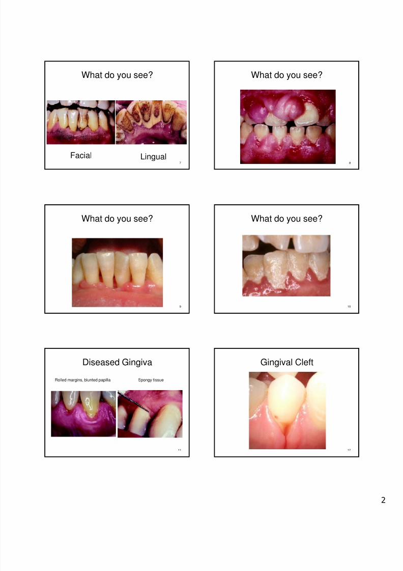
What do you see?
7
Facial Lingual
What do you see?
8
What do you see?
9
What do you see?
10
Diseased Gingiva
Rolled margins, blunted papilla Spongy tissue
11
Gingival Cleft
12

8/3/2019 Perio Exam 2010
http://slidepdf.com/reader/full/perio-exam-2010 3/10
To what are the arrows pointing?
13
Mucogingival Examination
• Evaluate width ofattached gingiva usingprobe
14
• Gingival recession
• MG defect
MG Defects
15
Gingival Recession
Measure recession fromCEJ to GM Tension test
16
Frenum Attachment
17
Health or Disease?
18
8/3/2019 Perio Exam 2010
http://slidepdf.com/reader/full/perio-exam-2010 4/10
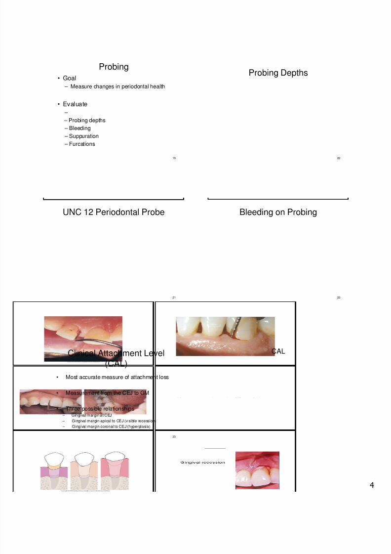
Probing
• Goal
– Measure changes in periodontal health
• Evaluate
19
–
– Probing depths
– Bleeding
– Suppuration
– Furcations
Probing Depths
20
UNC 12 Periodontal Probe
21
Bleeding on Probing
22
Clinical Attachment Level(CAL)
• Most accurate measure of attachment loss
23
• Measurement from the CEJ to GM
• Three possible relationships – Gingival margin at CEJ
– Gingival margin apical to CEJ (visible recession)
– Gingival margin coronal to CEJ (hyperplasia)
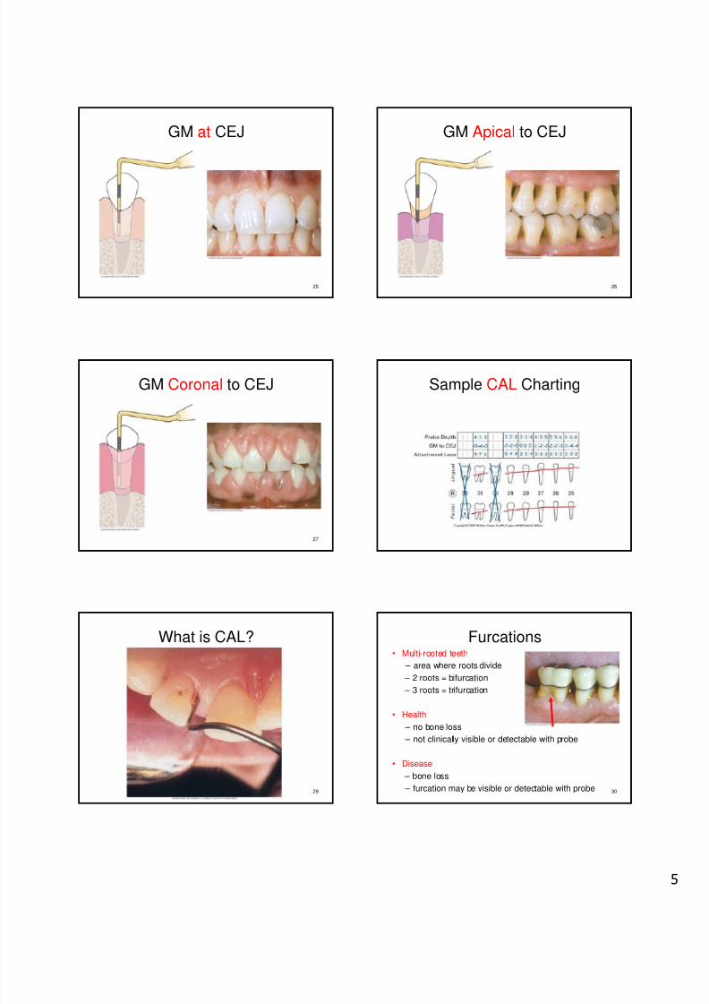
CAL
8/3/2019 Perio Exam 2010
http://slidepdf.com/reader/full/perio-exam-2010 5/10
GM at CEJ
25
GM Apical to CEJ
26
GM Coronal to CEJ
27
Sample CAL Charting
What is CAL?
29
Furcations• Multi-rooted teeth
– area where roots divide
– 2 roots = bifurcation
– 3 roots = trifurcation
30
• Health
– no bone loss
– not clinically visible or detectable with probe
• Disease
– bone loss
– furcation may be visible or detectable with probe

8/3/2019 Perio Exam 2010
http://slidepdf.com/reader/full/perio-exam-2010 6/10
Detection Radiographic Evidence
Furc at ion Morphology
Mandibular Molars
Maxillary First Premolars
.
Maxillary Molars: B and L
8/3/2019 Perio Exam 2010
http://slidepdf.com/reader/full/perio-exam-2010 7/10
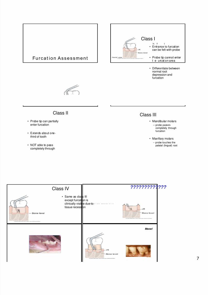
Furca t ion Assessment
Class I
• Entrance to furcationcan be felt with probe
• Probe tip cannot entert e urcat on area
• Differentiate betweennormal rootdepression andfurcation
Class II
• Probe tip can partiallyenter furcation
• Extends about one- third of tooth
• NOT able to passcompletely through
Class III
• Mandibular molars
– probe passescompletely throughfurcation
• Maxillary molars
– probe touches thepalatal (lingual) root
Class IV
• Same as class IIIexcept furcation isclinically visible due to
tissue recession
?????????????
Meow!

8/3/2019 Perio Exam 2010
http://slidepdf.com/reader/full/perio-exam-2010 8/10
SymbolsClass I Class II
SymbolsClass III Class IV
Sample Furcation Charting Bone Loss: Health vs. Disease
Normal level of alveolar crest is about 2 mm apical to CEJ
Where is the junctional epithelium in health?
Tooth Mobility
• Distinguish between physiologic orpathologic
•
47
– normal or expected mobility
• Pathologic – horizontal or vertical movement of tooth
beyond physiologic limit
Causes of Mobility
• Alveolar bone loss
• Inflammation
• Occlusal Forces
– Primary
– Secondary
48

8/3/2019 Perio Exam 2010
http://slidepdf.com/reader/full/perio-exam-2010 9/10
Tooth Mobility
• Evaluate with mirrorhandles
• Horizontal mobility
– ucco ngua or
faciolingual) movement
– Adjacent tooth is observedas a point of reference
• Vertical mobility
– Depressible in socket
Measuring Mobility
• 1 = Slight mobility, greaterthan normal; up to 1mmbuccolingually
50
= , -mm buccolingually, novertical displacement
• 3 = Severe mobility, >2 mmbuccolingually withvertical displacement
Mobility Check
51
Depressible in Socket
Fremitus
• Press index fingers besidemaxillary teeth
• Ask patient to “tap-tap-tap”
teeth together
• Vibration may be felt indicatingpremature contact which may
indicate mobility
• Technique used with occlusalanalysis or adjustment
53
Biofilm ID
54
8/3/2019 Perio Exam 2010
http://slidepdf.com/reader/full/perio-exam-2010 10/10
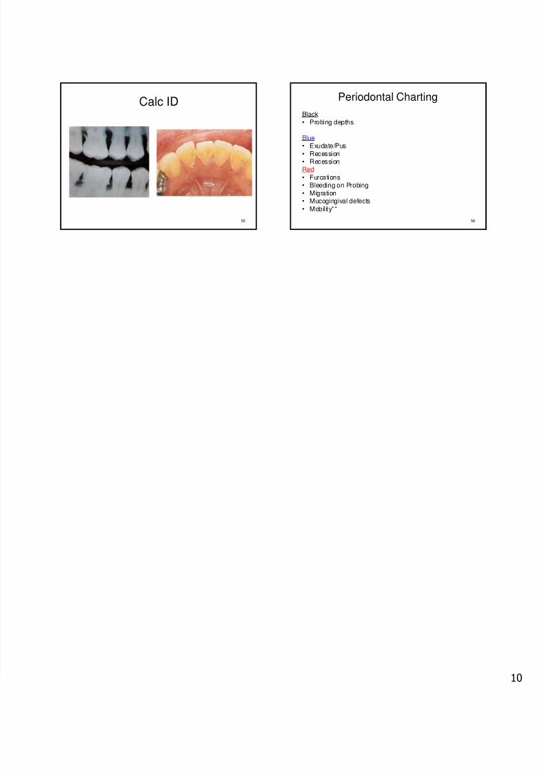
Calc ID
55
Periodontal Charting
Black• Probing depths
Blue• Exudate/Pus• Recession
56
• RecessionRed• Furcations• Bleeding on Probing• Migration• Mucogingival defects• Mobility**