PDF (1.3MB)
13
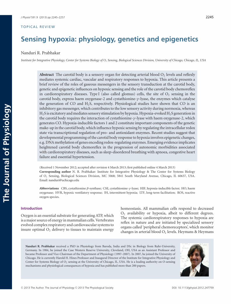
J Physiol 591.9 (2013) pp 2245–2257 2245 The Journal of Physiology TOPICAL REVIEW Sensing hypoxia: physiology, genetics and epigenetics Nanduri R. Prabhakar Institute for Integrative Physiology, Center for Systems Biology of O 2 Sensing, Biological Sciences Division, University of Chicago, Chicago, IL, USA Abstract The carotid body is a sensory organ for detecting arterial blood O 2 levels and reflexly mediates systemic cardiac, vascular and respiratory responses to hypoxia. This article presents a brief review of the roles of gaseous messengers in the sensory transduction at the carotid body, genetic and epigenetic influences on hypoxic sensing and the role of the carotid body chemoreflex in cardiorespiratory diseases. Type I (also called glomus) cells, the site of O 2 sensing in the carotid body, express haem oxygenase-2 and cystathionine-γ -lyase, the enzymes which catalyse the generation of CO and H 2 S, respectively. Physiological studies have shown that CO is an inhibitory gas messenger, which contributes to the low sensory activity during normoxia, whereas H 2 S is excitatory and mediates sensory stimulation by hypoxia. Hypoxia-evoked H 2 S generation in the carotid body requires the interaction of cystathionine-γ -lyase with haem oxygenase-2, which generates CO. Hypoxia-inducible factors 1 and 2 constitute important components of the genetic make-up in the carotid body, which influence hypoxic sensing by regulating the intracellular redox state via transcriptional regulation of pro- and antioxidant enzymes. Recent studies suggest that developmental programming of the carotid body response to hypoxia involves epigenetic changes, e.g. DNA methylation of genes encoding redox-regulating enzymes. Emerging evidence implicates heightened carotid body chemoreflex in the progression of autonomic morbidities associated with cardiorespiratory diseases, such as sleep-disordered breathing with apnoea, congestive heart failure and essential hypertension. (Received 1 November 2012; accepted after revision 4 March 2013; first published online 4 March 2013) Corresponding author N. R. Prabhakar: Institute for Integrative Physiology & The Center for Systems Biology of O 2 Sensing, Biological Sciences Division, MC 5068; 5841 South Maryland Avenue, Chicago, IL 60637, USA. Email: [email protected] Abbreviations CBS, cystathionine β-synthase; CSE, cystathionine-γ-lyase; HIF, hypoxia-inducible factor; HO, haem oxygenase; HVR, hypoxic ventilatory response; IH, intermittent hypoxia; LTF, long-term facilitation; ROS, reactive oxygen species. Introduction Oxygen is an essential substrate for generating ATP, which is a major source of energy in mammalian cells. Vertebrates evolved complex respiratory and cardiovascular systems to insure optimal O 2 delivery to tissues to maintain energy Nanduri R. Prabhakar received a PhD in Physiology from Baroda, India and DSc in Biology from Ruhr-University, Germany. In 1984, he joined the Case Western Reserve University, Cleveland, OH, USA as an Assistant Professor and became Professor and Vice-Chairman of the Department of Physiology (1997–2007). In 2007, he joined the University of Chicago. He is currently Harold H. Hines Professor and Inaugural Director of the Institute for Integrative Physiology and Center for Systems Biology of O 2 sensing at the University of Chicago, IL, USA. He is a leading authority on O-sensing mechanisms and physiological consequences of hypoxia and has published more than 200 papers. homeostasis. All mammalian cells respond to decreased O 2 availability or hypoxia, albeit to different degrees. The systemic cardiorespiratory responses to hypoxia are reflex in nature and are initiated by specialized sensory organs called ‘peripheral chemoreceptors’, which monitor changes in arterial blood O 2 levels. Heymans & Heymans C 2013 The Author. The Journal of Physiology C 2013 The Physiological Society DOI: 10.1113/jphysiol.2012.247759
Transcript of PDF (1.3MB)
J Physiol 591.9 (2013) pp 2245–2257 2245
The
Jou
rnal
of
Phys
iolo
gy
TOP ICAL REV IEW
Sensing hypoxia: physiology, genetics and epigenetics
Nanduri R. Prabhakar
Institute for Integrative Physiology, Center for Systems Biology of O2 Sensing, Biological Sciences Division, University of Chicago, Chicago, IL, USA
Abstract The carotid body is a sensory organ for detecting arterial blood O2 levels and reflexlymediates systemic cardiac, vascular and respiratory responses to hypoxia. This article presents abrief review of the roles of gaseous messengers in the sensory transduction at the carotid body,genetic and epigenetic influences on hypoxic sensing and the role of the carotid body chemoreflexin cardiorespiratory diseases. Type I (also called glomus) cells, the site of O2 sensing in thecarotid body, express haem oxygenase-2 and cystathionine-γ-lyase, the enzymes which catalysethe generation of CO and H2S, respectively. Physiological studies have shown that CO is aninhibitory gas messenger, which contributes to the low sensory activity during normoxia, whereasH2S is excitatory and mediates sensory stimulation by hypoxia. Hypoxia-evoked H2S generation inthe carotid body requires the interaction of cystathionine-γ-lyase with haem oxygenase-2, whichgenerates CO. Hypoxia-inducible factors 1 and 2 constitute important components of the geneticmake-up in the carotid body, which influence hypoxic sensing by regulating the intracellular redoxstate via transcriptional regulation of pro- and antioxidant enzymes. Recent studies suggest thatdevelopmental programming of the carotid body response to hypoxia involves epigenetic changes,e.g. DNA methylation of genes encoding redox-regulating enzymes. Emerging evidence implicatesheightened carotid body chemoreflex in the progression of autonomic morbidities associatedwith cardiorespiratory diseases, such as sleep-disordered breathing with apnoea, congestive heartfailure and essential hypertension.
(Received 1 November 2012; accepted after revision 4 March 2013; first published online 4 March 2013)Corresponding author N. R. Prabhakar: Institute for Integrative Physiology & The Center for Systems Biologyof O2 Sensing, Biological Sciences Division, MC 5068; 5841 South Maryland Avenue, Chicago, IL 60637, USA.Email: [email protected]
Abbreviations CBS, cystathionine β-synthase; CSE, cystathionine-γ-lyase; HIF, hypoxia-inducible factor; HO, haemoxygenase; HVR, hypoxic ventilatory response; IH, intermittent hypoxia; LTF, long-term facilitation; ROS, reactiveoxygen species.
Introduction
Oxygen is an essential substrate for generating ATP, whichis a major source of energy in mammalian cells. Vertebratesevolved complex respiratory and cardiovascular systems toinsure optimal O2 delivery to tissues to maintain energy
Nanduri R. Prabhakar received a PhD in Physiology from Baroda, India and DSc in Biology from Ruhr-University,Germany. In 1984, he joined the Case Western Reserve University, Cleveland, OH, USA as an Assistant Professor andbecame Professor and Vice-Chairman of the Department of Physiology (1997–2007). In 2007, he joined the University ofChicago. He is currently Harold H. Hines Professor and Inaugural Director of the Institute for Integrative Physiology andCenter for Systems Biology of O2 sensing at the University of Chicago, IL, USA. He is a leading authority on O-sensingmechanisms and physiological consequences of hypoxia and has published more than 200 papers.
homeostasis. All mammalian cells respond to decreasedO2 availability or hypoxia, albeit to different degrees.The systemic cardiorespiratory responses to hypoxia arereflex in nature and are initiated by specialized sensoryorgans called ‘peripheral chemoreceptors’, which monitorchanges in arterial blood O2 levels. Heymans & Heymans
C© 2013 The Author. The Journal of Physiology C© 2013 The Physiological Society DOI: 10.1113/jphysiol.2012.247759

2246 N. R. Prabhakar J Physiol 591.9
(1927) were some of the first to report that stimulationof breathing by hypoxia is a reflex triggered by the carotidbody, and they proposed the existence of similar structuresin the aortic arch (aortic bodies) (Heymans et al. 1931).A subsequent study by Comroe (1939) provided firmevidence for reflex stimulation of breathing by aorticbodies as well. For the discovery of the sensory natureof the carotid body, C. F. Heymans received the NobelPrize in Physiology in 1938, while J. H. Comroe Jr wasgiven an honorary doctorate from the Karolinska Institute,Stockholm for elucidating the functional role of aorticbodies. Tissues with morphology similar to carotid andaortic bodies have also been described in the thorax andabdomen, and might serve as ancillary chemoreceptors(Deane et al. 1975; Easton & Howe, 1983).
Much of the information on the mechanisms of hypo-xic sensing by the peripheral chemoreceptors has comefrom the studies on the carotid body. Innumerable studieshave investigated how the carotid body detects hypo-xia. An account of these early studies can be found inprevious reviews (Fidone & Gonzalez, 1986; Gonzalezet al. 1994; Prabhakar, 2000). A more comprehensive andcontemporary analysis of the structure and function ofthe carotid body and the physiological significance ofthe chemoreflex is available in a recent review (Kumar& Prabhakar, 2012). The present article focuses on recentstudies addressing the following factors: (i) the roles ofgaseous messengers in the hypoxic sensing by the carotidbody; (ii) modulation of hypoxic sensing by genetic andepigenetic factors; and (iii) the role of the carotid bodychemoreflex in cardiorespiratory diseases.
Physiology of carotid body hypoxic sensing
Carotid bodies, which reside bilaterally in the bifurcationof the common carotid arteries, receive sensoryinnervation from a branch of the glossopharengeal nervecalled the ‘carotid sinus nerve’. The sensory dischargefrequency of the carotid sinus nerve is low duringnormoxia (arterial PO2 ∼100 mmHg), but increasesdramatically with even a modest drop in arterial PO2 (e.g.PO2 80–60 mmHg). The sensory response to low oxygenis rapid and occurs within seconds after the onset ofhypoxia. The remarkable sensitivity and the speed withwhich it responds to hypoxia make the carotid body aunique sensory receptor for monitoring changes in thearterial blood PO2 . The chemoreceptor tissue is composedof two major cell types, called type I (also called glomus)cells and type II cells. A substantial body of evidencesuggests that type I cells are the initial sites of hypoxicsensing, and they work in concert with the nearby sensorynerve ending as a ‘sensory unit’ (see Kumar & Prabhakar,2012 for references). The general consensus is that hypo-xia inhibits certain K+ channels in type I cells, and the
resulting depolarization leads to Ca2+-dependent releaseof excitatory neurotransmitter(s), which stimulates thenearby sensory nerve ending, leading to an increase insensory discharge (Fig. 1). Despite intensive investigations,neither the mechanism(s) by which hypoxia inhibits K+
channels nor the identity of the chemical messenger(s)mediating the sensory excitation by low O2 are certain.
Role of gaseous messengers in hypoxic sensing
Nearly four decades ago, Brian Lloyd, Daniel Cunninghamand their colleagues from the University of Oxford madean intriguing observation that brief inhalation of carbonmonoxide inhibits ventilatory stimulation by hypoxia inconscious human subjects (Lloyd et al. 1968). Given thatthe affinity of haemoglobin for CO is much greater thanfor O2, it was proposed that the deoxy- conformation of ahaem-containing protein(s) might be critical for initiatingthe carotid body response to hypoxia. Interestingly, CO,which was once used as an experimental tool to under-stand hypoxic sensing by the carotid body, is now knownto be produced by the same chemoreceptor tissue. Thefollowing section provides a brief summary of our currentunderstanding of the role of endogenous CO in the carotidbody.
Carbon monoxide contributes to the low sensory activityof the carotid body during normoxia. Carbon mono-xide is generated during degradation of haem by haemoxygenases (HOs), with NADPH and cytochrome P450reductase as cofactors (Maines, 1997). Molecular O2 isessential for enzymatic generation of CO. The affinity(K m) of O2 for the haem–HO complex ranges between30 and 80 μM (Migita et al. 1998). Two HO isoforms havebeen identified, namely a constitutively expressed HO-2and an inducible HO-1 (also called heat shock protein 32or Hsp32; Maines, 1997). Haem oxygenase-2 is expressedin type I cells of cat, rat (Prabhakar et al. 1995), mouse(Ortega-Saenz et al. 2006) and human carotid bodies(Mkrtchian et al. 2012), but not in the nerve fibres andtype II cells (Prabhakar et al. 1995). Haem oxygenase-1expression is not evident in the carotid body in basalconditions.
The following evidence suggests that endogenous COis a physiological inhibitor of carotid body activity.Zinc protoporphyrin-9, an inhibitor of HO, increasescarotid body sensory activity in a dose-dependentmanner, whereas copper protoporphyrin-9, which doesnot inhibit HO activity, has no influence (Prabhakaret al. 1995). Haem oxygenase-2 knockout mice exhibitelevated baseline carotid body sensory activity and anaugmented sensory response to hypoxia (Prabhakar,2012). Exogenous application of low concentrations ofCO, similar to O2, inhibits the carotid body activity
C© 2013 The Author. The Journal of Physiology C© 2013 The Physiological Society

J Physiol 591.9 Carotid body hypoxic sensing 2247
(Prabhakar et al. 1995). Disruption of HO-2 function intype I cells, like hypoxia, inhibits the maxi-K+ channelactivity (Riesco-Fagundo et al. 2001; Williams et al.2004) and elevates [Ca2+]i (Prabhakar, 1998). Biochemicalstudies have shown that carotid bodies generate sub-stantial levels of CO during normoxia, whereas hypo-xia inhibits CO generation (Prabhakar, 2012). Based onthese findings, it was proposed that high levels of COgenerated during normoxia keep the sensory activitylow, and that hypoxia-evoked carotid body stimulationis in part due to reduced formation of CO, therebyremoving its inhibitory influence on the sensory activity(Prabhakar, 1999).
Recent studies have argued against a role for end-ogenous CO in hypoxic sensing in the carotid body.For instance, Ortega-Saenz et al. (2006) found that thetype I cell neurosecretory response to hypoxia is unalteredin HO-2 knockout mice. This study utilized carotidbody slices cultured for 20–96 h. Whether the long-termculture conditions masked the expected hypoxia-evokedincrease in the secretory response in HO-2 knockout mice,however, is not clear from this study. If CO is a physio-logical inhibitor of the carotid body, then the hypoxicventilatory response (HVR), a hallmark reflex triggered bythe carotid body, should be augmented. Contrary to thispossibility, Adachi et al. (2004) reported a blunted HVR inHO-2 knockout mice. However, in their study the authorsmeasured the HVR for 15–20 s (see Adachi et al. 2004,p. 515), which is too short a duration to monitor breathingreliably in conscious mice. Thus, these unanticipatedfindings from the studies described above appear to berelated to the experimental conditions employed and maytherefore not provide a critical assessment of the role ofCO in carotid body function.
In striking contrast to inhibition of the sensoryactivity by low doses of CO (Prabhakar et al. 1995),exogenous application of extremely high concentrationsof CO (partial pressure of CO ∼320 mmHg) stimulatecarotid body activity, depolarize type I cells andfacilitate voltage-gated Ca2+ influx, and these effects areantagonized by light (Barbe et al. 2002). As early as 1928,Warburg and Negelein demonstrated an interaction of COwith the mitochondrial respiratory chain, which is alsoantagonized by light. Inhibitors of mitochondrial oxidativephosphorylation are known for their stimulatory effects onthe carotid body (see Lahiri et al. 2006 for references). It isconceivable that, like other metabolic poisons, the effect ofa high concentration of CO on the carotid body is due to itsinhibition of the mitochondrial electron transport chain.Thus, the findings of Barbe et al. (2002) do not necessarilynegate the proposed inhibitory role of endogenous CO inthe carotid body.
Hydrogen sulfide mediates the sensory excitation byhypoxia. As early as in 1931, Heymans et al. reportedthat systemic administration of an H2S donor stimulatesbreathing, and this effect is mediated by the carotidbody chemoreflex. It is now well established that H2Sis produced endogenously and functions as a gaseousmessenger in various physiological processes (Gadalla& Snyder, 2010; Wang, 2012). Cystathionine β-synthase(CBS) and cystathionine γ-lyase (CSE) are the twomajor enzymes that catalyse the biosynthesis of H2S.Cystathionine β-synthase is abundant in the centralnervous system, whereas CSE is preponderant in peri-pheral tissues. Carotid body type I cells express CBS (Li
Figure 1. Schematic illustration of transduction ofthe hypoxic stimulus in a type I cell of the carotidbodyAbbreviations: NT, neurotransmitter(s); and NT-R,neurotransmitter receptor.
C© 2013 The Author. The Journal of Physiology C© 2013 The Physiological Society

2248 N. R. Prabhakar J Physiol 591.9
et al. 2010; Telezhkin et al. 2010; Fitzgerald et al. 2011) aswell as CSE (Peng et al. 2010; Mkrtchian et al. 2012).
Cystathionine γ-lyase-deficient mice and rats treatedwith DL-propargylglycine, an inhibitor of CSE, exhibitabsence or marked attenuation of carotid body sensoryfunction, as well as the ventilatory response to hypo-xia (Peng et al. 2010), suggesting that CSE-catalysedH2S is an excitatory gas messenger and mediatesthe sensory excitation by hypoxia. Mice treated withaminooxyacetic acid, a putative inhibitor of CBS, alsoexhibit impaired carotid body and ventilatory responsesto hypoxia (Li et al. 2010). However, in additionto inhibiting CBS, aminooxyacetic acid also inhibitsother pyridoxal phosphate-dependent enzymes, includingCSE and 4-aminobutyrate aminotransferase (Beeler &Churchich, 1976), and disrupts mitochondrial function(Kauppinen et al. 1987). Given the broad spectrum ofactions of aminooxyacetic acid, the role of CBS-catalysedH2S in the carotid body remains uncertain at present.
Further evidence supporting an excitatory role forH2S in the carotid body comes from studies with H2Sdonors. Exogenous application of NaHS, an H2S donor,produces robust sensory excitation of the carotid body ina concentration-dependent manner in mice and rats (Liet al. 2010; Peng et al. 2010). Like the response to hypoxia,the sensory response to NaHS is rapid in onset, occurswithin seconds, and returns promptly to baseline aftertermination of its application (Peng et al. 2010).
How might H2S activate the carotid body? Inhibitionof K+ channels and the resulting depolarization-inducedvoltage-gated Ca2+ influx in type I cells are critical stepsfor producing sensory excitation by hypoxia. Sodiumhydrosulfide, like hypoxia, inhibits maxi-K+ (Li et al.2010; Telezhkin et al. 2010) and TASK-like K+ channel(Tandem Pore K+ channel family) (Buckler, 2012) anddepolarizes type I cells (Buckler, 2012). Also, NaHSleads to robust elevation of [Ca2+]i in type I cells,and this effect is absent in the absence of extracellularCa2+ (Buckler, 2012; Makarenko et al. 2012) or ifthe depolarization is prevented by voltage clampingthe cell at the resting membrane potential (Buckler,2012). Furthermore, nifedipine, a blocker of L-type Ca2+
channels, prevents [Ca2+]i elevation by NaHS as wellas by hypoxia (Makarenko et al. 2012). Sodium hydro-sulfide increases NADH autofluorescence in type I cells,suggesting that H2S might mediate its actions in part byits effects on the mitochondrial electron transport chain(Buckler, 2012). These studies taken together demonstratethat H2S, like hypoxia, depolarizes type I cells by inhibitingcertain K+ channels, facilitates voltage-gated Ca2+ influxand thus produces sensory excitation of the carotid body.
The generation of H2S in the carotid body is regulatedby O2. Levels of H2S are low during normoxia, and hypoxiaincreases its levels in a stimulus-dependent manner in thecarotid body (Peng et al. 2010). A similar increase in H2S
generation was also seen in isolated type I cells challengedwith hypoxia (Makarenko et al. 2012). Hypoxia-evokedH2S generation was absent in CSE knockout mice andin rats treated with the CSE inhibitor DL-propargylglycine(Peng et al. 2010). These observations, taken together withthe finding that disruption of H2S generation preventsthe carotid body and type I cell responses to hypoxia (Liet al. 2010; Peng et al. 2010; Makarenko et al. 2012), ledto the suggestion that H2S is a physiological mediator ofthe carotid body response to hypoxia. However, as theconcentrations of H2S required for eliciting type I celland ventilatory responses are much greater than thoseproduced endogenously, Buckler (2012) and Haouze et al.(2011) argued against a physiological role for H2S inthe carotid body. The development of sensitive methodsfor simultaneous measurements of H2S in the carotidbody along with the sensory discharge is needed in orderfirmly to establish the physiological role of H2S in thecarotid body. Notwithstanding this limitation, currentevidence from studies involving pharmacological andgenetic disruption of endogenous H2S generation doprovide a rather compelling case for a physiological role ofH2S in mediating the carotid body response to hypoxia. Inthis context, it is noteworthy that H2S is an ancient gaseousmessenger system that is well conserved across phyla, asevidenced by its participation in hypoxic responses of troutgill chemoreceptors (Olson et al. 2008) and Caenorhabditiselegans (Ma et al. 2012).
Mechanism(s) underlying increased H2S generation byhypoxia in the carotid body: a role for CO. Howdoes hypoxia increase H2S generation? The findingsthat CO levels are relatively high and H2S levelslow during normoxia prompted Peng et al. (2010)to examine whether CO keeps the generation of H2Ssuppressed during normoxia. Indeed, reducing CO levelsby inhibition of HO markedly increased H2S generationduring normoxia. Conversely, a CO donor inhibited thehypoxia-evoked H2S generation in the carotid body in aconcentration-dependent manner. Given that the effects ofthe HO inhibitor were absent in CSE knockout mice, it wasproposed that CO reduces H2S generation by inhibitingCSE enzyme activity (Peng et al. 2010). Based on thesefindings, it was suggested that low sensory dischargeduring normoxia is due to an inhibitory influence of COon CSE, resulting in low H2S generation, whereas reducedCO generation during hypoxia lifts the inhibition onCSE, leading to elevated H2S levels and increased sensorydischarge (Fig. 2). Further studies, however, are neededto delineate the mechanisms by which CO inhibits CSEactivity during normoxia. In addition, as proposed byDombkowski et al. (2006), inhibition of H2S oxidationmight also contribute to its elevated levels during hypoxia,a possibility that remains to be investigated.
C© 2013 The Author. The Journal of Physiology C© 2013 The Physiological Society

J Physiol 591.9 Carotid body hypoxic sensing 2249
Is H2S an ‘O2 sensor’ or ‘mediator’ of the carotidbody response to hypoxia? Hydrogen sulfide has beenproposed as an ‘O2 sensor’ in trout gill chemoreceptors(Olson et al. 2008). Can H2S be regarded as an ‘O2 sensor’in the carotid body? Although H2S is critical for sensoryexcitation by hypoxia, the available evidence suggests thatits generation in the carotid body is regulated by CO.Thus, interactions between the HO-2–CO and CSE–H2Ssystems preclude a role for H2S as an ‘O2 sensor’ in thecarotid body. Rather, it would be more appropriate toregard H2S as an important ‘mediator’ of the sensoryexcitation during hypoxia. It was proposed that interactingproteins working in concert as a ‘chemosome’ accountfor the curvilinear response of the carotid body to hypo-xia, providing a fail-proof redundancy of chemoreceptorresponse to low O2 (Prabhakar, 2006). It is likely thatthe enzymes generating CO and H2S, in concert with K+
channels in type I cells constitute important componentsof the ‘chemosome’. Recent studies suggest that AMPkinase signalling also plays a role in hypoxic sensing by the
carotid body (Evans et al. 2012). It would be interesting toexamine the potential cross-talk between the HO-2–COand CSE–H2S systems with AMP kinase signalling in thecarotid body in future studies.
Genetic influence on carotid body hypoxic sensing
John Weil and his co-workers from the University ofColorado were some of the first to recognize variations inthe HVR in human subjects, with familial clusters and inmono- and dizygotic twins (see Weil, 2003 for references).As HVR is a hallmark reflex initiated by the carotid body,these variations were attributed to the influence of geneticfactors on intrinsic hypoxic sensitivity of the chemo-receptors (Weil, 2003). Studies on spontaneously hyper-tensive rats and F-344 rats, which come from two geneticbackgrounds, showed that the carotid body responseto hypoxia was markedly augmented in spontaneouslyhypertensive rats, whereas it was severely impaired in
Figure 2. Schematic illustration of interactions between haem oxygenase-2 (HO-2)-generated carbonmonoxide and cystathionine-γ-lyase (CSE)-regulated hydrogen sulfide generation and their effects oncarotid body sensory activity during normoxia and hypoxiaAbbreviations: NT, neurotransmitter(s); and NT-R, neurotransmitter receptor.
C© 2013 The Author. The Journal of Physiology C© 2013 The Physiological Society

2250 N. R. Prabhakar J Physiol 591.9
F-344 rats (Weil et al. 1998), suggesting that genetic factorsdo influence hypoxic sensing by the carotid body. However,neither the identity of the genetic mechanism(s) nor thegenes influencing the intrinsic hypoxic sensitivity of thecarotid body have been explored further.
Regulation of carotid body hypoxic sensing by trans-criptional activators: role of hypoxia-inducible factors(HIFs). Hypoxia-inducible factors 1 and 2 are thewell-studied members of the HIF family of transcriptionalactivators, which are essential for maintaining O2 homeo-stasis (Semenza, 2012). Hypoxia-inducible factors 1and 2 are heterodimeric proteins, which consist of aconstitutively expressed HIF-1β subunit and O2-regulatedHIF-1α or HIF-2α subunits, respectively. Type I cellsexpress both HIF-1α and HIF-2α, and the relativeabundance of the latter is higher than the former (Tianet al. 1998; Roux et al. 2005).
Given that homozygous deficiency of either HIF-1αor HIF-2α is embryonically lethal (Iyer et al. 1998;Scortegagna et al. 2003), the roles of HIFs in thecarotid body response to hypoxia were examined in micewith partial deficiencies in either HIF-1α (Hif-1a+/−) orHIF-2α (Hif-2a+/−). Carotid body responses to hypo-xia were severely impaired in Hif-1a+/− mice (Klineet al. 2002; Peng et al. 2006), whereas responsesto cyanide, a potent chemoreceptor stimulant, or toCO2, another physiological stimulus, were unaffected.In striking contrast, Hif-2a+/− mice exhibit selectiveheightening of the carotid body response to hypoxia(Peng et al. 2011). The enhanced hypoxic sensitivityof the carotid body in Hif-2a+/− mice was reflected inexaggerated chemoreflex function manifested by irregularbreathing, with apnoea, hypertension and elevated plasmacatecholamines. These observations demonstrate thatcarotid bodies from Hif-1a+/− mice are hyposensitive tohypoxia, whereas carotid bodies from Hif-2a+/− mice arehypersensitive.
How might HIFs contribute to hypoxic sensing bythe carotid body? The HIFs regulate a variety of geneproducts associated with the maintenance of O2 homeo-stasis (Prabhakar & Semenza, 2012). Hypoxia-induciblefactor-regulated gene products may affect carotid bodyfunction either by affecting its morphology and/orindependent of structural changes. The defective hypo-xic sensing in Hif-1a+/− mice and the exaggeratedhypoxic response in Hif-2a+/− mice appear not to besecondary to changes in carotid body morphology (Klineet al. 2002; Peng et al. 2011). Although HIFs regulate avariety of gene products, including some ion channels(see Prabhakar & Semenza, 2012 for references), recentstudies have shown that HIF-1 and HIF-2 regulate theexpression of gene products with opposing functions thatregulate the redox state. For instance, HIF-1 regulates the
expression of pro-oxidant enzymes, including NADPHoxidases (Diebold et al. 2010; Yuan et al. 2011), whereasHIF-2 regulates the expression of antioxidant enzymes(Scortegagna et al. 2003; Nanduri et al. 2009). Given thathypoxia alters the redox equilibrium in cells towards amore reduced state, it is likely that the contrasting effectsof carotid body responses to hypoxia in HIF-1a+/− versusHIF-2a+/− mice might be due to differential expression ofpro- and antioxidant enzymes and the ensuing changes inthe redox state. Indeed, carotid bodies from Hif-2a+/−
mice showed reduced levels of mRNAs encoding anti-oxidant enzymes and oxidative stress, and antioxidanttreatment prevented the augmented hypoxic sensitivityof the carotid body and corrected the oxidative stress inthese mice (Peng et al. 2011). Given that HIF-1 regulatesthe transcription of pro-oxidant enzymes (Diebold et al.2010; Yuan et al. 2011), it is likely that reduced pro-oxidantenzyme expression and the resulting reduction in theintracellular redox state might contribute to the impairedhypoxic sensitivity in Hif-1a+/− mice, a possibility thatremains to be established. Taken together, these studiessuggest that a subset of HIF-regulated genes associatedwith the maintenance of the intracellular redox state mightaccount in part for genetic variability in the carotid bodyresponse to hypoxia. The effects of partial deficiencies ofHIF-α isoforms on the carotid body activity and the down-stream redox-regulating enzyme genes are summarized inFig. 3. Further studies are needed to determine whetherHIFs also regulate the enzymes generating CO and H2S aswell as ion channels and to elucidate the role of other trans-criptional activators that may also contribute to geneticvariability of the carotid body response to hypoxia.
Epigenetic regulation of hypoxic sensing
Systemic or environmental perturbations in O2 levelsin the early neonatal period influence the carotid body
Figure 3. Molecular mechanisms underlying the carotid bodyresponses to hypoxia in mice with partial deficiencies ofhypoxia-inducible factors HIF-1α and HIF-2α
C© 2013 The Author. The Journal of Physiology C© 2013 The Physiological Society

J Physiol 591.9 Carotid body hypoxic sensing 2251
response to hypoxia in adulthood. For instance, adultrats treated with perinatal intermittent hypoxia (IH),simulating the apnoea of prematurity, exhibit carotidbody hypersensitivity to hypoxia (Peng et al. 2004; Pawaret al. 2008, 2009). The mechanisms underlying thelong-term consequences of O2 perturbations in the earlydevelopmental period are not known.
Emerging evidence suggests that aberrant epigeneticregulation is an important mechanism mediating theeffects of environmental perturbations in the neonatalperiod in adulthood (Feinberg, 2007). Epigeneticmechanisms are heritable modifications of DNA thatdo not involve changes in the DNA primary sequence(Barker et al. 1993; Gluckman et al. 2005). SilencingRNAs, DNA methylation and histone modificationsare three well-studied epigenetic mechanisms, whicheither individually or collectively determine whethera gene is activated or silenced (Feinberg, 2007).Of the three mechanisms, DNA methylation hasbeen implicated in mediating the gene expressionassociated with developmental programming (Waterlandand Jirtle, 2003). The DNA methylation is catalysedby DNA methyltransferases or Dnmts and occursat the 5′ position of cytosine of CpG dinucleotideclusters in the gene promoter called ‘CpG’ islands(Gardiner-Garden & Frommer, 1987; Feinberg, 2007).In general, DNA hypermethylation leads to repressionof gene transcription, whereas hypomethylation causestranscriptional activation.
A recent study examined the role of DNA methylationin mediating the long-term effect of neonatal IH oncarotid body responses to hypoxia in adult rats (Nanduriet al. 2012). Adult rats exposed to neonatal IH exhibitaugmented hypoxic sensitivity of the carotid body,respiratory abnormalities manifested by a greater numberof spontaneous apnoeas, hypertension and elevatedplasma catecholamines. The mRNA levels of Dnmt1 andDnmt3b mRNA and the corresponding protein levels wereelevated and global DNA methylation was increased inthe carotid bodies from adult rats exposed to neonatalIH. Genes encoding antioxidant enzymes were down-regulated, and there was increased oxidative stress inthe carotid bodies. Given that DNA hypermethylationsuppresses gene expression, whether increased DNAmethylation contributes to the reduced antioxidantgene expression was examined. A detailed analysis ofthe promoter region of the superoxide dismutase 2gene (Sod2) revealed marked hypermethylation of asingle CpG dinucleotide close to the transcription startsite in adult rats exposed to neonatal IH. Remarkably,neonatal treatment with decitabine, an inhibitor of DNAmethylation, eliminated DNA hypermethylation of thesingle CpG dinucleotide in the Sod2 gene, correctedthe oxidative stress, prevented the enhanced hypoxicsensitivity of the carotid body, and normalized the cardio-
respiratory functions in adult rats exposed to neonatalIH (Nanduri et al. 2012). These findings implicate ahitherto uncharacterized role for epigenetic mechanismsmediating the effects of perturbations in O2 levels inthe neonatal period on the carotid body response tohypoxia in adulthood (Fig. 4). These studies are onlythe beginning in this area, and further investigationsare certainly needed to delineate the role of otherepigenetic mechanisms, including the role of histonemodifications and/or silencing RNAs, and to identify thetarget genes associated with developmental programmingof the carotid body response to hypoxia.
Carotid body chemoreflex in cardiorespiratorydiseases
The importance of the carotid body chemoreflexin maintaining cardiorespiratory homeostasisduring a variety of physiological conditions is welldocumented. For instance, the chemoreflex is critical forventilatory adaptations to high altitude, as well as for
Figure 4. Schematic illustration of epigenetic mechanisms(e.g. DNA hypermethylation) contributing to augmentedcarotid body sensitivity to hypoxia in adult rats exposed toneonatal intermittent hypoxia and its impact oncardiorespiratory homeostasis
C© 2013 The Author. The Journal of Physiology C© 2013 The Physiological Society

2252 N. R. Prabhakar J Physiol 591.9
cardiorespiratory responses to exercise (see Kumar &Prabhakar, 2012). Emerging evidence suggests thatthe carotid body chemoreflex plays a more prominentrole than hitherto appreciated in the progression ofautonomic morbidities associated with cardiorespiratorydiseases. For example, heightened chemoreflex functionhas been implicated in autonomic dysfunction caused bysleep-disordered breathing with apnoea (Kara et al. 2003;Prabhakar et al. 2007), congestive heart failure (Schultz& Li, 2007) and neurogenic hypertension (Abdala et al.2012). Owing to space constraints, the following sectionwill focus on studies assessing the effects of chronicintermittent hypoxia, a hallmark of sleep-disorderedbreathing with apnoea, on carotid body and chemoreflexfunction.
Carotid body function in patients with recurrent apnoeaSleep-disordered breathing with obstructive sleep apnoeais a major clinical disorder that is reaching epidemicproportions (Nieto et al. 2000; Shahar et al. 2001). Inseverely affected patients, the frequency of apnoea mayexceed 60 episodes h−1, and arterial blood O2 saturationcan be reduced to as low as 50%. Patients with recurrentapnoea exhibit elevated sympathetic nerve activity, plasmaand urinary catecholamines, and are prone to develophypertension. It has been proposed that carotid bodiesconstitute the ‘frontline’ defense system for detecting peri-odic hypoxaemia associated with apnoea and that thechemoreflex contributes to elevated sympathetic activityand blood pressure (Cistulli & Sullivan, 1994). Patientswith recurrent apnoea exhibit an augmented hypoxicventilatory response (Narkiewicz et al. 1999; Kara et al.2003), a reflex initiated by the carotid body. Obstructivesleep apnoea patients exhibit a more pronounced decreasein sympathetic activity in response to brief hyperoxia(Dejour’s test; an indirect measure of peripheral chemo-receptor sensitivity) than control subjects (Tafil-Klaweet al. 1991; Kara et al. 2003), suggesting augmented chemo-reflex function. Remarkably, carotid body-resected sub-jects with sleep apnoea do not develop hypertension (seediscussion by Somers & Abboud, 1993). These studiessuggest heightened carotid body chemoreflex function inpatients with recurrent apnoea.
Effects of chronic intermittent hypoxia on carotid bodyresponse to hypoxia. Recurrent apnoea produces chronicIH and hypercapnia. A major advance in the field isthe demonstration that exposure of experimental animalsto chronic IH alone is sufficient to cause autonomicmorbidities similar to those described in recurrent apnoeapatients (Fletcher, 2001; Peng et al. 2006). Rats exposed tochronic IH also develop hypertension similar to that seenin patients with recurrent apnoea, and bilateral sectioningof the carotid sinus nerves prevents this response (Lesske
et al. 1997). Direct recording of carotid body sensoryactivity has shown selective enhancement of the hypo-xic response in animals exposed to chronic IH (Penget al. 2003, 2006; Peng & Prabhakar, 2004; Rey et al.2004, 2006). Furthermore, repetitive hypoxia producesa long-lasting increase in baseline sensory activity inrodents exposed to chronic IH, and this phenomenonwas termed ‘sensory long-term facilitation’ or sensory LTF(Peng et al. 2003, 2006). Chronic IH had no significanteffect on carotid body morphology (Peng et al. 2003).In striking contrast, the carotid body response to hypo-xia was normal in rats exposed to chronic continuoushypoxia (Peng et al. 2003), suggesting that the effectsare unique to IH. The absence of a heightened carotidbody response to hypoxia with chronic continuous hypo-xia might explain why high-altitude residents, who areadapted to a low-O2 environment, do not exhibit auto-nomic dysfunction, as opposed to patients with recurrentapnoea, who experience chronic IH.
Reactive oxygen species (ROS) mediate the effects ofchronic IH on the carotid body. The following findingssuggest that ROS signalling mediates the effects ofchronic IH on the carotid body. First, ROS levels wereelevated in carotid bodies from chronic IH-treated rodents(Peng et al. 2003, 2006; Pawar et al. 2009). Second,antioxidant treatment prevented the chronic IH-evokedaugmented hypoxic sensitivity and sensory LTF (Penget al. 2003; Pawar et al. 2009). Available evidencesuggests that increased transcription and activationof the pro-oxidant enzyme, NADPH oxidase-2, anddecreased transcription and activity of the antioxidantenzymes (e.g. superoxide dismutase 2) contribute to theincrease in ROS levels induced by IH (Peng et al.2009; Nanduri et al. 2009). In addition, IH increasesROS in the mitochondrial compartment by inhibitingcomplex I of the mitochondrial electron transport chain(Peng et al. 2003). A recent study by Khan et al.(2011) showed that ROS generated by NADPH oxidase-2inhibit the mitochondrial complex I in IH-exposed cells,involving S-glutathionylation of the complex I subunits.The selective contribution of ROS generated in themitochondrial compartment to the altered carotid bodyfunction, however, remains to be investigated.
Analysis of the molecular mechanisms revealed thatactivation of HIF-1 mediates the increase in NADPHoxidase-2 transcription induced by IH (Peng et al. 2006;Yuan et al. 2011). On the contrary, IH decreases HIF-2activity, which in turn leads to insufficient transcriptionof superoxide dismutase 2 (Nanduri et al. 2009). Thus,the imbalance between HIF-1 and HIF-2 and resultingchanges in pro- and antioxidant enzyme expressionscontribute to IH-induced oxidative stress in the carotidbody. How might ROS mediate the effects of chronic
C© 2013 The Author. The Journal of Physiology C© 2013 The Physiological Society

J Physiol 591.9 Carotid body hypoxic sensing 2253
IH on the carotid body? Available evidence suggeststhat ROS-dependent recruitment of endothelin-1 and5-HT signalling contribute to the chronic IH-evokedaugmented hypoxic response and sensory LTF, respectively
(Pawar et al. 2009; Peng et al. 2009). However, theeffects of chronic IH on recently identified HO-2–COand CSE–H2S signalling in the carotid body remain to beinvestigated.
Figure 5. Contribution of carotid body chemoreflex to cardiorespiratory responses to chronic inter-mittent hypoxia (CIH)A, the heightened carotid body response to hypoxia induced by CIH leads to a greater number of spontaneousapnoeas. B, example of breathing responses in awake rats before and after exposure to CIH with intact anddenervated carotid bodies (CB). Arrow represents irregular breathing with apnoea. C, CIH-induced sensorylong-term facilitation (LTF) of the carotid body reflexly stimulates sympathetic activity. D, examples of splanchnicnerve activity (SNA) in normoxia (Nx) and hypoxia (Hx, 12% O2) in anaesthetized, mechanically ventilated ratsexposed to normoxia (control) or CIH with intact carotid bodies and following carotid body denervation. Note theenhanced SNA response to Hx in the CIH-exposed rat and the absence of this response in the CB-denervated ratexposed to CIH.
C© 2013 The Author. The Journal of Physiology C© 2013 The Physiological Society

2254 N. R. Prabhakar J Physiol 591.9
Heightened carotid body chemoreflex mediates auto-nomic dysfunction caused by chronic IH. What mightbe the functional significance of chronic IH-inducedchanges in carotid body function? It was proposed thatduring each episode of apnoea the increased carotid bodysensitivity to hypoxia can lead to a greater magnitude ofhyperventilation, thus driving the respiratory controllerbelow the apnoeic threshold for CO2, leading to a greaternumber of apnoeas (Prabhakar, 2001). In other words,the heightened hypoxic sensitivity of the carotid bodymight act as a ‘positive feedback’, thereby exacerbatingthe occurrence of apnoeas (Fig. 5A). Supporting such apossibility is the finding that chronic IH-exposed ratswith intact carotid bodies exhibit a greater incidenceof spontaneous apnoeas, and this effect was absentin carotid body-sectioned rats exposed to chronic IH(Fig. 5B). Patients with sleep-disordered breathing exhibitelevated sympathetic nerve activity during the day time,wherein apnoeas are absent (Kara et al. 2003). Giventhat the carotid body chemoreflex stimulates sympatheticnerve activity, it was proposed that the sensory LTFcontributes to the day-time increase in sympathetic nerveactivity in patients with recurrent apnoea (Prabhakar,2001; Prabhakar et al. 2012; Fig. 5C). Indeed, ratsexposed to chronic IH showed elevated sympathetic nerveactivity, and this effect was absent following chronicbilateral sectioning of the carotid sinus nerves (Fig. 5D).These findings suggest that the heightened carotidbody chemoreflex contributes to autonomic dysfunctionin patients with sleep-disordered breathing withapnoea.
Concluding remarks
Vertebrates are endowed with a variety of sensoryreceptors for detecting diverse sensory modalities,including mechanical, thermal, visual, olfactory and tastesensations. Two principal categories of sensory trans-duction machineries have been indentified. One involvesion channels, which mediate the mechanical and thermalsensations, and the other is G-protein-coupled receptors,which contribute to the transduction of visual, olfactoryand taste sensations (Julius & Nathans, 2012). Althoughion channels were implicated in carotid body hypo-xic sensing, emerging evidence suggests a biochemicalmechanism associated with O2-dependent enzymaticgeneration of gas messengers (e.g. CO and H2S) as animportant initial step in the transduction cascade ofthe hypoxic stimulus in the carotid body. Thus, thetransduction machinery in the carotid body appearsto be distinctly different from other known sensoryreceptors. Evidence is emerging that genetic and epigeneticmechanisms profoundly impact the carotid body response
to hypoxia by altering the intracellular redox state.Elucidation of the mechanisms by which redox state affectsthe transduction machinery of the carotid body is animportant area for future research.
Emerging evidence suggests that either hypo- or hyper-sensitivity of the carotid body to hypoxia has adversephysiological consequences. For instance, people withblunted HVR, a hallmark reflex response of the carotidbody, exhibit impaired ventilatory adaptations to highaltitude, manifested by pulmonary oedema (Hohenhauset al. 1995). On the contrary, hypersensitivity to hypo-xia contributes to the autonomic dysfunction associatedwith highly prevalent cardiorespiratory diseases, such assleep-disordered breathing with apnoea, congestive heartfailure and neurogenic hypertension. Future developmentof new therapeutic interventions targeting the carotidbody to correct either the hypo- or hypersensitivityto hypoxia is of greater translational significance thanhitherto appreciated.
References
Abdala AP, McBryde FD, Marina N, Hendy EB, Engelman ZJ,Fudim M, Sobotka PA, Gourine AV & Paton JF (2012).Hypertension is critically dependent on the carotid bodyinput in the spontaneously hypertensive rat. J Physiol 590,4269–4277.
Adachi T, Ishikawa K, Hida W, Matsumoto H, Masuda T, DateF, Ogawa K, Takeda K, Furuyama K, Zhang Y, Kitamuro T,Ogawa H, Maruyama Y & Shibahara S (2004). Hypoxemiaand blunted hypoxic ventilatory responses in mice lackingheme oxygenase-2. Biochem Biophys Res Commun. 320,514–522.
Barbe C, Al-Hashem F, Conway AF, Dubuis E, Vandier C &Kumar P (2002). A possible dual site of action for carbonmonoxide-mediated chemoexcitation in the rat carotidbody. J Physiol. 543, 933–945.
Barker DJ, Osmond C, Simmonds SJ & Wield GA (1993). Therelation of small head circumference and thinness at birth todeath from cardiovascular disease in adult life. BMJ 306,422–426.
Beeler T & Churchich JE (1976). Reactivity of thephosphopyridoxal groups of cystathionase. J Biol Chem 251,5267–5271.
Buckler KJ (2012). Effects of exogenous hydrogen sulphide oncalcium signalling, background (TASK) K channel activityand mitochondrial function in chemoreceptor cells. PflugersArch 463, 743–754.
Cistulli P & Sullivan C (1994). Pathophysiology of sleep apnea.In Sleep and Breathing , ed. Saunders N & Sullivan C,pp. 405–448. Marcel Dekker, New York.
Comroe JH (1939). The location and function of thechemoreceptors of the aorta. Am J Physiol 127,176–191.
Deane BM, Howe A & Morgan M (1975). Abdominal vagalparaganglia: distribution and comparison with carotid body,in the rat. Acta Anat (Basel) 93, 19–28.
C© 2013 The Author. The Journal of Physiology C© 2013 The Physiological Society
J Physiol 591.9 Carotid body hypoxic sensing 2255
Diebold I, Petry A, Djordjevic T, BelAiba RS, Fineman J, BlackS, Schreiber C, Fratz S, Hess J, Kietzmann T & Gorlach A(2010). Reciprocal regulation of Rac1 and PAK-1 by HIF-1α:a positive-feedback loop promoting pulmonary vascularremodeling. Antioxid Redox Signal 13, 399–412.
Dombkowski RA, Doellman MM, Head SK & Olson KR(2006). Hydrogen sulfide mediates hypoxia-inducedrelaxation of trout urinary bladder smooth muscle. J ExpBiol 209, 3234–3240.
Easton J & Howe A (1983). The distribution of thoracicglomus tissue (aortic bodies) in the rat. Cell Tissue Res 232,349–356.
Evans AM, Peers C, Wyatt CN, Kumar P & Hardie DG (2012).Ion channel regulation by the LKB1-AMPK signallingpathway: the key to carotid body activation by hypoxia andmetabolic homeostasis at the whole body level. Adv Exp MedBiol 758, 81–90.
Feinberg AP (2007). Phenotypic plasticity and the epigeneticsof human disease. Nature 447, 433–440.
Fidone SJ & Gonzalez C (1986). Initiation and control ofchemoreceptor activity in the carotid body. In Handbook ofPhysiology, section 3, The Respiratory System, vol. II, Controlof Breathing , pp. 247–312. American Physiological Society,Bethesda, MD, USA.
Fitzgerald RS, Shirahata M, Chang I, Kostuk E & Kiihl S (2011).The impact of hydrogen sulfide (H2S) on neurotransmitterrelease from the cat carotid body. Respir Physiol Neurobiol176, 80–89.
Fletcher EC (2001). Physiological consequences of intermittenthypoxia: systemic blood pressure. J Appl Physiol 90,1600–1605.
Gadalla MM & Snyder SH (2010). Hydrogen sulfide as agasotransmitter. J Neurochem 113, 14–26.
Gardiner-Garden M & Frommer M (1987). CpG islands invertebrate genomes. J Mol Biol 196, 261–282.
Gluckman PD, Cutfield W, Hofman P & Hanson MA (2005).The fetal, neonatal, and infant environments—thelong-term consequences for disease risk. Early Hum Dev81, 51–59.
Gonzalez C, Almaraz L, Obeso A & Rigual R (1994). Carotidbody chemoreceptors: from natural stimuli to sensorydischarges. Physiol Rev 74, 829–898.
Haouzi P, Bell H & Van de Louw A (2011). Hypoxia-inducedarterial chemoreceptor stimulation and hydrogen sulfide: toomuch or too little? Respir Physiol Neurobiol. 179, 97–102.
Heymans C, Bouckaert JJ & Dautrebande L (1931). Sinuscarotidien et reflexes respiratoires: sensibilite des sinuscarotidiens aux substances chimiques. Action stimulanterespiratoire reflexe du sulfure de sodium, du cyanure depotassium, de la nicotine et de la lobeline. Arch IntPharmacodyn Ther 40, 54–91.
Heymans J & Heymans C (1927). Sur les modifications directeset sur la regulation reflexede l’activitie du centre respiratoryde la tete isolee du chien. Arch Int Pharmacodyn Ther 33,273–372.
Hohenhaus E, Paul A, McCullough RE, Kucherer H & BartschP (1995). Ventilatory and pulmonary vascular response tohypoxia and susceptibility to high altitude pulmonaryoedema. Eur Respir J 8, 1825–1833.
Iyer NV, Kotch LE, Agani F, Leung SW, Laughner E, WengerRH, Gassmann M, Gearhart JD, Lawler AM, Yu AY &Semenza GL (1998). Cellular and developmental control ofO2 homeostasis by hypoxia-inducible factor 1α. Genes Dev12, 149–162.
Julius D & Nathans J (2012). Signaling by sensory receptors.Cold Spring Harb Perspect Biol 4, a005991.
Kara T, Narkiewicz K & Somers VK (2003). Chemoreflexes –physiology and clinical implications. Acta Physiol Scand 177,377–384.
Kauppinen RA, Sihra TS & Nicholls DG (1987).Aminooxyacetic acid inhibits the malate-aspartate shuttle inisolated nerve terminals and prevents the mitochondria fromutilizing glycolytic substrates. Biochim Biophys Acta 930,173–178.
Khan SA, Nanduri J, Yuan G, Kinsman B, Kumar GK, Joseph J,Kalyanaraman B & Prabhakar NR (2011). NADPH oxidase 2mediates intermittent hypoxia-induced mitochondrialcomplex I inhibition: relevance to blood pressure changes inrats. Antioxid Redox Signal 14, 533–542.
Kline DD, Peng YJ, Manalo DJ, Semenza GL & Prabhakar NR(2002). Defective carotid body function and impairedventilatory responses to chronic hypoxia in mice partiallydeficient for hypoxia-inducible factor 1α. Proc Natl Acad SciU S A 99, 821–826.
Kumar P & Prabhakar NR (2012). Peripheral chemoreceptors:function and plasticity of the carotid body. Compr Physiol 2,141–219.
Lahiri S, Roy A, Baby SM, Hoshi T, Semenza GL & PrabhakarNR (2006). Oxygen sensing in the body. Prog Biophys MolBiol 91, 249–286.
Lesske J, Fletcher EC, Bao G & Unger T (1997). Hypertensioncaused by chronic intermittent hypoxia – influence ofchemoreceptors and sympathetic nervous system. JHypertens 15, 1593–1603.
Li Q, Sun B, Wang X, Jin Z, Zhou Y, Dong L, Jiang LH & RongW (2010). A crucial role for hydrogen sulfide in oxygensensing via modulating large conductance calcium-activatedpotassium channels. Antioxid Redox Signal 12, 1179–1189.
Lloyd BB, Cunningham DJC & Goode RC (1968). Depressionof hypoxic hyperventilation in man by sudden inspiration ofcarbon monoxide. In Arterial Chemoreceptors, ed. TorranceRW, pp. 145–150. Blackwell, Oxford.
Ma DK, Vozdek R, Bhatla N & Horvitz HR (2012). CYSL-1interacts with the O2-sensing hydroxylase EGL-9 to promoteH2S-modulated hypoxia-induced behavioral plasticity in C.elegans. Neuron 73, 925–940.
Maines MD (1997). The heme oxygenase system: a regulator ofsecond messenger gases. Annu Rev Pharmacol Toxicol 37,517–554.
Makarenko VV, Nanduri J, Raghuraman G, Fox AP, GadallaMM, Kumar GK, Snyder SH & Prabhakar NR (2012).Endogenous H2S is required for hypoxic sensing by carotidbody glomus cells. Am J Physiol Cell Physiol 303,C916–C923.
Migita CT, Matera KM, Ikeda-Saito M, Olson JS, Fujii H,Yoshimura T, Zhou H & Yoshida T (1998). The oxygen andcarbon monoxide reactions of heme oxygenase. J Biol Chem273, 945–949.
C© 2013 The Author. The Journal of Physiology C© 2013 The Physiological Society

2256 N. R. Prabhakar J Physiol 591.9
Mkrtchian S, Kahlin J, Ebberyd A, Gonzalez C, Sanchez D,Balbir A, Kostuk EW, Shirahata M, Fagerlund MJ & ErikssonLI (2012). The human carotid body transcriptome withfocus on oxygen sensing and inflammation – a comparativeanalysis. J Physiol 590, 3807–3819.
Nanduri J, Makarenko V, Reddy VD, Yuan G, Pawar A, Wang N,Khan SA, Zhang X, Kinsman B, Peng YJ, Kumar GK, Fox AP,Godley LA, Semenza GL & Prabhakar NR (2012). Epigeneticregulation of hypoxic sensing disrupts cardiorespiratoryhomeostasis. Proc Natl Acad Sci U S A 109, 2515–2520.
Nanduri J, Wang N, Yuan G, Khan SA, Souvannakitti D, PengYJ, Kumar GK, Garcia JA & Prabhakar NR (2009).Intermittent hypoxia degrades HIF-2α via calpains resultingin oxidative stress: implications for recurrent apnea-inducedmorbidities. Proc Natl Acad Sci U S A 106,1199–1204.
Narkiewicz K, van de Borne PJ, Pesek CA, Dyken ME, MontanoN & Somers VK (1999). Selective potentiation of peripheralchemoreflex sensitivity in obstructive sleep apnea.Circulation 99, 1183–1189.
Nieto FJ, Young TB, Lind BK, Shahar E, Samet JM, Redline S,D’Agostino RB, Newman AB, Lebowitz MD & Pickering TG(2000). Association of sleep-disordered breathing, sleepapnea, and hypertension in a large community-based study.Sleep Heart Health Study. JAMA 283, 1829–1836.
Olson KR, Healy MJ, Qin Z, Skovgaard N, Vulesevic B, DuffDW, Whitfield NL, Yang G, Wang R & Perry SF (2008).Hydrogen sulfide as an oxygen sensor in trout gillchemoreceptors. Am J Physiol Regul Integr Comp Physiol 295,R669–R680.
Ortega-Saenz P, Pascual A, Gomez-Dıaz R & Lopez-Barneo J(2006). Acute oxygen sensing in heme oxygenase-2 nullmice. J Gen Physiol 128, 405–411.
Pawar A, Nanduri J, Yuan G, Khan SA, Wang N, Kumar GK &Prabhakar NR (2009). Reactive oxygen species-dependentendothelin signaling is required for augmented hypoxicsensory response of the neonatal carotid body byintermittent hypoxia. Am J Physiol Regul Integr Comp Physiol296, R735–R742.
Pawar A, Peng YJ, Jacono FJ & Prabhakar NR (2008).Comparative analysis of neonatal and adult rat carotid bodyresponses to chronic intermittent hypoxia. J Appl Physiol104, 1287–1294.
Peng YJ, Nanduri J, Khan SA, Yuan G, Wang N, Kinsman B,Vaddi DR, Kumar GK, Garcia JA, Semenza GL & PrabhakarNR (2011). Hypoxia-inducible factor 2α (HIF-2α)heterozygous-null mice exhibit exaggerated carotid bodysensitivity to hypoxia, breathing instability, andhypertension. Proc Natl Acad Sci U S A 108,3065–3070.
Peng YJ, Nanduri J, Raghuraman G, Souvannakitti D, GadallaMM, Kumar GK, Snyder SH & Prabhakar NR (2010). H2Smediates O2 sensing in the carotid body. Proc Natl Acad SciU S A 107, 10719–10724.
Peng YJ, Nanduri J, Yuan G, Wang N, Deneris E, Pendyala S,Natarajan V, Kumar GK & Prabhakar NR (2009). NADPHoxidase is required for the sensory plasticity of the carotidbody by chronic intermittent hypoxia. J Neurosci 29,4903–4910.
Peng YJ, Overholt JL, Kline D, Kumar GK & Prabhakar NR(2003). Induction of sensory long-term facilitation in thecarotid body by intermittent hypoxia: implications forrecurrent apneas. Proc Natl Acad Sci U S A 100, 10073–10078.
Peng YJ & Prabhakar NR (2004). Effect of two paradigms ofchronic intermittent hypoxia on carotid body sensoryactivity. J Appl Physiol 96, 1236–1242; discussion 1196.
Peng YJ, Rennison J & Prabhakar NR (2004). Intermittenthypoxia augments carotid body and ventilatory response tohypoxia in neonatal rat pups. J Appl Physiol 97,2020–2025.
Peng YJ, Yuan G, Ramakrishnan D, Sharma SD, Bosch-MarceM, Kumar GK, Semenza GL & Prabhakar NR (2006).Heterozygous HIF-1α deficiency impairs carotidbody-mediated systemic responses and reactive oxygenspecies generation in mice exposed to intermittent hypoxia. JPhysiol 577, 705–716.
Prabhakar NR (1998). Endogenous carbon monoxide incontrol of respiration. Respir Physiol 114, 57–64.
Prabhakar NR (1999). NO and CO as second messengers inoxygen sensing in the carotid body. Respir Physiol 115,161–168.
Prabhakar NR (2000). Oxygen sensing by the carotid bodychemoreceptors. J Appl Physiol 88, 2287–2295.
Prabhakar NR (2001). Oxygen sensing during intermittenthypoxia: cellular and molecular mechanisms. J Appl Physiol90, 1986–1994.
Prabhakar NR (2006). O2 sensing at the mammalian carotidbody: why multiple O2 sensors and multiple transmitters?Exp Physiol 91, 17–23.
Prabhakar NR (2012). Carbon monoxide (CO) and hydrogensulfide (H2S) in hypoxic sensing by the carotid body. RespirPhysiol Neurobiol 184, 165–169.
Prabhakar NR, Dinerman JL, Agani FH & Snyder SH (1995).Carbon monoxide: a role in carotid body chemoreception.Proc Natl Acad Sci U S A 92, 1994–1997.
Prabhakar NR, Kumar GK, Nanduri J & Semenza GL (2007).ROS signaling in systemic and cellular responses to chronicintermittent hypoxia. Antioxid Redox Signal 9,1397–1403.
Prabhakar NR, Kumar GK & Peng YJ (2012).Sympatho-adrenal activation by chronic intermittenthypoxia. J Appl Physiol 113, 1304–1310.
Prabhakar NR & Semenza GL (2012). Adaptive andmaladaptive cardiorespiratory responses to continuous andintermittent hypoxia mediated by hypoxia-inducible factors1 and 2. Physiol Rev 92, 967–1003.
Rey S, Del Rio R, Alcayaga J & Iturriaga R (2004). Chronicintermittent hypoxia enhances cat chemosensory andventilatory responses to hypoxia. J Physiol 560, 577–586.
Rey S, Del Rio R & Iturriaga R (2006). Contribution ofendothelin-1 to the enhanced carotid body chemosensoryresponses induced by chronic intermittent hypoxia. BrainRes 1086, 152–159.
Riesco-Fagundo AM, Perez-Garcıa MT, Gonzalez C &Lopez-Lopez JR (2001). O2 modulates large-conductanceCa2+-dependent K+ channels of rat chemoreceptor cells by amembrane-restricted and CO-sensitive mechanism. Circ Res89, 430–436.
C© 2013 The Author. The Journal of Physiology C© 2013 The Physiological Society

J Physiol 591.9 Carotid body hypoxic sensing 2257
Roux JC, Brismar H, Aperia A & Lagercrantz H (2005).Developmental changes in HIF transcription factor incarotid body: relevance for O2 sensing by chemoreceptors.Pediatr Res 58, 53–57.
Schultz HD & Li YL (2007). Carotid body function in heartfailure. Respir Physiol Neurobiol 157, 171–185.
Scortegagna M, Ding K, Oktay Y, Gaur A, Thurmond F, Yan LJ,Marck BT, Matsumoto AM, Shelton JM, Richardson JA,Bennett MJ & Garcia JA (2003). Multiple organ pathology,metabolic abnormalities and impaired homeostasis ofreactive oxygen species in Epas1−/− mice. Nat Genet 35,331–340.
Semenza GL (2012). Hypoxia-inducible factors in physiologyand medicine. Cell 148, 399–408.
Shahar E, Whitney CW, Redline S, Lee ET, Newman AB, JavierNieto F, O’Connor GT, Boland LL, Schwartz JE & Samet JM(2001). Sleep-disordered breathing and cardiovasculardisease: cross-sectional results of the Sleep Heart HealthStudy. Am J Respir Crit Care Med 163, 19–25.
Somers VK & Abboud FM (1993). Chemoreflexes–responses,interactions and implications for sleep apnea. Sleep 16,S30–S33; discussion S33–S34.
Tafil-Klawe M, Thiele AE, Raschke F, Mayer J, Peter JH & vonWichert W (1991). Peripheral chemoreceptor reflex inobstructive sleep apnea patients; a relationship betweenventilatory response to hypoxia and nocturnal bradycardiaduring apnea events. Pneumologie 45(Suppl 1), 309–311.
Telezhkin V, Brazier SP, Cayzac SH, Wilkinson WJ, Riccardi D& Kemp PJ (2010). Mechanism of inhibition by hydrogensulfide of native and recombinant BKCa channels. RespirPhysiol Neurobiol 172, 169–178.
Tian H, Hammer RE, Matsumoto AM, Russell DW &McKnight SL (1998). The hypoxia-responsive transcriptionfactor EPAS1 is essential for catecholamine homeostasis andprotection against heart failure during embryonicdevelopment. Genes Dev 12, 3320–3324.
Wang R (2012). Physiological implications of hydrogen sulfide:a whiff exploration that blossomed. Physiol Rev 92,791–896.
Warburg O & Negelin E (1928). Uber die photochemischeDissoziation bei intermittierender Beilichtung und dasabsolute Absorptionsspektrum des Atmungs ferments.Biochem. Z. 202, 202–228.
Waterland RA & Jirtle RL (2003). Transposable elements:targets for epigenetic gene regulation. Mol Cell Biol 23,5293–5300.
Weil JV (2003). Variation in human ventilatorycontrol—genetic influence on the hypoxic ventilatoryresponse. Respir Physiol Neurobiol 135, 239–246.
Weil JV, Stevens T, Pickett CK, Tatsumi K, Dickinson MG,Jacoby CR & Rodman DM (1998). Strain-associateddifferences in hypoxic chemosensitivity of the carotid bodyin rats. Am J Physiol Lung Cell Mol Physiol 274,L767–L774.
Williams SE, Wootton P, Mason HS, Bould J, Iles DE, RiccardiD, Peers C & Kemp PJ (2004). Hemoxygenase-2 is an oxygensensor for a calcium-sensitive potassium channel. Science306, 2093–2097.
Yuan G, Khan SA, Luo W, Nanduri J, Semenza GL & PrabhakarNR (2011). Hypoxia-inducible factor 1 mediates increasedexpression of NADPH oxidase-2 in response to intermittenthypoxia. J Cell Physiol 226, 2925–2933.
Acknowledgements
This article is based on 2012 J.H. Comroe Distinguished Lectureof the American Physiological Society. It is a great privilegeto acknowledge the collaborations of Professors Solomon H.Snyder and Gregg L. Semenza of the Johns Hopkins Schoolof Medicine. The studies presented in this article would havenot been possible without the participation of my colleaguesProfessor G. K. Kumar, Professor J. Nanduri and Drs Y-J. Pengand G. Yuan. I am greatly indebted to Ms Shanna L. Williamsfor preparing the figures. The research in my laboratory issupported by grants from National Institutes of Health, Heart,Lung and Blood Institute, R01-HL76537, P01-HL90554 andR01-HL86493.
C© 2013 The Author. The Journal of Physiology C© 2013 The Physiological Society


![PFI brochure [PDF - 1.3MB]](https://static.fdocuments.in/doc/165x107/586a1ccd1a28ab847d8bcc1c/pfi-brochure-pdf-13mb.jpg)
![Year ended March 31, 2006 [PDF 1.3MB]](https://static.fdocuments.in/doc/165x107/5854869d1a28abfa39906cc2/year-ended-march-31-2006-pdf-13mb.jpg)