Pathogenetic Features of Hematopoiesis in MDS: Focus …€¦ · Pathogenetic Features of...
41
Pathogenetic Features of Hematopoiesis in MDS: Focus on Aging Irving L. Weissman, MD Stanford University School of Medicine Director, Institute for Stem Cell Biology and Regenerative Medicine Director, Stanford Ludwig Center for Cancer Stem Cell Research and Medicine Professor of Pathology and Developmental Biology Stanford, California
-
Upload
duongtuyen -
Category
Documents
-
view
216 -
download
1
Transcript of Pathogenetic Features of Hematopoiesis in MDS: Focus …€¦ · Pathogenetic Features of...
Pathogenetic Features of Hematopoiesis in MDS: Focus on Aging
Irving L. Weissman, MDStanford University School of Medicine
Director, Institute for Stem Cell Biology
and Regenerative Medicine
Director, Stanford Ludwig Center for Cancer
Stem Cell Research and Medicine
Professor of Pathology and Developmental Biology
Stanford, California

DISCLOSURE
I have no relevant financial relationships to disclose.
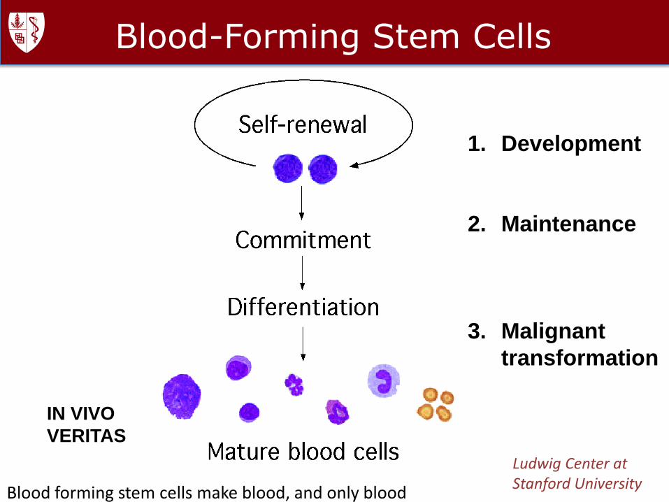
Blood-Forming Stem Cells
Ludwig Center at Stanford University
IN VIVO
VERITAS
1. Development
2. Maintenance
3. Malignant
transformation
Blood forming stem cells make blood, and only blood
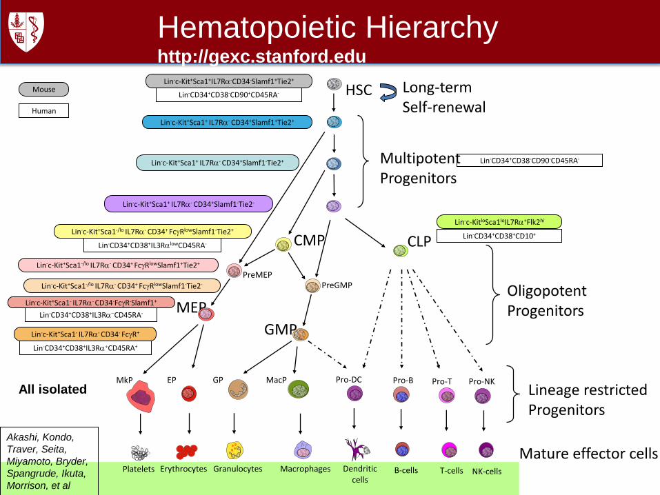
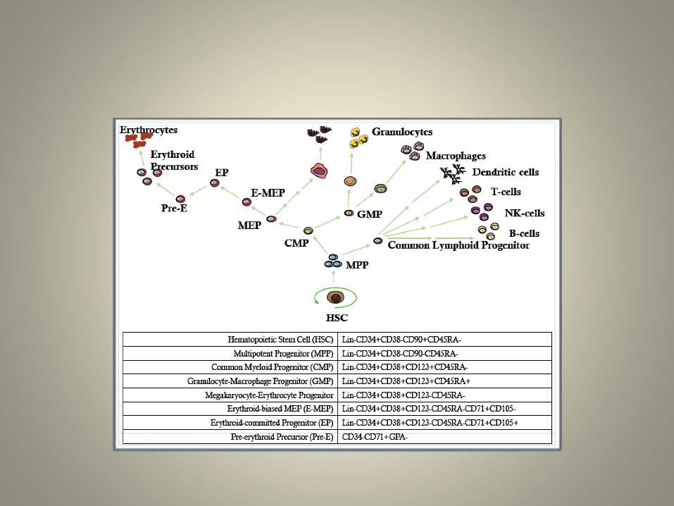
HSC
MultipotentProgenitors
CLP
T-cells NK-cellsB-cells
MEP
GMP
CMP
OligopotentProgenitors
Mature effector cells
Pro-T Pro-NKPro-B
Dendriticcells
Pro-DC
Lineage restrictedProgenitors
GranulocytesErythrocytesPlatelets Macrophages
Hematopoietic Hierarchyhttp://gexc.stanford.edu
Lin-c-Kit+Sca1- IL7Ra- CD34- FcgR+
Lin-c-Kit+Sca1- IL7Ra- CD34-FcgR-Slamf1+
Lin-c-Kit+Sca1-/lo IL7Ra- CD34+ FcgRlowSlamf1-Tie2+
Lin-c-Kit+Sca1+ IL7Ra- CD34+Slamf1+Tie2+
Lin-c-Kit+Sca1+IL7Ra-CD34-Slamf1+Tie2+
Lin-c-Kit+Sca1+ IL7Ra- CD34+Slamf1-Tie2-
Lin-c-KitloSca1loIL7Ra+Flk2hi
Lin-c-Kit+Sca1+ IL7Ra- CD34+Slamf1-Tie2+
Lin-CD34+CD38-CD90+CD45RA-Mouse
Human
Lin-CD34+CD38-CD90-CD45RA-
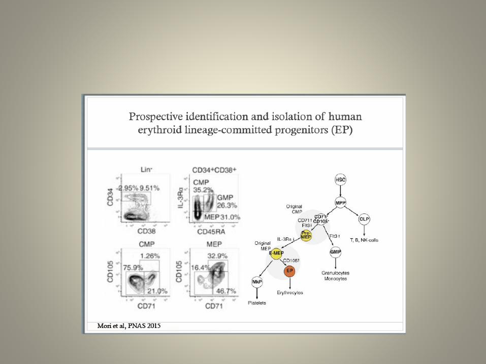
Lin-CD34+CD38+CD10+
Lin-CD34+CD38+IL3RaCD45RA-
Lin-CD34+CD38+IL3RaCD45RA+
Lin-CD34+CD38+IL3RalowCD45RA-
EPMkP
Long-termSelf-renewal
GP MacP
PreGMPPreMEP
Lin-c-Kit+Sca1-/lo IL7Ra- CD34+ FcgRlowSlamf1+Tie2+
Lin-c-Kit+Sca1-/lo IL7Ra- CD34+ FcgRlowSlamf1-Tie2-
All isolated
Akashi, Kondo,
Traver, Seita,
Miyamoto, Bryder,
Spangrude, Ikuta,
Morrison, et al
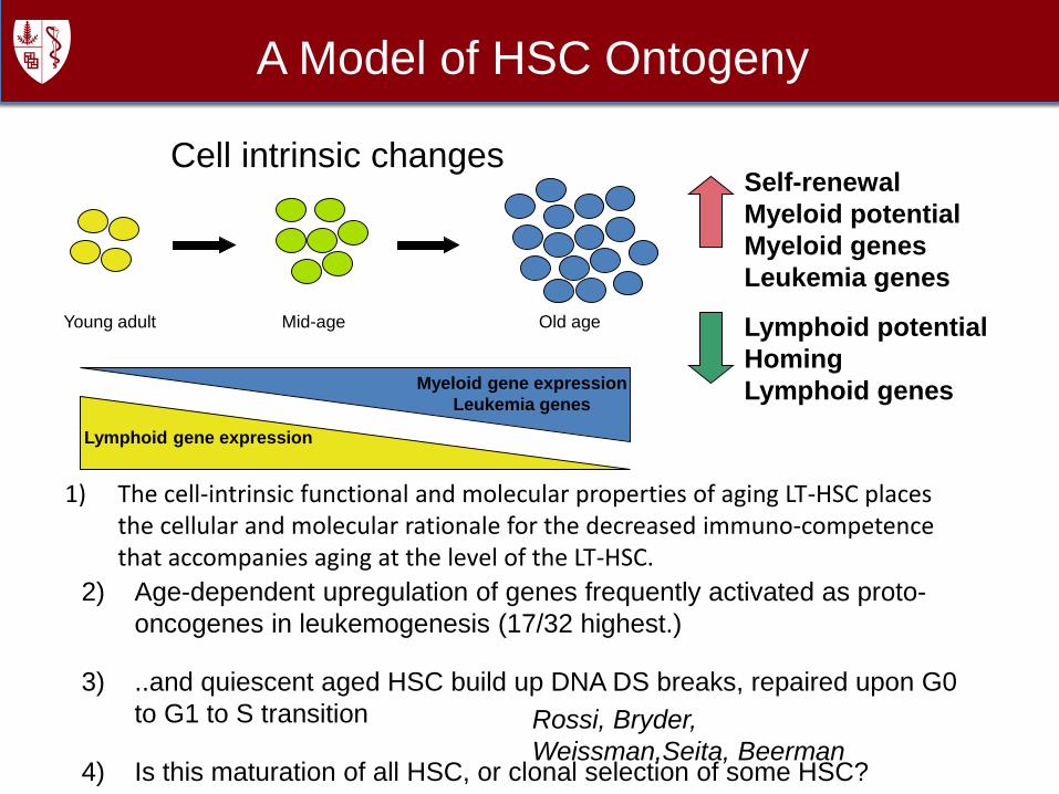
A Model of HSC Ontogeny
Young adult Mid-age Old age
Lymphoid gene expression
Myeloid gene expression
Leukemia genes
Self-renewal
Myeloid potential
Myeloid genes
Leukemia genes
Lymphoid potential
Homing
Lymphoid genes
Rossi, Bryder,
Weissman,Seita, Beerman
Cell intrinsic changes
)
1) The cell-intrinsic functional and molecular properties of aging LT-HSC places the cellular and molecular rationale for the decreased immuno-competence that accompanies aging at the level of the LT-HSC.
2) Age-dependent upregulation of genes frequently activated as proto-
oncogenes in leukemogenesis (17/32 highest.)
3) ..and quiescent aged HSC build up DNA DS breaks, repaired upon G0
to G1 to S transition
4) Is this maturation of all HSC, or clonal selection of some HSC?
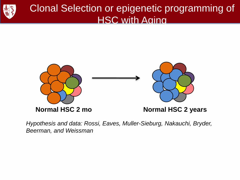
Clonal Selection or epigenetic programming of
HSC with Aging
Normal HSC 2 mo Normal HSC 2 years
Hypothesis and data: Rossi, Eaves, Muller-Sieburg, Nakauchi, Bryder,
Beerman, and Weissman
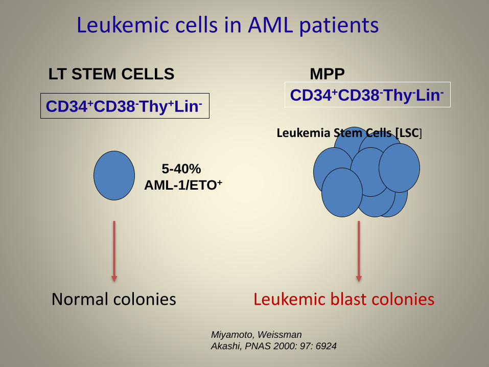
Leukemic cells in AML patients
CD34+CD38-Thy+Lin-CD34+CD38-Thy-Lin-
5-40%
AML-1/ETO+
Normal colonies Leukemic blast colonies
Miyamoto, Weissman
Akashi, PNAS 2000: 97: 6924
LT STEM CELLS MPP
Leukemia Stem Cells [LSC]

Cell of origin – progression to leukemia
Jak2
W
e
i
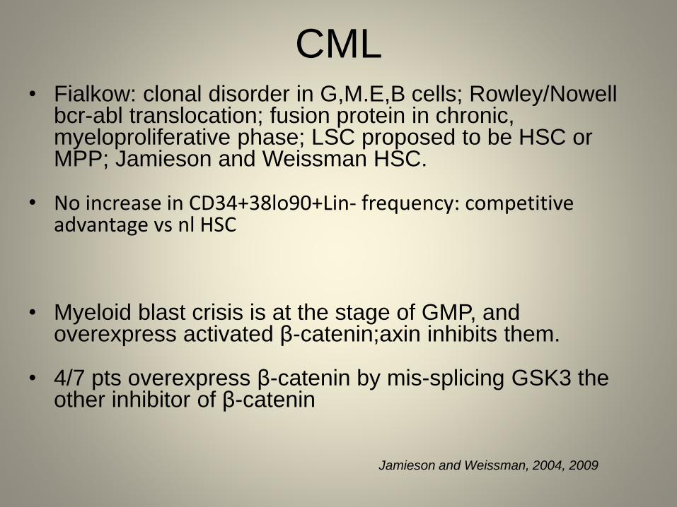
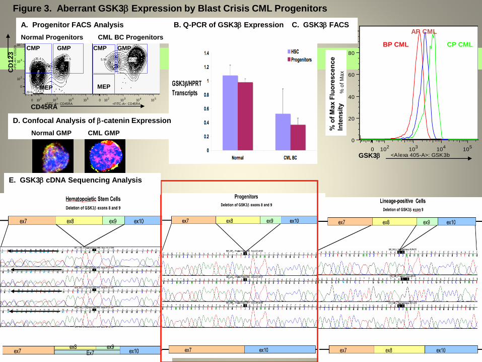
CML• Fialkow: clonal disorder in G,M.E,B cells; Rowley/Nowell
bcr-abl translocation; fusion protein in chronic, myeloproliferative phase; LSC proposed to be HSC or MPP; Jamieson and Weissman HSC.
• No increase in CD34+38lo90+Lin- frequency: competitive advantage vs nl HSC
• Myeloid blast crisis is at the stage of GMP, and overexpress activated β-catenin;axin inhibits them.
• 4/7 pts overexpress β-catenin by mis-splicing GSK3 the other inhibitor of β-catenin
Jamieson and Weissman, 2004, 2009
GSK30 10
210
310
410
5
<Alexa 405-A>: GSK3b
0
20
40
60
80
100
% o
f M
ax
CP CML
AP CML
BP CML
0 102
103
104
105
<FITC-A>: CD45RA
0
102
103
104
105
<P
E-A
>:
CD
12
3
5.98 90.3
0 102
103
104
105
<FITC-A>: CD45RA
0
102
103
104
105
<P
E-A
>:
CD
12
3
29.6
35.4 27.5
CD45RA
CD
123
GMPCMP
MEP
CMP GMP
MEP
A. Progenitor FACS Analysis B. Q-PCR of GSK3 Expression C. GSK3 FACS
Normal Progenitors CML BC Progenitors
D. Confocal Analysis of -catenin Expression
Normal GMP CML GMP
E. GSK3 cDNA Sequencing Analysis
Figure 3. Aberrant GSK3Expression by Blast Crisis CML Progenitors

AML Gene Annotation
SU070 PXDN V616I
KALRN S44P
TET2 Y1649stop
TET2 T1884A
TMEM8B nt G471A
NCRNA00200 nt G354A
TMEM20 A143T
ZRANB1 nt G4659A
SCN4B H227N
GABARAPL1 nt C1583T
DOCK9 A1475V PLAG2G4D P246A
CACNA1H R1069stop
CTCF R339Q GZF1 nt G3835C
PRPF6 R527H
CXorf36 I225L CXorf66 G321S
FLT3 599-610 ITD !
AML HSC
FACS
Normal
Tissue
(T Cells)
Next generation
exome sequencing
N Mutations
In AML
Identification of Somatic Mutations by Exome
Sequencing
Max Jan
Thomas Snyder
Ryan Corces-
Zimmerman
Steve Quake
Ravi Majeti
Irv Weissman Science Translatinal Medicine
2012 4(149): 149ra18.
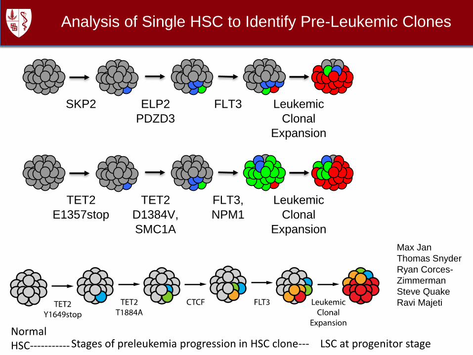
Analysis of Single HSC to Identify Pre-Leukemic Clones
Max Jan
Thomas Snyder
Ryan Corces-
Zimmerman
Steve Quake
Ravi Majeti
TET2
E1357stop
TET2
D1384V,
SMC1A
FLT3,
NPM1
Leukemic
Clonal
Expansion
SKP2 ELP2
PDZD3
FLT3 Leukemic
Clonal
Expansion
Normal HSC----------- Stages of preleukemia progression in HSC clone--- LSC at progenitor stage

Ryan corces –Zimmerman, Ravi Majeti, and IW
Now 21 AMLs PNAS 2014
If you want to know which genes are likely oncogenes, ask the cancer.
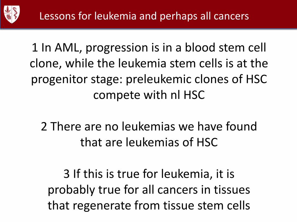
1 In AML, progression is in a blood stem cell clone, while the leukemia stem cells is at the progenitor stage: preleukemic clones of HSC
compete with nl HSC
2 There are no leukemias we have foundthat are leukemias of HSC
3 If this is true for leukemia, it isprobably true for all cancers in tissuesthat regenerate from tissue stem cells
Lessons for leukemia and perhaps all cancers

CD47 was discovered as a marker of aging RBC by Oldenborg. We found it on m/h AML LSC
Macrophage
SIRPa
Receptor+
AMLLSC
CD47
Stimulus
---
Net Result: No Phagocytosis
Hypothesis: Increased expression of CD47 on myeloid leukemia cells contributes to pathogenesis by facilitating evasion of phagocytosis
Prediction: Increased expression of CD47 on human AML is associated with a worse clinical outcome
Eat me
Don’teat me
Programmed cell removal
Traver and IW 1998; Jaiswal, Majeti, Chao,and IW 2008.
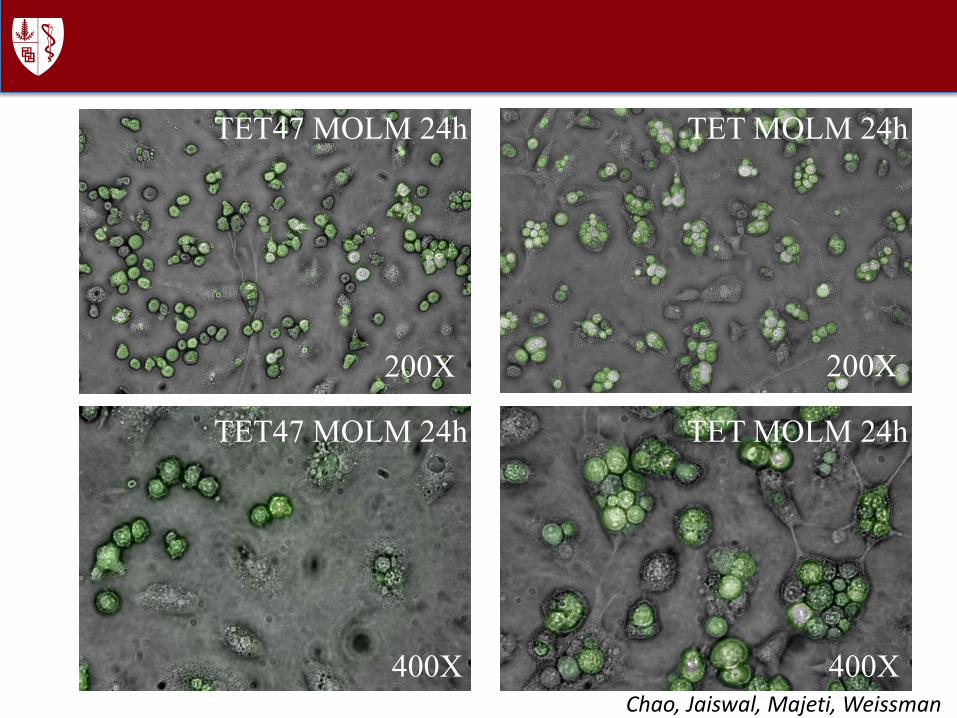
TET47 MOLM 24h
TET47 MOLM 24h
200X
400X
TET MOLM 24h
TET MOLM 24h
200X
400XChao, Jaiswal, Majeti, Weissman
liv e4 7 M O L M R f e m u r
E v e n t C o u n t : 3 2 1 4 6
0 1 03
1 04
1 05
< P E - C y 7 - A > : h u C D 4 5
0
1 03
1 04
1 05
<A
PC
-A
>:
m
uC
D4
5
9 7 . 3
0 . 7
liv e4 7 M O L M R t ib ia
E v e n t C o u n t : 5 2 9 9 7
0 1 03
1 04
1 05
< P E - C y 7 - A > : h u C D 4 5
0
1 03
1 04
1 05
<A
PC
-A
>:
m
uC
D4
5
3 8 . 3
5 5 . 9
liv e4 7 M O L M R h u m e r u s
E v e n t C o u n t : 4 0 5 7 5
liv e4 7 M O L M L f e m u r
E v e n t C o u n t : 3 9 9 5 8
0 1 03
1 04
1 05
< P E - C y 7 - A > : h u C D 4 5
0
1 03
1 04
1 05
<A
PC
-A
>:
m
uC
D4
5
9 7 . 4
1 . 6 8
liv e4 7 M O L M L t ib ia
E v e n t C o u n t : 4 2 5 4 8
0 1 03
1 04
1 05
< P E - C y 7 - A > : h u C D 4 5
0
1 03
1 04
1 05
<A
PC
-A
>:
m
uC
D4
5
6 3 . 9
3 1 . 1
liv e4 7 M O L M L h u m e r u s
E v e n t C o u n t : 1 6 4 0 7
0 1 03
1 04
1 05
< P E - C y 7 - A > : h u C D 4 5
0
1 03
1 04
1 05
<A
PC
-A
>:
m
uC
D4
5
7 3 . 6
2 3 . 8
liv e4 7 M O L M S p
E v e n t C o u n t : 3 5 0 3 0
0 1 03
1 04
1 05
< P E - C y 7 - A > : h u C D 4 5
0
1 03
1 04
1 05
<A
PC
-A
>:
m
uC
D4
5
4 0 . 8
2 0 . 7
liv e
T E T M O L M R f e m u rE v e n t C o u n t : 1 0 9 3 9 8
0 1 03
1 04
1 05
< P E - C y 7 - A > : h u C D 4 5
0
1 03
1 04
1 05
<A
PC
-A
>:
m
uC
D4
5
1 . 3 2
9 6
liv e
T E T M O L M R t ib iaE v e n t C o u n t : 4 5 8 5 0
0 1 03
1 04
1 05
< P E - C y 7 - A > : h u C D 4 5
0
1 03
1 04
1 05
<A
PC
-A
>:
m
uC
D4
5
0
9 8 . 5
liv e
T E T M O L M R h u m e r u sE v e n t C o u n t : 3 8 0 3 9
0 1 03
1 04
1 05
< P E - C y 7 - A > : h u C D 4 5
0
1 03
1 04
1 05
<A
PC
-A
>:
m
uC
D4
5
2 . 6 3 e - 3
9 7 . 3
liv e
T E T M O L M L f e m u rE v e n t C o u n t : 6 5 8 3 5
0 1 03
1 04
1 05
< P E - C y 7 - A > : h u C D 4 5
0
1 03
1 04
1 05
<A
PC
-A
>:
m
uC
D4
5
1 . 5 2 e - 3
9 7
liv e
T E T M O L M L t ib iaE v e n t C o u n t : 5 0 4 1 4
0 1 03
1 04
1 05
< P E - C y 7 - A > : h u C D 4 5
0
1 03
1 04
1 05
<A
PC
-A
>:
m
uC
D4
5
3 . 9 7 e - 3
9 7 . 8
liv e
T E T M O L M L h u m e r u sE v e n t C o u n t : 3 2 7 4 9
0 1 03
1 04
1 05
< P E - C y 7 - A > : h u C D 4 5
0
1 03
1 04
1 05
<A
PC
-A
>:
m
uC
D4
5
0
9 7 . 5
liv e
T E T M O L M S pE v e n t C o u n t : 1 4 2 9 3
0 1 03
1 04
1 05
< P E - C y 7 - A > : h u C D 4 5
0
1 03
1 04
1 05
<A
PC
-A
>:
m
uC
D4
5
7 . e - 3
7 7 . 7
0 1 03
1 04
1 05
< P E - C y 7 - A > : h u C D 4 5
0
1 03
1 04
1 05
<A
PC
-A
>:
m
uC
D4
5
9 8 . 6
0 . 4 7
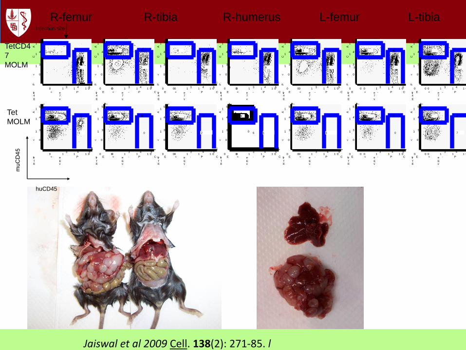
R-femur R-tibia R-humerus L-femur L-tibia LInjection site
TetCD4
7
MOLM
Tet
MOLM
huCD45
Jaiswal et al 2009 Cell. 138(2): 271-85. l
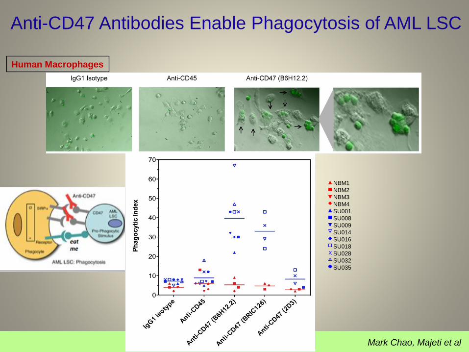
Anti-CD47 Antibodies Enable Phagocytosis of AML LSC
Human Macrophages
NBM1
NBM2
NBM3
NBM4
SU001
SU008
SU009
SU014
SU016
SU018
SU028
SU032
SU035
X
Mark Chao, Majeti et al
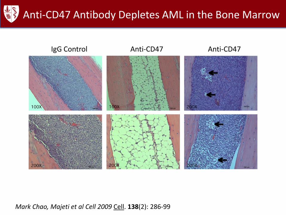
Anti-CD47 Antibody Depletes AML in the Bone Marrow
IgG Control Anti-CD47 Anti-CD47
Mark Chao, Majeti et al Cell 2009 Cell. 138(2): 286-99

Inject Tumor Cells
Into Peritoneal Cavity
Initiate Treatment Evaluate Tumor Growth
With Bioluminescence
Imaging
2.5 Months2 Weeks
Volkmer, Willingham, Chin, and IW
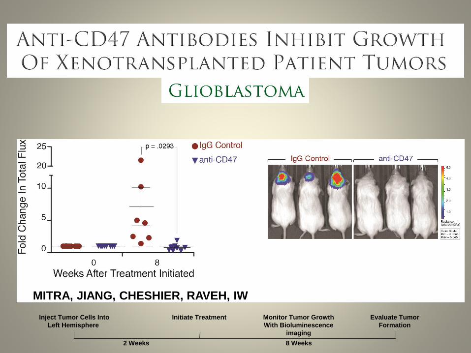
Inject Tumor Cells Into
Left Hemisphere
Initiate Treatment Evaluate Tumor
Formation
8 Weeks2 Weeks
Monitor Tumor Growth
With Bioluminescence
imaging
MITRA, JIANG, CHESHIER, RAVEH, IW

Investigation and Targeting of CD47 in Human Cancers
GlioblastomaMedulloblastomaOligodendrogliomaHepatocellular CarcinomaGastric CancerMultiple MyelomaChronic Myeloid LeukemiaAcute Myeloid LeukemiaNon-Hodgkin’s Lymphoma
T-Acute Lymphoblastic LeukemiaB-Acute Lymphoblastic Leukemia
In all cases tested with metastatic tumors, the metastases areeliminated.
Breast OvarianBladderPancreaticColonProstateLungKidneyLeiomyosarcomaHead & NeckMelanoma
Monotherapy trials

Precancer cells express calreticulin, and emergent
cancer clones overcome this with CD47
lrl
Macrophage
SIRPa
Receptor+
AMLLSC
CD47
Stimulus
---
Increased expression of CD47 on myeloid leukemia cells contributes to
pathogenesis by facilitating evasion of phagocytosis
Eat me
Don’t
eat me
Lrp 1 calreticulin
Chao, Jaiswal, Weissman-Tsukamoto, Majeti, and IW
Science Transl Med 2010. 2(63)

Toll like receptor agonist incubation of macrophages increases anti-CD47
mediated phagocytosisof cancer cells
Mingye Feng, Volkmer, Weissman
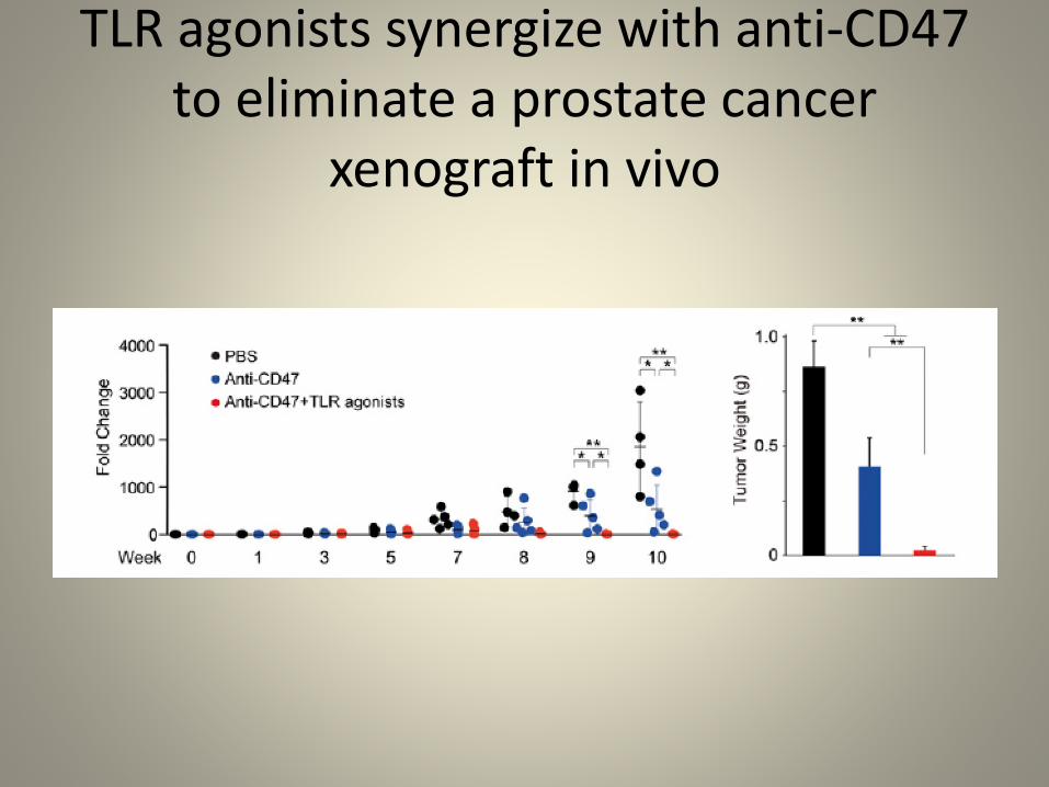
TLR agonists synergize with anti-CD47to eliminate a prostate cancer
xenograft in vivo
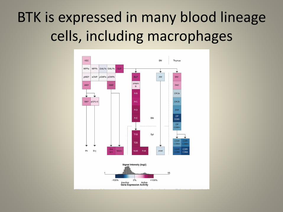
BTK is expressed in many blood lineage cells, including macrophages

Phagocytosis of tumor cells is inhibited by the BTK inhibitor, ibrutinib
Mingye Feng, Volkmer, Weissman
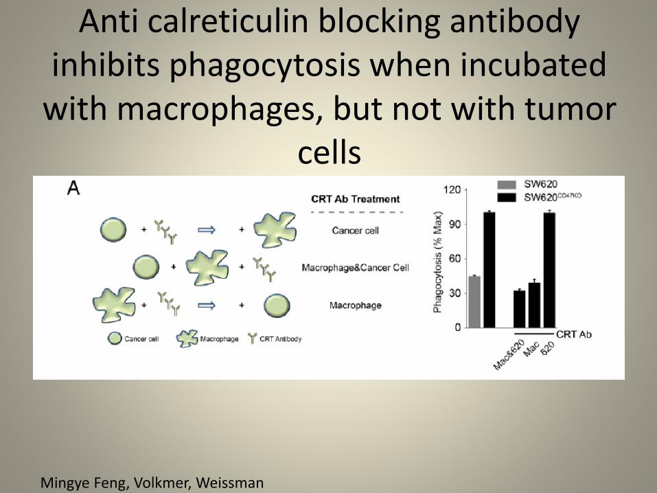
Anti calreticulin blocking antibody inhibits phagocytosis when incubated
with macrophages, but not with tumor cells
Mingye Feng, Volkmer, Weissman
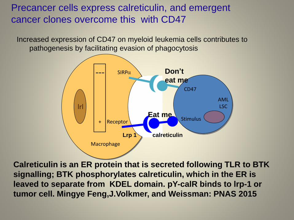
Precancer cells express calreticulin, and emergent
cancer clones overcome this with CD47
lrl
Macrophage
SIRPa
Receptor+
AMLLSC
CD47
Stimulus
---
Increased expression of CD47 on myeloid leukemia cells contributes to
pathogenesis by facilitating evasion of phagocytosis
Eat me
Don’t
eat me
Lrp 1 calreticulin
Calreticulin is an ER protein that is secreted following TLR to BTK
signalling; BTK phosphorylates calreticulin, which in the ER is
leaved to separate from KDEL domain. pY-calR binds to lrp-1 or
tumor cell. Mingye Feng,J.Volkmer, and Weissman: PNAS 2015
0%
10%
20%
30%
40%
Normal age-matched MDS
HS
C /
CD
34+
F
requency
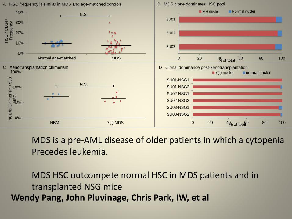
A HSC frequency is similar in MDS and age-matched controls
N.S.
B MDS clone dominates HSC pool
0%
1%
10%
100%
NBM 7(-) MDS
hC
D45 C
him
erism
/ 5
00
HS
C
N.S.
0 20 40 60 80 100
SU03
SU02
SU01
% of total
7(-) nuclei Normal nuclei
C Xenotransplantation chimerism D Clonal dominance post-xenotransplantation
0 20 40 60 80 100
SU03-NSG2
SU03-NSG1
SU02-NSG2
SU02-NSG1
SU01-NSG2
SU01-NSG1
% of total
7(-) nuclei normal nuclei
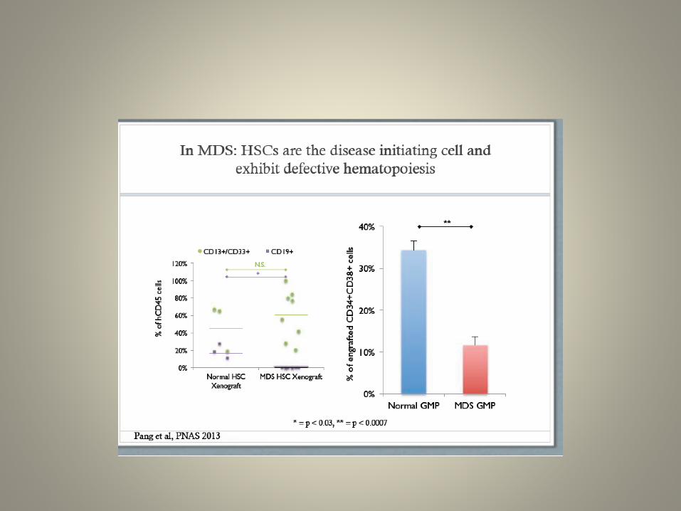
Wendy Pang, John Pluvinage, Chris Park, IW, et al
MDS is a pre-AML disease of older patients in which a cytopeniaPrecedes leukemia.
MDS HSC outcompete normal HSC in MDS patients and in transplanted NSG mice

NBM 34+38+ MDS 34+38+
0
10
20
30
40
50
60
Normal HSC MDS HSC
Rela
tive M
FI
0
10
20
30
40
50
60
70
Normal 34+38+ MDS 34+38+
Rela
tive M
FI
0
20
40
60
80
100
Normal GMP MDS GMP
Rela
tive M
FI
0
20
40
60
80
100
Normal 34+38+ MDS 34+38+
Phagocytic I
nex
0
10
20
30
40
Normal 34+38+ MDS 34+38+
Phagocytic I
nex
E CRT-blocking peptide abrogates phagocytosis of MDS CD34+CD38+ cells
D Increased phagocytosis of MDS CD34+CD38+ cells C Increased CRT on MDS GMP
A CRT not increased on MDS HSC B Increased cell-surface CRT on MDS CD34+CD38+ cells
N.S. *
** ***
****
% o
f
ma
xim
um
CRT Expression
Normal + PBS
MDS + PBS
Normal + CRT-blocking
MDS + CRT-blocking
Wendy Pang, John Pluvinage,Chris Park, IW, et al
Calreticulin expressed onMDS progenitors, but notHSC, causes programmedcell removal
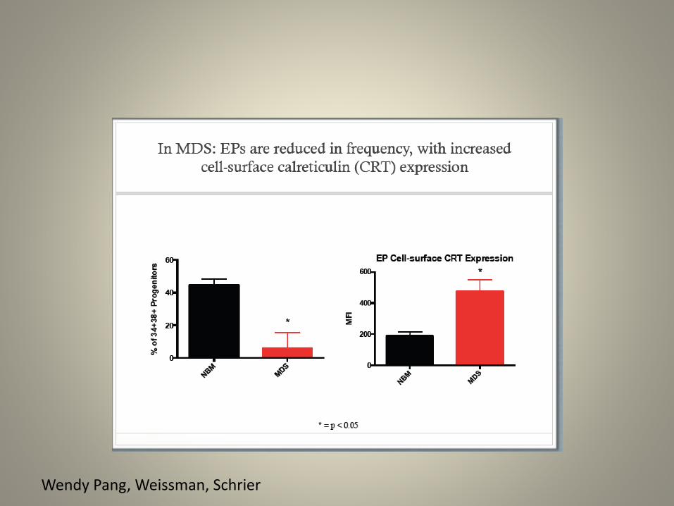
Wendy Pang, Weissman, Schrier

Conclusions
MDS HSC outcompete normal HSC in patient and xenotransplants
The MDS-initiating cell resides in the HSC compartment
High CRT predisposes MDS myeloid [GMP, MkP, EP] progenitors for programmed cell removal
Increased CD47 expression is a crucial step in the progression from MDS to AML
• Calreticulin and LRP1 are potential therapeutic targets
Wendy Pang, John Pluvinage, Chris Park, IW, et al
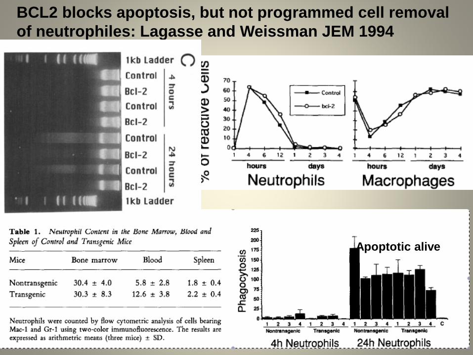
BCL2 blocks apoptosis, but not programmed cell removal
of neutrophiles: Lagasse and Weissman JEM 1994
Apoptotic alive
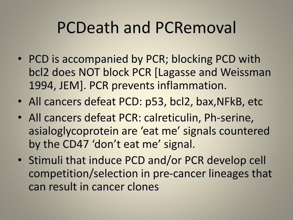
PCDeath and PCRemoval
• PCD is accompanied by PCR; blocking PCD with bcl2 does NOT block PCR [Lagasse and Weissman 1994, JEM]. PCR prevents inflammation.
• All cancers defeat PCD: p53, bcl2, bax,NFkB, etc
• All cancers defeat PCR: calreticulin, Ph-serine, asialoglycoprotein are ‘eat me’ signals countered by the CD47 ‘don’t eat me’ signal.
• Stimuli that induce PCD and/or PCR develop cell competition/selection in pre-cancer lineages that can result in cancer clones
Lokey Stem Cell Institute at Stanford
Ludwig Center at Stanford
Institute for Stem Cell Biology and Regenerative Medicine
Supported grants from NCI, NHLBI, Calif Inst Reg Medicine, Lacob Foundation, Siebel SCI

Wendy Pang, Weissman, Schrier

Wendy Pang, Weissman, Schrier