Pathogen-Associated Molecular Pattern-Triggered Immunity ... › content › plantcell › 32 › 4...
22
Pathogen-Associated Molecular Pattern-Triggered Immunity Involves Proteolytic Degradation of Core Nonsense-Mediated mRNA Decay Factors During the Early Defense Response [OPEN] Ho Won Jung, a Gagan Kumar Panigrahi, b,c,d Ga Young Jung, a Yu Jeong Lee, a Ki Hun Shin, b,c Annapurna Sahoo, b,c Eun Su Choi, a Eunji Lee, a Kyung Man Kim, b,c Seung Hwan Yang, e Jong-Seong Jeon, f Sung Chul Lee, g and Sang Hyon Kim b,c,1 a Department of Applied Bioscience, Dong-A University, Busan 49315, Korea b Department of Biosciences and Bioinformatics, Myongji University, Yongin 17058, Korea c RNA Genomics Center, Myongji University, Yongin 17058, Korea d School of Applied Sciences, Centurion University of Technology and Management, Odisha 752050, India e Department of Biotechnology, Chonnam National University, Yeosu 59626, Korea f Graduate School of Biotechnology and Crop Biotech Institute, Kyung Hee University, Yongin 17104, Korea g School of Biological Sciences, Chung-Ang University, Seoul 06974, Korea ORCID IDs: 0000-0001-6408-4168 (H.W.J.); 0000-0003-3908-3379 (G.K.P.); 0000-0001-7829-3825 (G.Y.J.); 0000-0002-8145-9520 (Y.J.L.); 0000-0002-7530-6229 (K.H.S.); 0000-0002-5079-0261 (A.S.); 0000-0001-9503-6441 (E.S.C.); 0000-0001-6777-0876 (E.L.); 0000-0001-5105-922X (K.M.K.); 0000-0003-0603-2209 (S.H.Y.); 0000-0001-6221-4993 (J.-S.J.); 0000-0003-2725-0854 (S.C.L.); 0000-0003-3444-4446 (S.H.K.). Nonsense-mediated mRNA decay (NMD), an mRNA quality control process, is thought to function in plant immunity. A subset of fully spliced (FS) transcripts of Arabidopsis (Arabidopsis thaliana) resistance (R) genes are upregulated during bacterial infection. Here, we report that 81.2% and 65.1% of FS natural TIR-NBS-LRR (TNL) and CC-NBS-LRR transcripts, respectively, retain characteristics of NMD regulation, as their transcript levels could be controlled posttranscriptionally. Both bacterial infection and the perception of bacteria by pattern recognition receptors initiated the destruction of core NMD factors UP- FRAMESHIFT1 (UPF1), UPF2, and UPF3 in Arabidopsis within 30 min of inoculation via the independent ubiquitination of UPF1 and UPF3 and their degradation via the 26S proteasome pathway. The induction of UPF1 and UPF3 ubiquitination was delayed in mitogen-activated protein kinase3 (mpk3) and mpk6, but not in salicylic acid-signaling mutants, during the early immune response. Finally, previously uncharacterized TNL-type R transcripts accumulated in upf mutants and conferred disease resistance to infection with a virulent Pseudomonas strain in plants. Our findings demonstrate that NMD is one of the main regulatory processes through which PRRs fine-tune R transcript levels to reduce fitness costs and achieve effective immunity. INTRODUCTION Plant immunity, a counterattack mechanism against microbial infection, is exquisitely controlled by two different immune re- ceptors known as extracellular immune receptors (pattern rec- ognition receptors, PRRs) and intracellular immune receptors (resistance [R] proteins; nucleotide binding oligomerization domain– like receptors [NOD-like receptors or NLRs]; or nucleotide bind- ing site–leucine-rich repeat [NBS-LRR] proteins) that recognize microbe-associated molecular patterns (MAMPs) and pathogen- derived effectors, respectively. In general, pattern-triggered im- munity (PTI), which is controlled by PRRs, confers moderate disease resistance to a broad spectrum of pathogens, and effector- triggered immunity (ETI), which is controlled by R proteins, is responsible for resistance to a specific pathogen carrying a cognate avirulence gene (Chisholm et al., 2006; Jones and Dangl, 2006). In addition to their predominance, R genes are also in- dispensable for establishing basal immunity to virulent pathogen infection and maintaining the balance between growth and defense (Li et al., 2001; Shirano et al., 2002; Palma et al., 2010; Maekawa et al., 2011; Karasov et al., 2017). The maintenance of the switched-off state and posttranslational modification of R proteins by other cellular components are crucial for maintaining the proper homeostasis of R activity (DeYoung and Innes, 2006; Li et al., 2015; Karasov et al., 2017). Transcriptional controls, such as histone modification and DNA methylation, are fundamental for fine-tuning R transcript levels (Halter and Navarro, 2015; Lai and Eulgem, 2018). Alternative splicing (AS) can secure the accu- mulation of diverse transcript isoforms for full immunity (Dinesh- Kumar and Baker, 2000; Zhang and Gassmann, 2007; Xu et al., 2012; Yang et al., 2014). A few fully spliced (FS) and AS versions of R transcripts are targets of miRNAs and nonsense-mediated mRNA decay (NMD; Zhai et al., 2011; Shivaprasad et al., 2012; Gloggnitzer et al., 2014; Zhang et al., 2016), indicating that R gene expression is also under posttranscriptional control in plants. 1 Address correspondence to [email protected]. The author responsible for distribution of materials integral to the findings presented in this article in accordance with the policy described in the instructions for authors (www.plant cell.org) is Sang Hyon Kim ([email protected]). [OPEN] Articles can be viewed without a subscription. www.plantcell.org/cgi/doi/10.1105/tpc.19.00631 The Plant Cell, Vol. 32: 1081–1101, April 2020, www.plantcell.org ã 2020 ASPB.
Transcript of Pathogen-Associated Molecular Pattern-Triggered Immunity ... › content › plantcell › 32 › 4...
Pathogen-Associated Molecular Pattern-Triggered ImmunityInvolves Proteolytic Degradation of Core Nonsense-MediatedmRNA Decay Factors During the Early Defense Response[OPEN]
HoWon Jung,a Gagan Kumar Panigrahi,b,c,d Ga Young Jung,a Yu Jeong Lee,a Ki Hun Shin,b,c Annapurna Sahoo,b,c
Eun Su Choi,a Eunji Lee,a Kyung Man Kim,b,c Seung Hwan Yang,e Jong-Seong Jeon,f Sung Chul Lee,g andSang Hyon Kimb,c,1
a Department of Applied Bioscience, Dong-A University, Busan 49315, KoreabDepartment of Biosciences and Bioinformatics, Myongji University, Yongin 17058, Koreac RNA Genomics Center, Myongji University, Yongin 17058, Koread School of Applied Sciences, Centurion University of Technology and Management, Odisha 752050, Indiae Department of Biotechnology, Chonnam National University, Yeosu 59626, Koreaf Graduate School of Biotechnology and Crop Biotech Institute, Kyung Hee University, Yongin 17104, KoreagSchool of Biological Sciences, Chung-Ang University, Seoul 06974, Korea
ORCID IDs: 0000-0001-6408-4168 (H.W.J.); 0000-0003-3908-3379 (G.K.P.); 0000-0001-7829-3825 (G.Y.J.); 0000-0002-8145-9520(Y.J.L.); 0000-0002-7530-6229 (K.H.S.); 0000-0002-5079-0261 (A.S.); 0000-0001-9503-6441 (E.S.C.); 0000-0001-6777-0876 (E.L.);0000-0001-5105-922X (K.M.K.); 0000-0003-0603-2209 (S.H.Y.); 0000-0001-6221-4993 (J.-S.J.); 0000-0003-2725-0854 (S.C.L.);0000-0003-3444-4446 (S.H.K.).
Nonsense-mediated mRNA decay (NMD), an mRNA quality control process, is thought to function in plant immunity. A subsetof fully spliced (FS) transcripts of Arabidopsis (Arabidopsis thaliana) resistance (R) genes are upregulated during bacterialinfection. Here, we report that 81.2% and 65.1% of FS natural TIR-NBS-LRR (TNL) and CC-NBS-LRR transcripts, respectively,retain characteristics of NMD regulation, as their transcript levels could be controlled posttranscriptionally. Both bacterialinfection and the perception of bacteria by pattern recognition receptors initiated the destruction of core NMD factors UP-FRAMESHIFT1 (UPF1), UPF2, and UPF3 in Arabidopsis within 30 min of inoculation via the independent ubiquitination of UPF1and UPF3 and their degradation via the 26S proteasome pathway. The induction of UPF1 and UPF3 ubiquitination was delayedin mitogen-activated protein kinase3 (mpk3) and mpk6, but not in salicylic acid-signaling mutants, during the early immuneresponse. Finally, previously uncharacterized TNL-type R transcripts accumulated in upf mutants and conferred diseaseresistance to infection with a virulent Pseudomonas strain in plants. Our findings demonstrate that NMD is one of the mainregulatory processes through which PRRs fine-tune R transcript levels to reduce fitness costs and achieve effectiveimmunity.
INTRODUCTION
Plant immunity, a counterattack mechanism against microbialinfection, is exquisitely controlled by two different immune re-ceptors known as extracellular immune receptors (pattern rec-ognition receptors, PRRs) and intracellular immune receptors(resistance [R] proteins; nucleotidebindingoligomerization domain–like receptors [NOD-like receptors or NLRs]; or nucleotide bind-ing site–leucine-rich repeat [NBS-LRR] proteins) that recognizemicrobe-associated molecular patterns (MAMPs) and pathogen-derived effectors, respectively. In general, pattern-triggered im-munity (PTI), which is controlled by PRRs, confers moderatedisease resistance to a broad spectrum of pathogens, and effector-triggered immunity (ETI), which is controlled by R proteins, is
responsible for resistance to a specific pathogen carrying acognate avirulence gene (Chisholm et al., 2006; Jones and Dangl,2006). In addition to their predominance, R genes are also in-dispensable for establishing basal immunity to virulent pathogeninfection and maintaining the balance between growth anddefense (Li et al., 2001; Shirano et al., 2002; Palma et al., 2010;Maekawa et al., 2011; Karasov et al., 2017). The maintenance ofthe switched-off state and posttranslational modification of Rproteins by other cellular components are crucial for maintainingthe proper homeostasis of R activity (DeYoung and Innes, 2006; Liet al., 2015;Karasov et al., 2017). Transcriptional controls, such ashistone modification and DNA methylation, are fundamental forfine-tuning R transcript levels (Halter and Navarro, 2015; Lai andEulgem, 2018). Alternative splicing (AS) can secure the accu-mulation of diverse transcript isoforms for full immunity (Dinesh-Kumar and Baker, 2000; Zhang and Gassmann, 2007; Xu et al.,2012; Yang et al., 2014). A few fully spliced (FS) andAS versions ofR transcripts are targets of miRNAs and nonsense-mediatedmRNA decay (NMD; Zhai et al., 2011; Shivaprasad et al., 2012;Gloggnitzer et al., 2014; Zhang et al., 2016), indicating thatR geneexpression is also under posttranscriptional control in plants.
1 Address correspondence to [email protected] author responsible for distribution of materials integral to the findingspresented in this article in accordance with the policy described in theinstructions for authors (www.plant cell.org) is Sang Hyon Kim([email protected]).[OPEN]Articles can be viewed without a subscription.www.plantcell.org/cgi/doi/10.1105/tpc.19.00631
The Plant Cell, Vol. 32: 1081–1101, April 2020, www.plantcell.org ã 2020 ASPB.
NMD is a translation-coupled mRNA surveillance process ineukaryotes that prevents mRNAs containing premature termi-nation codons (PTCs) from being translated. PTC-containingtranscripts can arise from mutations or AS, and NMD is usuallytriggered by the presence of a downstream splice junction or long39untranslated region (UTR;RebbapragadaandLykke-Andersen,2009; Peccarelli and Kebaara, 2014). FS natural transcripts canalso be subject to NMD by virtue of containing NMD-triggeringcharacteristics such as one or more introns in the 39UTR, long39UTRs, or upstream open reading frames (uORFs) encoding nofewer than 35 amino acids (Kertész et al., 2006; Kerényi et al.,2008; Nyikó et al., 2009; Kalyna et al., 2012; Le Hir et al., 2016).
The canonical NMD process occurs when a translating ribo-someencounters aPTCand fails toproperly terminate translation.This leads to the phosphorylation and activation of the core NMDfactor UP-FRAMESHIFT1 (UPF1), a process promoted by theinteraction of UPF1 with UPF2 and UPF3, which associate witha downstream exon-junction complex (EJC; Kim et al., 2001;Banihashemi et al., 2006; Schweingruber et al., 2013). In a tran-sient wild tobacco (Nicotiana benthamiana) assay, NMD factorSUPPRESSORFORMORPHOLOGICALDEFECTSOFGENITALIA7(SMG7) bound to the phosphorylated C-terminal region of UPF1for subsequent recruitment of the decapping and deadenylationcomplexes before the decay of the target mRNA (Mérai et al.,2013). A recent study provided evidence that the dephosphory-lation ofUPF1 involves anSMG7 function (Kesarwani et al., 2019).NMD is involved in flowering, Suc metabolism, and stress re-sponses in plants (Yoine et al., 2006; Jeong et al., 2011; Raysonetal., 2012;Riehs-Kearnanetal., 2012;Shi et al., 2012;Garciaet al.,2014; Gloggnitzer et al., 2014; Filichkin et al., 2015; Sureshkumaret al., 2016).
The most noteworthy function of NMD is its participation in theregulation of plant immunity. A null mutation in UPF1 or UPF3confers disease resistance via increased levels of salicylic acid(SA) and elevated expression of defense genes, but this mutationalso causes seedling lethality and late flowering in Arabidopsis(Arabidopsis thaliana; Jeongetal., 2011;Raysonetal., 2012;Riehs-Kearnan et al., 2012; Shi et al., 2012). A subset of FS natural TIR-NBS-LRR (TNL)–type R gene transcripts carrying NMD-elicitingfeatures accumulated with known AS-coupled NMD (AS-NMD)transcripts in both smg7-1, an NMD-compromised Arabidopsismutant showing autoimmunity, and a wild-type plant infectedwith Pseudomonas syringae pv tomato DC3000 (PstDC3000;Gloggnitzer et al., 2014). In addition, the autoimmune phenotypeof smg7-1 requiring RESISTANT TO P. SYRINGAE6 (RPS6) wassuppressed by the simultaneous mutation of PHYTOALEXINDEFICIENT4 (PAD4) or ENHANCEDDISEASESUSCEPTIBILITY1(EDS1), which are important for TNL-dependent signaling (Gloggnitzeret al., 2014). These findings suggest that autoimmunity in NMD-compromised mutants results from the activation of TNL-typeR-dependent immunity and that bacterial infection might imme-diately suppress theNMDmachinery inArabidopsis.However, thepathway and timing of the decrease in NMD efficiency duringPseudomonas infection are still unclear.
Here, we analyzed the architecture of R genes expressed inNMD-compromised Arabidopsis mutants and found that most Rgenes produce transcripts that are targets of NMD. The recog-nition of bacterial infection by PRRs leads to the accumulation of
a group of TNL- and CC-NBS-LRR (CNL)–type transcripts bytriggering the decay of UPF proteins through the ubiquitin-26Sproteasome system (UPS). These findings demonstrate that theexpression of a subset ofR genes is controlled byNMD in the faceof pathogen infection.
RESULTS
Simultaneous Mutation of UPF1 and UPF3 Causes SevereAutoimmune Responses
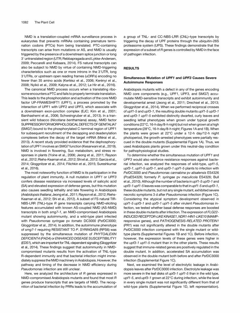
Arabidopsis mutants with a defect in any of the genes encodingNMD core components (e.g., UPF1, UPF3, and SMG7) accu-mulate NMD-sensitive transcripts and exhibit autoimmunity anddevelopmental arrest (Jeong et al., 2011; Drechsel et al., 2013;Gloggnitzer et al., 2014). When we performed reciprocal crossesof upf1-5 and upf3-1, the resulting double mutants upf1-5 upf3-1and upf3-1 upf1-5 exhibited distinctly dwarfed, curly leaves andseedling lethal phenotypes when grown under typical growthconditions (22°C, 16-hday/8-hnight) but notwhengrownat ahightemperature (28°C, 16-h day/8-h night; Figures 1A and 1B). Whenthe plants were grown at 22°C under a 12-h day/12-h nightphotoperiod, the growth-arrested phenotypes were partially res-cued in the double mutants (Supplemental Figure 1A). Thus, weused Arabidopsis plants grown under this neutral-day conditionfor pathophysiological studies.To determine whether the simultaneous mutation of UPF1 and
UPF3 would also reinforce resistance responses against bacte-rial infection, we analyzed the responses of wild-type, upf1-5,upf3-1, upf1-5 upf3-1, and upf3-1 upf1-5 plants to infection withPstDC3000 and Pseudomonas cannabina pv alisalensis ES4326(PcaES4326; formerly P. syringae pv maculicola ES4326; Bullet al., 2010). Although the number of bacteria in upf1-5 upf3-1 andupf3-1upf1-5 leaveswascomparable to that inupf1-5andupf3-1,these doublemutants, but not any singlemutant, exhibited severenecrotic symptoms 3 d after Pseudomonas infection (Figure 1C).Considering the atypical symptom development observed inupf1-5 upf3-1 and upf3-1 upf1-5 after virulent Pseudomonas in-fection, we tested whether basal defense responses are boostedin these doublemutants after infection. The expression of FLG22-INDUCEDRECEPTOR-LIKEKINASE1,NDR1-HIN1-LIKE10 (MAMP-responsive genes), and PATHOGENESIS-RELATED PROTEIN1(PR1) was not significantly altered in the double mutants afterPstDC3000 infection compared with the single mutant or wild-type plants (Supplemental Figures 1B and 1C). Before infection,however, the expression levels of these genes were higher inthe upf3-1 upf1-5 mutant than in the other plants. These resultssuggest that immune-related genes are positively regulated in thedouble mutant. In addition, accelerated SA accumulation wasobserved in the double mutant both before and after PstDC3000infection (Supplemental Figure 1C).Next, we examined the level of electrolytic leakage in Arabi-
dopsis leaves after PstDC3000 infection. Electrolyte leakage wasmore severe in the leaf disks of upf3-1 upf1-5 than in the wild type,upf1-5, and upf3-1 grown at 22°C during infection, while the levelin every single mutant was not significantly different from that ofwild-type plants (Supplemental Figure 1D, left representation).
1082 The Plant Cell
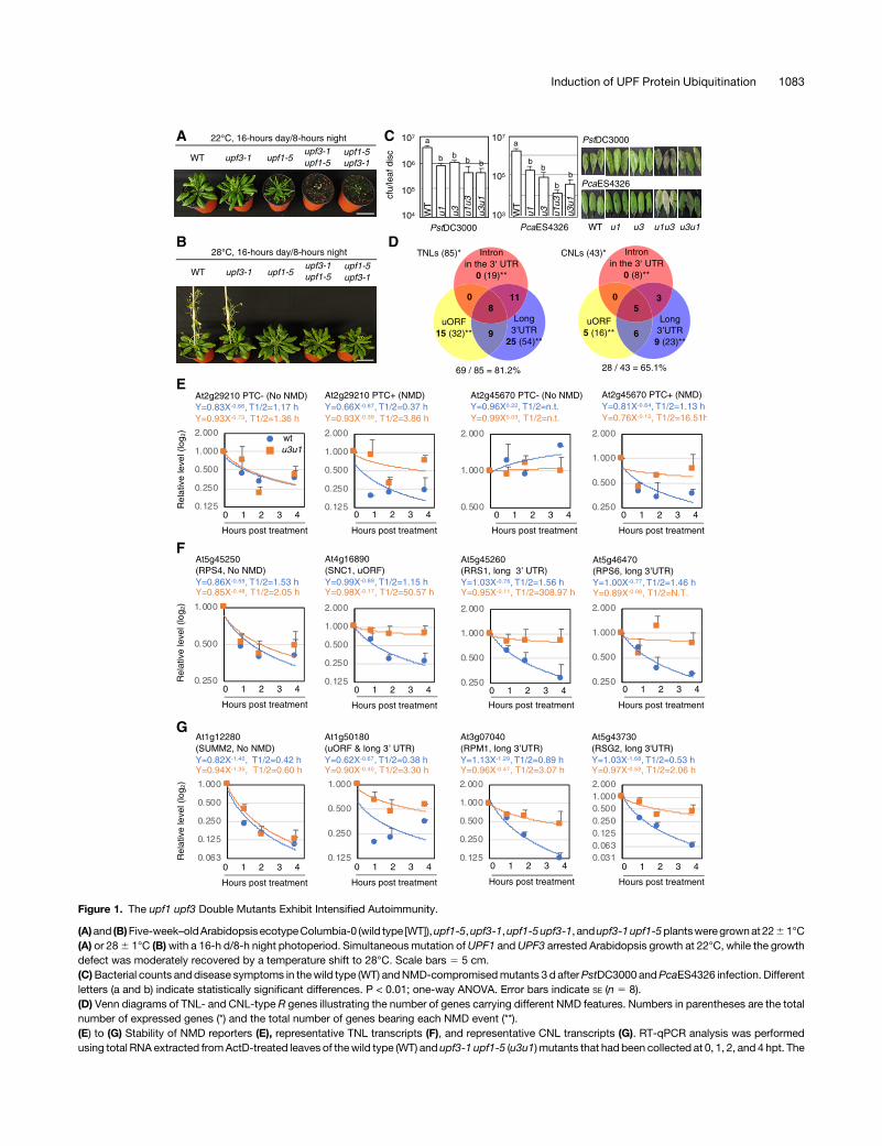
Figure 1. The upf1 upf3 Double Mutants Exhibit Intensified Autoimmunity.
(A)and (B)Five-week–oldArabidopsis ecotypeColumbia-0 (wild type [WT]),upf1-5,upf3-1,upf1-5upf3-1, andupf3-1upf1-5plantsweregrownat2261°C(A) or 286 1°C (B)with a 16-h d/8-h night photoperiod. Simultaneous mutation ofUPF1 andUPF3 arrested Arabidopsis growth at 22°C, while the growthdefect was moderately recovered by a temperature shift to 28°C. Scale bars 5 5 cm.(C)Bacterial counts and disease symptoms in thewild type (WT) andNMD-compromisedmutants 3 d afterPstDC3000 andPcaES4326 infection. Differentletters (a and b) indicate statistically significant differences. P < 0.01; one-way ANOVA. Error bars indicate SE (n 5 8).(D) Venn diagrams of TNL- and CNL-type R genes illustrating the number of genes carrying different NMD features. Numbers in parentheses are the totalnumber of expressed genes (*) and the total number of genes bearing each NMD event (**).(E) to (G) Stability of NMD reporters (E), representative TNL transcripts (F), and representative CNL transcripts (G). RT-qPCR analysis was performedusing total RNA extracted fromActD-treated leaves of thewild type (WT) and upf3-1 upf1-5 (u3u1) mutants that had been collected at 0, 1, 2, and 4 hpt. The
Induction of UPF Protein Ubiquitination 1083

However, an increase in electrolyte leakage was not observed inthe double mutant grown at 28°C (Supplemental Figure 1D, rightrepresentation). The transcripts of 39 TNL-type R genes weremore stable in smg7 pad4 than in pad4, 20 of which showed NMD-sensitive features (Gloggnitzer et al., 2014). To determinewhetherthe accumulation of TNLs is a representative physiologicalcharacteristic in theseupfmutants,wemeasured themRNA levelsof selected TNLs in plants under both NMD-compromised con-ditions (upf1-5, upf3-1, upf1-5 upf3-1, upf3-1 upf1-5, and cy-cloheximide [CHX]-treated wild type) and wild-type plants grownat two different temperatures.We analyzedwell-characterized FSR gene transcripts showing NMD sensitivity (Gloggnitzer et al.,2014). We treated wild-type and mutant plants with actinomycin D(ActD) for 4 h to inhibit transcriptional elongation (Sobell, 1985), asdescribed in a previous analysis of upf3-1, in which the 4-htreatment was found to be optimal without damaging plant tis-sues (Hori and Watanabe, 2005). This blockage of de novotranscription enables the NMD sensitivity of individual transcriptsto be evaluated at the post-transcriptional level (Kurosaki et al.,2014). The known NMD-sensitive TNL transcripts (Gloggnitzeret al., 2014) accumulated under potent NMD-compromisedconditions (in the double mutants and CHX-treated wild-typeplants) regardless of temperature (Supplemental Figure 2). Theseresults suggest that double mutants possessing an excessiveamount ofTNL transcripts remainunder thecontrol of temperature-dependent EDS1-mediated immune responses, as describedfor smg7-1 (Wang et al., 2009; Alcázar and Parker, 2011; Heidrichet al., 2013; Carstens et al., 2014;Gloggnitzer et al., 2014; Stuttmannet al., 2016). Notably, the differences in transcript levels amongthe wild type and upf mutants were reduced when de novotranscription continued to occur compared with ActD-treatedplants (DMSO in Supplemental Figure 2), indicating that contin-uous transcription is necessary for maintaining a steady-statelevel of R gene transcripts in Arabidopsis (Drechsel et al., 2013).Altogether,wepropose that theaccumulation of a variety ofNMD-sensitiveR transcripts leads to enhanced basal immunity in thesemutants.
A Significant Number of R Gene Transcripts RetainNMD-Sensitive Characteristics
To comprehensively examine the extent towhich the expressedRtranscripts differ both quantitatively and qualitatively across wild-type and single– and double–mutant plants, we analyzed the FSnatural transcripts and the AS variants of TNLs and CNLs basedon our RNA-sequencing (RNA-seq) data of ActD-treated plants(Supplemental Data Set 1) and determined whether each tran-script carried NMD-eliciting features (Supplemental Data Set 2;Supplemental Files 1 to 7). Among the 85 TNL and 43 CNL genesexpressed, 19 and 8 of the TNL (22.3%) and CNL genes (18.6%)analyzed, respectively, contained at least one intron in the 39UTR.
A long 39UTR was the most common NMD feature of the FS Rtranscripts (63.5%, 54 TNLs out of 85; 53.4%, 23 out of 43 CNLs),ranging from 350 to 3,180 nucleotides in length. Thirty-two TNLs(37.6%) and 16 CNLs (37.2%) carried uORFs that either over-lapped with the main ORFs or encoded peptides composed of nofewer than 35 amino acids (Table 1). In summary, the FS naturaltranscripts of 69 TNLs (81.2%) and 28 CNLs (65.1%) retainedat least one of the three NMD-sensitive features (Figure 1D;Supplemental Data Set 2; Supplemental Files 6 and 7). The FSnatural transcripts of 16 TNL and 15 CNL genes carried no NMDsignals, but a range of their splicing variants (12 TNLs and 5CNLs)were targets of AS-NMD (Table 1; Supplemental Data Set 2).Overall, 81 TNLs (95.3%) and 33CNLs (76.7%) are potential NMDsubstrates. Previously, inArabidopsis studies, 1 to 2%of the naturaltranscripts and 13 to 17.4% of intron-containing genes wereshown to be upregulated in NMD-deficient plants (Yoine et al.,2006; Kurihara et al., 2009; Kalyna et al., 2012; Drechsel et al.,2013), demonstrating that plant R genes are highly enriched inNMD regulation.Next, we tested the stability of these NMD-susceptible FS R
transcripts inupf3-1upf1-5mutants bymeasuring thehalf-lives ofR transcripts from leaf samples taken from wild-type and mutantplants at 0, 1, 2, and 4 h after blocking de novo transcription. Weused At2g29210 and LYSOPHOSPHATIDYLETHANOLAMINEACYLTRANSFERASE2 (LPEAT2) AS variants (PTC1) as NMDcontrols (Gloggnitzer et al. 2014). The half-lives of these NMDmarkers and R transcripts carrying the NMD-sensitive featureswere elevated in upf3-1 upf1-5 compared with normal-lookingtranscripts showing similar levels of stability between wild typeand upf3-1 upf1-5 (Figures 1E to 1G; Supplemental Figure 3),indicating that NMD is one of the main processes controlling theexpression of TNL and CNL genes. Other transcripts bearingNMD-sensitive features, however, showed no differences inRNA stability between wild type and upf3-1 upf1-5, e.g., RPP5(Supplemental Figure 3). This result suggests that not all R genesbearing NMD-sensitive features are genuine NMD targets, aswidely observed in eukaryotic genes (Rebbapragada and Lykke-Andersen, 2009; Kurosaki et al., 2014; Peccarelli and Kebaara,2014).To determine whether FS natural TNL and CNL transcripts are
post-transcriptionally regulated, we measured the transcriptionrates of the randomly selected genes by performing a chromatinimmunoprecipitation (ChIP) assay of wild-type and upf3-1 upf1-5plants using an a-RNA polymerase II (a-RNAPII) C-terminal do-main antibody, and primer sets specific to regions between thetranscriptionand translation initiation sites.TheChIPsignals in theTNL geneswere largely constant between thewild type and upf3-1 upf1-5 (Supplemental Figure 4A), indicating that most TNL-typeR geneswith NMD-sensitive features are constitutively transcribedin Arabidopsis leaves and that the resulting transcripts are subjectto immediate turnover by NMD in thewild type. Unlike TNLs, half of
Figure 1. (continued).
half-lives were calculated by nonlinear least-squares regression analysis (average 6 SD, n 5 3; three biological replicates with three technical repeats).Stability analyses of other R transcripts are provided in Supplemental Figure 3. Sequences of the individual primers used in this study are presented inSupplemental Data Set 3.
1084 The Plant Cell
the CNL genes tested here showed higher enrichment of theRNAPII elongation complex in upf3-1 upf1-5 than in wild type, e.g.,RPP7 andRPM1 (Supplemental Figure 4B), suggesting that thesegenes are transcriptionally upregulated in the upf3-1 upf1-5mutant. Taken together, these findings support the notion thatNMD is one of the regulatory pathways controlling the steady-state levels of both FS and AS R transcripts carrying NMD-sensitive features (Gloggnitzer et al., 2014).
Bacterial Infection Results in Turnover of UPF Proteins
Because bacterial infection upregulates a subset of NMD-sensitive transcripts (Gloggnitzer et al., 2014), we monitored thetranscript levels of core NMD factors inPstDC3000-infectedwild-type leaves to detect any decrease in transcript levels uponbacterial infection. TheFSnatural transcriptsencodingArabidopsisUPF1, UPF3, and SMG7 are known NMD substrates (Nyikó et al.,2013;Gloggnitzer etal., 2014;Degtiar et al., 2015;Kesarwani etal.,2019). Our RNA-seq data also revealed that the UPF1 transcript,a possible NMD target, carries a 508 nucleotides–long 39UTR(Supplemental Figure 5A). These transcripts were upregulatedafterPstDC3000 infection,while the levelof theFSUPF2 transcriptremainedconstant (Supplemental Figure5A). Theupperproducts,which accumulated during infection, were identified as variantsof UPF1 and UPF2 produced by unspliced introns 26 and 14,respectively, and they eventually retained NMD-triggering fea-tures owing to the long 39UTR containing two introns (single anddouble asterisks, Supplemental Figures 5A and 5B). The resultsindicate that the plant NMD system becomes impaired at an earlystage of infection, despite the increasing levels of transcripts ofNMD factors (Kerényi et al., 2008; Nyikó et al., 2013; Gloggnitzeret al., 2014; Degtiar et al., 2015; Shaul, 2015).
The inconsistency between the increased transcript levels ofUPF genes and the reduction in NMD activity after infection(Gloggnitzer et al., 2014) suggests that bacterial infection mightdisrupt the plant NMD system by affecting the integrity of themachinery at the protein level. To address this hypothesis, weprepared monoclonal antibodies against UPF1 (a-UPF1), UPF2,and UPF3 and used them for immunoblot analysis (SupplementalFigures 6B and 6C). a-UPF1 detected a faint signal of UPF1 in theknockdownmutant upf1-5 and in the putative knockdownmutantupf1-4. However, the protein was undetectable in the amino acidsubstitution mutant upf1-1 for unknown reasons (SupplementalFigure 6B). We examined the levels of UPF1, UPF2, and UPF3 inwild-type leaves infected with PstDC3000 using a-UPF1, a-UPF2,
anda-UPF3. The levels ofUPF1andUPF3were reduced to <40%(UPF1) to 50% (UPF3) within 0.5 h post inoculation (hpi) andbecame undetectable from 10 to 30 hpi, whereas the level of UPF2was maintained for up to 6 hpi. All three NMD factors were un-detectable by 20 hpi (Figure 2A). The turnover of UPF1 occurredmore quickly than that of UPF3, i.e., UPF3 was still detectable at10 hpi when UPF1 was absent (Figure 2A, upper representation).BecauseUPF1 decay began as early as 30min post inoculation
(mpi; Figure2A,upper representation),wewonderedhowearly theturnover of UPF proteins was initiated. Wemonitored the stabilityof UPF proteins at various time points beginning at 1 mpi withPstDC3000. Surprisingly, the reduction in UPF1 levels was clearlyobserved at 5 mpi, and a sharp decrease occurred between 15 and20 mpi, after which only ;50% UPF1 remained. A drastic de-crease in UPF3 levels was observed between 15 and 25 mpi,during which >90% of UPF2 remained (Figure 2A, lower repre-sentation). These findings suggest that the turnover of UPFproteins is an early response to PstDC3000 infection.To determine whether other protein components involved
in NMD (Kerényi et al., 2008) are also unstable upon bacterialinfection, we generated transgenic Arabidopsis plants over-expressing the SMG7 and EJC proteins. SMG7 and the otherEJC components—Y14, MAGO, eIF4AIII, and CASC3/Barentsz(BTZs)—were stably maintained during infection compared withthe overexpressed UPF proteins, although the overexpressedversions of UPF proteins (the first to third rows of the left repre-sentation in Figure 2B) were destabilized later than the nativeproteins (Figure 2B). Thus, we conclude that bacterial infectioninitiates the specific destruction of UPF proteins.To determine whether the decay of UPF proteins is initiated by
only virulent Pseudomonas infection, we inoculated wild-typeplants with an attenuated strain, PstDC3000 hrcC2, and twoavirulent derivatives of PstDC3000 carrying AvrRpm1 or AvrRps4and subjected them to immunoblot analysis using an anti-UPF1polyclonal antibody. Like the turnover pattern of UPF1 inPstDC3000-infected plants, infection with these different strainsalso triggered the decay of UPF1 proteins in Arabidopsis(Supplemental Figures 7A and 7B), indicating that the sensing of P.syringae is sufficient to initiate the decay of the core NMD factors.Unlike the immunoblotting results obtained using monoclonala-UPF1 antibody, the UPF1 level at 6 hpi was somehow re-covered, which was more pronounced after infection with thePstDC3000 hrcC- strain. Furthermore, a putative UPF1 degra-dation product (single asterisk in Supplemental Figure 7) was alsoobserved in parallel with UPF1 dynamics and was not detected in
Table 1. Detailed NMD-Sensitive Characteristics of 85 TNL and 43 CNL Genes Expressed in Arabidopsis Leaves
Number of Genes (%)
TNLs CNLs
NMD-Sensitive Features Intron in the 39 UTR 19 (22.3%) 8 (18.6%)Long 39 UTR 54 (63.5%) 23 (53.4%)uORF 32 (37.6%) 16 (37.2%)
AS-NMD 43 (50.6%) 13 (30.2%)Alternative promoters to NMD 12 (27.9%) 7 (16.3%)Alternative polyadenylation to NMD 15 (17.6%) 3 (7.0%)Fusion transcripts between the flanking genes resulting in NMD 9 (10.6%) 7 (16.3%)
Induction of UPF Protein Ubiquitination 1085

the blots using monoclonal a-UPF1 antibody. We speculate thatUPF1 might undergo a conformational change during the decayprocess and that the monoclonal antibody is unable to crossreact with this transitional form of UPF1. The detection of a smallproduct by the polyclonal antibody even before infection (singleasterisk in Supplemental Figure 7) suggests that the UPF1 levelmay be controlled posttranslationally under normal conditions.
UPF1 and UPF3 Are Destroyed Via Ubiquitination at an EarlyStage of Infection
To examine whether UPF proteins are post-translationally de-graded during an early stage of infection, MG132, an inhibitor ofthe proteasome, was coinfiltrated into wild-type Arabidopsisleaves with PstDC3000. While the coapplication of the solvent
DMSO alone with PstDC3000 did not affect the protein dynamicsof UPF1, UPF2, or UPF3, MG132 coinfiltrated with PstDC3000interrupted the infection-induced destruction of these proteins(Figure 3A). These results support our observation that the re-duction and subsequent disappearance of UPF proteins resultingfrom Pseudomonas infection (Figure 2) is due to the decay of UPFproteins rather than the translational inhibition of mRNAs en-coding these NMD factors.Toconfirmthat thedecayof theUPFproteinsoccursvia theUPS
duringbacterial infection,wemonitored the levelsofubiquitinatedUPFproteins in thea-UPF1,a-UPF2,anda-UPF3 immunoprecipitatesby immunoblot analysis using an a-ubiquitin (a-UBQ) antibody.The ubiquitination of UPF1 and UPF3 was strongly induced at30 mpi and 1 hpi, respectively (Figure 3B; Supplemental Figure8A), which is consistent with the dynamics of these proteins
Figure 2. UPF1, UPF2, and UPF3 Decay during an Early Phase of PstDC3000 Infection.
(A)Dynamics of UPF1, UPF2, and UPF3 proteins up to 30 hpi (top) and 50mpi (bottom). Immunoblot analyses (left) were performed for leaf samples takenfromwild-type Col-0 plants that had been infected with Pseudomonas and collected at the indicated time points using an anti-UPF1monoclonal antibody(a-UPF1),a-UPF2, ora-UPF3. The right showsUPFprotein levels in infected leaves at the indicated timepoints (average6 SD,n54). Different letters abovethebars (i.e., a, b, c, d, ab, andbc) indicate statistically significantdifferences (P<0.05, one-wayANOVA). Successful infectionwasverifiedbyexamining thelevels of PR1. Both experiments were performed with four biological replicates.(B)TheNMD factor SMG7and the EJCcoreprotein components, but notUPF1, UPF2, orUPF3, remained stable duringPstDC3000 infection. The leaves oftransgenic Arabidopsis plants (T3) stably expressing protein A-fusedUPF1, UPF2, UPF3, SMG7, Y14,MAGO, elF4A-III, BTZ1, andBTZ2were infectedwithPstDC3000 and collected at the indicated time points after infection for immunoblot analysis. GFP-protein A was used as a representative stable protein.The recombinant proteins were detected using an a-PAP antibody. M, prestained protein ladder.
1086 The Plant Cell
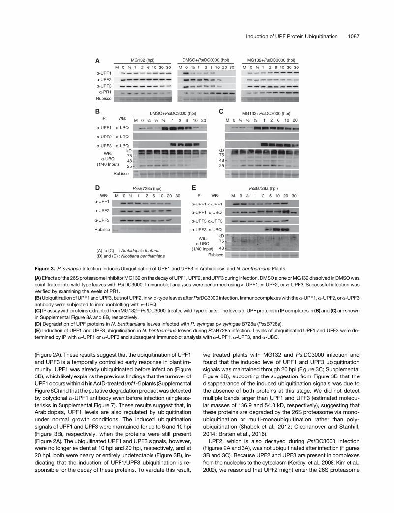
(Figure 2A). These results suggest that the ubiquitination of UPF1and UPF3 is a temporally controlled early response in plant im-munity. UPF1 was already ubiquitinated before infection (Figure3B), which likely explains the previous findings that the turnover ofUPF1occurswithin4h inActD-treatedupf1-5plants (SupplementalFigure6C)and that theputativedegradationproductwasdetectedby polyclonal a-UPF1 antibody even before infection (single as-terisks in Supplemental Figure 7). These results suggest that, inArabidopsis, UPF1 levels are also regulated by ubiquitinationunder normal growth conditions. The induced ubiquitinationsignals of UPF1 and UPF3 were maintained for up to 6 and 10 hpi(Figure 3B), respectively, when the proteins were still present(Figure 2A). The ubiquitinated UPF1 and UPF3 signals, however,were no longer evident at 10 hpi and 20 hpi, respectively, and at20 hpi, both were nearly or entirely undetectable (Figure 3B), in-dicating that the induction of UPF1/UPF3 ubiquitination is re-sponsible for the decay of these proteins. To validate this result,
we treated plants with MG132 and PstDC3000 infection andfound that the induced level of UPF1 and UPF3 ubiquitinationsignals was maintained through 20 hpi (Figure 3C; SupplementalFigure 8B), supporting the suggestion from Figure 3B that thedisappearance of the induced ubiquitination signals was due tothe absence of both proteins at this stage. We did not detectmultiple bands larger than UPF1 and UPF3 (estimated molecu-lar masses of 136.9 and 54.0 kD, respectively), suggesting thatthese proteins are degraded by the 26S proteasome via mono-ubiquitination or multi-monoubiquitination rather than poly-ubiquitination (Shabek et al., 2012; Ciechanover and Stanhill,2014; Braten et al., 2016).UPF2, which is also decayed during PstDC3000 infection
(Figures 2A and 3A), was not ubiquitinated after infection (Figures3B and 3C). Because UPF2 and UPF3 are present in complexesfrom the nucleolus to the cytoplasm (Kerényi et al., 2008; Kim et al.,2009), we reasoned that UPF2 might enter the 26S proteasome
Figure 3. P. syringae Infection Induces Ubiquitination of UPF1 and UPF3 in Arabidopsis and N. benthamiana Plants.
(A)Effects of the 26Sproteasome inhibitorMG132on the decayofUPF1,UPF2, andUPF3during infection. DMSOaloneorMG132dissolved inDMSOwascoinfiltrated into wild-type leaves with PstDC3000. Immunoblot analyses were performed using a-UPF1, a-UPF2, or a-UPF3. Successful infection wasverified by examining the levels of PR1.(B)UbiquitinationofUPF1andUPF3, but notUPF2, inwild-type leavesafterPstDC3000 infection. Immunocomplexeswith thea-UPF1,a-UPF2, ora-UPF3antibody were subjected to immunoblotting with a-UBQ.(C) IP assaywith proteins extracted fromMG1321PstDC3000-treatedwild-type plants. The levels of UPFproteins in IP complexes in (B) and (C) are shownin Supplemental Figure 8A and 8B, respectively.(D) Degradation of UPF proteins in N. benthamiana leaves infected with P. syringae pv syringae B728a (PssB728a).(E) Induction of UPF1 and UPF3 ubiquitination in N. benthamiana leaves during PssB728a infection. Levels of ubiquitinated UPF1 and UPF3 were de-termined by IP with a-UPF1 or a-UPF3 and subsequent immunoblot analysis with a-UPF1, a-UPF3, and a-UBQ.
Induction of UPF Protein Ubiquitination 1087

complex with UPF3 during infection. Indeed, UPF2 coimmuno-precipitated with UPF3 under both uninfected and PstDC3000-infected conditions (Supplemental Figure 9). To test whetherUPF1/UPF3 decay upon bacterial challenge is universal amongplants, we subjected N. benthamiana plants to infection withthe known bacterial pathogen P. syringae pv syringae B728a(PssB728a). The UPF proteins of the N. benthamiana plants werealso destroyed by 30 hpi (Figure 3D). The induction of UPF1 andUPF3 ubiquitination was detected at 2 and 6 hpi, respectively(Figure 3E), suggesting that plant immunity involves the sup-pression of NMD via the control of UPF protein levels in membersof the Solanaceae family.
Because TNL genes are known to be deregulated upon Pseu-domonas infection due to decreased NMD efficiency (Gloggnitzeret al., 2014), we investigated whether the kinetics of the NMD-sensitive TNL and CNL transcripts reflect the decay of UPFproteins upon PstDC3000 infection. We monitored the stabilityof TNL and CNL transcripts from ActD-treated infected leavestakenat 0, 1, 2, and4hpi byRT-qPCRanalysis. The transcripts ofNMD markers as controls, namely, At2g29210 and LPEAT2retainingPTC (At2g29210PTC1andLPEAT2PTC1),weremorestably maintained in infected wild-type leaves than in non-infected leaves (Figure 4A). Similarly, the TNL and CNL tran-scripts bearing NMD-sensitive features, e.g., SIKIC3, RPS6,RPM1, and RPP7, were more long-lived in infected leaves thanin noninfected leaves (Figures 4B and 4C; Supplemental Fig-ure 10). The half-lives of SNC1 and RPP5, which are repre-sentative targets of small-interfering RNA (Yi and Richards,2007), and non-NMD targets, namely, SUMM2 and RPS4, weresimilar between infected and noninfected leaves (Figures 4Band 4C; Supplemental Figure 10). The gaps between the NMDtarget R transcripts in infected and noninfected plants wereless distinct than those between upf3-1 upf1-5 and wild type(Figures 1F, 1G, 4B, and C4C; Supplemental Figures 3 and 10),suggesting that NMD may have still occurred by 4 hpi, whenUPF1 is still present (Figures 2A and 3B).
In parallel, we analyzed the accumulation of R transcripts ininfected leaf samples taken at the same timepoints (Figures 2 and3) used for protein analysis. As a result, we were able to sort the Rgenes bearing NMD-sensitive features into three groups. Theearly-response R genes, including RPM1 and At3g44400, wereupregulated as early as 30 mpi of PstDC3000 (Figure 4; Supple-mental Figure 11), suggesting that the transcripts of these genesare instantly stabilized when the UPF1 level is reduced to <40%.The second group, the so-called late-response genes, e.g., SOC3,At3g14470, At4g14610, and At5g38340, appeared to be upre-gulated later than 10 hpi, when UPF1 is completely decayed(Supplemental Figure 10). The nonresponsive genes, includingRRS1 and At5g35450, were not regulated by Pseudomonasinfection (Supplemental Figure 10).
The miRNA target CNL-type R genes At1g50180 and RSG2(Boccara et al., 2015; Cai et al. 2018), carrying uORF/long 39UTRand long 39UTR, respectively, are thought to also be targets ofNMD, because their transcripts are stabilized upon Pseudomonasinfection (Supplemental Figure 10B). By contrast, the half-lives ofother known miRNA targets including RPS5, At1g12290, andAt1g61180 were not altered or shortened by the mutation of UPF1/UPF3 or Pseudomonas infection (Supplemental Figure 11A),
suggesting that the steady-state level of a subset of CNLtranscripts bearing NMD-sensitive features may be under thecontrol of miRNA-mediated gene silencing rather than NMD.Moreover, three TNL-type R transcripts, At3g44400, At4g14370,and At5g41740, which were upregulated as early as 30 mpi byPseudomonas infection, were more stable in infected versusnoninfected plants. By contrast, the stability of these tran-scripts was reduced in upf3-1 upf1-5 versus wild-type plants(Supplemental Figure 11B). These results suggest that suscepti-bility to NMD varies across FS R transcripts carrying NMD-sensitive features (Rebbapragada and Lykke-Andersen, 2009;Kurosaki et al., 2014; Peccarelli and Kebaara, 2014) and that thecontrol of the expression of these genes might involve other pro-cesses, e.g., transcriptional regulation, under a variety of conditions.Based on these results, we propose that NMD is one of the mainposttranscriptional control pathways involved in maintaining Rtranscript levels in Arabidopsis during the immunity response.
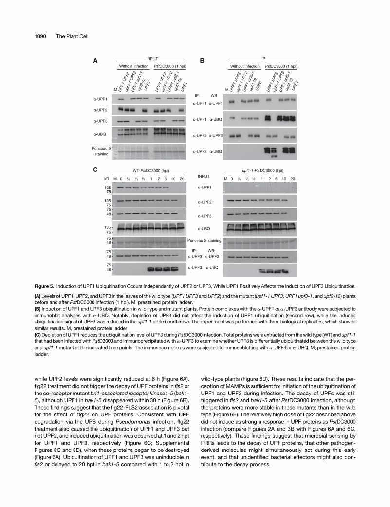
The Induction of UPF3 Ubiquitination Partially Dependson UPF1
The Cys- and His-rich domain of UPF1, which serves as a ReallyInteresting New Gene-related E3 ligase, is responsible for theUPF3-dependent self-ubiquitination required for activation ofNMD in yeast (Takahashi et al., 2008). To determinewhether UPF3is required for UPF1 ubiquitination and vice versa in Arabidopsis,we monitored the ubiquitination and turnover of UPF proteins inupf1-1, upf2-12, and upf3-1 plants at 1 h after PstDC3000 in-fection. The ubiquitination level of UPF1 was similar across thewild type, upf2-12, and upf3-1 (Figure 5A; second row inFigure 5B), suggesting that the induction of UPF1 ubiquitinationupon PstDC3000 infection occurs independently of UPF2 orUPF3. By contrast, the level of ubiquitinated UPF3 decreased1.246 0.042-fold in upf1-1 compared with the wild type (fourthrow in Figure 5B; sixth row in Figure 5C). The UPF3 level sta-bilized from 1 to 10 hpi in upf1-1 compared with wild type (thirdand sixth rows in Figure 5C), most likely due to decreasedubiquitination. These results are consistentwith theobservationthat the decay of UPF1 occurs before that of UPF3 (Figures 2Aand 3B and 3C), and that UPF1 protein itself or ubiquitinatedUPF1 modulates the ubiquitination of UPF3 upon bacterial in-fection. Bacterial infection induces the ubiquitination of UPF1/UPF3 for the decay of three UPF proteins, which differs from theUPF3-dependent self-ubiquitination of UPF1 required for NMDin yeast (Takahashi et al., 2008).
Microbial Perception by PRRs Initiates the Decay ofUPF Proteins
NMD-sensitive transcript levels also increased in wild-type plantstreated with 1 mM of the bacterial elicitor flg22, while the ex-pression of these transcripts was suppressed in flagellin sensitive2(fls2) mutants (Gloggnitzer et al., 2014), suggesting that MAMPrecognition can also affect the integrity of UPF proteins. Com-pared with mock treatment, in which the levels of UPF proteinswereunchanged,bothUPF1andUPF3started todegradeat1and2hpost treatment (hpt)with flg22 inwild-typeplants, respectively,
1088 The Plant Cell
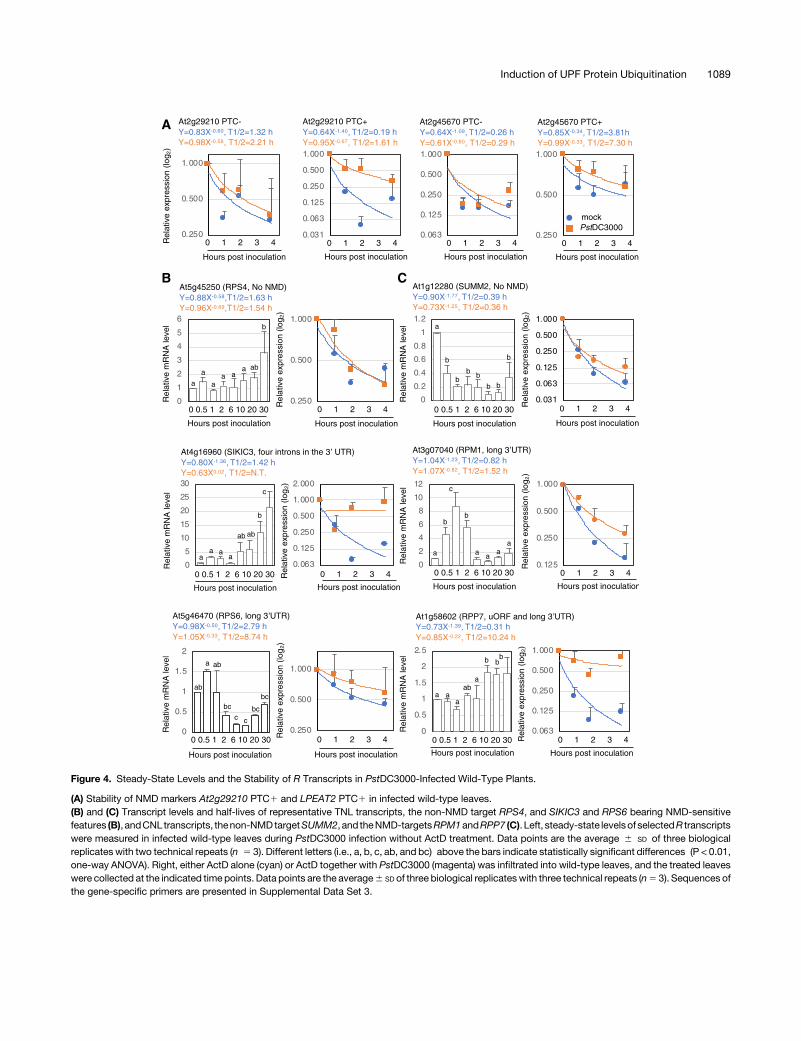
Figure 4. Steady-State Levels and the Stability of R Transcripts in PstDC3000-Infected Wild-Type Plants.
(A) Stability of NMD markers At2g29210 PTC1 and LPEAT2 PTC1 in infected wild-type leaves.(B) and (C) Transcript levels and half-lives of representative TNL transcripts, the non-NMD target RPS4, and SIKIC3 and RPS6 bearing NMD-sensitivefeatures (B), andCNL transcripts, thenon-NMDtargetSUMM2, and theNMD-targetsRPM1andRPP7 (C). Left, steady-state levelsof selectedR transcriptswere measured in infected wild-type leaves during PstDC3000 infection without ActD treatment. Data points are the average 6 SD of three biologicalreplicates with two technical repeats (n 5 3). Different letters (i.e., a, b, c, ab, and bc) above the bars indicate statistically significant differences (P < 0.01,one-way ANOVA). Right, either ActD alone (cyan) or ActD together with PstDC3000 (magenta) was infiltrated into wild-type leaves, and the treated leaveswere collected at the indicated time points. Data points are the average6 SD of three biological replicateswith three technical repeats (n5 3). Sequences ofthe gene-specific primers are presented in Supplemental Data Set 3.
Induction of UPF Protein Ubiquitination 1089
while UPF2 levels were significantly reduced at 6 h (Figure 6A).flg22 treatment did not trigger the decay of UPF proteins in fls2 orthe co-receptormutantbri1-associated receptor kinase1-5 (bak1-5), although UPF1 in bak1-5 disappeared within 30 h (Figure 6B).These findings suggest that the flg22-FLS2 association is pivotalfor the effect of flg22 on UPF proteins. Consistent with UPFdegradation via the UPS during Pseudomonas infection, flg22treatment also caused the ubiquitination of UPF1 and UPF3 butnotUPF2, and induced ubiquitinationwas observed at 1 and 2hptfor UPF1 and UPF3, respectively (Figure 6C; SupplementalFigures 8C and 8D), when these proteins began to be destroyed(Figure 6A). Ubiquitination of UPF1 and UPF3 was uninducible infls2 or delayed to 20 hpt in bak1-5 compared with 1 to 2 hpt in
wild-type plants (Figure 6D). These results indicate that the per-ception ofMAMPs is sufficient for initiation of the ubiquitination ofUPF1 and UPF3 during infection. The decay of UPFs was stilltriggered in fls2 and bak1-5 after PstDC3000 infection, althoughthe proteins were more stable in these mutants than in the wildtype (Figure 6E). The relatively high dose of flg22 described abovedid not induce as strong a response in UPF proteins as PstDC3000infection (compare Figures 2A and 3B with Figures 6A and 6C,respectively). These findings suggest that microbial sensing byPRRs leads to the decay of UPF proteins, that other pathogen-derived molecules might simultaneously act during this earlyevent, and that unidentified bacterial effectors might also con-tribute to the decay process.
Figure 5. Induction of UPF1 Ubiquitination Occurs Independently of UPF2 or UPF3, While UPF1 Positively Affects the Induction of UPF3 Ubiquitination.
(A) Levels of UPF1, UPF2, and UPF3 in the leaves of the wild type (UPF1 UPF3 andUPF2) and themutant (upf1-1 UPF3,UPF1 upf3-1, and upf2-12) plantsbefore and after PstDC3000 infection (1 hpi). M, prestained protein ladder.(B) Induction of UPF1 and UPF3 ubiquitination in wild-type andmutant plants. Protein complexes with the a-UPF1 or a-UPF3 antibody were subjected toimmunoblot analyses with a-UBQ. Notably, depletion of UPF3 did not affect the induction of UPF1 ubiquitination (second row), while the inducedubiquitination signal of UPF3 was reduced in the upf1-1 allele (fourth row). The experiment was performed with three biological replicates, which showedsimilar results. M, prestained protein ladder(C)DepletionofUPF1 reduces theubiquitination level ofUPF3duringPstDC3000 infection. Total proteinswere extracted from thewild type (WT) andupf1-1that had been infected with PstD3000 and immunoprecipitated with a-UPF3 to examine whether UPF3 is differentially ubiquitinated between the wild typeand upf1-1mutant at the indicated time points. The immunocomplexes were subjected to immunoblotting with a-UPF3 or a-UBQ. M, prestained proteinladder.
1090 The Plant Cell

MITOGEN-ACTIVATED PROTEIN KINASE3 andMITOGEN-ACTIVATED PROTEIN KINASE6 Regulate theInduction of UPF1 and UPF3 Ubiquitination
The perception of microbial infection rapidly triggers an oxidativeburst and dual phosphorylation of MITOGEN-ACTIVATED PRO-TEIN KINASE3 (MPK3) and MPK6 in plants (Nicaise et al., 2009).To examine whether the degradation of UPF proteins is associatedwith these early events, we observed the decay of these proteins
in wild-type and mutant plants defective in early signaling. UPF1turnover occurred by 20 hpi after PstDC3000 infection in mpk3 andmpk6 compared with 10 hpi in the wild type. UPF1 was still de-tectable until 20 hpt after flg22 treatment inmpk6 cells (Figure 7A).To address whether the induction of UPF1/UPF3 ubiquitination isaffected by mutations in MPK3 and MPK6, we monitored thea-UPF1 and a-UPF3 immunoprecipitates from flg22-treatedmpk3 and mpk6 leaves by immunoblotting using an a-UBQ an-tibody. The induction of UPF1 ubiquitinationwas initiated at 2 and
Figure 6. The Recognition of the Representative MAMP flg22 Is Sufficient To Trigger UPF Protein Decay in Arabidopsis.
(A)Dynamics of UPF1, UPF2, andUPF3 proteins inwild-type (WT) leaves treatedwith 10mMofMgSO4 (WT-mock) or 1mMof flg22 (WT-flg22) up to 30 hpt.M, prestained protein ladder.(B) The extracellular immune receptor (FLS2) and the coreceptor (BAK1) are required to initiate degradation of the UPF proteins upon flg22 treatment. M,prestained protein ladder.(C) Perception of flg22 induces ubiquitination of UPF1 and UPF3 in wild-type (WT) leaves. Levels of the UPF proteins in the IP complexes are shown inSupplemental Figures 8C and 8D. M, prestained protein ladder.(D)Noordelayed inductionofUPF1/UPF3ubiquitination isobservedafterflg22 treatment in thefls2andbak1-5mutants, respectively.M,prestainedproteinladder.(E) Levels of UPF1, UPF2, and UPF3 proteins in wild-type (WT), fls2, and bak1-5 leaves during PstDC3000 infection. M, prestained protein ladder.
Induction of UPF Protein Ubiquitination 1091

6 hpt inmpk3 andmpk6, respectively, comparedwith 1 hpt in wildtype (Figures 6C, 6D, and 7B). The ubiquitination of UPF3 wasunaffected inmpk3, but it was delayed to 6 hpt inmpk6 from 2 hptin wild type (Figures 6C, 6D, and 7B). By contrast, the induction ofUPF1/UPF3 ubiquitination was unaffected by mutations in theRESPIRATORYBURSTOXIDASEHOMOLOGUED (RBOHD) andRBOHF genes, although UPF2 was not degraded in the flg22-treated rbohD rbohFmutant compared with the responses in wildtype (Figures 7A and 7B; Supplemental Figure 12A). These find-ings indicate that the induction of UPF1/UPF3 ubiquitination re-quires the mitogen-activated protein kinase (MAPK) signalingcascade and the crucial early response in PTI, and that reactiveoxygen species might stimulate the turnover of UPF2. Indeed,transgenic plants expressing constitutively active MPK6(MPK6CA) showed a low level of UPF1 compared with the wildtype (Supplemental Figure 12B), supporting the idea that MAPKsignaling participates in the decay of UPF proteins after sensingmicrobial infection.
We examined the effects of SA-related defense responseson the decay event. The levels of UPF proteins were not signifi-cantly altered in the SA signaling mutants eds1-1, sa inductiondeficient2-1 (sid2-1) and nonexpresser of pr genes1 (npr1-1)
compared to the wild type after PstDC3000 infection and flg22treatment (Figure 7C). Exogenous treatment with BTH, a SA ag-onist, did not have any effect on the stability of UPF proteins inwild-type plants (Supplemental Figure 12C). The induction ofUPF1/UPF3 ubiquitination upon flg22 treatment was also un-altered in these mutants (Supplemental Figure 12D). However,UPF2 persisted in sid2-1 and npr1-1 leaves for up to 20 h. Theseresults suggest that the complete decay of UPF2 might involveSA-related events during a late stage of bacterial infection,which would explain the stability of UPF2 during early stages ofPstDC3000 infection, i.e., 0.5 to 6 hpi (Figures 2A and 7C). Basedon these results, we suggest that the decay of UPF1 and UPF3 bythe UPS involves as-yet unidentified events downstream of theMAPK pathway triggered by bacterial recognition rather thansubsequent SA-related events.
Unannotated Fused TNL-type Transcripts Accumulate inWild-Type Plants after Bacterial Infection
Finally, we aimed to establish a model explaining why thesuppression of NMD and the resulting upregulation of
Figure 7. Initiation of UPF Protein Decay Involves a MAPK Pathway.
(A) Levels of UPF1, UPF2, and UPF3 in the leaves ofmpk3,mpk6 (defective in the MAPK cascade), and rbohD rbohF (defective in the ROS burst) mutantsafter recognition of flg22 and PstDC3000 infection. M, prestained protein ladder; WT, wild type.(B)MPK3 andMPK6, but not RBOHD or RBOHF, are genetically required for the induction of UPF1 and UPF3 ubiquitination. M, prestained protein ladder.(C)EDS1 (a key player in TNL-typeR-triggered immunity andSA regulation), ICS1/SID2 (SA biosynthesis), andNPR1 (SA response) are dispensable for thedecay of UPF1, UPF2, and UPF3. M, prestained protein ladder; WT, wild type.Wild-typeandmutant plantswereeither inoculatedwithPstDC3000or treatedwith1mMofflg22and thenused for immunoblotting.Col-0 is theparental lineof mpk3, mpk6, rbohD rbohF, sid2-1, and npr1-1, while Wassilewskija (Ws) is the parental line of eds1-1.
1092 The Plant Cell
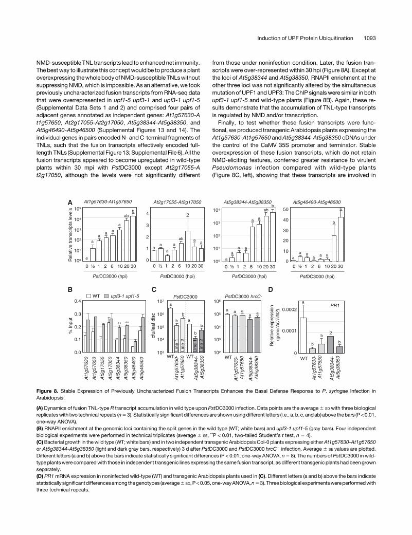
NMD-susceptible TNL transcripts lead to enhanced net immunity.The bestway to illustrate this conceptwould be toproduce aplantoverexpressing thewholebodyofNMD-susceptibleTNLswithoutsuppressingNMD,which is impossible. As an alternative, we tookpreviously uncharacterized fusion transcripts from RNA-seq datathat were overrepresented in upf1-5 upf3-1 and upf3-1 upf1-5(Supplemental Data Sets 1 and 2) and comprised four pairs ofadjacent genes annotated as independent genes: At1g57630-At1g57650, At2g17055-At2g17050, At5g38344-At5g38350, andAt5g46490-At5g46500 (Supplemental Figures 13 and 14). Theindividual genes in pairs encoded N- and C-terminal fragments ofTNLs, such that the fusion transcripts effectively encoded full-length TNLs (Supplemental Figure 13; Supplemental File 6). All thefusion transcripts appeared to become upregulated in wild-typeplants within 30 mpi with PstDC3000 except At2g17055-At2g17050, although the levels were not significantly different
from those under noninfection condition. Later, the fusion tran-scripts were over-represented within 30 hpi (Figure 8A). Except atthe loci of At5g38344 and At5g38350, RNAPII enrichment at theother three loci was not significantly altered by the simultaneousmutation of UPF1 andUPF3: TheChIP signalswere similar in bothupf3-1 upf1-5 and wild-type plants (Figure 8B). Again, these re-sults demonstrate that the accumulation of TNL-type transcriptsis regulated by NMD and/or transcription.Finally, to test whether these fusion transcripts were func-
tional, we produced transgenic Arabidopsis plants expressing theAt1g57630-At1g57650 andAt5g38344-At5g38350 cDNAs underthe control of the CaMV 35S promoter and terminator. Stableoverexpression of these fusion transcripts, which do not retainNMD-eliciting features, conferred greater resistance to virulentPseudomonas infection compared with wild-type plants(Figure 8C, left), showing that these transcripts are involved in
Figure 8. Stable Expression of Previously Uncharacterized Fusion Transcripts Enhances the Basal Defense Response to P. syringae Infection inArabidopsis.
(A) Dynamics of fusion TNL-type R transcript accumulation in wild type upon PstDC3000 infection. Data points are the average6 SD with three biologicalreplicateswith two technical repeats (n53). Statistically significant differencesare shownusingdifferent letters (i.e., a, b, c, andab) above thebars (P<0.01,one-way ANOVA).(B) RNAPII enrichment at the genomic loci containing the split genes in the wild type (WT; white bars) and upf3-1 upf1-5 (gray bars). Four independentbiological experiments were performed in technical triplicates (average 6 SE, **P < 0.01, two-tailed Student’s t test, n 5 4).(C)Bacterial growth in thewild type (WT; white bars) and in two independent transgenic Arabidopsis Col-0 plants expressing eitherAt1g57630-At1g57650or At5g38344-At5g38350 (light and dark gray bars, respectively) 3 d after PstDC3000 and PstDC3000 hrcC2 infection. Average 6 SE values are plotted.Different letters (a and b) above the bars indicate statistically significant differences (P < 0.01, one-way ANOVA, n5 8). The numbers ofPstDC3000 in wild-type plantswere comparedwith those in independent transgenic lines expressing the same fusion transcript, as different transgenic plants had beengrownseparately.(D) PR1mRNA expression in noninfected wild-type (WT) and transgenic Arabidopsis plants used in (C). Different letters (a and b) above the bars indicatestatistically significantdifferencesamong thegenotypes (average6 SD,P<0.05,one-wayANOVA,n53).Threebiological experimentswereperformedwiththree technical repeats.
Induction of UPF Protein Ubiquitination 1093
basal resistance to bacterial infection. However, the bacterialnumbers of PstDC3000 hrcC2 from these transgenic lines werecomparable to those from wild-type plants (Figure 8C, right). Incontrast to upf3-1 upf1-5, which produces a high level of thePR1 transcript even before pathogen infection (SupplementalFigure 1C), the PR1mRNA levels were reduced in the transgenicplants in the absence of pathogen infection (Figure 8D). Theconformational changes in translated NLRs crucial for NLR ac-tivation may not occur under infection by the disarmed strain andnoninfection conditions (Sukarta et al., 2016). Overall, we suggestthat the suppression of NMD via the degradation of UPFs and theresulting stabilization of TNL transcript levels from an early stageof bacterial infection contribute to the basal immunity of plants tovirulent pathogen infection.
DISCUSSION
Microbial perception by PRRs and R proteins occurs at theforefront of the battle between plants andmicrobes. In this study,we demonstrated that MAMP recognition by PRRs controls thelevels of R transcripts carrying NMD-sensitive features via theinduction of UPF1/UPF3 ubiquitination, followed by their 26S-proteasome-dependent degradation at anearly stageof infection.
After their export from the nucleus, virtually all mRNAs undergomRNA surveillance processes, including NMD and miRNA-mediated gene silencing, by which only those certified as “nor-mal” are deemed competent at translation (Eulalio et al., 2007; Laiand Eulgem, 2018). Because most FS R transcripts retain one ofthe NMD-triggering characteristics, they are thought to be eitherprevented from undergoing translation or immediately decayedby the NMD machinery, which may be essential for avoiding theunnecessary costs of immunity under normal growth conditions(Maekawa et al., 2011; Karasov et al., 2017). The observationthat a majority of TNL and CNL genes carry NMD-sensitive fea-tures suggests that the NMD machinery and R genes coevolvedin plants. The coexistence of both normal genes and thosewith NMD-regulated features in a few single R gene clusters(Supplemental Figure 15) suggests that gene duplication eventsincluding unequal crossing-over (Meyers et al., 2003) may haveoccurred before genetic drift, followed by subsequent positiveselection. Such gene clustersmight have evolved in land plants tofacilitate the switching of R transcript levels (Raxwal and Riha,2016).
Most FS R transcripts of the genes bearing NMD-elicitingfeatures that we tested were highly stable in upf3-1 upf1-5, ex-cept for those of a few genes, supporting previous observations(Gloggnitzer et al., 2014). However, the gaps in the stability ofthese FS R transcripts between noninfected and Pseudomonas-infected plants were smaller than those between wild type andupf3-1 upf1-5 under our experimental conditions. In addition, thesteady-state levels of a few R transcripts carrying typical NMDfeaturesmight also be regulated by the putativemiRNA-mediatedcontrol system during Pseudomonas infection. Based on thesefindings,we speculate thatNMD inPseudomonas-infected plantsstill occurs until the UPF proteins are entirely eliminated. It is alsolikely that other mRNA control processes, including miRNA-mediated gene silencing, are deregulated in Arabidopsis duringinfection, because the intergenic pri-mRNAs may contain uORFs
or long 39UTRs due to their weak protein-encoding capability asnoncodingmRNAs or primary transcripts of small nucleolar RNAsand are thus NMD targets (Kurihara et al., 2009; Drechsel et al.,2013). The predicted NMD-sensitivity ofR transcripts did not fullyexplain their actual biological NMD susceptibility, as the R tran-scripts were upregulated, unregulated, or even downregulated ata variety of time points after Pseudomonas infection, althoughmost were upregulated or highly stable in the upf3-1 upf1-5mutant.In addition to the FS R transcripts, their AS variants also pre-
dominantly accumulated inupfmutants (Supplemental Files 6and7), which suggests that the atypical cell death-like symptomsobserved in upf1-5 upf3-1 and upf3-1 upf1-5 after virulentPseudomonas infection resulted from the increased hetero-genicity of the NMD-sensitive R transcript repertoire as well asRPS6 (Gloggnitzer et al., 2014). Therefore, we suggest that thediversified expression of R genes, not their canonical functionsin ETI, confers basal immunity in NMD-compromised mutants.NMD-sensitive R transcripts also accumulate in wild-type leavesafter virulent P. syringae infection by suppressing NMD. How dovirulent P. syringae strains overcome the induced barriers re-sulting from the accumulated R proteins? One possible expla-nation is that wild-type Col plants do not produce any functionalR proteins capable of recognizing effectors delivered fromPstDC3000 and PcaES4326 and are unable to trigger ETI in re-sponse to infectionwith these virulent pathogens, evenwhen theyaccumulatehigh levelsofNMD-sensitiveR transcripts. If this is thecase, these R transcripts may not function in the susceptibleresponse inplants. DuringETI, theaccumulationof various sensorand helper NLRs is necessary for full immunity against avirulentpathogen infections (Jones et al., 2016; Wu et al., 2017). Toachieve full immunity against a countless number of differentpathogens, plants may have evolved diverse NLRs, known as theNLRome and pan-NLRome, via an arms race against a variety ofpathogens (Meyers et al., 2003; van deWeyer et al., 2019). UnlikemiRNAs that directly regulate the levels of their target R tran-scripts, NMD covers both AS and FS R transcripts as long asthey carry NMD-triggering characteristics. Thus, NMD-mediatedposttranscriptional regulation can diversify the expressed Rrepertoire to maximize the chances to achieve full resistance tounexpectedpathogen infection.Ourdatadonotprovidedetails onevery upregulated FS R transcript and its AS variants duringpathogen infection, although we described their RNA-seq cov-erage data extracted from upfmutants (Supplemental Files 6 and7). It would be interesting to monitor the dynamic changes in ASvariants in real time after infection, because most splicing factorgenes have AS forms, and many are also NMD-sensitive (Palusaand Reddy, 2010).The UPS regulates diverse fundamental processes in plants,
including hormonal signaling and immune responses (Kelley andEstelle, 2012; Marino et al., 2012; Dudler, 2013). The latter hasbeen referred to as a double-edged sword, in which E3 ligasesfunction as either positive or negative regulators of plant immunity(Marino et al., 2012). The best examples are the E3 ligases PLANTU-BOX12 (PUB12)/PUB13 and PUB22, which are involved in thedecay of flagellin-induced FLS2 and EXOCYST SUBUNIT EXO70FAMILY PROTEIN B2, respectively, resulting in attenuated plantimmunity (Lu et al., 2011; Stegmann et al., 2012). Thehomologous
1094 The Plant Cell
triplet PUB22/PUB23/PUB24 can dampen early immune re-sponses stimulated by MAMPs (Trujillo et al., 2008). By contrast,the transcription of ARABIDOPSIS TOXICOS EN LEVADURA9,encoding a Really Interesting New Gene zinc-finger E3 ligase, isinduced by a variety of external stimuli, including chitin, and themutant shows enhanced susceptibility to Golovinomyces ci-choracearum and reduced reactive oxygen species productionafter infection (Ramonell et al., 2005; Berrocal-Lobo et al., 2010).Thus, theearly immune response is either suppressedoractivatedby ubiquitination (Marino et al., 2012). We report here that thedecay of UPF1 and UPF3 via the UPS is linked to the suppressionof NMD. These processes are governed by the roles of PRRs assensors, reporting dire circumstances to the downstream com-ponents of PTI, leading to the suppression of NMD and involvingthe decay of even nonubiquitinated UPF2.
Unlike the well-characterized target proteins of the UPS, e.g.,receptors and transcription factors, UPF proteins participate indiverse cellular processes in plants by controlling the tran-scriptome (Kurihara et al., 2009; Drechsel et al., 2013). Why mustUPF proteins decay upon bacterial infection at the expense ofsuch pleiotropic effects? The most likely answer is that a con-siderable variety of R proteins must accumulate during an earlystage of infection to protect the host plants from attack by un-predictable pathogens before they successfully colonize theplants.WhenRproteinsmonitor their cognate avirulence proteinsderived from the pathogen, plants may only have to pay a rea-sonable cost for subsequent immunity. If not, the accumulated Rproteins can confer a defense response to plants. Thus, wesuggest that the consecutive events between MAMP perceptionby PRRs and the accumulation of R transcripts are well pro-grammed in the “black box.”
The decay of UPF proteins and the accumulation of R tran-scripts occur in infected leaves at an early phase after bacterialinfection. Interestingly, the significant delay in the induction ofubiquitination and subsequent decay of UPF1 inmpk3 andmpk6suggests that the MAPK cascade is associated with these events.Because thempk3 mpk6 double mutant was lethal, we could notquantify the ubiquitination of UPF1 and UPF3 in these plants.However, the results sufficiently indicate that the MAPK cascadenot only modulates transcription activators/coactivators thatregulate target genes involved in plant immunity (Rasmussenet al., 2012), it also links bacterial sensing and themaintenance ofR transcripts by participating in the induction of the ubiquitinationof proteins, including UPF1 and UPF3. Therefore, our findingspoint to an additional layer of plasticity between the PTI and ETIby NMD.
METHODS
Plants and Pathogens
Wild-type Arabidopsis (Arabidopsis thaliana) Columbia-0 (Col-0) andthe NMD-compromised mutants upf1-1 (Yoine et al., 2006), upf1-4(Salk_022721), upf1-5 (Salk_112922), upf2-12 (SAIL_512_G03), upf3-1(Salk_025175), upf3-2 (Salk_097931), upf3-3 (Salk_061923), smg7-1,upf1-5upf3-1, andupf3-1upf1-5weregrown inenvironmentally controlledgrowth chambers (22 6 1°C, 16-h day [120 mmol photons m22 s21]/8-hnight for developmental studies or 12-h day/12-h night for
pathophysiological experiments; Hori andWatanabe, 2005; Arciga-Reyeset al., 2006; Riehs et al., 2008; Merchante et al., 2015). The upf1-5 upf3-1and upf3-1 upf1-5mutants were maintained at a high temperature (28°C,16-h day/8-h night) until the flowering stage. The fls2 (Salk_093905), bak1-5, rbohD robhF,mpk3-1,mpk6-2, eds1-1, sid2-1, and npr1-1mutants andMPK6CA transgenic plants used, as described by Parker et al. (1996), Caoet al. (1997), Nawrath and Métraux (1999), Torres et al. (2002), Wang et al.(2007); Boutrot et al. (2010), Schwessinger et al. (2011), and Hudik et al.(2014), were grown at 22°C6 1°Cwith a 12-h day/12-h night photoperiod.
The ORFs of UPF1, UPF2, UPF3, SMG7, Y14, MAGO, elF4AIII, BTZ1,and BTZ2 were cloned into CTAPi, a plant transformation vector (Rohilaet al., 2004), for stable expression in Arabidopsis. The oligonucleotidesequences used to amplify the cDNA fragments are listed in SupplementalData Set 3. Transgenic plants were produced by the floral dip method(Clough and Bent, 1998). Seeds were screened on Murashige and Skoog(MS) medium (Duchefa) containing 50 mg mL21 of DL-phosphinothricin(Duchefa). T3 homozygotes were isolated and used for analysis.
Freshly incubated bacterial strains PstDC3000, PcaES4326,PstDC3000/AvrRpm1, PstDC3000/AvrRps4, and PstDC3000 hrcC2 inKing’s B medium supplemented with the appropriate antibiotics werediluted in 10mM ofMgSO4 to different concentrations as follows: OD6005
0.05 to measure ion leakage, OD6005 0.01 to evaluate pathophysiologicalresponses, and OD6005 0.0001 (PstDC3000 and PcaES4326), or OD6005
0.001 (PstDC3000 hrcC-) to count the number of bacteria in leaf disks. Thenumber of bacteria in infected leaves was determined 3 d after inoculationafter a typical serial dilution method, and the infected leaves were pho-tographed on the same day.
RNA-Seq
Three-week–old Arabidopsis plants were vacuum-infiltrated for 5 min in0.53MSmediumcontaining80mMActDand/or 20mMCHXand incubatedin thedark for 4 hbeforeRNA isolation (Ambion;Hori andWatanabe, 2005).After confirming theRNApuritywith abioanalyzer using theRNA6000PicoKit (Agilent), the total RNA was processed for mRNA sequencing librarypreparation using a TruSeq Stranded mRNA Sample Preparation Kit ac-cording to the manufacturer’s instructions (Illumina). In brief, mRNAs wereisolated from400ngof total RNAwithRNApurification beads usingpoly(A)capture followedby enzymeshearing. After first- and second-strand cDNAsynthesis, A-tailing and end repair were performed for the ligation ofproprietary primers incorporating unique sequencing adaptors with anindex for tracking Illumina reads from multiplexed samples run on a singlesequencing lane. All samples were processed in two biological replicatesper genotype, each from individual plants. For each library, an insert size of;270 bp was confirmed with a bioanalyzer, and the library was quantifiedbyRT-PCRusing theCFX96System (BioRad). Each librarywassequencedfor 75-bp paired-end reads with a NextSeq500 High Output 150 Cycle Kiton the NextSeq500 platform (Illumina).
The raw image data were transformed by base-calling into sequencedata and stored in FASTQ format. The RNA-seq data were aligned by thesoftwares TopHat2 andBowTie2 todetect thecanonical andnoncanonicalintron motifs (Langmead and Salzberg, 2012; Kim et al., 2013). Unalignedreads using the above platforms were mapped by the software BLAST(Trapnell et al., 2010). AS events were extracted from The ArabidopsisInformationResource 10 (TAIR10) genemodels (www.arabidopsis.org). Toevaluate NMD-triggering features (see “Evaluation of R Transcripts forNMD-Sensitive Features”), the data sets were mapped in the IntegrativeGenomics Viewer browser (Robinson et al., 2011; Thorvaldsdóttir et al.,2013). To quantify the total transcript mass in fragments per kilobase oftranscript per million mapped reads, biological replicates of ActD-treatedwild-type Col-0, ActD/CHX-treated wild-type Col-0, upf1-5, upf3-1, andupf3-1 upf1-5 were separately aligned with the Arabidopsis TAIR10 genemodel using the Cufflinks package (Saeed et al., 2003).
Induction of UPF Protein Ubiquitination 1095

Evaluation of R Transcripts for NMD-Sensitive Features
Although many normal-looking mRNAs can be regulated by NMD via lowtranslation efficiency of the main ORF or out-of-frame translation due toribosome read-through (Celik et al., 2017), we ruled out these possibilitiesdue to the absence of sufficient evidence in Arabidopsis genes. We ex-amined three NMD-triggering features to judge whether the individualTNL or CNL transcripts are putatively NMD-regulated:
Intron in the 39UTR
Intronsoccurringmore than55nucleotidesdownstreamof the termination-codon trigger NMD, by which the ribosome-bound UPF1 and EJC-boundUPF2–UPF3 complex are well spaced to activate UPF1 (Kertész et al.,2006; Nyikó et al., 2013; Le Hir et al., 2016).
Long 39UTR
Long 39UTR ($350 nucleotides)-containing transcripts are also NMDtargets in Arabidopsis (Kertész et al., 2006; Kerényi et al., 2008; Kalynaet al., 2012), wherein UPF1, which otherwise falls off the normal mRNAtemplate due to its ownATPase activity, binds in a size-dependentmannerfor the phosphorylation and activation of NMD (Kurosaki et al., 2014;Peccarelli and Kebaara, 2014).
uORFs
Although uORF-driven NMD has also been considered an EJC type, ex-actly how it can trigger NMD is still unclear, because the translation ofuORFs is often leaky. Approximately 20% of Arabidopsis genes containuORFs (Kochetov et al., 2002) of which only up to 2% are putative NMDtargets that contain uORFs encoding$ 35-amino acid peptides for stableexpression. (Nyikó et al., 2009, 2013). The MAGNESIUM/PROTON EX-CHANGER (AtMHX ) uORF, encoding a 13-amino–acid peptide, inducesNMD in a downstream intron-dependent manner (Saul et al., 2009). Here,however, we considered CNL and TNL transcripts containing only uORFsencoding peptide(s) $ 35 amino acids as NMD targets based on generalobservations (Nyikó et al., 2009). The transcripts carrying uORFs thatoverlap with the authentic initiation codons were also considered to haveputative NMD-sensitive features because this event showed strong cor-relations with sensitivity to NMD, even in the transcripts with uORFs en-coding # 35 amino acids (Kalyna et al., 2012).
RNA Analysis
Leaves of 4-week–old Arabidopsis (;0.1 g) infiltrated with the PstDC3000strain or 80 mM of ActD/20 mM CHX were harvested at the indicated timepoints after treatment. RNA was extracted from the leaves following themanufacturer’s instructions (TRIzol reagent; Thermo Fisher Scientific).TURBO DNase (2 units; Thermo Fisher Scientific) was used to removecontaminating genomic DNA. After phenol/chloroform/isoamyl alcoholextraction, the purified RNA was dissolved in diethyl pyrocarbonate-treated water. Five micrograms of total RNA were used to synthesizefirst-strand cDNA by SuperScript-II RTase (Thermo Fisher Scientific), andthe first-strand cDNA was diluted 10-fold for standard RT-PCR or RT-qPCR. RT-qPCR was performed with SYBR Premix Ex Taq (TaKaRa Bio)as described previously by Jung et al. (2009). All experiments were per-formed in three biological replicates with two or three technical repeats.The oligonucleotide sequences used in this study are presented inSupplemental Data Set 3.
Quantification of Cell-Death–Like Responses
Electrolyte leakage measurements were performed to quantify the hy-persensitive response in plants after infection (Sohn et al., 2012). After
infiltrating PstDC3000 into the leaves of wild-type and mutant plants, 10leaf disks per biological replicate (n 5 4) were collected from the infectedleaves of different plants, rinsed with distilled water for 30 min, and sub-merged in 2 mM of MgSO4. Conductivity was measured from 1 to 36 hpiwith a LAQUAtwin Compact Water Quality Meter (HORIBA Life ScienceSolutions).
flg22 Treatment and SA Measurement
flg22 (1 mM; Peptron) was infiltrated into Arabidopsis leaves, and thetreated plants were transferred to a growth chamber (226 1°C, 12-h day/12-h night). The leaves were collected at the indicated time points aftertreatment to examine the levels of UPF proteins. Free-SA levels were alsoquantified using HPLC coupled with a fluorescence detector (Agilent) asdescribed previously by Seskar et al. (1998) and Jung et al. (2009).
Monoclonal and Polyclonal Antibodies against UPF Proteins, IP, andImmunoblot Analysis
After analysis of the UPF1, UPF2, and UPF3 primary sequences usingthe software package SEAL (http://www.ab-mart.com), four peptidefragments per protein were designed to yield monoclonal antibodies(Supplemental Figure 16). After vigorous tests for 3 to 4 antibodies raisedfrom each peptide using enzyme-linked immunosorbent assay and im-munoblot analysis, one epitope each from the four peptides against eachprotein appeared to generate a specific antibody: 41-GSPTAWPTPSDS-52(UPF1), UPF2 (933-ENGEAHGEESDS-944) andUPF3 (192-NKPSPRPSKRNS-203). These antibodies were used for further immunoblot and IP analyses.The specificity and sensitivity of the selected monoclonal antibodies wereverified by immunoblotting using different upf1, upf2, and upf3 mutantalleles (Supplemental Figure 6).
GST-UPF1(1-232), covering the N-terminal and Cys- and His-richdomains, was bacterially expressed and purified by glutathione-agarosechromatography (GreenandSambrook, 2012), followedbyextraction fromSDS-polyacrylamidegels, andused to raise ananti-UPF1 rabbit polyclonalantibody (Youngin Biotech).
For IP, leaf samples were ground in extraction buffer containing 20 mMofTris-Cl atpH8.0, 1mMofEDTA,100mMofNaCl (Figure5;SupplementalFigure 9) or 150 mM of NaCl (Figures 3, 6, and 7; Supplemental Figures 8and 12), 1 mM of PMSF, and 13 proteinase inhibitor, and clarified at15,000g for 10 min at 4°C. The homogenate was incubated with proteinG-magnetic beads (Invitrogen) thatwere coatedwith thea-UPF1,a-UPF2,ora-UPF3monoclonal antibody for 1hat 4°C. The immunecomplexon themagnetic beads waswashed five times in buffer containing 20mMof Tris-Cl at pH 8.0, 1 mM of EDTA, 100 mM (or 150 mM for high stringency) ofNaCl, and 0.5% (v/v) Triton X-100, resuspended in SDS sample buffer, andloaded on SDS-polyacrylamide gels for subsequent immunoblot analysis.
Finely ground leaf powder was homogenized in an equal volume ofprotein extraction buffer containing 20 mM of Tris-HCl at pH 7.5, 1 mM ofEDTA, 150 mM of NaCl, 0.1% (v/v) Triton X-100, 0.1% (w/v) SDS, 5 mM ofdithiothreitol, and 13 proteinase inhibitor (Roche Applied Science) andloaded onto SDS-polyacrylamide gels after clarification for immunoblotanalysis as described by Green and Sambrook (2012). The antibodies andmanufacturers were as follows:a-UBQ (AS08 307, Agrisera), a-PR1 (AS10687, Agrisera), and a-PAP (P1291; Sigma-Aldrich). The dilution of primaryantibodies used in this study was 1:5,000. The signal was visualized witha chemiluminescent substrate (Intron).
ChIP-qPCR
Nuclei from the leaves of 4-week–old wild-type and upf3-1 upf1-5 plantswere extracted and purified as described in Jaskiewicz et al. (2011). Thepurified nuclei were resuspended in nucleus lysis buffer (50 mM of Tris-Cl at
1096 The Plant Cell
pH8.0, 10mMof EDTA, 1% [w/v] SDS, and 13 proteinase inhibitor [RocheApplied Science]), and the resulting chromatin was sheared by sonicationtoobtain fragment sizes ranging from200 to800bp (Biorupter;Diagenode).TheChIPassaywasperformedusingananti-C-terminal domain (phospho-S5) antibody (ab5131; Abcam) as described by the manufacturer (PierceAgarose ChIP Kit; Thermo Fisher Scientific). RNAPII enrichment betweenthe transcription start sites and translation initiation sites of representativeTNL- and CNL-type R genes was determined by RT-qPCR and calculatedby the percent input method (Lin et al., 2012). The primers used in thisstudy are listed in Supplemental Data Set 3.
Statistical Analysis
One-way ANOVA with a post hoc Tukey honest significant difference testwas used for multiple comparisons. To analyze significant differencesbetween thecontrol and treatmentgroups,a two-tailedStudent’s t testwasemployed in this study. The sample size (n) and type of error bar are in-dicated in the figure legends. All pathophysiological experiments wereperformed in at least triplicate. The results of all statistical analyses per-formed in this study are presented in Supplemental Data Sets 4 and 5.
Accession Numbers
The accession numbers for the genes examined and discussed in thisarticle are as follows: ACTIN2 (At3g18780), BTZ1 (At1g80000), BTZ2(At1g15280), EDS1 (At3g48090), eIF4AIII (At3g19760), FLG22-INDUCEDRECEPTOR-LIKEKINASE1 (At2g19190), ICS1/SID2 (At1g74710),LPEAT2(At2g45670),MAGO (At1g02140),MPK3 (At3g45640),MPK6 (At2g43790),FLG22-INDUCED RECEPTOR-LIKE KINASE1, NDR1-HIN1-LIKE10(At2g35980), NPR1 (At1g64280), PAD4 (At3g52430), PR1 (At2g14610),RBOHD (At5g47910), RBOHF (At1g64060), RPM1 (At3g07040), RPP5(At4g16950), RPP7 (At1g58602), RPS2 (At4g26090), RPS4 (At5g45250),RPS5 (At1g12220), RPS6 (At5g46740), RRS1 (At5g45260), RSG2(At5g43730),SIKIC3 (At4g16960),SMG7 (At5g19400),SNC1 (At4g16890),SOC3 (At1g17600), SUMM2 (At1g12280), UPF1 (At5g47010), UPF2(At2g39260), UPF3 (At1g33980), and Y14 (At1g51510).
RNA-seq data have been deposited in The National Agricultural Bio-technology Information Center (http://nabic.rda.go.kr/) under accessionnumbers NN-5098, NN-5102, NN-5103, NN-5104, and NN-5105.
Supplemental Data
Supplemental Figure 1. upf1-5 upf3-1 and upf3-1 upf1-5 exhibitsevere necrotic responses and enhanced immune responses againstinfection with virulent Pseudomonas strains.
Supplemental Figure 2. Representative R transcripts that accumu-lated in the leaves of NMD-compromised mutants.
Supplemental Figure 3. Stability of the mRNAs of TNLs and CNLs inwild-type and upf3-1 upf1-5 plants.
Supplemental Figure 4. RNAPII enrichment at FS R genes in wild-type and upf3-1 upf1-5 plants.
Supplemental Figure 5. Upregulation of the FS natural UPF1, UPF3,and SMG7 transcripts and AS versions of UPF1 and UPF2 in wild-typeleaves during infection.
Supplemental Figure 6. Morphology of the wild-type and NMD-compromised mutants and the specificities of monoclonal antibodiesused in this study.
Supplemental Figure 7. Stability of UPF1 in wild-type leaves infectedwith different P. syringae strains.
Supplemental Figure 8. Levels of UPF proteins in the immunecomplexes shown in Figures 3B, 3C, and 6C.
Supplemental Figure 9. UPF2 physically associates with UPF3regardless of bacterial infection.
Supplemental Figure 10. Steady-state levels and stability of themRNAs of TNLs and CNLs in wild-type leaves infected with a virulentPstDC3000 strain.
Supplemental Figure 11. Steady-state levels and stability of putativemiRNA-target R transcripts.
Supplemental Figure 12. Induction of UPF1 and UPF3 ubiquitinationoccurs independently of reactive oxygen species burst or SAsignaling.
Supplemental Figure 13. Structures of the fusion transcripts encodedby two separately annotated genes and their protein products.
Supplemental Figure 14. RNA-seq coverage plots of the At1g57630-57650 fusion transcript.
Supplemental Figure 15. Physical locations of Arabidopsis R genes.
Supplemental Figure 16. Amino acid sequences of UPF1, UPF2,and UPF3.
Supplemental Data Set 1. Read counts and fragments per kilobase oftranscript per million mapped reads values of the Arabidopsis NLRgenes determined by RNA-seq in the wild-type and NMD-compromised mutants 4 h after ActD treatment.
Supplemental Data Set 2. NMD-sensitive features in the TNL- andCNL-type genes expressed in Arabidopsis leaves
Supplemental Data Set 3. Oligonucleotides used in this study.
Supplemental Data Set 4. Statistical analysis of the results of shownin Figures 1, 2, 4, and 8.
Supplemental Data Set 5. Statistical analysis of the results of shownin the Supplemental Figures 1, 2, 4, 10, and 11.
Supplemental File 1. Nucleotide sequences of the genomic DNAregion of each TNL-type gene in the Arabidopsis genome.
Supplemental File 2. Genomic DNA sequences of Arabidopsis CNL-type genes.
Supplemental File 3. Nucleotide sequences of the FS natural tran-scripts and the AS variants of each TNL-type gene obtained by RNA-seq analysis.
Supplemental File 4. Transcripts of the CNL-type genes identified byRNA-seq analysis.
Supplemental File 5. Legends of Supplemental Files 1 and 2.
Supplemental File 6. The 85 FS natural transcripts and their ASvariants from the Arabidopsis TNL genes expressed in leaves of thewild-type and upf mutants.
Supplemental File 7. The 43 FS natural transcripts and their ASvariants from the Arabidopsis CNL genes expressed in leaves of thewild-type and upf mutants.
ACKNOWLEDGMENTS
We thank John W.S. Brown (Dundee University, United Kingdom), SangBong Choi (Myongji University), Ki Hoon Sohn (Postech University), andDoil Choi (Seoul National University) for critical reading and discussions.We thank Woo Taek Kim (Yonsei University) and Okmae Kim (KoreaUniversity) for the rbohD rbohF seeds, Jae -Heung Ko (Kyung Hee Uni-versity) for theMPK6CA seeds, andCyril Zipfel (The Sainsbury Laboratory)for the bak1-5 seeds. This work was carried out with the support of the
Induction of UPF Protein Ubiquitination 1097

Cooperative Research Program for Agriculture Science & TechnologyDevelopment, Rural Development Administration, Republic of Korea(Project PJ01365101 to S.H.K.), and the Basic Science Research Pro-gram, National Research Foundation of Korea (2019R1I1A3A01063543to H.W.J.).
AUTHOR CONTRIBUTIONS
H.W.J. and S.H.K. conceived the study, designed the experiments,analyzed and interpreted the data, and wrote the article; H.W.J., G.K.P.,G.Y.J., Y.J.L., K.H.S., A.S., E.S.C., E.L., K.M.K., S.H.Y., J.S.J., and S.C.L.performed the experiments.
Received August 15, 2019; revised February 4, 2020; accepted February18, 2020; published February 21, 2020.
REFERENCES
Alcázar, R., and Parker, J.E. (2011). The impact of temperature onbalancing immune responsiveness and growth in Arabidopsis.Trends Plant Sci. 16: 666–675.
Arciga-Reyes, L., Wootton, L., Kieffer, M., and Davies, B. (2006).UPF1 is required for nonsense-mediated mRNA decay (NMD) andRNAi in Arabidopsis. Plant J. 47: 480–489.
Banihashemi, L., Wilson, G.M., Das, N., and Brewer, G. (2006).Upf1/Upf2 regulation of 39 untranslated region splice variants ofAUF1 links nonsense-mediated and A1U–rich element-mediatedmRNA decay. Mol. Cell. Biol. 26: 8743–8754.
Berrocal-Lobo, M., Stone, S., Yang, X., Antico, J., Callis, J.,Ramonell, K.M., and Somerville, S. (2010). ATL9, a RING zincfinger protein with E3 ubiquitin ligase activity implicated in chitin-and NADPH oxidase-mediated defense responses. PLoS One 5:e14426.
Boccara, M., Sarazin, A., Thiébeauld, O., Jay, F., Voinnet, O.,Navarro, L., and Colot, V. (2015). Correction: The ArabidopsismiR472-RDR6 silencing pathway modulates PAMP- and effector-triggered immunity through the post-transcriptional control of dis-ease resistance genes. PLoS Pathog. 11: e1004814.
Boutrot, F., Segonzac, C., Chang, K.N., Qiao, H., Ecker, J.R.,Zipfel, C., and Rathjen, J.P. (2010). Direct transcriptional control ofthe Arabidopsis immune receptor FLS2 by the ethylene-dependenttranscription factors EIN3 and EIL1. Proc. Natl. Acad. Sci. USA 107:14502–14507.
Braten, O., et al. (2016). Numerous proteins with unique character-istics are degraded by the 26S proteasome following mono-ubiquitination. Proc. Natl. Acad. Sci. USA 113: E4639–E4647.
Bull, C.T., Manceau, C., Lydon, J., Kong, H., Vinatzer, B.A., andFischer-Le Saux, M. (2010). Pseudomonas cannabina pv. canna-bina pv. nov., and Pseudomonas cannabina pv. alisalensis (CintasKoike and Bull, 2000) comb. nov., are members of the emendedspecies Pseudomonas cannabina (ex Sutic & Dowson 1959) Gardan,Shafik, Belouin, Brosch, Grimont & Grimont 1999. Syst. Appl. Microbiol.33: 105–115.
Cai, Q., Liang, C., Wang, S., Hou, Y., Gao, L., Liu, L., He, W., Ma, W.,Mo, B., and Chen, X. (2018). The disease resistance protein SNC1represses the biogenesis of microRNAs and phased siRNAs. Nat.Commun. 9: 5080.
Cao, H., Glazebrook, J., Clarke, J.D., Volko, S., and Dong, X.(1997). The Arabidopsis NPR1 gene that controls systemic acquired
resistance encodes a novel protein containing ankyrin repeats. Cell88: 57–63.
Carstens, M., McCrindle, T.K., Adams, N., Diener, A., Guzha, D.T.,Murray, S.L., Parker, J.E., Denby, K.J., and Ingle, R.A. (2014).Increased resistance to biotrophic pathogens in the Arabidopsisconstitutive induced resistance1 mutant is EDS1 and PAD4-dependent and modulated by environmental temperature. PLoS One9: e109853.
Celik, A., Baker, R., He, F., and Jacobson, A. (2017). High-resolutionprofiling of NMD targets in yeast reveals translational fidelity asa basis for substrate selection. RNA 23: 735–748.
Chisholm, S.T., Coaker, G., Day, B., and Staskawicz, B.J. (2006).Host-microbe interactions: Shaping the evolution of the plant im-mune response. Cell 124: 803–814.
Ciechanover, A., and Stanhill, A. (2014). The complexity of recog-nition of ubiquitinated substrates by the 26S proteasome. Biochim.Biophys. Acta 1843: 86–96.
Clough, S.J., and Bent, A.F. (1998). Floral dip: A simplified method forAgrobacterium-mediated transformation of Arabidopsis thaliana.Plant J. 16: 735–743.
Degtiar, E., Fridman, A., Gottlieb, D., Vexler, K., Berezin, I., Farhi,R., Golani, L., and Shaul, O. (2015). The feedback control of UPF3is crucial for RNA surveillance in plants. Nucleic Acids Res. 43:4219–4235.
DeYoung, B.J., and Innes, R.W. (2006). Plant NBS-LRR proteins inpathogen sensing and host defense. Nat. Immunol. 7: 1243–1249.
Dinesh-Kumar, S.P., and Baker, B.J. (2000). Alternatively spliced Nresistance gene transcripts: Their possible role in tobacco mosaicvirus resistance. Proc. Natl. Acad. Sci. USA 97: 1908–1913.
Drechsel, G., Kahles, A., Kesarwani, A.K., Stauffer, E., Behr, J.,Drewe, P., Rätsch, G., and Wachter, A. (2013). Nonsense-mediated decay of alternative precursor mRNA splicing variants isa major determinant of the Arabidopsis steady state transcriptome.Plant Cell 25: 3726–3742.
Dudler, R. (2013). Manipulation of host proteasomes as a virulencemechanism of plant pathogens. Annu. Rev. Phytopathol. 51:521–542.
Eulalio, A., Behm-Ansmant, I., and Izaurralde, E. (2007). P bodies:At the crossroads of post-transcriptional pathways. Nat. Rev. Mol.Cell Biol. 8: 9–22.
Filichkin, S.A., Cumbie, J.S., Dharmawardhana, P., Jaiswal, P.,Chang, J.H., Palusa, S.G., Reddy, A.S., Megraw, M., andMockler, T.C. (2015). Environmental stresses modulate abun-dance and timing of alternatively spliced circadian transcripts inArabidopsis. Mol. Plant 8: 207–227.
Garcia, D., Garcia, S., and Voinnet, O. (2014). Nonsense-mediateddecay serves as a general viral restriction mechanism in plants. CellHost Microbe 16: 391–402.
Gloggnitzer, J., Akimcheva, S., Srinivasan, A., Kusenda, B., Riehs,N., Stampfl, H., Bautor, J., Dekrout, B., Jonak, C., Jiménez-Gómez, J.M., Parker, J.E., and Riha, K. (2014). Nonsense-mediatedmRNA decay modulates immune receptor levels to regulate plantantibacterial defense. Cell Host Microbe 16: 376–390.
Green, M.R., and Sambrook, J. (2012). Molecular Cloning: A Labo-ratory Manual, Fourth Edition. (Cold Spring Harbor, NY: Cold SpringHarbor Laboratory Press).
Halter, T., and Navarro, L. (2015). Multilayer and interconnectedpost-transcriptional and co-transcriptional control of plant NLRs.Curr. Opin. Plant Biol. 26: 127–134.
Heidrich, K., Tsuda, K., Blanvillain-Baufumé, S., Wirthmueller, L.,Bautor, J., and Parker, J.E. (2013). Arabidopsis TNL-WRKY do-main receptor RRS1 contributes to temperature-conditioned RPS4auto-immunity. Front Plant Sci 4: 403.
1098 The Plant Cell

Hori, K., and Watanabe, Y. (2005). UPF3 suppresses aberrant splicedmRNA in Arabidopsis. Plant J. 43: 530–540.
Hudik, E., Berriri, S., Hirt, H., and Colcombet, J. (2014). Identifica-tion of constitutively active AtMPK6 mutants using a functionalscreen in Saccharomyces cerevisiae. Methods Mol. Biol. 1171: 67–77.
Jaskiewicz, M., Peterhansel, C., and Conrath, U. (2011). Detectionof histone modifications in plant leaves. J. Vis. Exp. 55: 3096.
Jeong, H.J., Kim, Y.J., Kim, S.H., Kim, Y.H., Lee, I.J., Kim, Y.K., andShin, J.S. (2011). Nonsense-mediated mRNA decay factors, UPF1and UPF3, contribute to plant defense. Plant Cell Physiol. 52:2147–2156.
Jones, J.D., and Dangl, J.L. (2006). The plant immune system. Nature444: 323–329.
Jones, J.D., Vance, R.E., and Dangl, J.L. (2016). Intracellular innateimmune surveillance devices in plants and animals. Science 354:aaf6395.
Jung, H.W., Tschaplinski, T.J., Wang, L., Glazebrook, J., andGreenberg, J.T. (2009). Priming in systemic plant immunity. Sci-ence 324: 89–91.
Kalyna, M., et al. (2012). Alternative splicing and nonsense-mediateddecay modulate expression of important regulatory genes inArabidopsis. Nucleic Acids Res. 40: 2454–2469.
Karasov, T.L., Chae, E., Herman, J.J., and Bergelson, J. (2017).Mechanisms to mitigate the trade-off between growth and defense.Plant Cell 29: 666–680.
Kelley, D.R., and Estelle, M. (2012). Ubiquitin-mediated control ofplant hormone signaling. Plant Physiol. 160: 47–55.
Kerényi, Z., Mérai, Z., Hiripi, L., Benkovics, A., Gyula, P.,Lacomme, C., Barta, E., Nagy, F., and Silhavy, D. (2008). Inter-kingdom conservation of mechanism of nonsense-mediated mRNAdecay. EMBO J. 27: 1585–1595.
Kertész, S., Kerényi, Z., Mérai, Z., Bartos, I., Pálfy, T., Barta, E.,and Silhavy, D. (2006). Both introns and long 39-UTRs operate ascis-acting elements to trigger nonsense-mediated decay in plants.Nucleic Acids Res. 34: 6147–6157.
Kesarwani, A.K., Lee, H.C., Ricca, P.G., Sullivan, G., Faiss, N.,Wagner, G., Wunderling, A., and Wachter, A. (2019). Multifactorialand species-specific feedback regulation of the RNA surveillancepathway nonsense-mediated decay in plants. Plant Cell Physiol. 60:1986–1999.
Kim, D., Pertea, G., Trapnell, C., Pimentel, H., Kelley, R., andSalzberg, S.L. (2013). TopHat2: accurate alignment of transcriptomesin the presence of insertions, deletions and gene fusions. GenomeBiol. 14: R36.
Kim, S.H., Koroleva, O.A., Lewandowska, D., Pendle, A.F., Clark,G.P., Simpson, C.G., Shaw, P.J., and Brown, J.W. (2009). Aber-rant mRNA transcripts and the nonsense-mediated decay proteinsUPF2 and UPF3 are enriched in the Arabidopsis nucleolus. PlantCell 21: 2045–2057.
Kim, V.N., Kataoka, N., and Dreyfuss, G. (2001). Role of thenonsense-mediated decay factor hUpf3 in the splicing-dependentexon-exon junction complex. Science 293: 1832–1836.
Kochetov, A.V., Syrnik, O.A., Rogozin, I.B., Glazko, G.V.,Komarova, M.L., and Shumnyĭ, V.K. (2002). Context organization ofmRNA 59-untranslated regions of higher plants [in Russian]. Mol. Biol.(Mosk.) 36: 649–656.
Kurihara, Y., et al. (2009). Genome-wide suppression of aberrantmRNA-like noncoding RNAs by NMD in Arabidopsis. Proc. Natl.Acad. Sci. USA 106: 2453–2458.
Kurosaki, T., Li, W., Hoque, M., Popp, M.W., Ermolenko, D.N., Tian,B., and Maquat, L.E. (2014). A post-translational regulatory switchon UPF1 controls targeted mRNA degradation. Genes Dev. 28:1900–1916.
Lai, Y., and Eulgem, T. (2018). Transcript-level expression control ofplant NLR genes. Mol. Plant Pathol. 19: 1267–1281.
Langmead, B., and Salzberg, S.L. (2012). Fast gapped-read align-ment with BowTie 2. Nat. Methods 9: 357–359.
Le Hir, H., Saulière, J., and Wang, Z. (2016). The exon junctioncomplex as a node of post-transcriptional networks. Nat. Rev. Mol.Cell Biol. 17: 41–54.
Li, X., Clarke, J.D., Zhang, Y., and Dong, X. (2001). Activation of anEDS1-mediated R-gene pathway in the snc1 mutant leads to con-stitutive, NPR1-independent pathogen resistance. Mol. Plant Mi-crobe Interact. 14: 1131–1139.
Li, X., Kapos, P., and Zhang, Y. (2015). NLRs in plants. Curr. Opin.Immunol. 32: 114–121.
Lin, X., Tirichine, L., and Bowler, C. (2012). Protocol: Chromatinimmunoprecipitation (ChIP) methodology to investigate histonemodifications in two model diatom species. Plant Methods 8: 48.
Lu, D., Lin, W., Gao, X., Wu, S., Cheng, C., Avila, J., Heese, A.,Devarenne, T.P., He, P., and Shan, L. (2011). Direct ubiquitinationof pattern recognition receptor FLS2 attenuates plant innate im-munity. Science 332: 1439–1442.
Maekawa, T., Kufer, T.A., and Schulze-Lefert, P. (2011). NLRfunctions in plant and animal immune systems: so far and yet soclose. Nat. Immunol. 12: 817–826.
Marino, D., Peeters, N., and Rivas, S. (2012). Ubiquitination duringplant immune signaling. Plant Physiol. 160: 15–27.
Mérai, Z., Benkovics, A.H., Nyikó, T., Debreczeny, M., Hiripi, L.,Kerényi, Z., Kondorosi, É., and Silhavy, D. (2013). The late stepsof plant nonsense-mediated mRNA decay. Plant J. 73: 50–62.
Merchante, C., Brumos, J., Yun, J., Hu, Q., Spencer, K.R.,Enríquez, P., Binder, B.M., Heber, S., Stepanova, A.N., and Alonso,J.M. (2015). Gene-specific translation regulation mediated by thehormone-signaling molecule EIN2. Cell 163: 684–697.
Meyers, B.C., Kozik, A., Griego, A., Kuang, H., and Michelmore,R.W. (2003). Genome-wide analysis of NBS-LRR-encoding genes inArabidopsis. Plant Cell 15: 809–834.
Nawrath, C., and Métraux, J.P. (1999). Salicylic acid induction-deficient mutants of Arabidopsis express PR-2 and PR-5 and ac-cumulate high levels of camalexin after pathogen inoculation. PlantCell 11: 1393–1404.
Nicaise, V., Roux, M., and Zipfel, C. (2009). Recent advances inPAMP-triggered immunity against bacteria: pattern recognitionreceptors watch over and raise the alarm. Plant Physiol. 150:1638–1647.
Nyikó, T., Kerényi, F., Szabadkai, L., Benkovics, A.H., Major, P.,Sonkoly, B., Mérai, Z., Barta, E., Niemiec, E., Kufel, J., andSilhavy, D. (2013). Plant nonsense-mediated mRNA decay is con-trolled by different autoregulatory circuits and can be induced by anEJC-like complex. Nucleic Acids Res. 41: 6715–6728.
Nyikó, T., Sonkoly, B., Mérai, Z., Benkovics, A.H., and Silhavy, D.(2009). Plant upstream ORFs can trigger nonsense-mediatedmRNA decay in a size-dependent manner. Plant Mol. Biol. 71:367–378.
Palma, K., Thorgrimsen, S., Malinovsky, F.G., Fiil, B.K., Nielsen,H.B., Brodersen, P., Hofius, D., Petersen, M., and Mundy, J.(2010). Autoimmunity in Arabidopsis acd11 is mediated by epige-netic regulation of an immune receptor. PLoS Pathog. 6: e1001137.
Palusa, S.G., and Reddy, A.S. (2010). Extensive coupling of alter-native splicing of pre-mRNAs of serine/arginine (SR) genes withnonsense-mediated decay. New Phytol. 185: 83–89.
Parker, J.E., Holub, E.B., Frost, L.N., Falk, A., Gunn, N.D., andDaniels, M.J. (1996). Characterization of eds1, a mutation in Arabidopsissuppressing resistance to Peronospora parasitica specified by severaldifferent RPP genes. Plant Cell 8: 2033–2046.
Induction of UPF Protein Ubiquitination 1099
Peccarelli, M., and Kebaara, B.W. (2014). Regulation of naturalmRNAs by the nonsense-mediated mRNA decay pathway. Eukar-yot. Cell 13: 1126–1135.
Ramonell, K., Berrocal-Lobo, M., Koh, S., Wan, J., Edwards, H.,Stacey, G., and Somerville, S. (2005). Loss-of-function mutationsin chitin responsive genes show increased susceptibility to thepowdery mildew pathogen Erysiphe cichoracearum. Plant Physiol.138: 1027–1036.
Rasmussen, M.W., Roux, M., Petersen, M., and Mundy, J. (2012).MAP kinase cascades in Arabidopsis innate immunity. Front PlantSci 3: 169.
Raxwal, V.K., and Riha, K. (2016). Nonsense mediated RNA decayand evolutionary capacitance. Biochim. Biophys. Acta 1859:1538–1543.
Rayson, S., Arciga-Reyes, L., Wootton, L., De Torres Zabala, M.,Truman, W., Graham, N., Grant, M., and Davies, B. (2012). A rolefor nonsense-mediated mRNA decay in plants: Pathogen responsesare induced in Arabidopsis thaliana NMD mutants. PLoS One 7:e31917.
Rebbapragada, I., and Lykke-Andersen, J. (2009). Execution ofnonsense-mediated mRNA decay: What defines a substrate? Curr.Opin. Cell Biol. 21: 394–402.
Riehs, N., Akimcheva, S., Puizina, J., Bulankova, P., Idol, R.A.,Siroky, J., Schleiffer, A., Schweizer, D., Shippen, D.E., and Riha,K. (2008). Arabidopsis SMG7 protein is required for exit from mei-osis. J. Cell Sci. 121: 2208–2216.
Riehs-Kearnan, N., Gloggnitzer, J., Dekrout, B., Jonak, C., andRiha, K. (2012). Aberrant growth and lethality of Arabidopsis de-ficient in nonsense-mediated RNA decay factors is caused byautoimmune-like response. Nucleic Acids Res. 40: 5615–5624.
Robinson, J.T., Thorvaldsdóttir, H., Winckler, W., Guttman, M.,Lander, E.S., Getz, G., and Mesirov, J.P. (2011). Integrative ge-nomics viewer. Nat. Biotechnol. 29: 24–26.
Rohila, J.S., Chen, M., Cerny, R., and Fromm, M.E. (2004). Improvedtandem affinity purification tag and methods for isolation of proteinheterocomplexes from plants. Plant J. 38: 172–181.
Saeed, A.I., et al. (2003). TM4: A free, open-source system formicroarray data management and analysis. Biotechniques 34:374–378.
Saul, H., et al. (2009). The upstream open reading frame of the Ara-bidopsis AtMHX gene has a strong impact on transcript accumu-lation through the nonsense-mediated mRNA decay pathway. PlantJ. 60: 1031–1042.
Schweingruber, C., Rufener, S.C., Zünd, D., Yamashita, A., andMühlemann, O. (2013). Nonsense-mediated mRNA decay—mechanismsof substrate mRNA recognition and degradation in mammalian cells.Biochim. Biophys. Acta 1829: 612–623.
Schwessinger, B., Roux, M., Kadota, Y., Ntoukakis, V., Sklenar, J.,Jones, A., and Zipfel, C. (2011). Phosphorylation-dependent dif-ferential regulation of plant growth, cell death, and innate immunityby the regulatory receptor-like kinase BAK1. PLoS Genet. 7: e1002046.
Seskar, M., Shulaev, V., and Raskin, I. (1998). Endogenous methylsalicylate in pathogen-inoculated tobacco plants. Plant Physiol.116: 387–392.
Shabek, N., Herman-Bachinsky, Y., Buchsbaum, S., Lewinson, O.,Haj-Yahya, M., Hejjaoui, M., Lashuel, H.A., Sommer, T., Brik, A.,and Ciechanover, A. (2012). The size of the proteasomal substratedetermines whether its degradation will be mediated by mono- orpolyubiquitylation. Mol. Cell 48: 87–97.
Shaul, O. (2015). Unique aspects of plant nonsense-mediated mRNAdecay. Trends Plant Sci. 20: 767–779.
Shi, C., Baldwin, I.T., and Wu, J. (2012). Arabidopsis plants havingdefects in nonsense-mediated mRNA decay factors UPF1, UPF2,
and UPF3 show photoperiod-dependent phenotypes in developmentand stress responses. J. Integr. Plant Biol. 54: 99–114.
Shirano, Y., Kachroo, P., Shah, J., and Klessig, D.F. (2002). A gain-of-function mutation in an Arabidopsis Toll Interleukin1 receptor-nucleotide binding site-leucine-rich repeat type R gene triggersdefense responses and results in enhanced disease resistance.Plant Cell 14: 3149–3162.
Shivaprasad, P.V., Chen, H.M., Patel, K., Bond, D.M., Santos, B.A.,and Baulcombe, D.C. (2012). A microRNA superfamily regulatesnucleotide binding site-leucine-rich repeats and other mRNAs.Plant Cell 24: 859–874.
Sobell, H.M. (1985). Actinomycin and DNA transcription. Proc. Natl.Acad. Sci. USA 82: 5328–5331.
Sohn, K.H., Hughes, R.K., Piquerez, S.J., Jones, J.D., and Banfield,M.J. (2012). Distinct regions of the Pseudomonas syringae coiled-coil effector AvrRps4 are required for activation of immunity. Proc.Natl. Acad. Sci. USA 109: 16371–16376.
Stegmann, M., Anderson, R.G., Ichimura, K., Pecenkova, T.,Reuter, P., Zársky, V., McDowell, J.M., Shirasu, K., and Trujillo, M.(2012). The ubiquitin ligase PUB22 targets a subunit of the exocystcomplex required for PAMP-triggered responses in Arabidopsis. PlantCell 24: 4703–4716.
Stuttmann, J., Peine, N., Garcia, A.V., Wagner, C., Choudhury,S.R., Wang, Y., James, G.V., Griebel, T., Alcázar, R., Tsuda, K.,Schneeberger, K., and Parker, J.E. (2016). Arabidopsis thalianaDM2h (R8) within the Landsberg RPP1-like resistance locus un-derlies three different cases of EDS1-conditioned autoimmunity.PLoS Genet. 12: e1005990.
Sukarta, O.C.A., Slootweg, E.J., and Goverse, A. (2016). Structure-informed insights for NLR functioning in plant immunity. Semin. CellDev. Biol. 56: 134–149.
Sureshkumar, S., Dent, C., Seleznev, A., Tasset, C., andBalasubramanian, S. (2016). Nonsense-mediated mRNA decaymodulates FLM-dependent thermosensory flowering response inArabidopsis. Nat. Plants 2: 16055.
Takahashi, S., Araki, Y., Ohya, Y., Sakuno, T., Hoshino, S., Kontani,K., Nishina, H., and Katada, T. (2008). Upf1 potentially serves asa RING-related E3 ubiquitin ligase via its association with Upf3 inyeast. RNA 14: 1950–1958.
Thorvaldsdóttir, H., Robinson, J.T., and Mesirov, J.P. (2013). In-tegrative Genomics Viewer (IGV): High-performance genomics datavisualization and exploration. Brief. Bioinform. 14: 178–192.
Torres, M.A., Dangl, J.L., and Jones, J.D. (2002). Arabidopsisgp91phox homologues AtrbohD and AtrbohF are required for ac-cumulation of reactive oxygen intermediates in the plant defenseresponse. Proc. Natl. Acad. Sci. USA 99: 517–522.
Trapnell, C., Williams, B.A., Pertea, G., Mortazavi, A., Kwan, G.,van Baren, M.J., Salzberg, S.L., Wold, B.J., and Pachter, L.(2010). Transcript assembly and quantification by RNA-seq revealsunannotated transcripts and isoform switching during cell differ-entiation. Nat. Biotechnol. 28: 511–515.
Trujillo, M., Ichimura, K., Casais, C., and Shirasu, K. (2008). Neg-ative regulation of PAMP-triggered immunity by an E3 ubiquitin li-gase triplet in Arabidopsis. Curr. Biol. 18: 1396–1401.
van de Weyer, A.-L., Monteiro, F., Furzer, O.J., Nishimura, M.T.,Cevik, V., Witek, K., Jones, J.D.G., Dangl, J.L., Weigel, D., andBemm, F. (2019). A species-wide inventory of NLR genes and al-leles in Arabidopsis thaliana. Cell 178: 1260–1272.e14.
Wang, H., Ngwenyama, N., Liu, Y., Walker, J.C., and Zhang, S.(2007). Stomatal development and patterning are regulated by en-vironmentally responsive mitogen-activated protein kinases inArabidopsis. Plant Cell 19: 63–73.
1100 The Plant Cell
Wang, Y., Bao, Z., Zhu, Y., and Hua, J. (2009). Analysis of temper-ature modulation of plant defense against biotrophic microbes. Mol.Plant Microbe Interact. 22: 498–506.
Wu, C.H., Abd-El-Haliem, A., Bozkurt, T.O., Belhaj, K., Terauchi, R.,Vossen, J.H., and Kamoun, S. (2017). NLR network mediates immunityto diverse plant pathogens. Proc. Natl. Acad. Sci. USA 114: 8113–8118.
Xu, F., Xu, S., Wiermer, M., Zhang, Y., and Li, X. (2012). The cyclin Lhomolog MOS12 and the MOS4-associated complex are required forthe proper splicing of plant resistance genes. Plant J. 70: 916–928.
Yang, S., Tang, F., and Zhu, H. (2014). Alternative splicing in plantimmunity. Int. J. Mol. Sci. 15: 10424–10445.
Yi, H., and Richards, E.J. (2007). A cluster of disease resistancegenes in Arabidopsis is coordinately regulated by transcriptionalactivation and RNA silencing. Plant Cell 19: 2929–2939.
Yoine, M., Ohto, M.A., Onai, K., Mita, S., and Nakamura, K.(2006). The lba1 mutation of UPF1 RNA helicase involved innonsense-mediated mRNA decay causes pleiotropic phenotypicchanges and altered sugar signalling in Arabidopsis. Plant J. 47:49–62.
Zhai, J., et al. (2011). MicroRNAs as master regulators of the plantNB-LRR defense gene family via the production of phased, trans-acting siRNAs. Genes Dev. 25: 2540–2553.
Zhang, X.C., and Gassmann, W. (2007). Alternative splicing andmRNA levels of the disease resistance gene RPS4 are inducedduring defense responses. Plant Physiol. 145: 1577–1587.
Zhang, Y., Xia, R., Kuang, H., and Meyers, B.C. (2016). The di-versification of plant NBS-LRR defense genes directs the evolution ofmicroRNAs that target them. Mol. Biol. Evol. 33: 2692–2705.
Induction of UPF Protein Ubiquitination 1101

DOI 10.1105/tpc.19.00631; originally published online February 21, 2020; 2020;32;1081-1101Plant CellSang Hyon Kim
Eun Su Choi, Eunji Lee, Kyung Man Kim, Seung Hwan Yang, Jong-Seong Jeon, Sung Chul Lee and Ho Won Jung, Gagan Kumar Panigrahi, Ga Young Jung, Yu Jeong Lee, Ki Hun Shin, Annapurna Sahoo,
Core Nonsense-Mediated mRNA Decay Factors During the Early Defense ResponsePathogen-Associated Molecular Pattern-Triggered Immunity Involves Proteolytic Degradation of
This information is current as of June 13, 2020
Supplemental Data /content/suppl/2020/02/26/tpc.19.00631.DC2.html
References /content/32/4/1081.full.html#ref-list-1
This article cites 106 articles, 37 of which can be accessed free at:
Permissions https://www.copyright.com/ccc/openurl.do?sid=pd_hw1532298X&issn=1532298X&WT.mc_id=pd_hw1532298X
eTOCs http://www.plantcell.org/cgi/alerts/ctmain
Sign up for eTOCs at:
CiteTrack Alerts http://www.plantcell.org/cgi/alerts/ctmain
Sign up for CiteTrack Alerts at:
Subscription Information http://www.aspb.org/publications/subscriptions.cfm
is available at:Plant Physiology and The Plant CellSubscription Information for
ADVANCING THE SCIENCE OF PLANT BIOLOGY © American Society of Plant Biologists