Part 2: Logbook Investigation on Alzheimer ’s Disease · Investigation on Alzheimer’s disease -...
23
Investigation on Alzheimer’s disease - Logbook Zac Petersen, Year 6, STANSW Young Scientist Award 015 Page 1 of 23 Part 2: Logbook Investigation on Alzheimer ’s Disease Zac Petersen, Year 6, STANSW Young Scientist Awards 2015 Table of Contents Part 2: Logbook ....................................................................................................................................... 1 Investigation on Alzheimer ’s Disease .................................................................................................... 1 Logbook Summary .............................................................................................................................. 1 General Results ................................................................................................................................... 2 Specific Results in Response to the Research Questions .................................................................... 3 Results of Research Question 1 ...................................................................................................... 3 Results in Pursuit of Research Question 2 .................................................................................... 13 Results in Pursuit of Research Question 3 .................................................................................... 17 End of Logbook ................................................................................................................................. 23 Logbook Summary I kept a hand-written logbook and photo journal to record my observations during the course of my scientific investigation. I have relied upon and reproduced my logbook observations and photo journal in this electronic logbook to communicate my findings.
Transcript of Part 2: Logbook Investigation on Alzheimer ’s Disease · Investigation on Alzheimer’s disease -...

Investigation on Alzheimer’s disease - Logbook
Zac Petersen, Year 6, STANSW Young Scientist Award 015 Page 1 of 23
Part 2: Logbook
Investigation on Alzheimer ’s Disease
Zac Petersen, Year 6, STANSW Young Scientist Awards 2015
Table of Contents Part 2: Logbook ....................................................................................................................................... 1
Investigation on Alzheimer ’s Disease .................................................................................................... 1
Logbook Summary .............................................................................................................................. 1
General Results ................................................................................................................................... 2
Specific Results in Response to the Research Questions .................................................................... 3
Results of Research Question 1 ...................................................................................................... 3
Results in Pursuit of Research Question 2 .................................................................................... 13
Results in Pursuit of Research Question 3 .................................................................................... 17
End of Logbook ................................................................................................................................. 23
Logbook Summary I kept a hand-written logbook and photo journal to record my observations during the course of my
scientific investigation. I have relied upon and reproduced my logbook observations and photo
journal in this electronic logbook to communicate my findings.

Investigation on Alzheimer’s disease - Logbook
Zac Petersen, Year 6, STANSW Young Scientist Award 015 Page 2 of 23
General Results On the first day of my investigation, I thawed the 5 lamb’s brains and mum and I had a look and feel
of them to figure out what we could do with them. These are the photos I took:
I saw the two hemispheres, and the hindbrain cerebellum, responsible for motor control:
I could also see the brain stem. As we explored, one brain accidentally split in two:
Investigation on Alzheimer’s disease - Logbook
Zac Petersen, Year 6, STANSW Young Scientist Award 015 Page 3 of 23
Specific Results in Response to the Research Questions The following sections document the results of my investigations into research questions 1, 2 and 3.
Results of Research Question 1 Research Question 1: What is the impact of alcohol, salt and sugar solutions on the structure of
lamb’s brains?
The following table combines the photos we took for each numbered brain hemispheres, with the
matrix my mum and I devised for allocating those hemispheres (LHS for left and RHS for right) to
solutions:

Investigation on Alzheimer’s disease - Logbook
Zac Petersen, Year 6, STANSW Young Scientist Award 015 Page 4 of 23
Day 0 | Date: 17/8/2015, Time: 10:30pm.
Initial brains, prior to soaking
Container Column 1: Green Container Column 2: Blue Container Column 3: Pink
Row 1
1 Brain 1 LHS
4 Brain 1 RHS
7 Brain 2 LHS
Row 2
2 Brain 2 RHS
5 Brain 3 LHS
8 Brain 3 RHS
Row 3
3 Brain 4 LHS
6 Brain 4 RHS
9 Brain 5 LHS
Container Column 1: Green Container Column 2: Blue Container Column 3: Pink

Investigation on Alzheimer’s disease - Logbook
Zac Petersen, Year 6, STANSW Young Scientist Award 015 Page 5 of 23
In the next few sections I document the results of my investigation into the effect of soaking lambs
brains in each of the solutions. Each section reviews the findings at a point in time after the brains
were first soaked.
Here is the layout of my tubs with the vodka, sugar and salt soaked brain hemispheres:
At the starting date and time of my investigation, what I noticed on spooning the brain hemispheres
into the different solutions was as follows:
Container Column 1: Green
Container Column 2: Blue
Container Column 3: Pink
Application Vodka (37.5% alcohol) 50% Saturated Sugar Solution
25% Saturated Salt Solution
Observation All three brains immediately sank in
the vodka
All three brains immediately floated in
the sugar solution
All three brains immediately floated in
the salt solution

Investigation on Alzheimer’s disease - Logbook
Zac Petersen, Year 6, STANSW Young Scientist Award 015 Page 6 of 23
Day 1 | Date: 18/8/2015, Time 8:30pm.
After 22 hours, I examined my hemispheres in solution, photographed them in pair-wise
comparisons while mum held them up, and I was able to make these further comparisons:
Container Column 1: Green Vodka
Container Column 2: Blue Sugar
Container Column 3: Pink Salt
Row 1
1 Brain 1 LHS
4 Brain 1 RHS
7 Brain 2 LHS
Green > Blue
Green: sink, Blue: float Green: grey colour, Blue: bloody
Note: LHS and RHS hemispheres of same brain
Blue < Pink
Pink and Blue: both floating Pink: with fleshy appearance, Blue: bloody

Investigation on Alzheimer’s disease - Logbook
Zac Petersen, Year 6, STANSW Young Scientist Award 015 Page 7 of 23
Container Column 1: Green Vodka
Container Column 2: Blue Sugar
Container Column 3: Pink Salt
Row 2
2 Brain 2 RHS
5 Brain 3 LHS
8 Brain 3 RHS
Green > Blue
Green: sink, Blue: float Green: grey colour, Blue: bloody
Blue ~< Pink
Pink and Blue: both floating Pink: with fleshy appearance, Blue: bloody
Note: LHS and RHS hemispheres of same brain

Investigation on Alzheimer’s disease - Logbook
Zac Petersen, Year 6, STANSW Young Scientist Award 015 Page 8 of 23
Container Column 1: Green Vodka
Container Column 2: Blue Sugar
Container Column 3: Pink Salt
Row 3
3 Brain 4 LHS
6 Brain 4 RHS
9 Brain 5 LHS
Green > Blue
Green: sink, Blue: float Green: grey colour, Blue: bloody
Note: LHS and RHS hemispheres of same brain
Blue ~= Pink
Pink and Blue: both floating Pink: with fleshy appearance, Blue: bloody

Investigation on Alzheimer’s disease - Logbook
Zac Petersen, Year 6, STANSW Young Scientist Award 015 Page 9 of 23
My summary at 22 hours for the brain hemispheres in solution was that in general, the suspended
volume of the hemispheres in vodka seemed almost double the volume of the suspended
hemispheres in salt, which seemed slightly greater or about the same as the volume of the
suspended hemispheres in sugar. In other words, the sugar and salt brains seemed compressed by
their floating action.
The hemispheres in vodka had a greyish appearance with the lines of definition on the hemispheres
remaining clear, where-as the hemispheres in sugar were bloody and jelly looking, with the
characteristic brain lines beginning to look less definite. The hemispheres in salt weren’t as bloody
as the hemispheres in sugar, but nevertheless appeared fleshy and slightly decayed compared to the
hemispheres in vodka.
We spooned the brains out onto the grid paper to compare their cross-sectional height, width and
area, so that we could later chart the dimensions of the soaked hemispheres over time:
Here is the last row of my spooned out hemispheres demonstrating the vodka, sugar and salt soaked
brains:

Investigation on Alzheimer’s disease - Logbook
Zac Petersen, Year 6, STANSW Young Scientist Award 015 Page 10 of 23
Day 3 | Date: 20/8/2015, Time 9:00am.
After 58.5 hours, I again examined my hemispheres in solution, and took them out for observation
and measurement.
Green: Vodka – sunken, well defined, and about the same size as the initially soaked brains
Blue: Sugar – floating, bloody, compressed and shrunken compared to the initially soaked brains
Pink: Salt – floating, well defined, not as bloody, but compressed and shrunken

Investigation on Alzheimer’s disease - Logbook
Zac Petersen, Year 6, STANSW Young Scientist Award 015 Page 11 of 23
I am taking the photos and writing down the observations while mum is laying out the brain
hemispheres for inspection:

Investigation on Alzheimer’s disease - Logbook
Zac Petersen, Year 6, STANSW Young Scientist Award 015 Page 12 of 23
I saw a significant difference in the way the brain tissue had responded to each of the chemical
solutions in which it was bathed. The sugar, and salt soaked brains appeared to have grown smaller
than the vodka soaked brains compared to their original selves. A possible exception was
hemisphere 7, a salt soaked hemisphere, which seemed large. It seemed that we had left some of
the brain stem tissue attached to that one.
Investigation on Alzheimer’s disease - Logbook
Zac Petersen, Year 6, STANSW Young Scientist Award 015 Page 13 of 23
Results in Pursuit of Research Question 2 Research Question 2: Could osmosis be responsible for the destruction of living cells?
The next two sections show my photographic log of the results of my investigation into whether
osmosis could damage living cells.
In summary, 16 hours, 21 hours, and 36 hours after soaking:
The fern fronds soaked in vodka sucked up the alcohol and almost completely died, while
maintaining their structural integrity.
The fern fronds soaked in sugar lost significant structural integrity, progressively shrinking
with the leaves that formed contact with the solution drooping all over the place in a similar
jam- and jelly- like response to the sugar soaked brains.
The fern fronds soaked in salt responded in a similar way to the salt soaked brains,
remaining stiff but progressively curling up and shrinking.

Investigation on Alzheimer’s disease - Logbook
Zac Petersen, Year 6, STANSW Young Scientist Award 015 Page 14 of 23
Day 3 | Date: 21/8/2015, Time: 12:00 noon
The following table shows the photographic results of my investigation into whether osmosis could
damage living cells. 16 hours after soaking:
Time Vodka Sugar Salt
16 hours after dunking in
solution Date: 21/8/2015 Time: 12:00 noon

Investigation on Alzheimer’s disease - Logbook
Zac Petersen, Year 6, STANSW Young Scientist Award 015 Page 15 of 23
Day 3 | Date: 21/8/2015, Time: 5:00 pm
A further examination of the soaked fern fronds was made 21 hours after soaking. The following
table shows the results of my investigation:
Time Vodka Sugar Salt
21 hours after dunking in
solution Date: 21/8/2015
Time: 5:00 pm
Investigation on Alzheimer’s disease - Logbook
Zac Petersen, Year 6, STANSW Young Scientist Award 015 Page 16 of 23
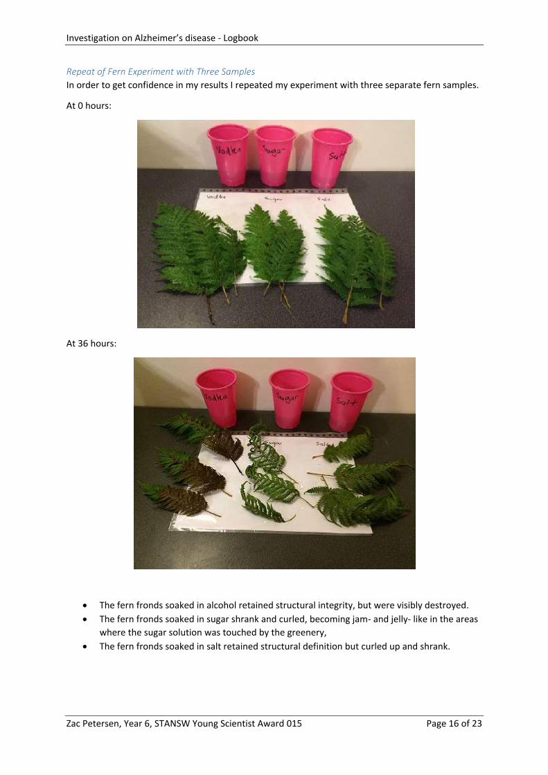
Repeat of Fern Experiment with Three Samples
In order to get confidence in my results I repeated my experiment with three separate fern samples.
At 0 hours:
At 36 hours:
The fern fronds soaked in alcohol retained structural integrity, but were visibly destroyed.
The fern fronds soaked in sugar shrank and curled, becoming jam- and jelly- like in the areas
where the sugar solution was touched by the greenery,
The fern fronds soaked in salt retained structural definition but curled up and shrank.

Investigation on Alzheimer’s disease - Logbook
Zac Petersen, Year 6, STANSW Young Scientist Award 015 Page 17 of 23
Results in Pursuit of Research Question 3 Research Question 3: Is it feasible that prions get entrapped in a damaged Alzheimer’s brain?
In pursuit of an answer to this third research question, we dissected our tenth lamb’s brain
hemisphere, to make slides from it for examination under a microscope.
Day 1 | Date: 18/8/2015, Time 8:30pm.
Our first incision appeared to slice right through one of the ventricles:
I wasn’t really sure what I would find, but I knew that saliva can contain pathogens, which could
potentially enter the brain, for example through the bloodstream when a tooth falls out. I wondered
what kind of pathogens human saliva might contain.
With the spare 10th brain hemisphere, mum and I decided to make three columns of slides to be
covered up over several days. Slides 1, 2, and 3 would be covered up immediately, and the later
slides on later days. One column would have no saliva and just brain tissue that had been left out a
while. The second column would have brain tissue in my spit. The third column would have brain
tissue in mum’s spit.

Investigation on Alzheimer’s disease - Logbook
Zac Petersen, Year 6, STANSW Young Scientist Award 015 Page 18 of 23
Day 0 | Date: 17/8/2015, Time: 10:30pm.
I noticed that my spit (slides 2, 5, 8, 11, 14) was much more watery and runny than mum’s spit
(slides 3, 6, 9, 12, 15), which seemed thicker. We used the disposable scalpel to create really thin
slivers of brain for the slides, but it was hard to get the really thin slices, and we weren’t completely
sure what we were doing.
The next few sections document the results of my investigation into the structure of brain tissue
under the microscope, mixed with saliva.
Day 1 | Date: 18/8/2015, Time 8:30pm.
After 22 hours of slide exposure, we covered up slides 4, 5, 6, 7, 8 and 9.
Investigation on Alzheimer’s disease - Logbook
Zac Petersen, Year 6, STANSW Young Scientist Award 015 Page 19 of 23
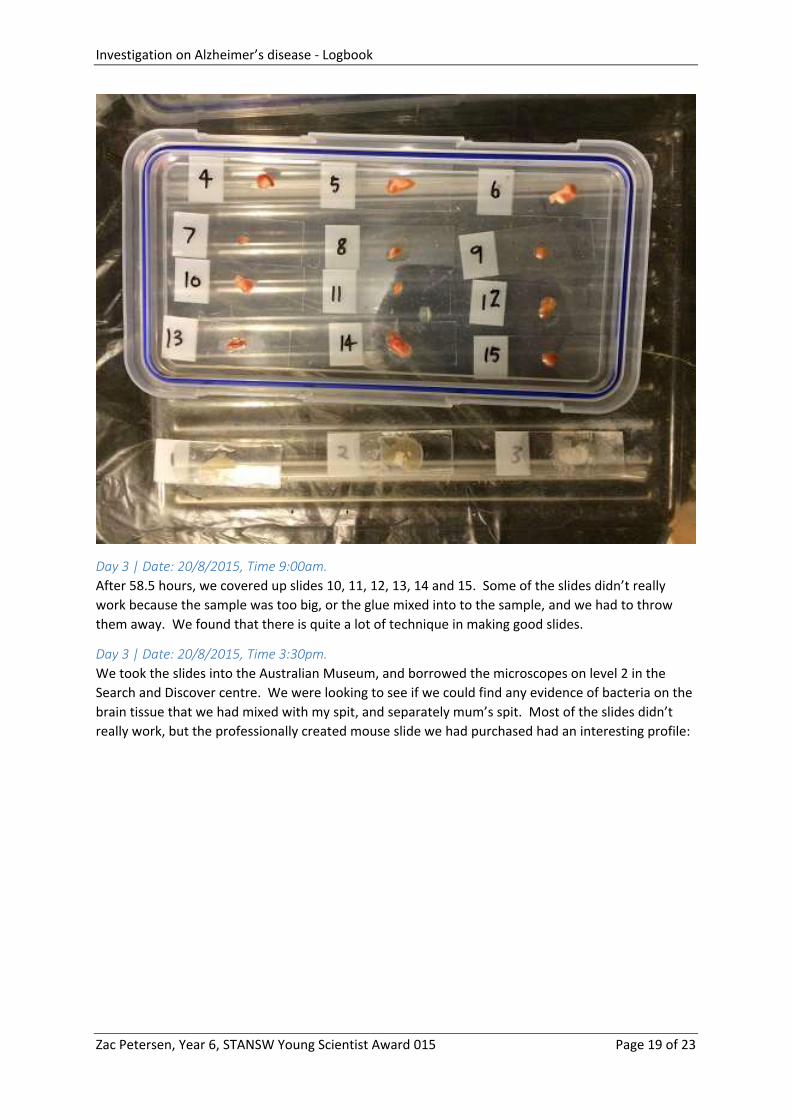
Day 3 | Date: 20/8/2015, Time 9:00am.
After 58.5 hours, we covered up slides 10, 11, 12, 13, 14 and 15. Some of the slides didn’t really
work because the sample was too big, or the glue mixed into to the sample, and we had to throw
them away. We found that there is quite a lot of technique in making good slides.
Day 3 | Date: 20/8/2015, Time 3:30pm.
We took the slides into the Australian Museum, and borrowed the microscopes on level 2 in the
Search and Discover centre. We were looking to see if we could find any evidence of bacteria on the
brain tissue that we had mixed with my spit, and separately mum’s spit. Most of the slides didn’t
really work, but the professionally created mouse slide we had purchased had an interesting profile:

Investigation on Alzheimer’s disease - Logbook
Zac Petersen, Year 6, STANSW Young Scientist Award 015 Page 20 of 23

Investigation on Alzheimer’s disease - Logbook
Zac Petersen, Year 6, STANSW Young Scientist Award 015 Page 21 of 23
I was even able to magnify my own skin on the 30x TV microscope:
Investigation on Alzheimer’s disease - Logbook
Zac Petersen, Year 6, STANSW Young Scientist Award 015 Page 22 of 23
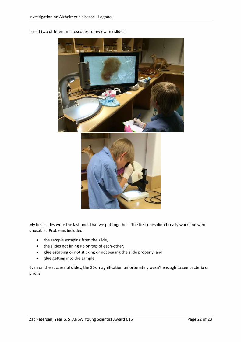
I used two different microscopes to review my slides:
My best slides were the last ones that we put together. The first ones didn’t really work and were
unusable. Problems included:
the sample escaping from the slide,
the slides not lining up on top of each-other,
glue escaping or not sticking or not sealing the slide properly, and
glue getting into the sample.
Even on the successful slides, the 30x magnification unfortunately wasn’t enough to see bacteria or
prions.
Investigation on Alzheimer’s disease - Logbook
Zac Petersen, Year 6, STANSW Young Scientist Award 015 Page 23 of 23
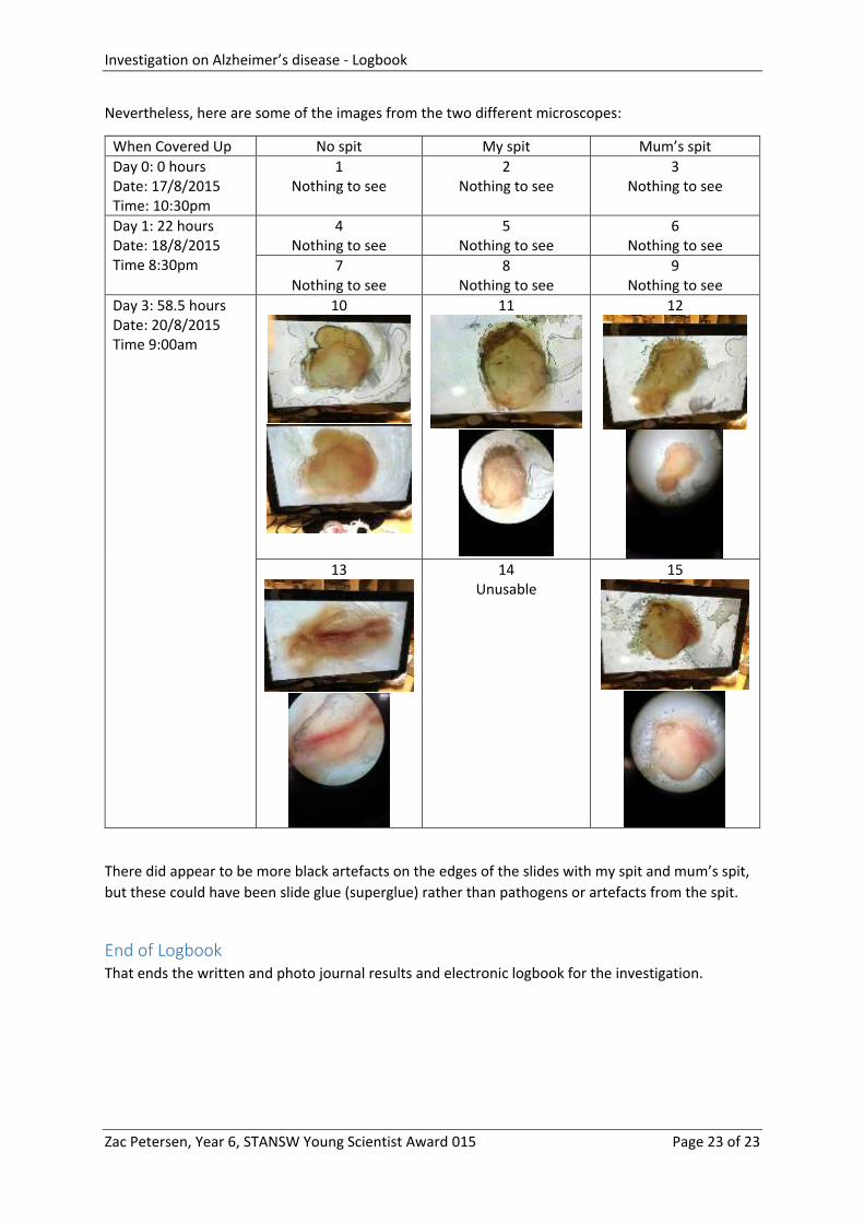
Nevertheless, here are some of the images from the two different microscopes:
When Covered Up No spit My spit Mum’s spit
Day 0: 0 hours Date: 17/8/2015 Time: 10:30pm
1 Nothing to see
2 Nothing to see
3 Nothing to see
Day 1: 22 hours Date: 18/8/2015 Time 8:30pm
4 Nothing to see
5 Nothing to see
6 Nothing to see
7 Nothing to see
8 Nothing to see
9 Nothing to see
Day 3: 58.5 hours Date: 20/8/2015 Time 9:00am
10
11
12
13
14 Unusable
15
There did appear to be more black artefacts on the edges of the slides with my spit and mum’s spit,
but these could have been slide glue (superglue) rather than pathogens or artefacts from the spit.
End of Logbook That ends the written and photo journal results and electronic logbook for the investigation.