Palaeontologia Electronica VISUALISING MUSCLE ANATOMY USING THREE-DIMENSIONAL
18
Palaeontologia Electronica http://palaeo-electronica.org PE Article Number: 12.3.7T Copyright: Palaeontological Association December 2009 Submission: 9 January 2009. Acceptance: 13 October 2009 Curtis, Neil, Jones, Marc E.H., Evans, Susan E., O’Higgins, Paul, and Fagan, Michael J. 2009. Visualising Muscle Anatomy Using Three-dimensional Computer Models - An Example Using the Head and Neck Muscles of Sphenodon. Palaeontologia Electronica Vol. 12, Issue 3; 7T: 18p; http://palaeo-electronica.org/2009_3/194/index.html VISUALISING MUSCLE ANATOMY USING THREE-DIMENSIONAL COMPUTER MODELS - AN EXAMPLE USING THE HEAD AND NECK MUSCLES OF SPHENODON Neil Curtis, Marc E. H. Jones, Susan E. Evans, Paul O’Higgins, and Michael J. Fagan ABSTRACT We demonstrate how the computer-based technique of multi-body dynamics anal- ysis (MDA) can be used to create schematic, but informative three-dimensional (3D) representations of complex muscle anatomy. As an example we provide an overview of the head and neck muscles present in Sphenodon (Diapsida: Lepidosauria: Rhyn- chocephalia). First a computer model based on micro-computed tomography datasets provides a detailed and anatomically correct three-dimensional (3D) framework to work from. Secondly, muscles are represented by groups of cylinders that can be colour coded as desired. This allows muscle positions, attachment areas, and 3D orientation to be visualised clearly. This method has advantages over imaging techniques such as two-dimensional drawings and permits the form and function of the muscles to be understood in a way that is not always possible with more classical visualisation tech- niques. Neil Curtis. Department of Engineering, University of Hull, Hull, HU6 7RX, United Kingdom. [email protected]. Marc E. H. Jones. Research Department of Cell and Developmental Biology, Gower Street, UCL, University College London, London, WCIE 6BT, United Kingdom. [email protected]. Susan E. Evans. Research Department of Cell and Developmental Biology, Gower Street, UCL, University College London, London, WCIE 6BT, United Kingdom. [email protected]. Paul O’Higgins. The Hull York Medical School, University of York, York, YO10 5DD, United Kingdom. [email protected]. Michael J. Fagan. Department of Engineering, University of Hull, Hull, HU6 7RX, United Kingdom. [email protected]. KEY WORDS: 3D; imaging; muscle; myology; head; neck
Transcript of Palaeontologia Electronica VISUALISING MUSCLE ANATOMY USING THREE-DIMENSIONAL

Palaeontologia Electronica http://palaeo-electronica.org
VISUALISING MUSCLE ANATOMY USING THREE-DIMENSIONAL COMPUTER MODELS - AN EXAMPLE USING THE HEAD AND NECK
MUSCLES OF SPHENODON
Neil Curtis, Marc E. H. Jones, Susan E. Evans, Paul O’Higgins,and Michael J. Fagan
ABSTRACT
We demonstrate how the computer-based technique of multi-body dynamics anal-ysis (MDA) can be used to create schematic, but informative three-dimensional (3D)representations of complex muscle anatomy. As an example we provide an overview ofthe head and neck muscles present in Sphenodon (Diapsida: Lepidosauria: Rhyn-chocephalia). First a computer model based on micro-computed tomography datasetsprovides a detailed and anatomically correct three-dimensional (3D) framework to workfrom. Secondly, muscles are represented by groups of cylinders that can be colourcoded as desired. This allows muscle positions, attachment areas, and 3D orientationto be visualised clearly. This method has advantages over imaging techniques such astwo-dimensional drawings and permits the form and function of the muscles to beunderstood in a way that is not always possible with more classical visualisation tech-niques.
Neil Curtis. Department of Engineering, University of Hull, Hull, HU6 7RX, United Kingdom. [email protected] E. H. Jones. Research Department of Cell and Developmental Biology, Gower Street, UCL, University College London, London, WCIE 6BT, United Kingdom. [email protected] E. Evans. Research Department of Cell and Developmental Biology, Gower Street, UCL, University College London, London, WCIE 6BT, United Kingdom. [email protected] O’Higgins. The Hull York Medical School, University of York, York, YO10 5DD, United Kingdom. [email protected] J. Fagan. Department of Engineering, University of Hull, Hull, HU6 7RX, United Kingdom. [email protected].
KEY WORDS: 3D; imaging; muscle; myology; head; neck
PE Article Number: 12.3.7TCopyright: Palaeontological Association December 2009Submission: 9 January 2009. Acceptance: 13 October 2009
Curtis, Neil, Jones, Marc E.H., Evans, Susan E., O’Higgins, Paul, and Fagan, Michael J. 2009. Visualising Muscle Anatomy Using Three-dimensional Computer Models - An Example Using the Head and Neck Muscles of Sphenodon. Palaeontologia Electronica Vol. 12, Issue 3; 7T: 18p; http://palaeo-electronica.org/2009_3/194/index.html

Curtis, et al.: VISUALISING MUSCLE ANATOMY
INTRODUCTION
In the past, muscles were mainly describedand illustrated using monochrome line drawingsoften in a standard anatomical plane such as lat-eral view (e.g., Byerly 1925; Oelrich 1956; Haas1973; Schumacher 1973; Gomes 1974; Wu 2003).The limitations are obvious when describing com-plex three-dimensional (3D) structures and maylead to ambiguous communication or misunder-standings. For example, Gorniak et al. (1982) pro-vide a detailed illustration of the jaw muscles inSphenodon (Günther 1867), but it is ambiguous asto the path of the muscle labelled as m. AdductorMandibulae Externus Posterior and, indeed, it isuncertain whether this muscle is correctly identified(Wu 2003; Jones et al. 2009). From drawingsmade in lateral view alone it is also often difficult toappreciate the mediolateral orientation of muscles,for example the complexities of the pterygoideusmuscle in the lizard Uromastyx (e.g., Haas 1973;Throckmorton 1978).
Hypotheses for the muscle arrangement infossil taxa have long been carried out (e.g., Adams1919; Anderson 1935; Fox 1964; Barghusen 1973;Rieppel 2002), with the depiction and communica-tion of such hypotheses being subject to similarproblems. More recent descriptions of muscleanatomy include photographic images on to whichmuscle groups are drawn and colour coded (e.g.,Sniverly and Russell 2007; Tsuihiji 2007), andimprove the clarity and understanding of musclepositioning and function.
Computer-based data collection and imaginghas been used to some extent by palaeontologistsand comparative anatomists for at least 25 years(e.g., Conroy and Vannier 1984; Conroy et al.1990), but only with the recent advances in compu-tational power have 3D imaging and functionalanalysis become more widespread (e.g., Hutchin-son et al. 2005; Motani 2005; Grosse et al. 2007;Wickens 2007; Strait and Evans 2008; Sutton2008). Examples include the description of thecranial anatomy of dinosaurs, which has beenimpressively presented in recent publications (Wit-mer and Ridgely 2008, 2009; Evans et al. 2009),and 3D bony anatomy derived from computedtomography (CT) data of dinosaur skulls that hasbeen used as a frame on which to present muscleanatomy (e.g., Holliday 2009).
The modelling and analysis software knownas multi-body dynamics analysis (MDA) is an engi-neering technique that has recently been used tocalculate the kinetic and kinematic behaviour withinskulls (e.g., Curtis et al. 2008, 2009; Moazen et al.
2008, 2009). Although not the primary purpose ofthis software, the way in which muscles are gener-ated has the potential to make the muscle anatomymore clear and to help understand the function ofindividual muscle groups. Others apply this tech-nology to understand function (e.g., Hutchinson etal. 2005), but the potential of this technique as apure visualisation tool has not yet been explored orhighlighted.
In order to demonstrate this potential, wepresent a thorough treatment of the head and neckmuscles in Sphenodon. As the only extant rhyn-chocephalian (sensu Gauthier et al. 1988) thisgenus is an important reference taxon for work onthe muscles of other amniotes (Schwenk 1986;Bryant and Russell 1992; Witmer 1995; Abdalaand Moro 2003; Holliday and Witmer 2007), and itis therefore important that anatomical interpreta-tions are both clear and detailed. The musclearrangements of Sphenodon have been repeatedlyexamined and discussed (e.g., Nishi 1916; Byerly1925; Anderson 1936; Poglayen-Neuwall 1953;Haas 1973; Gorniak et al. 1982; Wu 2003),although, as for many other taxa, they have mainlybeen described and illustrated in lateral view usingline drawings. Apart from a few recent examples(e.g., Tsuihiji 2005, 2007; Holliday and Witmer2007; Jones et al. 2009), authors were restricted tomonochromatic images (e.g., Poglayen-Neuwall1953; Haas 1973), and as descriptions havebecome more detailed, the limitations of thesemethods have become more apparent.
Here accurate 3D models of the skull, lowerjaw, and neck of Sphenodon form a base on towhich muscle groups are positioned. The musclesare represented as simple cylinders and thus allowthe origins and insertions to be easily identified.Complex muscle groups are divided into clear,colour coded sections. A variation in bone trans-parency and orientation are used to achieve thebest view of the muscle attachment, and the effec-tive lines of action can be determined for eachmuscle group in both lateral and anterior views.
This paper visually presents all muscle groupsin Sphenodon with only limited descriptive text; themuscle arrangements are based on a comprehen-sive descriptive review published by Jones et al.(2009).
MATERIALS AND METHODS
A dry Sphenodon skull (specimen LDUCZx036) was subjected to micro-computed tomogra-phy (micro-CT) at the University of Hull, UK. Then,using image segmentation and analysis software
2

PALAEO-ELECTRONICA.ORG
(AMIRA 4.1, Mercury Computer Systems Inc.,USA), the micro-CT dataset was converted into 3Dmodels of the skull and lower jaws. An additionalmicro-CT dataset (University of Texas, Austin,USA) was used in the construction of the neck(specimen YPM 9194). This additional datasetrequired some manipulation as the neck wastwisted and only part of the 6th vertebra wasscanned. With reference to other dry Sphenodonmaterial (LDUCZ x036) the proatlas and the firsttwo vertebrae (atlas and axis) were subsequentlyrealigned during the 3D model construction, andthe right-hand portion of the most posterior verte-bra and girdle were duplicated and mirrored to forma complete and symmetrical structure. The soft tis-sue fascia sheets that cover the lower temporalfenestra (onto which part of the adductor muscula-ture attaches) were created manually in AMIRA,and the 7th and 8th vertebrae of the neck were sim-ply represented by cylinders and cuboids posi-tioned posterior to the 6th vertebra. These 3Dmodels were imported into ADAMS multi-bodydynamics software (MSC Software Corp, USA), inwhich a representation of the muscle groups wereadded.
Each muscle group is represented by severalstraight cylinders extending between the skull andlower jaw, or skull and neck. The exact origin andattachment point of each cylinder was positionedaccording to descriptions in Jones et al. (2009),which was, in turn, based on descriptions in theworks of previous authors (e.g., Nishi 1916; Byerly1925; Anderson 1936; Poglayen-Neuwall 1953;Haas 1973; Gorniak et al. 1982; Wu 2003; Al-Has-sawi 2004, 2007; Tsuihiji 2005, 2007; Holliday andWitmer 2007) in combination with first-hand obser-vations. When a muscle wraps around other mus-cles or bone, it is represented by two or threecylinders joined end-to-end. The colour coding andabbreviations used in this paper follow as closelyas possible those of Jones et al. (2009).
MUSCULATURE
Jaw Muscles
Table 1 summarises all major jaw closing andopening muscle groups associated with Spheno-don. Brief descriptions of the main function of eachindividual muscle group are presented in Table 1,but for a comprehensive review of muscle functionand anatomy see Jones et al. (2009). Figure 1presents all adductor muscles covered in thispaper, while Figures 2 - 12 each represent selectgroups of muscles, as identified in Table 1.
Neck Muscles
Portions of some neck muscles extendbetween neighbouring vertebrae and provide sta-bility for the axial skeleton (e.g., Byerly 1925; Gasc1981; Tsuihiji 2005, 2007); however, it is only thesections of the muscle that extend anteriorly andattach directly to the skull that will be consideredhere. Table 2 lists these neck muscles along withtheir main contribution towards head movements.Figure 13 presents all neck muscles in Sphenodon,while Figures 14 - 20 represent select groups ofneck muscles, as identified in Table 2.
SUMMARY
Multi-body dynamics analysis (MDA), a 3Dengineering technique, was used here as a visuali-sation tool to present the head and neck muscula-ture of Sphenodon. Figure 21 displays the entiremuscular anatomy of the Sphenodon head andincludes all individual muscle groups covered inthis paper. Alternatively, a rotating movie of the 3DSphenodon model with all musculature is shown inFigure 22, while a rotating movie with superficialmuscle groups removed is shown in Figure 23. Thecolour coding is consistent in all images andmatches, as far as possible, that was used else-where (e.g., Tsuihiji 2005, 2007; Holliday and Wit-mer 2007; Jones et al. 2009). The detailed muscleanatomy is presented in a clear, simple manner,where muscle groups are divided into a finite num-ber of sections, each represented by one to threestraight cylinders. The ability to display a 3D repre-sentation (from any view) of colour coded musclegroups, and the option of removing or altering thetransparency of specific objects means that themuscle origin/ insertion locations and muscle forcelines of action can be visualised clearly.
In reality muscles bulge, wrap, and blend intotendons; they are therefore more complex in termsof their structure than represented here. Neverthe-less, the presentation of muscles in this paperallows the general role of each individual musclegroup to be determined based on its location, andthe relative interaction between muscle groups tobe inferred. For example, from Figure 14 we seethat the m. Episternocleidomastoid (mEscm, red)follows on from the m. Clavicle Dorsalis (mClDo,peach) and the m. Trapezius (mTrap, purple) toform a strong collar of muscle that connects the gir-dle and neck with the posterior regions of the skull.We can then infer that these muscles together con-tribute to raising and turning/ twisting the head, aswell as contributing to general head stability during
3
Curtis, et al.: VISUALISING MUSCLE ANATOMY
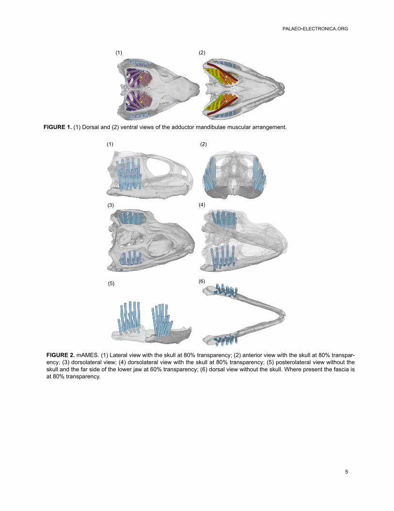
activities such as feeding or locomotion. Assessingthe jaw adductor muscles, we can suggest that thevertical alignment of the m. Adductor MandibulaeExternus Superficialis (mAMES, Figure 2.1) ren-ders it well placed to move the lower jaw orthally(Olson 1961), whereas the more angled m. Ptery-goideus Typicus (mPtTyp, Figures 6 and 7) willcontribute more towards anterior translations of themandible.
The method of representing muscle anatomypresented here is not intended to replace moreclassical techniques such as line drawings fordepicting observed or hypothesised muscle anat-omy. The visual representations do however offeran additional and complimentary means of commu-nicating such morphological information, and may
appeal to scientists who do not have extensiveexperience in muscle anatomy and function.
ACKNOWLEDGEMENTS
For access to the Sphenodon skull (specimenLDUCZ x036) we thank J. Ashby, M. Carnall, andN. McEnroe (Grant Museum of Zoology, UCL, UK),and for the micro-CT dataset (specimenYPM9194), we thank L.K. Murray and C.J. Bell(University of Texas, Austin, USA). For her generalhelp and in this instance the scanning of specimenLDUCZ x036 we thank S. Taft at the University ofHull, UK. We also gratefully acknowledge the Bio-technology and Biological Sciences ResearchCouncil (BBSRC - grants BB/E007465/1, BB/E009204/1 and BB/E007813/1) who funded thisresearch.
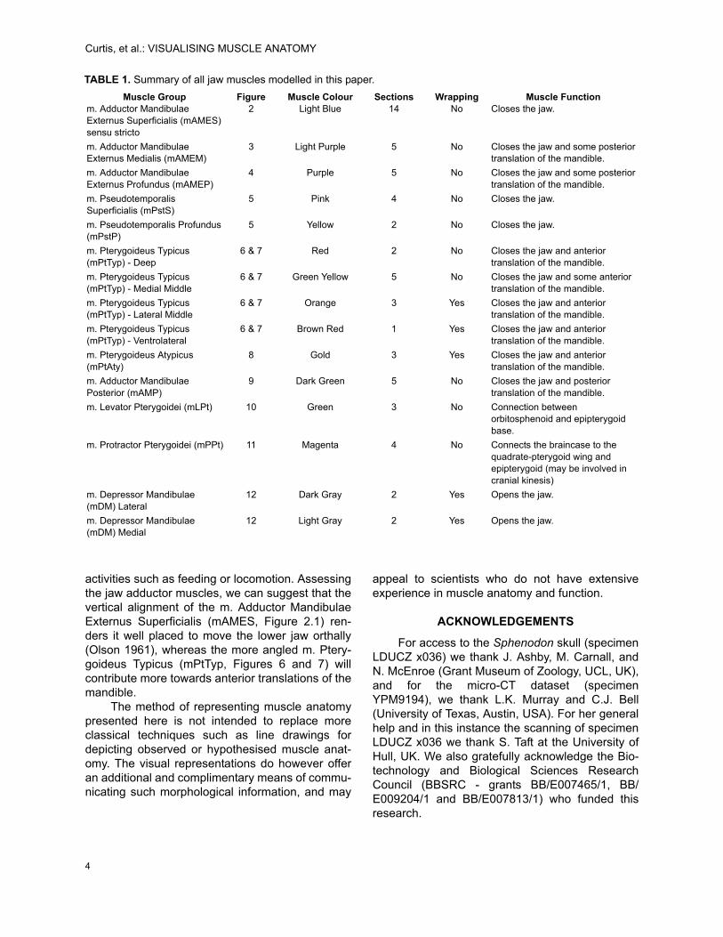
TABLE 1. Summary of all jaw muscles modelled in this paper.
Muscle Group Figure Muscle Colour Sections Wrapping Muscle Functionm. Adductor Mandibulae Externus Superficialis (mAMES) sensu stricto
2 Light Blue 14 No Closes the jaw.
m. Adductor Mandibulae Externus Medialis (mAMEM)
3 Light Purple 5 No Closes the jaw and some posterior translation of the mandible.
m. Adductor Mandibulae Externus Profundus (mAMEP)
4 Purple 5 No Closes the jaw and some posterior translation of the mandible.
m. Pseudotemporalis Superficialis (mPstS)
5 Pink 4 No Closes the jaw.
m. Pseudotemporalis Profundus (mPstP)
5 Yellow 2 No Closes the jaw.
m. Pterygoideus Typicus (mPtTyp) - Deep
6 & 7 Red 2 No Closes the jaw and anterior translation of the mandible.
m. Pterygoideus Typicus (mPtTyp) - Medial Middle
6 & 7 Green Yellow 5 No Closes the jaw and some anterior translation of the mandible.
m. Pterygoideus Typicus (mPtTyp) - Lateral Middle
6 & 7 Orange 3 Yes Closes the jaw and anterior translation of the mandible.
m. Pterygoideus Typicus (mPtTyp) - Ventrolateral
6 & 7 Brown Red 1 Yes Closes the jaw and anterior translation of the mandible.
m. Pterygoideus Atypicus (mPtAty)
8 Gold 3 Yes Closes the jaw and anterior translation of the mandible.
m. Adductor Mandibulae Posterior (mAMP)
9 Dark Green 5 No Closes the jaw and posterior translation of the mandible.
m. Levator Pterygoidei (mLPt) 10 Green 3 No Connection between orbitosphenoid and epipterygoid base.
m. Protractor Pterygoidei (mPPt) 11 Magenta 4 No Connects the braincase to the quadrate-pterygoid wing and epipterygoid (may be involved in cranial kinesis)
m. Depressor Mandibulae (mDM) Lateral
12 Dark Gray 2 Yes Opens the jaw.
m. Depressor Mandibulae (mDM) Medial
12 Light Gray 2 Yes Opens the jaw.
4
PALAEO-ELECTRONICA.ORG
5
FIGURE 1. (1) Dorsal and (2) ventral views of the adductor mandibulae muscular arrangement.
FIGURE 2. mAMES. (1) Lateral view with the skull at 80% transparency; (2) anterior view with the skull at 80% transpar-ency; (3) dorsolateral view; (4) dorsolateral view with the skull at 80% transparency; (5) posterolateral view without theskull and the far side of the lower jaw at 60% transparency; (6) dorsal view without the skull. Where present the fascia isat 80% transparency.

Curtis, et al.: VISUALISING MUSCLE ANATOMY
6
FIGURE 3. mAMEM. (1) Lateral view with the skull at 80% transparency; (2) anterior view with the skull at 80%transparency; (3) anterolateral view; (4) anterolateral view with the skull at 80% transparency. The fascia is at 80%transparency.
FIGURE 4. mAMEP. (1) Anterodorsolateral view; (2) lateral view with all bone at 80% transparency; (3) anterior viewwith the skull at 80% transparency; (4) posterolateral view showing only the lower jaw. The fascia is not visible in thisfigure.

PALAEO-ELECTRONICA.ORG
7
FIGURE 5. mPstS (pink) and mPstP (yellow). (1) Lateral view showing both the mPstS and mPstP with the skull at80% transparency; (2) lateral view showing only the mPstP with the skull at 80% transparency; (3) anterior viewshowing both the mPstS and mPstP with the skull at 80% transparency; (4) posterolateral view of only the lower jawshowing both the mPstS and mPstP; (5) posterodorsolateral view of the mPstS; (6) posterodorsolateral view of themPstP. The fascia is not included in this figure.
FIGURE 6. m. Pterygoideus. (1) Lateral view with all bone at 80% transparency; (2) posterior view with the skull andlower jaws at 80% transparency; (3) ventral view. Figure includes the dorsal (red), middle (orange and green yellow)and ventrolateral (brown red) portions of the mPtTyp. No fascia is present in this figure.
Curtis, et al.: VISUALISING MUSCLE ANATOMY
8
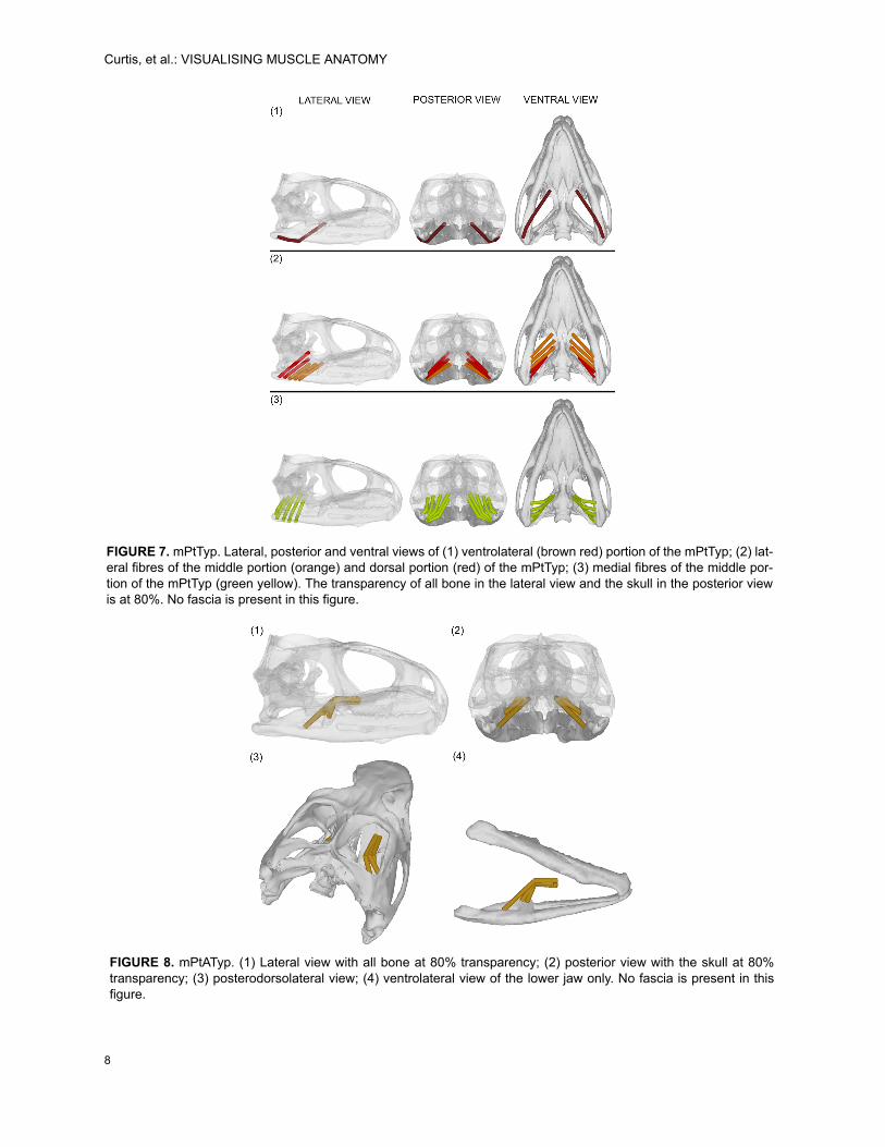
FIGURE 7. mPtTyp. Lateral, posterior and ventral views of (1) ventrolateral (brown red) portion of the mPtTyp; (2) lat-eral fibres of the middle portion (orange) and dorsal portion (red) of the mPtTyp; (3) medial fibres of the middle por-tion of the mPtTyp (green yellow). The transparency of all bone in the lateral view and the skull in the posterior viewis at 80%. No fascia is present in this figure.
FIGURE 8. mPtATyp. (1) Lateral view with all bone at 80% transparency; (2) posterior view with the skull at 80%transparency; (3) posterodorsolateral view; (4) ventrolateral view of the lower jaw only. No fascia is present in thisfigure.

PALAEO-ELECTRONICA.ORG
9
FIGURE 9. mAMP. (1) Anteroventrolateral view with the left side of the lower jaw at 80% transparency; (2) lateralview with all bone at 80% transparency; (3) posterior view with the skull at 80% transparency; (4) posterolateral viewshowing only the lower jaw. The fascia is not visible in this figure.
FIGURE 10. mLPt (1) Anterodorsolateral view; (2) anterodorsolateral view with the skull at 80% transparency; (3)lateral view with the skull at 80% transparency. Fascia at 80% transparency
Curtis, et al.: VISUALISING MUSCLE ANATOMY
10
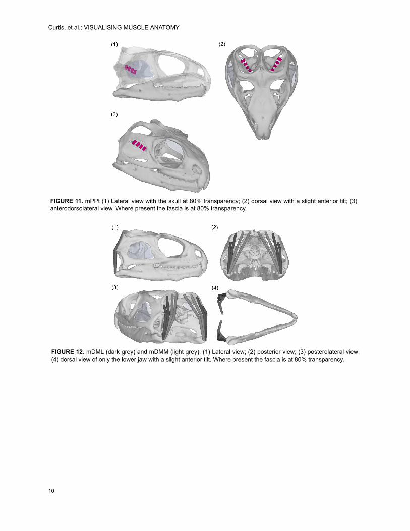
FIGURE 11. mPPt (1) Lateral view with the skull at 80% transparency; (2) dorsal view with a slight anterior tilt; (3)anterodorsolateral view. Where present the fascia is at 80% transparency.
FIGURE 12. mDML (dark grey) and mDMM (light grey). (1) Lateral view; (2) posterior view; (3) posterolateral view;(4) dorsal view of only the lower jaw with a slight anterior tilt. Where present the fascia is at 80% transparency.

PALAEO-ELECTRONICA.ORG
11
TABLE 2. Summary of all neck muscles modelled in this paper.
Muscle Group Figure Muscle Colour Sections Branching Muscle Functionm. Trapezius (mTrap) 14 Purple 2 No Holds or raises the head and some
rotational control.
m. Episternocleidomastoid (mEscm)
14 Red 2 No Holds or raises the head and some rotational control.
m. Clavicle Dorsalis (mClDo) 14 Peach 1 No Holds or raises the head and some rotational control.
m. Spinalis Capitis (nSpCa) 15 Green 3 No Holds or raises the head.
m. Axis-Supraoccipital (mAxSu) 16 Pink 3 No Holds or raises the head.
m. Rectus Capitis Posterior Superficialis (mReCaPS)
16 Bright Blue 2 No Holds or raises the head and slight rotational control.
m. Rectus Capitis Posterior Profundus (mReCaPP)
16 Light Blue 3 No Holds or raises the head and rotational control.
m. Obliquus Capitis Magnus (mReCaM)
16 Blue Gray 3 No Rotational control with limited contribution to raising the head.
m. Semispinalis Capitis (mSSpCa)
17 Brown 4 Yes Holds or raises the head and rotational control.
m. Longissimus Capitis Lateralis (mLCaL)
17 Violet Red 1 Yes Rotational control.
m. Longissimus Capitis Medialis (mLCaM)
17 Dark Red 2 No Rotational control with limited contribution to raising the head.
m. Longissimus Capitis Pars Transversalis Cervicus (mLCaPTCe)
18 Navy Blue 1 Yes Lowering the head and some rotational control.
m. Iliocostalis capitis (mlcosCa) 19 Bright Green 2 Yes Lowering the head and some rotational control.
m. Longus Colli (mLoCol) 20 Yellow 7 No Lowering the head and some rotational control.
FIGURE 13. (1) Doral view and (2) ventral view of all major neck muscles.

Curtis, et al.: VISUALISING MUSCLE ANATOMY
12
FIGURE 14. mTrap (purple), mEscm (red) and mClDo (peach). (1) Dorsal view; (2) ventral view; (3) lateral view; (4)posterior view; (5) posterolateral view; (6) anterolateral view. Fascia at 80% transparency.
FIGURE 15. mSpCa. (1), (2) and (3) show posterior to anterior attachment locations on lateral views of the neck andposterior section of the skull; (4) posterodorsal view. Fascia at 80% transparency.

PALAEO-ELECTRONICA.ORG
13
FIGURE 16. mAxSu (pink - [5]), mReCaPS (bright blue - [6]), mReCaPP (light blue [7]), mObCaM (blue gray [8]). (1)Posterodorsal view; (2) lateral view with the skull at 80% transparency; (3) anterodorsolateral view with the skull at80% transparency; (4) anterodorsolateral view of only the neck showing all muscle groups; (5) anterodorsolateralview of only the neck showing the mAxSu; (6) anterodorsolateral view of only the neck showing the mReCaPS; (7)anterodorsolateral view of only the neck showing the mReCaPP; (8) anterodorsolateral view of only the neck show-ing the mObCaM. Where present the fascia is at 80% transparency.
FIGURE 17. mSSpCa (brown [5]), mLCaL (violet_red [6]) and mLCaM (dark red [7]). (1) Dorsal view; (2) posteroven-trolateral view; (3)ventral view; (4) posteroventrolateral view without the neck; (5) anterodorsolateral view of themSSpCa without the skull; (6) anterodorsolateral view of the mLCaL without the skull; (7) anterodorsolateral view ofthe mLCaM without the skull. Where present the fascia is at 80% transparency.

Curtis, et al.: VISUALISING MUSCLE ANATOMY
14
FIGURE 18. mLCaPTCe. (1) Posterodorsolateral view; (2) posteroventrolateral view with the neck at 80% transpar-ency; (3) lateral view with all bone at 80% transparency. Fascia is at 80% transparency.
FIGURE 19. mlcosCa. (1) Posterodorsolateral view; (2) posteroventrolateral view with the neck at 80% transparency;(3) lateral view with all bone at 80% transparency. Fascia is at 80% transparency.
PALAEO-ELECTRONICA.ORG
15
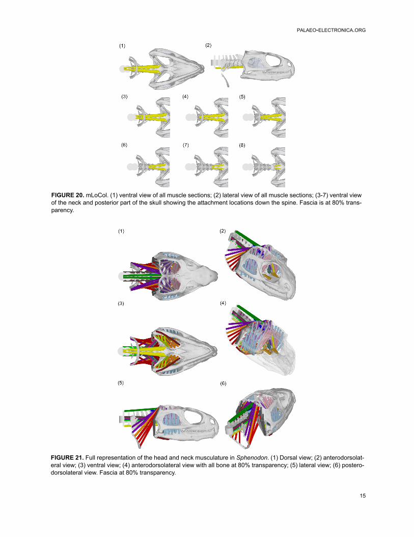
FIGURE 20. mLoCol. (1) ventral view of all muscle sections; (2) lateral view of all muscle sections; (3-7) ventral viewof the neck and posterior part of the skull showing the attachment locations down the spine. Fascia is at 80% trans-parency.
FIGURE 21. Full representation of the head and neck musculature in Sphenodon. (1) Dorsal view; (2) anterodorsolat-eral view; (3) ventral view; (4) anterodorsolateral view with all bone at 80% transparency; (5) lateral view; (6) postero-dorsolateral view. Fascia at 80% transparency.
Curtis, et al.: VISUALISING MUSCLE ANATOMY
16
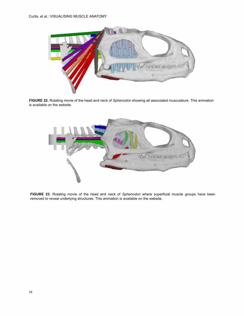
FIGURE 22. Rotating movie of the head and neck of Sphenodon showing all associated musculature. This animationis available on the website.
FIGURE 23. Rotating movie of the head and neck of Sphenodon where superficial muscle groups have beenremoved to reveal underlying structures. This animation is available on the website.

PALAEO-ELECTRONICA.ORG
REFERENCES
Abdala, V. and Moro, S. 2003. A cladistic analysis of tenlizard families (Reptilia: Squamata) based on cranialmusculature. Russian Journal of Herpetology, 10:53-73.
Adams, L.A. 1919. A memoir on the phylogeny of the jawmuscles in recent and fossil vertebrates, Annals ofthe New York Academy of Science, 28:51-166.
Al-Hassawi, A.M. 2004. The osteology and myology ofthe craniocervical region in squamate reptiles: acomparative study. Unpublished Ph.D. Thesis, Uni-versity of London, UK.
Al-Hassawi, A.M. 2007. Comparative Anatomy of theNeck Region in Lizards. Trafford Publishing, Victoria,Canada.
Anderson, H.T. 1936. The jaw musculature of the phyto-saur, Machaeroprosopus. Journal of Morphology,59(3):549-587.
Barghusen, H.R. 1973. The adductor jaw musculature ofDimetrodon (Reptilia, Pelycosauria), Journal of Pale-ontology, 47:823-834.
Bryant, H.N., and Russell, A.P. 1992. The role of phylo-genetic analysis in the inference of unpreserved attri-butes of extinct taxa. Philosophical Transactions ofthe Royal Society of London B 337:405-418.
Byerly, T.C. 1925. The myology of Sphenodon punc-tatum. University of Iowa Studies in Natural History,9(6):1-51.
Conroy G.C., and Vannier, M.W. 1984. Noninvasivethree-dimensional computer imaging of matrix-filledfossil skulls by high-resolution computed tomogra-phy. Science, 226 (4673):456-458.
Conroy, G., Vannier, M., and Tobias, P. 1990. Endocra-nial features of Australopithecus africanus revealedby 2- and 3-D computed tomography. Science,247:838-841.
Curtis, N., Kupczik, K., O’Higgins, P., Moazen, M., andFagan, M.J. 2008. Predicting skull loading: applyingdynamics analysis to a macaque skull. The Anatomi-cal Record, 291:491-501.
Curtis, N., Jones, M.E.H., Evans, S.E., Shi, J., O’Hig-gins, P., and Fagan, M.J. 2009. Predicting muscleactivation patterns from motion and anatomy: model-ling the skull of Sphenodon (Diapsida: Rhyn-chocephalia). Journal of the Royal Society Interface,published online. doi:10.1098/rsif.2009.0139.
Evans, D.C., Ridgely, R., and Witmer, L.M. 2009.Endocranial anatomy of Lambeosaurine Hadrosau-rids (Dinosauria: Ornithischia): A sensorineural per-spective on cranial crest function. The AnatomicalRecord, 292:1315-1337.
Fox, R.C. 1964. The adductor muscles of the jaw insome primitive reptiles. University of Kansas Publish-ing Museum of Natural History, 12:657-680.
Gasc, J.-P. 1981. Axial musculature, p. 355-435. In GansC. and Parsons, T.S. (eds.). Biology of the Reptiliavolume 11. Academic Press, New York and London.
Gauthier, J.A., Estes, R., de Queiroz, K. 1988. A phylo-genetic analysis of the Lepidosauromorpha, p. 15-98.In Estes, R. and Pregill, G., (ed). Phylogenetic Rela-tionships of the Lizard Families: Essays Commemo-rating Charles L. Camp. Stanford University Press,Stanford.
Gomes, N. 1974. Anatomie comparée de la musculaturetrigeminale des lacertilians, Memoirs du MuseumNational d’Histoire Naturelle, Paris (Zoology), 90:1-107.
Gorniak, G.C., Rosenberg, H.I., and Gans, C. 1982.Mastication in the tuatara, Sphenodon punctatus(Reptilia: Rhynchocephalia): Structure and activity ofthe motor system. Journal of Morphology, 171:321-353.
Grosse, I.R., Dumont, E.R., Coletta, C., Tolleson, A.2007. Techniques for modelling muscle-inducedforces on finite element models of skeletal structures.The Anatomical Record Part A, 290:1069-1088.
Günther, A. 1867. Contribution to the anatomy of Hatte-ria (Rhynchocephalus, Owen), Philosophical Trans-actions of the Royal Society, 157:1-34.
Haas, G. 1973. Muscles of the jaws and associatedstructures in the Rhynchocephalia and Squamata, p.285-490. In Gans, C. and Parsons, T.S. (eds.), Biol-ogy of the Reptilia volume 4. Academic Press, NewYork and London.
Holliday, C.M. and Witmer, L.M. 2007. Archosaur adduc-tor chamber evolution: integration of musculoskeletaland topological criteria in jaw muscle homology.Journal of Morphology, 268:457-484.
Holliday, C.M. 2009. New insights into dinosaur jaw mus-cle anatomy. The Anatomical Record, 292:1246-1265.
Hutchinson, J.R., Anderson, F.C., Blemker, S.S., andDelp, S.L. 2005. Analysis of hindlimb muscle momentarms in Tyrannosaurus rex using a three-dimensionalmusculoskeletal computer model: implications forstance, gait, and speed. Paleobiology, 31:676-701.
Jones, M.E.H., Curtis, N., Evans, S.E., O’Higgins, P.,and Fagan, M.J. 2009. The head and neck musclesassociated with feeding in Sphenodon (Reptilia: Lep-idosauria: Rhynchocephalia). Palaeontologia Elec-tronica 12 (2, 7A):1-56.
Moazen, M., Curtis, N., Evans, S.E., O’Higgins, P., andFagan, M.J. 2008. Rigid-body analysis of a lizardskull: Modelling the skull of Uromastyx hardwickii.Journal of Biomechanics, 41:1274-1280.
Moazen, M., Curtis, N., Evans, S.E., O’Higgins, P., andFagan, M.J. 2009. Assessment of the role of suturesin a lizard skull: a computer modelling study. Pro-ceedings of the Royal Society B, 276:39-46.
Motani, R. 2005. Detailed tooth morphology in adurophagous ichthyosaur captured by 3D laser scan-ner. Journal of Vertebrate Paleontology, 25:462-465.
Nishi, S. 1916. Zur vergleichenden Anatomie der eigent-lichen (genuinen) Rückenmuskeln. GegenbauersMorphologisches Jahrbuch, 50:168-318.
17

Curtis, et al.: VISUALISING MUSCLE ANATOMY
Oelrich, T.M. 1956. The anatomy of the head of Cteno-saura pectinata (Iguanidae). Miscellaneous Publica-tions of Zoology University of Michigan, 94:1-122.
Olson, E.C. 1961. Jaw mechanisms in rhipidistians,amphibians, reptiles. American Zoologist, 1:205-215.
Poglayen-Neuwall, I. 1953. Untersuchungen über die Tri-geminusmuskulatur von Hatteria. Zeitschrift wissen-schaft Zoologie, 157:57-76.
Rieppel, O. 2002. Feeding mechanics in Triassic stem-group sauropterygians: the anatomy of a successfulinvasion of Mesozoic seas. Zoological Journal of theLinnean Society, 135:33-63.
Schumacher, G.-H. 1973. The head muscles and hyo-laryngal skeleton of turtles and crocodilians, chapter2, p. 101-199. In Gans, C. and Parsons, T.H. (eds.),Biology of the Reptilia 4 Morphology. AcademicPress, London and New York.
Schwenk, K. 1986. Morphology of the tongue in the tuat-ara, Sphenodon punctatus (Reptilia: Lepidosauria),with comments on function and phylogeny. Journal ofMorphology 188:129-156
Snively, E. and Russell, A.P. 2007. Functional variationof neck muscles and their relation to feeding style inTyrannosauridae and other large theropod dinosaurs.The Anatomical Record, 290:934-957.
Strait, S.G. and Evans, A.R. 2008. Special Issue: Three-Dimensional imaging in vertebrate paleontology.Palaeontologia Electronica, 11(2).
Sutton, M.D. 2008. Tomographic techniques for thestudy of exceptionally preserved fossils. Proceedingsof the Royal Society B: Biological Sciences, 275:1-7.
Throckmorton, G.S. 1978. Action of the pterygoideusmuscle during feeding in the lizard Uromastix aegyp-tius (Agamidae). The Anatomical Record. 190:217-222.
Tsuihiji, T. 2005. Homologies of the transversospinalismuscles in the anterior presacral region of Sauria(crown Diapsida). Journal of Morphology, 263:151-178.
Tsuihiji, T. 2007. Homologies of the longissimus, iliocos-talis, and hypaxial muscles in the anterior presacralregion of extant Diapsida. Journal of Morphology,268:986-1020.
Wickens, Z. 2007. Computer aided visualisation in palae-ontology (Meeting report). Palaeontological newslet-ter, 66:37-40.
Witmer, L.M. 1995. The extant phylogenetic bracket andthe importance of reconstructing soft tissues in fos-sils, p. 19-33. In Thomason, J.J. (ed), FunctionalMorphology in Vertebrate Palaeontology. CambridgeUniversity Press, Cambridge.
Witmer, L.M. and Ridgely, R.C. 2008. The paranasal airsinuses of predatory and armoured dinosaurs(Archosauria: Theropoda and Ankylosauria) and theircontribution to cephalic structure. The AnatomicalRecord, 291:1362-1388.
Witmer, L.M. and Ridgely, R.C. 2009. New insights intothe brain, braincase, and ear region of tyrannosaurs(Dinosauria, Theropoda), with implications for sen-sory organization and behavior. The AnatomicalRecord, 292:1266-1296.
Wu, X.-C. 2003. Functional morphology of the temporalregion in the Rhynchocephalia. Canadian Journal ofEarth Sciences, 40:589-607.
18