Ozurdex (dexamethasone intravitreal implant) · drug-delivery system (DDS) a drug delivery system,...
25
Page 1 of 25 DISCLAIMER This Molina Clinical Policy (MCP) is intended to facilitate the Utilization Management process. It expresses Molina's determination as to whether certain services or supplies are medically necessary, experimental, investigational, or cosmetic for purposes of determining appropriateness of payment. The conclusion that a particular service or supply is medically necessary does not constitute a representation or warranty that this service or supply is covered (i.e., will be paid for by Molina) for a particular member. The member's benefit plan determines coverage. Each benefit plan defines which services are covered, which are excluded, and which are subject to dollar caps or other limits. Members and their providers will need to consult the member's benefit plan to determine if there are any exclusion(s) or other benefit limitations applicable to this service or supply. If there is a discrepancy between this policy and a member's plan of benefits, the benefits plan will govern. In addition, coverage may be mandated by applicable legal requirements of a State, the Federal government or CMS for Medicare and Medicaid members. CMS's Coverage Database can be found on the CMS website. The coverage directive(s) and criteria from an existing National Coverage Determination (NCD) or Local Coverage Determination (LCD) will supersede the contents of this MCP document and provide the directive for all Medicare members. SUMMARY OF EVIDENCE/POSITION This policy addresses the coverage of Ozurdex (dexamethasone intravitreal implant), solid polymer sustained-release drug-delivery system (DDS) a drug delivery system, is surgically implanted in the vitreous of the eye for sustained release of a corticosteroid when appropriate criteria are met. The intent of this drug policy is to ensure appropriate selection of patients for therapy based on product labeling, clinical guidelines, and clinical studies. Diabetic macular edema (DME) is defined as retinal thickening within 2 disc diameters of the center of the macula, and results from retinal microvascular changes that compromise the blood-retinal barrier, causing leakage of plasma constituents into the surrounding retina and, consequently, retinal edema. Diabetes is a leading cause of new blindness in the United States, with clinically significant macular edema greatly contributing to this vision loss. Treatment options Laser photocoagulation, pharmacotherapy with intravitreal injection of corticosteroids or anti-vascular endothelial growth factor (VEGF) are options for treating DME. Dexamethasone, a potent corticosteroid, suppresses inflammation in the eye by inhibiting edema, fibrin deposition, capillary leakage and phagocytic migration. Corticosteroids inhibit the expression of vascular endothelial growth factor (VEGF), a cytokine that is a potent promoter of vascular permeability and expressed at increased concentrations in macular edema. Dexamethasone is a corticosteroid which is five times more potent than triamcinolone acetonide (TA). The lipophilicity of dexamethasone is less than that of TA and fluocinolone acetonide (FA) causing less binding to the trabecular meshwork and the lens. 15 This decreases the risks of high IOP and cataract formation. The half-life of intravitreally injected dexamethasone in the human vitreous humor is 5.5 hours, which limits the clinical use; 15 therefore, to extend the activity of dexamethasone, an intravitreal sustained release form was developed. 15 Subject: Intravitreal corticosteroid implants: Ozurdex (dexamethasone intravitreal implant) Original Effective Date: 10/24/2016 Policy Number: MCP-282 Revision Date(s): 12/13/2017
Transcript of Ozurdex (dexamethasone intravitreal implant) · drug-delivery system (DDS) a drug delivery system,...

Page 1 of 25
DISCLAIMER
This Molina Clinical Policy (MCP) is intended to facilitate the Utilization Management process. It expresses Molina's
determination as to whether certain services or supplies are medically necessary, experimental, investigational, or
cosmetic for purposes of determining appropriateness of payment. The conclusion that a particular service or supply is
medically necessary does not constitute a representation or warranty that this service or supply is covered (i.e., will be
paid for by Molina) for a particular member. The member's benefit plan determines coverage. Each benefit plan defines
which services are covered, which are excluded, and which are subject to dollar caps or other limits. Members and their
providers will need to consult the member's benefit plan to determine if there are any exclusion(s) or other benefit
limitations applicable to this service or supply. If there is a discrepancy between this policy and a member's plan of
benefits, the benefits plan will govern. In addition, coverage may be mandated by applicable legal requirements of a
State, the Federal government or CMS for Medicare and Medicaid members. CMS's Coverage Database can be found on
the CMS website. The coverage directive(s) and criteria from an existing National Coverage Determination (NCD) or
Local Coverage Determination (LCD) will supersede the contents of this MCP document and provide the directive for all
Medicare members.
SUMMARY OF EVIDENCE/POSITION
This policy addresses the coverage of Ozurdex (dexamethasone intravitreal implant), solid polymer sustained-release
drug-delivery system (DDS) a drug delivery system, is surgically implanted in the vitreous of the eye for sustained release
of a corticosteroid when appropriate criteria are met. The intent of this drug policy is to ensure appropriate selection of
patients for therapy based on product labeling, clinical guidelines, and clinical studies.
� Diabetic macular edema (DME) is defined as retinal thickening within 2 disc diameters of the center of the macula,
and results from retinal microvascular changes that compromise the blood-retinal barrier, causing leakage of plasma
constituents into the surrounding retina and, consequently, retinal edema. Diabetes is a leading cause of new blindness
in the United States, with clinically significant macular edema greatly contributing to this vision loss.
� Treatment options
Laser photocoagulation, pharmacotherapy with intravitreal injection of corticosteroids or anti-vascular endothelial
growth factor (VEGF) are options for treating DME.
� Dexamethasone, a potent corticosteroid, suppresses inflammation in the eye by inhibiting edema, fibrin deposition,
capillary leakage and phagocytic migration. Corticosteroids inhibit the expression of vascular endothelial growth
factor (VEGF), a cytokine that is a potent promoter of vascular permeability and expressed at increased
concentrations in macular edema.
Dexamethasone is a corticosteroid which is five times more potent than triamcinolone acetonide (TA). The
lipophilicity of dexamethasone is less than that of TA and fluocinolone acetonide (FA) causing less binding to the
trabecular meshwork and the lens.15 This decreases the risks of high IOP and cataract formation. The half-life of
intravitreally injected dexamethasone in the human vitreous humor is 5.5 hours, which limits the clinical use;15
therefore, to extend the activity of dexamethasone, an intravitreal sustained release form was developed.15
Subject: Intravitreal corticosteroid implants: Ozurdex
(dexamethasone intravitreal implant)
Original Effective Date: 10/24/2016
Policy Number: MCP-282 Revision Date(s): 12/13/2017

Page 2 of 25
� Ozurdex (dexamethasone intravitreal implant) is a dexamethasone implant using a solid polymer delivery system,
in which dexamethasone is combined with biodegradable material in the form of a small rod, which is injected into
the vitreous cavity using a specific injector.a,15,16 Dexamethasone is released in a biphasic manner over 6 months, with
higher concentrations released for the first 6 weeks, followed by lower concentrations for the following months.16
After this time, the implant dissolves to CO2 and H2O leaving no residue within the eye.16
� Ozurdex is FDA-approved for various ocular etiologies:
1) Macular edema due to branch or central retinal vein occlusion (BRVO/CRVO) in June 2009,
2) Non-infectious posterior uveitis in September 2010, and
3) Diabetic macular edema (DME) in 2014, first with restrictions to patients who are pseudophakic or are phakic
(have an artificial lens or have a cataract requiring removal and placement of an artificial lens) and scheduled
for cataract surgery. Later in 2014, the FDA broadened the approval to all patients with DME.
The goal of therapy is to reduce the inflammatory process in the eye while minimizing the adverse effects of the
therapeutic regimen.
� An intravitreal implant may an appropriate treatment alternative in members/individuals who are intolerant or
refractory to other therapies or in patients who are judged likely to experience severe adverse events from systemic
corticosteroids. Selection of the route of corticosteroid administration (topical, systemic, periocular or intraocular
injection) is based on the cause, location, and severity of the disease. Due to the differing benefits and risks of each
therapeutic approach, members/individuals should be informed about the potential adverse effects of a corticosteroid
intravitreal implant, including cataracts, increased intraocular pressure or hypotony, endophthalmitis, and risk of need
for additional surgical procedures.
CLASSIFICATION: Anti-inflammatory Agent, Corticosteroid, Ophthalmic
FDA INDICATIONS
The covered FDA-approved indications are conditions that are considered medically necessary; however it is not
inclusive of all conditions which may be approved by the Medical Reviewer. At the discretion of the Medical Director and
on a case-by-case basis, Molina Healthcare may consider authorization of the biologic therapy addressed in this Policy
for members with exceptional circumstances and for members with severe disease who may fall outside of the defined
criteria. Molina Healthcare reserves the right to update this Policy and revise coverage criteria to include or omit any
off-label condition(s) as necessary based on medical literature and clinical studies that may become available.
� Retinal Vein Occlusion [FDA approved: June 17, 2009]
Treatment of macular edema following branch retinal vein occlusion (BRVO) or central retinal vein occlusion
(CRVO).c
� Posterior Segment Uveitis [FDA approved: September 24, 2010]
Treatment of non-infectious uveitis affecting the posterior segment of the eye.c
� Diabetic macular edema [FDA approved: June 2014 and updated indication on September 2014 for DME]
Treatment of diabetic macular edemac
Available as: Ozurdex 0.7 mg (700 µg) and 0.35 mg (350 µg) biodegradable dexamethasone intravitreal implant
Ozurdex employs the Novadur™ solid polymer drug delivery system. Each implant comes preloaded in a specially
designed, single-use applicator. The implant provides intravitreal dexamethasone for up to 6 months.
Black Box Warnings: None at the time of this writing
REMS: None at the time of this writing

Page 3 of 25
Warnings and precautions in the product label include the following (Allergan Inc., 2014):
• Intravitreal injections have been associated with endophthalmitis, eye inflammation, increased IOP, and retinal
detachments. Patients should be monitored following the intravitreal injection.
• Use of corticosteroids may produce posterior subcapsular cataracts, increased IOP, and glaucoma, and may
enhance the establishment of secondary ocular infections due to bacteria, fungi, or viruses.
• The implant may migrate into the anterior chamber if the posterior lens capsule is not intact.
RECOMMENDATIONS/COVERAGE CRITERIA
Ozurdex (Dexamethasone Intravitreal Implant) for initial treatment of each affected eye may be authorized for members
who meet ALL of the following criteria [ALL]
1. Prescriber specialty [ALL]
� Prescribed by board-certified ophthalmologists or retinal specialist experienced in the administration of
intravitreal injections. Treatment and monitoring must be retained by the specialist.
2. Diagnosis/Indication [ALL]
Documentation of diagnosis required and may include clinical notes from the member’s medical records including
any relevant labs and/or tests, supporting the diagnosis [ALL]
� Diagnosis of ONE (1) of the following: [ONE: A, B, C, OR D]
A. Diabetic Macular Edema (DME) [ALL]
NOTE: DME indicated by the presence of clinically significant macular edema as defined by the
Early Treatment Diabetic Retinopathy Study (ETDRS): Retinal thickening within 500 micrometers
(µm) of the center of the fovea, OR Hard exudates within 500 µm (≤ 500 micrometers) of the fovea
center with adjacent retinal thickening, OR At least 1 disc area of retinal thickening, any part of
which is within 1 disc diameter of the center of the fovea
B. Macular Edema due to Branch Retinal Vein Occlusion (BRVO)
C. Macular Edema due to Central Retinal Vein Occlusion (CRVO)
D. Chronic (duration of 1 year or more) Non-infectious Posterior Segment Uveitis
NOTE: Ozurdex is not for use in anterior uveitis or in uveitis caused by infection
� Diagnosis and disease progression (history of progressive visual loss or worsening of anatomic appearance) as
confirmed/determined by fluorescein angiography, Optical Coherence Tomography (OCT) or Scanning
Computerized Ophthalmic Diagnostic Imaging (SCODI) Fraser CE
MOLINA REVIEWER: Baseline labs (prior to treatment with requested implant) noted in member’s profile
to review for reauthorization of treatment Informational Note
� Refer to ‘Summary of Evidence’ section discussing DME for additional information on the diagnosis of DME.
� The evaluation of RVO includes a history and a complete ophthalmic examination. Important components of the
ophthalmic examination include visual acuity and dilated fundus examination looking for hemorrhage, edema,
and dilatation of the retinal veins. Other testing such as fluorescein angiogram and coherence tomography may
be performed to confirm the diagnosis and follow disease progression or response to treatment.23
� Vision impairment (sight-threatening or sight-losing) caused by condition

Page 4 of 25
3. Age/Gender/Restrictions [ALL]
� 18 years of age or older � Safety and efficacy not established in pediatric patients 18 years of age and younger
4. Conventional Therapy/Concurrent Therapy/Other Requirements [ALL]
� Requested Dexamethasone Intravitreal Implant (Ozurdex) will NOT be concurrently prescribed or
administered with other intravitreal implants [i.e. fluocinolone acetonide intravitreal implant (Iluvien® and
Retisert®)]
NOTE: Molina staff: Verify medical/pharmacy claims data for the above drug therapies or implants within
the last 30 days, OR for new members to Molina Healthcare, confirm above mentioned therapies in medical
chart history
� Previously treated with a course of corticosteroids and did not have a clinically significant rise in intraocular
pressure
� Condition-specific criteria as applicable to member’s diagnosis [ONE: A, B, OR C]
A. Diabetic Macular Edema (DME) ONLY [ALL]
Inadequate response, clinically significant adverse effects, labeled contraindication, or clinical rationale
supporting the inappropriateness of the following (include date(s) of failed therapy or clinical event).
Documentation required. [ALL]
� Triamcinolone acetonide, intravitreal injection
Informational Note � There is no current preparation of triamcinolone acetonide (TA) approved for the treatment of DME,
although historically it has been studied and clinically used extensively for this purpose.
� Intravitreal TA is available in four preparations: triamcinolone acetonide injectable suspension 40 mg/mL
(Triescence, Alcon); triamcinolone acetonide 80 mg/mL (Trivaris, Allergan); triamcinolone acetonide
injectable suspension 40 mg/mL or 10 mg/mL (Kenalog, Bristol-Myers Squibb) formulated for intramuscular
or intra-articular use; and preservative-free TA prepared by a compounding pharmacy. All formulations are
used off-label for DME. The 40-mg/mL and 80-mg/mL formulations of TA have been approved for
intravitreal injection and are prepackaged and preservative-free, thus avoiding potential sterile inflammatory
reaction to preservative or to contaminants in compounded TA. The formulation of TA for intramuscular or
intra-articular use contains preservatives and is not approved by the US FDA for intraocular use, but it is
nevertheless commonly used by ophthalmologists off-label Injections of intravitreal TA are generally
repeated every 2 to 4 months to maintain effect.
� Vascular Endothelial Growth Factor (VEGF) Inhibitors [ONE]
� bevacizumab (Avastin): PREFERRED/NO PA REQUIRED
� ranibizumab (Lucentis)
� pegaptanib (Macugen)
� aflibercept (Eylea)B,16,F
Informational Note B,16,F � VEGF inhibitor therapy is indicated for DME [American Diabetes Association (ADA) Grade A].F
� NICE Appraisal Committee recognizes that there are currently no clinical criteria for determining whether a
treatment is unsuitable for a person with DME.B However, clinical experts suggested that treatment with an
anti-VEGF agent (i.e. ranibizumab) is likely to be unsuitable for individuals who cannot or unable to attend
monthly appointments, individuals who have had a recent cardiovascular event or stroke, and individuals
who have a phobia of needles.
� Laser Photocoagulation
Page 5 of 25
B. Macular Edema due to BRVO and CRVO [ALL]
Inadequate response, clinically significant adverse effects, labeled contraindication, or clinical rationale
supporting the inappropriateness of the following (include date(s) of failed therapy or clinical event).
Documentation required. [ALL]
� Vascular Endothelial Growth Factor (VEGF) Inhibitors21 [ONE]
� bevacizumab (Avastin): PREFERRED/NO PA REQUIRED
� ranibizumab (Lucentis)
� pegaptanib (Macugen)
� aflibercept (Eylea)
� In patients with macular edema from BRVO or CRVO that has caused visual loss, intravitreal anti-VEGF
treatment as first-line therapy is recommended.21,23
� Triamcinolone acetonide, intravitreal injection [FOR CRVO ONLY (BRVO not required)]
Informational Note � Based on safety and efficacy findings from the SCORE-CRVO trial, administering intravitreal triamcinolone
in a 1-mg dose and following the retreatment criteria applied in the SCORE study should be considered for
up to 1 year, and possibly 2 years, in patients with vision loss associated with macular edema secondary to
CRVO.18,19
� In the Standard Care versus Corticosteroid for Retinal Vein Occlusion (SCORE) trial for BRVO, intravitreal
triamcinolone in doses of 1 or 4 mg were compared with macular grid laser photocoagulation in 411 patients
with visual acuity loss from BRVO-related macular edema.19 There was no difference in visual acuity
between the treatment arms. Increasing intraocular pressure and cataract formation were more common in
the triamcinolone arms, particularly the 4 mg treatment group. Given these complications, other therapies
such as intravitreal anti-VEGF agents or macular grid laser photocoagulation are preferred options for
patients with persistent vision loss secondary to BRVO-related macular edema. Therefore, triamcinolone is
not a criterion for BRVO.19
� The Ophthalmic Technology Assessment Committee Retina/Vitreous panel of the American Academy of
Ophthalmology evaluated available literature regarding efficacy of available pharmacotherapies in the
treatment of macular edema due to CRVO. The panel reported that intravitreal anti-VEGF therapy is safe
and effective over 2 years for macular edema and that delayed treatment is associated with worse visual
outcomes. Intravitreal corticosteroid therapy yielded short-term efficacy but was associated with a higher
frequency of adverse events.E
� Laser Photocoagulation [*AS APPLICABLE ONLY]
*Generally not suitable due to macular hemorrhageA (NICE 2011)
NOTE: For the treatment of macular edema from RVO, grid laser photocoagulation is generally
considered to be second-line to anti-VEGF and intravitreal glucocorticoid therapies and often
reserved for patients who are averse to intravitreal injections
Informational Note � In patients with BRVO and visual loss who are averse to intravitreal injection treatments, macular grid
laser photocoagulation treatment rather than observation is recommended.21,18,1
Page 6 of 25
C. Non-infectious Posterior Segment Uveitis ONLY
Inadequate response, clinically significant adverse effects, labeled contraindication, or clinical rationale
supporting the inappropriateness of the following (include date(s) of failed therapy or clinical event).
Documentation required. [BOTH: 1 AND 2]
1) ONE (1) of the following: [ONE]
� Injectable or systemic corticosteroids
� Immunosuppressives, including but not limited to:G [ONE]
� Antimetabolites: azathioprine, mycophenolate mofetil (CellCept; Myfortic), or
methotrexate
� Calcineurin inhibitors: cyclosporine or tacrolimus
2) Triamcinolone acetonide injection
Informational Note: Currently, systemic immunomodulation with oral corticosteroids is the mainstay of treatment to
control the inflammation. Systemic steroid sparing immunomodulators such as antimetabolites (methotrexate,
azathioprine, and mycophenolate mofetil) and calcineurin inhibitors (cyclosporine and tacrolimus), among others,
are often included in the treatment plan [Pasadhika , Rosenbaum 2014]
5. Contraindications/Exclusions/Discontinuations
Authorization for Ozurdex (dexamethasone intravitreal implant) will not be authorized if ANY of the following
conditions apply [ANY]
� Hypersensitivity to dexamethasone or any component of the formulationc � Documentation of allergenic cross-reactivity for corticosteroids is limited. However, due to similarities in
chemical structure and/or pharmacologic actions, the possibility of cross-sensitivity cannot be ruled out with
certainty.
� Ocular or periocular infections (viral, bacterial, or fungal): Active or suspected ocular or periocular infections
including most viral diseases of the cornea and conjunctiva, including active epithelial herpes simplex
keratitis (dendritic keratitis), vaccinia, varicella, mycobacterial infections, or fungal infections of the eye � Corticosteroids are not recommended to be used in patients with a history of ocular herpes simplex because of
the potential for reactivation of the viral infection.
� Advanced glaucoma: Glaucoma with cup to disc ratios of greater than 0.8
� Aphakic eyes with rupture of the posterior lens capsule
� ACIOL (Anterior Chamber Intraocular Lens) and Rupture of the Posterior Lens Capsule � Ozurdex is contraindicated in patients whose posterior lens capsule is torn or ruptured because of the risk of
migration into the anterior chamber.
� Laser posterior capsulotomy in pseudophakic patients is not a contraindication for use.
� Concurrent treatment with other intravitreal implants [i.e. Fluocinolone acetonide intravitreal implant
(Iluvien® and Retisert®)]

Page 7 of 25
6. Labs/Reports/Documentation required [ALL]
All documentation for determination of medical necessity must be submitted for review. Prescriber to submit
documentation as indicated in the criteria above, including but not limited to chart notes, applicable lab values and/or
tests, adverse outcomes, treatment failures, or any other additional clinical information or clinical notes from the
member’s medical records supporting the diagnosis. Letters of support and/or explanation are often useful, but are
not sufficient documentation unless ALL specific information required by this MCP is included.
NOTE: Additional documentation, rationale, and/or supporting evidence may be requested for review as deemed
necessary or appropriate by Molina Medical/Pharmacy staff.
� Member has been informed about the potential adverse effects of a corticosteroid intravitreal implant,
including cataracts, increased intraocular pressure, or hypotony, endophthalmitis, and risk of need for
additional surgical procedures.
� Requested dexamethasone intravitreal implant for use in affected eye: [APPLICABLE]
� Right eye
� Left eye

Page 8 of 25
CONTINUATION OF THERAPY
Ozurdex (dexamethasone intravitreal implant) may be reauthorized if meet ALL of the following criteria are met: [ALL]
1. Initial Coverage Criteria [ALL]
� Reauthorization request is for the same eye as initial authorization AND six months since the previous
intravitreal implant
NOTE: The continuation of therapy criteria is only for the same previously treated eye. If member has
developed condition in an untreated eye, Prescriber must submit new request in accordance to ‘Initial
Coverage’ criteria.
� Member continues to meet initial coverage criteria AND member’s continued need for Ozurdex has been
formally assessed and documented
2. Compliance: N/A
3. Labs/Reports/Documentation required [ALL APPLICABLE]
Prescriber submit ALL supporting documentation and clinical rationale [ALL APPLICABLE]
� Initial positive response to treatment; however subsequently experience a loss in visual acuity, including but
not limited to the following: [AS APPLICABLE]
� Diabetic Macular Edema (DME): Member experienced decreased vision and/or an increase in
retinal thickness, secondary to recurrent or worsening DME
� Non-infectious Posterior Segment Uveitis ONLY [ONE]
o Greater than (>) 15 letters (3 lines) in BCVA from baseline after 12 weeks following
administration or the patient achieves driving visual acuity
o Visual acuity is maintained to at least 50% of the best recorded following diagnosis of uveitis
� Response to treatment (including disease progression or history of progressive visual loss or worsening of
anatomic appearance) as determined by fluorescein angiography, Optical Coherence Tomography (OCT)23 or
Scanning Computerized Ophthalmic Diagnostic Imaging (SCODI) [DOCUMENTATION REQUIRED]
NOTE:
• Retreatment is usually not necessary for patients that have maintained vision improvement.
Exceptions may be reviewed on a case-by-case basis with relevant, supporting documentation from
Prescriber.
• Ozurdex treatment should be discontinued (and patient monitored) in absence of macular edema or stable
visual acuity. Treatment (and monitoring intervals) may be resumed at prescribing specialist’s discretion
and submission of authorization request with presence of macular edema or visual acuity is decreasing at
any time.
� Member is likely to benefit from re-treatment without being exposed to significant risk, according to
Prescriber’s clinical judgment
Page 9 of 25
4. Discontinuation of Treatment [ANY]
� Loss of visual acuity from baseline (pre-treatment with Ozurdex) values
� Severely raised intraocular pressure (IOP) occurs in the treated eye, or moderately raised IOP in the treated
eye is considered to be related to Ozurdex
� Limited clinically meaningful benefit of treatment: Maximal gain in visual acuity is less than five letters on a
standard sight chart in the presence of limited anti-inflammatory effect
� Absence of macular edema or stable visual acuity
NOTE: If absence of macular edema or stable visual acuity, Ozurdex treatment should be discontinued and
patient monitored. Treatment and monitoring intervals may be resumed at prescribing specialist’s discretion and
submission of authorization request if there is presence of macular edema or visual acuity is decreasing at any
time.
� Contraindications/Exclusions to therapy
Discontinue treatment if ANY of the following conditions applies: [ANY]
� Hypersensitivity to dexamethasone or any component of the formulationc � Documentation of allergenic cross-reactivity for corticosteroids is limited. However, because of
similarities in chemical structure and/or pharmacologic actions, the possibility of cross-sensitivity
cannot be ruled out with certainty.
� Ocular or periocular infections (viral, bacterial, or fungal): Active or suspected ocular or periocular
infections including most viral diseases of the cornea and conjunctiva, including active epithelial
herpes simplex keratitis (dendritic keratitis), vaccinia, varicella, mycobacterial infections, or fungal
infections of the eye � Corticosteroids are not recommended to be used in patients with a history of ocular herpes simplex
because of the potential for reactivation of the viral infection.
� Advanced glaucoma: Glaucoma with cup to disc ratios of greater than 0.8
� Aphakic eyes with rupture of the posterior lens capsule
� ACIOL (Anterior Chamber Intraocular Lens) and Rupture of the Posterior Lens Capsule � Ozurdex is contraindicated in patients whose posterior lens capsule is torn or ruptured because of the
risk of migration into the anterior chamber.
� Laser posterior capsulotomy in pseudophakic patients is not a contraindication for use.
� Concurrent treatment with other intravitreal implants [i.e. Fluocinolone acetonide intravitreal implant
(Iluvien® and Retisert®)]

Page 10 of 25
ADMINISTRATION, QUANTITY LIMITATIONS, AND AUTHORIZATION PERIOD
Consult the manufacturer's labeling for more detailed information on dosage and administration of this drug, cautions,
precautions, contraindications, potential drug interactions, laboratory test interferences, and monitoring.
1. Recommended Dosage [ONE]
� Adult: 0.7 mg (700 µg) intravitreal implant injected intravitreally in affected eyec � Dexamethasone is released over approximately 6 months, after which the implant dissolves, leaving no residue.
� Pediatric: Safety and efficacy in pediatric patients have not been established.c
2. Authorization Limit [ALL]
� ONE (1) dexamethasone intravitreal implant may be administered every 6 months in the affected eye
EXCEPTION: For requests more frequently than 6 months, clinical rationale and relevant supporting
documentation must be submitted to Molina Medical Director for review.
Note: � In the efficacy DME studies discussed in the FDA-approved labeling, subjects were to be evaluated for retreatment
eligibility every three months starting from Month 6; however received successive treatments at least 6 months
apart.a
� GENEVA trial5 patients with macular edema associated with BRVO and CRVO: A retreatment was not allowed
before 6 months after the initial implantation according to the protocol of this trial.5
� According to NICE (TA349; 2015), ‘The summary of product characteristics states that, after initial treatment,
retreatment can be performed after approximately 6 months if the patient experiences decreased vision with or
without an increase in retinal thickness with recurrent or worsening diabetic macular edema.’
� NICE Appraisal Committee noted that ‘dexamethasone intravitreal implant is licensed for use every 6 months in line
with the MEAD trials, however noted from physician experts that it is often given more frequently than this in
practice (every 4 months).’ Over a 3-year period, the mean number of injections reported in the MEAD trial was
4.1,4 but given the duration of action being reported to be anywhere between 3 and 6 months, therefore the injection
number is expected to be higher in real-world clinical usage.
� TOTAL: Up to six implants total in affected eyeA
� There is currently no experience of the efficacy or safety of repeat administrations in DME beyond
seven implantsk according to the ‘Summary of Product Characteristics’ for Ozurdex.
� One dexamethasone intravitreal implant is administered usually every 6 months in the affected eye
and up to six implants may be given.A [NICE TA229]
3. Route of Administration [ALL]
� Ozurdex (dexamethasone intravitreal implant) is considered a provider-administered by a qualified
ophthalmologist experienced in intravitreal injections.
� Administration of intravitreal therapy (record in the procedure or post-procedure note following the
completion of treatments). Documentation of the following information required for review and submission of
requests for subsequent treatment(s):
� Name of the intravitreal therapy
� Dose and frequency
� Treated eye: right eye, left eye, or both eyes

Page 11 of 25
COVERAGE EXCLUSIONS
This policy only addresses the FDA approved indications of Ozurdex (Dexamethasone Intravitreal Implant) when
appropriate criteria are met.
All other uses of Ozurdex (Dexamethasone Intravitreal Implant) that are not an FDA-approved indication or not included
in the ‘Coverage Criteria’ section of this policy are considered experimental/investigational or not a covered benefit of
this policy. This subject to change based on research and medical literature, or at the discretion of Molina Healthcare.
Ozurdex is considered investigational and experimental for the following indications (not an all-inclusive list):
� Ozurdex (Dexamethasone Intravitreal Implant) therapy initiated with samples and the member does not meet
policy criteria for Initial and Continuation of coverage The use of pharmaceutical samples (from the prescriber or manufacturer assistance program) will not be
considered when evaluating the medical condition, prior prescription history, or as continuation of therapy. Initial
and Continuation of Therapy coverage criteria must be met for authorization.
� Combined cataract surgery with intravitreal dexamethasone implant (Ozurdex) � Molina Healthcare considers combined cataract surgery and Ozurdex experimental and investigational for the
treatment of cataract and macular edema due to insufficient evidence of effectiveness of this approach.
� The safety and effectiveness of intravitreal dexamethasone implant in patients with cataract and ME undergoing
phacoemulsification and intra-ocular lens (IOL) implantation was evaluated (Sze et al 2015).
• A total of 24 eyes with ME secondary to DME and RVO were retrospectively reviewed. These eyes
underwent phacoemulsification with IOL implantation and intravitreal dexamethasone implant 0.7 mg at
the same setting between September 2012 and September 2013.
• Demographic data, BCVA, CMT, (IOP, surgical time, and complications were recorded.
• Twelve eyes had DME and 12 eyes had RVO (10 central RVO and 2 branch RVO). Median baseline
logMAR BCVA was 1.0 (Snellen 20/200) and mean baseline CMT was 530.2 ± 218.9 µm. Median follow-up
duration was 13 months. At last follow-up, median VA improved significantly to 0.523 (Snellen 20/66) (p =
0.003) and CMT decreased to 300.7 ± 78.1 µm (p = 0.000). Median surgical time was 23 minutes.
• There were no intraoperative complications. However, in 12 eyes, ME recurred, requiring further
treatment, and median time to recurrences was 21 weeks. One eye had raised IOP after second
dexamethasone implant for recurrent ME. No major complication such as vitreous hemorrhage, retinal
detachment, or endophthalmitis occurred.
• The investigators of the study concluded that combined cataract surgery with intra-vitreal dexamethasone
implant appeared to be safe and effective in treating patients with cataract and ME in this small case
series. Furthermore, a larger prospective study with longer follow-up is required to demonstrate the long-
term benefit of this combined procedure.
� Coats’ disease
� Macular edema secondary to idiopathic retinal vasculitis, Eneurysms, Neuroretinitis (IRVAN) syndrome, or
retinitis Pigmentosa
� Non-arteritic anterior ischemic optic neuropathy
� Proliferative vitreoretinopathy
� Pseudophakic macular edema (Irvine-Gass syndrome) except for Pseudophakic persons with DME
� Radiation maculopathy
� Age-Related Macular Degeneration • The ERIE Study Group (2015) published a single-masked, sham-controlled, multicenter trial on the use of a
dexamethasone intravitreal implant as adjunctive therapy to treat age-related macular edema.10 All patients
(n=243) in this industry-sponsored study received 2 ranibizumab injections, with the next injection given as needed
based on established study criteria. The primary efficacy end point was the ranibizumab injection-free interval.
Ozurdex increased the injection interval based on Kaplan-Meier survival analysis (p=0.016). A small, but
statistically significant percentage of patients did not require rescue ranibizumab over the 6-month study period
(8.3% vs 2.5%, p=0.048). There was a very small reduction in the mean number of as-needed ranibizumab
injections over the 6 months of the study (3.15 vs. 3.37), but patients in the Ozurdex group received an additional
injection of the implant. There were no significant differences between the groups in mean change from baseline
BCVA. More patients in the Ozurdex group had increased IOP (13.2% vs 4.2%; p=0.014), but there were no
differences between the groups in cataract-related events.
Page 12 of 25
SUMMARY OF EVIDENCE
� SAFETY
Five studies evaluated the safety of the dexamethasone implant compared with intravitreal injection of anti-VEGF
agents, including bevacizumab (3 studies) (Gillies et al., 2014; Moisseiev et al., 2014; Shah et al., 2016) and
ranibizumab (2 studies) (Callanan et al., 2016; Thomas et al., 2016). Two studies found significantly increased IOP in
dexamethasone-treated eyes compared with either intravitreal bevacizumab injection (P=0.049) (Shah et al., 2016) or
intravitreal ranibizumab injection (P<0.001) (Callanan et al., 2016). One study reported a greater incidence of cataract
(P<0.001), ocular hypertension (P=0.018), vitreous floaters (P=0.011), and vitreous hemorrhage (P=0.007) in
dexamethasone-treated eyes compared with intravitreal ranibizumab injection (Callanan et al., 2016). No differences
between groups were noted for pain or serious ocular or systemic AEs in any study. (Hayes 2016)
� ADVERSE EVENTS
Kiddee et al (2013) conducted a systematic review of IOP following intravitreal corticosteroid administration.8 The
review included 7 studies on the fluocinolone intravitreal implant and 6 studies on the dexamethasone implant. Meta-
analysis of ocular hypertension with intravitreal corticosteroid implants was conducted using a threshold of a 10 mm
Hg rise from baseline or an IOP greater than 21 mm Hg for fluocinolone or 25 mm Hg or more for dexamethasone.
Pooled analysis found a rise in IOP in 65.9% of patients with a fluocinolone 0.59-mg implant and 10.9% of patients
with a dexamethasone 0.7-mg implant. Most cases of ocular hypertension can be controlled medically.
NON-INFECTIOUS UVEITIS: Treatment of non-infectious uveitis affecting the posterior segment of the eye
� Uveitis is a general term that refers to inflammation of the part of the eye known as the uvea. The uvea is a relatively
thick, strong layer of fibrous tissue that encloses and protects the eyeball. It consists of three parts: the iris, the ciliary
body, and the choroid. Uveitis: An inflammation of part or all of the uvea, the middle (vascular) tunic of the eye and
commonly involving the other tunics (the sclera and cornea and the retina). Uveitis is classified anatomically
according to the part of the uvea that is affected as:
� Anterior uveitis: Localized primarily to the anterior segment of the eye, includes iritis (inflammation in
the anterior chamber alone) and iridocyclitis (inflammation in the anterior chamber and anterior vitreous)
� Intermediate uveitis: Localized to the vitreous cavity and/or pars plana
� Posterior uveitis: Any form of retinitis, choroiditis, or inflammation of the optic disk. Posterior uveitis is
the rare form of the disorder and is the type of uveitis most associated with loss of vision.
� Panuveitis: Inflammation involving anterior, intermediate, and posterior structures
Uveitis encompasses a variety of conditions, of either infectious or non-infectious etiologies, that are characterized by
inflammation of any part of the uveal tract of the eye (iris, ciliary body, choroid).
• Infectious etiologies include syphilis, toxoplasmosis, cytomegalovirus retinitis, and candidiasis
• Non-infectious etiologies include sarcoidosis, Behcet’s disease, and “white dot” syndromes such as multifocal
choroiditis or “birdshot” chorioretinopathy
Uveitis may also be idiopathic, have a sudden or insidious onset, a duration that is limited (less than 3 months) or
persistent, and a course that may be acute, recurrent, or chronic.
The standard of care for non-infectious uveitis is local, topical, or oral corticosteroids in combination with
immunosuppressive therapy, when indicated.3 The goal of therapy is to reduce the inflammatory process in the eye
while minimizing the adverse effects of the therapeutic regimen.
� Ozurdex is indicated for the treatment of non-infectious ocular inflammation affecting the posterior segment of the
eye. This dexamethasone implant has been studied in a single multicenter double masked RCT in patients with
intermediate or posterior uveitis, which showed a significant increase in the percentage of patients who gained 15 or
more letters compared with sham treatment.
Page 13 of 25
� FDA approval Ozurdex is primarily based on HURON, a Phase III posterior segment uveitis study (Lowder et al.
2011)3 was a double-masked, randomized, controlled trial that compared the effect of two implant doses (0.7 mg and
0.35 mg) with sham injection.
� The Ozurdex HURON study group (46 study sites in 18 countries) reported safety and efficacy outcomes of a
double-masked RCT of the dexamethasone intravitreal implant in 229 participants (mean age 45 years) with
uveitis (non-infectious intermediate or posterior uveitis).
� Patients were randomized to dexamethasone 0.7 mg intravitreal implant (n=77) vs. dexamethasone 0.35 mg
intravitreal implant (n=76) vs. sham and followed for 26 weeks (n=76).
� Patients continued on existing medication regimens, including nonsteroidal anti-inflammatory agents,
systemic corticosteroids, and systemic immunosuppressants.
� Key exclusion criteria: Active ocular disease or infection; uveitis unresponsive to prior corticosteroid
treatment; the use of IOP-lowering medications within the last month and a history of glaucoma, ocular
hypertension, or clinically significant IOP elevation in response to corticosteroid treatment; IOP more than 21
mm Hg at baseline; BCVA less than 34 letters in the non-study eye; or any uncontrolled systemic disease.
� Conclusion:
� In this study a single dose of the DEX implant was well-tolerated and produced significant
improvements in intraocular inflammation and visual acuity that persisted for 6 months. The implant
was still effective at the 6-month time point.
� Ozurdex was found to be safe with a low incidence of cataract reported over the 26-week period (15%
of patients developed clinically evident lens opacity, but none sufficient to require surgery). The key
potential side effect of raised intraocular pressure (IOP) was also low with IOP >25 mmHg occurring
in less than 10% of patients.3
� The 0.7 mg DEX implant demonstrated greater efficacy than the 0.35 implant, with similar safety.
Both implant doses proved effective in controlling vitreous inflammation and improving visual acuity
with reduction in cystoid macular edema, but the higher-dose implant (0.7 mg DEX implant) had a
longer duration of action without a significant increase in side effects.
� The 0.35-mg implant group showed significant improvement compared with sham at 8 weeks
(p=0.012) but not at 26 weeks. Interpretation of these results is limited by the higher baseline scores
for the sham group.
� No significant difference was seen in cataract progression or in the proportion of patients with an IOP
> 25 mmHg between the groups. The duration of the study was 6 months and no repeat injections
were performed.
MACULAR EDEMA (ME) FOLLOWING RETINAL VEIN OCCLUSION (BRVO or CRVO)5,6,7
� The classification of RVO is based on the site of retinal vein involvement. Branch retinal vein occlusion (BRVO)
occurs when a vein in the distal retinal venous system is occluded, leading to blockage of a relatively small portion of
the retina. Central retinal vein occlusion (CRVO) occurs due to thrombus within the central retinal vein, leading to
involvement of the entire retina. The goals of pharmacotherapy are to reduce morbidity and to prevent complications.
� Ozurdex is the first FDA-approved treatment for macular edema secondary to BRVO and CRVO.
� Macular edema (ME) is one of the prominent treatable causes of decreased visual acuity in patients with CRVO.
• The exact mechanism of macular edema is unclear; however multiple factors involved include increased
venous pressure, elevated levels of VEGF, and deregulation of multiple inflammatory mediators leading to
increased capillary permeability and leakage.
• No known effective medical treatment is available for either the prevention of or the treatment of CRVO.
Identifying and treating any systemic medical problems to reduce further complications is important. Since
the exact pathogenesis of the CRVO is not known, various medical modalities of treatment have been
advocated by multiple authors with varying success in preventing complications and in preserving vision.
• There is no cure for this condition, thus the primary goal of treatment is to improve or prevent further loss of
visual acuity and reduce macular edema.

Page 14 of 25
� Intraocular steroid injection has been shown to be effective in decreasing macular edema. Although the exact
mechanism of action is unknown, steroids work by targeting various inflammatory pathways and decreasing
expression of VEGF, reducing vascular permeability, stabilizing endothelial tight junctions, and decreasing macular
edema.
� The Ophthalmic Technology Assessment Committee Retina/Vitreous panel of the American Academy of
Ophthalmology (AAO)E The technology assessment (AAO 2015) found that intravitreal anti-VEGF treatment was
safe and effective for treatment of macular edema associated with CRVO, and that earlier treatment was associated
with improved visual outcomes.E Studies included treatment with ranibizumab, aflibercept, and bevacizumab.E
Conclusion: Level I evidence indicates that intravitreal anti-VEGF pharmacotherapy is safe and effective over 2 years
for ME associated with CRVO and that delay in treatment is associated with worse visual outcomes. In addition, level
I evidence demonstrates short-term efficacy of intravitreal corticosteroid but also an association with a higher
frequency of adverse events.
� Efficacy and safety of intravitreal therapy in macular edema due to BRVO and CRVO: a systematic review. A systematic review published by Pielen et al.12 compared anti-VEGF agents (ranibizumab, bevacizumab,
aflibercept) versus steroids (triamcinolone and Ozurdex) for macular edema in CRVO or BRVO. All anti-VEGF
agents showed a better visual acuity gain compared to steroids at month 12. The downside was that anti-VEGF
therapy requires more frequent injections (around 8 injections per year, compared to 2 injections in the steroid group).
IOP increase and cataract progression were also significantly higher in the patients treated with steroids compared to
patients treated with anti-VEGF agents. Prospective studies comparing ranibizumab versus Ozurdex are ongoing
[COMO and COMRADE (http://www.clinicaltrials.gov)]. The dexamethasone implant may be of value in
vitrectomized eyes, where anti-VEGFs have shown significantly reduced half-life compared to non-vitrectomized
eyes in previous reports13, although some authors argue there is no difference.14
� There were 2 publications identified that compared dexamethasone with sham treatment (Haller et al., 2010 and Sadda
et al., 2013) and an associated open label study (Haller et al., 2011):
• The Haller (2010)5 publication included 2 identical RCTs of people with CRVO or BRVO that compared
dexamethasone with sham treatment in 1267 eyes. At 6 months, there were statistically significantly more
eyes with an improvement of 15 or more letters and statistically significantly fewer eyes with a 15 or more
letter loss in the dexamethasone group (both p<0.001). The time needed to achieve an improvement in BCVA
of 15 letters or greater was statistically significantly less with dexamethasone than sham (p<0.001). At day
180 there was no significant difference in the percentage of patients with intraocular pressure or cataracts, or
needing cataract surgery.
• The Haller (2011)6 open label extension study of the trials reported in Haller (2010) concluded that single and
repeated dexamethasone treatment had a favorable safety profile over 12 months. The Haller (2011) study
provides longer-term treatment data than the GENEVA study used in TA229 and may help to provide
additional safety evidence. However, it does not provide any additional efficacy evidence than was included
in the TA229 appraisal. The Sadda publication7 was a post hoc analysis of data from 2 phase 3 clinical trials
that compared dexamethasone with sham treatment in 329 eyes with CRVO or BRVO. There were
statistically significantly fewer unreadable assessments because of hemorrhage at day 180 with
dexamethasone compared to sham (p=0.029). There was a statistically significant difference in rate of
neovascularisation between the groups by day 180 (p=0.026). There was no statistically significant difference
in overall non-perfusion or mean area of macular capillary non-perfusion.
Page 15 of 25
� GENEVA trials: Global Evaluation of ImplaNtable DExamethasone in Retinal Vein Occlusion With Macular
EdemA (Haller et al., 2010)5
• The safety and efficacy of dexamethasone intravitreal implant for the treatment of macular edema (ME) after
BRVO or CRVO was studied in two identical, randomized, prospective, multicenter, masked, sham-
controlled, 6-month, phase 3 trials involving 853 patients with macular edema following branch or central
retinal vein occlusion who received Ozurdex 0.7 mg or sham.5
• Participants were randomized to a single treatment with dexamethasone intravitreal implant (DEX implant)
0.7 mg (n=427), DEX implant 0.35 mg (n=414), or sham (n=426).
• Patients receiving the dexamethasone implant had a statistically significant improvement in vision compared
to the sham group.
• The primary outcome measure for the combined data from the two studies was time to achieve a greater than
or equal to 15-letter improvement in BCVA.
• Central retinal thickness, BCVA and safety were the secondary endpoints.
• The greatest improvement was seen at day 60 with the 700 mg implant, with 29% of patients achieving a 15-
letter improvement in vision. The proportion of eyes achieving at least a 15-letter improvement from baseline
was significantly greater in patients receiving the injection at months 1 and 3, but no difference was seen at 6
months.
• Cataracts were not increased in any group, and although 30% of eyes were treated with intraocular pressure
(IOP) lowering medication, the IOP returned to baseline after 6 months of the procedure in all groups. After 6
months, all study patients were eligible to receive an implant if they experienced a drop in vision or persistent
macular edema.
• The results of the study demonstrated that the dexamethasone implant reduced the risk of further vision loss
and increased the chance of improvement in visual acuity in eyes with CRVO. The same efficacy and a
similar effect on the IOP were seen in the 997 patients who received an implant after 6 months. The only
difference was seen in cataract progression, which occurred in 29.8% of patients who received two implants
versus 5.7% in patients who had previously belonged to the sham group. However, only one patient required
cataract surgery.
� The Standard Care versus Corticosteroid for Retinal Vein Occlusion (SCORE) Study (Ip et al, 2009)
� The (SCORE) study is a National Eye Institute (NEI) at the National Institutes of Health (NIH) randomized
controlled trial comparing 1-mg and 4-mg doses of preservative-free intravitreal triamcinolone acetonide
versus observation for visual acuity loss due to macular edema associated with perfused CRVO.
A phase III clinical trial conducted at 84 clinical sites included participants with CRVO (n = 271) and BRVO
(n = 411) in 2 separate trials.18,19
� The SCORE study reported that intravitreal triamcinolone (both 1-mg and 4-mg dose groups) yielded better
visual acuity outcomes over 12 months than observation alone. Beyond 12 months, the greater likelihood of
visual acuity gain with triamcinolone persists, although there is a mild attenuation of the effect of
triamcinolone with respect to mean change in visual acuity.
� Both triamcinolone groups had a similar change in mean visual acuity letter score compared with the
observation group, but the 4-mg group had the highest rate of cataract formation, cataract surgery, and
intraocular pressure (IOP) elevation. The risk factors for IOP-related events included higher treatment dose,
younger age, and higher baseline IOP. IOP-related events may take several months from the time of first
triamcinolone injection to occur, so it is recommended to assess the risks and benefits of triamcinolone
therapy and to maintain long-term follow-up of patients at risk for this complication.
� Conclusion: Based on safety and efficacy findings from the SCORE-CRVO trial, administering intravitreal
triamcinolone in a 1-mg dose and following the retreatment criteria applied in the SCORE study should be
considered for up to 1 year, and possibly 2 years, in patients with vision loss associated with macular edema
secondary to CRVO.18,19

Page 16 of 25
� National Institute for Health and Clinical Excellence (NICE 2011)A
Dexamethasone intravitreal implant for the treatment of macular edema secondary to retinal vein occlusion
Technology Appraisal Guidance (TA229); Published Date: 27 July 2011. Evidence reviewed: January 2015 and no
changes were made to guidance.
A technology appraisal was published in 2011 NICE on ‘Dexamethasone intravitreal implant for the treatment of
macular edema secondary to retinal vein occlusion.’A
� Dexamethasone intravitreal implant is recommended as an option for macular edema following central retinal
vein occlusion (CRVO).
� It is recommended as an option for macular edema following branch retinal vein occlusion (BRVO) when
treatment with laser photocoagulation has not been beneficial or is not considered suitable because of macular
hemorrhage.
DIABETIC MACULAR EDEMA (DME)
FDA approved: June 2014 for diabetic macular edema in persons who are pseudophakic or are phakic and scheduled for
cataract surgery and FDA approved updated indication: September 2014 for DME without any additional qualifications.
� DME is characterized by increased vascular permeability mediated by many inflammatory cells, cytokines, and
inflammatory mediators including VEGF, resulting in accumulation of intraretinal fluid and macular thickening. Although
the multifactorial nature of DME has become clearer, the most commonly used treatments now largely focus on VEGF.
� DME is diagnosed by funduscopic examination. The following studies can also be performed, to provide information
for treatment and follow-up:
• Optical coherence tomography (OCT): Captures reflected light from retinal structures to create a cross-
sectional image of the retina, which is comparable to histologic sections as seen with a light microscope; it
can demonstrate 3 basic structural changes of the retina from diabetic macular edema: retinal swelling,
cystoid edema, and serous retinal detachment
• Fluorescein angiography: Distinguishes and localizes areas of focal versus diffuse leakage, thereby guiding
the placement of laser photocoagulation
• Color stereo fundus photographs: Can be used to evaluate long-term changes in the retina
Visual acuity should also be measured. Although it does not aid in the diagnosis of clinically significant macular
edema (CSME) initially, at least, patients may have a visual acuity of 20/20—it is an essential parameter in following
the progression of macular edema.
� Treatments for DME include:
Intravitreal administration of anti-VEGF agents (bevacizumab, ranibizumab, aflibercept, and pegaptanib);
� Intravitreal anti-VEGF injections, with or without laser, have become first-line treatment for
DME, corticosteroids are important second-line treatments
� Treatment with anti-VEGF agents is not effective for all patients with DME due to underlying
mechanisms of pathogenesis. Cases of DME that do not respond well to regular anti-VEGF injections
may be driven by pro-inflammatory cytokines other than VEGF.20
� The limitations of anti-VEGF injections include frequent injections, induction of resistance, and
tachyphylaxis due to the long-term nature of the treatment.
Laser photocoagulation
� Laser photocoagulation is effective in some cases but may damage central and color vision, and is not
indicated in eyes with perifoveal edema. Although the mechanism of action of corticosteroids is not
fully understood, it is known that steroids suppress inflammation, reduce leukostasis, support the
barrier function of retinal endothelial cells, and regulate proteins associated with transport of water
out of the cell, thereby reducing edema.
� The Diabetic Retinopathy Clinical Research Network Protocol I trial reported that IVTA plus laser
had efficacy similar to ranibizumab (Lucentis, Genentech) plus laser in pseudophakic patients at 2
years.

Page 17 of 25
Intravitreal administration of corticosteroids (dexamethasone, fluocinolone, and triamcinolone)
� Corticosteroids are associated with substantial side effects, in particular, formation of cataracts and
increases in intraocular pressure (IOP). The limitations of steroids for treatment of DME have led to
the development of several types of long-acting, sustained-release, intravitreal implants that provide
continuous delivery of a low dose of steroids without need for repeated intravitreal injections.
� Three synthetic corticosteroids have been used mainly in the treatment of DME: triamcinolone
acetonide (TA), DEX, and fluocinolone acetonide (FA).15
TRIAMCINOLONE ACETONIDE (TA)22
The use of intravitreal TA for DME has been investigated in multiple clinical trials. Intravitreal triamcinolone has
been shown to be more effective than placebo for improving vision in patients with refractory DME, and its
efficacy has been studied in multiple clinical trials.
The Diabetic Retinopathy Clinical Research Network Protocol I trial reported that IVTA plus laser had efficacy
similar to ranibizumab (Lucentis, Genentech) plus laser in pseudophakic patients at 2 years. More recently,
intravitreal triamcinolone combined with anti-VEGF injections has been shown to provide more benefit than anti-
VEGF injections alone for some patients with DME.vii
Intravitreal TA is available in four preparations: triamcinolone acetonide injectable suspension 40 mg/mL
(Triescence, Alcon); triamcinolone acetonide 80 mg/mL (Trivaris, Allergan); triamcinolone acetonide injectable
suspension 40 mg/mL or 10 mg/mL (Kenalog, Bristol-Myers Squibb) formulated for intramuscular or intra-
articular use; and preservative-free TA prepared by a compounding pharmacy. All formulations are used off-
label for DME. The 40-mg/mL and 80-mg/mL formulations of TA have been approved for intravitreal injection
and are prepackaged and preservative-free, thus avoiding potential sterile inflammatory reaction to preservative or
to contaminants in compounded TA. The formulation of TA for intramuscular or intra-articular use contains
preservatives and is not approved by the US Food and Drug Administration for intraocular use, but it is
nevertheless commonly used by ophthalmologists off-label. Injections of intravitreal TA are generally repeated
every 2 to 4 months to maintain effect.
� Ozurdex was the first intravitreally injectable biodegradable implant drug approved for the treatment of DME.
Refer ‘Appendix 1’ for a summary of main clinical trials with DEX Intravitreal Implant for the treatment of DME.
� Macular Edema: Assessment of Implantable Dexamethasone in Diabetes (MEAD) trial4 (Boyer 2014)
The MEAD trial evaluated the safety and efficacy of dexamethasone intravitreal implant (Ozurdex, DEX
implant) 0.7 and 0.35 mg for the treatment of DME in two randomized, multicenter, masked, sham-
controlled, phase III clinical trials with identical protocols.
1048 subjects with DME, best-corrected visual acuity (BCVA) of 20/50 to 20/200 (Snellen equivalent), and
central retinal thickness (CRT) of ≥300 µm by optical coherence tomography
3 year follow up
Randomized to treatment with dexamethasone intravitreal implant 0.7 mg , dexamethasone implant 0.35 mg
or sham procedure (1:1:1)
Individuals who met re-treatment eligibility criteria could be retreated no more than every 6 months.
Results:
• The percentage of patients with ≥ 15-letter improvement (3 lines) in BCVA from baseline at study
end was greater with DEX implant 0.7 mg (22.2%) and DEX implant 0.35 mg (18.4%) than sham
(12.0%; P ≤ 0.018).
• Mean average reduction in CRT from baseline was greater with DEX implant 0.7 mg (-111.6 mm)
and DEX implant 0.35 mg (-107.9 mm) than sham (-41.9 mm; P < 0.001).
• Rates of cataract-related adverse events in phakic eyes were 67.9%, 64.1%, and 20.4% in the DEX
implant 0.7 mg, DEX implant 0.35 mg, and sham groups, respectively.
• Increases in IOP were usually controlled with medication or no therapy; only 2 patients (0.6%) in the
DEX implant 0.7 mg group and 1 (0.3%) in the DEX implant 0.35 mg group required trabeculectomy.
Conclusions: The DEX implant 0.7 mg and 0.35 mg met the primary efficacy endpoint for improvement in
BCVA. The safety profile was acceptable and consistent with previous reports.

Page 18 of 25
� Refer to Appendix 1 for ‘Summary of main clinical trials with DEX Intravitreal Implant for the treatment of Diabetic
Macular Edema’
� PLACID: dexamethasone (6 monthly) + laser photocoagulation versus laser photocoagulation
The PLACID study randomized patients (N = 253) with diffuse DME to 0.7 mg Ozurdex implant followed by laser
photocoagulation at 1 month (n = 126) or to sham injection followed by laser photocoagulation at 1 month (n = 127).
• Subjects were eligible for an additional Ozurdex implant or sham injection 6 months after the first injection.
Macular laser could be applied to both groups as needed every 3 months.
• There was no significant difference in BCVA between the groups at month 12. However, there was a
significantly greater improvement in BCVA from baseline to various time points up to 9 months in the group
receiving the Ozurdex implant.
• The percentage of patients who gained 10 letters or more was greater at 1 month (31.7% vs 11.0%, p<0.001)
and 9 months (31.7% vs 17.3%, p=0.007) than at 12 months (27.8% vs 23.6%). More patients in the laser-
alone group discontinued the study due to lack of efficacy (8.7% vs 0.8%), which may have biased results.
• An increase in IOP of at least 10 mm Hg was observed in 15.2% of eyes treated with Ozurdex. In addition,
cataract-related adverse events were more common after treatment with Ozurdex (22.2% vs 9.5%,
p=0.017).Cataract surgery was performed in 3.2% of eyes over the 12 months of this study.
� UpToDate16
• Vascular endothelial growth factor (VEGF) (pegaptanib, bevacizumab, ranibizumab, aflibercept) is favored as
initial therapy for the majority of patients with DME
• Focal photocoagulation is an established treatment for clinically significant ME. Focal laser photocoagulation
is an option for initial therapy in poorly compliant patients with clinically significant ME, who may not return
for follow-up appointments.
• The precise role for anti-VEGF therapy in combination with focal photocoagulation is unclear, and such
treatment decisions should be made on an individual basis. The addition of focal laser photocoagulation to
anti-VEGF therapy may be beneficial in eyes that do not respond to or have an incomplete response to anti-
VEGF therapy.
� HAYES
Hayes assigned a rating of ‘C’ for the use of dexamethasone (DEX) intravitreal implant for treatment of diabetic
macular edema (DME)
• ‘This Rating reflects a low-quality body of evidence suggesting that the DEX implant is efficacious, and
when compared with intravitreal injections of bevacizumab or ranibizumab, produces similar improvements
in visual acuity, better anatomical outcomes, and requires fewer treatment applications.’
• This Rating also reflects safety concerns, particularly regarding increased risk of cataract and elevation of
intraoperative pressure.
• Decisions to treat should be made on a case-by-case basis and may be most appropriate for pseudophakic
patients or patients with persistent DME that is refractory to other treatments with a more moderate safety
profile.’ (Hayes 2016)
� Cochrane Collaboration
A 2008 Cochrane review11 evaluated the efficacy of intravitreal steroids for macular edema in diabetes.
Seven studies, involving 632 eyes with diabetic macular edema (DME) were included.
Three of the total seven trials examined intravitreal steroids implantation with either fluocinolone acetonide
(Retisert®) or the dexamethasone drug delivery system (Kupperman 2007).9
• Two trials evaluated fluocinolone implant versus standard of care or observation. At 12 months, there
was no evidence of effect on three or more lines improvement in visual acuity (RR 2.73, 95% CI 0.63
to 11.93). The other trial demonstrated a marginal statistically significant effect on three or more lines
in visual acuity at 36 months (RR 1.93, 95% CI 1.02 to 3.66). Data was insufficient to combine for a
meta-analysis and both trials had a high risk of bias.
Page 19 of 25
• The dexamethasone implant trial included participants with various underlying causes of macular
edema. In a subgroup analysis of DME patients, 58% in the dexamethasone group showed a two-line
improvement in vision compared to 21% in the observation group (RR 2.75, 95% CI 1.59 to 4.76) at
3 months. This suggests a beneficial effect from dexamethasone and possible evidence of benefit with
the use of fluocinolone. There were many methodology limitations in the fluocinolone data, making it
difficult to draw strong conclusions on the magnitude of effect.
Conclusion: The authors concluded that evidence suggests that steroids administered either by intravitreal
injection or surgical implantation may improve visual outcomes in eyes with persistent or refractory DME.
However, questions remained about whether intravitreal steroids could be of value in other (earlier) stages of
DME or in combination with other therapies, such as laser photocoagulation.
� National Institute for Health and Clinical Excellence (NICE 2015)
Guidance: Dexamethasone Intravitreal Implant for Treating Diabetic Macular Edema (DME)B
Technology Appraisal Guidance (TA349); 22 July 2015. Next review: July 2018
Dexamethasone intravitreal implant is recommended as an option for treating diabetic macular edema only if:
• the implant is to be used in an eye with an intraocular (pseudophakic) lens AND
• DME has not improved with non-corticosteroid treatment, or such treatment is unsuitable
The Appraisal Committee concluded that, based on current practice, the relevant comparators for dexamethasone
intravitreal implant in the 4 subpopulations of people with DME are as follows and made the recommendations for
each group ineligible for treatment with dexamethasone intravitreal implant :
• ranibizumab in people with a pseudophakic lens with a CRT of 400 micrometres or more
• laser photocoagulation or bevacizumab in people with a pseudophakic lens with a CRT less than
400 micrometres
• fluocinolone acetonide intravitreal implant in people with a pseudophakic lens and whose chronic DMO does
not respond to non-corticosteroid treatment
• watch-and-wait for people who do not have a pseudophakic lens and whose DMO does not respond to
non-corticosteroid treatment, or for whom such treatment is unsuitable.
Page 20 of 25
DEFINITIONS
• Biodegradable: The Ozurdex implant uses a biodegradable* polymer implant that releases dexamethasone over an
extended period of time to suppress inflammation, which plays a key role in the development of DME (Allergan,
2014). The most common adverse events in the studies of Ozurdex for DME included cataracts and elevated
intraocular pressure. An increase in mean intra-ocular pressure (IOP) was seen with each treatment cycle; mean
pressure generally returned to baseline between treatment cycles. The labeling states that Ozurdex should not be used
in persons with glaucoma.
� Corticosteroid implants may be either biodegradable or non-biodegradable. Non-biodegradable systems are
thought to be preferable for treating chronic, long-term disease, while biodegradable products may be
preferred for conditions that require short-term therapy.
� Although the continuous local release of steroid with an implant may reduce or eliminate the need for
intravitreal injections and/or long-term systemic therapy, surgical implantation of the device carries its own
risks, and the implant could potentially increase ocular toxicity due to increased corticosteroid concentrations
in the eye over a longer duration. With any route of administration, cataracts are a frequent complication of
long-term corticosteroid therapy.
• Branch retinal vein occlusion: An occlusion near the retina in a branch retinal vein.
• Central retinal vein occlusion: An occlusion of the central retinal vein where it enters the eye.
• Diabetic macular edema (DME): The leakage of fluid from retinal blood vessels which in turn causes the macula to
swell.
• Diabetic retinopathy (DR): The progressive damage to the blood vessels in the back of the eye.
• Intravitreal: refers to that which is injected into the eye's vitreous humor between the lens and the retina.
• Intravitreal implants deliver a continuous concentration of drug over a prolonged period of time. Intravitreal
corticosteroid implants are being studied for a variety of eye conditions leading to macular edema, including uveitis,
diabetic retinopathy, and retinal venous occlusions. The goal of therapy is to reduce the inflammatory process in the
eye while minimizing the adverse effects of the therapeutic regimen.
• Phakic: An eye containing the natural lens.
• Pseudophakic: An eye in which a natural lens is replaced with an artificial lens implant.
• Retinal vein occlusion (RVO): A blockage of one or more veins that carry blood away from the retina. Central retinal
vein occlusion (CRVO) occurs when the blockage is in the main vein in the retina. Branch retinal vein occlusion
(BRVO) occurs when the blockage is one of the smaller veins attached to the main vein in the retina.
• Retinopathy: Damage to the retina.

Page 21 of 25
APPENDIX
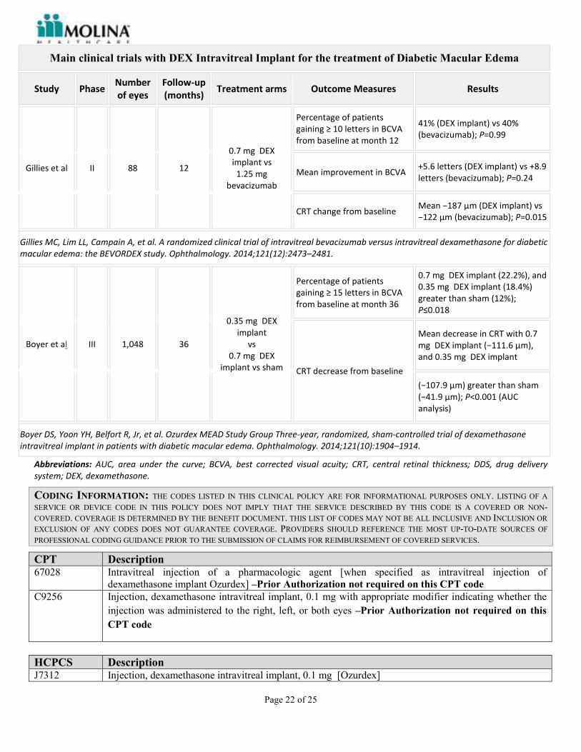
Main clinical trials with DEX Intravitreal Implant for the treatment of Diabetic Macular Edema
Study Phase Number
of eyes
Follow-up
(months) Treatment arms Outcome Measures Results
Haller et al. II 171 6
0.35 mg DEX
implant
vs
0.7 mg DEX
implant vs
observation
Percentage of patients
gaining ≥ letters in BCVA
from baseline at day 90
0.7 mg DEX implant (33.3%), and
0.35 mg DEX implant (21.1%)
greater than observation
(12.3%); P=0.007
CRT decreases from baseline
at day 90 Percentage of
patients with ≥2 levels
decrease in fluorescein
leakage at day 90
−132.3 µm (0.7 mg ) vs −30.2
µm; (observation) P<0.001 36.4%
(0.7 mg ) vs 5.4% (observation);
P<0.001
Haller JA, Kuppermann BD, Blumenkranz MS, et al. Dexamethasone DDS Phase II Study Group Randomized controlled trial of an intravitreous
dexamethasone drug delivery system in patients with diabetic macular edema. Arch Ophthalmol. 2010;128(3):289–296.
Boyer et al. II 55 6
0.7 mg DEX
implant
(vitrectomized
patients)
BCVA increase from baseline
at month 6 Percentage of
patients gaining a ≥ letters in
BCVA from baseline
Mean +3.0 letters; P=0.046 21%
at month 6
CRT change from baseline at
month 6 Percentage of
patients with fluorescein
leakage in the macula
Mean −38.9 µm at month 6;
P=0.004 96.4% at baseline vs
84.0% at month 6
Boyer DS, Faber D, Gupta S, et al. Ozurdex CHAMPLAIN Study Group Dexamethasone intravitreal implant for treatment of diabetic macular edema in
vitrectomized patients. Retina. 2011;31(5):915–923.
Callanan et
al II 253 12
0.7 mg DEX
implant + laser
vs laser
Percentage of patients
gaining ≥ 10 letters in BCVA
from baseline at month 12
27.8% (0.7 mg DEX implant +
laser) vs 23.6% (laser); P=0.453
BCVA change from baseline
Greater improvement in DEX
implant + laser than laser alone,
in patients with diffuse DME over
12 months (AUC analysis);
P<0.001
CRT change from baseline
No significant difference
between arms in mean CRT
change from baseline at month
12
Fluorescein leakage area
from baseline
Mean change in leakage area
decreased greater in DEX implant
+ laser than laser alone, in all
time points; P≤0.041
Callanan DG, Gupta S, Boyer DS, et al. Ozurdex PLACID Study Group Dexamethasone intravitreal implant in combination with laser
photocoagulation for the treatment of diffuse diabetic macular edema. Ophthalmology. 2013;120(9):1843–1851.
Page 22 of 25
Main clinical trials with DEX Intravitreal Implant for the treatment of Diabetic Macular Edema
Study Phase Number
of eyes
Follow-up
(months) Treatment arms Outcome Measures Results
Gillies et al II 88 12
0.7 mg DEX
implant vs
1.25 mg
bevacizumab
Percentage of patients
gaining ≥ 10 letters in BCVA
from baseline at month 12
41% (DEX implant) vs 40%
(bevacizumab); P=0.99
Mean improvement in BCVA +5.6 letters (DEX implant) vs +8.9
letters (bevacizumab); P=0.24
CRT change from baseline Mean −187 µm (DEX implant) vs
−122 µm (bevacizumab); P=0.015
Gillies MC, Lim LL, Campain A, et al. A randomized clinical trial of intravitreal bevacizumab versus intravitreal dexamethasone for diabetic
macular edema: the BEVORDEX study. Ophthalmology. 2014;121(12):2473–2481.
Boyer et al III 1,048 36
0.35 mg DEX
implant
vs
0.7 mg DEX
implant vs sham
Percentage of patients
gaining ≥ 15 letters in BCVA
from baseline at month 36
0.7 mg DEX implant (22.2%), and
0.35 mg DEX implant (18.4%)
greater than sham (12%);
P≤0.018
CRT decrease from baseline
Mean decrease in CRT with 0.7
mg DEX implant (−111.6 µm),
and 0.35 mg DEX implant
(−107.9 µm) greater than sham
(−41.9 µm); P<0.001 (AUC
analysis)
Boyer DS, Yoon YH, Belfort R, Jr, et al. Ozurdex MEAD Study Group Three-year, randomized, sham-controlled trial of dexamethasone
intravitreal implant in patients with diabetic macular edema. Ophthalmology. 2014;121(10):1904–1914.
Abbreviations: AUC, area under the curve; BCVA, best corrected visual acuity; CRT, central retinal thickness; DDS, drug delivery
system; DEX, dexamethasone.
CODING INFORMATION: THE CODES LISTED IN THIS CLINICAL POLICY ARE FOR INFORMATIONAL PURPOSES ONLY. LISTING OF A
SERVICE OR DEVICE CODE IN THIS POLICY DOES NOT IMPLY THAT THE SERVICE DESCRIBED BY THIS CODE IS A COVERED OR NON-
COVERED. COVERAGE IS DETERMINED BY THE BENEFIT DOCUMENT. THIS LIST OF CODES MAY NOT BE ALL INCLUSIVE AND INCLUSION OR
EXCLUSION OF ANY CODES DOES NOT GUARANTEE COVERAGE. PROVIDERS SHOULD REFERENCE THE MOST UP-TO-DATE SOURCES OF
PROFESSIONAL CODING GUIDANCE PRIOR TO THE SUBMISSION OF CLAIMS FOR REIMBURSEMENT OF COVERED SERVICES.
CPT Description 67028 Intravitreal injection of a pharmacologic agent [when specified as intravitreal injection of
dexamethasone implant Ozurdex] –Prior Authorization not required on this CPT code
C9256 Injection, dexamethasone intravitreal implant, 0.1 mg with appropriate modifier indicating whether the
injection was administered to the right, left, or both eyes –Prior Authorization not required on this
CPT code
HCPCS Description J7312 Injection, dexamethasone intravitreal implant, 0.1 mg [Ozurdex]
Page 23 of 25
REFERENCES
PACKAGE INSERT, FDA, DRUG COMPENDIA
Ozurdex (dexamethasone) [Product Information], Irvine, CA. Allergan, Inc.; September 2014. Available at:
http://www.allergan.com/assets/pdf/ozurdex_pi.pdf. Accessed September 2017
Iluvien (fluocinolone) [Product Information], Alpharetta, GA. Alimera Sciences, Inc.; September 2014. Available at:
http://www.alimerasciences.com/wp-content/uploads/2014/09/iluvien-prescribing-information.pdf. Accessed September
2017
Retisert (fluocinolone) [Product Information], Rochester, NY. Bausch and Lomb, Inc.; May 2012. Available at:
http://www.bausch.com/Portals/107/-/m/BL/United%20States/USFiles/Package%20Inserts/Pharma/retisert-prescribing-
information.pdf. Accessed September 2017.
DynaMed Plus [Internet]. Ipswich (MA): EBSCO Information Services. 1995 - . Record No. 326288, Posterior uveitis;
[updated 2017 Jul 31, cited September 2017]; [about 19 screens]. Available from
http://www.dynamed.com/login.aspx?direct=true&site=DynaMed&id=326288. Registration and login required. Accessed
September 2017.
DynaMed Plus [Internet]. Ipswich (MA): EBSCO Information Services. 1995 - . Record No. 116611, Diabetic
retinopathy; [updated 2017 Jul 27, cited September 2017]; [about 29 screens]. Available from
http://www.dynamed.com/login.aspx?direct=true&site=DynaMed&id=116611. Registration and login required.
Drug Facts and Comparisons. Facts and Comparisons eAnswers [online]. Ozurdex (dexamethasone). Clinical Drug
Information LLC, 2017. Available from Wolters Kluwer Health, Inc. [via subscription only] Accessed September 2017.
Clinical Pharmacology [database online]. Ozurdex (dexamethasone). Tampa, FL: Gold Standard, Inc.; 2016. URL:
http://www.clinicalpharmacology.com. [via subscription only] Accessed September 2017.
American Hospital Formulary Service (AHFS). Drug Information 2016. [STAT!Ref Web site]. Available at:
http://online.statref.com. [via subscription only].
Lexi-Drugs Compendium. [Lexicomp Online Web site via UpToDate subscription]. Accessed September 2017.
Micromedex® Healthcare Series [Internet database]. DRUGDEX® Evaluations. Greenwood Village, CO: Thomson
Micromedex. [via subscription only] Accessed September 2017.
Summary of Product Characteristics for OZURDEX 700 micrograms intravitreal implant in applicator. Available from:
https://www.medicines. org.uk/emc/medicine/23422. Accessed September 2017.
CLINICAL TRIALS, DEFINITIONS, PEER-REVIEWED PUBLICATIONS
1. [DEFINITION] Jabs DA, Nussenblatt RB, Rosenbaum JT, et al: Standardization of uveitis nomenclature for
reporting clinical data. Results of the First International Workshop. Am J Ophthalmol 140(3):509-16, 2005.
2. [DEFINITION] United States National Library of Medicine. Medline Plus. Medical Encyclopedia. Uveitis. August
16, 2004. Available at: http://www.nlm.nih.gov/medlineplus/ency/article/001005.htm. Accessed October 2016.
3. Lowder C, Belfort R Jr, Lightman S, et al. Ozurdex HURON Study Group. Dexamethasone Intravitreal Implant for
Noninfectious Intermediate or Posterior Uveitis. Arch Ophthalmol, 2011, 129(5):545-53. [PubMed 21220619]
4. Boyer DS, Yoon YH, Belfort R Jr, et al. Ozurdex MEAD Study Group. Three-year, randomized, sham-controlled
trial of dexamethasone intravitreal implant in patients with diabetic macular edema. Ophthalmology.
2014;121(10):1904-1914.

Page 24 of 25
5. Haller JA, Bandello F, Belfort R Jr, et al. Ozurdex GENEVA Study Group. Randomized, sham-controlled trial of
dexamethasone intravitreal implant in patients with macular edema due to retinal vein occlusion. Ophthalmology.
2010;117(6):1134-1146.
6. Haller, J.A., Bandello, F., Belfort, R. Jr., et al, Ozurdex GENEVA Study Group. Dexamethasone intravitreal
implant in patients with macular edema related to branch or central retinal vein occlusion twelve-month study results.
Ophthalmology 118(12):2453-60, 2011.
7. Sadda S, Danis RP, Pappuru RR, et al. Vascular changes in eyes treated with dexamethasone intravitreal implant for
macular edema after retinal vein occlusion. Ophthalmology. 2013; 120(7):1423-1431.
8. Kiddee W, Trope GE, Sheng L, et al. Intraocular pressure monitoring post intravitreal steroids: a systematic review.
Surv Ophthalmol. Jul-Aug 2013;58(4):291-310. PMID 23768920
9. Kuppermann BD, Blumenkranz MS, Haller JA, et al. Randomized controlled study of an intravitreous
dexamethasone drug delivery system in patients with persistent macular edema. Arch Ophthalmol. Mar
2007;125(3):309-317. PMID 17353400
10. Kuppermann BD, Goldstein M, Maturi RK, et al. Dexamethasone intravitreal implant as adjunctive therapy to
ranibizumab in neovascular age-related macular degeneration: a multicenter randomized controlled trial.
Ophthalmologica. Sep 2015; 234(1):40-54. PMID 26088793
11. Grover D, Li TJ, Chong CC. Intravitreal steroids for macular edema in diabetes. Cochrane Database Syst Rev.
2008(1):CD005656. PMID 18254088 Accessed October 2016.
12. Pielen, N. Feltgen, C. Isserstedt, J. Callizo, B. Junker, and C. Schmucker. Efficacy and safety of intravitreal therapy
in macular edema due to branch and central retinal vein occlusion: a systematic review. PloS ONE, vol. 8, no. 10,
Article ID e78538, 2013.
13. E. Moisseiev, M. Waisbourd, E. Ben-Artsi, et al. Pharmacokinetics of bevacizumab after topical and intravitreal
administration in human eyes. Graefe's Archive for Clinical and Experimental Ophthalmology. vol. 252, no. 2, pp.
331–337, 2014.
14. J. Ahn, H. Kim, S. J. Woo et al. Pharmacokinetics of intravitreally injected bevacizumab in vitrectomized eyes.
Journal of Ocular Pharmacology and Therapeutics, vol. 29, no. 7, pp. 612–618, 2013.
15. Cebeci Z, Kir N. Role of implants in the treatment of diabetic macular edema: focus on the dexamethasone
intravitreal implant. Diabetes, Metabolic Syndrome and Obesity: Targets and Therapy. 2015;8:555-566.
doi:10.2147/DMSO.S73540. Available at: https://www.ncbi.nlm.nih.gov/pmc/articles/PMC4655951/ Accessed
September 2017.
16. Fraser, Claire E. Diabetic retinopathy: Prevention and treatment. In: UpToDate. Nathan, DM (Ed), UpToDate,
Waltham, MA, 2016. Aug 2017. [via subscription only] Accessed September 2017.
17. Apte RS. What is chronic or persistent diabetic macular edema and how should it be treated? JAMA ophthalmol.
2016;134(3):285-6.
18. Ip MS, Scott IU, VanVeldhuisen PC, et al; SCORE Study Research Group. A randomized trial comparing the
efficacy and safety of intravitreal triamcinolone with observation to treat vision loss associated with macular edema
secondary to central retinal vein occlusion: The Standard Care vs Corticosteroid for Retinal Vein Occlusion
(SCORE) Study Report 5. Arch Ophthalmol. 2009;127(9):1101-1114.
19. Scott IU, Ip MS, VanVeldhuisen PC, et al; SCORE Study Research Group. A randomized trial comparing the
efficacy and safety of intravitreal triamcinolone with standard care to treat vision loss associated with macular edema
secondary to branch retinal vein occlusion: The Standard Care vs Corticosteroid for Retinal Vein Occlusion
(SCORE) Study Report 6. Arch Ophthalmol. 2009;127(9):1115-1128.
20. Abcouwer SF. Angiogenic factors and cytokines in diabetic retinopathy. J Clin Cell Immunol 2013; Suppl
1(11).doi:10.4172/2155-9899.
21. Covert, Douglas J. Retinal vein occlusion: Treatment. In: UpToDate, Trobe, J (Ed), UpToDate, Waltham, MA,
August 2017. Accessed September 2017.
22. Diabetic Retinopathy Clinical Research Network.
• Elman MJ, Aiello LP, Beck RW, et al; Diabetic Retinopathy Clinical Research Network. Randomized trial
evaluating ranibizumab plus prompt or deferred laser or triamcinolone plus prompt laser for diabetic
macular edema. Ophthalmology. 2010;117(6):1064-1077.

Page 25 of 25
Elman MJ, Bressler NM, Qin H, et al; Diabetic Retinopathy Clinical Research Network. Expanded 2-year
follow-up of ranibizumab plus prompt or deferred laser or triamcinolone plus prompt laser for diabetic macular
edema. Ophthalmology. 2011;118(4):609-614.
23. Covert, Douglas J. Retinal vein occlusion: Epidemiology, clinical manifestations, and diagnosis. In: UpToDate,
Trobe, J (Ed), UpToDate, Waltham, MA, August 2017. Accessed September 2017.
24. Pasadhika S, Rosenbaum JT. Update on the use of systemic biologic agents in the treatment of noninfectious uveitis.
Biologics. 2014;8:67–81. Accessed October 2017.
Government Agencies, Professional Societies, and Other Authoritative Publications
A. National Institute for Health and Clinical Excellence (NICE). Dexamethasone intravitreal implant for the
treatment of macular edema secondary to retinal vein occlusion. NICE Technology appraisal guidance [TA229]
Published date: 27 July 2011. Available at: https://www.nice.org.uk/Guidance/ta229. Accessed September 2017.
B. National Institute for Health and Care Excellence (NICE). Dexamethasone intravitreal implant for treating
diabetic macular edema. 22 July 2015; Available at: www.nice.org.uk/guidance/ta349.
https://www.nice.org.uk/guidance/ta349/chapter/1-Guidance Accessed September 2017.
C. National Institute for Health and Clinical Excellence (NICE). Fluocinolone acetonide intravitreal implant for
treating chronic diabetic macular edema after an inadequate response to prior therapy TA 301. 2013. Available at:
http://guidance.nice.org.uk/TA301. Accessed September 2017.
D. Puliafito CA, Cousins SW, Bacharach J, et al. Forming a consensus: data and guidance for physicians treating diabetic
macular edema. Ophthalmic Surg Lasers Imaging Retina. 2016;47(4):S4–S15.
E. Yeh S, Kim SJ, Ho AC, Schoenberger SD, Bakri SJ, Ehlers JP, et al. Therapies for macular edema associated with
central retinal vein occlusion: a report by the American Academy of Ophthalmology. Ophthalmology. 2015 Apr.
122 (4):769-78. Available at: https://www.ncbi.nlm.nih.gov/pubmed/25576994 Accessed October 2016.
F. American Diabetes Association. Standards of medical care in diabetes-- 2015. Diabetes Care 2015 Jan;38 Suppl
1:S1-93 Available at:
http://care.diabetesjournals.org/content/suppl/2014/12/23/38.Supplement_1.DC1/January_Supplement_Combined_Fi
nal.6-99.pdf Accessed September 2017.
G. Jabs DA, Nussenblatt RB, Rosenbaum JT, Standardization of Uveitis Nomenclature (SUN) Working Group.
Standardization of uveitis nomenclature for reporting clinical data. Results of the First International Workshop. Am J
Ophthalmol 2005; 140: 509.
H. Hayes. Health Technology Brief. Dexamethasone Intravitreal Implant (Ozurdex; Allergan Inc.) for Treatment of
Diabetic Macular Edema. December 2016. Accessed September 2017.
I. Ehlers J.P., Kim S.J., et al. Therapies for Macular Edema Associated with Branch Retinal Vein Occlusion: A Report
by the American Academy of Ophthalmology. (2017) Ophthalmology, 124 (9) , pp. 1412-1423. Available at:
http://www.aaojournal.org/article/S0161-6420(17)30980-6/fulltext Accessed September 2017.


![Bimodal Gastroretentive Drug Delivery Systems of ......a gastroretentive floating drug delivery system[12]. The drug concentrations can be controlled by formulating bimodal drug delivery](https://static.fdocuments.in/doc/165x107/5e6f0293269d113bd9170da6/bimodal-gastroretentive-drug-delivery-systems-of-a-gastroretentive-floating.jpg)