OSTEOARTHRITIS and CARTILAGE - COnnecting REpositories · Osteoarthritis staging: ... gross...
12
Osteoarthritis and Cartilage (1995) 3, 169-180 O 1995 Osteoarthritis Research Society 1063-4584/95/030169 + 12 $08.00/0 OSTEOARTHRITIS and CARTILAGE Osteoarthritis staging: comparison between magnetic resonance imaging, gross pathology and histopathology in the rhesus macaque BY H. K. GAHUNIA*, P. BABYNT, C. LEMAIRE:~, M. J. KESSLER§ AND K. P. H. PRITZKER* *Connective Tissue Research Group, Department of Pathology and Laboratory Medicine, and Samuel Lunenfeld Research Institute, Mount Sinai Hospital and University of Toronto, Toronto, Ontario, Canada; TDepartment of Radiology, Hospital for Sick Children, University of Toronto, Toronto, Ontario, Canada; SMagnetic Resonance Imaging Research Laboratory, Department of Medical Imaging and University of Toronto, Toronto, Ontario, Canada; §Caribbean Primate Research Centre, University of Puerto Rico, Sebana Seca, Puerto Rico, U.S.A. Summary Although osteoarthritis (OA) is the most common cause of articular skeletal disability in humans, assessing progression (staging) with noninvasive methods remains a major clinical problem. Using the rhesus macaque animal model, the objective of this study was to compare OA staging by noninvasive magnetic resonance imaging (MRI) against gross pathology and histopathology. Right knee joints from 18 rhesus macaques were used in this study. Using a four-point ordinal scale for each of the above-mentioned modalities, the lateral and medial femoral condyle and tibial plateau of each knee joint was independently scored for OA severity, i.e. normal, mild OA, moderate OA and severe OA. Correlation between each staging system was performed using Stuart's Tauc correlation coefficient. By our criteria, MRI staging correlated as well with gross pathology (v = 0.75) and histopathology (~ = 0.80) as did gross pathology with histopathology (~ = 0.78). Our study shows that MRI is a promising noninvasive modality to evaluate the severity of OA. MRI appears to be sensitive for demarcating the presence and extent of focal OA cartilage lesions. However, at this time, while MRI is sensitive for detecting OA change it cannot distinguish between certain lesions such as superficial cartilage matrix fibrillation and hypertrophy both of which show elevated signal intensity. Key words: Articular cartilage, Osteoarthritis, Disease staging, Magnetic resonance imaging, Gross pathology, Histopathology. Introduction OSTEOARTHRITIS (OA) is a group of degenerative joint diseases leading to joint instability or joint failure. OA is characterized by progressive architectural and compositional deterioration and remodeling of articular cartilage and subchondral bone along with proliferative and fibroplastic changes in synovium. The definition of osteoar- thritis is complicated by the range of manifes- tations of the disease [1]. Altman et al. [2] defined osteoarthritis as 'a heterogenous group of con- ditions that lead to joint symptoms and signs which are associated with defective integrity of articular cartilage, in addition to related changes in the underlying trabeculae and cortical bone and at the joint margins, in particular the synovium'. Submitted 2 May 1994; accepted 21 November 1994. Address correspondence to: Dr Kenneth P. H. Pritzker, Department of Pathology and Laboratory Medicine, Suite 600, Mount Sinai Hospital, 600 University Avenue, Toronto, Ontario M5G lX5, Canada. Dieppe [3] recently proposed that OA should be viewed as a dynamic disease process, which goes through temporary phases of activity which may be dominated by either cartilage destruction or repair. Currently, one of the difficulties faced by OA clinical and research groups is the lack of a standard methodology and classification system for assessing changes associated with OA and to evaluate the efficacy of drug treatment in OA patients. Classifications of OA severity are often unclearly defined and depend upon the modality used. The terms used to describe disease, 'staging' and 'grading', have often been used interchange- ably. However, the term 'grade' more appropriately reflects the index of disease activity, whereas 'stage' is the term used for disease progression [4]. Grading of OA activity of articular cartilage includes transient features such as cartilage edema, alteration in proteoglycan and collagen degradation products as well as altered synthetic activity of chondrocytes. Staging of OA 169
-
Upload
duongquynh -
Category
Documents
-
view
224 -
download
0
Transcript of OSTEOARTHRITIS and CARTILAGE - COnnecting REpositories · Osteoarthritis staging: ... gross...
Osteoarthritis and Cartilage (1995) 3, 169-180 O 1995 Osteoarthritis Research Society 1063-4584/95/030169 + 12 $08.00/0
OSTEOARTHRITIS and
CARTILAGE Osteoarthri t i s staging: compar i son between m a g n e t i c re sonance imaging , gross patho logy and h i s topatho logy in the rhesus m a c a q u e
BY H. K. GAHUNIA*, P. BABYNT, C. LEMAIRE:~, M. J. KESSLER§ AND K. P. H. PRITZKER* *Connective Tissue Research Group, Department of Pathology and Laboratory Medicine, and Samuel
Lunenfeld Research Institute, Mount Sinai Hospital and University of Toronto, Toronto, Ontario, Canada; TDepartment of Radiology, Hospital for Sick Children, University of Toronto, Toronto, Ontario,
Canada; SMagnetic Resonance Imaging Research Laboratory, Department of Medical Imaging and University of Toronto, Toronto, Ontario, Canada; §Caribbean Primate Research Centre, University of
Puerto Rico, Sebana Seca, Puerto Rico, U.S.A.
Summary
Although osteoarthritis (OA) is the most common cause of articular skeletal disability in humans, assessing progression (staging) with noninvasive methods remains a major clinical problem. Using the rhesus macaque animal model, the objective of this study was to compare OA staging by noninvasive magnetic resonance imaging (MRI) against gross pathology and histopathology. Right knee joints from 18 rhesus macaques were used in this study. Using a four-point ordinal scale for each of the above-mentioned modalities, the lateral and medial femoral condyle and tibial plateau of each knee joint was independently scored for OA severity, i.e. normal, mild OA, moderate OA and severe OA. Correlation between each staging system was performed using Stuart 's T a u c correlation coefficient. By our criteria, MRI staging correlated as well with gross pathology (v = 0.75) and histopathology (~ = 0.80) as did gross pathology with histopathology (~ = 0.78). Our study shows that MRI is a promising noninvasive modality to evaluate the severity of OA. MRI appears to be sensitive for demarcating the presence and extent of focal OA cartilage lesions. However, at this time, while MRI is sensitive for detecting OA change it cannot distinguish between certain lesions such as superficial cartilage matrix fibrillation and hypertrophy both of which show elevated signal intensity. Key words: Articular cartilage, Osteoarthritis, Disease staging, Magnetic resonance imaging, Gross pathology, Histopathology.
Introduction
OSTEOARTHRITIS (OA) is a group of degene ra t i ve jo in t diseases l ead ing to jo in t ins tab i l i ty or jo in t fai lure. OA is cha rac t e r i zed by progress ive a r c h i t e c t u r a l and compos i t iona l d e t e r i o r a t i o n and r emode l ing of a r t i c u l a r ca r t i l age and subchond ra l bone a long wi th p ro l i f e ra t ive and f ibroplas t ic changes in synovium. T h e def in i t ion of os teoar- th r i t i s is compl ica ted by the r ange of manifes- t a t ions of the disease [1]. A l tman et al. [2] defined o s t eoa r th r i t i s as 'a h e t e r o g e n o u s group of con- d i t ions t h a t lead to j o in t symptoms and signs which are assoc ia ted wi th defect ive i n t eg r i t y of a r t i c u l a r car t i lage , in add i t ion to r e l a t ed changes in the u n d e r l y i n g t r a b e c u l a e and cor t i ca l bone and a t the jo in t margins , in p a r t i c u l a r the synov ium' .
Submitted 2 May 1994; accepted 21 November 1994. Address correspondence to: Dr Kenneth P. H. Pritzker,
Department of Pathology and Laboratory Medicine, Suite 600, Mount Sinai Hospital, 600 University Avenue, Toronto, Ontario M5G lX5, Canada.
Dieppe [3] r e c e n t l y proposed t ha t OA shou ld be viewed as a dynamic disease process, wh ich goes t h r o u g h t e m p o r a r y phases of ac t iv i ty wh ich may be domina t ed by e i t he r ca r t i l age d e s t r u c t i o n or repair .
Cur ren t ly , one of the difficulties faced by OA cl inical and r e s e a r c h groups is the l ack of a s t anda rd m e t h o d o l o g y and c lass i f ica t ion sys tem for assessing changes assoc ia ted wi th OA and to eva lua t e the efficacy of d rug t r e a t m e n t in OA pat ients . Class i f ica t ions of OA sever i ty a re o f ten u n c l e a r l y defined and depend upon the m o d a l i t y used. The te rms used to descr ibe disease, ' s tag ing ' and 'grading ' , h a v e of ten been used i n t e r change - ably. However , the t e rm 'g rade ' more a p p r o p r i a t e l y ref lects the index of disease ac t iv i ty , whe rea s ' s tage ' is the t e rm used for disease p rog re s s ion [4]. Grad ing of OA a c t i v i t y of a r t i cu l a r c a r t i l age inc ludes t r a n s i e n t f ea tu res such as c a r t i l age edema, a l t e r a t i o n in p r o t e o g l y c a n and co l l agen d e g r a d a t i o n p roduc t s as well as a l t e red syn the t i c ac t iv i ty of chondrocy tes . S t ag ing of OA
169

170 Gahunia e t a l . : Staging of osteoarthritic articular cartilage
progression in art icular cartilage is primarily based on established morphologic features such as matrix fibrillation, delamination, changes in carti- lage thickness, changes in collagen architecture, changes in chondrocyte population such as hyper- cellularity, hypocellularity, cloning and necrosis as well as t idemark reduplication.
Magnetic resonance imaging (MRI) has shown unique applications in the diagnosis of cartilagi- nous lesions in OA [5-7] and can demonstrate cartilage lesions prior to actual loss in carti lage thickness [8]. Based on criteria such as cartilage loss, subchondral sclerosis, osteophyte formation and subchondral cysts in classifying OA severity, Chan et al. [9] compared MRI findings with plain radiographs and computed tomography. Sabiston et al. [10] used gross pathology and MR images to classify OA severity based on changes in the menisci, capsule and osteophyte formation. Other MRI studies have concentrated on the variat ion in cartilage thickness [11-13] and depth of the focal cartilage lesions [14 16] present in OA as well as variat ion in MR signal intensi ty of articu- lar cartilage with the OA progression [17-19].
Histopathologic assessment of OA has been pro- posed by several authors [20-22]. Byers et a l . ' s [20] histopathologic assessment of hip joint OA carti- lage was based on cartilage margin ossification and breakdown and loss of cartilage. Bennett et al. [21] proposed a grading system to assess OA which emphasized such cartilage lesions as matrix ero- sion and decrease in cartilage thickness. Collins' [22] grading system of ar t icular carti lage and bone changes in OA was based on criteria of disease progression and by our criteria would be referred as OA staging.
Mankin and colleagues [23] proposed tha t the histopathologic sequence in the evolution of OA starts with tangent ial fissures of ar t icular cartilage, a process which is now known to occur later in the course of the disease. Prior to the cartilage surface disruption, it is now well known that the earlier stages of OA are associated with increased hydrat ion (edema) of carti lage with more avid water binding to OA affected tissue than to normal tissue [24]. Cartilage edema is followed by alterations in proteoglycan concentrat ion and composition [25, 26].'Byers et al. [27] and Meachim and Emery [28] described the histological patterns of fibrillation in OA. Lust and Summers' [29] histologic criteria of OA assessment was based on cartilage lesions and safranin O' s taining intensity. Although the above-mentioned authors provided a severity scoring system to assess OA, they described their criteria as a grading system rather than a staging system. Further, more
explicit criteria of disease progression was lacking in these studies. Moskowitz and Goldberg [30] proposed a classification system of OA based on changes in synovium, vascular i ty and cell prolifer- ation. Other aspects of cart i lage destruction such as surface integri ty and reduplication of t idemark were omitted in their classification system. Based on morphological variat ions in ar t icular cartilage, Mohr and Lehmann [31] provided a good evalu- at ion system for the severity of carti lage changes in OA. However, they excluded important features such as the various aspects of chondrocyte cellularity, reduplication of t idemark and vascular invasion. The histopathologic criteria provided by Colombo et al. [32] is subjectiv& Their scoring system is based on the lesion p e r se ra ther than a set of lesions per stage as proposed in our staging system.
It has not been possible to define the sequence of events leading to the changes in OA cartilage and bone due to the difficulties in identifying clinically early stages of OA or in obtaining joint tissue from patients in an early stage of the disease [33]. Therefore, experimental models of OA have been utilized to elucidate the pathogenesis of OA. We chose the Macaca mulat ta (Caribbean Primate Research Centre) animal model because a high incidence of spontaneous degenerative arthri t is is known to develop in this population of rhesus macaques. The natural life span of Macaca mulat ta is between 18 and 30 years. A 30-year-old monkey corresponds approximately to an 85-year-old human [34]. Advantages of this model include availabili ty of sufficient diseased animals, suffi- cient tissues for biochemical and histopathological studies, availability of a control population, known matril ineages and a uniform free ranging environment [35]. The merit in using this popu- lation of rhesus macaques as a model of OA is well documented [35, 36].
The purpose of this study was to compare OA progression staged by MRI with the disease assessed by gross pathology and histopathology. We also wanted to document the quali tat ive MRI changes in the art icular cartilage in the various stages of OA and to compare MRI and histopatho- logical observations in specific carti lage lesions.
Mater ia l s and M e t h o d s
E X P E R I M E N T A L A N I M A L M O D E L
Fully encapsulated r ight knee joints of 10 female and eight male rhesus monkeys (age 7-25 years) were obtained post mortem from the Caribbean Primate Research Centre (CPRC) in Puerto Rico.

O s t e o a r t h r i t i s and Cart i lage Vol . 3 No . 2 171
The knee joints were kept sealed in plastic bags, shipped frozen ( -10°C) to Toronto and stored frozen at -20°C unti l the day of experiment.
E X P E R I M E N T A L P R O C E D U R E
OA sever i ty was independent ly assessed for the medial and lateral femoral condyles and tibial plateaus using MRI, gross pathology and his- tology. To clarify the anatomic location, each knee joint was divided into four quadrants , i.e. medial femoral condyle, medial t ibial plateau, la teral femoral condyle and la teral t ibial plateau.
The first step involved in the exper iment was thawing of the knee joints by keeping them in a re f r igera tor (4°C) for 5-6 h. The completely thawed joints were then processed in the chronological sequence given below.
Magnetic resonance imaging
MRI scans of the in tac t knee joints were per- formed at 85 MHz with a 55 mm birdcage coil [37] using a Bruker 2.0 Tesla, CSI Omega imaging uni t with Acustar self shielded gradients, hor izontal magnet bore of 18 cm in diameter and a maximum gradient s t rength of 100 mT/m. The experiments were performed at a magnet bore tempera ture of 12+_2°C, which was control led by the magnet cooling system. A multislice spin echo sequence (TR = 1000 ms; TE = 20 ms; field of view = 50 mm) was used to obtain sagit tal MR images after posi t ioning from coronal and axial images. Four MR slices (1 mm thick and 2 mm apar t from the adjacent slice) were taken on each of the lateral and the medial compar tment of the joint. A multi- echo sa tura t ion / recovery sequence was employed for the MR imaging. Each pulse cycle consists of a 90 ° hard pulse fo l lowed by a 90 ° slice selective pulse and this in tu rn was followed by a CPMG tra in [38] of 180 ° hard pulses. In the chosen sequence, the pre-saturat ion time (TP) corresponds to the time between the 90 ° hard pulse and the 90 ° slice selective pulse. The repet i t ion time (TR) (not to be confused with T P ) is the time between two consecut ive 90 ° hard pulses. The next cycle begins with a very short in terval of 1 ms after the last image acquisition. Each image was acquired with 128 phase-encoding gradients and four ac- cumulations. The data matr ix was 256 × 128 and the pixel size was 0.20 m m x 0.39 mm. The spatial resolut ion was 0.078 mm 3. To maximize the spatial resolut ion across the carti lage, the readout gradi- ent was oriented perpendicular to the surface of the a r t icu la r cartilage. The qual i ta t ive evaluat ion was performed from at least five of the 19 images
(TP=200 , 500, 1500, 3000ms and TE = 10ms; TP = 3000 ms and TE = 50 ms). The features used for MRI diagnosis of OA included a r t i cu la r carti- lage and bone changes as shown in Table I. Each quadran t of the knee jo int was scored individual ly for OA severi ty depending upon the overall appearance of the quadran t as given in Table II.
Registration of M R I images to histopathology
The mid-parasagit tal MR image from each compar tment was chosen for comparison with the corresponding mid-parasagit tal gross ana tomy and histological sections. The l inear dis tance of the chosen mid-parasagit tal plane from the medial to the la teral margin of the compar tment was taken into account in deciding the posit ion of the 3-mm thick mid-parasagit tal histological block so tha t the 1-mm thick MR slice is approximate ly in the middle of the histological block where the histologic slices were chosen for comparison.
Gross pathology
Immediately after MRI scanning, the jo ints were dissected, studied grossly and photographed. Care was taken to minimize the time these joints were exposed to room tempera ture to prevent excessive cart i lage evaporat ion. Each quadran t of the knee
Table I Magnetic resonance imaging: OA diagnostic features
1. Joint space (a) Normal (b) Widening with effusion (c) Narrowing
2. Articular cartilage thickness (a) Normal (b) Focal cartilage thickening (c) Focal cartilage thinning or loss (d) Diffuse cartilage thinning
3. Articular cartilage signal intensity (a) Normal (b) Increased signal intensity (c) Decreased signal intensity
4. Subchondral and cortical bone/bone marrow (a) Normal (b) Focal sclerosis with decreased signal intensity in
T1- and T2-weighted images (c) Thickening of subchondral bone (d) Eburnation in subchondral bone indicated by
decreased signal intensity in T1- and T2-weighted images with absence of overlying articular cartilage
(e) Osteophyte formation noted along the anterior and/or posterior margin
(f) Cyst formation with increased signal intensity on T2-weighted images
(g) Deformity of bone shape

172 Gahunia et al.: Staging of osteoarthritic articular cartilage
Table II OA staging: magnetic resonance imaging criteria
Stage Severity Criteria
0 Normal
1 Mild
2 Moderate
3 Severe
Normal joint space with smooth cartilage contour; no abnormality in signal intensity in cartilage or bone.
Slight narrowing of the joint space in the load-bearing region; slight irregularity in cartilage contour; slight focal variation in signal intensity in articular cartilage; slight thickening of the subchondral bone.
Distinct focal cartilaginous lesions reflected by changes in cartilage thickness and signal intensity; diffuse cartilage thinning; subchondral thickening and/or sclerosis; slight deformity of bone ends; distinct osteophytes.
Marked irregularity in cartilage contour along with distinct regions of focal lesions or focal thickening of the articular cartilage; distinct regions of marked focal variation in the articular cartilage Signal intensity; severe subchondral sclerosis; marked abnormality in bone shape; cysts may be present; large osteophyte(s).
joint was assessed individually and scored grossly for the severity of OA based upon the cr i ter ia for OA assessment of Byers et al. and Bennet t et al. [20, 21] (Table III).
His t o logy
After gross pathological examination, the patella and menisci were discarded and the tibial plateau detached from the femoral condyle. Mid- parasagit tal cart i lage tissue of each quadrant of the knee joint was demarcated (3 mm thick) with a knife and cut with a Buehler Isomet low speed saw to closely correspond to the MR mid-parasagittal slices. These tissue blocks were cut perpendicular to the ar t icular surface and extended to the depth of the bone marrow (approximately 2 cm). After decalcification of the tissue blocks and histochem- ical processing, 5/~m thick sections were stained with Haematoxyl in & Eosin (to assess cart i lage cellularity) and Toluidine blue dyes (to assess the proteoglycan content). Using a modification of Mankin 's classification [23], histological sections from the four quadrants of the knee joint were
assessed on a four-point ordinal scale by two observers and a severi ty score was al located to each quadrant (Table IV). This his topathological scoring system was used as a s tandard in this experiment.
STATISTICAL ANALYSIS
To eliminate biased assessment, the macroscopic and microscopic diagnosis and al locat ion of sever- i ty scores were made using MRI, gross pathology and histology on different occasions. Of the 72 quadrants examined, two lateral tibial plateaus, a lateral femoral condyle and a media l femoral condyle were excluded from the OA sever- i ty analysis due to problems associated with noisy MR images or chemical shift artifact. Table V shows the summary of number of quad- rants used per OA severi ty stage. Stuar t ' s Tau-c (~) was used to determine the extent to which the various staging systems of OA (MRI, gross pathol- ogy and histopathology) are correlated [39]. Stuar t ' s Tau-c can take any v a l u e between + 1 (perfectly positively correlated) and - 1
Table III OA staging: gross pathologic criteria (from Byers et al. [20]; Bennett et al. [21])
Stage Severity Gross observations
0 Normal
1 Mild
2 Moderate
3 Severe
Normal/no visible changes.
Slight unevenness; granularity of the surface; discoloration of cartilage; shallow furrows or streaking; superficial fraying, splitting and pitting of cartilage with or without small erosions.
Focal loss of cartilage; marked degrees of the above changes associated with extensive ulceration and/or haemorrhagic areas.
Large areas of complete cartilage degeneration; exposure and eburnation of the subchondral bone; definite abnormality in bone shape.

Osteoarthri t i s and Carti lage Vol. 3 No. 2
Table IV OA staging: histopathologic criteria
Stage Severity Mankin scale Criteria
173
0 Normal 0-4.9
1 Mild 5.0-6.9
2 Moderate 7.0-8.9
3 Severe 9.0--14.0
Smooth intact surface; uniform distribution of chondrocytes with one or two cells/lacuna; uniform blue stained extracellular matrix.
Mild fibrillation of surface; clefts not extending beyond the superficial zone; slight hypercellularity; slight reduction of Toluidine blue staining intensity especially superficial zone.
Regions of cartilage thickening or thinning; deep fibrillation; clefts extend into the middle and occasionally into the calcified zones; hypercellularity usually in the form of chondrocyte clusters (clones); moderate reduction of Toluidine blue staining intensity extending up to middle zone; reduplication of tidemark with or without vascular invasion; presence of osteophytes with cartilage caps usually at the margins of the femoral condyle/tibial plateau.
Regions with full thickness of cartilage eroded; clefts extend down to the calcified cartilage; variable regions of cartilage hypo- and hypercellularity; markedly reduced Toluidine blue staining intensity of proteoglycans throughout the entire tissue; marked reduplication of tidemark and definite vascular invasion; large osteophytes with marrow spaces.
(perfectly negatively correlated). A value of 0 would mean that there is no linear relationship between the two staging systems being correlated. A stat ist ically significant value was chosen at P ~< 0.O5.
R e s u l t s
Histological assessment of the stage of OA was most comparable to the MRI findings (T = 0.80, P = 0.0001) and followed by gross pathological findings (3 ~- 0.78, P = 0.0001). MRI staging of OA correlated with gross pathology with Stuart 's
= 0.75 ( P -- 0.0001). Table V outlines the number of OA quadrants with each stage as assessed by each modality. Table VI shows direct comparison of MRI vs gross pathology severity score and Table VII shows the comparison of MRI vs histology. Examples of M R I , gross pathology and histopathology grading are i l lustrated in Figs 1, 2 and 3, respectively. In the majority of the quadrants, cartilage i r regular i t ies , t h inn ing or erosions detected by MRI were also apparent by gross examinat ion. . In Fig. l(c), the medial com- partment of moderately OA cartilage revealed the
Table V Number of quadrants analysed for each OA severity
stage With different modalities
OA severity MRI Gross pathology Histology
Normal 16 24 20 Mild OA 26 22 22 Moderate OA 14 13 12 Severe OA 12 9 14 Total 68 68 68
irregular i ty o f t h e ar t icular cartilage contour which was more pronounced in the weight-bearing region of the tibial plateau. Altered bone shape in most moderate and all severely OA quadrants were obvious in the MR images.
In mild OA carti lage the most common MRI findings were focal regions of reduced signal inten- sity compared with the intermediate signal inten- sity of normal cart i lag e [Fig. l(b)]. With increasing severity intracar t i laginous focal lesions with high and low signal intensities were apparent and well contrasted when both were present within the same quadrant. An increase in the thickness of the region of low signal intensi ty of the subchondral bone (when compared with normal) was noted in some moderate OA and severe OA quadrants (Fig. 4). This change was more pronounced in the tibial plateau compared with the femoral condyle. This observation was compatible with histologic sclerotic regions.
Regions of focal lesions were demarcated on MR images and compared with the corresponding histologic regions. Regions of interest with decreased signal intensi ty in some moderate OA quadrants corresponded to histologic regions of collagen condensation. Increased cartilage thick- ness and brightness in MRI was noted to corre- spond to histologic regions of hypertrophy and hypercellularity. Histologically fibrillated regions of carti lage also showed MRI high signal intensi ty (Fig. 4).
Interestingly, some MR images of the normal carti lage indicated the existence of three MR zones parallel to the ar t icular surface characterized by high, low and high signal intensi ty extending from the ar t icular surface to the subchondral bone. This

174 Gahunia e t a l . : Staging of osteoarthritic articular cartilage
Table VI Quadrants assessed by magnetic resonance imaging and gross pathology
Gross pathology Severity score (MRI) Normal Mild OA Moderate OA Severe OA
Normal 13 3 0 0 Mild OA 11 14 1 0 Moderate OA 0 5 9 0 Severe OA 0 0 3 9
Stuart's Tau-c = 0,745.
pat tern could also be seen in the regions of normal ar t icular car t i lage of the moderate and severely OA quadrants but were disrupted towards the periphery of the focal lesions where most of the ar t icular cart i lage thickness had eroded (Fig. 4).
D i s c u s s i o n
To assess the act ivi ty and progression of OA, accurate evaluat ion of the severity of the disease is essential. Using various modalities, the classifi- cation cr i ter ia of OA proposed by most authors [10, 23] has been based mainly on the advanced stages of OA. However to assess OA progression accurately, it is important to develop precise, non- invasive methods for staging early OA. MRI assessment and staging of OA is still in its juvenile stage and requires fur ther invest igat ion to provide a sat isfactory classification of OA severity, in par t icular the early stage of OA. The purpose of the quali tat ive MRI scoring system in our study was to provide a non-invasive staging system for the severity of OA which would encompass the lesions of car t i lage and bone at each OA stage. In our study, macroscopical ly and microscopically evident cart i lage degenerat ion compared well with findings on MR images in the major i ty of the quadrants. The quadrants in which the MRI was normal and the his topathology showed mild OA suggest tha t ei ther the morphologic changes observed were beyond the MRI spatial resolut ion or may be due to a slight shift in the anatomic
correspondence of MRI slices and histological sections. Funct ional ly , the possibility should be considered tha t the lack of MRI r signal in tensi ty change could indicate tha t the very mild gross or histologic lesions observed in fact may not represent OA. The quadrants in which the MRI showed signal in tensi ty changes in the absence of gross pathology changes suggest tha t earl iest changes in OA, namely changes in water quality, may be detected more readily by MRI than by pathologic techniques.
Bongartz et al. [19] documented MRI detectable signs of OA in hip. However their assessment cr i ter ia was limited to few types of lesions (i.e. cart i lage th inning or loss, var ia t ion in signal intensi ty in ar t icular cartilage, cort ical th ickening and joint effusion) and the lesions were not grouped according to the severi ty level. Chan et al. [9] evaluated OA severi ty based on four diagnostic cri ter ia individually. Cart i lage abnormal i ty was reflected only in terms of decreased thickness. Increased cart i lage thickness in OA, as a conse- quence of repair process, was omitted. Recently, Broderick et al. [40] used a five-category scale to assess the severity of ar t icular car t i lage lesions in OA. However, they classified OA severi ty by a single cr i ter ion namely, cart i lage th inning or loss. OA may not be a single disease entity, therefore classification of OA severi ty based on single cr i ter ia r a the r than a group of cr i ter ia is inappropriate.
Our MRI assessment of the stage of OA was comparable with th~ gross pathological findings.
Table VII Quadrants assessed by magnetic resonance imaging and histology
Severity score (MRI)
Histology
Normal Mild OA Moderate OA Severe OA
Normal 11 5 0 0 Mild OA 9 17 0 0 Moderate OA 0 0 12 2 Severe OA 0 0 0 12
Stuart's Tau c = 0.795.

Osteoarthrit is and Cartilage Vol. 3 No. 2 175
This is c o n g r u e n t wi th K a r v o n e n et al.'s [41] s tudy on h u m a n OA knee and Ho et al.'s [42] s tudy in goa t model for OA, whoich showed ca r t i l age erosions , t h i n n i n g or i r r egu l a r i t i e s t h a t were de t ec t ed by MRI had the same defects obse rved by gross exam- ina t ions . On the c o n t r a r y , Hod le r et al.'s [13] s tudy on a r t i c u l a r ca r t i l age defec ts showed poor corre- l a t ion be tween MR a p p e a r a n c e and gross a n a t o m i c findings. Hi s to log ica l c o r r e l a t i o n of MRI f indings
wi th respec t to the type of les ion (e.g. f ib r i l l a t ion or fibrosis) was l ack ing in the above s tudy and th i s may have c o n t r i b u t e d to the r epor t ed incons is t - ency in c o r r e l a t i o n be tween the type of s ignal i n t ens i t y and dep th of the focal defect. F u r t h e r , h ighe r spa t ia l MR re so lu t i on (smal ler field of v iew and t h i n n e r MR slices) m a y have improved the c o r r e l a t i o n be tween MR findings and gross ana tomic findings.
F~G. 1. Mid-parasagittal magnetic resonance images (TP = 3000 ms; TE = 10 ms)o f right knee joint of rhesus monkey showing the characteristic MR changes with the evolution of OA in the quadrants. The posterior aspect of the knee joint is on the same side as the external doped water reference (WR). (a) Normal. (b) Mild OA; note loss of signal intensity in the load-bearing region of tibial plateau. (c) Moderate OA; note variability in the cartilage thickness in the various regions. (d) Severe OA; note the well contrasted high and low signal intensity in the articular cartilage, subchondral bone and bone marrow.

176 G a h u n i a et al.: Stag ing o f o s t e o a r t h r i t i c ar t icu lar cart i lage
Histological cr i ter ia for classification of OA is considered as the 'Gold Standard' , but the infrequent availabil i ty of affected joints is a l imitation to this approach. In experimental animal models and humans, the Mankin score is frequently used as a his topathological grading tool [23]. However this system lacks a score for the progressive stages of OA classifying the joint as normal for a score of less than 6 and as osteoarthr i t ic for a score of 6-14. In addition, recognized features of OA remodeling such as osteophyte formation in bone and t idemark reduplicat ion are not included in this classifi- cation. As well, van der Sluijs et al . ' s modification [43] of the Mankin score does not account for the various OA stages, i.e. mild OA, moderate OA ana severe OA. With our modification of the Mankin score, we were able to al locate a severity score for staging from score 0 (normal) to score 3 (severe OA) to the various quadrants of the knee joint. Reduplication of t idemark was included in our classification system. However, unlike Engel 's [44] result in mild OA, in our experiment, reduplicat ion
of t idemark in histological sections were observed in the late stage of the disease, i.e. moderate OA and severe OA.
MRI demonstrated signal intensi ty var ia t ions even in the absence of cart i lage thickness and contour abnormalities. In some mild OA quad- rants, a focal decrease in MR signal intensi ty (TP = 500 ms; TE = 10 ms) was observed. Gluckert et al. [8] have also documented focal decrease in signal intensi ty on Tl-weighted images suggesting edematous regions of the cartilage. Our obser- r a t i on of focal decrease in MR signal intensi ty in moderate and severe OA quadrants (TP = 3000 ms; TE = 10 ms), corresponding to histological regions of condensed collagen is compatible with Hayes et a l . ' s [45] observation. Engel [44] reported that a low signal intensi ty region on spin echo TI- weighted images corresponded to histologic fibrotic cartilage. Using human cadaver lumber facet joints, Monson et al. [46] also reported a corre la t ion between MR image signal intensi ty pat terns with histologically normal cart i lage and three histologically different pat terns of
Fro. 2. Gross pathological macrographs of the femoral condyles (lateral and medial) at various stages of OA. (a) Normal condyles showing the whitish smooth articular cartilage. (b) Mild OA; note the region of discoloration of the articular cartilage in the anterior nonload-bearing region. (c) Moderate OA; note the regions of focal lesions (arrow). (d) Severe OA; note the extent of irregularity in the cartilage contour and the eburnated surface (arrowhead).

Osteoarthrit is and Carti lage Vol. 3 No. 2 177
degeneration based on changes in collagen fibre orientation.
In our experiment, regions of interest showing enhanced MR signal intensity (TP = 1500 or 3000 ms; TE = 10ms) in OA quadrants corresponded to histologic regions of fibrillated carti lage or carti lage with hypertrophic and pro- liferating chondrocytes [47]. The enhanced signal intensi ty can be at tr ibuted to the increased tissue fluid in the regions of interest [48]. Konig et al. [49] and Lehner et al. [50] observed enhanced signal intensi ty in the region of fibrillated carti lage
(human and bovine, respectivei:y) and was inter- preted as a result of an increased tissue fluid. Konig et a l . ' s [51] observation of focal hyperintense regions in the OA carti lage was interpreted as focal regions of edema.
Changes in MR signal intensi ty of ar t icular carti lage have been correlated with var iat ion in its macromolecular content [52-56]. In normal human knee, Paul et al. [52] reported a positive correlat ion between the signal intensi ty of individual pixels on sagittal MR images to carti lage proteoglycan content. Konig et al. [49] reported a positive corre-
:~iiii!iiiiii
FIG. 3. Photomicrographs of Toluidine blue stained histological sections of the femoral condyle articular cartilage with progression of OA. (a) Normal. (b) Mild OA; note slight loss of staining intensity especially towards the articular surface. (c) Moderate OA. (d) Severe OA; note marked reduction of staining intensity towards the fibrillated cartilage (arrow). (Original magnification × 60).
178 Gahunia e t a l . : Staging of osteoarthritic articular cartilage
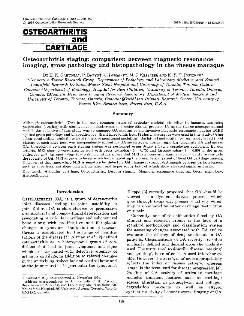
Fic. 4. Mid-parasagittal magnetic resonance image (TP = 3000 ms; TE = 10 ms) of moderately osteoarthritic quadrants of an 18-year-old female rhesus monkey show- ing the existence of three MR zones (arrowhead) extend- ing from the articular surface to the subchondral bone. Note that in the periphery of the region of focal lesion (black arrow) the MR zones are not seen. The high signal intensity in the tibial cartilage surface (white arrow) showed histologic fibrillated region. The thickened sub- chondral bone of tibial plateau (SC) corresponded to histologic sclerotic region. The posterior aspect of the knee joint is on the same side as the external doped water reference (WR).
lation between the proteoglycan reduction seen with histologic studies and signal reduction of the affected carti lage seen on MR images. Tyrell et al. [55] proposed tha t the variations in the signal intensity on MR images may be due to structural changes in the collagen within the ar t icular carti- lage. Variat ion in collagen orientat ion in cartilage maybe another factor contributing to MR signal intensity [56]. In the interpretat ion of MR focal variation in cartilage signal intensity, at tent ion should be given to the pulse sequence used, i.e. TR, TP and TE values, and the image weighting. For example, a decreased signal intensity on Tl-weighted images (short TR and short TE) is proposed to be due to increased water content whereas increased water content is reflected as increased signal intensity on T2-weighted images. A decreased signal intensity on a T2-weighted
image may be due to carti lage fibrosis, as observed in our study.
On our proton density-weighted MR images, an increase in the thickness of the low signal inten- sity band between the ar t icular cartilage and the bone marrow was observed in the OA quadrants and compared well with the corresponding histo- logic region of subchondral sclerosis. Decreased signal intensity of subchondral sclerosis was also reported by Hodler et al. [13] on the T1 and proton density-weighted images and by Engle in moderate and severe OA joints on spin echo Tl-weighted images [44].
Our study shows tha t focal variat ion in the cartilage signal intensi ty provides an indication of early degenerative process of the ar t icular cartilage. Focal or diffuse areas of increased or decreased signal intensi ty of the art icular carti- lage appears to precede art icular cartilage contour deformity or carti laginous thinning observed in the advanced stage of the disease. However, al though MRI is highly sensitive in assessing moderate and severe OA quadrants, some dis- crepancy in the assessment of early stage of the disease still exists. Although MRI is capable of revealing focal regions of high signal intensity corresponding to histologic regions of fibrillated cartilage and of proliferating (and hypertrophic) chondrocytes, to date it is not capable of segregating the two components in the evolution of OA, i.e. cartilage degeneration and repair. The explanation considered for the observed feature is elevated tissue fluid content both in regions of extensive fibrillation and repair process. Another type of pathological lesion was observed in osteo- arthri t ic cartilage corresponding to regions of col- lagen condensation (fibrosis). Fibrotic lesions were visualized as regions of MRI low signal intensity.
Our MRI data provided an indication of the loss of heterogeneity (laminar appearance) of ar t icular carti lage in the vicinity o f the severely osteoarthri t ic lesions where the cartilage has degraded to almost its entire width, i.e. segmental homogeneity is seen at the periphery of osteo- arthri t ic lesion. Two possibilities exist for observed homogeneity of the art icular cartilage in the region of focal lesions. The first possibility is due to the inabili ty of the present resolution to resolve the various layers in the region where cartilage thinning has occurred. The second possibility is due to the disruption of the ordered structure of carti lage consti tuents namely colla- gen and/or proteoglycan and/or due to the macro- molecular interact ion with the water content.
In conclusion, this study documents the various morphological MR changes observed in OA

O s t e o a r t h r i t i s and Cartilage Vol. 3 No. 2 179
c a r t i l a g e and the c o r r e s p o n d i n g gross p a t h o l o g i c a l and h i s to log ica l y a r i a t i o n s . The c a p a b i l i t y of non- i n v a s i v e M R I to v i sua l i ze a r t i c u l a r c a r t i l a g e and to show soft t i s sue c h a n g e s / p a t h o l o g y m a k e s i t idea l for a s sess ing the s e v e r i t y of OA b o t h in c l in ica l s tud ies as well as e x p e r i m e n t a l a n i m a l models . F u r t h e r i m p r o v e m e n t in M R I t e c h n i q u e (e.g. pu lse s eque nce p a r a m e t e r s ) and i n s t r u m e n t a - t ion (e.g. r a d i o f r e q u e n c y coils) as well as improve- m e n t of spa t i a l M R r e s o l u t i o n m a y e n h a n c e M R a s s e s s m e n t of OA c a r t i l a g e les ions and m a y p rove v a l u a b l e for a s se s s ing the effects of d rug t h e r a p y on a r t i c u l a r ca r t i l age .
Acknowledgments
The f inanc ia l s u p p o r t of the Ar th r i t i s Soc ie ty of C a n a d a is g ra t e fu l ly a cknowl edged . The a u t h o r s wish to t h a n k N e l s o n A m o r a n t o for the t e c h n i c a l a s s i s t a n c e in the p r e p a r a t i o n of the h i s to log ica l slides. We also t h a n k A l be r t Cross and N o r m a n K o n y e r for t he i r t e c h n i c a l he lp in m a g n e t i c reson- ance i m a g i n g and Geo rge T h o m l i n s o n for his ad- vice wi th r e g a r d to the s t a t i s t i ca l ana lys i s .
References
1. Dieppe P. Creating a glossary of terms, methods and instruments for clinical research in osteoarthritis. Osteoarthritis Cart 1993;1:149-50.
2. Altman R, Asch E, Bloch D et al. Development of criteria for the classification and reporting of osteo-arthritis. Classification of osteoarthrit is of the knee. Arthritis Rheum 1986;29:1039-49.
3. Dieppe P. The clinical spectrum of osteoarthrit is (Abstract). Osteoarthritis Cart 1993;1:18,
4. Pritzker KPH. Cartilage histopathology in human and rhesus m acaque osteoarthritis. In Kuettner KE, Schleyerback R, Peyron JG, Hascall VC, Eds. Articular cartilage and osteoarthritis New York: Raven Press 1992:473-83.
5. McKeag D, Smith BWH, Edmins te r R, Laird JC, Herron S. Estimating the severity of osteoarthrit is with magnetic resonance spectroscopy. Semin Arthritis Rheum 1992;21:227-38.
6. Adams ME. Cartilage hypertrophy following canine anterior cruciate ligament transection differs among areas of the joint, J Rheumatol 1986;16:818-24.
7. Reicher MA, Bassett LW, Gold RH. High-resolution magnetic resonance imaging of the knee joint: Pathologic correlations. Am J Roentgen 1985;145:903-9.
8. Gluckert K, Kladny B, Ponsel O, Deimling M. MRI of the knee joint with measurement of cartilage signal intensity and relaxation times to determine different stages of cartilage hydrat ion--a future aspect to identify human early osteoarthrosis (Abstract). Osteoarthritis Cart 1993;1:24.
9. Chan WP, Lang P, Stevens MP et al. Osteoarthritis of the knee: Comparison of radiography, CT and
MR imaging to assess extent and severity. Am J Roentgen 1991;157:799-806.
10. Sabiston CP, Adams ME, Li DKB. Magnetic reson- ance imaging of osteoarthritis: Correlation with gross pathology using an experimental model. J Orthrop Res 1987;5:164-72
11. Wojtys E, Wilson M, Buckwalter K, Braunstein E, Martel W. Magnetic resonance imaging of knee hyaline cartilage and intraart icular pathology. Am J Sports Med 1987;15-455-63.
12. Speer KP, Spritzer CE, Goldner JL, Garret t WE. Magnetic resonance imaging of t raumatic knee articular cartilage injuries. Am J Sports Med 1991;19:396-402.
13. Hodler J, Berthiaume MJ, Schweitzer ME, Resnick D. Knee joint hyaline cartilage defects: A comparative study of MR and anatomic sections. J Comput Assist Tomogr 1992;16:597-603.
14. Gylys-Morin VM, Hajek PC, Sartoris DJ, Resnick D. Articular cartilage defects: detectability incadaver knees with MR. Am J Roentgen 1987;148:1153-7.
15. Chandnani VP, Ho C, Chu P, Trudell D, Resnick D. Knee hyaline cartilage evaluated with MR imaging: a cadaveric study involving multiple imaging sequences and intraart icular injection of gadolinium and saline solution. Radiology 1991;178:557 61.
16. Adams ME, Li DKB, McConkey JP, Davidson RG, Day B, Duncan CP, Tron V. Evaluation of cartilage lesions by magnetic resonance imaging at 0.15T: comparison with anatomy and concordance with arthroscopy. J Rheumatol 1991;18:1573-80.
17. Adam G, Nolte-Ernsting C, Prescher A e t al. Exper- imental hyaline cartilage lesions: two-dimensional spin-echo versus three-dimensional gradient-echo MR imaging. Am Magn Reson Imaging 1991;1:665-72.
18. Adam G, Buhne M, Prescher A, et al. Stability of osteochondral fragments of the femoral condyle: magnetic resonance imaging with histopathologic correlation in an animal model. Skeletal Radiol 1991;20:601-6.
19. Bongartz G, Bock E, Horbach T, Requardt H. Degenerative cartilage lesions of the hip--mag- netic resonance evaluation. Magn Reson Imaging 1989;7:179-86.
20. Byers PD, Contepomi CA, Farkas TA. A post mortem study of the hip joint. Including the prevalence of the features of the right side. Ann Rheum Dis 1970;29:15-31.
21. Bennett GA, Waine H, Bauer W. Changes in the knee joint at various ages with particular reference to
.,.~ the nature and development of degenerative joint disease. New York: The Commonwealth Fund. 1942:1-93.
22. Collins DH. Osteoarthritis. In The pathology ofartic. ular and spinal diseases. London: Edward Arnold 1949:74-115.
23. Mankin HJ, Dorfman H, Lippiello L, Zarins A. Biochemical and metabolic abnormalities in artic- ular cartilage from osteoarthritic human hips. II. Correlation of morphology with biochemical and metabolic data. J Bone Joint Surg 1971:53A:523-37.
24. Mankin HJ, Thrasher AZ. Water content and bind- ing in normal and osteoarthritic human cartilage. J Bone Joint Surg 1975;57A:76 80.

180 Gahunia et al.: Staging of osteoarthritic articular cartilage
25. Bollet AJ, Nance JL. Biochemical findings in normal and osteoarthritic articular cartilage: chondroitin sulphate concentration and chain length, water and ash content. J Clin Invest 1966;45:1170-7.
26. Venn M, Maroudas A. Chemical composition and swelling of normal and osteoarthritic femoral head cartilage. Ann Rheum Dis 1977;36:121-9.
27. Byers PF, Contempomi CA, Farkas TA. Post-mortem study of the hip joint. II. Histological basis for limited and progressive cartilage alterations. Ann Rheum Dis 1976;35:114-21.
28. Meachim G, Emery IH. Quantitative aspects of patello-femoral cartilage fibrillation in Liverpool necropsies. Ann Rheum Dis 1974;33:39-47.
29. Lust G, Summers BA. Early, asymptomatic stage of degenerative joint disease in canine hip joints. Am J Vet Res 1981;42:1849-55.
30. Moskowitz RW, Goldberg VM. Studies of osteo- phyte pathogenesis in experimentally induced osteoarthritis. J Rheum 1987;14:311-20.
31. Mohr W, Lehmann H. Osteoarthritis of the ankle joints in old rats. Zeitschrift Fur Rheumatologie 1992;51:35-40.
32. Colombo C, Butler M, O'Byrne E et al. A new model of osteoarthritis in rabbits. I. Develop- ment of knee joint pathology following lateral meniscectomy and section of the fibular collateral and sesamoid ligaments. Arthritis Rheum 1983;26:875 86.
33. Pritzker KPH. Animal models for osteoarthritis: processes, problems and prospects (Review). Ann Rheum Dis 1994;53:406-20.
34. Bite LZ, DeRousseau CJ, Kaufman PL, Bito JW. Age-dependent loss of accommodative amplitude in rhesus monkeys: an animal model for presbyopia. Invest Ophthal Vis Sci 1982;23:23-31.
35. DeRousseau CJ, Rawlins RG, Denlinger JL. Aging in the musculoskeletal system of rhesus monkeys: I. Passive joint excursion. Am J Phys Anthropol 1983;61:483-94.
36. Chateauvert JMD, Grynpas MD, Kessler MJ, Pritzker KPH. Spontaneous osteoarthritis in rhesus macaques. I. Chemical and biochemical studies. J Rheumatol 1989;16; 1098 104.
37. Hayes CE, Edelstein WA, Schenck JF, Mueller OM, Eash M. An efficient, highly homogeneous r.f. coil for whole-body imaging at 1.5 T. J Magn Reson 1985;63:622-8.
38. Maudsley AA. Modified Carr-Purcell Meiboom-Gill sequence for NMR fourier imaging application. J Magn Reson 1986;69:488-91.
39. SAS Institute Inc. S A S / S T A T ° User's Guide, Version 6, 4th ed, Vol. 1. Cary, NC: SAS Institute Inc. 1989:860-7.
40. Broderick LS, Turner DA, Renfrew DL, Schnitzer TJ, Huff JP, Harris C. Severity of articular carti- lage abnormality in patients with osteoarthritis: evaluation with fast spin-echo MR vs arthroscopy. Am J Roentgen 1994;162; 99-103.
41. Karvonen RL, Negendank WG, Fraser SM, Mayes MD, An T, Fernandez-Madrid F. Articular cartilage defects of the knee: correlation between magnetic resonance imaging and gross pathology. Ann Rheum Dis 1990;49:672-5.
42. Ho C, Cervilla V, Kjellin Iet al. Magnetic resonance imaging in assessing cartilage changes in exper- imental osteoarthrosis of the knee. Invest Radiol 1992;27:84-90.
43. Van der Sluijs JA, Geesink RGT, Van der Linden AJ, Bulstra SK, Kuyer R, Drukker J. The reliability of the Mankin Score for osteoarthritis. J Orthop Res 1992;10:58-61.
44. Engel A. Magnetic resonance knee arthrography. Enhanced contrast by gadolinium complex in the rabbit and in humans. Acta Orthop Scand 1990;61 (Suppl. 240):1-57.
45. Hayes CW, Sawyer RW, Conway WF. Patellar carti- lage lesions: in vitro detection and staging with MR imaging and pathologic correlation. Radiology 1990;176:479-83.
46. Monson NL, Haughton VM, Modl JM, Sether LA, Ho KC. Normal and degenerating articular carti- lage: in-vitro correlation of MR imaging and histo-
/ logic findings. J Magn Reson Imaging 1992;2:41-5. ~7. Gahunia HK, Lemaire C, Cross AR, Babyn P, Kessler
MJ, Pritzker KPH. Osteoarthritis in rhesus macaques: assessment of cartilage matrix quality by quantitative magnetic resonance imaging. Agents Actions Suppl 1993;39:255-9.
48. Gahunia HK, Lemaire C, Cross AR, Babyn P, Kessler MJ, Pritzker KPH. Osteoarthritis in rhesus macaque knee joint: magnetic resonance imaging tissue characterization of articular cartilage. Rheumatol: (in press).
49. Konig H, Sauter R, Deimling M, Vogt M. Cartilage disorders: comparison of spin echo, CHESS, and FLASH sequence MR images. Radiology 1987;164:753-8.
50. Lehner KB, Rechl HP, Gmeinwieser JK, Heuck AF, Lukas HP, Kohl HP. Structure, function and degeneration of bovine hyaline cartilage: assess- ment with MR imaging in vitro. Radiology 1989;170:495 9.
51. Konig H, Aicher K, Klose U, Saal J. Quantitative evaluation of hyaline cartilage disorders using FLASH sequence: I. Method and animal exper- iments. Acta Radiol 1990;31:371-5.
52. Paul PK, Jasani MK, Sebok D, Rakhit A, Dunton AW, Douglas FL. Variation in MR signal intensity across normal human knee cartilage. J Magn Reson Imaging 1993;3:569-74.
53. Wilson D, Paul PK, Roberts ED et al. Magnetic resonance imaging and morphometric quanti- tation of cartilage histology after chronic infusion of interleukin 1 in rabbit knees. Proc Soc Exp Biol Med 1993;203:30-7.
54. O'Byrne EM, Paul PK, Blancuzzi Ve t al. Magnetic resonance imagir/g of the rabbit knee: detection of cartilage proteoglycan degradation. Agents and Actions 1991;34:214-16.
55. Tyrell RL, Gluckert K, Pathria M, M0dic MT. Fast three-dimensional MR imaging of the knee: comparison with arthroscopy. Radiology 1988;166:865-2.
56. Rubinstein JD, Kim JK, Morava-Protzner I, Stanchev PL, Henkelman RM. Effects of collagen orientation on MR imaging character- istics of bovine articular cartilage. Radiology 1993;188:219 26.
Edited by P. Dieppe