A dual role for NOTCH signaling in joint cartilage ... · maintenance and osteoarthritis ......
15
PHYSIOLOGY A dual role for NOTCH signaling in joint cartilage maintenance and osteoarthritis Zhaoyang Liu, 1,2 Jianquan Chen, 3 Anthony J. Mirando, 1,3 Cuicui Wang, 1,4 Michael J. Zuscik, 1 Regis J. O’Keefe, 1,5 Matthew J. Hilton 1,3 * Loss of NOTCH signaling in postnatal murine joints results in osteoarthritis, indicating a requirement for NOTCH during maintenance of joint cartilage. However, NOTCH signaling components are substantially increased in abundance in posttraumatic osteoarthritis in humans and mice, suggesting either a repara- tive or a pathological role for NOTCH activation in osteoarthritis. We investigated a potential dual role for NOTCH in joint maintenance and osteoarthritis by generating two mouse models overexpressing the NOTCH1 intracellular domain (NICD) within postnatal joint cartilage. The first mouse model exhibited sus- tained NOTCH activation to resemble pathological NOTCH signaling, whereas the second model had tran- sient NOTCH activation, which more closely reflected physiological NOTCH signaling. Sustained NOTCH signaling in joint cartilage led to an early and progressive osteoarthritic-like pathology, whereas transient NOTCH activation enhanced the synthesis of cartilage matrix and promoted joint maintenance under normal physiological conditions. Through RNA-sequencing, immunohistochemical, and biochemical approaches, we identified several targets that could be responsible for NOTCH-mediated cartilage degra- dation, fibrosis, and osteoarthritis progression. These targets included components of the interleukin-6 (IL-6)–signal transducer and activator of transcription 3 (STAT3) and mitogen-activated protein kinase signaling pathways, which may also contribute to the posttraumatic development of osteoarthritis. Together, these data suggest a dual role for the NOTCH pathway in joint cartilage, and they identify downstream effectors of NOTCH signaling as potential targets for disease-modifying osteoarthritis drugs. INTRODUCTION Osteoarthritis (OA) is the most common joint disorder observed world- wide and is a major cause of disability, which carries an extremely high socioeconomic burden (1). In the United States alone, nearly 25% of the population is expected to have physician-diagnosed OA by 2030 (2), which is estimated to cost between $100 billion and $200 billion annually (3, 4). OA is characterized clinically by fibrosis and degradation of joint cartilage, osteophyte formation, subchondral bone sclerosis, and synovial tissue hyperplasia (5–7). Most of these disease outcomes are determined by abnormal differentiation of chondrocytes coupled with an imbalance in the turnover of cartilaginous extracellular matrix (ECM). Under physiolog- ical conditions, articular and joint chondrocytes maintain a dynamic balance between the synthesis and degradation of ECM components. Normal joint cartilages are largely composed of an ECM rich in type II collagen (COL2A1) and the proteoglycan aggrecan (ACAN), which in the context of OA are degraded by specific collagenases [including matrix metallopro- teinase 13 (MMP13), MMP9, and MMP3] and aggrecanases [including a disintegrin and metalloproteinase with thrombospondin motif 4 (ADAMTS4) and ADAMTS5] (8–10). The disease progression of OA can also be ex- acerbated by inflammatory cytokines, such as interleukin-1b (IL-1b), IL-6, and tumor necrosis factor–a (TNF-a)(11–13), which both suppress ma- trix synthesis and promote matrix degradation. Although several factors have been implicated in the pathogenesis of OA, the genetic pathways that precisely regulate the catabolic (degradative) or anabolic (build up) carti- laginous response or the balance of these processes is just beginning to be understood (5, 14, 15). Understanding the molecular mechanisms that reg- ulate cartilage anabolism and catabolism to maintain joint cartilage homeostasis will be extremely important in developing future disease- modifying OA drugs (DMOADs). The NOTCH pathway was identified as a potential regulator of both catabolic and anabolic molecules in the cartilage ECM during develop- ment (16–18). In mammals, NOTCH signaling is primarily initiated when a ligand of NOTCH interacts with one of the NOTCH family receptors, which leads to a series of receptor cleavage events that ultimately release the NOTCH intracellular domain (NICD) into the cytoplasm. The NICD then translocates to the nucleus and activates downstream target gene ex- pression, including genes of the Hes/Hey families, through its interactions with the recombination signal binding protein for immunoglobulin kJ re- gion and mastermind-like (RBPj k-MAML) transcriptional complex ( 19–21). NOTCH signaling components are found in the developing growth plate car- tilage and in adult articular cartilage (22, 23), which suggests a functional role for NOTCH in regulating both cartilage development and homeostasis. We previously found that loss of RBPjk-dependent NOTCH signaling in all joint tissues, as well as in postnatal joint cartilages, results in an early and progressive OA-like pathology (24), indicating a requisite role for NOTCH in the maintenance of articular cartilage and joints. Studies have also demonstrated that the NOTCH pathway is highly activated in mouse and human joint tissues during posttraumatic OA (25–27) and that tem- porary suppression of NOTCH signaling in murine joints leads to the de- layed progression of OA (25). Together, these data suggest that physiological NOTCH signaling within joint tissues is essential for joint maintenance; however, when NOTCH signaling is abnormally activated, such as occurs during posttraumatic OA, temporary inhibition of the NOTCH pathway or its downstream effectors may provide a means for altering the progression of posttraumatic OA. Because the NOTCH pathway is just beginning to be 1 Department of Orthopaedics and Rehabilitation, Center for Musculoskeletal Research, University of Rochester Medical Center, Rochester, NY 14642, USA. 2 Department of Biology, University of Rochester, Rochester, NY 14642, USA. 3 Department of Orthopaedic Surgery, Duke Orthopaedic Cellular, Develop- mental, and Genome Laboratories, Duke University School of Medicine, Durham, NC 27710, USA. 4 Department of Pathology and Laboratory Medicine, University of Rochester, Rochester, NY 14642, USA. 5 Department of Orthopaedic Surgery, Washington University School of Medicine, St. Louis, MO 63110, USA. *Corresponding author. E-mail: [email protected] RESEARCHARTICLE www.SCIENCESIGNALING.org 21 July 2015 Vol 8 Issue 386 ra71 1 on May 26, 2019 http://stke.sciencemag.org/ Downloaded from
-
Upload
hoangquynh -
Category
Documents
-
view
214 -
download
0
Transcript of A dual role for NOTCH signaling in joint cartilage ... · maintenance and osteoarthritis ......

R E S E A R C H A R T I C L E
P H Y S I O L O G Y
A dual role for NOTCH signaling in joint cartilagemaintenance and osteoarthritisZhaoyang Liu,1,2 Jianquan Chen,3 Anthony J. Mirando,1,3 Cuicui Wang,1,4 Michael J. Zuscik,1
Regis J. O’Keefe,1,5 Matthew J. Hilton1,3*
http:D
ownloaded from
Loss of NOTCH signaling in postnatal murine joints results in osteoarthritis, indicating a requirement forNOTCH during maintenance of joint cartilage. However, NOTCH signaling components are substantiallyincreased in abundance in posttraumatic osteoarthritis in humans and mice, suggesting either a repara-tive or a pathological role for NOTCH activation in osteoarthritis. We investigated a potential dual role forNOTCH in joint maintenance and osteoarthritis by generating two mouse models overexpressing theNOTCH1 intracellular domain (NICD) within postnatal joint cartilage. The first mouse model exhibited sus-tained NOTCH activation to resemble pathological NOTCH signaling, whereas the second model had tran-sient NOTCH activation, which more closely reflected physiological NOTCH signaling. Sustained NOTCHsignaling in joint cartilage led to an early and progressive osteoarthritic-like pathology, whereas transientNOTCH activation enhanced the synthesis of cartilage matrix and promoted joint maintenance undernormal physiological conditions. Through RNA-sequencing, immunohistochemical, and biochemicalapproaches, we identified several targets that could be responsible for NOTCH-mediated cartilage degra-dation, fibrosis, and osteoarthritis progression. These targets included components of the interleukin-6(IL-6)–signal transducer and activator of transcription 3 (STAT3) and mitogen-activated protein kinasesignaling pathways, which may also contribute to the posttraumatic development of osteoarthritis.Together, these data suggest a dual role for the NOTCH pathway in joint cartilage, and they identifydownstream effectors of NOTCH signaling as potential targets for disease-modifying osteoarthritis drugs.
//s
on May 26, 2019tke.sciencem
ag.org/
INTRODUCTION
Osteoarthritis (OA) is the most common joint disorder observed world-wide and is a major cause of disability, which carries an extremely highsocioeconomic burden (1). In the United States alone, nearly 25% of thepopulation is expected to have physician-diagnosed OA by 2030 (2),which is estimated to cost between $100 billion and $200 billion annually(3, 4). OA is characterized clinically by fibrosis and degradation of jointcartilage, osteophyte formation, subchondral bone sclerosis, and synovialtissue hyperplasia (5–7). Most of these disease outcomes are determinedby abnormal differentiation of chondrocytes coupled with an imbalance inthe turnover of cartilaginous extracellular matrix (ECM). Under physiolog-ical conditions, articular and joint chondrocytes maintain a dynamic balancebetween the synthesis and degradation of ECM components. Normal jointcartilages are largely composed of an ECM rich in type II collagen(COL2A1) and the proteoglycan aggrecan (ACAN), which in the contextof OA are degraded by specific collagenases [including matrix metallopro-teinase 13 (MMP13), MMP9, and MMP3] and aggrecanases [including adisintegrin and metalloproteinase with thrombospondin motif 4 (ADAMTS4)and ADAMTS5] (8–10). The disease progression of OA can also be ex-acerbated by inflammatory cytokines, such as interleukin-1b (IL-1b), IL-6,and tumor necrosis factor–a (TNF-a) (11–13), which both suppress ma-trix synthesis and promote matrix degradation. Although several factorshave been implicated in the pathogenesis of OA, the genetic pathways that
1Department of Orthopaedics and Rehabilitation, Center for MusculoskeletalResearch, University of Rochester Medical Center, Rochester, NY 14642,USA. 2Department of Biology, University of Rochester, Rochester, NY 14642,USA. 3Department of Orthopaedic Surgery, Duke Orthopaedic Cellular, Develop-mental, and Genome Laboratories, Duke University School of Medicine, Durham,NC 27710, USA. 4Department of Pathology and Laboratory Medicine, Universityof Rochester, Rochester, NY 14642, USA. 5Department of Orthopaedic Surgery,Washington University School of Medicine, St. Louis, MO 63110, USA.*Corresponding author. E-mail: [email protected]
precisely regulate the catabolic (degradative) or anabolic (build up) carti-laginous response or the balance of these processes is just beginning to beunderstood (5, 14, 15). Understanding the molecular mechanisms that reg-ulate cartilage anabolism and catabolism to maintain joint cartilagehomeostasis will be extremely important in developing future disease-modifying OA drugs (DMOADs).
The NOTCH pathway was identified as a potential regulator of bothcatabolic and anabolic molecules in the cartilage ECM during develop-ment (16–18). In mammals, NOTCH signaling is primarily initiated whena ligand of NOTCH interacts with one of the NOTCH family receptors,which leads to a series of receptor cleavage events that ultimately releasethe NOTCH intracellular domain (NICD) into the cytoplasm. The NICDthen translocates to the nucleus and activates downstream target gene ex-pression, including genes of the Hes/Hey families, through its interactionswith the recombination signal binding protein for immunoglobulin kJ re-gion and mastermind-like (RBPjk-MAML) transcriptional complex (19–21).NOTCH signaling components are found in the developing growth plate car-tilage and in adult articular cartilage (22, 23), which suggests a functionalrole for NOTCH in regulating both cartilage development and homeostasis.
We previously found that loss of RBPjk-dependent NOTCH signalingin all joint tissues, as well as in postnatal joint cartilages, results in an earlyand progressive OA-like pathology (24), indicating a requisite role forNOTCH in the maintenance of articular cartilage and joints. Studies havealso demonstrated that the NOTCH pathway is highly activated in mouseand human joint tissues during posttraumatic OA (25–27) and that tem-porary suppression of NOTCH signaling in murine joints leads to the de-layed progression of OA (25). Together, these data suggest that physiologicalNOTCH signaling within joint tissues is essential for joint maintenance;however, when NOTCH signaling is abnormally activated, such as occursduring posttraumatic OA, temporary inhibition of the NOTCH pathway orits downstream effectors may provide a means for altering the progressionof posttraumatic OA. Because the NOTCH pathway is just beginning to be
www.SCIENCESIGNALING.org 21 July 2015 Vol 8 Issue 386 ra71 1

R E S E A R C H A R T I C L E
understood in the context of OA and joint cartilage maintenance, identify-ing potential downstream effectors that may also serve as better drug tar-gets will be crucial in both our understanding of the disease and thedevelopment of future therapeutics. To assess the potential dual role forNOTCH signaling in joint cartilage and to determine whether the amplitude,duration, or frequency of NOTCH activation influences cartilage physiologyor pathology, we generated mouse models overexpressing NICD1 withinpostnatal joint cartilages in either a sustained or a transient manner. We fur-ther investigated the underlying mechanisms and downstream targets ofNOTCH signaling during OA development and cartilage degeneration.
on May 26, 2019
http://stke.sciencemag.org/
Dow
nloaded from
RESULTS
Sustained activation of NOTCH1 signaling in postnatalchondrocytes results in a progressive OA-like pathologyTo determine whether NOTCH activation in OAwas a reparative responseor contributed to the pathology, we generated a NOTCH gain-of-function(GOF) genetic mouse model using the tetracycline-on (Tet-On) system incombination with the Cre-recombinase system: Col2a1Cre; tetO-NICD1;Rosa-rtTAf/+ (fig. S1A). Only in the presence of the reverse tetracycline trans-activator (rtTA) and tetracycline [or its commercially available alternativedoxycycline (DOX)] can the tetO promoter drive overexpression of NICD1within cartilage. Therefore, we can control the robustness and duration ofNOTCH signaling by adjusting the dose and frequency of DOX adminis-tration. We first generated and characterized the NOTCH1 overexpressionprofile in a sustained NOTCH GOF model with high doses and frequen-cies of DOX injections. Knee joints of Col2a1Cre; tetO-NICD1; Rosa-rtTAf/+
(sGOF NICD1) and Rosa-rtTAf/+ littermate controls (wild type) were in-jected with a single high dose of DOX (100 mg/g) and harvested at days 1 and3 after injection. Immunofluorescence analysis revealed that NOTCH1was increased in abundance in all zones of the articular cartilage in sGOFNICD1 mice at day 1 after injection, and maintained high abundance onday 3 after DOX delivery, suggesting sustained NOTCH1 activation (Fig. 1A).
To confirm these results, RNAwas isolated from the articular chondro-cytes of 1-month-old wild-type and sGOFNICD1mice on day 0 and on days 1,3, and 7 after receiving a single high-dose DOX injection. Real-time quan-titative polymerase chain reaction (qPCR) analysis revealed that NICD1expression peaked on day 1 after DOX injection (eightfold increased relativetowild type) and maintained a high level of expression at days 3 and 7 (four-and sixfold increased, respectively) (Fig. 1B). Expression of the NOTCHtarget gene Hes1 was also increased on days 1 and 3 (~1.6-fold increasedrelative to wild type at both time points), although expression returned tobaseline by day 7 (Fig. 1B). Therefore, we injected 1-month-old sGOF NICD1mice and control mice with high doses of DOX at high frequency (100 mg/gthree times per week) to obtain sustained NOTCH activation (fig. S1B)and then harvested knee joints at 2 months of age. Histological analysis re-vealed the hallmark features of an OA-like pathology in the sGOF NICD1mice, including (i) joint cartilage fibrosis and degeneration, (ii) meniscusdegradation, and (iii) synovial tissue hyperplasia (Fig. 1C). Alcian blue,hematoxylin, and orange G (ABH/OG) staining demonstrated a loss ofproteoglycan content in the articular cartilage of knee sections from thesGOF NICD1 mice, with the development of chondrocyte clusters in regionsretaining some stain, a hallmark of early OA (Fig. 1C). Severe synovialhyperplasia was also observed in the sGOF NICD1 mice. Additionally, thegrowth plates often collapsed in sGOF NICD1 mice, which led to analtered architecture of the subchondral bone. Histomophometry performedon 2-month-old ABH/OG-stained knee sections established that articularcartilage thickness and area were substantially reduced (by ~30 and ~40%,respectively) in sGOF NICD1 mice compared to wild-type mice, and the
number of chondrocytes was decreased by ~50% (Fig. 1D). Terminaldeoxynucleotidyl transferase–mediated deoxyuridine triphosphate nickend labeling (TUNEL) staining revealed an almost 60% increase in TUNEL-positive cells in knee sections from sGOF NICD1 mice, especially in theregions in which chondrocytes formed cell clusters (fig. S2A). These datasuggest that sustained NOTCH1 activation in joint cartilage led to earlyand severe cartilage loss, which was potentially mediated by the inductionof apoptosis in articular chondrocytes.
To identify specific changes in the abundances of ECM-related mole-cules, we performed immunohistochemical analysis for COL2A1, COL10A1,SOX9, COL3A1, and MMP13 in knee sections from 2-month-old sGOFNICD1 mice and control mice (Fig. 1C). This analysis demonstrated thatfibrotic cartilage regions exhibited enhanced COL3A1 abundance, whichwas suggestive of chondrocyte dedifferentiation and fibrosis. The abundanceof COL2A1 was moderately reduced in the articular cartilage in sectionsfrom sGOF NICD1 mice, whereas COL10A1 abundance was increased inthe articular cartilage deep zone. Note that SOX9 was largely absent fromthe superficial zones of sGOF NICD1 articular cartilage. In regions thathad severe cartilage degeneration and fibrosis, SOX9 was completely ab-sent (Fig. 1C). Consistent with articular cartilage loss, we detected an increasein the catabolic marker MMP13 within areas of cartilage degeneration.
Real-time qPCR analysis of RNA isolated from the articular chondro-cytes of 2-month-old sGOF NICD1 mice and control mice (Fig. 1E) dem-onstrated that the expression of the anabolic gene Sox9 was decreased by~2-fold in sGOF NICD1 mice, whereas the expression of the catabolic genesMmp13 and Adamts5 was substantially increased (by 132- and 6-fold, re-spectively). Consistent with the immunohistochemical analysis, the de-differentiation marker Col1a1 was increased ~35-fold in expression in sGOFNICD1 mice compared to that in control mice. Moderate increases in theexpression of Col2a1 (~2-fold) and Acan (~4-fold) were also observed insGOF NICD1 mice; however, the expression of Col10a1 was decreased(~3-fold), which was suggestive of abnormal synthesis of cartilage ECM(Fig. 1E). Together, these data suggest that sGOF NICD1 mice exhibitedaltered ECM synthesis, increased cartilage degradation, and cartilage fi-brosis, consistent with a progressive OA-like pathology. These phenotypeswere largely replicated at 2 and 8 months of age in another sustained GOFNICD1 genetic mouse model for which tamoxifen (1 mg/10 g for 5 days at1 month of age) was used to decrease the cartilage-specific sustained ex-pression of NICD1 from the Rosa26 locus, which was induced by theAcanCreERT2 driver (NICD1AcanTM) (fig. S3), further suggesting thatenhanced NOTCH signaling specifically within chondrocytes was the driv-ing force for OA development in these mice.
Transient activation of NOTCH1 signaling in postnatalchondrocytes results in increased synthesis of cartilageECM and joint maintenanceBecause sustained NOTCH activation caused by both low and high amountsof NICD1 resulted in an early and progressive OA-like pathology, we setout to determine whether transient NOTCH activation in joint cartilage ledto a different bioactivity by more closely mimicking physiologic NOTCHsignaling in immature cartilage during development. To generate a transientNOTCH GOF model, we injected the Col2a1Cre; tetO-NICD1; Rosa-rtTAf/+
mice (tGOF NICD1) and Rosa-rtTAf/+ littermate controls (wild type) withlow doses of DOX at a low frequency (1 mg/g once per week for 1 month)at 1 month of age (fig. S1B) and then harvested knee joints at 2 and 4months of age. Immunofluorescence and real-time qPCR analyses per-formed on 1-month-old tGOF NICD1 mice and control mice confirmedthat a single low dose of DOX only transiently activated NOTCH1 signal-ing for 1 to 3 days in sparse chondrocytes of the articular cartilage (Fig. 2,A and B). Joint integrity analyses demonstrated that by 2 months of age,
www.SCIENCESIGNALING.org 21 July 2015 Vol 8 Issue 386 ra71 2

R E S E A R C H A R T I C L E
on May 26, 2019
http://stke.sciencemag.org/
Dow
nloaded from
Col1a1 Adamts5
Adamts4 Mmp13
Acan Col2a1 Sox9
Relative cartilage thickness
0
1.0
2 months
1.5
0.5
Relative cartilage area
0
1.0
2 months
1.5
0.5
WT
AB
H/O
G
CO
L2
A1
S
OX
9
CO
L1
0A
1
sGOF NICD1
CO
L3
A1
2 months
MM
P1
3
A
E
C
WT sGOF NICD1
Day 1
WT
sG
OF
NIC
D1
NOTCH1 DAPI
Day 3
Day 0 Day 1 Day 3 Day 7 0
2
4
6
8
10
NICD1
Day 0 Day 1 Day 3 Day 7 0
0.5
1.0
1.5
2.0
Hes1
WT sGOF NICD1 B
0
1.0
2 months
1.5
Relative cartilage cell number
0.5
WT sGOF NICD1 D
Col10a1
Fig. 1. Sustained activation of NOTCH1 signaling in postnatal chondrocytes month-old WT and sGOF NICD1 mice. High magnification of a centralized
results in a progressive, OA-like pathology. (A and B) Wild-type (WT) miceandCol2a1Cre; tetO-NICD1;Rosa-rtTAf/+mice (sGOFNICD1)were subjectedto high-dose administration of DOX (100 mg/g), and then sGOFNICD1andWTsamples were analyzed at the indicated times by (A) immunofluorescencestaining for NOTCH1 and (B) real-time qPCR assay to determine the relativeabundances of NICD1 and Hes1 mRNAs. All mRNA abundances were nor-malized to that of the gene encoding b-actin (Actb) and then were normalizedto thecontrols.Dataaremeans±SDofat least three independentexperiments.*P < 0.05 by two-tailed Student’s t test. Images are representative of at leastthree independent experiments. Scale bars, 50 mm. DAPI, 4′,6-diamidino-2-phenylindole. (C) ABH/OG staining and immunohistochemical analysis ofCOL2A1, SOX9, COL10A1, COL3A1, and MMP13 in knee sections from 2-region of articular cartilage is shown in the yellow boxes.White arrows indicatecell clusters. Scale bars, 50 mm. Data are representative of at least threeindependent experiments. (D) Histomorphometry analysis of articular cartilagethickness, area, and chondrocyte number was performed on knee sectionsfrom 2-month-old WT and sGOF NICD1 mice. All results were normalized toWT controls, which were set at 1. Data are means ± SD of at least threeindependent experiments. *P<0.05by two-tailedStudent’s t test. (E) Real-timeqPCR analysis was performed to compare the relative expression of the indi-catedgenes inarticular chondrocytes isolated from2-month-oldWTandsGOFNICD1 mice. All mRNA abundances were normalized to that of Actb mRNAand thenwerenormalized to thecontrols.Data aremeans±SDof at least threeindependent experiments. *P < 0.05 by two-tailed Student’s t test.
www.SCIENCESIGNALING.org 21 July 2015 Vol 8 Issue 386 ra71 3

R E S E A R C H A R T I C L E
on May 26, 2019
http://stke.sciencemag.org/
Dow
nloaded from
Adamts5
Adamts4 Mmp13 Col10a1
Acan Col2a1
Hes1 NICD1
Relative cartilage area
0
1.0
2 months
1.5
0.5
WT
AB
H/O
G
CO
L2
A1
S
OX
9
CO
L1
0A
1
tGOF NICD1
CO
L3
A1
2 months
MM
P1
3
A
E
C
WT tGOF NICD1
Day 1
WT
tG
OF
NIC
D1
NOTCH1 DAPI
Day 3
WT tGOF NICD1 B
0
1.0
2 months
1.5
0.5
Relative cartilage thickness
Relative cartilage cell number
0
1.0
2 months
1.5
0.5
WT tGOF NICD1D
Sox9
Col1a1
Fig. 2. Transient activation of NOTCH1 signaling in postnatal chondrocytes in knee sections of 2-month-oldWTand tGOFNICD1mice. Highmagnification
results in increased synthesis of cartilage ECM and joint maintenance. (A andB) WT mice and Col2a1Cre; tetO-NICD1; Rosa-rtTAf/+ mice (tGOF NICD1)were subjected to low-dose administration of DOX (1 mg/g), and then tGOFNICD1 andWT samples were analyzed at the indicated times by (A) immuno-fluorescence staining for NOTCH1 and (B) real-time qPCR assay to determinethe relative abundances of NICD1 and Hes1mRNAs. All mRNA abundanceswere normalized to that of Actb and then normalized to the controls. Data aremeans±SDof at least three independent experiments. *P<0.05 by two-tailedStudent’s t test. Images are representative of at least three independentexperiments. Scale bars, 50 mm. (C) ABH/OG staining and immuno-histochemical analysis of COL2A1, SOX9, COL10A1, COL3A1, and MMP13of acentralizeddomainof articular cartilage is shown in theyellowboxes.Scalebars, 50mm.Dataare representativeofat least three independentexperiments.(D) Histomorphometry analysis of articular cartilage thickness, area, and chon-drocyte number was performed on knee sections from 2-month-old WT andtGOF NICD1 mice. Data are means ± SD of at least three independentexperiments. *P < 0.05 by two-tailed Student’s t test. (E) Real-time qPCR anal-ysis was performed to compare the relative expression of the indicated genesinarticularchondrocytes isolated from2-month-oldWTandsGOFNICD1mice.All mRNA abundances were normalized to that of ActbmRNA and then werenormalized to the controls. Data aremeans ± SD of at least three independentexperiments. *P < 0.05 by two-tailed Student’s t test.
www.SCIENCESIGNALING.org 21 July 2015 Vol 8 Issue 386 ra71 4

R E S E A R C H A R T I C L E
on May 26, 2019
http://stke.sciencemag.org/
Dow
nloaded from
tGOF NICD1 mice that received a single injection of DOX per week ex-hibited normal knee joint architecture with increased synthesis of cartilageECM (Fig. 2C). Histomophometry analysis revealed that articular cartilagethickness and area were increased by 23 and 31%, respectively, in tGOFNICD1 mice as compared to control mice (Fig. 2D). Note that the numberof articular chondrocytes in the tGOF NICD1 mice was increased by 27%(Fig. 2D). By 4 months of age, the tGOF NICD1 mice exhibited the con-tinuation of joint cartilage maintenance, with trends for increased cartilagearea and chondrocyte number, as well as a 13% increase in articular cartilagethickness as compared to that in control mice (fig. S4, A and B). TUNELstaining revealed no substantial change in the extent of apoptosis in tGOFNICD1 mice at both 2 and 4 months of age (fig. S2, B and C).
To identify specific changes in the abundances of ECM-related mole-cules, we performed immunohistochemical analysis of COL2A1, COL10A1,SOX9, COL3A1, and MMP13 in knee sections of tGOF NICD1 mice andcontrol mice at 2 and 4 months of age (Fig. 2C and fig. S4A). By 2months of age, COL2A1 abundance was substantially increased in the ar-ticular cartilage of knee sections of tGOF NICD1 mice. The number ofchondrocytes expressing SOX9 was also increased in the superficial andtangential zones of tGOF NICD1 mouse knee sections, whereas a modestincrease in COL10A1 abundance was observed strictly in the deep zone.The increased amounts of COL2A1 and SOX9 were also observed in4-month-old tGOF NICD1 mice. Both COL3A1 and MMP13 were almostundetectable in tGOF NICD1 mice and control mice at 2 and 4 months ofage (Fig. 2C and fig. S4A). RNAwas isolated from the articular chondro-cytes of 2- and 4-month-old tGOF NICD1 mice and control mice to ac-cess changes in the expression of ECM-related genes (Fig. 2E and fig.S4C). Real-time qPCR analysis demonstrated that by the age of 2 months,the expression of Sox9, Col2a1, and Acan was substantially increased (be-tween two- and threefold for each) in tGOF NICD1 mutant mice, whereasthe expression of Col10a1 was decreased by more than twofold. Moderateincreases in the expression of Col1a1 andMmp13were also observed in thetGOF NICD1 mice at this time (by 3.5- and 6.2-fold, respectively) (Fig. 2E).We did not observe any substantial changes in the expression of Adamts4and Adamts5 (Fig. 2E). The anabolic effect observed in 2-month-old tGOFNICD1 mice was maintained until 4 months of age, which was 2 monthsafter the last administration of DOX. Gene expression analysis revealedthe increased expression of Sox9, Col2a1, and Acan in the tGOF NICD1mice (by 4-, 3.2-, and 3-fold, respectively), whereas the previously in-creased expression of Col1a1 and Mmp13 in 2-month-old tGOF NICD1mice was now reduced by 2-fold in 4-month-old tGOF NICD1 mice ascompared to age-matched wild-type mice (fig. S4C). Together, these datawere consistent with the immunohistochemical analysis, and they dem-onstrated that transient activation of NOTCH1 signaling in adult jointcartilages enhanced the synthesis of articular cartilage ECM and pro-moted joint maintenance for at least 2 months after the last administrationof DOX.
To investigate whether transient NOTCH1 activation protected joint in-tegrity against trauma-induced OA, we performed a meniscal-ligamentousinjury (MLI) surgery on 8-week-old tGOF NICD1 mice and control mice,which was followed by four low doses of DOX by injection (1 mg/g onceper week). Knee joints were harvested 8 weeks after surgery, and joint in-tegrity was analyzed by ABH/OG-stained histology (fig. S4D). AcceleratedOA progression was observed in the tGOF NICD1 mice, including jointcartilage degeneration and clefting, meniscus degradation, osteophyte for-mation, and severe synovial tissue expansion (fig. S4D). These data suggestthat transient NOTCH activation promoted cartilage and joint maintenanceonly under physiological conditions, but in pathological situations, such asthe trauma-induced inflammatory environment, even transient NOTCHactivation accelerated OA development and led to rapid joint degradation,
which was potentially a result of synergistic effects with other proinflam-matory factors within the injured joint environment.
Large-scale temporal gene expression profiling revealspotential NOTCH1 target genes responsible for cartilagefibrosis and degradationWe demonstrated that NOTCH signaling was a critical regulator of carti-lage and joint maintenance under physiological conditions and that sus-tained activation of NOTCH in postnatal cartilages led to a progressiveOA-like pathology; however, the downstream NOTCH targets responsiblefor OA development remained unclear. To address this issue, we devel-oped an in vitro sustained NOTCH GOF model with costal chondrocytesisolated from Col2a1Cre; tetO-NICD1; Rosa-rtTAf/+mice and control mice,and we performed RNA-sequencing (RNA-seq) analysis at 6 and 48 hoursafter the cells were treated with DOX in culture. These data revealed thatNOTCH activation induced the expression of 451 genes at 6 hours and1249 genes at 48 hours, such as NOTCH pathway genes (including HeyL,Notch3, and Jag1) and genes encoding cartilage-degradation enzymes (in-cludingMmp13,Mmp9, Adamts4, and Adamts5), fibrous collagens (includ-ing Col3a1, Col4a1, and Col5a3), growth factors (including Pdgfb, Tgfb1,and Tgfb2), and specific cytokines and chemokines (including Il6, Ccl20,and Ccl17), whereas the expression of genes encoding other inflammatoryfactors often associated with OA remained relatively low (including Il1a,Il1b, and Tnf) (Fig. 3).
NOTCH activation also resulted in the decreased expression of 75 genesat 6 hours and 867 genes at 48 hours, such as those encoding cartilage-related collagens (including Col2a1, Col9a1, and Col11a1) and otherchondrogenic factors (including Sox9, Sox5, Sox6, Acan, and Comp). Inparticular, we highlight here the profiles of NOTCH target genes, chon-drogenic genes, catabolic genes, and genes encoding fibrous collagensand inflammatory factors that are likely associated with the NOTCH-induced progression of OA in vivo (Fig. 3). Most of the chondrogenic geneswere decreased in expression, whereas the expression of Mmp and Adamtsfamily genes was substantially increased at 48 hours after treatment withDOX. Furthermore, the expression of genes encoding collagens was dif-ferentially affected: the expression of genes encoding cartilage-relatedcollagens, such asCol2a1, Col9a1, Col9a2, Col9a3, Col11a1, and Col11a2was substantially reduced, whereas the expression of genes encoding fibrouscollagens, such as Col3a1, Col4a1, Col4a2, Col5a3, Col6a2, and Col14a1,was markedly increased (Fig. 3).
These results identified previously uncharacterized NOTCH-regulatedgenes in chondrocyte cultures and demonstrated that sustained NOTCHsignaling suppressed the chondrogenic phenotype and promoted cartilagefibrosis and degradation, implicating the NOTCH pathway as a critical reg-ulator of the pathogenesis of OA. We also found that the expression of somegenes encoding inflammatory factors was modestly increased, such as sev-eral cytokine-encoding genes. In particular, the expression of Il6 was 5- and54-fold increased at 6 and 48 hours, respectively (Fig. 3), which was one ofthe most robustly responsive genes in DOX-treated chondrocytes fromsGOFNICD1mice. In addition, pathway analysis identified several clusteredgenes related to tyrosine kinase signaling, G protein (heterotrimeric guaninenucleotide–binding protein)–coupled receptor (GPCR) signaling, and nitricoxide (NO) signaling pathways as being substantially increased in expres-sion in the DOX-treated chondrocytes from sGOF NICD1 mice, many ofwhich have been implicated in OA, cartilage catabolism, or both (Fig. 3).
Sustained NOTCH1 signaling activates the IL-6–STAT3pathway in OA cartilageIL-6 is a proinflammatory cytokine that is produced by various types of cellsand is associated with OA in humans (28). It is found in normal articular
www.SCIENCESIGNALING.org 21 July 2015 Vol 8 Issue 386 ra71 5

R E S E A R C H A R T I C L E
on May 26, 2019
http://stke.sciencemag.org/
Dow
nloaded from
Gene Log2 FC
(6 h)
Log2 FC
(48 h)
Hes1 1.9 2.7
Hes5 3.6 3.7
Hey1 4.1 6.6
Hey2 5.1 8.3
Heyl 9.7 11.0
Jag1 3.1 6.4
Notch1 6.0 6.3
Notch3 5.4 7.9
Notch4 0.1 3.3
Gene Log2 FC
(6 h)
Log2 FC
(48 h)
Sox5 –0.6 –1.7
Sox6 –0.4 –2.5
Sox9 –0.7 –3.6
Acan 0 –3.1
Col2a1 0.3 –5.1
Comp –0.2 –3.9
Fgfr3 –0.1 –2.3
Col9a1 –0.1 –3.2
Col11a1 –0.1 –2.6
Gene Log2 FC
(6 h)
Log2 FC
(48 h)
Mmp9 1.3 6.1
Mmp11 0.4 3.0
Mmp13 0.6 1.7
Mmp14 0.4 1.3
Timp3 –0.7 –2.4
Adamts4 1.8 3.7
Adamts5 0.3 1.2
Adamts15 –0.1 6.6
Tnfsf11 2.0 4.3
ChondrogenicNotch pathway Catabolic
Gene Log2 FC
(6 h)
Log2 FC
(48 h)
Col3a1 2.1 3.1
Col4a1 1.2 4.5
Col4a2 0.8 3.3
Col5a1 0.4 0.6
Col5a2 –0.2 –0.3
Col5a3 1.5 4.1
Coll6a1 0.5 1.8
Col6a2 0.7 2.5
Col14a1 1.2 2.1
Fibrous collagens Gene Log2
FC (6 h)
Log2 FC
(48 h)
Il6 2.3 5.8
Il1a 0.1 0.9
Il1b 0 0
Tnf 0.6 1.4
Ccl20 3.3 5.5
Tnfsf11 2.0 4.3
Ccl17 –0.3 3.3
Cxcr4 0.1 3.1
Cx3cl1 1.3 2.8
Inflammatory signaling Gene Log2
FC (6 h)
Log2 FC
(48 h)
Flt1 3.8 7.9
Pgf 3.3 6.7
Pdgfb 2.5 6.7
Angpt2 1.0 5.0
Ngfr 0.8 4.6
Ngf 0.9 3.6
Hbegf 0.6 2.2
Egfr 1.3 2.2
Vegfc 0.1 1.8
Tyrosine kinase signaling
Gene Log2 FC
(6 h)
Log2 FC
(48 h)
Gpbar1 7.6 10.3
Rgs5 0.8 9.7
Adra1a 0.3 6.9
Gpr20 –1.1 6.8
Gpr133 2.3 6.1
Ednra 2.8 6.4
Sctr 0 5.8
Adora2a 0.3 5.3
Gipr –2.7 5.3
GPCR signaling NO signaling Gene Log2
FC (6 h)
Log2 FC
(48 h)
Gucy1a3 5.0 9.6
Gucy1b3 1.9 8.5
Gucy1a2 2.9 7.1
Pde9a 4.3 8.0
Pde2a 0.9 7.2
Pde1b 1.9 6.0
Ednra 2.8 6.4
Pde1a 0.5 2.9
Pde3a 0.4 2.4
Fig. 3. Large-scale temporal geneexpression profiling re-veals potential NOTCH1 target genes responsible for car-tilage fibrosis and degradation. P2 costal chondrocytesisolated from control and Col2a1Cre; tetO-NICD1f/+; Rosa-rtTAf/+ mice were treated with DOX (10 mg/ml) for 6 or48 hours in culture, after which RNA was collected fromthe chondrocytes for RNA-seq experiments. Each groupcontained three biological repeats. The log2 of the foldchange in gene expression (Log2 FC) in the Col2a1Cre;tetO-NICD1f/+; Rosa-rtTAf/+ mice relative to that in thecontrol mice of the indicated genes of interest at the indi-cated times are shown, including Notch pathway genes,typical chondrogenic and catabolic genes, genes en-coding fibrous collagens, as well as genes whoseproducts are involved in inflammatory signaling, tyrosinekinase signaling, GPCR signaling, andNO signaling. *P<0.05 after Benjamini-Hochberg correction for multipletesting (Cufflinks version 2.0.2). Negative values repre-sent a reduction in gene expression as compared tocontrols at each time indicated.
www.SCIENCESIGN
ALING.org 21 July 2015 Vol 8 Issue 386 ra71 6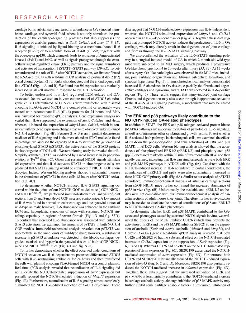
R E S E A R C H A R T I C L E
on May 26, 2019
http://stke.sciencemag.org/
Dow
nloaded from
cartilage but is substantially increased in abundance in OA synovial mem-brane, cartilage, and synovial fluid, where it not only stimulates the pro-duction of the cartilage-degrading proteases but also suppresses theexpression of anabolic genes, such as Sox9, Col2a1, and Acan (7, 9, 11).IL-6 signaling is initiated by ligand binding to a membrane-bound IL-6receptor (IL-6R) or to a soluble form of IL-6R (sIL-6R) together withthe co-receptor gp130, which ultimately leads to activation of Janus-activatedkinase 1 (JAK1) and JAK2, as well as signals propagated through the extra-cellular signal–regulated kinase (ERK) pathway and the signal transducerand activator of transcription 1 (STAT1)–STAT3 pathway (29–32). To bet-ter understand the role of IL-6 after NOTCH activation, we first confirmedthe RNA-seq results with real-time qPCR analysis of postnatal day 2 (P2)costal chondrocytes, P21 articular chondrocytes, and the chondrogenic cellline ATDC5 (Fig. 4, A and B). We found that Il6 expression was markedlyincreased in all cell models in response to NOTCH activation.
To better understand how IL-6 regulated ECM-related and OA-associated factors, we used an in vitro culture model of ATDC5 chondro-genic cells. Differentiated ATDC5 cells were transfected with plasmidencoding FLAG-tagged NICD1 or a control plasmid or separately weretreated with recombinant IL-6 (rIL-6) proteins for 24 hours, and RNAwas harvested for real-time qPCR analyses. Gene expression analysis re-vealed that rIL-6 suppressed the expression of Sox9, Cola2a1, and Acan,whereas it induced the expression of Mmp13 and Col3a1, which is con-sistent with the gene expression changes that were observed under sustainedNOTCH activation (Fig. 4B). Because STAT3 is an important downstreammediator of IL-6 signaling and is the most abundant STAT molecule foundin cartilage, we assessed the capacity of IL-6 to stimulate the generation ofphosphorylated STAT3 (pSTAT3), the active form of the STAT3 protein,in chondrogenic ATDC5 cells. Western blotting analysis revealed that rIL-6 rapidly activated STAT3, as evidenced by a rapid increase in its phospho-rylation at Tyr705 (Fig. 4C). Given that sustained NICD1 signals stimulateIl6 expression and that IL-6 activates STAT3 in chondrogenic cells, wepredicted that STAT3 signaling would be enhanced in NICD1 GOF chon-drocytes. Indeed, Western blotting analysis showed a substantial increasein the abundance of pSTAT3 in these cells 48 hours after NOTCH activa-tion (Fig. 4C).
To determine whether NOTCH-induced IL-6–STAT3 signaling oc-curred within the joints of our NOTCH GOF model mice (sGOF NICD1and NICD1AcanTM), we performed immunohistochemical analysis of jointsections from 2- and 8-month-old GOFmice and control mice. A low amountof IL-6 was found in normal articular cartilage and the synovial tissues ofwild-type animals; however, IL-6 abundance was enhanced in the cartilageECM and hyperplastic synovium of mice with sustained NOTCH sig-naling, especially in regions of severe fibrosis (Fig. 4D and fig. S3D).To confirm that increased IL-6 abundance was associated with enhancedSTAT3 activation, we examined the amounts of pSTAT3 in both NOTCHGOF models. Immunohistochemical analysis revealed that pSTAT3 wasundetectable in the knee joints of wild-type mice; however, a substantialincrease in pSTAT3 abundance was detected in the fibrotic cartilages, de-graded menisci, and hyperplastic synovial tissues of both sGOF NICD1mice and NICD1AcanTM mice (Fig. 4D and fig. S3D).
To further demonstrate whether the effect observed under conditions ofNOTCH activation was IL-6–dependent, we pretreated differentiated ATDC5cells with IL-6–neutralizing antibodies for 24 hours and then transfectedthe cells with plasmid encoding FLAG-tagged NICD1 or a control plasmid.Real-time qPCR analysis revealed that neutralization of IL-6 signaling didnot alleviate the NOTCH-mediated suppression of Sox9 expression butpartially reduced the NOTCH-mediated induction of Mmp13 expression(Fig. 4E). Furthermore, neutralization of IL-6 signaling almost completelyeliminated the NOTCH-mediated induction of Col3a1 expression. These
data suggest that NOTCH-mediated Sox9 expression was IL-6–independent,whereas the NOTCH-stimulated expression of Mmp13 and Col3a1occurred in an IL-6–dependent manner (Fig. 4E). Together, these data sug-gest that prolonged NOTCH activity induces the production of IL-6 in OAcartilage, which may directly result in the degeneration of joint cartilageand fibrosis through the IL-6–STAT3 signaling pathway.
We also confirmed the activation of the IL-6–STAT3 signaling path-way in a surgical-induced model of OA in which 2-month-old wild-typemice were subjected to an MLI surgery, which produces a progressiveOA-like phenotype from 4 to 20 weeks after injury (33, 34). At 12 weeksafter surgery, OA-like pathologies were observed in the MLI mice, includ-ing joint cartilage degeneration and fibrosis, osteophyte formation, andsynovial hyperplasia (Fig. 5). Immunohistochemical analysis demonstratedincreased IL-6 abundance in OA tissues, especially the fibrotic and degen-erative cartilages and synovium, and pSTAT3 was detected in IL-6–positiveregions (Fig. 5). These results suggest that injury-induced joint cartilagefibrosis and degeneration may also occur through inappropriate activationof the IL-6–STAT3 signaling pathway, a mechanism that may be sharedwith NOTCH-induced OA.
The ERK and p38 pathways likely contribute to theNOTCH1-induced OA-related phenotypesIn addition to STAT3, the ERK and p38 mitogen-activated protein kinase(MAPK) pathways are important mediators of pathological IL-6 signaling,as well as of numerous other cytokines and growth factors. To test whetherIL-6 activated both pathways in chondrogenic cells, we examined the effectsof rIL-6 on the phosphorylation (and thus activation) of ERK and p38MAPK in ATDC5 cells. Western blotting analysis showed that the abun-dances of phosphorylated ERK1/2 (pERK1/2) and phosphorylated p38(pp38) increased markedly within 30 min of treatment with rIL-6 and thenrapidly declined, indicating that IL-6 can simultaneously activate both ERKand p38 MAPK pathways in ATDC5 cells (Fig. 6A). Consistent with theNOTCH-induced increase in IL-6 abundance in costal chondrocytes, theabundances of pERK1/2 and pp38 were also substantially increased inthese NICD GOF primary cells (Fig. 6A). Similar to our analysis of pSTAT3abundance, immunohistochemical analysis of articular cartilage sectionsfrom sGOF NICD1 mice further confirmed the increased abundance ofpp38 in vivo (Fig. 6B). Unfortunately, the available anti-pERK1/2 antibo-dies were not compatible with our immunohistochemical analysis of par-affin sections of adult mouse knee joints. Therefore, further in vivo studiesmay be needed to elucidate the potential contributions of p38 and ERK1/2to NOTCH-induced OA-like phenotypes.
To determine whether ERK and p38 MAPK contributed to the OA-associated phenotypes caused by sustained NICD1 signals in vitro, we eval-uated the effects of the MEK inhibitor U0126 (which thus prevents theactivation of ERK) and the p38 MAPK inhibitor SB202190 on the expres-sion of anabolic (Sox9 and Acan), catabolic (Adamts5 and Mmp13), andfibrotic (Col3a1) genes. Real-time qPCR analysis revealed that bothU0126 and SB202190 had no substantial effect on the NOTCH-mediatedincrease in Col3a1 expression or the suppression of Sox9 expression (Fig.6, C and D). Whereas U0126 had no effect on the NOTCH-mediated sup-pression of Acan expression (Fig. 6C), SB202190 exacerbated the NOTCH-mediated suppression of Acan expression (Fig. 6D). Furthermore, bothU0126 and SB202190 substantially reduced the NOTCH-induced expres-sion of Mmp13 (Fig. 6, C and D). Moreover, SB202190 also partially re-duced the NOTCH-mediated increase in Adamts4 expression (Fig. 6D).Together, these data suggest that the increased activation of ERK andp38 MAPK at least partially contributes to the NOTCH-mediated increasein cartilage catabolic activity, although inhibition of p38 MAPK activity mayfurther inhibit some cartilage anabolic factors. Furthermore, inhibition of
www.SCIENCESIGNALING.org 21 July 2015 Vol 8 Issue 386 ra71 7

R E S E A R C H A R T I C L E
on May 26, 2019
http://stke.sciencemag.org/
Dow
nloaded from
C
WT
AB
H/O
G
sGOF NICD1
2 months
IL-6
pS
TA
T3
Il6
P2 costal chondrocytes
WT sGOF NICD1 0
10
20
30 6 h 48 h
Il6
P21 articular chondrocytes
WT sGOF NICD1 0
10
20
40 6 h 48 h
5
A B
pSTAT3
STAT3
100 ng/ml rIL-6
D
Sox9 Col2a1 Acan Mmp13
0 30 60 min
Il6
Adamts5 Col3a1
24 h
0
1
4
3
Control rIL-6
2
FLAG NICD1
24 h
4
2
0
6
8
E
FLAG NICD1 NICD1 + anti-IL-6
pSTAT3
STAT3
Ad-GFP Ad-Cre
f/+R26-NICD1
Sox9
Mmp13 Col3a1
Fig. 4. Sustained NOTCH1
signaling activates the IL-6–STAT3pathway in cultured cellsand OA cartilages. (A) WT andCol2a1Cre; tetO-NICD1f/+;Rosa-rtTAf/+mice (sGOFNICD1)were treatedwithDOX(10mg/ml)for 6 or 48 hours. RNA was thencollected from P2 costal chon-drocyte cultures (top) or P21articular chondrocyte cultures(bottom) isolated from the mice,which was subjected to real-timeqPCRanalysis to determinethe relative abundance of Il6mRNA. All mRNA abundanceswere normalized to that of Actband thenwere normalized to thecontrols. Data are means ± SDof three independent experi-ments. *P < 0.05 by two-tailedStudent’s t test. (B) ATDC5 cellswere transfected with FLAGcontrol plasmid or with plasmidencoding FLAG-NICD1 (top)or were separately treated withcontrol diluents or rIL-6 protein(1 ng/ml) for 24 hours (bottom),and then the relative abun-dancesof the indicatedmRNAswere determined by real-timeqPCRanalysis. All mRNAabun-dances were normalized to thatof Actb and then were normal-ized to the controls. Data aremeans ± SD of three indepen-dent experiments. *P < 0.05 bytwo-tailed Student’s t test. (C)ATDC5 cells that were left un-treated or were treated withrIL-6 (100 ng/ml) for 30 or 60 min(top) and R26-NICD1f/+ prima-ry chondrocytes that wereinfected with adenoviruses ex-pressing either green fluores-cent protein (Ad-GFP) or Cre(Ad-CRE) (bottom) were sub-jected to Western blotting anal-ysis with antibodies specific forthe indicated proteins. Westernblots are representative of threeindependent experiments. (D)ABH/OG staining and immuno-histochemical analysis of IL-6 and pSTAT3 in knee sections from 2-month-old WT and sGOF NICD1mice. Red arrowheads indicate pSTAT3-positive cells. Scale bars, 50 mm.Data are representative of at least three independent experiments. (E)ATDC5 cells that were transfected with FLAG control plasmid or that weretransfected with plasmid encoding FLAG-NICD1 and then were either leftuntreated or treated with IL-6–neutralizing antibody (anti–IL-6) for 24 hoursw
were subjected to real-time qPCRanalysis to determine the relative abun-dances of the indicatedmRNAs. All mRNA abundances were normalizedto that ofActb and then were normalized to the controls, which were set at1. Data are means ± SD of three independent experiments. *P < 0.05 byone-way analysis of variance (ANOVA), followed by the Bonferronimethod.
ww.SCIENCESIGNALING.org 21 July 2015 Vol 8 Issue 386 ra71 8

R E S E A R C H A R T I C L E
on May 26, 2019
http://stke.sciencemag.org/
Dow
nloaded from
either ERK or p38 MAPK did not affect NOTCH-induced cartilage fi-brotic gene expression, as indicated by the lack of a change in Col3a1expression in the presence or absence of inhibitors after NOTCH activa-tion (Fig. 6D).
Alternative signaling pathways are activated bysustained NOTCH1 signaling in chondrocytesIn addition to highlighting inflammatory signaling, the gene and pathwayanalysis of our RNA-seq data revealed that multiple alternative signalingpathways were markedly increased in extent in sGOF NICD1 chondrocytescompared to control chondrocytes. We highlight here the top three pathwaysin our analyses that have also been previously reported to be relevant in
cartilage biology and arthritis-related pathologies. These include tyrosinekinase signaling, GPCR signaling, and NO signaling pathways (Fig. 3).
Many genes related to tyrosine kinase signaling were induced by sus-tained NOTCH1 signaling in chondrocytes (Fig. 3). Several of these, includ-ing Pgf, Flt1, and Ngf, are involved in the pathology of OA and cartilagecatabolism (35–39). To investigate whether tyrosine kinase signalingcontributed to NOTCH1-induced, OA-related molecular phenotypes invitro, we treated chondrocytes with genistein, a tyrosine kinase inhibitor.Our real-time qPCR analysis revealed that genistein did not alleviate theNOTCH1-dependent suppression of Sox9 and Acan expression; however,genistein substantially attenuated the NOTCH1-induced expression of cat-abolic (Adamts4 andMmp13) and fibrotic (Col3a1) genes (fig. S5). Thesedata indicate that tyrosine kinase signaling may partially mediateNOTCH1-induced catabolic and fibrotic processes.
Because several components of the GPCR signaling pathways wereidentified in our RNA-seq data set (Fig. 3) and because several are alsoimplicated in cartilage biology and joint disease (40, 41), we next testedwhether enhanced GPCR signaling contributed to the NOTCH1-induced,OA-associated molecular phenotypes. We individually inhibited three ma-jor downstream pathways of GPCR signaling, the Gas–adenylate cyclase(AC) pathway, the Gai pathway, and the Gaq/11–phospholipase Cb (PLCb)pathway, with the AC inhibitor SQ-22536 (fig. S6A), the Gai inhibitorpertussis toxin (PTx) (fig. S6B), and the PLC inhibitor edelfosine (fig.S6C), respectively. Inhibition of any of these three pathways did not inhibitthe NOTCH1-mediated induction of Adamts4 andMmp13 or the NOTCH1-mediated suppression of Sox9 and Acan expression (fig. S6, A to C);however, both PTx and edelfosine slightly reduced the extent of theNOTCH1-dependent expression of Col3a1 (fig. S6, B and C), suggestingthat GPCR signaling may partially mediate the NOTCH1-dependent in-duction of Col3a1 expression.
NO signaling is implicated in human OA and experimental models ofOA (42–45). The expression of a group of genes related to NO signaling,including genes encoding endothelin receptor A, soluble guanylyl cyclase(sGC; the only known receptor for NO), and some phosphodiesterases(PDEs), was increased by sustained NICD1 signals (Fig. 3). However, theendothelin receptor A antagonist BQ-123 and the sGC inhibitor ODQ onlyslightly inhibited the NOTCH1-mediated induction of Col3a1 expression(fig. S7, A and B). Similarly, the PDE inhibitor isobutylmethylxanthine(IBMX) only modestly inhibited the NOTCH1-mediated suppression ofSox9 and Acan expression (fig. S7C). In contrast, none of these inhibitorsblocked the NOTCH1-dependent induction of Adamts4 and Mmp13 ex-pression (fig. S7, A to C), suggesting that NO-related signaling is unlikelyto mediate the effect of NOTCH1 activation on the increased expression ofcatabolic genes, but that it may have an effect on the NOTCH1-dependentanabolic and fibrotic responses in cartilage.
DISCUSSION
Here, we have provided genetic evidence that sustained versus transientNOTCH activation in postnatal joint cartilages leads to opposing effectson articular cartilage and joint maintenance. This study establishes thatsustained NOTCH activation in adult joint cartilage results in a severe,early, and progressive OA-like pathology, whereas transient NOTCH acti-vation results in increased synthesis of cartilage ECM and joint maintenanceonly under physiological conditions. In vitro and in vivo studies demon-strated the capability of NOTCH signaling to regulate the expression ofgenes required for anabolic, catabolic, and fibrotic processes, and RNA-seq experiments determined that sustained NOTCH activation suppressedthe expression of chondrogenic genes but promoted the expression of genesencoding cartilage-related proteases (MMPs and ADAMTSs), fibrotic
12 weeks post-surgery
Sham MLI
AB
H/O
G
IL-6
pS
TAT
3
Fig. 5. The IL-6–STAT3 pathway is activated in a trauma-induced mousemodel of OA. ABH/OG staining and immunohistochemical analysis ofIL-6 and pSTAT3 in knee sections from 5-month-old WT mice 12 weeksafter they were subjected to sham treatment or MLI surgery. Red arrow-heads indicate pSTAT3-positive cells. Scale bars, 50 mm. Data are repre-sentative of at least five independent experiments.
www.SCIENCESIGNALING.org 21 July 2015 Vol 8 Issue 386 ra71 9

R E S E A R C H A R T I C L E
on May 26, 2019
http://stke.sciencemag.org/
Dow
nloaded from
collagens, inflammatory factors (including IL-6), and components of a hostof broad signaling pathways (tyrosine kinase, GPCR, and NO signaling)that affect cartilage biology and potentially contribute to OA. Together, thesedata suggest that NOTCH signaling is a critical pathway that regulates jointcartilage homeostasis, and suggest that in pathological situations of sus-
www.SCIENCESIGNALING.org
tained signaling, NOTCH is involved in jointcartilage degradation and fibrosis, likelythrough activating at least the IL-6–STAT3,ERK, and p38 MAPK pathways, and po-tentially others. Therefore, an appropriatebalance of NOTCH signalingmust be achievedto maintain normal articular cartilage ho-meostasis and joint integrity.
NOTCH signaling components are wide-ly found in human adult articular cartilage,indicating a potential role for NOTCH sig-naling in articular cartilage maintenanceduring adult life (46, 47). Furthermore, theabundances of NOTCH signaling compo-nents are substantially increased in OAcartilage compared to normal cartilage(25–27, 46), indicating a role for NOTCHin the onset and progression of OA. Wedemonstrated here that sustained NOTCHactivation in postnatal joint cartilages ledto an early and progressive OA-like pathol-ogy, including joint cartilage degradation,fibrosis, and synovial expansion, which waslikely a result of the marked increase in theabundances of cartilage-degrading enzymesand proinflammatory factors. Loss of or re-ductions in cartilage-specific NOTCH sig-naling are capable of reducing the abundanceof MMP13 within murine joints, as wellas delaying cartilage degeneration over theshort term (8 weeks) (25). Our previous datademonstrated that although the decreasedamounts of both anabolic and catabolic fac-tors were observed with short-term inhibi-tion of NOTCH signaling in joint cartilages,long-term (6- to 8-month) reduction inNOTCH signaling disrupts the normal phys-iology of the cartilage and ultimately resultsin cartilage degeneration (24). Together,these data suggest that NOTCH signalingmay play a complex role in cartilage homeo-stasis, such that both sustained activation orpermanent reduction of NOTCH signalingleads to cartilage degradation and joint failure.
NOTCH signaling is involved in boththe anabolism and catabolism of cartilage.We and others previously showed that sus-tained NICD activation in committed growthplate chondrocytes (17, 48) and articularchondrocytes (49) stimulates the expressionof Mmp13, whereas permanent inhibitionof NOTCH signaling within the growth plateand primary articular chondrocyte culturesreducesMmp13 expression (17, 48). Sever-al lines of evidence have demonstrated that
sustained NOTCH signaling in mesenchymal progenitors and growthplate chondrocytes suppresses the expression of Sox9, Col2a1, and Acan(17, 48, 50, 51). Data presented by Mead and Yutzey (18) indicated thatloss of NOTCH signaling leads to the inappropriate expression of Sox9in hypertrophic chondrocytes. Furthermore, we demonstrated here that
Adamts4 Mmp13
D
A B
C
pERK
ERK
100 ng/ml rIL-6
0 30 60 min Ad-GFP Ad-Cre
R26-NICD1 f/+
pERK
pp38
ERK
p38
pp38
p38
Ad-GFP Ad-Cre Ad-Cre + U0126
Ad-GFP Ad-Cre Ad-Cre + SB202190
pERK
WT
sG
OF
NIC
D1
Sox9 Acan Col3a1
Adamts4 Mmp13 Sox9 Acan Col3a1
2 months
Fig. 6. Sustained NOTCH1 signaling activates the ERK and p38 pathways in cultured cells and OA carti-lages, leading to selected effects on the expression of catabolic, chondrogenic, and fibrous genes. (A)ATDC5 cells that were left untreated or were treated with rIL-6 (100 ng/ml) for 30 or 60 min (left) andR26-NICD1f/+ primary chondrocytes that were infected with Ad-GFP or Ad-Cre viruses (right) weresubjected to Western blotting analysis with antibodies specific for the indicated proteins. Western blotsare representative of three independent experiments. (B) Immunohistochemical analysis of pERK in kneesections from 2-month-old WT and sGOF NICD1 mice. Red arrowheads indicate pERK-positive cells.Scale bars, 50 mm. Data are representative of three independent experiments. (C and D) Primary chon-drocytes from R26-NICD1f/+ mice were infected with Ad-GFP virus or with Ad-Cre virus alone or in thepresence of (C) the MAPK kinase (MEK) inhibitor U0126 or (D) the p28 MAPK inhibitor SB202190. Seventy-two hours later, RNAwas isolated from the cells and was analyzed by real-time qPCR analysis to determine therelative abundances of the indicated mRNAs. All mRNA abundances were normalized to that of Actb and thenwere normalized to the control cells (Ad-GFP–infected cells; set at 1). Data are means ± SD of threeindependent experiments. *P < 0.05 by one-way ANOVA followed by the Bonferroni method.
21 July 2015 Vol 8 Issue 386 ra71 10

R E S E A R C H A R T I C L E
on May 26, 2019
http://stke.sciencemag.org/
Dow
nloaded from
sustained overexpression of NICD1 leads to the suppression of Sox9,Col2a1, and Acan expression in adult articular cartilage and chondrogeniccell cultures, whereas transient overexpression of NICD1 in adult jointcartilages promotes the expression of Sox9, Col2a1, and Acan in vivo.Other studies have demonstrated that short-term or transient NOTCHsignaling promotes chondrocyte anabolism by inducing Sox9 expressionin vitro (52), although the underlying mechanisms remain unclear. To-gether, these data suggest that transient or physiological NOTCH signalingin chondrocytes favors a balanced anabolic and catabolic cartilage-maintenanceresponse, whereas sustained or enhanced NOTCH activity elicits a path-ological response through the simultaneous suppression of chondrogenicgenes and the induction of genes encoding catabolic factors.
All of the sustained NICD1 GOF mouse models exhibited robust in-creases in IL-6 and pSTAT3 signaling in degenerating and fibrotic regionsof joint cartilages and synovial tissues. Previous studies identified a con-served RBPjk-binding site within the promoter of Il6, which overlaps witha nuclear factor kB (NF-kB)–binding site (53). RBPjk can directly bind tothis site, and NOTCH signaling can induce Il6 transcription through inter-actions with the NF-kB pathway (53, 54). Consistent with these findings,we found that IL-6 production was increased in response to NOTCH ac-tivation in vivo and in vitro. RNA-seq analysis revealed that the abundanceof Il6 mRNA was 5- and 54-fold increased after 6 and 48 hours ofNOTCH1 activation, respectively, compared to that in control cells, whichsuggests that Il6 may be a direct NOTCH target gene. An in vitro studyperformed in chondrocyte cultures also suggested that the neutralization ofIL-6 cannot modify the NOTCH-mediated suppression of Sox9 and Col2a1expression, but can oppose the NOTCH-mediated induction of Mmp13expression (55). Here, we further demonstrated that neutralization of IL-6at least partially inhibited the NOTCH-dependent induction of Mmp13 ex-pression and also abolished the NOTCH-mediated induction of Col3a1expression. Furthermore, STAT3 activity can be modulated by NOTCHand the NOTCH effectors HES1 and HES5 in cultured neural cells inwhich both the NICD and the HES proteins associated with and facilitatedcomplex formation between JAK2 and STAT3 (56). Therefore, these datasuggest a potential role for NOTCH and HES proteins in promoting car-tilage degeneration in both an IL-6–independent and an IL-6–dependentmanner by regulating the phosphorylation and activity of STAT3.
We also determined that sustained NOTCH1 signaling activated theERK and p38 MAPK pathways in chondrocytes both in vitro and in vivo.ERK and p38 MAPK can function as downstream effectors of IL-6signaling, but they are also regulated independently of IL-6 through nu-merous pathways, including those mediated by other inflammatory cyto-kines, as well growth factors and growth factor receptor signaling pathways,including transforming growth factor–b (TGF-b) and TGF-b receptors, re-ceptor tyrosine kinases (RTKs), and GPCRs. Although the mechanism bywhich NOTCH1 activates ERK and p38 MAPK in chondrocytes may oc-cur through the induction of IL-6 signaling, we also demonstrated the abilityof NOTCH1 to induce the expression of genes encoding other inflamma-tory cytokines (including Ccl20, Ccl17, and Cxcr4), RTK components (in-cluding Flt1, Pgf, Pdgfb, Ngf, and Ngfr), and GPCR family members(including Gpbar1, Rgs5, Gpr20, and Ednra). Several of these factors sep-arately activate ERK and p38 MAPK signaling cascades that are also im-plicated in cartilage biology and joint disease (35–41, 57). Attenuation ofspecific branches of these broad signaling pathways (RTKs and GPCRs),as well as others induced by NOTCH1 in chondrocytes (for example, NO),has demonstrated the complex nature by which NOTCH signaling regu-lates cartilage anabolism, catabolism, and fibrotic gene regulation. Futurestudies will be directed at dissecting this complex regulation to uncoverthe precise mechanism(s) involved in NOTCH-induced OA and to identifypotential targets for the development of DMOADs.
w
MATERIALS AND METHODS
MiceAnimal studies were approved by the University of Rochester Committeeon Animal Resources. All mouse strains, including AcanCreERT2 (58), Rosa-NICD1f/f (59), Col2a1Cre (60), tetO-NICD1 (61), and Rosa-rtTAf/+ (62)mice, were described previously. Col2a1Cre; tetO-NICD1; Rosa-rtTAf/+miceand AcanCreERT2; Rosa-NICD1f/f (NICD1AcanTM) mice were viable andproduced in Mendelian ratios. DOX was administrated to Col2a1Cre;tetO-NICD1; Rosa-rtTAf/+ mice and littermate controls with two strategiesstarting at 1 month of age: (i) 1 mg/g, once per week, or (ii) 100 mg/g, threetimes per week. Mice were harvested at 2 and 4 months of age. Tamoxifen(1 mg/10 g) was administered daily by intraperitoneal injection to allNICD1AcanTM mice and their littermate controls for five continuous daysstarting at 1 month of age. Mice were then harvested at 2 and 8 months of age.
Trauma-induced OA model and surgical proceduresWe used a well-established mouse MLI model to mimic the clinical situa-tion of trauma-induced OA (34). All experiments were performed accord-ing to the protocol approved by the Institutional Animal Care and UseCommittee at the University of Rochester Medical Center (URMC). MLIsurgery was performed on 8-week-old Col2a1Cre; tetO-NICD1; Rosa-rtTAf/+
(tGOF NICD1) mice and littermate controls. Briefly, the bilateral hind-limbs were shaved and prepared for aseptic surgery. The right knee jointwas exposed, and the medial collateral ligament was transected. The jointspace was then opened slightly, and the medial meniscus was detachedfrom its anterior-medial tibial attachment through a microsurgical tech-nique. The contralateral knee joint was sham-operated without any ligamenttransection or meniscus detachment. The skin incision was closed after thesurgery. DOX was administered to both tGOF NICD1 mice and controlmice through intraperitoneal injection (1 mg/g, once per week) for 4 weeksafter MLI surgery. Knee joints were harvested 8 weeks after MLI surgeryby the age of 4 months.
Analysis of mouse tissue sectionsKnee joints were harvested and fixed in 10% neutral-buffered formalin for3 days, decalcified in Formic Acid Bone Decalcifier (Immunocal, DecalChemical Corp.) for 7 to 10 days, paraffin-processed, and embedded forsectioning. Tissues were sectioned at 5 mm and stained with ABH/OG.Immunohistochemical analyses were performed on sections with traditionalantigen retrieval and colorimetric development methodologies with the fol-lowing primary antibodies: anti-SOX9 (Santa Cruz Biotechnology), anti-COL2A1 (Thermo Scientific), anti-COL10A1 (Quartett), anti-COL1A1(Abcam), anti-COL3A1 (Abcam), anti–MMP-13 (Thermo Scientific), anti–IL-6 (Abcam), and anti-pSTAT3 (Cell Signaling). The TUNEL cell deathassay was performed on sections with the In Situ Cell Death DetectionKit, Fluorescein (Roche) according to the manufacturer’s instructions. Im-munofluorescence analysis and b-galactosidase staining were performedon frozen sections. Knee joints were harvested and fixed in 4% para-formaldehyde for 2 hours at 4°C and decalcified with 14% EDTA at 4°Cfor 10 days. Tissues were washed in sucrose gradient, embedded withTissue-Tek OCT medium, snap-frozen in liquid nitrogen, and sectionedat 10 mm with a Leica CM1850 cryotome. An anti-NOTCH1 primary anti-body (Santa Cruz Biotechnology) was used for immunofluorescence analy-sis. b-Galactosidase staining was performed as previously described (63).
Murine costal chondrocyte isolation and RNA-seqMurine costal chondrocytes were isolated as previously described (15), withmodifications. Briefly, rib cages were dissected from 2-day-old Col2a1Cre;tetO-NICD1; Rosa-rtTAf/+ or control pups with the soft tissue removed and
ww.SCIENCESIGNALING.org 21 July 2015 Vol 8 Issue 386 ra71 11

R E S E A R C H A R T I C L E
on May 26, 2019
http://stke.sciencemag.org/
Dow
nloaded from
were plated in 1× phosphate-buffered saline (PBS). Rib cages were di-gested with Pronase (2 mg/ml, Roche) in 1× PBS for 1 hour in a 37°Cshaking water bath and then were digested with collagenase D (3 mg/ml,Roche) in high-glucose Dulbecco’s modified Eagle’s medium (DMEM;Gibco) for 1 hour at 37°C. The rib cages were transferred into a petri dishand digested in collagenase D (3 mg/ml) in high-glucose DMEM for 4 to6 hours. Murine costal chondrocyte cell suspensions were then filteredthrough 40-mm filters and seeded at a density of 500,000 cells per well insix-well tissue culture plates in high-glucose DMEM supplemented with10% fetal bovine serum (FBS; Sigma) and 1% penicillin/streptomycin.Both sets of cell cultures were treated with DOX (10 mg/ml) for 6 or 48 hours.RNAwas extracted with the RNeasy Mini Kit (Qiagen) and the RNase-FreeDNase Set (Qiagen) according to the manufacturer’s instructions. Threebiological replicates were prepared for both the Col2a1Cre; tetO-NICD1;Rosa-rtTAf/+ and control groups. One microgram of total RNA from eachsample was sent to the Genomics Research Center (GRC) at URMC formRNA sequencing and data processing. According to the GRC, RNAconcentration was determined with a NanoDrop 1000 spectrophotometer,whereas RNA quality was assessed with an Agilent Bioanalyzer. The TruSeqRNA Sample Preparation Kit V2 (Illumina) was used for next-generationsequencing library construction according to the manufacturer’s protocols.Briefly, mRNAwas purified from 100 ng of total RNAwith oligo(dT) mag-netic beads and then was fragmented. First-stand complementary DNA(cDNA) synthesis was performed with random hexamer priming followedby second-strand cDNA synthesis. End repair and 3′ adenylation were per-formed on the double-stranded cDNA. Illumina adaptors were ligated toboth ends of the cDNA, which was then purified by gel electrophoresisand amplified with PCR primers specific to the adaptor sequences to gen-erate amplicons of about 200 to 500 base pairs in size. The amplified li-braries were hybridized to the Illumina single-end flow cell and amplifiedwith the cBot (Illumina) at a concentration of 8 pmol per lane. Single-endreads of 100 nucleotides were generated for each sample and aligned to theorganism-specific reference genome. Raw reads generated from the Illumi-na HiSeq2500 sequencer were de-multiplexed with configurebcl2fastq.plversion 1.8.3 software. Low-complexity reads and vector contaminationwere removed with sequence cleaner (“SeqClean”) and the National Centerfor Biotechnology Information (NCBI) UniVec database, respectively. TheFASTX toolkit (fastq_quality_trimmer) was applied to remove bases withquality scores below Q = 13 from the end of each read. Processed readswere then mapped to the UCSC hg38 genome build with SHRiMP version2.2.3, and differential expression analysis was performed with Cufflinks ver-sion 2.0.2 [specifically, cuffdiff2 and usage of the general transfer format(GTF) annotation file for the given reference genome].
ATDC5 cell culture and quantitative geneexpression analysesATDC5 cells (RIKEN BioResource Center) were maintained in DMEM/F-12 (1:1) medium (Gibco) supplemented with 5% FBS and 1% penicillin/streptomycin. For ATDC5 cell differentiation studies, ATDC5 cells werecultured with DMEM/F-12 (1:1) medium supplemented with 5% FBS,1% penicillin/streptomycin, and 0.1% insulin, transferrin, and sodiumselenite premix (BD Biosciences). Differentiated ATDC5 chondrogenic cellscultured for 7 to 14 days were then cultured in serum-free DMEM/F-12 (1:1)medium for 6 hours, transfected with plasmid encoding FLAG-NICD1 orwith the control FLAG plasmid with Lipofectamine 2000 (Invitrogen)according to the manufacturer’s instructions, or treated with vehicle con-trol or recombinant mouse IL-6 protein (R&D Systems) for 24 hours. Foracute signaling studies, differentiated ATDC5 cells were serum-starvedovernight and then were treated with vehicle control or mouse rIL-6 pro-tein (R&D Systems) for the times indicated in the figure legends. For
w
experiments with the IL-6–neutralizing antibody (Abcam), differentiatedATDC5 cells were pretreated with the antibody for 24 hours and then weretransfected with control plasmid or plasmid encoding FLAG-NICD1 withLipofectamine 2000. After 24 hours, RNA was isolated with the RNeasyMini Kit, and then cDNA synthesis and real-time qPCR analysis wereperformed as previously described (16). Sequences of primers specificfor NICD1, Hes1, Sox9, Col2a1, Acan, Col10a1, Col3a1, Col1a1, Mmp13,Adamts4, Adamts5, and Il6 are available upon request.
Infection of cells with adenovirusFor experiments with inhibitors, primary costal chondrocytes were isolatedfrom neonatal Rosa-NICD1f/+ pups and were cultured in DMEM con-taining 10% FBS and 1% penicillin/streptomycin as described earlier.Isolated chondrocytes were seeded in 12-well plates at 0.5 × 10⁶ cellsper well. After overnight culture, the cells were infected with Ad-GFPor Ad-CRE at a multiplicity of infection of 100 in the presence of polybrene(8 mg/ml). Twenty-four hours after infection, the cells were cultured either infresh medium (for protein analysis) or in fresh medium containing vehicleor the appropriate inhibitors (for RNA analysis). Forty-eight hours later,cells were harvested for isolation of protein or RNA.
InhibitorsThe ERK1/2 inhibitor U0126 was obtained from Sigma and was used at afinal concentration of 10 mM. The p38 MAPK inhibitor SB202190(Sigma) was used at a final concentration of 2 mM. The protein tyrosinekinase inhibitor genistein was obtained from Tocris and was used at a finalconcentration of 60 mM. The PLC inhibitor edelfosine was purchasedfrom Tocris and was used at a final concentration of 5 mM. The AC in-hibitor SQ-22536 was obtained from Sigma and was used at a final con-centration of 10 mM. PTx was obtained from Tocris and was used at afinal concentration of 100 ng/ml. The NO-sensitive GC inhibitor ODQwas purchased from Sigma and was used at a final concentration of 10 mM.The endothelin receptor A antagonist BQ-123 was obtained from Sigmaand was used at a final concentration of 5 mM. The PDE inhibitor IBMXwas purchased from Sigma and was used at a final concentration of 65 mM.
Western blotting analysisFor Western blotting analysis, total proteins were extracted from cells withradioimmunoprecipitation assay buffer [20 mM tris-HCl (pH 8.0), 150 mMNaCl, 0.1% SDS, 1% NP-40, 0.5% sodium deoxycholate] supplementedwith protease and phosphatase inhibitors (Roche). Thirty micrograms ofprotein from each sample was resolved by 10% SDS–polyacrylamide gelelectrophoresis and transferred to polyvinylidene difluoride membranes.Western blots were then blocked with 5% nonfat milk and incubated over-night with primary antibodies at a final dilution of 1:1000. Antibodies spe-cific for STAT3, pSTAT3 (Tyr705), ERK1/2, pERK1/2 (Thr202/Tyr204), p38MAPK, and pp38 MAPK (Thr180/Tyr182) were all purchased from CellSignaling Technology.
HistomorphometryQuantitative histomorphometry was performed on ABH/OG-stainedsections with an OsteoMeasure analysis system (OsteoMetrics). Cartilagethickness was measured from the middle of the femoral and tibial condyles.Cartilage area was traced from both articular cartilage surfaces with the areatool in OsteoMeasure software. Three to five mice from each group wereanalyzed, and at least three slides were examined for each mouse.
Statistical analysisStatistical analyses were performed using Student’s t test and one-wayANOVA followed by Bonferroni method as appropriate.
ww.SCIENCESIGNALING.org 21 July 2015 Vol 8 Issue 386 ra71 12

R E S E A R C H A R T I C L E
SUPPLEMENTARY MATERIALSwww.sciencesignaling.org/cgi/content/full/8/386/ra71/DC1Fig. S1. Development of a NOTCH1 activation model with the combined Tet-On/Cre system.Fig. S2. Sustained activation of NOTCH1 signaling in postnatal chondrocytes results inincreased chondrocyte apoptosis.Fig. S3. Sustained activation of NOTCH1 signaling in postnatal chondrocytes results in aprogressive, OA-like pathology.Fig. S4. Transient activation of NOTCH1 signaling in postnatal chondrocytes results inincreased synthesis of cartilage ECM and joint maintenance for as long as 3 months afterinjection with DOX, but does not protect from cartilage degradation after MLI surgery.Fig. S5. Suppression of tyrosine kinase signaling leads to selected effects on NOTCH-regulated genes.Fig. S6. Suppression of GPCR signaling leads to selected effects onNOTCH-regulated genes.Fig. S7. Suppression of NO signaling leads to selected effects on NOTCH-regulated genes.
on May 26, 2019
http://stke.sciencemag.org/
Dow
nloaded from
REFERENCES AND NOTES1. D. J. Hunter, D. Schofield, E. Callander, The individual and socioeconomic impact of
osteoarthritis. Nat. Rev. Rheumatol. 10, 437–441 (2014).2. J. M. Hootman, C. G. Helmick, Projections of US prevalence of arthritis and associated
activity limitations. Arthritis Rheum. 54, 226–229 (2006).3. R. Bitton, The economic burden of osteoarthritis. Am. J. Manag. Care 15, S230–S235
(2009).4. H. Kotlarz, C. L. Gunnarsson, H. Fang, J. A. Rizzo, Insurer and out-of-pocket costs of
osteoarthritis in the US: Evidence from national survey data. Arthritis Rheum. 60,3546–3553 (2009).
5. L. J. Sandell, Etiology of osteoarthritis: Genetics and synovial joint development. Nat.Rev. Rheumatol. 8, 77–89 (2012).
6. S. D. Bos, P. E. Slagboom, I. Meulenbelt, New insights into osteoarthritis: Early develop-mental features of an ageing-related disease.Curr. Opin. Rheumatol. 20, 553–559 (2008).
7. M. B. Goldring, S. R. Goldring, Osteoarthritis. J. Cell. Physiol. 213, 626–634 (2007).8. D. Heinegard, T. Saxne, The role of the cartilage matrix in osteoarthritis. Nat. Rev.
Rheumatol. 7, 50–56 (2011).9. A. S. Lee, M. B. Ellman, D. Yan, J. S. Kroin, B. J. Cole, A. J. van Wijnen, H. J. Im, A
current review of molecular mechanisms regarding osteoarthritis and pain. Gene 527,440–447 (2013).
10. M. Wang, J. Shen, H. Jin, H. J. Im, J. Sandy, D. Chen, Recent progress inunderstanding molecular mechanisms of cartilage degeneration during osteoarthritis.Ann. N. Y. Acad. Sci. 1240, 61–69 (2011).
11. M. Kapoor, J. Martel-Pelletier, D. Lajeunesse, J. P. Pelletier, H. Fahmi, Role of proin-flammatory cytokines in the pathophysiology of osteoarthritis. Nat. Rev. Rheumatol. 7,33–42 (2011).
12. M. Daheshia, J. Q. Yao, The interleukin 1b pathway in the pathogenesis of osteoarthritis.J. Rheumatol. 35, 2306–2312 (2008).
13. M. Kobayashi, G. R. Squires, A. Mousa, M. Tanzer, D. J. Zukor, J. Antoniou, U. Feige,A. R. Poole, Role of interleukin-1 and tumor necrosis factor a in matrix degradation ofhuman osteoarthritic cartilage. Arthritis Rheum. 52, 128–135 (2005).
14. L. N. Reynard, J. Loughlin, Insights from human genetic studies into the pathwaysinvolved in osteoarthritis. Nat. Rev. Rheumatol. 9, 573–583 (2013).
15. L. Wu, X. Huang, L. Li, H. Huang, R. Xu, W. Luyten, Insights on biology and pathologyof HIF-1a/-2a, TGFb/BMP, Wnt/b-catenin, and NF-kB pathways in osteoarthritis. Curr.Pharm. Des. 18, 3293–3312 (2012).
16. Y. Dong, A. M. Jesse, A. Kohn, L. M. Gunnell, T. Honjo, M. J. Zuscik, R. J. O’Keefe,M. J. Hilton, RBPjk-dependent Notch signaling regulates mesenchymal progenitorcell proliferation and differentiation during skeletal development. Development 137,1461–1471 (2010).
17. A. Kohn, Y. Dong, A. J. Mirando, A. M. Jesse, T. Honjo, M. J. Zuscik, R. J. O’Keefe,M. J. Hilton, Cartilage-specific RBPjk-dependent and -independent Notch signals regu-late cartilage and bone development. Development 139, 1198–1212 (2012).
18. T. J. Mead, K. E. Yutzey, Notch pathway regulation of chondrocyte differentiation andproliferation during appendicular and axial skeleton development. Proc. Natl. Acad.Sci. U.S.A. 106, 14420–14425 (2009).
19. R. Kopan, M. X. Ilagan, The canonical Notch signaling pathway: Unfolding the activationmechanism. Cell 137, 216–233 (2009).
20. K. Hori, A. Sen, S. Artavanis-Tsakonas, Notch signaling at a glance. J. Cell Sci. 126,2135–2140 (2013).
21. S. Artavanis-Tsakonas, M. D. Rand, R. J. Lake, Notch signaling: Cell fate control andsignal integration in development. Science 284, 770–776 (1999).
22. A. J. Hayes, G. P. Dowthwaite, S. V. Webster, C. W. Archer, The distribution of Notchreceptors and their ligands during articular cartilage development. J. Anat. 202, 495–502(2003).
23. G. P. Dowthwaite, J. C. Bishop, S. N. Redman, I. M. Khan, P. Rooney, D. J. Evans,L. Haughton, Z. Bayram, S. Boyer, B. Thomson, M. S. Wolfe, C. W. Archer, The
w
surface of articular cartilage contains a progenitor cell population. J. Cell Sci. 117,889–897 (2004).
24. A. J. Mirando, Z. Liu, T. Moore, A. Lang, A. Kohn, A. M. Osinski, R. J. O’Keefe, R. A. Mooney,M. J. Zuscik, M. J. Hilton, RBP-Jk–dependent Notch signaling is required for murinearticular cartilage and joint maintenance. Arthritis Rheum. 65, 2623–2633 (2013).
25. Y. Hosaka, T. Saito, S. Sugita, T. Hikata, H. Kobayashi, A. Fukai, Y. Taniguchi, M. Hirata,H. Akiyama, U. I. Chung, H. Kawaguchi, Notch signaling in chondrocytes modulatesendochondral ossification and osteoarthritis development. Proc. Natl. Acad. Sci. U.S.A.110, 1875–1880 (2013).
26. M. Mahjoub, N. Sassi, M. Driss, L. Laadhar, M. Allouche, M. Hamdoun, K. B. Romdhane,S. Sellami, S. Makni, Expression patterns of Notch receptors and their ligands in humanosteoarthritic and healthy articular cartilage. Tissue Cell 44, 182–194 (2012).
27. C. Karlsson, C. Brantsing, S. Egell, A. Lindahl, Notch1, Jagged1, and HES5 are abun-dantly expressed in osteoarthritis. Cells Tissues Organs 188, 287–298 (2008).
28. G. Livshits, G. Zhai, D. J. Hart, B. S. Kato, H. Wang, F. M. Williams, T. D. Spector,Interleukin-6 is a significant predictor of radiographic knee osteoarthritis: The Chingfordstudy. Arthritis Rheum. 60, 2037–2045 (2009).
29. S. O’Reilly, M. Ciechomska, R. Cant, T. Hugle, J. M. van Laar, Interleukin-6, its role infibrosing conditions. Cytokine Growth Factor Rev. 23, 99–107 (2012).
30. M. Mihara, M. Hashizume, H. Yoshida, M. Suzuki, M. Shiina, IL-6/IL-6 receptor sys-tem and its role in physiological and pathological conditions. Clin. Sci. 122, 143–159(2012).
31. M. Hashizume, N. Hayakawa, M. Mihara, IL-6 trans-signalling directly induces RANKLon fibroblast-like synovial cells and is involved in RANKL induction by TNF-a and IL-17.Rheumatology 47, 1635–1640 (2008).
32. M. Hashizume, M. Mihara, High molecular weight hyaluronic acid inhibits IL-6-inducedMMP production from human chondrocytes by up-regulating the ERK inhibitor, MKP-1.Biochem. Biophys. Res. Commun. 403, 184–189 (2010).
33. S. Kamekura, K. Hoshi, T. Shimoaka, U. Chung, H. Chikuda, T. Yamada, M. Uchida,N. Ogata, A. Seichi, K. Nakamura, H. Kawaguchi, Osteoarthritis development in novelexperimental mouse models induced by knee joint instability. Osteoarthritis Cartilage13, 632–641 (2005).
34. E. R. Sampson, C. A. Beck, J. Ketz, K. L. Canary, M. J. Hilton, H. Awad, E. M. Schwarz,D. Chen, R. J. O’Keefe, R. N. Rosier, M. J. Zuscik, Establishment of an index withincreased sensitivity for assessing murine arthritis. J. Orthop. Res. 29, 1145–1151(2011).
35. F. Iannone, C. De Bari, F. Dell’Accio, M. Covelli, V. Patella, G. Lo Bianco, G. Lapadula,Increased expression of nerve growth factor (NGF) and high affinity NGF receptor(p140 TrkA) in human osteoarthritic chondrocytes. Rheumatology 41, 1413–1418(2002).
36. T. Lowin, C.Weidler, Z. Jenei-Lanzl, S. Capellino, C. G. Baerwald, F. Buttgereit, R. H. Straub,Relationship between placenta growth factor 1 and vascularization, dehydroepiandrosteronesulfate to dehydroepiandrosterone conversion, or aromatase expression in patients withrheumatoid arthritis and patients with osteoarthritis. Arthritis Rheum. 64, 1799–1808(2012).
37. E. Pecchi, S. Priam, M. Gosset, A. Pigenet, L. Sudre, M. C. Laiguillon, F. Berenbaum,X. Houard, Induction of nerve growth factor expression and release by mechanicaland inflammatory stimuli in chondrocytes: Possible involvement in osteoarthritis pain.Arthritis Res. Ther. 16, R16 (2014).
38. Y. F. Ramos,W. den Hollander, J. V. Bovee, N. Bomer, R. van der Breggen, N. Lakenberg,J. C. Keurentjes, J. J. Goeman, P. E. Slagboom, R. G. Nelissen, S. D. Bos, I. Meulenbelt,Genes involved in the osteoarthritis process identified through genome wide expressionanalysis in articular cartilage; the RAAK study. PLOS One 9, e103056 (2014).
39. S. A. Yoo, H. J. Yoon, H. S. Kim, C. B. Chae, S. De Falco, C. S. Cho, W. U. Kim, Roleof placenta growth factor and its receptor flt-1 in rheumatoid inflammation: A link betweenangiogenesis and inflammation. Arthritis Rheum. 60, 345–354 (2009).
40. C. T. Appleton, C. G. James, F. Beier, Regulator of G-protein signaling (RGS) pro-teins differentially control chondrocyte differentiation. J. Cell. Physiol. 207, 735–745(2006).
41. E. Neumann, K. Khawaja, U. Muller-Ladner, G protein-coupled receptors in rheumatology.Nat. Rev. Rheumatol. 10, 429–436 (2014).
42. S. B. Abramson, M. Attur, A. R. Amin, R. Clancy, Nitric oxide and inflammatory mediatorsin the perpetuation of osteoarthritis. Curr. Rheumatol. Rep. 3, 535–541 (2001).
43. J. Pelletier, D. Jovanovic, J. C. Fernandes, P. Manning, J. R. Connor, M. G. Currie,J. Martel-Pelletier, Reduction in the structural changes of experimental osteoarthritis by anitric oxide inhibitor. Osteoarthritis Cartilage 7, 416–418 (1999).
44. J. P. Pelletier, V. Lascau-Coman, D. Jovanovic, J. C. Fernandes, P. Manning, J. R. Connor,M. G. Currie, J. Martel-Pelletier, Selective inhibition of inducible nitric oxide synthase inexperimental osteoarthritis is associated with reduction in tissue levels of catabolicfactors. J. Rheumatol. 26, 2002–2014 (1999).
45. R. Studer, D. Jaffurs, M. Stefanovic-Racic, P. D. Robbins, C. H. Evans, Nitric oxide inosteoarthritis. Osteoarthritis Cartilage 7, 377–379 (1999).
46. I. Ustunel, A. M. Ozenci, Z. Sahin, O. Ozbey, N. Acar, G. Tanriover, C. Celik-Ozenci,R. Demir, The immunohistochemical localization of notch receptors and ligands in
ww.SCIENCESIGNALING.org 21 July 2015 Vol 8 Issue 386 ra71 13

R E S E A R C H A R T I C L E
http://stke.sciencD
ownloaded from
human articular cartilage, chondroprogenitor culture and ultrastructural characteristicsof these progenitor cells. Acta Histochem. 110, 397–407 (2008).
47. S. P. Grogan, S. Miyaki, H. Asahara, D. D. Dlima, M. K. Lotz, Mesenchymal progenitorcell markers in human articular cartilage: Normal distribution and changes in osteo-arthritis. Arthritis Res. Ther. 11, R85 (2009).
48. M. J. Hilton, X. Tu, X. Wu, S. Bai, H. Zhao, T. Kobayashi, H. M. Kronenberg, S. L. Teitelbaum,F. P. Ross, R. Kopan, F. Long, Notch signaling maintains bone marrow mesenchymalprogenitors by suppressing osteoblast differentiation. Nat. Med. 14, 306–314 (2008).
49. R. Blaise, M. Mahjoub, C. Salvat, U. Barbe, C. Brou, M. T. Corvol, J. F. Savouret, F. Rannou,F. Berenbaum, P. Bausero, Involvement of the Notch pathway in the regulation of matrixmetalloproteinase 13 and the dedifferentiation of articular chondrocytes in murinecartilage. Arthritis Rheum. 60, 428–439 (2009).
50. N. Watanabe, Y. Tezuka, K. Matsuno, S. Miyatani, N. Morimura, M. Yasuda, R. Fujimaki,K. Kuroda, Y. Hiraki, N. Hozumi, K. Tezuka, Suppression of differentiation and prolifera-tion of early chondrogenic cells by Notch. J. Bone Miner. Metab. 21, 344–352 (2003).
51. S. Chen, J. Tao, Y. Bae, M. M. Jiang, T. Bertin, Y. Chen, T. Yang, B. Lee, Notch gainof function inhibits chondrocyte differentiation via Rbpj-dependent suppression ofSox9. J. Bone Miner. Res. 28, 649–659 (2013).
52. R. Haller, R. Schwanbeck, S. Martini, K. Bernoth, J. Kramer, U. Just, J. Rohwedel,Notch1 signaling regulates chondrogenic lineage determination through Sox9 activation.Cell Death Differ. 19, 461–469 (2012).
53. K. Miyazawa, A. Mori, K. Yamamoto, H. Okudaira, Transcriptional roles of CCAAT/enhancer binding protein-b, nuclear factor-kB, and C-promoter binding factor 1 in inter-leukin (IL)-1b-induced IL-6 synthesis by human rheumatoid fibroblast-like synoviocytes.J. Biol. Chem. 273, 7620–7627 (1998).
54. W. Wongchana, T. Palaga, Direct regulation of interleukin-6 expression by Notchsignaling in macrophages. Cell. Mol. Immunol. 9, 155–162 (2012).
55. S. Zanotti, E. Canalis, Interleukin 6 mediates selected effects of Notch in chondrocytes.Osteoarthritis Cartilage 21, 1766–1773 (2013).
56. S. Kamakura, K. Oishi, T. Yoshimatsu, M. Nakafuku, N. Masuyama, Y. Gotoh, Hesbinding to STAT3 mediates crosstalk between Notch and JAK–STAT signalling. Nat. CellBiol. 6, 547–554 (2004).
57. F. Wei, D. C. Moore, L. Wei, Y. Li, G. Zhang, X. Wei, J. K. Lee, Q. Chen, Attenuation ofosteoarthritis via blockade of the SDF-1/CXCR4 signaling pathway. Arthritis Res. Ther.14, R177 (2012).
58. S. P. Henry, C. W. Jang, J. M. Deng, Z. Zhang, R. R. Behringer, B. de Crombrugghe,Generation of aggrecan-CreERT2 knockin mice for inducible Cre activity in adult carti-lage. Genesis 47, 805–814 (2009).
w
59. L. C. Murtaugh, B. Z. Stanger, K. M. Kwan, D. A. Melton, Notch signaling controlsmultiple steps of pancreatic differentiation. Proc. Natl. Acad. Sci. U.S.A. 100,14920–14925 (2003).
60. F. Long, X. M. Zhang, S. Karp, Y. Yang, A. P. McMahon, Genetic manipulation ofhedgehog signaling in the endochondral skeleton reveals a direct role in the regulationof chondrocyte proliferation. Development 128, 5099–5108 (2001).
61. B. Z. Stanger, R. Datar, L. C. Murtaugh, D. A. Melton, Direct regulation of intestinalfate by Notch. Proc. Natl. Acad. Sci. U.S.A. 102, 12443–12448 (2005).
62. G. Belteki, J. Haigh, N. Kabacs, K. Haigh, K. Sison, F. Costantini, J. Whitsett, S. E. Quaggin,A. Nagy, Conditional and inducible transgene expression in mice through thecombinatorial use of Cre-mediated recombination and tetracycline induction. NucleicAcids Res. 33, e51 (2005).
63. M. J. Hilton, X. Tu, F. Long, Tamoxifen-inducible gene deletion reveals a distinct celltype associated with trabecular bone, and direct regulation of PTHrP expression andchondrocyte morphology by Ihh in growth region cartilage. Dev. Biol. 308, 93–105(2007).
Acknowledgments: We thank A. E. Kiernan for providing important mouse strains. Wewould like to gratefully acknowledge the technical expertise and assistance of S. Mack,K. Maltby, and A. Thomas within the Center for Musculoskeletal Research Histology, Bio-chemistry, and Molecular Imaging Core. We would also like to thank J. Ashton and colleagueswithin the GRC at URMC for RNA sequencing and data processing. Funding: This work wassupported in part by the following NIH grants: R01 grants AR057022 and AR063071 toM.J.H.; R21 grant AR059733 to M.J.H.; P50 Center of Research Translation grant AR054041to R.J.O.; and P30 Core Center grant AR061307, sub-award #7280 to M.J.H. Author con-tributions: M.J.H., Z.L., and J.C. conceived and designed experiments; Z.L., J.C., A.J.M.,and C.W. performed experiments; Z.L., J.C., A.J.M., C.W., M.J.Z., R.J.O., and M.J.H. ana-lyzed data and contributed to critical discussions; and Z.L., J.C., and M.J.H. wrote andedited the manuscript. Competing interests: The authors declare that they have nocompeting interests.
Submitted 26 November 2014Accepted 2 July 2015Final Publication 21 July 201510.1126/scisignal.aaa3792Citation: Z. Liu, J. Chen, A. J. Mirando, C. Wang, M. J. Zuscik, R. J. O’Keefe, M. J. Hilton,A dual role for NOTCH signaling in joint cartilage maintenance and osteoarthritis. Sci.Signal. 8, ra71 (2015).
em
ww.SCIENCESIGNALING.org 21 July 2015 Vol 8 Issue 386 ra71 14
on May 26, 2019
ag.org/

A dual role for NOTCH signaling in joint cartilage maintenance and osteoarthritisZhaoyang Liu, Jianquan Chen, Anthony J. Mirando, Cuicui Wang, Michael J. Zuscik, Regis J. O'Keefe and Matthew J. Hilton
DOI: 10.1126/scisignal.aaa3792 (386), ra71.8Sci. Signal.
protect joints or exacerbate joint injury depending on signal strength and duration and context.canexacerbated cartilage degradation. As Ratneswaran and Beier discuss in a Focus, these results suggest that NOTCH
osteoarthritis-like phenotype. However, after mice received a surgery-induced injury, transient NOTCH signalingthat transient NOTCH signaling promoted joint maintenance, whereas sustained NOTCH signaling led to an
. foundet alosteoarthritis. In experiments with mice with inducible and tunable expression of NICD in joint cartilage, Liu and maintenance of joint cartilage; however, suppression of NOTCH signaling after joint injury delays the progression ofwhich translocates to the nucleus to induce target gene expression. NOTCH signaling is required for the development
Binding of ligand to the transmembrane receptor NOTCH results in cleavage of its intracellular domain (NICD),Dual roles for NOTCH in joints
ARTICLE TOOLS http://stke.sciencemag.org/content/8/386/ra71
MATERIALSSUPPLEMENTARY http://stke.sciencemag.org/content/suppl/2015/07/17/8.386.ra71.DC1
CONTENTRELATED
http://stke.sciencemag.org/content/sigtrans/10/510/eaar7665.fullhttp://stke.sciencemag.org/content/sigtrans/10/483/eaag2989.fullhttp://stke.sciencemag.org/content/sigtrans/10/477/eaag1598.fullhttp://stke.sciencemag.org/content/sigtrans/10/473/eaah4679.fullhttp://stke.sciencemag.org/content/sigtrans/10/465/eaam9077.fullhttp://science.sciencemag.org/content/sci/338/6109/917.fullhttp://stke.sciencemag.org/content/sigtrans/8/405/ec360.abstracthttp://stm.sciencemag.org/content/scitransmed/5/167/167ra6.fullhttp://stm.sciencemag.org/content/scitransmed/6/251/251ra119.fullhttp://stke.sciencemag.org/content/sigtrans/4/181/rs7.fullhttp://stke.sciencemag.org/content/sigtrans/7/317/ra28.fullhttp://stke.sciencemag.org/content/sigtrans/4/172/ec134.abstracthttp://stke.sciencemag.org/content/sigtrans/6/286/ec176.abstracthttp://stke.sciencemag.org/content/sigtrans/8/386/fs14.full
REFERENCES
http://stke.sciencemag.org/content/8/386/ra71#BIBLThis article cites 63 articles, 13 of which you can access for free
PERMISSIONS http://www.sciencemag.org/help/reprints-and-permissions
Terms of ServiceUse of this article is subject to the
registered trademark of AAAS.is aScience Signaling Association for the Advancement of Science. No claim to original U.S. Government Works. The title
York Avenue NW, Washington, DC 20005. 2017 © The Authors, some rights reserved; exclusive licensee American (ISSN 1937-9145) is published by the American Association for the Advancement of Science, 1200 NewScience Signaling
on May 26, 2019
http://stke.sciencemag.org/
Dow
nloaded from