Osteitis and mucosal inflammation in a rabbit model of ... · J Otorhinolaryngol....
9
Braz J Otorhinolaryngol. 2015;81(3):312---320 www.bjorl.org Brazilian Journal of OTORHINOLARYNGOLOGY ORIGINAL ARTICLE Osteitis and mucosal inflammation in a rabbit model of sinusitis , Carlos Augusto Correia de Campos a,b,∗ , Eduardo Landini Lutaif Dolci a,b , Leonardo da Silva a,b , José Eduardo Lutaif Dolci a,b , Carlos Alberto Herrerias de Campos b , Ricardo Landini Lutaif Dolci a,b a Faculdade de Ciências Médicas, Santa Casa de São Paulo, São Paulo, SP, Brazil b Department of Otorhinolaryngology, Santa Casa de São Paulo, São Paulo, SP, Brazil Received 25 June 2014; accepted 5 August 2014 Available online 30 March 2015 KEYWORDS Sinusitis; Osteitis; Animal models Abstract Introduction: Several experimental studies have shown osteitis after the onset of sinusitis, sup- porting the idea that bone involvement could participate in the dissemination and perpetuation of this inflammatory disease. However, procedures commonly performed for the induction of sinusitis, such as antrostomies, can trigger sinusitis by themselves. Objective: To evaluate osteitis in an animal model of sinusitis that does not violate the sinus directly and verify whether this is limited to the induction side, or if it affects the contralateral side. Methods: Experimental study in which sinusitis was produced by inserting an obstructing sponge into the nasal cavity of 20 rabbits. After defined intervals, the animals were euthanized and maxillary sinus samples were removed for semi-quantitative histological analysis of mucosa and bone. Results: Signs of bone and mucosal inflammation were observed, affecting both the induc- tion and contralateral sides. Statistical analysis showed correlation between the intensity of osteitis on both sides, but not between mucosal and bone inflammation on the same side, sup- porting the theory that inflammation can spread through bone structures, regardless of mucosal inflammation. Please cite this article as: de Campos CA, Dolci EL, da Silva L, Dolci JE, de Campos CA, Dolci RL. Osteitis and mucosal inflammation in a rabbit model of sinusitis. Braz J Otorhinolaryngol. 2015;81:312---20. Institution: Department of Otorhinolaryngology, Santa Casa de São Paulo, São Paulo, SP, Brazil. ∗ Corresponding author. E-mail: [email protected] (C.A.C. de Campos). http://dx.doi.org/10.1016/j.bjorl.2015.03.003 1808-8694/© 2015 Associac ¸ão Brasileira de Otorrinolaringologia e Cirurgia Cérvico-Facial. Published by Elsevier Editora Ltda. All rights reserved.
Transcript of Osteitis and mucosal inflammation in a rabbit model of ... · J Otorhinolaryngol....
B
O
Os
CLC
a
b
RA
r
h1r
raz J Otorhinolaryngol. 2015;81(3):312---320
www.bjorl.org
Brazilian Journal of
OTORHINOLARYNGOLOGY
RIGINAL ARTICLE
steitis and mucosal inflammation in a rabbit model ofinusitis�,��
arlos Augusto Correia de Camposa,b,∗, Eduardo Landini Lutaif Dolcia,b,eonardo da Silvaa,b, José Eduardo Lutaif Dolcia,b,arlos Alberto Herrerias de Camposb, Ricardo Landini Lutaif Dolcia,b
Faculdade de Ciências Médicas, Santa Casa de São Paulo, São Paulo, SP, BrazilDepartment of Otorhinolaryngology, Santa Casa de São Paulo, São Paulo, SP, Brazil
eceived 25 June 2014; accepted 5 August 2014vailable online 30 March 2015
KEYWORDSSinusitis;Osteitis;Animal models
AbstractIntroduction: Several experimental studies have shown osteitis after the onset of sinusitis, sup-porting the idea that bone involvement could participate in the dissemination and perpetuationof this inflammatory disease. However, procedures commonly performed for the induction ofsinusitis, such as antrostomies, can trigger sinusitis by themselves.Objective: To evaluate osteitis in an animal model of sinusitis that does not violate the sinusdirectly and verify whether this is limited to the induction side, or if it affects the contralateralside.Methods: Experimental study in which sinusitis was produced by inserting an obstructing spongeinto the nasal cavity of 20 rabbits. After defined intervals, the animals were euthanized andmaxillary sinus samples were removed for semi-quantitative histological analysis of mucosa andbone.Results: Signs of bone and mucosal inflammation were observed, affecting both the induc-
tion and contralateral sides. Statistical analysis showed correlation between the intensity ofosteitis on both sides, but not between mucosal and bone inflammation on the same side, sup-porting the theory that inflammation can spread through bone structures, regardless of mucosalinflammation.� Please cite this article as: de Campos CA, Dolci EL, da Silva L, Dolci JE, de Campos CA, Dolci RL. Osteitis and mucosal inflammation in a
abbit model of sinusitis. Braz J Otorhinolaryngol. 2015;81:312---20.�� Institution: Department of Otorhinolaryngology, Santa Casa de São Paulo, São Paulo, SP, Brazil.∗ Corresponding author.E-mail: [email protected] (C.A.C. de Campos).
ttp://dx.doi.org/10.1016/j.bjorl.2015.03.003808-8694/© 2015 Associacão Brasileira de Otorrinolaringologia e Cirurgia Cérvico-Facial. Published by Elsevier Editora Ltda. All rightseserved.
Osteitis and mucosal inflammation in a rabbit sinusitis model 313
Conclusion: This study demonstrated that in an animal model of sinusitis that does not disturbthe sinus directly osteitis occurs in the affected sinus and that it also affects the contralateralside.© 2015 Associacão Brasileira de Otorrinolaringologia e Cirurgia Cérvico-Facial. Published byElsevier Editora Ltda. All rights reserved.
PALAVRAS-CHAVESinusite;Osteíte;Modelos animais
Osteíte e inflamacão mucosa em um modelo experimental de rinossinusite
ResumoIntroducão: Diversos estudos experimentais evidenciam osteíte após estabelecimento desinusite, corroborando para a ideia de que o envolvimento ósseo poderia participar nadisseminacão e perpetuacão do processo inflamatório. Porém procedimentos realizados parainducão da doenca nestes modelos, como antrostomias, podem, por si só, desencadear osteíte.Objetivo: Avaliar osteíte em um modelo de rinossinusite em que não ocorre manipulacão sinusale verificar se esta é limitada ao lado de inducão, ou se acomete o lado contralateral.Método: Estudo experimental em que induziu-se rinossinusite em 20 coelhos, por meio deobliteracão temporária com esponja de uma das cavidades nasais. Amostras de tecido sinusalforam submetidas à análise histológica semiquantitativa, após sacrifício dos animais em inter-valos regulares.Resultados: Foram observados sinais de inflamacão óssea e mucosa mais intensa no lado deinducão, mas também contralateral. Testes estatísticos evidenciaram correlacão entre a osteítede ambos os lados, porém não entre inflamacão óssea e mucosa de um mesmo lado, apoiandoa teoria de que a inflamacão poderia se disseminar através do tecido ósseo, independente dainflamacão mucosa.Conclusão: O presente estudo evidenciou a existência de osteíte, tanto no lado de inducãoquanto no contralateral, em modelo experimental em que não ocorre manipulacão sinusal.© 2015 Associacão Brasileira de Otorrinolaringologia e Cirurgia Cérvico-Facial. Publicado porElsevier Editora Ltda. Todos os direitos reservados.
ioas
ipi
ecdtsgafbintas
Introduction
Several factors may contribute to the onset and persistenceof sinonasal inflammation, leading to chronic rhinosinusitis(CRS). These range from alterations related to the host, suchas immunodeficiencies and mucociliary disease, to char-acteristics associated with etiological agents, such as thecapacity to form biofilms and bacterial superantigens.1
Among these, the involvement of the paranasal bones inCRS development and maintenance has been investigated.The close contact between bone and mucosa in this regionand radiological findings in patients with CRS suggest theinvolvement of this tissue.2---4
Several studies have disclosed the presence of sinusbone inflammation in patients with CRS, usually usingtomographic assessment or histological analysis.2,5,6 Thisincidence varies from 36% to 100%,4,7,8 depending on themethod chosen for patient inclusion and the form of assess-ment. Apparently, the incidence is greater when histologicalevaluation is performed, showing that, depending on theintensity of osteitis, there may not be evidence of inflam-mation on tomographic assessment.8,9
In this sense, Lee et al. prospectively evaluated 121
patients with CRS treated surgically. Based on tomography,they observed signs of osteitis affecting 36% of patients(82% ethmoid, 64% sphenoid, 45% maxillary, without eval-uation of the frontal sinus) but observed histological signsr
ut
n 53%.4 Other studies showed that tomographic signs ofsteitis are associated with greater disease intensity innatomopathological examinations7,9 and worse outcome inurgical treatments.6
The most commonly reported signs indicative of osteitisn patients with CRS are periosteal thickening, osteoblastroliferation, bone resorption and new bone formation, andnflammatory cell infiltration.2,4,5,7---9
Although these studies have provided evidence of thexistence of bone inflammation in cases of CRS and somelinical implications, others have pointed out that osteitisoes not occur in all patients. Also, they have indicatedhat it greatly increases if the patient has been previouslyubmitted to surgery (from 6.7% to 58%), which may sug-est the importance of other factors for its onset, suchs surgical trauma.4 Another important fact is that weound no clinical studies that evaluated the presence ofone inflammation in acute episodes of rhinosinusitis. Thats because acute rhinosinusitis (ARS) is usually treatedon-surgically, making it difficult to collect samples for his-ological analysis. Determining whether osteitis is presentt the early stages or if it arises only with the persistence ofinonasal inflammation would aid to further understand its
ole.For this purpose, experimental animal models are used,sually rabbit models, in which bone and mucosal inflamma-ion are assessed, and other factors commonly associated
3 de Campos CA et al.
wmaah1iaoa
mtgaiaospcio
itatIiap
wmist
M
Arbmb
bilTspt
wAuwte
M
Sp
S
LT
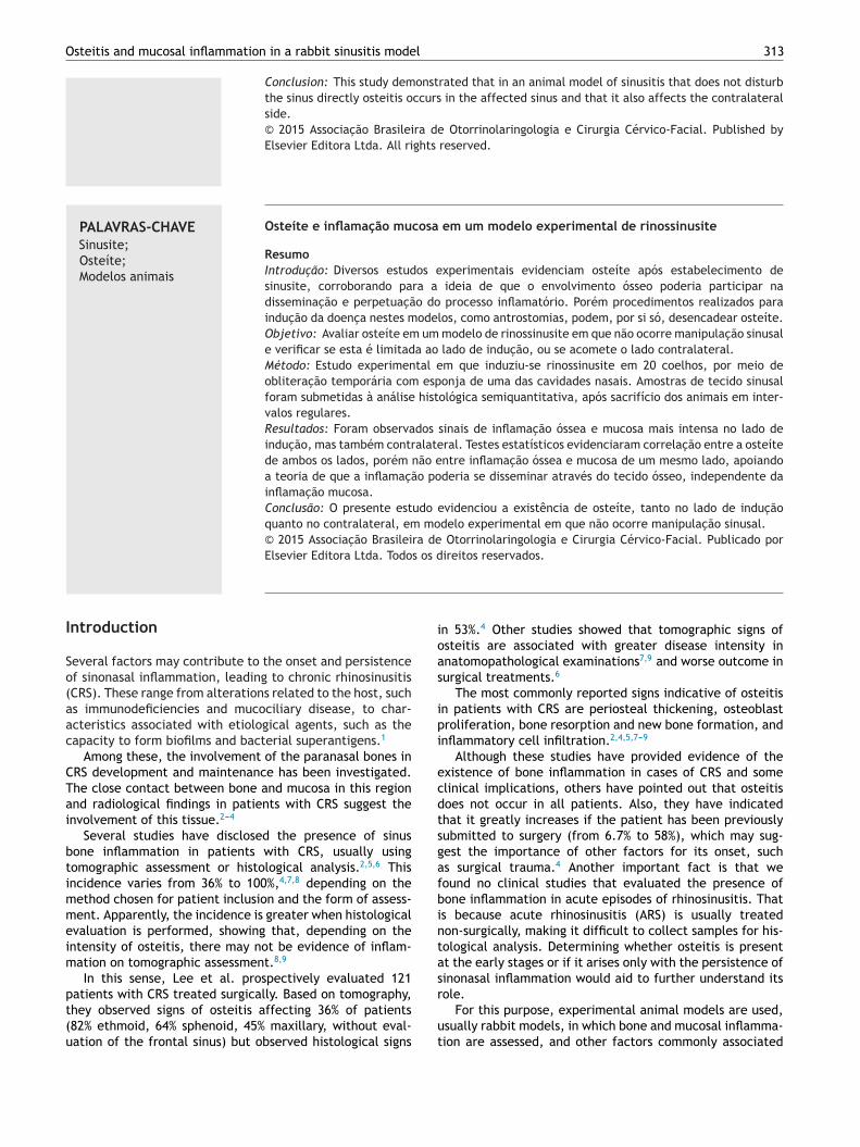
Figure 1 Anatomical specimen showing sponge (Sp) placedin the nasal cavity of rabbits. Observe the nasal septum (S),tt
sb(-i
nessTdiao
aTtla
14
ith sinusitis can be ruled out, such as allergic and inflam-atory disorders, drug use, previous surgery, and anatomical
lterations. These studies show bone involvement as earlys two weeks after the rhinosinusitis induction processas begun and persisting at varying intensities, for up to3 weeks. The most commonly described findings of bonenflammation are similar to those reported in clinical tri-ls: periosteal thickening, inflammatory infiltrate, increasedsteoclastic and osteoblastic activity, new bone formation,nd eventually fibrosis.3,10---13
The problem with these experimental models is theethods by which the sinusitis is induced. Normally, defini-
ive obliteration of the maxillary ostium drainage withlue and sinus inoculation of an infectious agent throughn external sinusotomy are performed. These proceduresnjure a certain area of the sinus wall and alter its physiologynd, by themselves, trigger tissue inflammation regardlessf the infection. Moreover, the definitive obliteration of theinus containing a pathogen leads to an intense infectiousrocess, that, if limited to the sinus cavity often does notorrespond to the pathophysiology of this disease.14 In ARS,nfection usually has a nasal origin and maxillary ostiumbstruction is reversible, as it is caused by mucosal edema.
There are experimental models where rhinosinusitis isnduced by procedures in which there is less manipulation ofhe animals’ nasal cavity. They are called rhinogenic modelsnd are based on the introduction of a sponge into one ofhe nasal cavities, that remains in place for a set period.n this method, the sinuses are not violated, thus limitingnflammation caused by the procedure, and the ostial block-ge is reversible. Therefore, these models better reflect thehysiopathogeny of this disease in humans.14---16
Therefore, the present study aimed to determinehether sinus bone inflammation occurs, to correlate it withucosal inflammation in an experimental model of rhinos-
nusitis in which there is no manipulation of the paranasalinus and to verify whether this inflammation is limited tohe induction side, or if it also affects the contralateral side.
ethods
total of 22 adult, white, male and female New Zealandabbits were used, weighing approximately 2500 g at theeginning of the experiment. Throughout the study, the ani-als were maintained in individual cages suitable for thereed and weight and had free access to food and water.
Bacterial rhinosinusitis was induced in 20 of 22 animals,y placing a small piece of porous polyvinyl sponge measur-ng 3.0 cm × 0.5 cm × 0.3 cm, previously sterilized with ethy-ene oxide, into the right nasal cavity of the animals (Fig. 1).he sponges were soaked in 1.0 mL solution containingtreptococcal and staphylococcal toxoid. No procedure waserformed in the left nasal cavity. Two animals used as con-rols were euthanized without undergoing any intervention.
After 10 days, the sponges were removed and six animalsere randomly euthanized (10th day of the experiment).fter another 7 days, during which the animals did not
ndergo any further intervention, seven additional animalsere euthanized (17th day of the experiment). Finally, onhe 30th day of the experiment, the last seven animals wereuthanized.
ie3e
he maxillary sinus (M), the middle turbinate (*), and the lowerurbinate on the right (LT).
All procedures were performed under anesthesia withpontaneous breathing, according to standards establishedy the Brazilian Society of Laboratory Animal ScienceSociedade Brasileira de Ciência em Animais de Laboratório-- SBCAL) and after approval by the ethics committee of thenstitution, under No. 2011-4.
After euthanization, opening of the outer wall of theasal cavity and paranasal sinuses was performed. Next, thentire medial wall of the maxillary sinus on both the inducedinusitis side and the contralateral side was removed andamples containing bone and mucosal tissue were obtained.he material of each sample was fixed in buffered formalin,ehydrated at increasing concentrations of ethanol, clearedn xylene, and embedded in paraffin. It was then sliced with
microtome into 4-mm thick sections that were mountedn slides, and stained with hematoxylin and eosin (HE).
The slides were evaluated using optical microscopy by pathologist blinded to each animal experiment protocol.he mucosal tissue and bone samples were graded accordingo inflammatory parameters, semi-quantitatively. The fol-owing was considered for mucosal inflammation: grade 0,bsence of inflammation; grade 1, mild inflammation (slightnflammatory cell infiltrate in the mucosa); grade 2, mod-
rate inflammation (diffuse inflammatory infiltrate); grade, intense inflammation (diffuse inflammatory infiltrate,pithelial cell injury, abnormal mucosal and submucosal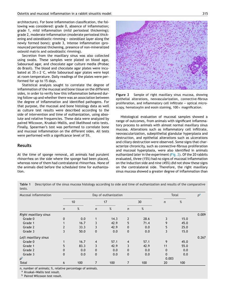
l 315
Figure 2 Sample of right maxillary sinus mucosa, showingepithelial alterations, neovascularization, connective-fibrousps
rtmndaaae
Osteitis and mucosal inflammation in a rabbit sinusitis mode
architecture). For bone inflammation classification, the fol-lowing was considered: grade 0, absence of inflammation;grade 1, mild inflammation (mild periosteal thickening);grade 2, moderate inflammation (moderate periosteal thick-ening and osteoblastic rimming --- osteoblast layer along thenewly formed bone); grade 3, intense inflammation (pro-nounced periosteal thickening, presence of non-mineralizedosteoid matrix and osteoblastic rimming).
Secretion from the maxillary sinus was also collectedusing swabs. These samples were plated on blood agar,Sabouraud agar, and chocolate agar culture media (Probacdo Brasil). The blood and chocolate agar plates were incu-bated at 35 ± 2 ◦C, while Sabouraud agar plates were keptat room temperature. Daily readings of the plates were per-formed for up to 15 days.
Statistical analysis sought to correlate the degree ofinflammation of the mucosal and bone tissue on the differentsides, in order to verify how this inflammation behaved dur-ing follow-up and whether there was an association betweenthe degree of inflammation and identified pathogens. Forthat purpose, the mucosal and bone histology data as wellas culture test results were described according to theside of intervention and time of euthanization, using abso-lute and relative frequencies. These data were analyzed bypaired Wilcoxon, Kruskal---Wallis, and likelihood ratio tests.Finally, Spearman’s test was performed to correlate boneand mucosal inflammation on the different sides. All testswere performed with a significance level of 5%.
Results
At the time of sponge removal, all animals had purulent
rhinorrhea on the side where the sponge had been placed,whereas none of them had contralateral rhinorrhea. None ofthe animals died before the scheduled time for euthaniza-tion.eoos
Table 1 Description of the sinus mucosa histology according to stests.
Mucosal inflammation Day of euthaniza
10 17
n % n %
Right maxillary sinus
Grade 0 0 0.0 1 14.3Grade 1 1 16.7 3 42.9Grade 2 2 33.3 3 42.9Grade 3 3 50.0 0 0.0
Left maxillary sinus
Grade 0 1 16.7 4 57.1Grade 1 5 83.3 3 42.9Grade 2 0 0.0 0 0.0Grade 3 0 0.0 0 0.0
pb
Total 6 100 7 100
n, number of animals; %, relative percentage of animals.a Kruskal---Wallis test result.b Paired Wilcoxon test result.
roliferation, and inflammatory cell infiltrate --- optical micro-copy, hematoxylin and eosin staining, 100× magnification.
Histological evaluation of mucosal samples showed aange of outcomes, from animals with significant inflamma-ory process to animals with almost normal maxillary sinusucosa. Alterations such as inflammatory cell infiltrate,
eovascularization, subepithelial glandular hyperplasia andestruction, and epithelial alterations such as ulcerationsnd ciliary destruction were observed. Some signs that char-cterize chronicity, such as connective-fibrous proliferationnd mucosal hyperplasia, were also identified in animalsuthanized later in the experiment (Fig. 2). Of the 20 rabbits
valuated, three (15%) had no signs of mucosal inflammationn the induction side and nine (45%) did not show these signsn the contralateral side. Therefore, the right maxillaryinus mucosa showed a greater degree of inflammation thanide and time of euthanization and results of the comparative
tion Total pa
30 n %
n %
0.009 2 28.6 3 15.0 5 71.4 9 45.0 0 0.0 5 25.0 0 0.0 3 15.0
0.267 4 57.1 9 45.0 3 42.9 11 55.0 0 0.0 0 0.0 0 0.0 0 0.0
0.0037 100 20 100
316 de Campos CA et al.
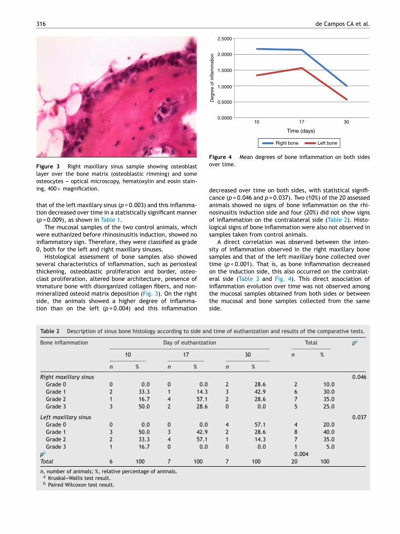
Figure 3 Right maxillary sinus sample showing osteoblastlayer over the bone matrix (osteoblastic rimming) and someoi
tt(
wi0
stcimst
Time (days)
10
2.5000
2.0000
1.5000
1.0000
0.5000
0.0000
Right bone
Deg
ree
of in
flam
mat
ion
Left bone
17 30
Figure 4 Mean degrees of bone inflammation on both sideso
dcanols
sstoe
steocytes --- optical microscopy, hematoxylin and eosin stain-ng, 400× magnification.
hat of the left maxillary sinus (p = 0.003) and this inflamma-ion decreased over time in a statistically significant mannerp = 0.009), as shown in Table 1.
The mucosal samples of the two control animals, whichere euthanized before rhinosinusitis induction, showed no
nflammatory sign. Therefore, they were classified as grade, both for the left and right maxillary sinuses.
Histological assessment of bone samples also showedeveral characteristics of inflammation, such as periostealhickening, osteoblastic proliferation and border, osteo-last proliferation, altered bone architecture, presence of
mmature bone with disorganized collagen fibers, and non-ineralized osteoid matrix deposition (Fig. 3). On the rightide, the animals showed a higher degree of inflamma-ion than on the left (p = 0.004) and this inflammation
itts
Table 2 Description of sinus bone histology according to side and
Bone inflammation Day of euthanizati
10 17
n % n %
Right maxillary sinusGrade 0 0 0.0 0 0.0
Grade 1 2 33.3 1 14.3
Grade 2 1 16.7 4 57.1
Grade 3 3 50.0 2 28.6
Left maxillary sinus
Grade 0 0 0.0 0 0.0
Grade 1 3 50.0 3 42.9
Grade 2 2 33.3 4 57.1
Grade 3 1 16.7 0 0.0
pb
Total 6 100 7 100
n, number of animals; %, relative percentage of animals.a Kruskal---Wallis test result.b Paired Wilcoxon test result.
ver time.
ecreased over time on both sides, with statistical signifi-ance (p = 0.046 and p = 0.037). Two (10%) of the 20 assessednimals showed no signs of bone inflammation on the rhi-osinusitis induction side and four (20%) did not show signsf inflammation on the contralateral side (Table 2). Histo-ogical signs of bone inflammation were also not observed inamples taken from control animals.
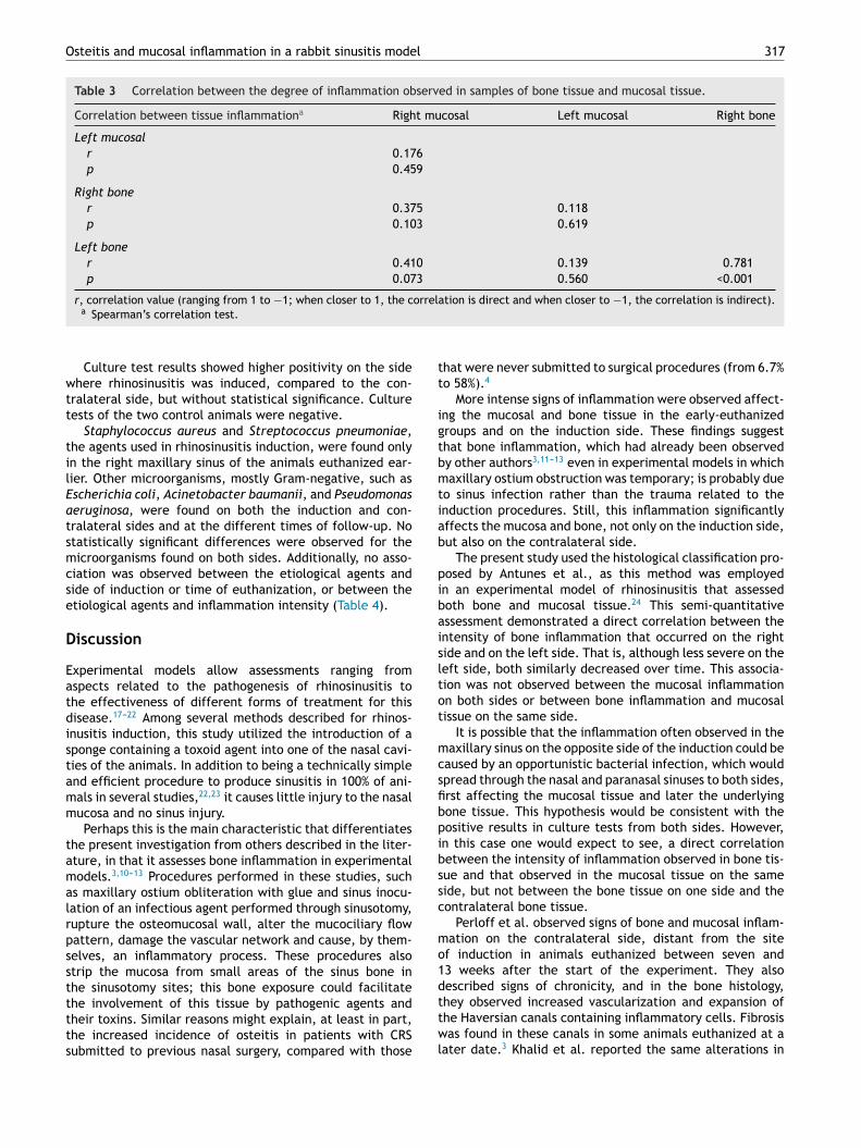
A direct correlation was observed between the inten-ity of inflammation observed in the right maxillary boneamples and that of the left maxillary bone collected overime (p < 0.001). That is, as bone inflammation decreasedn the induction side, this also occurred on the contralat-ral side (Table 3 and Fig. 4). This direct association ofnflammation evolution over time was not observed amonghe mucosal samples obtained from both sides or between
he mucosal and bone samples collected from the sameide.time of euthanization and results of the comparative tests.
on Total pa
30 n %
n %
0.0462 28.6 2 10.03 42.9 6 30.02 28.6 7 35.00 0.0 5 25.0
0.0374 57.1 4 20.02 28.6 8 40.01 14.3 7 35.00 0.0 1 5.0
0.0047 100 20 100
Osteitis and mucosal inflammation in a rabbit sinusitis model 317
Table 3 Correlation between the degree of inflammation observed in samples of bone tissue and mucosal tissue.
Correlation between tissue inflammationa Right mucosal Left mucosal Right bone
Left mucosalr 0.176p 0.459
Right boner 0.375 0.118p 0.103 0.619
Left boner 0.410 0.139 0.781p 0.073 0.560 <0.001
r, correlation value (ranging from 1 to −1; when closer to 1, the correlation is direct and when closer to −1, the correlation is indirect).
tt
igtbmtiab
pibaisltot
mcsfibpibssc
mo1d
a Spearman’s correlation test.
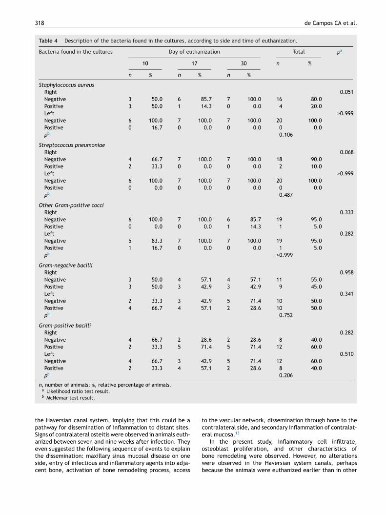
Culture test results showed higher positivity on the sidewhere rhinosinusitis was induced, compared to the con-tralateral side, but without statistical significance. Culturetests of the two control animals were negative.
Staphylococcus aureus and Streptococcus pneumoniae,the agents used in rhinosinusitis induction, were found onlyin the right maxillary sinus of the animals euthanized ear-lier. Other microorganisms, mostly Gram-negative, such asEscherichia coli, Acinetobacter baumanii, and Pseudomonasaeruginosa, were found on both the induction and con-tralateral sides and at the different times of follow-up. Nostatistically significant differences were observed for themicroorganisms found on both sides. Additionally, no asso-ciation was observed between the etiological agents andside of induction or time of euthanization, or between theetiological agents and inflammation intensity (Table 4).
Discussion
Experimental models allow assessments ranging fromaspects related to the pathogenesis of rhinosinusitis tothe effectiveness of different forms of treatment for thisdisease.17---22 Among several methods described for rhinos-inusitis induction, this study utilized the introduction of asponge containing a toxoid agent into one of the nasal cavi-ties of the animals. In addition to being a technically simpleand efficient procedure to produce sinusitis in 100% of ani-mals in several studies,22,23 it causes little injury to the nasalmucosa and no sinus injury.
Perhaps this is the main characteristic that differentiatesthe present investigation from others described in the liter-ature, in that it assesses bone inflammation in experimentalmodels.3,10---13 Procedures performed in these studies, suchas maxillary ostium obliteration with glue and sinus inocu-lation of an infectious agent performed through sinusotomy,rupture the osteomucosal wall, alter the mucociliary flowpattern, damage the vascular network and cause, by them-selves, an inflammatory process. These procedures alsostrip the mucosa from small areas of the sinus bone inthe sinusotomy sites; this bone exposure could facilitate
the involvement of this tissue by pathogenic agents andtheir toxins. Similar reasons might explain, at least in part,the increased incidence of osteitis in patients with CRSsubmitted to previous nasal surgery, compared with thosettwl
hat were never submitted to surgical procedures (from 6.7%o 58%).4
More intense signs of inflammation were observed affect-ng the mucosal and bone tissue in the early-euthanizedroups and on the induction side. These findings suggesthat bone inflammation, which had already been observedy other authors3,11---13 even in experimental models in whichaxillary ostium obstruction was temporary; is probably due
o sinus infection rather than the trauma related to thenduction procedures. Still, this inflammation significantlyffects the mucosa and bone, not only on the induction side,ut also on the contralateral side.
The present study used the histological classification pro-osed by Antunes et al., as this method was employedn an experimental model of rhinosinusitis that assessedoth bone and mucosal tissue.24 This semi-quantitativessessment demonstrated a direct correlation between thentensity of bone inflammation that occurred on the rightide and on the left side. That is, although less severe on theeft side, both similarly decreased over time. This associa-ion was not observed between the mucosal inflammationn both sides or between bone inflammation and mucosalissue on the same side.
It is possible that the inflammation often observed in theaxillary sinus on the opposite side of the induction could be
aused by an opportunistic bacterial infection, which wouldpread through the nasal and paranasal sinuses to both sides,rst affecting the mucosal tissue and later the underlyingone tissue. This hypothesis would be consistent with theositive results in culture tests from both sides. However,n this case one would expect to see, a direct correlationetween the intensity of inflammation observed in bone tis-ue and that observed in the mucosal tissue on the sameide, but not between the bone tissue on one side and theontralateral bone tissue.
Perloff et al. observed signs of bone and mucosal inflam-ation on the contralateral side, distant from the site
f induction in animals euthanized between seven and3 weeks after the start of the experiment. They alsoescribed signs of chronicity, and in the bone histology,
hey observed increased vascularization and expansion ofhe Haversian canals containing inflammatory cells. Fibrosisas found in these canals in some animals euthanized at aater date.3 Khalid et al. reported the same alterations in
318 de Campos CA et al.
Table 4 Description of the bacteria found in the cultures, according to side and time of euthanization.
Bacteria found in the cultures Day of euthanization Total pa
10 17 30 n %
n % n % n %
Staphylococcus aureusRight 0.051Negative 3 50.0 6 85.7 7 100.0 16 80.0Positive 3 50.0 1 14.3 0 0.0 4 20.0Left >0.999Negative 6 100.0 7 100.0 7 100.0 20 100.0Positive 0 16.7 0 0.0 0 0.0 0 0.0pb 0.106
Streptococcus pneumoniaeRight 0.068Negative 4 66.7 7 100.0 7 100.0 18 90.0Positive 2 33.3 0 0.0 0 0.0 2 10.0Left >0.999Negative 6 100.0 7 100.0 7 100.0 20 100.0Positive 0 0.0 0 0.0 0 0.0 0 0.0pb 0.487
Other Gram-positive cocciRight 0.333Negative 6 100.0 7 100.0 6 85.7 19 95.0Positive 0 0.0 0 0.0 1 14.3 1 5.0Left 0.282Negative 5 83.3 7 100.0 7 100.0 19 95.0Positive 1 16.7 0 0.0 0 0.0 1 5.0pb >0.999
Gram-negative bacilliRight 0.958Negative 3 50.0 4 57.1 4 57.1 11 55.0Positive 3 50.0 3 42.9 3 42.9 9 45.0Left 0.341Negative 2 33.3 3 42.9 5 71.4 10 50.0Positive 4 66.7 4 57.1 2 28.6 10 50.0pb 0.752
Gram-positive bacilliRight 0.282Negative 4 66.7 2 28.6 2 28.6 8 40.0Positive 2 33.3 5 71.4 5 71.4 12 60.0Left 0.510Negative 4 66.7 3 42.9 5 71.4 12 60.0Positive 2 33.3 4 57.1 2 28.6 8 40.0pb 0.206
n, number of animals; %, relative percentage of animals.a
tpSaetsc
tce
Likelihood ratio test result.b McNemar test result.
he Haversian canal system, implying that this could be aathway for dissemination of inflammation to distant sites.igns of contralateral osteitis were observed in animals euth-nized between seven and nine weeks after infection. They
ven suggested the following sequence of events to explainhe dissemination: maxillary sinus mucosal disease on oneide, entry of infectious and inflammatory agents into adja-ent bone, activation of bone remodeling process, accessobwb
o the vascular network, dissemination through bone to theontralateral side, and secondary inflammation of contralat-ral mucosa.13
In the present study, inflammatory cell infiltrate,
steoblast proliferation, and other characteristics ofone remodeling were observed. However, no alterationsere observed in the Haversian system canals, perhapsecause the animals were euthanized earlier than in other
l
eteoc
iCppibaibne
C
Inste
ti
C
T
R
Osteitis and mucosal inflammation in a rabbit sinusitis mode
studies and because the maxillary sinus clearance limitedthe inflammatory process. But even without histologicalalterations, this canal system may allow inflammatory medi-ators to spread to non-adjacent bone structures. This wouldexplain the fact that signs of inflammation were foundin the left maxillary sinus in this study and the findingthat the left osteitis intensity was correlated to the rightosteitis intensity rather than the underlying left mucosalinflammation. The dissemination of inflammation to dis-tant sites through bone tissue implies that these sitesonly improve after improvement of the site of the originalinflammation.
A higher percentage of S. aureus and S. pneumoniae, theagents inoculated into the rabbits, was found on the induc-tion side and in the animals that were euthanized earlier.Regarding other pathogens, the most frequently isolated wasE. coli. Several other bacilli were also observed, both Grampositive and Gram negative. Many of these micro-organismsare opportunistic pathogens of the respiratory and diges-tive system of rabbits, which after the prolonged courseof rhinosinusitis and the resulting alterations in the upperrespiratory tract, acquire the means to multiply and oftenreplace the original infection-causing agent, as described byother authors.23,25
Perloff et al. and Khalid et al. isolated the agents used forinduction in all animals euthanized at the end of the experi-ment. This is perhaps due to the fact that these authors usedmore pathogenic agents (P. aeruginosa and S. aureus), asso-ciated with definitive sinus obliteration.3,13 Westrin et al.used S. pneumoniae and Bacteroides fragilis for the induc-tion of experimental bacterial rhinosinusitis and analyzedthe subsequent bacteriologic alterations. On average, theyobserved the substitution of pneumococcus after 5 days ofculture. However, they identified B. fragilis on the day ofeuthanization of all animals that were inoculated.10
The present study found no correlation between the testsidentified in bacterial culture and the degree of inflamma-tion in the mucosal or bone tissue on both sides. This isexplained by the diverse flora found at those sites, mostlyconsisting of opportunistic agents that are capable of pro-liferating in the inflamed sinus, perhaps related to theinduction method.
The findings of this study show that the sinus bone inflam-mation occurs early after rhinosinusitis induction. They alsodemonstrate that prolonged maintenance of infection or sur-gical trauma is not necessary for the underlying bone tobe affected and for this involvement to extend to distantsites. It can be observed that, despite this initial involve-ment, bone inflammation at the induction site tends toimprove with sinus clearing and early ventilation, and thatthis improvement is accompanied by improvement in boneinflammation at distant sites. Finally this inflammation doesnot occur only in the presence of a specific etiological agent,but also in the presence of diverse flora.
The involvement of bone in the pathogenesis of rhi-nosinusitis, already addressed in previous clinical andexperimental research, needs to be better understood.This capacity to transmit inflammation to distant sites, as
suggested by the results of this study, could explain the char-acteristics observed in the clinical picture of this disease,such disease dissemination from a frontal or sphenoid sinusto the other, through the inter-sinus septum, or from the319
thmoid sinus to the middle turbinate. It could also explainhe reason for the persistence of symptoms in some patients,ven with medical treatment, and the need to remove notnly the mucosa but also the underlying bone, in specificases, in order to obtain clinical improvement.
However, care must be taken in extrapolating the find-ngs of experimental studies into daily clinical practice.RS is not only an infectious disease, but a multifactorialrocess, with environmental, individual, and host geneticredisposing factors. Even the inflammatory-infectious find-ngs of this study, compatible with the ARS picture, need toe tested in other models, in different periods, and evalu-ting other agents. However, it is evident that the sinonasalnflammation in this process is not limited to the mucosa,ut also exists in the underlying bone tissue. And its holeeeds to be better understood, so that treatments that re-stablish normality in both tissues can be formulated.
onclusion
n an experimental rhinosinusitis model in which there waso manipulation of the paranasal sinus, this study demon-trated the presence of inflammatory signs in the sinus boneissue, which affected both the induction and the contralat-ral side.
We documented a correlation between bone inflamma-ion on both sides, but not between bone and mucosalnflammation on the same side.
onflicts of interest
he authors declare no conflicts of interest.
eferences
1. Fokkens W, Lund V, Mullol J, Bachert C, Alobid I, Baroody F, et al.European position paper on rhinosinusitis and nasal polyps 2012.Rhinology. 2012;50:1---298.
2. Kennedy DW, Senior BA, Gannon FH, Montone KT, Hwang P, LanzaDC. Histology and histomorphometry of ethmoid bone in chronicrhinosinusitis. Laryngoscope. 1998;108:502---7.
3. Perloff JR, Gannon FH, Bolger WE, Montone KT, OrlandiR, Kennedy DW. Bone involvement in sinusitis: an apparentpathway for the spread of disease. Laryngoscope. 2000;110:2095---9.
4. Lee JT, Kennedy DW, Palmer JN, Feldman M, Chiu AG. Theincidence of concurrent osteitis in patients with chronicrhinosinusitis: a clinicopathological study. Am J Rhinol.2006;20:278---82.
5. Tovi F, Benharroch D, Gatot A, Hertzanu Y. Osteoblastic osteitisof the maxillary sinus. Laryngoscope. 1992;102:426---30.
6. Kim HY, Dhong HJ, Lee HJ, Chung YJ, Yin YJ, Oh JW, et al. Hyper-ostosis may affect prognosis after primary endoscopic sinussurgery for chronic rhinosinusitis. Otolaryngol Head Neck Surg.2006;135:94---9.
7. Giacchi RJ, Lebowitz RA, Yee HT, Light JP, Jacobs JB.Histopathologic evaluation of the ethmoid bone in chronic
sinusitis. Am J Rhinol. 2001;15:193---7.8. Biedlingmaier JF, Whelan P, Zoarski G, Rothman M. Histopath-ology and CT analysis of partially resected middle turbinates.Laryngoscope. 1996;106:102---4.
3
1
1
1
1
1
1
1
1
1
1
2
2
2
2
2
2
20
9. Cho SH, Min HJ, Han HX, Paik SS, Kim KR. CT analysis andhistopathology of bone remodeling in patients with chronic rhi-nosinusitis. Otolaryngol Head Neck Surg. 2006;135:404---8.
0. Westrin KM, Norlander T, Stierna P, Carlsoo B, Nord CE.Experimental maxillary sinusitis induced by Bacteroides frag-ilis. A bacteriological and histological study in rabbits. ActaOtolaryngol. 1992;112:107---14.
1. Norlander T, Forsgren K, Kumlien J, Stierna P, Carlsoo B. Cellu-lar regeneration and recovery of the maxillary sinus mucosa.An experimental study in rabbits. Acta Otolaryngol Suppl.1992;492:33---7.
2. Bolger WE, Leonard D, Dick EJ Jr, Stierna P. Gram negativesinusitis: a bacteriologic and histologic study in rabbits. Am JRhinol. 1997;11:15---25.
3. Khalid AN, Hunt J, Perloff JR, Kennedy DW. The role of bone inchronic rhinosinusitis. Laryngoscope. 2002;112:1951---7.
4. Marks SC. Acute sinusitis in the rabbit: a new rhinogenic model.Laryngoscope. 1997;107:1579---85.
5. Marks SC. Acute sinusitis in the rabbit model: histologic analysis.Laryngoscope. 1998;108:320---5.
6. Kara CO, Cetin CB, Demirkan N, Sengul M, Topuz B, Pinar HS,et al. Experimental sinusitis in a rhinogenic model. Laryngo-scope. 2004;114:273---8.
7. Min YG, Kim YK, Choi YS, Shin JS, Juhn SK. Mucociliary activity
and histopathology of sinus mucosa in experimental maxillarysinusitis: a comparison of systemic administration of antibioticand antibiotic delivery by polylactic acid polymer. Laryngo-scope. 1995;105:835---42.de Campos CA et al.
8. Bende M, Fukami M, Arfors KE, Mark J, Stierna P, IntagliettaM. Effect of oxymetazoline nose drops on acute sinusitis in therabbit. Ann Otol Rhinol Laryngol. 1996;105:222---5.
9. Maeyama T. A study of experimental sinusitis in rabbits. AurisNasus Larynx. 1981;8:87---98.
0. Cable BB, Wassmuth Z, Mann EA, Hommer D, Connely G, KlemC, et al. The effect of corticosteroids in the treatment of exper-imental sinusitis. Am J Rhinol. 2000;14:217---22.
1. Sutbeyaz Y, Aktan B, Yoruk O, Ozdemir H, Gundogdu C. Treat-ment of sinusitis with corticosteroids in combination withantibiotics in experimentally induced rhinosinusitis. Ann OtolRhinol Laryngol. 2008;117:389---94.
2. Cheng Y, Wei H, Li Z, Xue F, Jiang M, Chen W, et al. Effectsof intranasal corticosteroids in the treatment of experimentalacute bacterial maxillary sinusitis in rabbits. ORL J Otorhino-laryngol Relat Spec. 2009;71:57---65.
3. Costa HO, Luchi GER, Augusto AG, Castro M, Souza FC. Estudocomparativo entre diversas técnicas de confeccão de modeloexperimental de sinusite inflamatória em coelhos. Braz J Otorhi-nolaryngol. 2007;73:627---31.
4. Antunes MB, Feldman MD, Cohen NA, Chiu AG. Dose-dependenteffects of topical tobramycin in an animal model of Pseu-domonas sinusitis. Am J Rhinol. 2007;21:423---7.
5. Jyonouchi H, Sun S, Kennedy CA, Roche AK, Kajander KC,
Miller JR, et al. Localized sinus inflammation in a rabbitsinusitis model induced by Bacteroides fragilis is accompaniedby rigorous immune responses. Otolaryngol Head Neck Surg.1999;120:869---75.