Orthopedic Radiology Dr. W. Pacheco 2 XI 2010 Joyce and Cedes.
124
Orthopedic Radiology Dr. W. Pacheco 2 XI 2010 Joyce and Cedes
-
Upload
oswald-lucas -
Category
Documents
-
view
222 -
download
3
Transcript of Orthopedic Radiology Dr. W. Pacheco 2 XI 2010 Joyce and Cedes.

Orthopedic Radiology Dr. W. Pacheco
2 XI 2010Joyce and Cedes

Principles
• Orthogonal (90 degrees from last view- DEPTH)
• Joint above and below• Contralateral view for children• Splint! Because of pain. Secondary na ang
prevention of further injury• Weight-bearing vs. Dynamic joints• Primum non nocere.

Reporting
1) View, laterality, date2) Quality of X-ray (exposure, etc)3) Quality of bone and alignment (You’ll see
bone trabeculae dapat)4) Assess joint surfaces5) Assess soft tissues

Common Views for Upper Extremities
• Shoulder: AP, Scapular Y view• Humerus :AP, Lateral• Elbow: AP, Lateral• Forearm: AP, Lateral• Wrist: PA ,Lateral• Hand: oblique, AP, Lateral

Shoulderroutine: AP, axillary, transcapular,
scapular Y view

SHOULDERAP
Indications for imagingTrauma - dislocations, fractures, tendon calcificationsArthritis surveyBone pain.

SHOULDERAP
Shoulder X-ray, AP projection1, Clavicle. 2, Acromion. 3, Greater tubercle. 4,Lesser tubercle. 5, Neck of Humerus. 6, Humerus.7, Coracoid Process. 8, Axillary border of scapula.9, Rib.

SHOULDERAP

SHOULDERaxillary

SHOULDERscapular Y view
Indications for imagingTrauma - dislocations, fractures,
Anatomy DemonstratedDemonstrates relationship of humeral head to glenoid, spine of scapula and head of humerus in lateral profile.

SHOULDERscapular Y view
Shoulder X-ray: lateral view1, Coracoid Process. 2, Clavicle. 3, Acromion. 4,Head of Humerus. 5, Humerus. 6, Axillary border of scapula.

SHOULDERscapular Y view

SHOULDERscapular Y view

SHOULDERrecap

Abnormalities
1) Inferior Dislocation can’t say if it’s an anterior or posterior dislocation (xray has no depth)
2) Greater Tuberosity fracture: with humeral dislocation– Which view will you request? Scapular Y View (so you
don’t need to move the patient who’s in pain)3) Problem in scapular body w/ multiple rib fractures The Scapula is wrapped around muscle, so It’ll take a very large amount of energy to fracture it. Following such fracture, the rib cage is affected rib cage fracture pulmonary problems then arise (ex. pneumothorax…)
Humerusroutine: AP and lateral
Translateral!

HUMERUSAP
Indications for imagingTrauma - dislocations, fractures, soft tissue calcificationsArthritis surveyBone pain.

HUMERUSlateral
Indications for imagingTrauma - dislocations, fractures, soft tissue calcificationsArthritis surveyBone pain.
WALA AKONG MAKITANG HUMERUS LATERAL SA NET

Elbowroutine: AP and lateral

ELBOWAP
Indications for imagingTrauma, loose bodies, bone pain

ELBOWAP
Elbow Radiograph - AP projection
1, Humerus. 2, Medial epicondyle. 3, Lateral epicondyle. 4, Olecranon fossa. 5, capitellum. 6, Radius. 7, Radial Head. 8, Ulna. 9, Olecranon process. 10, Coronoid process.

ELBOWlateral
Indications for imagingTrauma, loose bodies, bone pain

ELBOWlateral
Elbow Radiograph - AP projection
1, Humerus. 2, Medial epicondyle. 3, Lateral epicondyle. 4, Olecranon fossa. 5, capitellum. 6, Radius. 7, Radial Head. 8, Ulna. 9, Olecranon process. 10, Coronoid process.11, anterior fat pad

ELBOWlateral

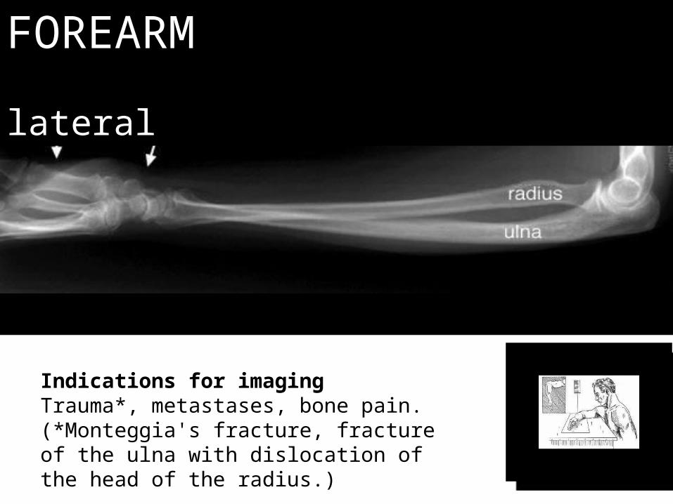
Forearmroutine: AP and lateral

FOREARMAP
Indications for imagingTrauma*, metastases, bone pain.(*Monteggia's fracture, fracture of the ulna with dislocation of the head of the radius.

FOREARMAP
FOREARMlateral
Indications for imagingTrauma*, metastases, bone pain.(*Monteggia's fracture, fracture of the ulna with dislocation of the head of the radius.)

FOREARMlateral

FOREARMAP
MONTEGGIA’s FRACTURE
- Giovanni Monteggia (1814) first described frx of proximal 1/3 of ulna in association w/ anterior dislocation of radial head; - hence dislocation of radial head w/ frx of proximal 1/3 of ulna is known as Monteggia's deformity.
- Mechanism: - proposed mechanisms include direct blow & hyperpronation injuries as well- as the hyperextension theory;
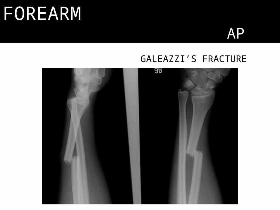
FOREARMAP
GALEAZZI’S FRACTURE

Fat Pad Sign
• More radiolucent area (less dense); seen in fractures wherein hematoma and bleeding pushes fat.
• Fracture hematomoa push out the normal fat visible radiolucent area

Wristroutine: PA and lateralSpecial: carpal tunnel view

WRISTPAIndications for imaging
Injury, pain, carpal tunnel syndrome,

WRISTlateral
Indications for imagingInjury, pain, carpal tunnel syndrome,

WRISTcarpal tunnel
view

WRISTothers
COLLES’ FRACTURE

Handroutine: AP and oblique
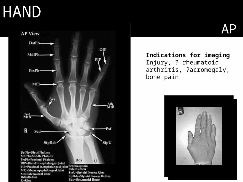
HANDAP
Indications for imagingInjury, ? rheumatoid arthritis, ?acromegaly, bone pain

HANDAP
Hand X-ray - AP1, Distal phalanx. 2, Distal interphalangeal joint. 3, Middle phalanx. 4, Proximal interphalangeal joint. 5, Proximal phalanx. 6, Metacarpophalangeal joint. 7, Head of 5th metacarpal. 8, Sesamoid bone.

HANDoblique

HANDothers

Common Views for Lower Extremities
• Both hips AP, frog leg lateral (for children, congenital problems)
• Cross table lateral (for hip fractures)• Femur AP, lateral• Knee AP, Lateral• Leg AP, Lateral• Ankle mortise, AP, Lateral• Foot AP, oblique, lateral• Pelvis AP

Pelvisroutine: AP only
Special: Pelvic Inlet, Pelvic OutletAcetabulum: Judets view (obturator & iliac)

PELVISAP

PELVISAP
Indications for imagingCongenital abnormalities, Trauma, degenerative disease, carcinoma primary and secondary, pathologies e.g. Perthes disease, slipped femoral epiphyses.
Anatomy DemonstratedIliac bones, femoral heads and necks, ishium, pubis and scrum.

PELVISAP
1 Superior Ramus of Right Pubis2 Symphysis Pubis3 Inferior Ramus of Left Pubis4 Left obturator foramen5 Left lesser Trochanter6 Left Greater Trochanter7 Left iliac wing8 Iliac crest9 Vertebral Pedicle (Lumbar Spine)10 Right Sacro-iliac joint 11 Head of right femur

PELVISAP

PELVISinlet vs outlet
view

PELVISmale vs
female

PELVISAP
Female pelvis. Note the sacro-iliac joints, the subpubic angle, and the continuous curvature of the margin of the obturator foramen and the neck of the femur (Shenton's line)

PELVISAP
This pelvis is of an 11 month old. To draw Shenton's line, the inferior border of the superior pubic ramus is traced laterally and should smoothly extend to the inferomedial border of the proximal femur.

PELVISacetabulum
1,Acetabular fossa.2, Head femoral.3, Greater trochanter.4, Lesser trochanter.5, Femur.6, Obturator foramen.7, Inferior pubic ramus.8, Superior pubic ramus.9, Sacrum.10, Iliac wing.

Sample Case• Asymmetrical (imagine picture- may obvious rami fracture and a
subtle SI fracture which John/.Joshua/Roel spotted)- Hole: symmetrical?- SI joint: normal? Widened? (if widened = Ala fracture)
• Request other views: outlet is dislocation up or down? Inlet did disloc hemipelvis move pa-front of back? *Sacral Wing problems involve Nerves S1-5 (which innervate the bladder, I.e. urinary and sexual function implications)
• True leg length (ASIS to medial malleolus), is it equal? Yes. • Apparent leg length (umbilicus [fixed portion in midline] to medial
malleolus) equal? No• hemi pelvis moved upwards and posteriorly

PELVISfrog’s view
Indications for imagingCongenital abnormalities, Perthes disease, slipped femoral epiphyses.
Anatomy DemonstratedFemoral heads and necks, acetabulum

PELVISfrog’s view
1, Symphysis pubis.2, Obturator foramen.3, Ischium.4, Lesser trochanter.5, Femur.6, Femoral head.7, Anterior inferior iliac spine.8, Acetabular fossa.9, Anterior superior iliac spine.

PELVIS- acetabulum visualization
obturator vs iliac view

PELVISobturator vs iliac

PELVISobturator vs illac

Additional Info
• Cross table lateral viewpatient’s opposite/unaffected limb is raised, plate is beside the involved hip, beam is at 30 degrees
• Frog leg view soles of the feet together then ask patient to make bukaka. Can see the relationship of the hip joint to the acetabulum. In kids, you can see if may dysplastic hip or a slipped capital femoral epiphysis.

Cervical SpineRoutine: AP and lateral
Special: Swimmer’s, Open Mouth

CERVICAL SPINE lateral
Indications for imagingTrauma, pain, rheumatoid arthritis, upper limb paraethesia, vertebral artery syndrome.

CERVICAL SPINE lateral

CERVICAL SPINE lateral
Cervical Spine X-ray: Lateral view. 1, Vertebral body (TH1). 2, Spinous process of C7. 3, Lamina. 4, Inferior articular process. 5, Superior articular process. 6,Spinous process of C2. 7, Odontoid process. 8, Anterior arch of C1 (Atlas). 9,Trachea.

CERVICAL SPINE lateral

CERVICAL SPINE lateral
Normal cervical spine?

CERVICAL SPINE lateral
A lateral radiograph of the cervical spine demonstrates a fracture through the posterior elements of C2 (yellow arrow) with forward subluxation of the anterior aspect of C2 on C3 (white arrow). This injury is caused by a combination of extension and compression

CERVICAL SPINE lateral

CERVICAL SPINE lateral
A lateral radiograph of the cervical spine demonstrates subluxation of C1 on C2, in this instance anterior subluxation most likely caused by severe hyperflexion (white arrow).
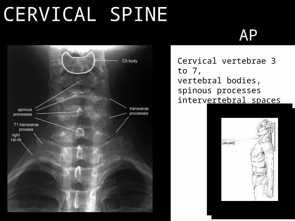
CERVICAL SPINE AP
Cervical vertebrae 3 to 7, vertebral bodies, spinous processes intervertebral spaces

CERVICAL SPINE AP
Cervical vertebrae 3 to 7, vertebral bodies, spinous processes intervertebral spaces

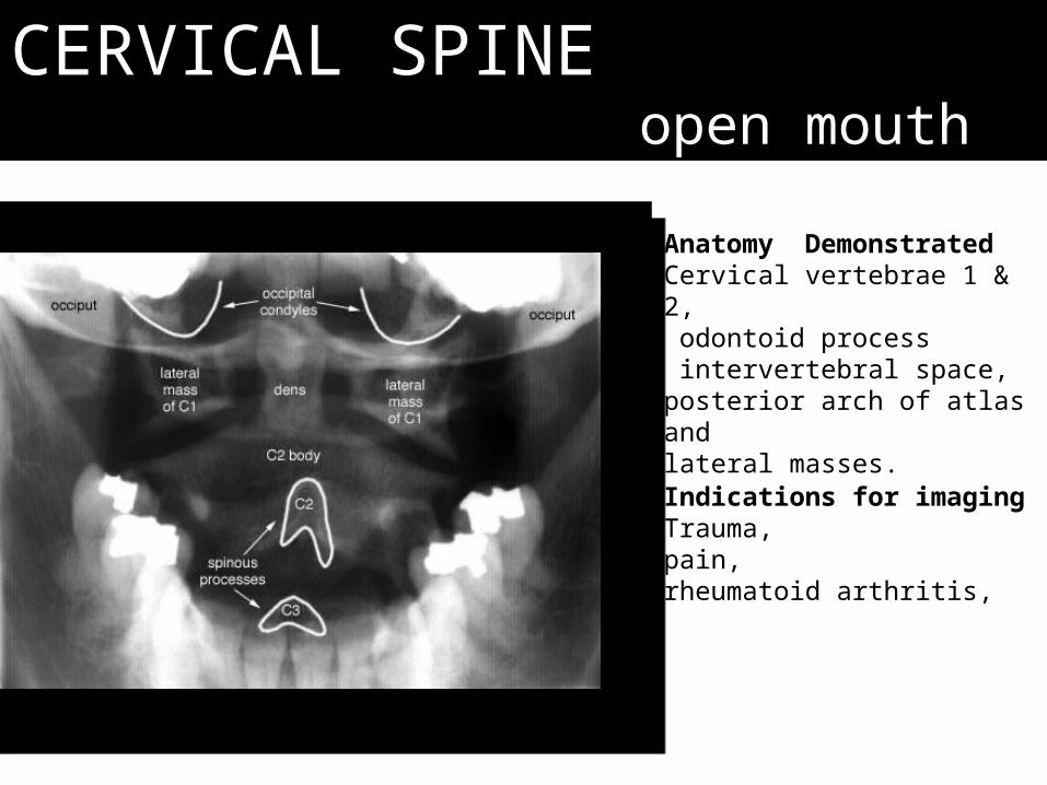
CERVICAL SPINE open mouth
CERVICAL SPINE open mouth
Indications for imagingTrauma, pain, rheumatoid arthritis,
Anatomy DemonstratedCervical vertebrae 1 & 2, odontoid process intervertebral space, posterior arch of atlas and lateral masses.

CERVICAL SPINE open mouth
Atlas and odontoid process: AP view (Mouth wide open). 1, Transverse process of C1. 2, Lateral mass of C1. 3, Odontoid. 4, Inferior articular process of C1.5, Superior articular process of C2.

Jefferson Fracture. There is bilateral offset of both the right and left lateral masses of C1 relative to the lateral masses of C2 on the open-mouth cervical spine view (above-white arrows).
This indicates a burst-type injury to the ring of C1. A single axial CT scan through the level of C1 shows fractures involving the right and left anterior ring of C1 and the right posterior ring (yellow arrows).
CERVICAL SPINE open mouth

CERVICAL SPINE oblique
Anatomy DemonstratedCervical vertebra bodies, intervertebral foramina, articular pillars apophysial joints and spinous processes. The intervertebral foramina demonstrated are those furthest from the film.
Indications for imagingTrauma, pain, rheumatoid arthritis, upper limb paraethesia, vertebral artery syndrome.

CERVICAL SPINE oblique
Anatomy Demonstrated
Cervical vertebra bodies, intervertebral foramina,articular pillars apophysial jointsspinous processes.

CERVICAL SPINE oblique
Cervical Spine X-ray, (Left Neural Foramina). 1, Rib. 2, Clavicle. 3, Neural Foramina. 4, Pedicle. 5, Trachea.

CERVICAL SPINE swimmer’s view

CERVICAL SPINE swimmer’s view

Thoracic Spineroutine: AP and lateral

AP VIEWTHORACIC SPINE AP
Indications for imagingCongenital abnormalities, scoliosis, trauma, pain, metastasis's.
Anatomy DemonstratedThoracic vertebra, medial ends of ribs.

THORACIC SPINE AP

THORACIC SPINE AP

THORACIC SPINE AP
Thoracic Spine X-ray: AP projection. 1, Left ventricle. 2, Gas in stomach. 3, Right hemidiaphragm. 4, Posterior rib. 5,Clavicle.
pedicles

THORACIC SPINE AP
Thoracic Spine X-ray: AP projection. 1, Gas in Colon (Splenic flexure). 2, Gas in stomach. 3, Left hemidiaphragm. 4, Posterior rib. 5, Pedicle. 6, Spinous process. 7, Transverse process.

THORACIC SPINE AP
Di pala thoracic to sorry! HEHE!Just to show scoliosis!

THORACIC SPINE AP
Spot the Winking Owl!

THORACIC SPINE AP

THORACIC SPINE lateral
Indications for imagingCongenital abnormalities, scoliosis, trauma, pain, metastasis's.
Anatomy DemonstratedThoracic vertebra,

THORACIC SPINE lateral
Thoracic Spine X-ray: Lateral view. 1, Right hemidiaphragm. 2, Left hemidiaphragm. 3, Vertebral body. 4, Rib
THORACIC SPINE lateral
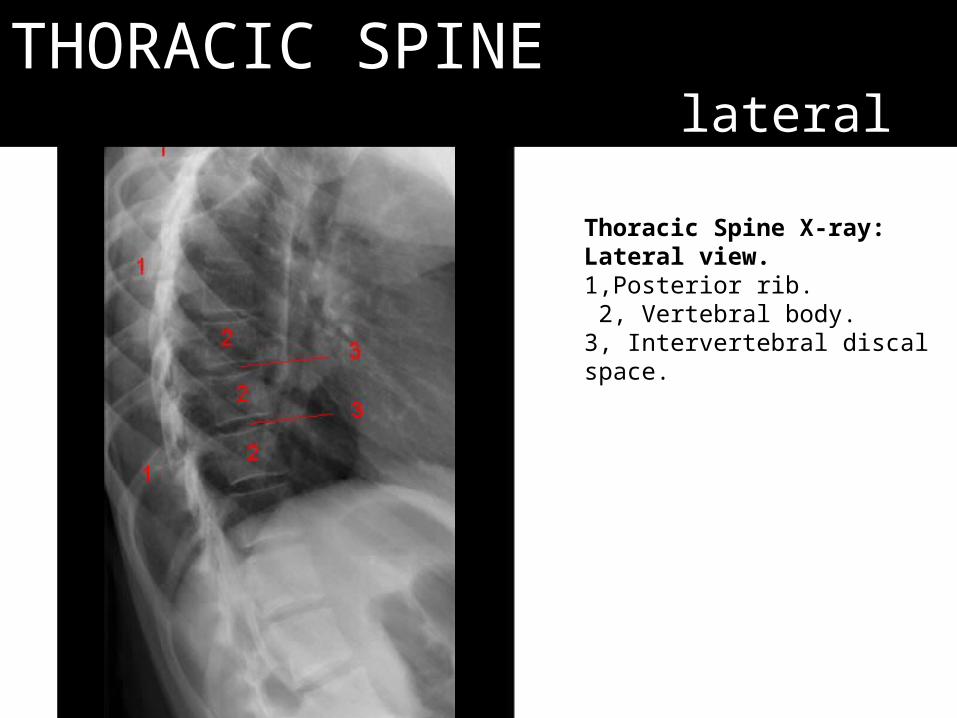
Thoracic Spine X-ray: Lateral view. 1,Posterior rib. 2, Vertebral body. 3, Intervertebral discal space.

THORACIC SPINE lateral

THORACIC SPINE lateral
A 73-year-old female with L1 vertebral compression fracture treated with kyphoplasty 6 weeks after fracture. The focal kyphosis was corrected from 16° to 5°.

Lumbosacral Spineroutine: AP and lateral

LUMBOSACRAL SPINE AP
Indications for imagingCongenital abnormalities, trauma, pain, metastasis's.
Anatomy DemonstratedLumbar vertebra, sacro iliac joints, Sacrumcoccyx

LUMBOSACRAL SPINE AP
Lumbar spine X-ray, AP projection1, rib. 2, Transverse process. 3, Pedicle. 4, Spinous Process. 5, Sacrum. 6, Sacroiliac joint

LUMBOSACRAL SPINE AP

LUMBOSACRAL SPINE AP

LUMBOSACRAL SPINE AP

LUMBOSACRAL SPINE lateral
Indications for imagingCongenital abnormalities, trauma, pain, metastasis's.
Anatomy DemonstratedLumbar vertebra.

LUMBOSACRAL SPINE lateral
Lumbar spine X-ray, lateral view1, Sacrum. 2, Spinous Process. 3, Vertebral body.4, Intervertebral disc space. 5, Intervertebral foramina. , Pedicle. 7, Inferior articulating facet. 8,Superior articulating facet. 9, Rib .

LUMBOSACRAL SPINE oblique

LUMBOSACRAL SPINE lateral

Kneeroutine: AP and lateral
Special: patella’s skyline view
s

APKNEE
AP

Standing APKNEE
AP
1
2
3
4

LATKNEE
lateral

KNEEskyline view
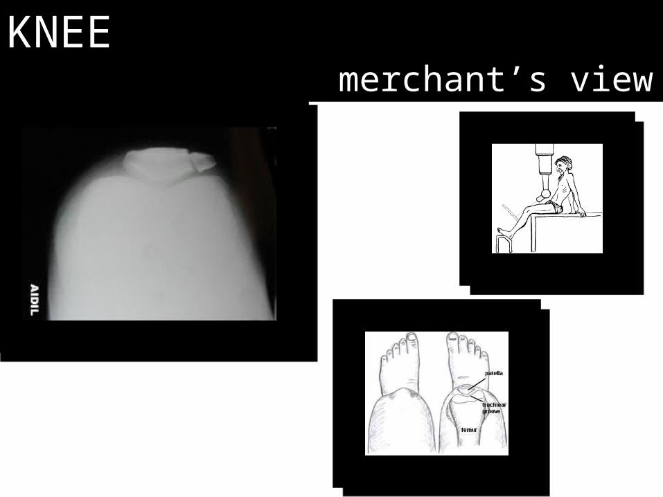
KNEEmerchant’s view

Ankleroutine: AP, mortise and lateral
s
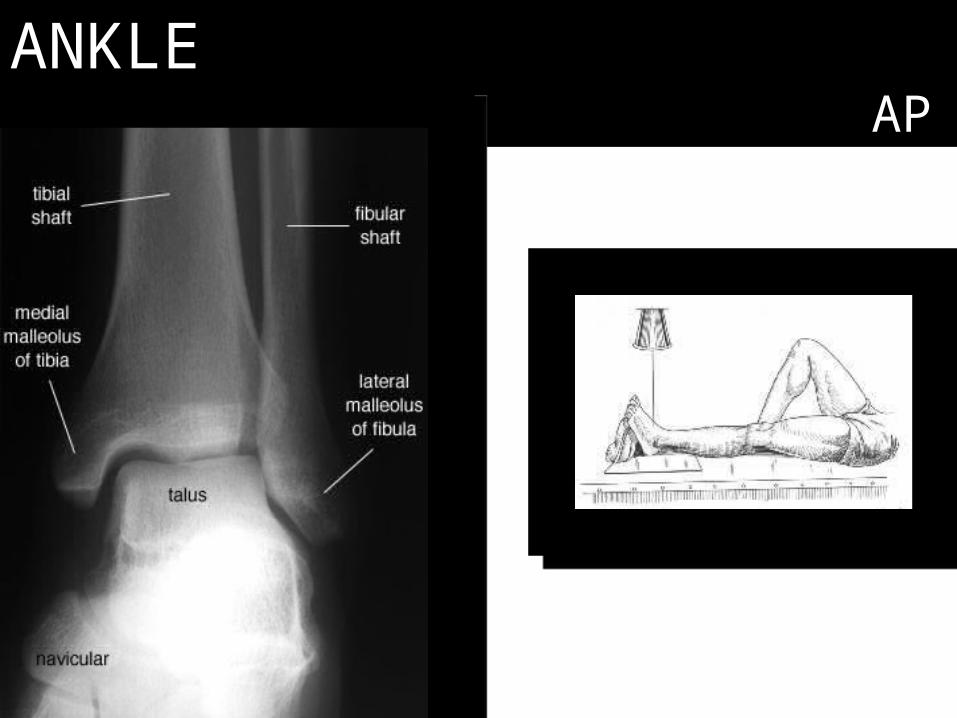
ANKLEAP

ANKLEmortise view

ANKLElateral view
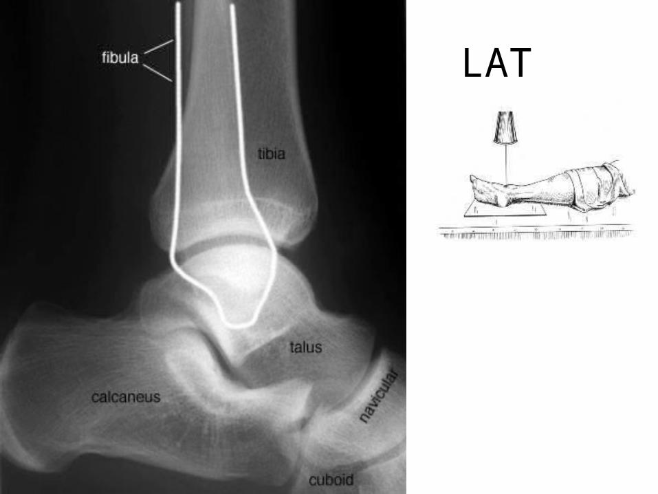
LAT

CalcaneusAxial , lateral, axial view
s

CALCANEUSlateral view

CALCANEUSlateral view

FootAP and oblique
s
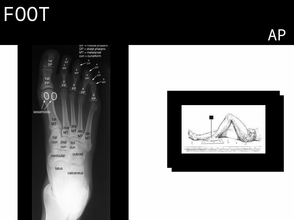
FOOTAP
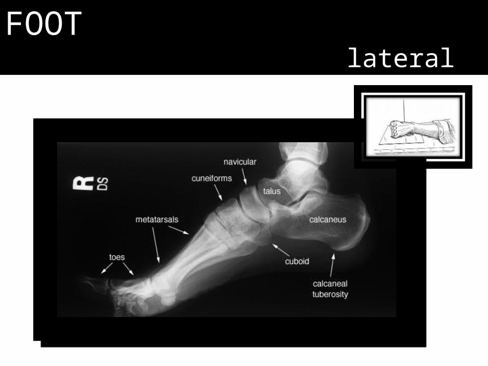
FOOTlateral

FOOToblique

Lisfranc’s Fracture

The end.