ORIGINAL ARTICLE Transgenerational Glucose...
10
Transgenerational Glucose Intolerance With Igf2/H19 Epigenetic Alterations in Mouse Islet Induced by Intrauterine Hyperglycemia Guo-Lian Ding, 1 Fang-Fang Wang, 1 Jing Shu, 1 Shen Tian, 1 Ying Jiang, 1 Dan Zhang, 1 Ning Wang, 1 Qiong Luo, 1 Yu Zhang, 1 Fan Jin, 1 Peter C.K. Leung, 2 Jian-Zhong Sheng, 3 and He-Feng Huang 1,4 Gestational diabetes mellitus (GDM) has been shown to be associated with high risk of diabetes in offspring. However, the mechanisms involved and the possibilities of transgenerational transmission are still unclear. We intercrossed male and female adult control and first-generation offspring of GDM (F1-GDM) mice to obtain the second-generation (F2) offspring in four groups: C♂-C♀,C♂-GDM♀, GDM♂-C♀, and GDM♂-GDM♀. We found that birth weight significantly increased in F2 offspring through the paternal line with impaired glucose tolerance (IGT). Regardless of birth from F1-GDM with or without IGT, high risk of IGT appeared as early as 3 weeks in F2 offspring and progressed through both parental lineages, especial the paternal line. IGT in male offspring was more obvious than that in females, with paren- tal characteristics and sex-specific transmission. In both F1 and F2 offspring of GDM, the expression of imprinted genes Igf2 and H19 was downregulated in pancreatic islets, caused by abnormal meth- ylation status of the differentially methylated region, which may be one of the mechanisms for impaired islet ultrastructure and func- tion. Furthermore, altered Igf2 and H19 gene expression was found in sperm of adult F1-GDM, regardless of the presence of IGT, indicating that changes of epigenetics in germ cells contributed to transgenerational transmission. Diabetes 61:1133–1142, 2012 A growing body of research suggests that expo- sure to an abnormal environment in the uterus can lead to chronic health problems later in life (1,2). Intrauterine hyperglycemia is a major characteristic of gestational diabetes mellitus (GDM) and has been suggested to be an important determinative fac- tor for the risk of diabetes in adulthood, in addition to the effects of genetic factors (3–6). Although many studies have investigated intrauterine growth retardation and later dia- betes, the mechanism involved in the association between intrauterine hyperglycemia and a high risk of diabetes in offspring remains unclear (7,8). In mammals, epigenetic reprogramming is involved in germ cells and early embryonic development (9,10). Evolutionarily and environmentally acquired genomic susceptibilities can induce epigenomic modulations early in life, which impact on the later development of human disease (11). Moreover, frequent tissue-specific and disease-specific de novo meth- ylation events occur during somatic cell development and differentiation (12). Because erasure and establishment of the genomic imprints for some imprinted genes begin when migratory primordial germ cells (PGCs) enter the embry- onic genital ridge through gametogenesis, epigenetic ab- normalities that occur during this phase may be involved in transgenerational transmission (13,14). We hypothesized that the hyperglycemic intrauterine environment of GDM could result in a high risk of diabetes in offspring by altering epigenetic modification. In addition to intergenerational transmission (F1 offspring), intrauterine hyperglycemia may also have effects on the second generation (F2 offspring). Since it is difficult to analyze the comparative parental contributions and the underlying mechanisms that result in F2 offspring outcomes in humans, in the current study we established a GDM mouse model of intrauterine hyper- glycemia. The female (♀) and male (♂) F1 adults of control and GDM mice were intercrossed to obtain F2 offspring of four groups: 1)C♂-C♀, 2)C♂-GDM♀, 3) GDM♂-C♀, and 4) GDM♂-GDM♀. Besides the phenotype, we examined the expression of imprinted genes Igf2, H19, and Plagl1 in islets. Abnormal Igf2 production has been shown to de- velop b-cell dysfunction and result in diabetes and appears to be an early landmark in the pathological sequence lead- ing to apoptosis of b-cells in the fetal Goto-Kakizaki (GK) rat (15–17). H19 is an imprinted gene at 90 kb 39 of Igf2 and is reciprocally imprinted with respect to Igf2, regulating its imprinting and expression (18,19). Plagl1, known as tran- scription factor zinc finger protein, is involved in the path- ogenesis of transient neonatal diabetes mellitus (20–22). We further analyzed the methylation status of differentially methylated regions (DMRs) in Igf2/H19 in mouse islets to investigate whether intrauterine hyperglycemia affected imprinted gene expression by regulating epigenetic modifi- cation. In addition, we examined imprinted genes in sperm of F1-GDM to explore the underlying mechanism of trans- generational transmission by germ cells. RESEARCH DESIGN AND METHODS All animal protocols were reviewed and approved by the Zhejiang University Animal Care and Use Committee. At the age of 8 weeks, virgin female ICR mice (n = 60) were mated with normal males. Onset of pregnancy was determined by the presence of a copulation plug after overnight mating (designated as day 0 [D0] of pregnancy). After a 12-h fast, the females were randomly divided into a control group and an intrauterine hyperglycemia group with GDM (GDM group). Mice in the GDM group were injected with a single intraperitoneal injection of streptozotocin (STZ; Sigma, St. Louis, MO) in 0.1 mmol/L citrate buffer (pH 4.5) at a dose of 150 mg/kg body wt. Control pregnant females received an equal volume of citrate buffer. On D3 of pregnancy, diabetes was confirmed by measurement of blood glucose concentration via the tail vein From the 1 Department of Reproductive Endocrinology, Women’s Hospital, School of Medicine, Zhejiang University, Hangzhou, China; the 2 Department of Obstetrics and Gynecology, Child and Family Research Institute, Univer- sity of British Columbia, Vancouver, British Columbia, Canada; the 3 Depart- ment of Pathophysiology, School of Medicine, Zhejiang University, Hangzhou, China; and the 4 Key Laboratory of Reproductive Genetics, Min- istry of Education, Hangzhou, China. Corresponding author: He-Feng Huang, [email protected], or Jian-Zhong Sheng, [email protected]. Received 23 September 2011 and accepted 21 January 2012. DOI: 10.2337/db11-1314 G.-L.D. and F-F.W. contributed equally to this study. Ó 2012 by the American Diabetes Association. Readers may use this article as long as the work is properly cited, the use is educational and not for profit, and the work is not altered. See http://creativecommons.org/licenses/by -nc-nd/3.0/ for details. diabetes.diabetesjournals.org DIABETES, VOL. 61, MAY 2012 1133 ORIGINAL ARTICLE
Transcript of ORIGINAL ARTICLE Transgenerational Glucose...
Transgenerational Glucose Intolerance With Igf2/H19Epigenetic Alterations in Mouse Islet Induced byIntrauterine HyperglycemiaGuo-Lian Ding,
1Fang-Fang Wang,
1Jing Shu,
1Shen Tian,
1Ying Jiang,
1Dan Zhang,
1Ning Wang,
1
Qiong Luo,1Yu Zhang,
1Fan Jin,
1Peter C.K. Leung,
2Jian-Zhong Sheng,
3and He-Feng Huang
1,4
Gestational diabetes mellitus (GDM) has been shown to beassociated with high risk of diabetes in offspring. However, themechanisms involved and the possibilities of transgenerationaltransmission are still unclear. We intercrossed male and femaleadult control and first-generation offspring of GDM (F1-GDM)mice to obtain the second-generation (F2) offspring in fourgroups: C♂-C♀, C♂-GDM♀, GDM♂-C♀, and GDM♂-GDM♀. Wefound that birth weight significantly increased in F2 offspringthrough the paternal line with impaired glucose tolerance (IGT).Regardless of birth from F1-GDM with or without IGT, high risk ofIGT appeared as early as 3 weeks in F2 offspring and progressedthrough both parental lineages, especial the paternal line. IGT inmale offspring was more obvious than that in females, with paren-tal characteristics and sex-specific transmission. In both F1 and F2offspring of GDM, the expression of imprinted genes Igf2 and H19was downregulated in pancreatic islets, caused by abnormal meth-ylation status of the differentially methylated region, which may beone of the mechanisms for impaired islet ultrastructure and func-tion. Furthermore, altered Igf2 and H19 gene expression wasfound in sperm of adult F1-GDM, regardless of the presence ofIGT, indicating that changes of epigenetics in germ cells contributedto transgenerational transmission. Diabetes 61:1133–1142, 2012
Agrowing body of research suggests that expo-sure to an abnormal environment in the uteruscan lead to chronic health problems later in life(1,2). Intrauterine hyperglycemia is a major
characteristic of gestational diabetes mellitus (GDM) andhas been suggested to be an important determinative fac-tor for the risk of diabetes in adulthood, in addition to theeffects of genetic factors (3–6). Although many studies haveinvestigated intrauterine growth retardation and later dia-betes, the mechanism involved in the association betweenintrauterine hyperglycemia and a high risk of diabetes inoffspring remains unclear (7,8).
In mammals, epigenetic reprogramming is involved in germcells and early embryonic development (9,10). Evolutionarilyand environmentally acquired genomic susceptibilities can
induce epigenomic modulations early in life, which impacton the later development of human disease (11). Moreover,frequent tissue-specific and disease-specific de novo meth-ylation events occur during somatic cell development anddifferentiation (12). Because erasure and establishment ofthe genomic imprints for some imprinted genes begin whenmigratory primordial germ cells (PGCs) enter the embry-onic genital ridge through gametogenesis, epigenetic ab-normalities that occur during this phase may be involved intransgenerational transmission (13,14). We hypothesizedthat the hyperglycemic intrauterine environment of GDMcould result in a high risk of diabetes in offspring by alteringepigenetic modification. In addition to intergenerationaltransmission (F1 offspring), intrauterine hyperglycemia mayalso have effects on the second generation (F2 offspring).
Since it is difficult to analyze the comparative parentalcontributions and the underlying mechanisms that result inF2 offspring outcomes in humans, in the current study weestablished a GDM mouse model of intrauterine hyper-glycemia. The female (♀) and male (♂) F1 adults of controland GDM mice were intercrossed to obtain F2 offspring offour groups: 1) C♂-C♀, 2) C♂-GDM♀, 3) GDM♂-C♀, and 4)GDM♂-GDM♀. Besides the phenotype, we examined theexpression of imprinted genes Igf2, H19, and Plagl1 inislets. Abnormal Igf2 production has been shown to de-velop b-cell dysfunction and result in diabetes and appearsto be an early landmark in the pathological sequence lead-ing to apoptosis of b-cells in the fetal Goto-Kakizaki (GK)rat (15–17). H19 is an imprinted gene at 90 kb 39 of Igf2 andis reciprocally imprinted with respect to Igf2, regulating itsimprinting and expression (18,19). Plagl1, known as tran-scription factor zinc finger protein, is involved in the path-ogenesis of transient neonatal diabetes mellitus (20–22). Wefurther analyzed the methylation status of differentiallymethylated regions (DMRs) in Igf2/H19 in mouse islets toinvestigate whether intrauterine hyperglycemia affectedimprinted gene expression by regulating epigenetic modifi-cation. In addition, we examined imprinted genes in spermof F1-GDM to explore the underlying mechanism of trans-generational transmission by germ cells.
RESEARCH DESIGN AND METHODS
All animal protocols were reviewed and approved by the Zhejiang UniversityAnimal Care and Use Committee. At the age of 8 weeks, virgin female ICR mice(n = 60) were mated with normal males. Onset of pregnancy was determinedby the presence of a copulation plug after overnight mating (designated as day0 [D0] of pregnancy). After a 12-h fast, the females were randomly divided intoa control group and an intrauterine hyperglycemia group with GDM (GDMgroup). Mice in the GDM group were injected with a single intraperitonealinjection of streptozotocin (STZ; Sigma, St. Louis, MO) in 0.1 mmol/L citratebuffer (pH 4.5) at a dose of 150 mg/kg body wt. Control pregnant femalesreceived an equal volume of citrate buffer. On D3 of pregnancy, diabetes wasconfirmed by measurement of blood glucose concentration via the tail vein
From the 1Department of Reproductive Endocrinology, Women’s Hospital,School of Medicine, Zhejiang University, Hangzhou, China; the 2Departmentof Obstetrics and Gynecology, Child and Family Research Institute, Univer-sity of British Columbia, Vancouver, British Columbia, Canada; the 3Depart-ment of Pathophysiology, School of Medicine, Zhejiang University,Hangzhou, China; and the 4Key Laboratory of Reproductive Genetics, Min-istry of Education, Hangzhou, China.
Corresponding author: He-Feng Huang, [email protected], or Jian-ZhongSheng, [email protected].
Received 23 September 2011 and accepted 21 January 2012.DOI: 10.2337/db11-1314G.-L.D. and F-F.W. contributed equally to this study.� 2012 by the American Diabetes Association. Readers may use this article as
long as the work is properly cited, the use is educational and not for profit,and the work is not altered. See http://creativecommons.org/licenses/by-nc-nd/3.0/ for details.
diabetes.diabetesjournals.org DIABETES, VOL. 61, MAY 2012 1133
ORIGINAL ARTICLE
and defined as a glucose level between 14 and 19 mmol/L (252–342 mg/dL),which was also measured on D7 and D20 of pregnancy to confirm the diabetescondition as previously described (23–26).
The pregnant mice were allowed to deliver spontaneously. The litter sizewas randomly reduced to 10 at birth to assure uniformity. The pups from theGDM groupwere fostered by normoglycemic females until they were weaned atthe age of 3 weeks.
The ♀ and ♂ F1 adults of control (F1-C) and GDM (F1-GDM) mice wereintercrossed, and the phenotypes of their F2 offspring were characterized. F2offspring were obtained from 4 groups: 1) C♂-C♀, 2) C♂-GDM♀, 3) GDM♂-C♀,and 4) GDM♂-GDM♀ (Fig. 1A).In vivo glucose and insulin analysis. Intraperitoneal glucose injection (2 g/kgbody wt) was performed in unrestrained conscious mice after a 12-h overnightfast. The area under the curve (AUC) of glucose against time was calculatedfor analysis of glucose tolerance as previously described (27). Blood samplesof fasted or fed condition were taken for evaluation of serum insulin concentra-tions by solid-phase enzyme-linked immunosorbent assay using commerciallyavailable kits (Linco Research., St. Charles, MI).Pancreatic islet isolation and insulin secretion. In anesthetized 8-week-oldmice, 2 mg/mL collagenase (Worthington Biochemical, Lakewood, NJ) wasinjected into the bile duct as previously described (28). The pancreas wasdissected out, with static incubation at 38°C for 20 min followed by shakingincubation at 38°C for 8 min. After ficoll gradient separation, freshly isolatedislets of similar size were handpicked under a stereomicroscope. Islets (20 perwell) were preincubated in RPMI-1640 (Sigma-Aldrich, St. Louis, MO), washed,and incubated in 1 mL fresh RPMI-1640 that contained indicated glucose for 30min (37°C). Fifty microliters of the medium were removed for insulin analysis.
Islets were extracted in acid ethanol (4°C) and stored (220°C) for insulincontent assay. In mice at embryonic day 17 (E17), the pancreas was directlydissected out and incubated in 2 mg/mL collagenase under the same conditionsas described above. Isolated islets were incubated in RPMI-1640 containingdifferent glucose concentrations (5 mmol/L or 25 mmol/L) for 24 h (37°C).Sperm collection. Sperm were obtained from the caudal epididymis of8-week-old adult male mice. Then, the sperm were washed in phosphate-bufferedsaline and centrifuged to pellet.Gene expression (quantitative PCR). Total RNA was isolated from islets orsperm using RNeasy (Qiagen, Valencia, CA). The cDNA was synthesized usingoligo-dT and random primers (TaKaRa, Dalian, China) for real-time quantitativePCR (ABI Prism 7900HT; Applied Biosystems, Foster City, CA). Glyceraldehyde-3-phosphate dehydrogenase served as an internal control.DNA methylation (bisulfite genomic sequencing PCR). Genomic DNAwas extracted from islets of 8-week-old mice in the control, F1-GDM, andGDM♂-GDM♀ groups. Bisulfite was converted using the EpiTect bisulfite kit(Qiagen, Valencia, CA) according to the manufacturer’s instructions to de-aminate cytosine to uracil; 5-methyl-cytosine was protected from deamination.Analysis of the methylation status of the Igf2 and H19 DMR was determinedby cloning and sequencing of bisulfite-treated DNA. The purified PCR productswere cloned using the pMD19-T vector system (TaKaRa, Dalian, China). Thesequence obtained by cloning was analyzed with 3730 DNA Analyzer polymers(Applied Biosystems, Carlsbad, CA).Electron microscopy. Samples were fixed in 2.5% glutaraldehyde and post-fixed in 1% osmium tetroxide and then stained with aqueous 2% uranyl acetatefor 30 min, dehydrated, and embedded in epoxy resin. Sections of 120 nm werestained with uranyl acetate and lead citrate. Samples were examined at 80 kV
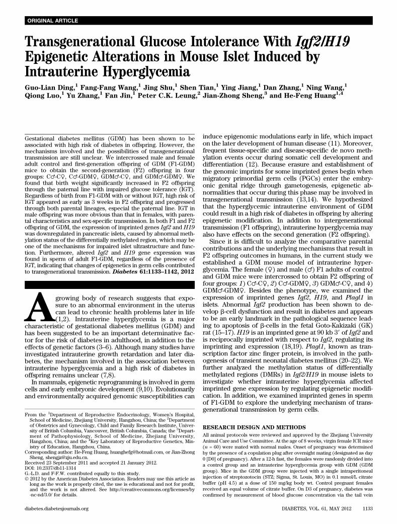
FIG. 1. Experimental design and islet ultrastructure of mice. A: Experimental design. Circles designate females and squares designate males. Notethat mating pairs were nonsiblings. The pups from the GDM group were fostered by normoglycemic females until weaned. B: Islet ultrastructure ofF1 offspring under transmission electron microscopy. Scale bar, 500 nm. C: Islet ultrastructure of F2 offspring under transmission electron mi-croscopy. Scale bar, 500 nm.
INTRAUTERINE HYPERGLYCEMIA AND EPIGENETICS
1134 DIABETES, VOL. 61, MAY 2012 diabetes.diabetesjournals.org
under a TECNAI 10 transmission electron microscope (Philips, Amsterdam,the Netherlands).Statistical analysis. Data are presented as means 6 SE. Statistical analysiswas performed by unpaired two-tailed Student t test, one-way ANOVA, or x2
test as described in the Table and Figure legends (version 16.0; SPSS). P , 0.05was considered statistically significant.
RESULTS
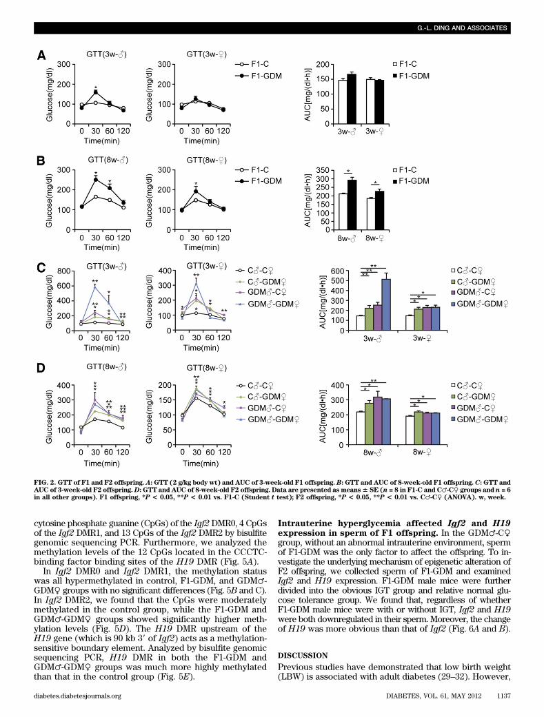
Intrauterine hyperglycemia induced transgenerationaleffect on birth weight and islet structure. We estab-lished a GDM mouse model by inducing moderate hyper-glycemia after pregnancy. Female and male F1 adults ofcontrol and GDM mice were intercrossed to obtain F2 off-spring (Fig. 1A). We found that the birth weight of F1-GDMoffspring was not changed significantly (Table 1). Afterfostering by the normoglycemic mice, the body weight ofboth F1-GDM males and females at 3 or 8 weeks of age wassimilar to that of F1-C offspring (Table 1). There was nosignificant difference of birth weight between control andF2-GDM mice born from F1-GDM with relative normal glu-cose tolerance (data not shown). After birth from F1-GDMwith impaired glucose tolerance (IGT), the birth weight inthe GDM♂-C♀ and GDM♂-GDM♀ groups significantly in-creased compared with that in C♂-C♀ (Table 1). However,the birth weight in the C♂-GDM♀ group did not increaseobviously. There was also no significant difference in birthweight between GDM♂-C♀ and GDM♂-GDM♀ groups(Table 1). The ratio of pancreas weight to body weight inF1-GDM offspring at 3 weeks was significantly higher thanthat in F1-C, although there was no difference at 8 weeks(Table 1). With the same tendency of F1-GDM mice, thepancreas weight–to–body weight ratio in F2-GDM offspringwhose one and/or two parents experienced intrauterinehyperglycemia was significantly higher than that in theC♂-C♀ group at 3 weeks (Table 1). There was no significantdifference of pancreatic islet morphology, insulin distri-bution, or glucagon distribution between F1-C and F1-GDM(data not shown). However, analyzed by transmissionelectron microscopy, the structure of the endoplasmic re-ticulum in islet cells was obviously swollen and disordered
in F1-GDM offspring at 3 weeks of age compared with thatof F1-C and progressed at 8 weeks of age (Fig. 1B). In F2offspring, compared with the C♂-C♀ group, the structure ofthe endoplasmic reticulum in islet cells was obviouslyswollen and disordered in F2-GDM groups at 3 weeks ofage, especially in GDM♂-C♀ and GDM♂-GDM♀ groups(Fig. 1C). At 8 weeks of age, the structure of endoplasmicreticulum in all the F2-GDM groups was almost recovered,with the exception of some fissures (Fig. 1C).Intrauterine hyperglycemia induced transgenerationaltransmission of glucose intolerance and abnormalinsulin level. Blood glucose levels of all the 3- and 8-week-old F1-GDM and F2-GDM offspring did not differ fromthose in the corresponding controls in either the fastingor random-fed condition (Table 2). We further performedglucose tolerance test (GTT) by intraperitoneal injection ofglucose (2 g/kg body wt). At 3 weeks, in both males andfemales, there were no differences in the GTT AUC betweenF1-GDM and F1-C, although the blood glucose level sig-nificantly increased in the males at 30 min after injection(Fig. 2A). However, at 8 weeks, IGT was found in both themale and female mice of the F1-GDM group, whose bloodglucose level significantly increased at 30 min after injection(Fig. 2B). F1-GDM males showed more evident IGT thanfemales (Fig. 2B). After GTT was performed by intraper-itoneal injection of glucose (2 g/kg body wt), IGT appearedin both 3- and 8-week-old male and female F2-GDM off-spring through both parental lineages. The male F2-GDMoffspring showed much more IGT than females (Fig. 2C andD). At 3 and 8 weeks of age, in both males and females,although fasting glucose levels were normal, fasting insulinlevels were much lower in F1-GDM than in F1-C (Table 2).In contrast, in the random-fed condition, the insulin levelsof F1-GDM mice significantly increased. The abnormal in-sulin level of the F1-GDM males was more obvious thanthat of the F1-GDM females (Table 2). In F2 offspring, thefasting insulin levels of the F2-GDM males at 3 and 8 weekswere significantly lower than those of the control group,and the fasting insulin levels in 8-week-old F2-GDM femaleswere also significantly decreased compared with the control
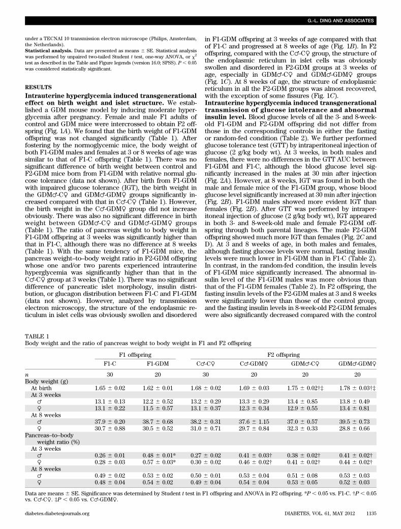
TABLE 1Body weight and the ratio of pancreas weight to body weight in F1 and F2 offspring
F1 offspring F2 offspring
F1-C F1-GDM C♂-C♀ C♂-GDM♀ GDM♂-C♀ GDM♂-GDM♀
n 30 20 30 20 20 20Body weight (g)At birth 1.65 6 0.02 1.62 6 0.01 1.68 6 0.02 1.69 6 0.03 1.75 6 0.02†‡ 1.78 6 0.03†‡At 3 weeks♂ 13.1 6 0.13 12.2 6 0.52 13.2 6 0.29 13.3 6 0.29 13.4 6 0.85 13.8 6 0.49♀ 13.1 6 0.22 11.5 6 0.57 13.1 6 0.37 12.3 6 0.34 12.9 6 0.55 13.4 6 0.81
At 8 weeks♂ 37.9 6 0.20 38.7 6 0.68 38.2 6 0.31 37.6 6 1.15 37.0 6 0.57 39.5 6 0.73♀ 30.7 6 0.88 30.5 6 0.52 31.0 6 0.71 29.7 6 0.84 32.3 6 0.33 28.8 6 0.66
Pancreas–to–bodyweight ratio (%)
At 3 weeks♂ 0.26 6 0.01 0.48 6 0.01* 0.27 6 0.02 0.41 6 0.03† 0.38 6 0.02† 0.41 6 0.02†♀ 0.28 6 0.03 0.57 6 0.03* 0.30 6 0.02 0.46 6 0.02† 0.41 6 0.02† 0.44 6 0.02†
At 8 weeks♂ 0.49 6 0.02 0.53 6 0.02 0.50 6 0.01 0.53 6 0.04 0.51 6 0.08 0.53 6 0.03♀ 0.48 6 0.04 0.54 6 0.02 0.49 6 0.04 0.54 6 0.04 0.53 6 0.05 0.52 6 0.03
Data are means 6 SE. Significance was determined by Student t test in F1 offspring and ANOVA in F2 offspring. *P , 0.05 vs. F1-C. †P , 0.05vs. C♂-C♀. ‡P , 0.05 vs. C♂-GDM♀.
G.-L. DING AND ASSOCIATES
diabetes.diabetesjournals.org DIABETES, VOL. 61, MAY 2012 1135
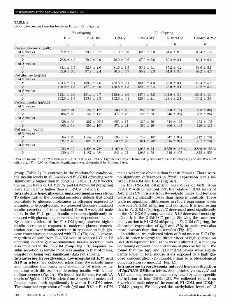
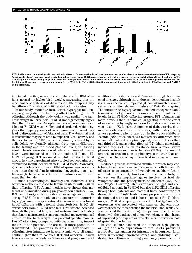
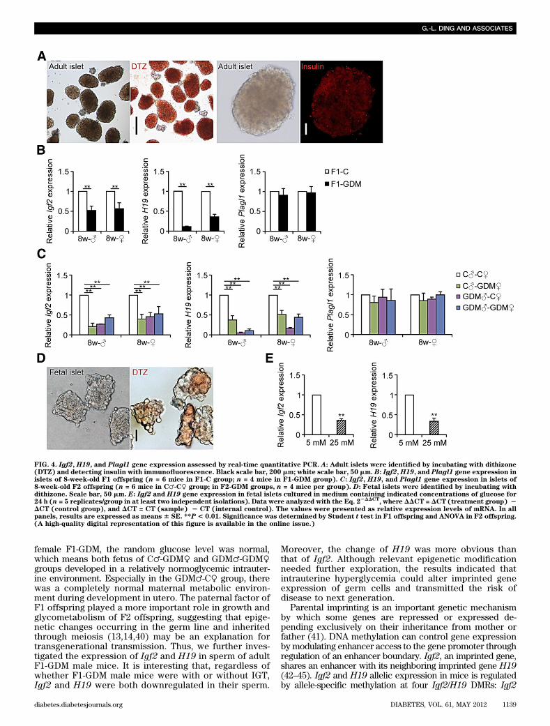
group (Table 2). In contrast, in the random-fed condition,the insulin levels in all 3-week-old F2-GDM offspring weresignificantly higher than in controls (Table 2). At 8 weeks,the insulin levels of GDM♂-C♀ and GDM♂-GDM♀offspringwere significantly higher than in C♂-C♀ (Table 2).Intrauterine hyperglycemia impaired insulin secretion.To define further the potential secretory defects that couldcontribute to glucose intolerance in offspring exposed tointrauterine hyperglycemia, we assessed glucose-stimulatedinsulin secretion of islets isolated from 8-week-old malemice. In the F1-C group, insulin secretion significantly in-creased with glucose exposure in a dose-dependent manner.By contrast, islets of the F1-GDM group showed higherinsulin secretion in response to moderate glucose concen-tration but lower insulin secretion in response to high glu-cose concentration compared with F1-C (Fig. 3A). Likewise,regardless of birth from F1-GDM with or without IGT, in F2offspring in vitro glucose-stimulated insulin secretion wasalso impaired in the F2-GDM group (Fig. 3B). Impaired in-sulin secretion in female mice was similar to that of males,despite not being very significant (data not shown).Intrauterine hyperglycemia downregulated Igf2 andH19 in islets. We collected islets from 8-week-old miceand identified the purity and specificity of islets by in-cubating with dithizone or detecting insulin with immu-nofluorescence (Fig. 4A). We found that the relative mRNAlevels of Igf2 and H19 in islets from 8-week-old males andfemales were both significantly lower in F1-GDM mice.The abnormal expression of both Igf2 and H19 in F1-GDM
males was more obvious than that in females. There wereno significant differences in Plagl1 expression levels be-tween F1-GDM and F1-C (Fig. 4B).
In the F2-GDM offspring, regardless of birth fromF1-GDM with or without IGT, the relative mRNA levels ofIgf2 and H19 in islets from 8-week-old males and femaleswere all significantly lower than those in controls. Therewere no significant differences in Plagl1 expression levelsbetween F2-GDM offspring and controls. It is interestingthat in F2-GDM offspring, Igf2 decreased most significantlyin the C♂-GDM♀ group, whereas H19 decreased most sig-nificantly in the GDM♂-C♀ group. Showing the same ten-dency as that in F1-GDM offspring, in F2-GDM offspring, theabnormal expression of Igf2 and H19 in males was alsomore obvious than that in females (Fig. 4C).
In addition, we collected islets of fetal mice at E17 (Fig.4D). In order to verify the direct effect of high glucose onislet development, fetal islets were cultured in a mediumcontaining different concentrations of glucose for 24 h. Wefound that the Igf2 and H19 mRNA levels were signifi-cantly lower in fetal mouse islets exposed to a high glu-cose concentration (25 mmol/L) than to a physiologicalconcentration (5 mmol/L) (Fig. 4E).Intrauterine hyperglycemia induced hypermethylationof Igf2/H19 DMRs in islets. As imprinted genes, Igf2 andH19 allelic expression in mice is regulated by allele-specificmethylation at four DMRs (21). We collected islets from8-week-old male mice of the control, F1-GDM, and GDM♂-GDM♀ groups. We analyzed the methylation levels of 12
TABLE 2Blood glucose and insulin levels in F1 and F2 offspring
F1 offspring F2 offspring
F1-C F1-GDM C♂-C♀ C♂-GDM♀ GDM♂-C♀ GDM♂-GDM♀
n 8 6 8 6 6 6Fasting glucose (mg/dL)At 3 weeks 82.2 6 1.5 79.2 6 3.7 81.0 6 2.0 82.2 6 3.3 81.6 6 2.6 80.4 6 1.5♂♀ 76.8 6 4.2 79.2 6 9.0 78.0 6 3.6 87.0 6 3.6 80.4 6 2.1 80.4 6 6.6
At 8 weeks♂ 83.4 6 1.5 82.8 6 2.0 81.6 6 1.5 83.4 6 3.1 85.2 6 4.2 84.6 6 8.1♀ 93.0 6 3.6 97.8 6 2.4 90.0 6 3.7 85.8 6 3.3 91.8 6 3.6 88.2 6 4.5
Fed glucose (mg/dL)At 3 weeks♂ 134.4 6 2.1 139.8 6 4.6 133.8 6 2.1 139.2 6 4.3 142.8 6 3.1 146.4 6 5.9♀ 129.0 6 1.5 127.2 6 2.6 130.8 6 1.5 129.0 6 2.4 132.0 6 2.1 142.2 6 5.4
At 8 weeks♂ 143.4 6 2.6 151.2 6 4.7 145.8 6 2.0 147.6 6 7.8 147.0 6 3.6 160.8 6 13♀ 132.6 6 1.5 133.8 6 4.3 132.6 6 3.1 126.6 6 2.1 128.4 6 2.1 130.2 6 3.9
Fasting insulin (pg/mL)At 3 weeks♂ 552 6 24 188 6 12* 508 6 19 208 6 26† 250 6 15† 188 6 36†♀ 496 6 26 135 6 11* 477 6 13 485 6 23 346 6 28† 182 6 26†
At 8 weeks♂ 628 6 36 207 6 20*‡ 653 6 17 320 6 29† 344 6 12† 153 6 13†♀ 605 6 13 303 6 19* 572 6 21 386 6 49† 300 6 14† 129 6 11†
Fed insulin (pg/mL)At 3 weeks♂ 585 6 20 1,157 6 22*‡ 535 6 37 752 6 18† 847 6 35† 1,142 6 79†♀ 597 6 49 822 6 11* 539 6 20 821 6 73† 1,015 6 92† 1,137 6 73†
At 8 weeks♂ 923 6 49 1,296 6 72* 1,169 6 39 1,036 6 74 2,539 6 115†‡ 2,839 6 156†‡♀ 939 6 13 1,187 6 68* 941 6 27 1,063 6 58 1,316 6 45† 1,875 6 55†
Data are means6 SE. *P, 0.05 vs. F1-C. †P, 0.05 vs. C♂-C♀. Significance was determined by Student t test in F1 offspring and ANOVA in F2offspring. ‡P , 0.05 vs. female. Significance was determined by Student t test.
INTRAUTERINE HYPERGLYCEMIA AND EPIGENETICS
1136 DIABETES, VOL. 61, MAY 2012 diabetes.diabetesjournals.org
cytosine phosphate guanine (CpGs) of the Igf2DMR0, 4 CpGsof the Igf2 DMR1, and 13 CpGs of the Igf2 DMR2 by bisulfitegenomic sequencing PCR. Furthermore, we analyzed themethylation levels of the 12 CpGs located in the CCCTC-binding factor binding sites of the H19 DMR (Fig. 5A).
In Igf2 DMR0 and Igf2 DMR1, the methylation statuswas all hypermethylated in control, F1-GDM, and GDM♂-GDM♀ groups with no significant differences (Fig. 5B and C).In Igf2 DMR2, we found that the CpGs were moderatelymethylated in the control group, while the F1-GDM andGDM♂-GDM♀ groups showed significantly higher meth-ylation levels (Fig. 5D). The H19 DMR upstream of theH19 gene (which is 90 kb 39 of Igf2) acts as a methylation-sensitive boundary element. Analyzed by bisulfite genomicsequencing PCR, H19 DMR in both the F1-GDM andGDM♂-GDM♀ groups was much more highly methylatedthan that in the control group (Fig. 5E).
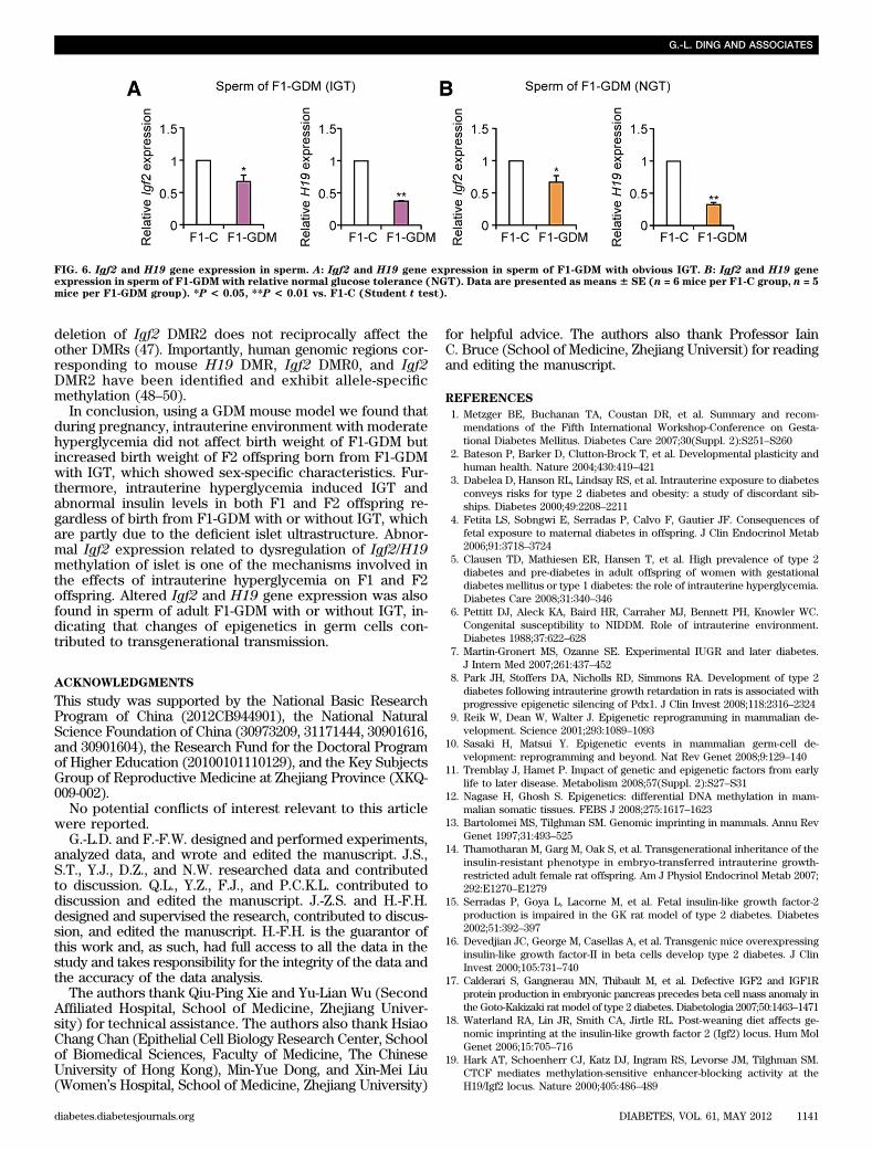
Intrauterine hyperglycemia affected Igf2 and H19expression in sperm of F1 offspring. In the GDM♂-C♀group, without an abnormal intrauterine environment, spermof F1-GDM was the only factor to affect the offspring. To in-vestigate the underlying mechanism of epigenetic alteration ofF2 offspring, we collected sperm of F1-GDM and examinedIgf2 and H19 expression. F1-GDM male mice were furtherdivided into the obvious IGT group and relative normal glu-cose tolerance group. We found that, regardless of whetherF1-GDM male mice were with or without IGT, Igf2 and H19were both downregulated in their sperm. Moreover, the changeof H19 was more obvious than that of Igf2 (Fig. 6A and B).
DISCUSSION
Previous studies have demonstrated that low birth weight(LBW) is associated with adult diabetes (29–32). However,
FIG. 2. GTT of F1 and F2 offspring. A: GTT (2 g/kg body wt) and AUC of 3-week-old F1 offspring.B: GTT and AUC of 8-week-old F1 offspring.C: GTT andAUC of 3-week-old F2 offspring.D: GTT and AUC of 8-week-old F2 offspring. Data are presented as means6 SE (n = 8 in F1-C and C♂-C♀ groups and n = 6in all other groups). F1 offspring, *P < 0.05, **P < 0.01 vs. F1-C (Student t test); F2 offspring, *P < 0.05, **P < 0.01 vs. C♂-C♀ (ANOVA). w, week.
G.-L. DING AND ASSOCIATES
diabetes.diabetesjournals.org DIABETES, VOL. 61, MAY 2012 1137
in clinical practice, newborns of mothers with GDM oftenhave normal or higher birth weight, suggesting that themechanism of high risk of diabetes in GDM offspring maybe different from that of LBW-related adult diabetes.
In our study, moderate intrauterine hyperglycemia dur-ing pregnancy did not obviously affect birth weight in F1offspring. Although the body weight was similar, the pan-creas weight in 3-week-old F1-GDM was significantly higherthan that of controls. Endoplasmic reticulum in pancreaticislets of F1-GDM was swollen and disordered, which sug-gests that hyperglycemia of intrauterine environment maylead to disorganization of fetal islet cells. The abnormal isletultrastructure may be related to impaired b-cell activity andthe development of IGT, which is primarily caused by in-sulin deficiency. Actually, although there was no differencein the fasting and fed blood glucose levels, the fastinginsulin levels were decreased while random-fed insulinlevels were increased in both the male and female F2-GDM offspring. IGT occurred in adults of the F1-GDMgroup. In vitro experiment also verified reduced glucose-stimulated insulin secretion in F1-GDM islets. Moreover,glucose intolerance of male GDM offspring was more ob-vious than that of female offspring, suggesting that malefetus might be more sensitive to the intrauterine environ-ment than female.
Human epidemiological investigation indicated a linkbetween mothers exposed to famine in utero with LBW intheir offspring (33). Animal models have shown that ma-ternal undernutrition during pregnancy could induce LBW,IGT, and obesity in both first- and second-generation off-spring (34,35). In our study, after exposure to intrauterinehyperglycemia, transgenerational transmission was foundin F2 offspring with parental characteristics. In F2 off-spring born from F1-GDM with IGT, birth weight increasedthrough the paternal line, but not maternal line, indicatingthat abnormal intrauterine environment had transgenerationaleffects on the birth weight in a parental-specific manner.For F2 offspring, compared with maternal line, impactfactors of the paternal line are much more prone to betransmitted. The pancreas weights in 3-week-old F2offspring after intrauterine hyperglycemia were all signifi-cantly higher than in controls. IGT and abnormal insulinlevels appeared as early as 3 weeks and progressed until
adulthood in both males and females, through both par-ental lineages, although the endoplasmic reticulum in adultislets was recovered. Impaired glucose-stimulated insulinsecretion in vitro showed in islets of F2-GDM offspring.The intrauterine hyperglycemia induced transgenerationaltransmission of glucose intolerance and abnormal insulinlevels. In all F2-GDM offspring groups, IGT of males wasmore obvious than in females, suggesting that the effectof intrauterine hyperglycemia on F2 males was more ob-vious than in F2 females. A number of diabetes-related an-imal models show sex differences, with males havinga more profound phenotype (36). In the Nagoya-Shibata-Yasuda (NSY) mice, there is a marked sex difference, withalmost all males developing hyperglycemia but less thanone-third of females being affected (37). Many geneticallyinduced forms of insulin resistance have a more severephenotype in males than in females (38,39). The sex dif-ferences in transmission of phenotypes indicate that epi-genetic mechanisms may be involved in transgenerationaleffect.
Reduced glucose-stimulated insulin secretion may con-tribute to impaired glucose tolerance in both F1 and F2offspring from intrauterine hyperglycemia. Many factorsare related to b-cell dysfunction. In the current study, wefocused on the imprinted genes involved in islet de-velopment and the pathogenesis of diabetes: Igf2, H19,and Plagl1. Downregulated expression of Igf2 and H19exhibited not only in F1-GDM but also in F2-GDM offspringthrough both paternal and maternal lines, confirming thatdysregulation of Igf2 leads to inappropriate insulin pro-duction and secretion and induces diabetes (15–17). More-over, in F2-GDM offspring, decreased level of Igf2 and H19expression was associated with parental characteristics.Igf2 reduced the most through the maternal line, while H19was reduced the most through the paternal line. In accor-dance with the tendency of phenotype changes, the changeof imprinted gene expression was also more obvious in maleoffspring than in female.
In vitro culture confirmed the effect of high glucoseon Igf2 and H19 expression in fetal islets, providinga probable explanation for intrauterine hyperglycemia di-rectly influencing imprinted genes and inducing b-celldysfunction. However, during pregnancy period of adult
FIG. 3. Glucose-stimulated insulin secretion in vitro. A: Glucose-stimulated insulin secretion in islets isolated from 8-week-old mice of F1 offspring(n = 5 replicates/group in at least two independent isolations). B: Glucose-stimulated insulin secretion in islets isolated from 8-week-old mice of F2offspring (n = 4 replicates/group in at least two independent isolations). Isolated islets were incubated with the indicated glucose concentrationfor 30 min. Results are expressed as means 6 SE. *P < 0.05; **P < 0.01. Significance was determined by Student t test in F1 offspring and ANOVAin F2 offspring.
INTRAUTERINE HYPERGLYCEMIA AND EPIGENETICS
1138 DIABETES, VOL. 61, MAY 2012 diabetes.diabetesjournals.org
female F1-GDM, the random glucose level was normal,which means both fetus of C♂-GDM♀ and GDM♂-GDM♀groups developed in a relatively normoglycemic intrauter-ine environment. Especially in the GDM♂-C♀ group, therewas a completely normal maternal metabolic environ-ment during development in utero. The paternal factor ofF1 offspring played a more important role in growth andglycometabolism of F2 offspring, suggesting that epige-netic changes occurring in the germ line and inheritedthrough meiosis (13,14,40) may be an explanation fortransgenerational transmission. Thus, we further inves-tigated the expression of Igf2 and H19 in sperm of adultF1-GDM male mice. It is interesting that, regardless ofwhether F1-GDM male mice were with or without IGT,Igf2 and H19 were both downregulated in their sperm.
Moreover, the change of H19 was more obvious thanthat of Igf2. Although relevant epigenetic modificationneeded further exploration, the results indicated thatintrauterine hyperglycemia could alter imprinted geneexpression of germ cells and transmitted the risk ofdisease to next generation.
Parental imprinting is an important genetic mechanismby which some genes are repressed or expressed de-pending exclusively on their inheritance from mother orfather (41). DNA methylation can control gene expressionby modulating enhancer access to the gene promoter throughregulation of an enhancer boundary. Igf2, an imprinted gene,shares an enhancer with its neighboring imprinted gene H19(42–45). Igf2 and H19 allelic expression in mice is regulatedby allele-specific methylation at four Igf2/H19 DMRs: Igf2
FIG. 4. Igf2, H19, and Plagl1 gene expression assessed by real-time quantitative PCR. A: Adult islets were identified by incubating with dithizone(DTZ) and detecting insulin with immunofluorescence. Black scale bar, 200 mm; white scale bar, 50 mm. B: Igf2,H19, and Plagl1 gene expression inislets of 8-week-old F1 offspring (n = 6 mice in F1-C group; n = 4 mice in F1-GDM group). C: Igf2, H19, and Plagl1 gene expression in islets of8-week-old F2 offspring (n = 6 mice in C♂-C♀ group; in F2-GDM groups, n = 4 mice per group). D: Fetal islets were identified by incubating withdithizone. Scale bar, 50 mm. E: Igf2 and H19 gene expression in fetal islets cultured in medium containing indicated concentrations of glucose for24 h (n = 5 replicates/group in at least two independent isolations). Data were analyzed with the Eq. 2
2DDCT, where DDCT = DCT (treatment group) 2
DCT (control group), and DCT = CT (sample) 2 CT (internal control). The values were presented as relative expression levels of mRNA. In allpanels, results are expressed as means 6 SE. **P< 0.01. Significance was determined by Student t test in F1 offspring and ANOVA in F2 offspring.(A high-quality digital representation of this figure is available in the online issue.)
G.-L. DING AND ASSOCIATES
diabetes.diabetesjournals.org DIABETES, VOL. 61, MAY 2012 1139
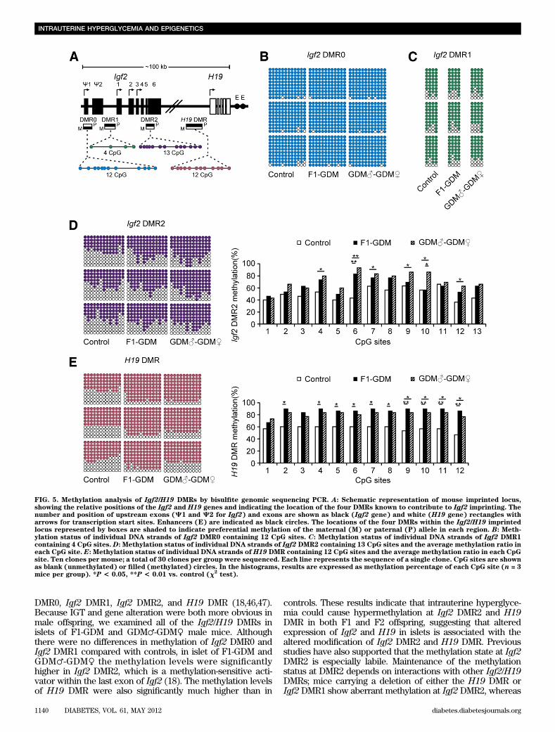
DMR0, Igf2 DMR1, Igf2 DMR2, and H19 DMR (18,46,47).Because IGT and gene alteration were both more obvious inmale offspring, we examined all of the Igf2/H19 DMRs inislets of F1-GDM and GDM♂-GDM♀ male mice. Althoughthere were no differences in methylation of Igf2 DMR0 andIgf2 DMR1 compared with controls, in islet of F1-GDM andGDM♂-GDM♀ the methylation levels were significantlyhigher in Igf2 DMR2, which is a methylation-sensitive acti-vator within the last exon of Igf2 (18). The methylation levelsof H19 DMR were also significantly much higher than in
controls. These results indicate that intrauterine hyperglyce-mia could cause hypermethylation at Igf2 DMR2 and H19DMR in both F1 and F2 offspring, suggesting that alteredexpression of Igf2 and H19 in islets is associated with thealtered modification of Igf2 DMR2 and H19 DMR. Previousstudies have also supported that the methylation state at Igf2DMR2 is especially labile. Maintenance of the methylationstatus at DMR2 depends on interactions with other Igf2/H19DMRs; mice carrying a deletion of either the H19 DMR orIgf2 DMR1 show aberrant methylation at Igf2 DMR2, whereas
FIG. 5. Methylation analysis of Igf2/H19 DMRs by bisulfite genomic sequencing PCR. A: Schematic representation of mouse imprinted locus,showing the relative positions of the Igf2 and H19 genes and indicating the location of the four DMRs known to contribute to Igf2 imprinting. Thenumber and position of upstream exons (C1 and C2 for Igf2) and exons are shown as black (Igf2 gene) and white (H19 gene) rectangles witharrows for transcription start sites. Enhancers (E) are indicated as black circles. The locations of the four DMRs within the Igf2/H19 imprintedlocus represented by boxes are shaded to indicate preferential methylation of the maternal (M) or paternal (P) allele in each region. B: Meth-ylation status of individual DNA strands of Igf2 DMR0 containing 12 CpG sites. C: Methylation status of individual DNA strands of Igf2 DMR1containing 4 CpG sites. D: Methylation status of individual DNA strands of Igf2 DMR2 containing 13 CpG sites and the average methylation ratio ineach CpG site. E: Methylation status of individual DNA strands of H19 DMR containing 12 CpG sites and the average methylation ratio in each CpGsite. Ten clones per mouse; a total of 30 clones per group were sequenced. Each line represents the sequence of a single clone. CpG sites are shownas blank (unmethylated) or filled (methylated) circles. In the histograms, results are expressed as methylation percentage of each CpG site (n = 3mice per group). *P < 0.05, **P < 0.01 vs. control (x2
test).
INTRAUTERINE HYPERGLYCEMIA AND EPIGENETICS
1140 DIABETES, VOL. 61, MAY 2012 diabetes.diabetesjournals.org
deletion of Igf2 DMR2 does not reciprocally affect theother DMRs (47). Importantly, human genomic regions cor-responding to mouse H19 DMR, Igf2 DMR0, and Igf2DMR2 have been identified and exhibit allele-specificmethylation (48–50).
In conclusion, using a GDM mouse model we found thatduring pregnancy, intrauterine environment with moderatehyperglycemia did not affect birth weight of F1-GDM butincreased birth weight of F2 offspring born from F1-GDMwith IGT, which showed sex-specific characteristics. Fur-thermore, intrauterine hyperglycemia induced IGT andabnormal insulin levels in both F1 and F2 offspring re-gardless of birth from F1-GDM with or without IGT, whichare partly due to the deficient islet ultrastructure. Abnor-mal Igf2 expression related to dysregulation of Igf2/H19methylation of islet is one of the mechanisms involved inthe effects of intrauterine hyperglycemia on F1 and F2offspring. Altered Igf2 and H19 gene expression was alsofound in sperm of adult F1-GDM with or without IGT, in-dicating that changes of epigenetics in germ cells con-tributed to transgenerational transmission.
ACKNOWLEDGMENTS
This study was supported by the National Basic ResearchProgram of China (2012CB944901), the National NaturalScience Foundation of China (30973209, 31171444, 30901616,and 30901604), the Research Fund for the Doctoral Programof Higher Education (20100101110129), and the Key SubjectsGroup of Reproductive Medicine at Zhejiang Province (XKQ-009-002).
No potential conflicts of interest relevant to this articlewere reported.
G.-L.D. and F.-F.W. designed and performed experiments,analyzed data, and wrote and edited the manuscript. J.S.,S.T., Y.J., D.Z., and N.W. researched data and contributedto discussion. Q.L., Y.Z., F.J., and P.C.K.L. contributed todiscussion and edited the manuscript. J.-Z.S. and H.-F.H.designed and supervised the research, contributed to discus-sion, and edited the manuscript. H.-F.H. is the guarantor ofthis work and, as such, had full access to all the data in thestudy and takes responsibility for the integrity of the data andthe accuracy of the data analysis.
The authors thank Qiu-Ping Xie and Yu-Lian Wu (SecondAffiliated Hospital, School of Medicine, Zhejiang Univer-sity) for technical assistance. The authors also thank HsiaoChang Chan (Epithelial Cell Biology Research Center, Schoolof Biomedical Sciences, Faculty of Medicine, The ChineseUniversity of Hong Kong), Min-Yue Dong, and Xin-Mei Liu(Women’s Hospital, School of Medicine, Zhejiang University)
for helpful advice. The authors also thank Professor IainC. Bruce (School of Medicine, Zhejiang Universit) for readingand editing the manuscript.
REFERENCES
1. Metzger BE, Buchanan TA, Coustan DR, et al. Summary and recom-mendations of the Fifth International Workshop-Conference on Gesta-tional Diabetes Mellitus. Diabetes Care 2007;30(Suppl. 2):S251–S260
2. Bateson P, Barker D, Clutton-Brock T, et al. Developmental plasticity andhuman health. Nature 2004;430:419–421
3. Dabelea D, Hanson RL, Lindsay RS, et al. Intrauterine exposure to diabetesconveys risks for type 2 diabetes and obesity: a study of discordant sib-ships. Diabetes 2000;49:2208–2211
4. Fetita LS, Sobngwi E, Serradas P, Calvo F, Gautier JF. Consequences offetal exposure to maternal diabetes in offspring. J Clin Endocrinol Metab2006;91:3718–3724
5. Clausen TD, Mathiesen ER, Hansen T, et al. High prevalence of type 2diabetes and pre-diabetes in adult offspring of women with gestationaldiabetes mellitus or type 1 diabetes: the role of intrauterine hyperglycemia.Diabetes Care 2008;31:340–346
6. Pettitt DJ, Aleck KA, Baird HR, Carraher MJ, Bennett PH, Knowler WC.Congenital susceptibility to NIDDM. Role of intrauterine environment.Diabetes 1988;37:622–628
7. Martin-Gronert MS, Ozanne SE. Experimental IUGR and later diabetes.J Intern Med 2007;261:437–452
8. Park JH, Stoffers DA, Nicholls RD, Simmons RA. Development of type 2diabetes following intrauterine growth retardation in rats is associated withprogressive epigenetic silencing of Pdx1. J Clin Invest 2008;118:2316–2324
9. Reik W, Dean W, Walter J. Epigenetic reprogramming in mammalian de-velopment. Science 2001;293:1089–1093
10. Sasaki H, Matsui Y. Epigenetic events in mammalian germ-cell de-velopment: reprogramming and beyond. Nat Rev Genet 2008;9:129–140
11. Tremblay J, Hamet P. Impact of genetic and epigenetic factors from earlylife to later disease. Metabolism 2008;57(Suppl. 2):S27–S31
12. Nagase H, Ghosh S. Epigenetics: differential DNA methylation in mam-malian somatic tissues. FEBS J 2008;275:1617–1623
13. Bartolomei MS, Tilghman SM. Genomic imprinting in mammals. Annu RevGenet 1997;31:493–525
14. Thamotharan M, Garg M, Oak S, et al. Transgenerational inheritance of theinsulin-resistant phenotype in embryo-transferred intrauterine growth-restricted adult female rat offspring. Am J Physiol Endocrinol Metab 2007;292:E1270–E1279
15. Serradas P, Goya L, Lacorne M, et al. Fetal insulin-like growth factor-2production is impaired in the GK rat model of type 2 diabetes. Diabetes2002;51:392–397
16. Devedjian JC, George M, Casellas A, et al. Transgenic mice overexpressinginsulin-like growth factor-II in beta cells develop type 2 diabetes. J ClinInvest 2000;105:731–740
17. Calderari S, Gangnerau MN, Thibault M, et al. Defective IGF2 and IGF1Rprotein production in embryonic pancreas precedes beta cell mass anomaly inthe Goto-Kakizaki rat model of type 2 diabetes. Diabetologia 2007;50:1463–1471
18. Waterland RA, Lin JR, Smith CA, Jirtle RL. Post-weaning diet affects ge-nomic imprinting at the insulin-like growth factor 2 (Igf2) locus. Hum MolGenet 2006;15:705–716
19. Hark AT, Schoenherr CJ, Katz DJ, Ingram RS, Levorse JM, Tilghman SM.CTCF mediates methylation-sensitive enhancer-blocking activity at theH19/Igf2 locus. Nature 2000;405:486–489
FIG. 6. Igf2 and H19 gene expression in sperm. A: Igf2 and H19 gene expression in sperm of F1-GDM with obvious IGT. B: Igf2 and H19 geneexpression in sperm of F1-GDM with relative normal glucose tolerance (NGT). Data are presented as means 6 SE (n = 6 mice per F1-C group, n = 5mice per F1-GDM group). *P < 0.05, **P < 0.01 vs. F1-C (Student t test).
G.-L. DING AND ASSOCIATES
diabetes.diabetesjournals.org DIABETES, VOL. 61, MAY 2012 1141
20. Gloyn AL, Mackay DJ, Weedon MN, et al. Assessment of the role ofcommon genetic variation in the transient neonatal diabetes mellitus(TNDM) region in type 2 diabetes: a comparative genomic and taggingsingle nucleotide polymorphism approach. Diabetes 2006;55:2272–2276
21. Gardner RJ, Mackay DJ, Mungall AJ, et al. An imprinted locus associatedwith transient neonatal diabetes mellitus. Hum Mol Genet 2000;9:589–596
22. Ma D, Shield JP, Dean W, et al. Impaired glucose homeostasis in transgenicmice expressing the human transient neonatal diabetes mellitus locus,TNDM. J Clin Invest 2004;114:339–348
23. Breyer MD, Böttinger E, Brosius FC 3rd, et al.; AMDCC. Mouse models ofdiabetic nephropathy. J Am Soc Nephrol 2005;16:27–45
24. Harris IS, Treskov I, Rowley MW, et al. G-protein signaling participates inthe development of diabetic cardiomyopathy. Diabetes 2004;53:3082–3090
25. Muller KA, Ryals JM, Feldman EL, Wright DE. Abnormal muscle spindleinnervation and large-fiber neuropathy in diabetic mice. Diabetes 2008;57:1693–1701
26. Kim SJ, Nian C, Doudet DJ, McIntosh CH. Inhibition of dipeptidyl peptidaseIV with sitagliptin (MK0431) prolongs islet graft survival in streptozotocin-induced diabetic mice. Diabetes 2008;57:1331–1339
27. Cederholm J, Wibell L. Evaluation of insulin release and relative peripheralresistance with use of the oral glucose tolerance test: a study in subjectswith normoglycaemia, glucose intolerance and non-insulin-dependent di-abetes mellitus. Scand J Clin Lab Invest 1985;45:741–751
28. Martinez SC, Tanabe K, Cras-Méneur C, Abumrad NA, Bernal-Mizrachi E,Permutt MA. Inhibition of Foxo1 protects pancreatic islet beta-cells ag-ainst fatty acid and endoplasmic reticulum stress-induced apoptosis. Dia-betes 2008;57:846–859
29. Oh W, Gelardi NL, Cha CJ. Maternal hyperglycemia in pregnant rats: itseffect on growth and carbohydrate metabolism in the offspring. Metabo-lism 1988;37:1146–1151
30. Kaijser M, Bonamy AK, Akre O, et al. Perinatal risk factors for diabetes inlater life. Diabetes 2009;58:523–526
31. Meier JJ. Linking the genetics of type 2 diabetes with low birth weight:a role for prenatal islet maldevelopment? Diabetes 2009;58:1255–1256
32. Chakravarthy MV, Zhu Y, Wice MB, et al. Decreased fetal size is associatedwith beta-cell hyperfunction in early life and failure with age. Diabetes2008;57:2698–2707
33. Lumey LH. Decreased birthweights in infants after maternal in utero ex-posure to the Dutch famine of 1944-1945. Paediatr Perinat Epidemiol 1992;6:240–253
34. Jimenez-Chillaron JC, Hernandez-Valencia M, Reamer C, et al. Beta-cellsecretory dysfunction in the pathogenesis of low birth weight-associateddiabetes: a murine model. Diabetes 2005;54:702–711
35. Jimenez-Chillaron JC, Isganaitis E, Charalambous M, et al. Intergenera-tional transmission of glucose intolerance and obesity by in utero under-nutrition in mice. Diabetes 2009;58:460–468
36. Rees DA, Alcolado JC. Animal models of diabetes mellitus. Diabet Med2005;22:359–370
37. Ueda H, Ikegami H, Yamato E, et al. The NSY mouse: a new animal modelof spontaneous NIDDM with moderate obesity. Diabetologia 1995;38:503–508
38. Corsetti JP, Sparks JD, Peterson RG, Smith RL, Sparks CE. Effect of di-etary fat on the development of non-insulin dependent diabetes mellitus inobese Zucker diabetic fatty male and female rats. Atherosclerosis 2000;148:231–241
39. Trevaskis JL, Meyer EA, Galgani JE, Butler AA. Counterintuitive effects ofdouble-heterozygous null melanocortin-4 receptor and leptin genes ondiet-induced obesity and insulin resistance in C57BL/6J mice. Endocri-nology 2008;149:174–184
40. Chong S, Vickaryous N, Ashe A, et al. Modifiers of epigenetic reprogram-ming show paternal effects in the mouse. Nat Genet 2007;39:614–622
41. Ferguson-Smith AC, Surani MA. Imprinting and the epigenetic asymmetrybetween parental genomes. Science 2001;293:1086–1089
42. DeChiara TM, Robertson EJ, Efstratiadis A. Parental imprinting of themouse insulin-like growth factor II gene. Cell 1991;64:849–859
43. Rainier S, Johnson LA, Dobry CJ, Ping AJ, Grundy PE, Feinberg AP. Re-laxation of imprinted genes in human cancer. Nature 1993;362:747–749
44. Cattanach BM, Beechey CV. Autosomal and X-chromosome imprinting.Dev Suppl 1990;108(Suppl):63–72
45. Bell AC, Felsenfeld G. Methylation of a CTCF-dependent boundary con-trols imprinted expression of the Igf2 gene. Nature 2000;405:482–485
46. Feil R, Walter J, Allen ND, Reik W. Developmental control of allelicmethylation in the imprinted mouse Igf2 and H19 genes. Development1994;120:2933–2943
47. Lopes S, Lewis A, Hajkova P, et al. Epigenetic modifications in an im-printing cluster are controlled by a hierarchy of DMRs suggesting long-range chromatin interactions. Hum Mol Genet 2003;12:295–305
48. Vu TH, Li T, Nguyen D, et al. Symmetric and asymmetric DNA methylationin the human IGF2-H19 imprinted region. Genomics 2000;64:132–143
49. Sullivan MJ, Taniguchi T, Jhee A, Kerr N, Reeve AE. Relaxation of IGF2imprinting in Wilms tumours associated with specific changes in IGF2methylation. Oncogene 1999;18:7527–7534
50. Reik W, Brown KW, Schneid H, Le Bouc Y, Bickmore W, Maher ER. Im-printing mutations in the Beckwith-Wiedemann syndrome suggested byaltered imprinting pattern in the IGF2-H19 domain. Hum Mol Genet 1995;4:2379–2385
INTRAUTERINE HYPERGLYCEMIA AND EPIGENETICS
1142 DIABETES, VOL. 61, MAY 2012 diabetes.diabetesjournals.org