Autoimmunity against INS-IGF2 expressed in human ...
30
Autoimmunity against INS-IGF2 expressed in human pancreatic islets. Kanatsuna, Norio; Taneera, Jalal; Vaziri Sani, Fariba; Wierup, Nils; Larsson, Helena; Delli, Ahmed; Skärstrand, Hanna; Balhuizen, Alexander; Bennet, Hedvig; Steiner, Donald F; Törn, Carina; Fex, Malin; Lernmark, Åke Published in: Journal of Biological Chemistry DOI: 10.1074/jbc.M113.478222 2013 Link to publication Citation for published version (APA): Kanatsuna, N., Taneera, J., Vaziri Sani, F., Wierup, N., Larsson, H., Delli, A., Skärstrand, H., Balhuizen, A., Bennet, H., Steiner, D. F., Törn, C., Fex, M., & Lernmark, Å. (2013). Autoimmunity against INS-IGF2 expressed in human pancreatic islets. Journal of Biological Chemistry, 288(40), 29013-29023. https://doi.org/10.1074/jbc.M113.478222 Total number of authors: 13 General rights Unless other specific re-use rights are stated the following general rights apply: Copyright and moral rights for the publications made accessible in the public portal are retained by the authors and/or other copyright owners and it is a condition of accessing publications that users recognise and abide by the legal requirements associated with these rights. • Users may download and print one copy of any publication from the public portal for the purpose of private study or research. • You may not further distribute the material or use it for any profit-making activity or commercial gain • You may freely distribute the URL identifying the publication in the public portal Read more about Creative commons licenses: https://creativecommons.org/licenses/ Take down policy If you believe that this document breaches copyright please contact us providing details, and we will remove access to the work immediately and investigate your claim.
Transcript of Autoimmunity against INS-IGF2 expressed in human ...

LUND UNIVERSITY
PO Box 117221 00 Lund+46 46-222 00 00
Autoimmunity against INS-IGF2 expressed in human pancreatic islets.
Kanatsuna, Norio; Taneera, Jalal; Vaziri Sani, Fariba; Wierup, Nils; Larsson, Helena; Delli,Ahmed; Skärstrand, Hanna; Balhuizen, Alexander; Bennet, Hedvig; Steiner, Donald F; Törn,Carina; Fex, Malin; Lernmark, ÅkePublished in:Journal of Biological Chemistry
DOI:10.1074/jbc.M113.478222
2013
Link to publication
Citation for published version (APA):Kanatsuna, N., Taneera, J., Vaziri Sani, F., Wierup, N., Larsson, H., Delli, A., Skärstrand, H., Balhuizen, A.,Bennet, H., Steiner, D. F., Törn, C., Fex, M., & Lernmark, Å. (2013). Autoimmunity against INS-IGF2 expressedin human pancreatic islets. Journal of Biological Chemistry, 288(40), 29013-29023.https://doi.org/10.1074/jbc.M113.478222
Total number of authors:13
General rightsUnless other specific re-use rights are stated the following general rights apply:Copyright and moral rights for the publications made accessible in the public portal are retained by the authorsand/or other copyright owners and it is a condition of accessing publications that users recognise and abide by thelegal requirements associated with these rights. • Users may download and print one copy of any publication from the public portal for the purpose of private studyor research. • You may not further distribute the material or use it for any profit-making activity or commercial gain • You may freely distribute the URL identifying the publication in the public portal
Read more about Creative commons licenses: https://creativecommons.org/licenses/Take down policyIf you believe that this document breaches copyright please contact us providing details, and we will removeaccess to the work immediately and investigate your claim.

INS-IGF2 in autoimmune diabetes
1
Autoimmunity against INS-IGF2 expressed in human pancreatic islets*.
Norio Kanatsuna1, Jalal Taneera
1, Fariba Vaziri-Sani
1, Nils Wierup
1, Helena Elding Larsson
1,
Ahmed Delli1, Hanna Skärstrand
1, Alexander Balhuizen
1, Hedvig Bennet
1, Donald F. Steiner
2,
Carina Törn1, Malin Fex
1, and Åke Lernmark
1.
1 Department of Clinical Sciences, Lund University Diabetes Center, Lund University, Skåne University
Hospital SUS, Malmö, Sweden;
2 Department of Medicine, University of Chicago, Chicago, IL 606 37
*Running title: INS-IGF2 in autoimmune diabetes
To whom correspondence should be addressed: Norio Kanatsuna MD PhD, Department of Clinical
Sciences, Lund University Diabetes Center, Lund University, Jan Waldenströms gata 35, Skåne
University Hospital SUS, SE-205 02 Malmö, Sweden, TEL.: +46 40 39 19 01; FAX: +46 40 39 19 19;
E-mail: [email protected]
Key words: islet cell autoantibodies, autoimmune diabetes, type 1 diabetes, radiobinding immunoassay,
insulin autoantibodies, in vitro transcription translation, glutamic acid decarboxylase, GAD65, IA-2, Zn
T8 transporter
Background: Islet INS-IGF2 was examined as a
possible autoantigen in type 1 diabetes.
Results: INS-IGF2 expression was inversely
related to donor HbA1c and glucose-stimulated
insulin release. Autoantibodies doubly reactive
with INS-IGF2 and insulin were more common in
type 1 diabetes patients than controls.
Conclusion: INS-IGF2 is recognized by
autoantibodies in type 1 diabetes.
Significance: Autoantibodies doubly reactive with
both INS-IGF2 and insulin may contribute to type
1 diabetes.
SUMMARY
Insulin is a major autoantigen in islet
autoimmunity and progression to type 1
diabetes. It has been suggested that the insulin
B-chain may be critical to insulin autoimmunity
in type 1 diabetes. INS-IGF2 consists of the
preproinsulin signal peptide, the insulin
B-chain and eight amino acids of the C-peptide
in addition to 138 amino acids from the IGF2
gene. We aimed to determine 1) expression of
INS-IGF2 in human pancreatic islets and 2)
autoantibodies in newly diagnosed type 1
diabetes children and controls. INS-IGF2,
expressed primarily in beta cells, showed higher

INS-IGF2 in autoimmune diabetes
2
levels of expression in islets from normal
compared to donors with either type 2 diabetes
(p=0.006) or high HbA1c levels (p<0.001).
INS-IGF2 autoantibody levels were increased in
newly diagnosed type 1 diabetes patients
(n=304) compared to healthy controls (n=355;
p<0.001). Displacement with cold insulin and
INS-IGF2 revealed that more patients than
controls had doubly reactive insulin-INS-IGF2
autoantibodies. These data suggest that
INS-IGF2, which contains the preproinsulin
signal peptide, the B-chain and eight amino
acids of the C-peptide may be an autoantigen in
type 1 diabetes. INS-IGF2 and insulin may
share autoantibody binding sites, thus
complicating the notion that insulin is the
primary autoantigen in type 1 diabetes.
Type 1 diabetes (T1D)3 is strongly associated
with a selective autoimmune destruction of the
pancreatic beta cells (1,2). The progressive loss of
beta cells is often triggered at an early age (1,3-5)
and autoantibodies directed against islet
autoantigens such as insulin, glutamic acid
decarboxylase 65 (GAD65), islet antigen-2 (IA-2)
and zinc transporter 8 (ZnT8) are currently the
best markers of the islet autoimmunity that
precede the clinical onset of T1D (6,7). Insulin
autoantibodies (IAA) may be the first islet
autoantibodies to appear (8,9). However, it is the
least frequent (10) as IAA are particularly
common in patients at younger age of diagnosis
(11). Persistent IAA in combination with any other
islet autoantibody predicts T1D (12). Insulin
iodinated with 125
I is primarily used to detect IAA
(13,14), however, IAA assays with iodinated
insulin have proven difficult to standardize (15,16).
It has been speculated that the primary autoantigen
is not matured insulin but rather proinsulin (17,18)
or perhaps preproinsulin (19,20). It was
hypothesized that beta-cell endoplasmic reticulum
(ER) fragments may become available to antigen
presenting cells following e.g. viral lysis of beta
cells (21).
The immune recognition of (pro)insulin is not
fully clarified as human studies are complicated by
the fact that circulating T and B cells as well as
antigen presenting cells (APC) may not reflect an
on-going insulitis. T cells reactive with insulin
have been detected in pancreatic lymphnodes from
T1D patients. Proinsulin epitopes recognized by
the T cell receptor (TCR) on CD4+ T cells have
been reported in children at HLA-risk for T1D
prior to the development of IAA (22). CD4+T
cells seem preferentially to recognize (pro)insulin
peptides from the B chain when presented on HLA
DR4 or DQ8 molecules (for a review see (21)).
IAA have been associated with HLA-DQ8 that is
in strong linkage disequilibrium with DR4 (23).
The B cell receptor (BCR) recognition may be
broader as the binding site for IAA includes
several B-chain amino acid residues (reviewed in
(21)). CD8+ T cells were reported to express TCR
recognizing B-chain residues 10-18 (20) and 9-23
(24) or signal peptide residues 15-24 (25,26).
These observations suggest that the B-chain of
insulin and perhaps also signal or B-C junction
peptides may contribute to a pre(pro)insulin
autoimmune response. It is expected that this
response would be reflected by the appearance of
pre(pro)insulin autoantibodies. The well
established radiobinding assay (RBA) using

INS-IGF2 in autoimmune diabetes
3
coupled in vitro transcription translation of
autoantigen cDNA effectively detects a number of
conformation-dependent autoantibodies (27-32).
The aim of the present study was to use coupled in
vitro transcription translation to test whether T1D
patients may have conformation-dependent
autoantibodies against the (signal
peptide)-(B-chain)-(B-C junction) of
pre(pro)insulin.
The insulin gene and insulin like growth factor
2 (IGF2) genes are located sequentially on
chromosome 11 (33). The recently described 200
amino acid long INS-IGF2 protein consists of the
preproinsulin signal peptide (24 amino acids), the
insulin B-chain (30 amino acids) and the
C-peptide (8 amino acids) in addition to 138
amino acids coded for in the IGF2 gene (34).
However, expression of short INS-IGF2 was
restricted to human fetal pancreas and eye (34).
The fact that the INS-IGF2 protein has the signal
peptide-B-chain-B-C junction of pre(pro)insulin
makes INS-IGF2 a candidate autoantigen in T1D.
Therefore, we first determined if INS-IGF2 was
expressed in adult human islets and then tested the
hypothesis that INS-IGF2 autoantibodies
measured by RBA were detected in newly
diagnosed T1D patients and controls.
EXPERIMENTAL PROCEDURES
Human pancreatic islets— Islets from cadaver
donors were provided by the Nordic Islet
Transplantation Programme
(www.nordicislets.org), Uppsala University,
Uppsala, Sweden. Islets were obtained from 66
non-diabetes donors (30 females, age 59 ± 10,
BMI 25.9 ± 3.5, HbA1c 5.5 ± 1.1 and days of
culture 3.5 ± 1.9 (mean values ± S.D.)) and 10
donors with type 2 diabetes (T2D) (4 females, 6
males, age 60.7 ± 12, BMI 28.1 ± 4.5, HbA1c 7.1
± 1.2 and days of culture 2 ± 0.9). Purity of the
islet preparations was assessed by dithizone
staining and amounted to 60.1 ± 20% in the T2D
and 70 ± 17% in the non-diabetic islets (p=0.10).
In addition, the contribution of exocrine and
endocrine tissues were assessed by measuring
expression of pancreatic lipase, alpha 2 amylase
and chymotrypsin 2 as markers of exocrine tissue,
and somatostatin and glucagon as markers of
endocrine tissue (probes for insulin were
unfortunately not included on the chip). Using this
approach, the estimated contribution of islet
endocrine tissue did not differ between
non-diabetic (72%) and T2D (68%) donors
(p=0.29). We also measured insulin content as a
surrogate marker for pancreatic islets -cell mass
in hyperglycemic (HbA1c >6.0%; 4.8 ± 3.2 ng/ml)
and normoglycemic (HbA1c <6.0%; 5.6±3.2
ng/ml; p=0.4). Prior to RNA isolation, the islets
were cultured at 37 °C (5% CO2) for 1–9 days in
CMRL 1066 (ICN Biomedicals, Costa Mesa, CA,
USA) supplemented with 10 mmol/l HEPES,
2 mmol/l L-glutamine, 50 μg/ml gentamicin,
0.25 μg/ml Fungizone (GIBCO, BRL,
Gaithersburg, MD, USA), 20 μg/ml ciprofloxacin
(Bayer Healthcare, Leverkusen, Germany), and
10 mmol/l nicotinamide. All procedures were
approved by Regional Ethics Boards at Uppsala
and Lund Universities, respectively.
RNA isolation— Total RNA was isolated with
the AllPrep DNA/RNA Mini Kit (Qiagen, Hilden,
Germany). RNA quality and concentration were
measured using an Agilent 2100 bioanalyzer
(Bio-Rad, Hercules, CA, USA) and Nanodrop

INS-IGF2 in autoimmune diabetes
4
ND-1000 spectrophotometer (NanoDrop
Technologies, Wilmington, DE, USA).
Microarray gene expression in human
pancreatic islets— The microarrays were
performed using the Affymetrix standard protocol.
Briefly, 100-200 ng of total RNA was processed
according to instructions of the GeneChip®
Expression 3’-Amplification Reagents One-cycle
cDNA synthesis kit (Affymetrix Inc, Santa Clara,
CA, USA) to produce double-stranded cDNA.
This was used as a template to generate
biotin-targeted cRNA following the
manufacturer’s specifications. A total of 15 µg of
the biotin-labeled cRNA was fragmented to
strands between 35 and 200 bases in length, 10 µg
of which was hybridized onto the GeneChip®
Human Gene 1.0 ST whole transcript based assay
overnight in the GeneChip® Hybridization oven
6400 using standard procedures. The arrays were
washed and stained in a GeneChip® Fluidics
Station 450. Scanning was carried out with the
GeneChip® Scanner 3000 and image analysis was
performed using GeneChip® Operating Software.
The array data was summarized and normalized
with Robust Multi-array Analysis (RMA) method
using affy R package. All Data are MIAME
compliant, and the raw data have been deposited
in a MIAME database (GEO, accession number:
GSE 38642 and GSE 44035).
Glucose-stimulated insulin release (GSIR)—
Islets were hand-picked under a stereomicroscope
at room temperature and first incubated for 30 min
at 37°C in Krebs Ringer bicarbonate (KRB) buffer
(pH 7.4) containing (in mmol/l) 120 NaCl, 25
NaHCO3, 4.7 KCl, 1.2 MgSO4, 2.5 CaCl2, 1.2
KH2PO, 10 HEPES supplemented with 0.1%
bovine serum albumin,
N-2-hydroxyethylpiperazine-N'-2-ethanesulfonic
acid (10 mmol/1) and 1 mmol/l glucose. Each
incubation vial contained 12 islets in 1.0 ml KRB
buffer solution and was treated with 95% O2-5%
CO2 to obtain constant pH 7.4 and oxygenation.
After the first incubation, the buffer was changed
to KRB buffer containing either 1 mM (basal
secretion) or 16.7 mM glucose (stimulated
secretion). The islets were then incubated for 1h at
37°C in a metabolic shaker (30 cycles per min).
Immediately after incubation an aliquot of the
medium was removed for analysis of insulin using
a radioimmunoassay kit (Euro-Diagnostica,
Malmö, Sweden). Insulin content in homogenised
human islets was assessed by ELISA (Mercodia,
Uppsala, Sweden) and values were normalized to
the total DNA in each sample as determined by a
fluorometric assay (Quant-iT Picogreen,
Invitrogen Molecular Probes, Stockholm,
Sweden).
Immunocytochemistry of human pancreas—
Human pancreatic specimens taken during
pancreatic surgery were used. Briefly, primary
antibodies were diluted in PBS containing 0.25%
bovine serum albumin and 0.25% Triton X-100
and applied overnight at 4°C. The primary
antibodies were mouse polyclonal anti-human
INS-IGF2 (dilution: 1:100, code: BO1P, Abnova
Corp. Taipei, Taiwan), guinea pig polyclonal
anti-human proinsulin (dilution: 1:5000, code:
9003, Euro-Diagnostica, Malmö, Sweden), guinea
pig polyclonal anti-human glucagon (dilution:
1:5000, code: 8708, Euro-Diagnostica), goat
polyclonal anti-human somatostatin (diluton:
1:800, code: SC7819, Santa Cruz Biotechnology,

INS-IGF2 in autoimmune diabetes
5
Inc., Santa Cruz, CA), sheep polyclonal
anti-human panreatic poplypeptide (PP) (dilution:
1:640, code: AHP 515, Serotecc, Oxford, UK) or
goat polyclonal anti-human ghrelin (dilution
1:1000, code: SC10368, Santa
Cruz,Biotechnology, Santa Cruz, CA). Secondary
antibodies specific for mouse, guinea pig, or goat
IgG and coupled to either Cy2, Texas red, or
7-amino-4-methyl coumarin-3-acetic acid
(AMCA) (Jackson, West Grove, PA) were applied
for 1 h at room temperature. Immunofluorescence
was examined in an epifluorescence microscope
(BX60; Olympus, Lund, Sweden). Images were
taken with a digital camera (DS-2Mv; Nikon,
Lund, Sweden).
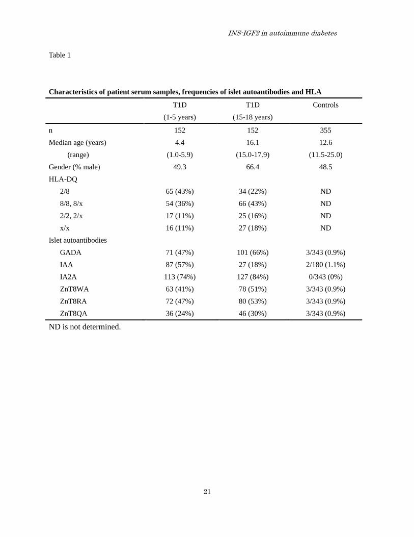
Patients— Serum samples from 304 (54%
male) patients with newly diagnosed T1D in the
Better Diabetes Diagnosis (BDD) study (10,35).
Patient sera were selected at random to represent
one group of 152 patients with clinical onset at 1-5
years of age and another group of 152 patients at
15-18 years of age (Table 1). The classification of
diabetes was clinically confirmed six months after
onset with T1D (10,35). HLA-DQ typing and
analyses of islet autoantibodies against GAD65,
insulin, IA-2 and ZnT8 (Table 1) were described in
detail elsewhere (28,35).
Controls— Serum samples were obtained from
a total of 355 healthy controls (Table 1). Among
these were 300 (47.3% males) 11.5-13.7 (range;
median age 12.5) years old school children
previously described (36) and analyzed for
multiple autoantibodies (37,38). In addition, 55
healthy adult blood donors (54.5% males, n=30)
aged 19.0-25.0 years (range; median age 23.0)
were also investigated (39).
Preparation of INS-IGF2— INS-IGF2 cDNA,
based on the sequence in the National Center for
Biotechnology Information (NCBI) was purchased
in the pJ201 vector from DNA 2.0 Inc. (DNA 2.0
Inc. Menlo Park, CA, USA).
Preparation of the pThINS-IGF2 vector— The
cDNA of INS-IGF2 was cut from the pJ201 vector
(DNA2.0 Inc.) with EcoRI and NotI (FastDigest™,
Fermentas Sweden AB, Helsingborg, Sweden)
using the FastDigest buffer 10X (Fermentas
Sweden AB, Helsingborg, Sweden). Following 30
min digestion at 37 °C, the pTNT™ vector was
dephosphorylated with Calf Intestinal Alkaline
Phosphatase (Fermentas) at a final concentration
of 0.04 U/μl of DNA termini according to
instructions from the supplier.
The linearized pTNT™vector and cDNA of
INS-IGF2 were analyzed by gel electrophoresis in
1% agarose and the two bands extracted using a
gel purification kit (QIAquick Gel Extraction Kit,
QIAGEN AB, Solna, Sweden) according to the
manufacturer's instructions. The ligation reaction
with T4 ligase (New England Biolabs, Inc.),
optimized to 1:3 (INS-IGF2) vector: insert molar
ratio, was carried out at room temperature for 1 h.
Subcloning efficiency DH5α competent cells were
used for transformation according to
manufacturer's instructions (Invitrogen AB,
Stockholm, Sweden). The ligation reaction (10 μl)
was added to the cells (50 μl), mixed gently, and
after 30 min incubation on ice, the cells were heat
shocked for 90 s in a 42 °C incubator. The cells
were immediately put on ice for 2 min, and then
incubated for 1 h at 37 °C in 500 μl LB/ampicillin
medium with shaking (300 rpm).
A total of 200 μl cell suspension were plated

INS-IGF2 in autoimmune diabetes
6
onto LB/ampicillin plates and incubated over night
at 37 °C. White colonies were selected and
transferred separately to a new set of
LB/ampicillin plates for overnight culture before
the cells were incubated for 16 h at 37 °C in 3 ml
LB/ampicillin medium. The bacterial cells were
harvested by centrifugation for 10 min at 4 °C at
4000rpm.
The pThINS-IGF2 plasmid DNA was extracted
using the QiaPrep Spin MiniPrep Kit (QIAGEN
AB).
The insert was sequenced by GATC Biotech
AG (Konstanz, Germany) using the 5′-TTA CGC
CAG CCC GGA TCC-3′and 5′-AAG GCT AGA
GTA CTT AAT ACG A-3′ as the reverse and
forward primers (DNA Technology A/S, Risskov,
Denmark), respectively.
Coupled in vitro transcription–translation of
pThINS-IGF2— The pTNT™ vector (Promega,
Madison, WI, USA), was used for in vitro coupled
transcription translation system in the presence of
[35
S]methionine and cysteine (28). Briefly, the
reaction mixture at a final volume of 100 μl
contained 3 μg of pThINS-IGF2 vector, 50 μl
TNT® Rabbit Reticulocyte Lysate, 4 μl TNT®
Reaction Buffer, 2 μl Amino Acid Mixture Minus
Methionine and Cystein, 2 μl RNasin®
Ribonuclease Inhibitor, 2 μl SP6 RNA Polymerase,
(all from Promega), 4 μl [35
S]methionine and
cystein (EasyTagTM
EXPRESS35
S Protein
Labeling Mix, [35
S]- (NEG772,
L-[35
S]-Methionine: 1175.0 Ci/mmol,
L-[35
S]-Cystein: 1075.0 Ci/mmol) from
PerkinElmer, Väsby, Sweden) and nuclease-free
water. The reaction mixture was incubated for 90
min at 30 °C with shaking at 300 rpm (Eppendorf
Thermomixercomfort, Eppendorf AG, Hamburg,
Germany).
The translation product was immediately
subjected to gel filtration on Illustra™ NAP-5
Columns (GE Healthcare Bio-Sciences AB,
Uppsala, Sweden). All fractions prior to the
hemoglobin band were discarded. Subsequent
fractions were one 300 μl fraction followed by
several 100 μl fractions. Radioactivity
incorporated into protein was determined
(1450MicroBeta TriLuxMicroplate Scintillation-
Luminescence Counter, PerkinElmer) and peak
fractions of radioactivity pooled.
Preparation of non-radioactive INS-IGF2 was
carried out essentially as described above but
without radioactive amino acids as 2 μl Complete
Amino Acid Mixture (Promega) was added to the
TNT® Rabbit Reticulocyte Lysate. The translation
product was filtered on the NAP-5 columns and
fractions corresponding to the typically peak
fractions of radioactivity labeling above were
collected.
Electrophoresis of pThINS-IGF2 translation
products— The labeled antigen produced in the in
vitro transcription translation kit was analyzed by
SDS-PAGE gel electrophoresis (ClearPage™
precast 16%, VWR, West Chester, PA, according
to manufacturers' recommendations). The samples
were incubated in sample buffer with reducing
agent (ClearPage™ accessories,VWR) for 10 min
at 70 °C and were loaded on the gel with a
14C-labeled molecular weight markers (Protein
Molecular Weight Markers
[Methyl-14
C]-Methylated, (NEC811, 26.4 Ci/mg),
PerkinElmer) and with unlabeled protein ladders
(PageRulerTM
Plus Prestained Protein Ladder

INS-IGF2 in autoimmune diabetes
7
(SM1811) Fermentas).
The gel was dried for 72 h at room temperature
using a Gel Drying Kit (Promega) according to the
manufacturers' instructions.
The gel was placed in contact with an X-ray
sensitive film (Amersham HyperfilmTM
ECL, GE
Healthcare Bio-Sciences AB) for 72 h at room
temperature. The film was treated with developing
and fixation solutions following the manufacturer's
instructions (DAB dental, Malmö, Sweden).
RBA for INS-IGF2 autoantibodies— The RBA
for INS-IGF2 autoantibodies was carried out over
night at 4 °C in duplicate samples of 5 μl of serum
incubated with 60 μl labeled antigen at around 425
cpm/μl diluted in antigen buffer (150 mM NaCl,
20 mM Tris, 0.15% (v/v) Tween 20 (MP
Biomedicals, LLC. Santa Ana, CA, USA), 1%
(v/v), TritonX-100 (Sigma-Aldrich Sweden AB,
Stockholm, Sweden), 0.1% (w/v) BSA (MP
Biomedicals, LLC.), pH 7.4). (All serum samples
were diluted by adding 3 parts of antigen buffer to
1 part of serum before incubated with labeled
antigen.) V-formed 96-well plates (Nunc V96
MicroWell™ plates, Nunc A/S, Roskilde,
Denmark) were used. A total of 50 μl reaction
mixure was then incubated for 1 h at 4 °C with 50
μl Protein A Sepharose 4B conjugated (20%,
washed four times at 4 °C by sedimentation in
antigen buffer) (Invitrogen) in a 96-well filtration
plate (MultiScreenHTS-DV Plate, Millipore AB,
Solna, Sweden). The plate was then washed 8
times with wash buffer (150 mM NaCl, 20 mM
Tris, 0.15% Tween 20 (MP Biomedicals, LLC.),
pH 7.4) using a microplate washer (ELx50TM
Microplate Strip Washer, Biotek Instruments, Inc.
Winooski, VT, USA). Antibody-bound
radio-activity was counted in a β-counter (1450
MicroBeta® TriLux, PerkinElmer).
Sepharose-bound radioactivity was converted into
units per ml (U/ml) using individual standard
curves generated by six step doubling dilutions
(1:128 – 1:4 dilutions) of high-titer T1D patient’s
sera with high reactivity for the INS-IGF2 antigen.
Three internal quality control samples, two from
healthy subjects and one from a long term diabetes
subjects were used in every assay.
Intra-assay coefficient of variation (CV) and
inter-assay CV for duplicate determinations were
4.1% and 5.1%, respectively.
RBA for GAD65A and IA-2A— These
autoantibodies were analyzed as described in
detail elsewhere (35). GAD65A and IA-2A levels
were expressed as units per milliliter derived from
the World Health Organization standard 97/550
(40). Samples were considered positive if
GAD65A levels were > 50 U/ml and IA-2A levels
> 10 U/ml. The intra-assay CV for duplicates in
the GAD65A assay was 7% and in the IA-2A 11%.
In the Diabetes Autoantibody Standardization
Program (DASP) 2010 workshop, the workshop
sensitivity was 80% and specificity 99% for
GAD65A and 60% sensitivity and 99% specificity
for IA-2A.
RBA for ZnT8 R, W and Q –variant
autoantibodies— These autoantibodies were
analyzed as previously described (28). The 2010
Diabetes Autoantibody Standardization Program
(DASP) workshop (41) showed that our laboratory
had a workshop sensitivity of 52% for ZnT8RA,
50% for ZnT8WA and 38% for ZnT8QA. The
workshop specificity was 100% for all three
variant autoantibodies.

INS-IGF2 in autoimmune diabetes
8
RBA for insulin autoantibodies (IAA)— Insulin
autoantibodies were determined essentially as
described (42). All samples were analyzed without
and with cold insulin (2 IU/ml; Actrapid®, Novo
Nordisk A/S, Bagsvaerd, Denmark) to control for
non-specific binding. Briefly, the RBA for IAA
was carried out 48h at 4 °C in duplicate samples of
7 μl of diluted serum samples incubated with 36 μl
human, recombinant [125
I]insulin (2200 Ci/mmol
from PerkinElmer) at around 40,000 cpm/36 μl
diluted in antigen buffer (50 mM Tris, 1% (v/v)
Tween 20 (Sigma-Aldrich), 1% (w/v) BSA
(Sigma-Aldrich), pH 8.0). V-formed 96-well plates
(Nunc V96 MicroWell™ plates, Nunc A/S,
Roskilde, Denmark) were used. A total of 25 μl
reaction mixure was then incubated for 1.5 h at
4 °C with 50 μl Protein A Sepharose 4B
conjugated (40%, washed four times at 4 °C by
sedimentation in antigen buffer) (Invitrogen) in a
96-well filtration plate (MultiScreenHTS-DV Plate,
Millipore AB). The plate was then washed 10
times with wash buffer (50 mM Tris, 1% Tween 20
(Sigma-Aldrich), pH 8.0) using a microplate
washer (ELx50TM
Microplate Strip Washer, Biotek
Instruments, Inc.). Supermix scintillation solution
(50 μl) was added to the wells after the plate had
dried for 20 min. The radioactivity was measured
in a β-counter (1450 MicroBeta® TriLux,
PerkinElmer). Sepharose-bound radioactivity was
converted into U/ml using individual standard
curves generated by six step doubling dilutions
(1:512 – 1:8 dilutions) of high-titer T1D patient’s
sera with high reactivity for IAA. Three internal
quality control samples, two from healthy subjects
and one from a long term diabetes subjects were
used in every assay. Sepharose-bound radioactivity
was converted into U/ml using individual standard
curves generated by six step doubling dilutions of
high-titer T1D patient’s sera with high reactivity
for each individual IAA.
Our assays showed comparable precision
(intra-assay CV was 6.0% for IAA) and
reproducibility (inter-assay CV) was 13.2%. Our
laboratory participated in the DASP (14) to show
26% sensitivity and 100% specificity on the DASP
samples.
Statistical analysis— The IBM® SPSS®
Statistics Version 20 (IBM Corp. Armonk, NY,
USA) and GraphPad Prism version 5.03 for
Windows (GraphPad Software, Inc. La Jolla, CA,
USA) were used for statistical analysis.
Differences in titers of INS-IGF2 antibody
between control and patients were assessed by
Mann-Whitney U test (two-tailed). Correlations
were assessed by Spearman’s rho and Fisher's
exact test to compare frequencies.
RESULTS
Microarray gene expression in human
pancreatic islets— The INS-IGF2 transcript
(Figure 1) was detected in human islets (Figure 2).
Levels of expression in islets from 66 normal
donors were 2.5 fold higher than that of IGF2
(p<0.001; Figure 2). The IA-2 autoantigen gene,
used for comparison, showed 3.2 fold higher
levels than INS-IGF2 (p<0.001; Figure 2, panel A).
In addition, in pancreatic islets from ten organ
donors with T2D, the expression level of
INS-IGF2 was lower compared to the controls
(p=0.006). Islets from T2D organ donors also
showed lower expression levels compared to
controls of both IGF2 (p=0.017) and IA-2
(p=0.009). Among the 66 control donors there

INS-IGF2 in autoimmune diabetes
9
were 23 with HbA1c above 6.0 % on the day of
islet isolation (referred to as donors with
hyperglycemia). The data in Panel B
demonstrate that INS-IGF2 gene expression
was reduced in islet from donors with 6.0 -
6.4% HbA1c (n=12) and from donors with T2D
who all had HbA1c ≥6.5% (n=10) compared to
donors with HbA1c <6% (n=43).
Data on glucose-stimulated insulin release
(GSIR) was available in human islets from 64
donors as determined in batch type incubations
(Figure 3). As INS-IGF2 gene expression was
affected by donor HbA1c levels, it could not be
excluded that a possible correlation between GSIR
and INS-IGF2 was similarly affected. The possible
interrelation was therefore depicted in
three-dimensional displays (Figure 3). It can be
seen from the data in Figure 3, panel A, that the
higher the INS-IGF2 expression, the lower the
HbA1c and the higher the GSIR. This conclusion
was supported by the observation (not illustrated)
that GSIR was positively correlated with increased
expression of INS-IGF2 (r=0.284; p=0.023)
primarily in donors with low HbA1c (r=-0.446;
p<0.001). Expression of IGF2 in relation to GSIR
and HbA1c showed a comparable pattern (Figure
3, panel B). However, IGF2 expression was not
correlated to GSIR (r=0.167; p=0.186) despite the
expression of IGF2 was lower in hyperglycemic
donors (r=-0.394; p=0.001). IA-2 expression in
relation to both GSIR and HbA1c showed a
pattern of higher GSIR in islets from donors with
low HBA1c (Figure 3, Panel C) Indeed, IA-2
expression correlated positively with GSIR
(r=0.501; p<0.001) especially in islets from
normoglycemic donors (r=-0.333; p=0.008).
Immunocytochemistry of human pancreas—
The expression of immunoreactive INS-IGF2 in
human islets was examined with a mouse
polyclonal antiserum against full-length human
INS-IGF2 (Figure 4). Using triple-
immunofluorescence and overlay, INS-IGF2 was
detected in beta cells (Figure 4, compare panels A,
B and D) and alpha cells (Figure 4, compare
panels A, C and D), in a subpopulation of delta
cells, but not in PP-cells or ghrelin cells (data not
shown).
In vitro transcription translation of INS-IGF2
cDNA— INS-IGF2 cDNA, subjected to in vitro
transcription translation in the presence of both
[35
S]methionine and [35
S]cysteine showed an
incorporation rate of 8-17% dependent on batch of
the rabbit reticulocyte lysate system compared to
2% in when the labeling reaction was run without
added cDNA. SDS-gel electrophoresis and
autoradiography revealed a major translation
product at the expected Mr 21.5K for INS-IGF2
(Figure 5, lane 2). Densitometric scanning showed
that 27.1% of the total radioactivity was found in
the major 21.5K band. The minor bands at higher
molecular mass may represent multimers of
INS-IGF2. There was a shadow of radioactivity
when the labeling reaction was carried out without
added cDNA (Figure 5, lane 3).
RBA for INS-IGF2 autoantibodies— Serum
from patient MD, used as standard in the
insulin autoantibody (IAA) assay showed the
highest binding of [125I]insulin at 1/2 dilution
(Figure 6, Panel A). Recombinant human insulin
(2 IU/ml) fully displaced the binding of
[125
I]insulin at all dilutions (Figure 6, panel A).
Patient MD also showed binding of

INS-IGF2 in autoimmune diabetes
10
[35S]INS-IGF2 in a dilution-dependent
manner with maximal binding at a dilution of
1/4 (Figure 6, Panel B). There was no further
increase at a dilution of 1/2 (data not shown).
The binding was displaced by about 50% at all
serum dilutions by cold INS-IGF2 produced by in
vitro transcription translation (Figure 6, panel B).
The data in Figure 6, panel B also demonstrate that
cold insulin was less efficient than cold INS-IGF2
to displace the binding of INS-IGF2 to the serum
of patient MD. The competition between
[35S]INS-IGF2 binding to the serum of patient
MD (tested at a dilution of 1/4) and various
concentrations of cold antigens showed that
only cold INS-IGF2 was effective to displace the
binding of [35
S]INS-IGF2 (Figure 6, panel C).
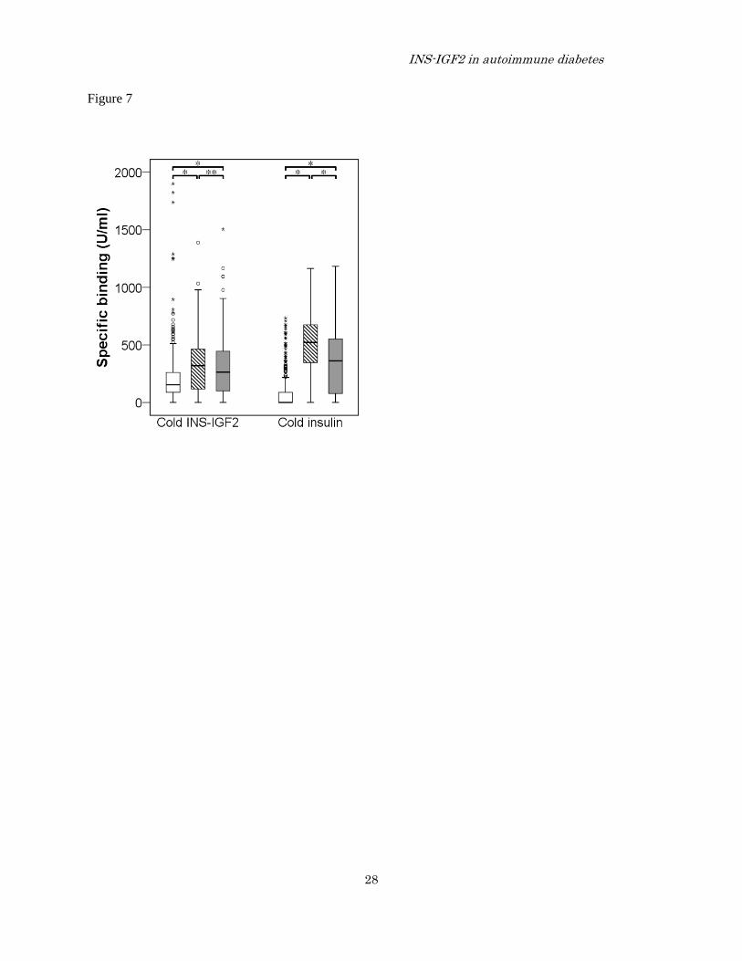
RBA for INS-IGF2 and insulin doubly reactive
autoantibodies— Autoantibodies against
INS-IGF2 (INS-IGF2A) were next examined in
serum from healthy controls (n=355) and newly
diagnosed T1D patients (n=304). In order to
correct for non-specific binding, all serum samples
were displaced with either cold INS-IGF2
(specific INS-IGF2A) or cold insulin (insulin
specific INS-IGF2A) (Figure 7). The median
binding for specific INS-IGF2A showed that both
1-5 (p<0.001) and 15-18 (p<0.001) year old T1D
patients differed from the controls. The specific
INS-IGF2A did not differed between the two
patient groups (p=0.204). The displacement with
cold insulin to reveal insulin-specific INS-IGF2A
showed that both 1-5 (p<0.001) and 15-18
(p<0.001) year old T1D patients also differed from
the controls (figure 7). However, the
insulin-specific INS-IGF2A showed higher median
levels in younger compared to older patients
(p<0.001).
Finally, specific INS-IGF2A (displaced with
cold INS-IGF2) and insulin-specific INS-IGF2A
(displaced with cold insulin) correlated in many
but not all serum samples from controls (r2=0.195,
p<0.001; Figure 8, panel A), T1D 1-5 year olds
(r2=0.380, p<0.001; panel B) and T1D 15-18 year
olds (r2=0.426, p<0.001; Figure 8, panel C).
Samples displaced by both cold INS-IGF2 and by
cold insulin are referred to as doubly reactive
INS-IGF2A. The data in Figure 8 suggest that
doubly reactive INS-IGF2A were found in 85%
(129/152) T1D 1-5 (p<0.001), in 74% (112/152)
T1D 15-18 year olds (p<0.001) compared to 48%
(169/355) among the controls.
DISCUSSION
The major finding in the present study is the
possibility that INS-IGF2 is a novel autoantigen in
T1D. This conclusion is based on several
observations. First, INS-IGF2 could be
specifically demonstrated in both insulin and
glucagon cells by immunocytochemistry as well as
by gene expression analyses of human islets from
several organ donors. Second, that the binding of
[35
S]INS-IGF2 was higher in serum from T1D
patients than in controls and that this binding
could be displaced both by cold INS-IGF2 and
cold insulin. Furthermore, our data suggest that
not only patients but also some control subjects
may have autoantibodies which were able to
recognize both [125
I]insulin and [35
S]INS-IGF2.
Indeed the displacement analyses (Figure 8)
suggest that as many as 47% of controls compared
to 85% among the 1-5 year olds and 74% among
the 15-18 year old patients may have doubly
reactive autoantibodies. The definition of a doubly

INS-IGF2 in autoimmune diabetes
11
reactive autoantibody sample would be that the
sample is binding both [125
I]insulin and
[35
S]INS-IGF2 and displaced by both cold insulin
and cold INS-IGF2. The doubly reactive
INS-IGF2A may be explained in part by the fact
that insulin and INS-IGF2 share the complete
B-chain sequence. The partial displacement of
INS-IGF2 binding by cold insulin may be
explained by such autoantibodies.
The fact that cold insulin in many samples did
not fully displace binding is likely to be explained
by autoantibody binding to non-B-chain epitopes
of the INS-IGF2 molecule. As 47% (controls) and
85% (patients) had insulin-INS-IGF2 doubly
reactive autoantibodies, it cannot be excluded that
INS-IGF2 is a more immunogenic entity than
insulin. This would be consistent with the report
that T1D patients may have specific T-cell
reactivity against the signal peptide (19,43) as well
as C-peptide sequences (44,45) that are part of
both preproinsulin and INS-IGF2 but not insulin
(Figure 1). The insulin-INS-IGF2 doubly reactive
sera complicate the understanding of insulin and
INS-IGF2 as autoantigens. It is undisputable that
they have sequence homologies. Although the
autoimmunogenicity of the C-terminal end of
INS-IGF2 remains to be determined, it cannot be
excluded that also this part of the molecule
contributes to the autoantibody binding.
Insulin autoantibodies were first described in
1983 using [125
I]insulin and acid-charcoal to
separate free from antibody-bund [125
I]insulin (46).
In early serum exchange exercise workshops only
radiobinding assays but not ELISA tests were
shown to detect disease-associated insulin
autoantibodies (IAA) (47-49). Despite efforts in
assay improvements (42), the IAA as opposed to
GADA, IA-2A and also ZnT8A remained the least
successful islet autoantibody assay to standardize
(14,39,41,50). We speculate that the inherent
difficulties to standardize the IAA assay may be
due to the presence of autoantibodies reactive with
INS-IGF2.
It has not yet been possible to identify the
mechanisms that trigger the formation of IAA.
Current data suggest that IAA are more common
in children developing T1D below the age of 5
compared to children older than 15 years of age
(51). Indeed, in children born to parents with T1D
who were followed from birth, IAA tend to be the
first islet autoantibody. This early insulin
autoantigenicity is still not understood but it
cannot be excluded that the combination of
HLA-DQ (IAA are associated with HLA-DQ8)
and insulin gene expression (reduced expression of
insulin in the thymus is associated with risk for
T1D through a genetic polymorphism in the INS
gene) promote insulin autoreactivity particularly in
young children. Our finding that INS-IGF2 was
expressed in human islets would support the
notion that INS-IGF2 may also become an
autoantigen based on similar mechanisms.
In a first report, INS-IGF2 gene expression in
human pancreas and eye was demonstrated (34).
In the present study, we therefore extend this
finding to demonstrate INS-IGF2 expression in
human islets. Our observation that the splice
variant expression was reduced in islets from T2D
patients or in islets from organ donors with
increased hemoglobin A1c suggest that the
INS-IGF2 gene regulation may be controlled by
mechanisms comparable to that of INS itself. This

INS-IGF2 in autoimmune diabetes
12
is not surprising as the INS and INS-IGF2 genes
share the same promoter. This promoter also
regulates INS gene expression in the thymus (52)
and we expect similar control of the INS-IGF2
expression. We noted that INS-IGF2 expression
was twice the expression of IGF2 but comparable
with IA-2 (Figure 1). The observation that the IA-2
autoantigen expression was similarly reduced in
islets from T2D patients suggested that
T2D-associated islet cell dysfunction may affect
the expression of both autoantigens. It was noted
that the expression of INS-IGF2 was correlated to
that of IA-2, both in healthy (r2=0.369, p<0.001)
and T2D (r2=0.888, p=0.001). Taken together, the
gene expression of INS-IGF2 in human islets and
it’s regulation by mechanisms similar to that of the
INS gene would be consistent with the possibility
that INS-IGF2 is an autoantigen that may be
important to T1D pathogenesis similar to that of
insulin.
The analysis of IAA requires the use of
[125
I]insulin (13,42,53) or possibly the use of
electrochemiluminescence technology (54,55). A
strength to assays using in vitro transcription
translation of cDNA to label the autoantigen with
radioactive amino acids has been the relative ease
of these radiobinding assays to be standardized
(15,56,57). Although we have not been able to in
vitro transcribe and translate native as well as
mutated variants of full length human
preproinsulin cDNA (data not shown), the present
truncated INS-IGF2 cDNA construct allowed us to
obtain consistently labeled autoantigen.
The specificity of the binding was tested with
increasing concentrations of insulin as well as
non-radioactive INS-IGF2 generated by in vitro
transcription translation. In the absence of native
or recombinant INS-IGF2 and a suitable antiserum
it has not been possible to establish a
radioimmunoassay that could be used to determine
the amount of cold INS-IGF2 generated. While
cold insulin could be used to fully block
[125
I]insulin binding, this was not possible with
INS-IGF2. This is a possible weakness to our
study and it cannot be excluded that we have
underestimated the proportion of sera with doubly
reactive autoantibodies. Another potential
weakness is that we have not compared
125I-labeled insulin with
125I-labeled INS-IGF2.
However, since [125
I]insulin for the detection of
IAA requires insulin labeled at the A14 tyrosine
such comparison would indeed not be possible.
In future investigations it will be important to
synthesize or produce recombinant INS-IGF2 in
order to determine maximal displacement of cold
INS-IGF2 and insulin alone as well as together. It
should then be possible to discern the contribution
of IAA alone, INS-IGF2A alone along with doubly
reactive sera. A dissection of whether the
autoreactivity to these two autoantigens with
shared amino acid sequences occurs
simultaneously or separately should be possible
using sera from longitudinal studies of at risk
children followed from birth (TEDDY, DIPP,
DAISY, BABY DIAB). Similarly, if T and B cells
have been collected in such studies it will be of
interest to determine whether the TCR of CD8+ T
cells express TCR recognizing B-chain residues
10-18 (20) or 9-23 (24) or signal peptide residues
15-24 (25,26) from preproinsulin, INS-IGF2, or
both.
The present study demonstrates that INS-IGF2

INS-IGF2 in autoimmune diabetes
13
may be a novel autoantigen in autoimmune T1D.
Our displacement experiments suggest that
autoantibodies against insulin and INS-IGF2 may
share autoantibody binding sites. Our findings
may shed light on several recent reports that
CD8+T cells recognize signal peptide and
C-peptide residues present in INS-IGF2 but not in
mature insulin. In addition, any epitope recognized
by T cells in the B-chain is likely to be shared
between mature insulin and INS-IGF2. It cannot
be excluded therefore that INS-IGF2 may augment
the ability of insulin to trigger the pathogenesis of
T1D.

INS-IGF2 in autoimmune diabetes
14
REFERENCES
1. Eisenbarth, G. S., and Jeffrey, J. (2008) Arquivos Brasileiros de Endocrinologia &
Metabologia 52, 146-155
2. Bach, J. F. (1994) Endocrine Reviews 15, 516-542
3. La Torre, D., and Lernmark, A. (2010) Immunology of beta-cell destruction. in Advances in
Experimental Medicine and Biology, 2010/03/11 Ed.
4. Pihoker, C., Gilliam, L. K., Hampe, C. S., and Lernmark, A. (2005) Diabetes 54 Suppl 2,
S52-61
5. Devendra, D., Liu, E., and Eisenbarth, G. S. (2004) BMJ 328, 750-754
6. Kulmala, P., Savola, K., Petersen, J. S., Vahasalo, P., Karjalainen, J., Lopponen, T., Dyrberg,
T., Akerblom, H. K., and Knip, M. (1998) J Clin Invest 101, 327-336
7. Wenzlau, J. M., Liu, Y., Yu, L., Moua, O., Fowler, K. T., Rangasamy, S., Walters, J., Eisenbarth,
G. S., Davidson, H. W., and Hutton, J. C. (2008) Diabetes 57, 2693-2697
8. Kupila, A., Keskinen, P., Simell, T., Erkkila, S., Arvilommi, P., Korhonen, S., Kimpimaki, T.,
Sjoroos, M., Ronkainen, M., Ilonen, J., Knip, M., and Simell, O. (2002) Diabetes 51, 646-651
9. Colman, P. G., Steele, C., Couper, J. J., Beresford, S. J., Powell, T., Kewming, K., Pollard, A.,
Gellert, S., Tait, B., Honeyman, M., and Harrison, L. C. (2000) Diabetologia 43, 203-209
10. Delli, A. J., Lindblad, B., Carlsson, A., Forsander, G., Ivarsson, S. A., Ludvigsson, J., Marcus,
C., Lernmark, A., and for the Better Diabetes Diagnosis Study, G. (2010) Pediatr Diabetes
11. Vardi, P., Ziegler, A. G., Mathews, J. H., Dib, S., Keller, R. J., Ricker, A. T., Wolfsdorf, J. I.,
Herskowitz, R. D., Rabizadeh, A., Eisenbarth, G. S., and et al. (1988) Diabetes Care 11,
736-739
12. Siljander, H. T., Simell, S., Hekkala, A., Lahde, J., Simell, T., Vahasalo, P., Veijola, R., Ilonen,
J., Simell, O., and Knip, M. (2009) Diabetes 58, 2835-2842
13. Diaz, J. L., and Wilkin, T. J. (1988) Clinical Chemistry 34, 356-359
14. Schlosser, M., Mueller, P. W., Torn, C., Bonifacio, E., and Bingley, P. J. (2010) Diabetologia 53,
2611-2620
15. Bingley, P. J., Bonifacio, E., and Mueller, P. W. (2003) Diabetes 52, 1128-1136
16. Wilkin, T., Palmer, J., Bonifacio, E., Diaz, J. L., and Kurtse, A. (1987) Diabetologia 30, 676-677
17. Toma, A., Haddouk, S., Briand, J. P., Camoin, L., Gahery, H., Connan, F., Dubois-Laforgue, D.,
Caillat-Zucman, S., Guillet, J. G., Carel, J. C., Muller, S., Choppin, J., and Boitard, C. (2005)
Proceedings of the National Academy of Sciences of the United States of America 102,
10581-10586
18. Mallone, R., Martinuzzi, E., Blancou, P., Novelli, G., Afonso, G., Dolz, M., Bruno, G., Chaillous,

INS-IGF2 in autoimmune diabetes
15
L., Chatenoud, L., Bach, J. M., and van Endert, P. (2007) Diabetes 56, 613-621
19. Skowera, A., Ellis, R. J., Varela-Calvino, R., Arif, S., Huang, G. C., Van-Krinks, C., Zaremba,
A., Rackham, C., Allen, J. S., Tree, T. I., Zhao, M., Dayan, C. M., Sewell, A. K., Unger, W. W.,
Drijfhout, J. W., Ossendorp, F., Roep, B. O., and Peakman, M. (2008) J Clin Invest 118,
3390-3402
20. Velthuis, J. H., Unger, W. W., Abreu, J. R., Duinkerken, G., Franken, K., Peakman, M.,
Bakker, A. H., Reker-Hadrup, S., Keymeulen, B., Drijfhout, J. W., Schumacher, T. N., and
Roep, B. O. (2010) Diabetes 59, 1721-1730
21. Kanatsuna, N., Papadopoulos, G. K., Moustakas, A. K., and Lernmark, A. (2012) Anat Res Int
2012, 457546
22. Durinovic-Bello, I., Boehm, B. O., and Ziegler, A. G. (2002) Journal of Autoimmunity 18, 55-66
23. Graham, J., Hagopian, W. A., Kockum, I., Li, L. S., Sanjeevi, C. B., Lowe, R. M., Schaefer, J. B.,
Zarghami, M., Day, H. L., Landin-Olsson, M., Palmer, J. P., Janer-Villanueva, M., Hood, L.,
Sundkvist, G., Lernmark, A., Breslow, N., Dahlquist, G., and Blohme, G. (2002) Diabetes 51,
1346-1355
24. Alleva, D. G., Crowe, P. D., Jin, L., Kwok, W. W., Ling, N., Gottschalk, M., Conlon, P. J.,
Gottlieb, P. A., Putnam, A. L., and Gaur, A. (2001) The Journal of Clinical Investigation 107,
173-180
25. Bulek, A. M., Cole, D. K., Skowera, A., Dolton, G., Gras, S., Madura, F., Fuller, A., Miles, J. J.,
Gostick, E., Price, D. A., Drijfhout, J. W., Knight, R. R., Huang, G. C., Lissin, N., Molloy, P. E.,
Wooldridge, L., Jakobsen, B. K., Rossjohn, J., Peakman, M., Rizkallah, P. J., and Sewell, A. K.
(2012) Nat Immunol
26. Wooldridge, L., Ekeruche-Makinde, J., van den Berg, H. A., Skowera, A., Miles, J. J., Tan, M.
P., Dolton, G., Clement, M., Llewellyn-Lacey, S., Price, D. A., Peakman, M., and Sewell, A. K.
(2012) J Biol Chem 287, 1168-1177
27. Falorni, A., Ortqvist, E., Persson, B., and Lernmark, A. (1995) J Immunol Methods 186, 89-99
28. Vaziri-Sani, F., Delli, A. J., Elding-Larsson, H., Lindblad, B., Carlsson, A., Forsander, G.,
Ivarsson, S. A., Ludvigsson, J., Marcus, C., and Lernmark, A. (2011) J Immunol Methods 371,
25-37
29. Elfving, A. M., Lindberg, B. A., Nystrom, L., Sundkvist, G., Lernmark, A., and Ivarsson, S. A.
(2003) Autoimmunity 36, 227-231
30. Falorni, A., Nikoshkov, A., Laureti, S., Grenback, E., Hulting, A. L., Casucci, G., Santeusanio,
F., Brunetti, P., Luthman, H., and Lernmark, A. (1995) J Clin Endocrinol Metab 80,
2752-2755
31. Agardh, D., Lynch, K., Brundin, C., Ivarsson, S. A., Lernmark, A., and Cilio, C. M. (2006) Clin
Exp Immunol 144, 67-75

INS-IGF2 in autoimmune diabetes
16
32. Wenzlau, J. M., Gardner, T. J., Frisch, L. M., Davidson, H. W., and Hutton, J. C. (2011)
Diabetes Metab Res Rev 27, 887-890
33. Weksberg, R., Shen, D. R., Fei, Y. L., Song, Q. L., and Squire, J. (1993) Nat Genet 5, 143-150
34. Monk, D., Sanches, R., Arnaud, P., Apostolidou, S., Hills, F. A., Abu-Amero, S., Murrell, A.,
Friess, H., Reik, W., Stanier, P., Constancia, M., and Moore, G. E. (2006) Hum Mol Genet 15,
1259-1269
35. Delli, A. J., Vaziri-Sani, F., Lindblad, B., Elding-Larsson, H., Carlsson, A., Forsander, G.,
Ivarsson, S. A., Ludvigsson, J., Kockum, I., Marcus, C., Samuelsson, U., Ortqvist, E., Groop,
L., Bondinas, G. P., Papadopoulos, G. K., and Lernmark, A. (2012) Diabetes 61, 2556-2564
36. Carlsson, A. K., Axelsson, I. E., Borulf, S. K., Bredberg, A. C., Lindberg, B. A., Sjoberg, K. G.,
and Ivarsson, S. A. (1999) Pediatrics 103, 1248-1252
37. Lindberg, B., Ahlfors, K., Carlsson, A., Ericsson, U. B., Landin-Olsson, M., Lernmark, A.,
Ludvigsson, J., Sundkvist, G., and Ivarsson, S. A. (1999) Pediatrics 104, e12
38. Agardh, D., Nilsson, A., Tuomi, T., Lindberg, B., Carlsson, A. K., Lernmark, A., and Ivarsson,
S. A. (2001) Pediatr Diabetes 2, 58-65
39. Torn, C., Mueller, P. W., Schlosser, M., Bonifacio, E., and Bingley, P. J. (2008) Diabetologia 51,
846-852
40. Mire-Sluis, A. R., Gaines Das, R., and Lernmark, A. (2000) Diabetologia 43, 1282-1292
41. Lampasona, V., Schlosser, M., Mueller, P. W., Williams, A. J., Wenzlau, J. M., Hutton, J. C.,
and Achenbach, P. (2011) Clin Chem 57, 1693-1702
42. Williams, A. J., Bingley, P. J., Bonifacio, E., Palmer, J. P., and Gale, E. A. (1997) Journal of
Autoimmunity 10, 473-478
43. Knight, R. R., Kronenberg, D., Zhao, M., Huang, G. C., Eichmann, M., Bulek, A., Wooldridge,
L., Cole, D. K., Sewell, A. K., Peakman, M., and Skowera, A. (2012) Diabetes
44. Arif, S., Tree, T. I., Astill, T. P., Tremble, J. M., Bishop, A. J., Dayan, C. M., Roep, B. O., and
Peakman, M. (2004) J Clin Invest 113, 451-463
45. Unger, W. W., Velthuis, J., Abreu, J. R., Laban, S., Quinten, E., Kester, M. G., Reker-Hadrup,
S., Bakker, A. H., Duinkerken, G., Mulder, A., Franken, K. L., Hilbrands, R., Keymeulen, B.,
Peakman, M., Ossendorp, F., Drijfhout, J. W., Schumacher, T. N., and Roep, B. O. (2011)
Journal of Autoimmunity 37, 151-159
46. Palmer, J. P., Asplin, C. M., Clemons, P., Lyen, K., Tatpati, O., Raghu, P. K., and Paquette, T. L.
(1983) Science 222, 1337-1339
47. Greenbaum, C. J., Palmer, J. P., Kuglin, B., and Kolb, H. (1992) The Journal of Clinical
Endocrinology & Metabolism 74, 1040-1044
48. Wilkin, T., Palmer, J., Kurtz, A., Bonifacio, E., and Diaz, J. L. (1988) Diabetologia 31, 449-450
49. Levy-Marchal, C., Bridel, M. P., Sodoyez-Goffaux, F., Koch, M., Tichet, J., Czernichow, P., and

INS-IGF2 in autoimmune diabetes
17
Sodoyez, J. C. (1991) Diabetes Care 14, 61-63
50. Verge, C. F., Stenger, D., Bonifacio, E., Colman, P. G., Pilcher, C., Bingley, P. J., and
Eisenbarth, G. S. (1998) Diabetes 47, 1857-1866
51. Andersson, C., Larsson, K., Vaziri-Sani, F., Lynch, K., Carlsson, A., Cedervall, E., Jonsson, B.,
Neiderud, J., Mansson, M., Nilsson, A., Lernmark, A., Elding Larsson, H., and Ivarsson, S. A.
(2011) Autoimmunity 44, 394-405
52. Noso, S., Kataoka, K., Kawabata, Y., Babaya, N., Hiromine, Y., Yamaji, K., Fujisawa, T.,
Aramata, S., Kudo, T., Takahashi, S., and Ikegami, H. (2010) Diabetes 59, 2579-2587
53. Stentz, F. B., Wright, R. K., and Kitabchi, A. E. (1982) Diabetes 31, 1128-1131
54. Yu, L., Miao, D., Scrimgeour, L., Johnson, K., Rewers, M., and Eisenbarth, G. S. (2012)
Diabetes 61, 179-186
55. Lo, B., Swafford, A. D., Shafer-Weaver, K. A., Jerome, L. F., Rakhlin, L., Mathern, D. R.,
Callahan, C. A., Jiang, P., Davison, L. J., Stevens, H. E., Lucas, C. L., White, J., von Borstel,
R., Todd, J. A., and Lenardo, M. J. (2011) J Transl Med 9, 203
56. Schlosser, M., Mueller, P. W., Achenbach, P., Lampasona, V., and Bingley, P. J. (2011) Diabetes
Care 34, 2410-2412
57. Bingley, P. J., Williams, A. J., Colman, P. G., Gellert, S. A., Eisenbarth, G., Yu, L., Perdue, L.
H., Pierce, J. J., Hilner, J. E., Nierras, C., Akolkar, B., and Steffes, M. W. (2010) Clin Trials 7,
S56-64

INS-IGF2 in autoimmune diabetes
18
Acknowledgements— We thank Anita Nilsson, Ingrid Wigheden, Ida Jönsson, and Ann-Helen Thorén
Fischer for expert technical assistance. Human islets were provided by the Nordic Center of Islet
Transplantation at the Uppsala University, Uppsala, Sweden by courtesy of professor Olle Korsgren.
Technical assistance was received from the staff in the Human Tissue Laboratory (HTL), governed by
Ulrika Krus, at the Lund University Diabetes Centre. The authors also thank the Swegene Center for
Integrative Biology at Lund University (SCIBLU) for performing the gene expression micorarrays. The
following members of the Better Diabetes Diagnosis study group, Annelie Carlsson (Lund), Gun
Forsander (Gothenburgh) Sten A. Ivarsson (Malmö), Johnny Ludvigsson (Linköping), Ingrid
Kockum (Stockholm), Claude Marcus (Stockholm), Ulf Samuelsson (Linköping), Eva Örtqvist
(Stockholm), Anita Nilsson (Malmö), Helena Desaix (Borås), Kalle Snellman (Eskilstuna), Anna
Olivecrona (Falun), Åke Stenberg (Gällivare), Lars Skogsberg (Gävle), Nils Östen Nilsson (Halmstad),
Jan Neiderud (Helsingborg), Åke Lagerwall (Hudiksvall), Kristina Hemmingsson (Härnösand), Karin
Åkesson (Jönköping), Göran Lundström (Kalmar), Magnus Ljungcrantz (Karlskrona), Eva Albinsson
(Karlstad), Karin Larsson (Kristianstad), Christer Gundewall (Kungsbacka), Rebecka Enander
(Lidköping), Agneta Brännström (Luleå), Maria Nordwall (Norrköping), Lennart Hellenberg (Nyköping),
Elena Lundberg (Skellefteå), Henrik Tollig (Skövde), Britta Björsell (Sollefteå), Björn Rathsman
(Stockholm/Sacchska), Torun Torbjörnsdotter (Stockholm/Huddinge), Björn Stjernstedt (Sundsvall), Nils
Wramner (Trollhättan), Ragnar Hanås (Uddevalla), Ingemar Swenne (Uppsala), Anna Levin (Visby),
Anders Thåström (Västervik), Carl-Göran Arvidsson (Västerås), Stig Edvardsson (Växjö), Björn Jönsson
(Ystad), Torsten Gadd (Ängelholm), Jan Åman (Örebro), Rein Florell (Örnsköldsvik), and Anna-Lena
Fureman (Östersund) provided serum samples from newly diagnosed type 1 diabetes patients.
FOOTNOTES
*This work was supported by the Swedish Research Council grant K2001-54X-15312-07-6, SUS Funds
& Donations, Diabetes Fund, and the Skåne County Council for Research and Development. The BDD
study group was supported by the Swedish Childhood Diabetes Fund. The LUDC/EXODIAB and the
Nordic Network for Clinical Islet Transplantation were supported by grants from the Swedish Research
Council (collaborative project grant Dnr. 521-2008-2974, strategic research area grant EXODIAB Dnr.
2009-1039, and Linnaeus grant Dnr. 349-2008-6589).
1To whom correspondence should be addressed: Norio Kanatsuna MD PhD, Department of Clinical
Sciences, Lund University Diabetes Center, Lund University, Jan Waldenströms gata 35, Skåne
University Hospital SUS, SE-205 02 Malmö, Sweden, TEL.: +46 40 39 19 01; FAX: +46 40 39 19 19;
E-mail: [email protected]
3The abbreviations used are: T1D, type 1 diabetes; GAD65, glutamic acid decarboxylase 65; IA-2, islet
antigen-2; ZnT8, zinc transporter 8; IAA, insulin autoantibodies; ER, endoplasmic reticulum; APC,

INS-IGF2 in autoimmune diabetes
19
antigen presenting cell; TCR, T cell receptor; BCR, B cell receptor; RBA, radiobinding assay; IGF2,
insulin like growth factor 2; GSIR, Glucose-stimulated insulin release; DASP, Diabetes Antibody
Standardization Program; IU, international unit; TEDDY, The Environmental Determinants of Diabetes in
the Young; DIPP, the Diabetes Prediction and Prevention AND DAISY, the Diabetes Autoimmunity Study
in the Young.
FIGURE LEGENDS
FIGURE 1. Genomic structure of the preproinsulin (INS) and IGF2 genes on chromosome 11.
The cDNA for the INS and INS-IGF2 genes are depicted essentially as described (34). The INS-IGF2
cDNA is a splice variant of INS (signal peptide, B chain and 8 amino acids of the C-peptide) and the
coding sequences of the two proximal ORF of the IGF2 gene. It should be noted that the IGF2 gene
ORFs are normally non-coding exons and thus insert a novel 138 aa C-terminal region unrelated to
preproIGF2 (34). Shaded boxes represent ORFs.
FIGURE 2. Gene expression of INS-IGF2, IGF2 and IA-2 in human pancreatic islets.
A. Mean expression (mean±SD) of INS-IGF2, IGF2 and IA-2 are shown in isolated human pancreatic
islets obtained from non-diabetes controls (n=66; open columns) and T2D patients (n=10; closed
columns). Gene expression, compared to controls, was lower in T2D islet for INS-IGF2 (**p=0.006),
IGF2 († p=0.017) as well as IA-2 (‡ p=0.009). Differences in gene expression between the three genes are
also indicated (* p<0.001). B. Mean expression (mean±SD) of INS-IGF2, IGF2 and IA-2 are shown for
isolated human pancreatic islets obtained from donors with HbA1c being <6.0% (n=41, open bars), 6.0 –
6.4% (n=12, hatched bars) or ≥ 6.5% (T2D; n=10; closed bars). Differences in gene expression between
the three groups are also indicated as follows: *1 p<0.001; *2 p=0.027; *3 p=0.059; *4 p=0.010; *5
p=0.015; *6 p=0.976; *7 p=0.006; *8 p=0.154; *9 p=0.104.
FIGURE 3. Mean gene expression of INS-IGF2, IGF2 and IA-2 in human pancreatic islets in relation to
Glucose –Stimulated Insulin Release (GSIR) and HbA1c levels (%) of the islet donors.
Mean gene expression is shown for INS-IGF2 (panel A), IGF2 (panel B) and IA-2 (panel C). The
expression of INS-IGF2 was correlated to GSIR (r=0.284; p=0.023) primarily in donors with low
HbA1c (r=0.446; p<0.001). IGF2 expression was not related to GSIR (p=0.186) despite the lower
expression in islets from donors with high HbA1c (r=0.394; p=0.001). Islets with increased IA-2
expression showed higher GSIR (r=0.501; p<0.001) especially in islets from donors with low
HbA1c (r=0.333; p=0.008).

INS-IGF2 in autoimmune diabetes
20
FIGURE 4. Triple immunofluorescence photomicrographs of sections of human pancreas.
Immunostaining for INS-IGF2 (A), insulin (B) and glucagon (B) and the overlay to show co-localization
with both glucagon and insulin (D). Co-localization with insulin is indicated with arrows. Co-localization
with glucagon is indicated with arrow-heads. The scale bar represents 50 µm.
FIGURE 5. SDS-gel electrophoresis and autoradiography of in vitro transcribed and translated
INS-IGF2.
Lane 1: Non-isotope protein ladder representing proteins of 250, 130, 100, 70, 55, 35 and 25 kDa. Lane 2:
[35
S]INS-IGF2 generated by in vitro transcription translation. The major band of 20.7 kDa represents
INS-IGF2. The minor bands indicated may represent non-specific components or an aggregate product.
Lane 3: Residual radioactivity after in vitro transcription translation without added cDNA.
FIGURE 6. Radiobinding assays for autoantibodies against insulin (IAA) and INS-IGF2 (INS-IGF2A) in
a type 1 diabetes serum (patient MD) used as in-house standard.
A. IAA assay with [125
I]insulin at various dilutions of the MD standard without (closed circles) and with
an excess of cold insulin (2 IU/ml; open circles). Mean values ± SE (n=4). B. INS-IGF2A assay with
[35
S]INS-IGF2 at various dilutions of the MD standard without (closed circles), with an excess of cold
insulin (11 IU/ml; open squares) and cold INS-IGF2 (2X of the labeled INS-IGF2). Mean values ± SE
(n=4). Error bars are within the symbols. C. [35
S]INS-IGF2A in the MD standard serum diluted 1:4 in the
presence of different concentrations of either cold INS-IGF2 (open circles), cold insulin (open squares) or
cold IA-2 (closed triangles). Error bars are mostly within the symbols. Mean ± SE) (n=6).
FIGURE 7. Specific INS-IGF2 autoantibody levels (U/ml) after displacement with either cold INS-IGF2,
generated by in vitro transcription translation and used at a concentration representing twice the labeled
INS-IGF2 , or with 11 IU/ml cold insulin.
The box plots with whiskers and outliers show the median levels of specific binding for 355 controls
(open box), 152 type 1 diabetes patients 1-5 years of age (hatched box) and 152 type 1 diabetes patients
15-18 years of age (gray box). Differences between the groups are indicated by * p<0.001 and **
p=0.204.
FIGURE 8. Correlation between specific INS-IGF2A (U/ml) in sera from controls and T1D patients
displaced with either cold INS-IGF2 or cold insulin (insulin-specific INS-IGF2) for specific binding.
A. Correlation between specific INS-IGF2A and insulin-specific INS-IGF2A in control subjects (n=355)
(r2=0.195, p<0.001). B. Correlation between specific INS-IGF2A and insulin-specific INS-IGF2A in 1-5
year old T1D patients (n=152) (r2=0.380, p<0.001). C. Correlation between specific INS-IGF2A and
insulin-specific INS-IGF2A in 15-18 year old T1D patients (n=152) (r2=0.426, p<0.001).
INS-IGF2 in autoimmune diabetes
21
Table 1
Characteristics of patient serum samples, frequencies of islet autoantibodies and HLA
T1D T1D Controls
(1-5 years) (15-18 years)
n 152 152 355
Median age (years) 4.4 16.1 12.6
(range) (1.0-5.9) (15.0-17.9) (11.5-25.0)
Gender (% male) 49.3 66.4 48.5
HLA-DQ
2/8 65 (43%) 34 (22%) ND
8/8, 8/x 54 (36%) 66 (43%) ND
2/2, 2/x 17 (11%) 25 (16%) ND
x/x 16 (11%) 27 (18%) ND
Islet autoantibodies
GADA 71 (47%) 101 (66%) 3/343 (0.9%)
IAA 87 (57%) 27 (18%) 2/180 (1.1%)
IA2A 113 (74%) 127 (84%) 0/343 (0%)
ZnT8WA 63 (41%) 78 (51%) 3/343 (0.9%)
ZnT8RA 72 (47%) 80 (53%) 3/343 (0.9%)
ZnT8QA 36 (24%) 46 (30%) 3/343 (0.9%)
ND is not determined.

INS-IGF2 in autoimmune diabetes
22
Figure 1

INS-IGF2 in autoimmune diabetes
23
Figure 2

INS-IGF2 in autoimmune diabetes
24
Figure 3

INS-IGF2 in autoimmune diabetes
25
Figure 4

INS-IGF2 in autoimmune diabetes
26
Figure 5

INS-IGF2 in autoimmune diabetes
27
Figure 6
INS-IGF2 in autoimmune diabetes
28
Figure 7

INS-IGF2 in autoimmune diabetes
29
Figure 8