ORIGINAL ARTICLE Role of transient receptor potential and...
10
ORIGINAL ARTICLE Role of transient receptor potential and pannexin channels in cigarette smoke-triggered ATP release in the lung Matthew Baxter, Suffwan Eltom, Bilel Dekkak, Liang Yew-Booth, Eric D Dubuis, Sarah A Maher, Maria G Belvisi, Mark A Birrell ▸ Additional material is published online only. To view please visit the journal online (http://dx.doi.org/10.1136/ thoraxjnl-2014-205467). Respiratory Pharmacology, Faculty of Medicine, National Heart and Lung Institute, Imperial College London, London, UK Correspondence to Dr Mark Birrell, Respiratory Pharmacology, Faculty of Medicine, National Heart and Lung Institute, Imperial College London, Exhibition Road, London, SW7 2AZ, UK; [email protected] Received 25 March 2014 Revised 4 August 2014 Accepted 13 September 2014 Published Online First 9 October 2014 To cite: Baxter M, Eltom S, Dekkak B, et al. Thorax 2014;69:1080–1089. ABSTRACT Background COPD is an inflammatory disease usually associated with cigarette smoking (CS) with an increasing global prevalence and no effective medication. Extracellular ATP is increased in the COPD affected lung and may play a key role in driving CS-induced airway inflammation, but the mechanism involved in ATP release has eluded researchers. Recently, the transient receptor potential (TRP) and pannexin-1 channels have been suggested to play a role in other experimental paradigms. Thus, the aim of this work is to investigate if these channels are involved in CS- induced ATP release in the lung. Methods Primary human cells were exposed to CS and extracellular ATP levels measured. Mice were exposed to mainstream CS and airway inflammation assessed. TRPV1/ 4 mRNA expression was assessed in human lung parenchyma. Results CS exposure caused a dose-related increase in ATP from primary airway bronchial epithelial cells. This was attenuated by blockers of TRPV1, TRPV4 and pannexin-1 channels. Parallel data was obtained using murine acute CS-driven model systems. Finally, TRPV1/4 mRNA expression was increased in lung tissue samples from patients with COPD. Conclusions Extracellular ATP is increased in the COPD affected lung and may play a key role in driving disease pathophysiology. These experiments uncover a novel mechanism which may be responsible for CS-induced ATP release. These findings highlight novel targets that could lead to the development of medicine to treat this devastating disease. INTRODUCTION COPD is an inflammatory airway disease usually associated with cigarette smoking (CS) with an increasing global prevalence and no effective medi- cation. 1–3 The Global Initiative for Chronic Obstructive Lung Disease guidelines state that COPD is characterised by airflow limitation that is not fully reversible and is attributed to small airways disease and parenchymal destruction. It is a steroid-resistant, inflammatory disease characterised by the infiltration of leucocytes including neutro- phils, macrophages and CD8 + T-cells. 45 Increased concentrations of many proinflammatory mediators are also observed including the caspase-1 related cytokines, IL-1β and IL-18, the levels of which are found to correlate with disease severity. 6 7 Caspase-1 activation is thought to be regulated by the NLRP3 inflammasome, and one of the key acti- vation mechanisms is through extracellular ATP (eATP)-dependent stimulation of the P2X 7 recep- tor. 8–10 Interestingly, increased levels of eATP have been reported in vitro and in vivo models of cigarette smoke-induced inflammation and in the lungs of patients with COPD. 11–14 The relevance of this pathway to the pathogenesis of COPD is supported by findings that P2X 7 receptor and caspase-1 activation are central to the development of CS-induced airway inflammation in murine models. 15–17 eATP is thought to be stored in the cytosol, but the mechanism by which it is released can depend on the cell/tissue type, the environment and the stimulus. 18–21 Recent evidence has suggested a role for the pannexin-1 channel/pore in the mechanism of ATP release. 22–27 Furthermore, transient receptor potential (TRP) channels have been suggested to play a pivotal role in the release of eATP and are associated with opening the pannexin-1 channel. 18 27 28 Of the TRP family, TRPV4 has been shown to be present in the airway and involved in the release of hypotonicity-induced eATP. 27 29 30 Endogenous TRPV1 activators (e.g. lipoxygenase products and mediators of the Protein Kinase-A, Protein Kinase-C and Phospholipase- C pathways) are known to be increased in the COPD affected Key messages What is the key question? ▸ COPD is an airway disease with an increasing global prevalence but no effective medication. Understanding the disease pathogenesis would be a major step towards developing new therapies. What is the bottom line? ▸ This study reveals for the first time the mechanism that leads to the increased ATP levels and the associated steroid-resistant inflammation observed in the airways of patients with COPD. Why read on? ▸ These data highlight novel targets which can be exploited for the development of effective therapeutics for the treatment of COPD. Chronic obstructive pulmonary disease 1080 Baxter M, et al. Thorax 2014;69:1080–1089. doi:10.1136/thoraxjnl-2014-205467 on 21 May 2018 by guest. Protected by copyright. http://thorax.bmj.com/ Thorax: first published as 10.1136/thoraxjnl-2014-205467 on 9 October 2014. Downloaded from
Transcript of ORIGINAL ARTICLE Role of transient receptor potential and...

ORIGINAL ARTICLE
Role of transient receptor potential and pannexinchannels in cigarette smoke-triggered ATP releasein the lungMatthew Baxter, Suffwan Eltom, Bilel Dekkak, Liang Yew-Booth, Eric D Dubuis,Sarah A Maher, Maria G Belvisi, Mark A Birrell
▸ Additional material ispublished online only. To viewplease visit the journal online(http://dx.doi.org/10.1136/thoraxjnl-2014-205467).
Respiratory Pharmacology,Faculty of Medicine, NationalHeart and Lung Institute,Imperial College London,London, UK
Correspondence toDr Mark Birrell, RespiratoryPharmacology, Faculty ofMedicine, National Heart andLung Institute, Imperial CollegeLondon, Exhibition Road,London, SW7 2AZ, UK;[email protected]
Received 25 March 2014Revised 4 August 2014Accepted 13 September 2014Published Online First9 October 2014
To cite: Baxter M, Eltom S,Dekkak B, et al. Thorax2014;69:1080–1089.
ABSTRACTBackground COPD is an inflammatory disease usuallyassociated with cigarette smoking (CS) with an increasingglobal prevalence and no effective medication. ExtracellularATP is increased in the COPD affected lung and may play akey role in driving CS-induced airway inflammation, but themechanism involved in ATP release has eluded researchers.Recently, the transient receptor potential (TRP) andpannexin-1 channels have been suggested to play a role inother experimental paradigms. Thus, the aim of this workis to investigate if these channels are involved in CS-induced ATP release in the lung.Methods Primary human cells were exposed to CS andextracellular ATP levels measured. Mice were exposed tomainstream CS and airway inflammation assessed. TRPV1/4 mRNA expression was assessed in human lungparenchyma.Results CS exposure caused a dose-related increase inATP from primary airway bronchial epithelial cells. This wasattenuated by blockers of TRPV1, TRPV4 and pannexin-1channels. Parallel data was obtained using murine acuteCS-driven model systems. Finally, TRPV1/4 mRNAexpression was increased in lung tissue samples frompatients with COPD.Conclusions Extracellular ATP is increased in the COPDaffected lung and may play a key role in driving diseasepathophysiology. These experiments uncover a novelmechanism which may be responsible for CS-induced ATPrelease. These findings highlight novel targets that couldlead to the development of medicine to treat thisdevastating disease.
INTRODUCTIONCOPD is an inflammatory airway disease usuallyassociated with cigarette smoking (CS) with anincreasing global prevalence and no effective medi-cation.1–3 The Global Initiative for ChronicObstructive Lung Disease guidelines state thatCOPD is characterised by airflow limitation that isnot fully reversible and is attributed to smallairways disease and parenchymal destruction. It is asteroid-resistant, inflammatory disease characterisedby the infiltration of leucocytes including neutro-phils, macrophages and CD8+ T-cells.4 5 Increasedconcentrations of many proinflammatory mediatorsare also observed including the caspase-1 relatedcytokines, IL-1β and IL-18, the levels of which arefound to correlate with disease severity.6 7
Caspase-1 activation is thought to be regulated by
the NLRP3 inflammasome, and one of the key acti-vation mechanisms is through extracellular ATP(eATP)-dependent stimulation of the P2X7 recep-tor.8–10 Interestingly, increased levels of eATPhave been reported in vitro and in vivo models ofcigarette smoke-induced inflammation and in thelungs of patients with COPD.11–14 The relevanceof this pathway to the pathogenesis of COPD issupported by findings that P2X7 receptor andcaspase-1 activation are central to the developmentof CS-induced airway inflammation in murinemodels.15–17
eATP is thought to be stored in the cytosol, butthe mechanism by which it is released can dependon the cell/tissue type, the environment and thestimulus.18–21 Recent evidence has suggested a rolefor the pannexin-1 channel/pore in the mechanismof ATP release.22–27 Furthermore, transient receptorpotential (TRP) channels have been suggested toplay a pivotal role in the release of eATP and areassociated with opening the pannexin-1channel.18 27 28 Of the TRP family, TRPV4 has beenshown to be present in the airway and involved inthe release of hypotonicity-induced eATP.27 29 30
Endogenous TRPV1 activators (e.g. lipoxygenaseproducts and mediators of the Protein Kinase-A,Protein Kinase-C and Phospholipase- C pathways)are known to be increased in the COPD affected
Key messages
What is the key question?▸ COPD is an airway disease with an increasing
global prevalence but no effective medication.Understanding the disease pathogenesis wouldbe a major step towards developing newtherapies.
What is the bottom line?▸ This study reveals for the first time the
mechanism that leads to the increased ATPlevels and the associated steroid-resistantinflammation observed in the airways ofpatients with COPD.
Why read on?▸ These data highlight novel targets which can
be exploited for the development of effectivetherapeutics for the treatment of COPD.
Chronic obstructive pulmonary disease
1080 Baxter M, et al. Thorax 2014;69:1080–1089. doi:10.1136/thoraxjnl-2014-205467
on 21 May 2018 by guest. P
rotected by copyright.http://thorax.bm
j.com/
Thorax: first published as 10.1136/thoraxjnl-2014-205467 on 9 O
ctober 2014. Dow
nloaded from

lung. Furthermore, low pH conditions often manifest in thelungs of patients with COPD as measured in exhaled breath con-densate and this is also a trigger for TRPV1 activation.31 TRPA1,has also been implicated in COPD-associated disease pathologies.Indeed, Trpa1−/− mice were found to be protected againstCS-mediated plasma protein extravasation and had reducedlevels of the murine neutrophil chemoattractant keratinocytechemoattractant (KC).32 Furthermore, TRPA1 is known to beactivated by some of the key inflammatory components of CS,including acrolein and crotonaldehyde.33
Therefore, in view of the evidence presented above, wehypothesised that CS exposure induces ATP release via activa-tion of specific TRP channels and the subsequent opening of thepannexin-1 pore. To test this hypothesis, we confirmed that CSexposure increased ATP levels in lavage in a murine model andthat these levels were unchanged in P2X7 receptor-deficientmice that were protected from the CS-induced neutrophilia.This suggested that in our system the P2X7 receptor was notinvolved in the eATP release and that the signal was not fromthe recruited neutrophils but probably from cells present in theairway i.e. alveolar macrophages (AM) and human bronchialepithelial cells (HBEC). To study the mechanism responsible forthe CS-induced release of ATP, we used primary human cell-based assays (AMs and HBECs) and pharmacological tools toinvestigate the role of TRPV1, TRPV4 and TRPA1 andpannexin-1. This was paralleled in our murine model usingTrpv1, Trpv4, Trpa1 and pannexin-1 gene knockout (KO) mice.Finally, we measured the expression of the TRP channels indisease-relevant lung tissue harvested from patients with COPDto provide convincing data implicating these ion channels in thepathophysiology of COPD and highlighting them as noveltargets for therapeutic intervention.
MATERIALS AND METHODSCell-based model systemCS bubbled medium (CSM) was prepared as described previ-ously34 and added to HBECs (Lonza, UK) or human AMs har-vested as previously described.34 Cell viability was assessedusing MTT. ATP levels were assessed using an ATPlite assay(Perkin Elmer, Cambridge, UK). Osmolarity and pH wasassessed using an Osmomat 030 osmometer (Gonotec,Germany) and pHep4pH meter (Hanna Instruments, UK),respectively. In some experiments, cells were pretreated withTRPV1, TRPV4 or TRPA1 antagonists (JNJ-17203212 (Sigma,UK), HC 067047 (Peakdale Molecular, UK) and HC 030031(Chembridge, USA)), respectively, or agonists capsaicin (Sigma,UK), GSK 1016790a (Sigma, UK) and acrolein (Sigma, UK),respectively. In separate experiments cells were pretreated withspecific pannexin antagonists carbenoxolone and probenecid orthe connexin inhibitor flufenamic acid (all from Sigma, UK).Concentrations used were based on previous studies.24 27 35
Measurement of mRNA expressionQuantitative RT-PCR was used to measure mRNA from humanlung samples and cell populations using TaqMan reagents andprotocols (Applied Biosystems) as previously described.36
Human donor and recipient lung tissue samples, surplus to clin-ical requirements, were obtained from a transplant programmesupported by the NIHR Respiratory Disease BiomedicalResearch Unit at the Royal Brompton and Harefield NHSFoundation Trust and Imperial College London. Informedwritten consent and ethical approval was obtained (EthicNumber: REC 09/H0708/72, active from Dec 2009). See table 1for demographic details.
In vivo studiesAll in vivo protocols were approved by a local ethical reviewprocess and strictly adhered to the Animals (ScientificProcedures) Act 1986 UK Home Office guidelines (ProjectLicence number: 70/7212, active from May 2012). Male wild-type C57BL/6 were bred inhouse alongside the genetic lines.Mice were either kind gifts or purchased from Jackson labs,USA, (P2X7—Professor Jean Kanellopoulos (UniversitéParis-Sud, France), Trpv1 and Trpa1—Jackson labs; Trpv4—Riken Bioresource Centre, (Japan)37 38 and Pannexin-1—DrVishva Dixit, (Genentech, USA)).
CS or lipopolysaccharide (LPS) exposure protocols have beendescribed previously.15 Briefly, mice were exposed to CS (1 h,twice a day for 3 days—acute model) or LPS (aerosol of1 mg/mL for 30 min) and the lungs lavaged 24 h or 6 h later,respectively. Bronchoalveolar lavage fluid (BALF) interleukin(IL)-1β (ELISA, assay range reported as 15.6–1000 pg/mL; tomeasure lower levels it is important to freshly prepare the assayplates) neutrophil and ATP levels (no increase detected in theLPS model) were assessed as previously described.15 A parallelsubchronic model was also performed where the wild-type orKO mice were exposed to CS for 14 days.
Data analysisData are expressed as mean±SEM of n observations. Statisticalsignificance was determined using either single or multiple com-parisons (specific tests used are described in the figure legends),using GraphPad Prism 5 software. A p value <0.05 was taken assignificant and all treatments were compared with the appropri-ate control group.
Online supplementary materialThe online supplementary material section contains the datafrom characterising the impact of smoke on cell/medium invitro, profiling the TRPV1, TRPV4 and pannexin-1 KO mice inthe LPS-driven model of airway inflammation. Additionally, itcontains the data from profiling the TRPA1 KO mice in thesmoke and LPS model systems.
RESULTSP2X7 receptor and neutrophils are not involved in acute CSmediated ATP release in vivoWild-type and P2X7
−/− mice were concurrently exposed to CStwice a day for three consecutive days. There was a significantincrease in ATP, IL-1β and neutrophils in the BALF inCS-challenged wild-type mice 24 h after the last exposure(figure 1). P2X7
−/− mice exhibited significantly reduced levels ofIL-1β and neutrophilia compared with the wild-type consistentwith previously published data15 16 (figures 1B, C). There was,however, no reduction in the levels of BALF ATP in CS-exposedP2X7
−/− mice compared with the wild-type controls. This
Table 1 Details on the donor/recipient
Disease group Age (years) Sex (M/F)
Non-smoking donor 27–72 3/9Smoking donor 22–62 7/3Emphysema 49–69 17/8
Non-smoking donor 27–72 3/9Smoking donor 22–62 7/3Emphysema 49–69 17/8
Chronic obstructive pulmonary disease
Baxter M, et al. Thorax 2014;69:1080–1089. doi:10.1136/thoraxjnl-2014-205467 1081
on 21 May 2018 by guest. P
rotected by copyright.http://thorax.bm
j.com/
Thorax: first published as 10.1136/thoraxjnl-2014-205467 on 9 O
ctober 2014. Dow
nloaded from

indicates that the ATP release is independent of the P2X7 recep-tor and that it is not released from the recruited neutrophils.This also suggests that CS-induced ATP is upstream of theP2X7-inflammasome-IL-1β/IL-18 axis and is released from struc-tural and/or resident cells in the airway, possibly AMs and bron-chiolar epithelial cells.
Development and characterisation of CS-induced ATPrelease in vitroTo begin to investigate the mechanism responsible forCS-induced ATP release we used cell-based assays. We selectedairway-relevant primary cells (HBECs and AMs) and exposedthem to CSM. CSM caused a dose-related release of ATP overtime in both cell types (figure 2A, C). CSM (2.5%) was chosen
for further analysis. CSM did not cause any cell death or alterthe pH or osmolarity of the culture medium (see online supple-mentary figure S1). Analysis of the mRNA levels of the TRPchannels under investigation in the two cell types showed thatboth cell types expressed significant levels of TRPV1 andTRPV4 mRNA but the levels of TRPA1 were below the detec-tion limit of the assay in AMs and detected only at very lowlevels in HBECs (figure 2D).
Role of TRPV1, TRPV4 and pannexin channels inCSM-mediated ATP release in vitroCSM-induced ATP release from HBECs was inhibited by aTRPV4 antagonist and, to a lesser extent, by a TRPV1 antagon-ist (figure 3). In parallel, the pannexin channel blockers
Figure 1 The role of P2X7 incigarette smoking (CS)-induced murineairway inflammation. P2X7
−/− micewere exposed to CS or room air(control) twice daily for 3 consecutivedays alongside wild-type controls.Bronchoalveolar lavage fluid wascollected 24 h after the last exposurefor measurement of neutrophil (C),interleukin (IL)-1β (B) and ATP (A)levels. Data are represented as mean±SEM for n=8 animals in each group.Statistical significance was determinedusing Mann–Whitney U test. #p<0.05,denoting a significant differencebetween the smoke-exposed andair-exposed wild-type groups;*p<0.05, denoting a significantdifference between the smoke-exposedknock-outs (KO) and wild-types.
Figure 2 Characterisation of ATPrelease from HBECs and AMs in vitro.For concentration-responseexperiments (A and C), HBECs andAMs were stimulated with increasingconcentrations (0.312%–10%) of CSMor vehicle (normal medium). Cells wereincubated for 3 h before removal ofsupernatant and measurement of ATPlevels. For time-course experiments (Band C), cells were stimulated with CSM(2.5/5%) or vehicle. Supernatants wereremoved from selected cell populationsat various time-points (1, 3, 6, 24 h)after stimulation for measurement ofATP. For both, concentration-responseand time-course experiments, data arerepresented as mean±SEM for n=3–5donors. (D) TRPA1, V1 and V4 mRNAexpression was measured in HBECsand AMs using TaqMan reagents andprotocols. Data are expressed as mean±SEM expression levels relative tointernal control (18S). TRP, transientreceptor potential; HBEC, humanbronchial epithelial cells; AM, alveolarmacrophages; CSM, cigarette smokingbubbled medium.
Chronic obstructive pulmonary disease
1082 Baxter M, et al. Thorax 2014;69:1080–1089. doi:10.1136/thoraxjnl-2014-205467
on 21 May 2018 by guest. P
rotected by copyright.http://thorax.bm
j.com/
Thorax: first published as 10.1136/thoraxjnl-2014-205467 on 9 O
ctober 2014. Dow
nloaded from
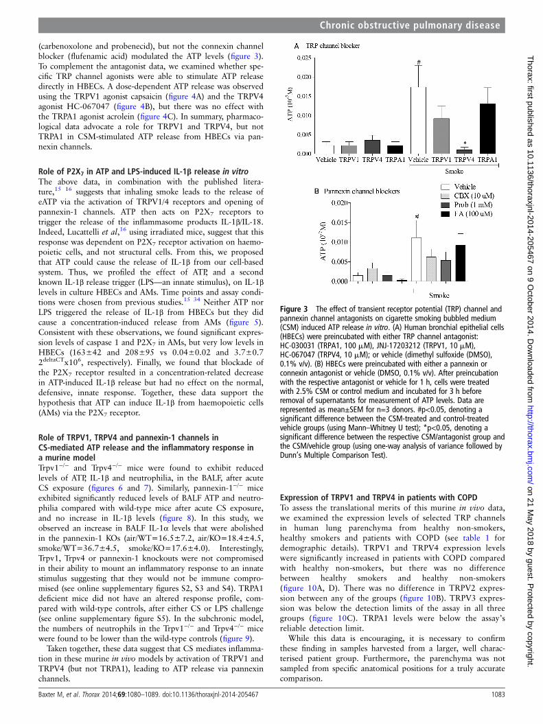
(carbenoxolone and probenecid), but not the connexin channelblocker (flufenamic acid) modulated the ATP levels (figure 3).To complement the antagonist data, we examined whether spe-cific TRP channel agonists were able to stimulate ATP releasedirectly in HBECs. A dose-dependent ATP release was observedusing the TRPV1 agonist capsaicin (figure 4A) and the TRPV4agonist HC-067047 (figure 4B), but there was no effect withthe TRPA1 agonist acrolein (figure 4C). In summary, pharmaco-logical data advocate a role for TRPV1 and TRPV4, but notTRPA1 in CSM-stimulated ATP release from HBECs via pan-nexin channels.
Role of P2X7 in ATP and LPS-induced IL-1β release in vitroThe above data, in combination with the published litera-ture,15 16 suggests that inhaling smoke leads to the release ofeATP via the activation of TRPV1/4 receptors and opening ofpannexin-1 channels. ATP then acts on P2X7 receptors totrigger the release of the inflammasome products IL-1β/IL-18.Indeed, Lucattelli et al,16 using irradiated mice, suggest that thisresponse was dependent on P2X7 receptor activation on haemo-poietic cells, and not structural cells. From this, we proposedthat ATP could cause the release of IL-1β from our cell-basedsystem. Thus, we profiled the effect of ATP, and a secondknown IL-1β release trigger (LPS—an innate stimulus), on IL-1βlevels in culture HBECs and AMs. Time points and assay condi-tions were chosen from previous studies.15 34 Neither ATP norLPS triggered the release of IL-1β from HBECs but they didcause a concentration-induced release from AMs (figure 5).Consistent with these observations, we found significant expres-sion levels of caspase 1 and P2X7 in AMs, but very low levels inHBECs (163±42 and 208±95 vs 0.04±0.02 and 3.7±0.72deltaCTx106, respectively). Finally, we found that blockade ofthe P2X7 receptor resulted in a concentration-related decreasein ATP-induced IL-1β release but had no effect on the normal,defensive, innate response. Together, these data support thehypothesis that ATP can induce IL-1β from haemopoietic cells(AMs) via the P2X7 receptor.
Role of TRPV1, TRPV4 and pannexin-1 channels inCS-mediated ATP release and the inflammatory response ina murine modelTrpv1−/− and Trpv4−/− mice were found to exhibit reducedlevels of ATP, IL-1β and neutrophilia, in the BALF, after acuteCS exposure (figures 6 and 7). Similarly, pannexin-1−/− miceexhibited significantly reduced levels of BALF ATP and neutro-philia compared with wild-type mice after acute CS exposure,and no increase in IL-1β levels (figure 8). In this study, weobserved an increase in BALF IL-1α levels that were abolishedin the pannexin-1 KOs (air/WT=16.5±7.2, air/KO=18.4±4.5,smoke/WT=36.7±4.5, smoke/KO=17.6±4.0). Interestingly,Trpv1, Trpv4 or pannexin-1 knockouts were not compromisedin their ability to mount an inflammatory response to an innatestimulus suggesting that they would not be immune compro-mised (see online supplementary figures S2, S3 and S4). TRPA1deficient mice did not have an altered response profile, com-pared with wild-type controls, after either CS or LPS challenge(see online supplementary figure S5). In the subchronic model,the numbers of neutrophils in the Trpv1−/− and Trpv4−/− micewere found to be lower than the wild-type controls (figure 9).
Taken together, these data suggest that CS mediates inflamma-tion in these murine in vivo models by activation of TRPV1 andTRPV4 (but not TRPA1), leading to ATP release via pannexinchannels.
Expression of TRPV1 and TRPV4 in patients with COPDTo assess the translational merits of this murine in vivo data,we examined the expression levels of selected TRP channelsin human lung parenchyma from healthy non-smokers,healthy smokers and patients with COPD (see table 1 fordemographic details). TRPV1 and TRPV4 expression levelswere significantly increased in patients with COPD comparedwith healthy non-smokers, but there was no differencebetween healthy smokers and healthy non-smokers(figure 10A, D). There was no difference in TRPV2 expres-sion between any of the groups (figure 10B). TRPV3 expres-sion was below the detection limits of the assay in all threegroups (figure 10C). TRPA1 levels were below the assay’sreliable detection limit.
While this data is encouraging, it is necessary to confirmthese finding in samples harvested from a larger, well charac-terised patient group. Furthermore, the parenchyma was notsampled from specific anatomical positions for a truly accuratecomparison.
Figure 3 The effect of transient receptor potential (TRP) channel andpannexin channel antagonists on cigarette smoking bubbled medium(CSM) induced ATP release in vitro. (A) Human bronchial epithelial cells(HBECs) were preincubated with either TRP channel antagonist:HC-030031 (TRPA1, 100 mM), JNJ-17203212 (TRPV1, 10 mM),HC-067047 (TRPV4, 10 mM); or vehicle (dimethyl sulfoxide (DMSO),0.1% v/v). (B) HBECs were preincubated with either a pannexin orconnexin antagonist or vehicle (DMSO, 0.1% v/v). After preincubationwith the respective antagonist or vehicle for 1 h, cells were treatedwith 2.5% CSM or control medium and incubated for 3 h beforeremoval of supernatants for measurement of ATP levels. Data arerepresented as mean±SEM for n=3 donors. #p<0.05, denoting asignificant difference between the CSM-treated and control-treatedvehicle groups (using Mann–Whitney U test); *p<0.05, denoting asignificant difference between the respective CSM/antagonist group andthe CSM/vehicle group (using one-way analysis of variance followed byDunn’s Multiple Comparison Test).
Chronic obstructive pulmonary disease
Baxter M, et al. Thorax 2014;69:1080–1089. doi:10.1136/thoraxjnl-2014-205467 1083
on 21 May 2018 by guest. P
rotected by copyright.http://thorax.bm
j.com/
Thorax: first published as 10.1136/thoraxjnl-2014-205467 on 9 O
ctober 2014. Dow
nloaded from

DISCUSSIONChronic exposure to CS is a key causative factor in the develop-ment of COPD.39 It is thought to result in a dysregulatedinflammatory response in the airways characterised by highlevels of leucocyte infiltration and inflammatory mediatorrelease which drive disease progression and lung functiondecline. Elucidation of the pathways which mediate CS-inducedinflammation is likely to uncover novel targets for the develop-ment of effective therapies. Patients with COPD exhibitincreased levels of eATP, IL-1β and IL-18 in the BALF, as well asincreased expression of P2X7 and caspase-1 in leucocytes andlung tissue, and importantly, these markers are found to correl-ate with disease severity.6 7 12 15 Murine in vivo models ofCS-induced airway inflammation have also been shown to bedependent on the P2X7-dependent activation of caspase-1 tomediate the maturation of cytokines IL-1β and IL-18, indicatingthat these models have disease relevance. Pharmacologicaland/or genetic manipulation of ATP and these proteins havebeen shown to attenuate CS-induced neutrophilia in both acuteand more chronic murine models, suggesting that modulation ofthis pathway could potentially reduce the steroid-resistantinflammation which drives the progression of COPD.15 16 40
Despite this wealth of data indicating a role for ATP, the mech-anism by which it is released after exposure to cigarette smokehas so far eluded researchers and, if identified, could revealnovel targets for the development of truly effective therapies.
The data presented herein strongly advocate a crucial role forTRPV1 and TRPV4 channels in the pannexin-1-mediatedrelease of ATP in response to acute CS exposure. This wasdemonstrated in human primary cells using TRPV1, TRPV4 andpannexin-1-specific antagonists to block CSE-induced ATP
release, as well as TRPV1 and TRPV4 agonists to directly elicitATP release. The importance of this TRP channel pannexin-1axis was recapitulated in vivo using colonies of Trpv1−/−,Trpv4−/−, and pannexin-1−/− genetically deficient mice.Importantly, these mice exhibited reduced ATP levels after acuteCS exposure, and they also exhibited attenuated IL-1β and neu-trophil levels in BALF compared with wild-type, demonstratingthat this pathway is integral to acute CS-induced inflammatoryresponse, which is thought to drive the progression of COPD.These findings provide a novel insight into the role of thesechannels in the generation of acute CS-induced airway inflam-mation. By comparison, genetic ablation of the P2X7 receptorwas found to protect CS-exposed mice against increased BALFIL-1β and neutrophilia, but did not alter ATP release. Theseobservations indicate that ATP is released upstream of P2X7
receptor activation in this model and help to unravel the rela-tionship between pannexin-1 and the inflammasome. These datasupport the hypothesis that in this system, pannexin-1 stimula-tion results in inflammasome activation indirectly, via the releaseof eATP resulting in subsequent P2X7 activation.21 While thesedata in the acute and subchronic models are very exciting,further work in chronic models is required to fully elucidate therole of this axis in the pathogenesis of clinical COPD.
Evidence suggests that this inflammatory pathway is importantin COPD, as translational data has shown increased levels ofP2X7 expression and caspase-1 activation in CS-exposed wild-type mice and in patients with COPD.12 15 Furthermore, thepreliminary demonstration that TRPV1 and TRPV4 geneexpression are also upregulated in lung tissue from patients withCOPD suggests that these channels may be important for theCS-mediated release of eATP in this disease. It could also
Figure 4 The effect of transient receptor potential (TRP) channel agonists on ATP release in vitro. Human bronchial epithelial cells (HBECs) werestimulated with increasing concentrations of agonist or vehicle. (A) TRPV1 agonist, capsaicin; (B) TRPV4 agonist, GSK 1016790A; (C) TRPA1 agonist,acrolein. Supernatants were collected 3 h after stimulation for assessment of ATP release. Data are expressed as mean±SEM for n=3 donors;*p<0.05, denoting a significant difference between the vehicle and agonist stimulation (using one-way analysis of variance followed by Dunn’sMultiple Comparison Test).
Chronic obstructive pulmonary disease
1084 Baxter M, et al. Thorax 2014;69:1080–1089. doi:10.1136/thoraxjnl-2014-205467
on 21 May 2018 by guest. P
rotected by copyright.http://thorax.bm
j.com/
Thorax: first published as 10.1136/thoraxjnl-2014-205467 on 9 O
ctober 2014. Dow
nloaded from

provide some preliminary insight into COPD susceptibility andindicate why only a certain percentage of smokers developCOPD (i.e. increased channel expression means higher levels ofATP). Indeed, TRPV4 single nucleotide polymorphisms havebeen associated with COPD susceptibility in two separatestudies.41 42
An interesting feature of this inflammatory pathway is the factthat release of eATP to activate the P2X7-inflammasome-caspase-1axis facilitates the production of IL-1β and IL-18 without the needfor transcriptional upregulation of the genes which encode theseproinflammatory cytokines. Rather, there are substantial levels ofthe proforms of these cytokines constitutively present in the
cytosol, for which caspase-1 facilitates the maturation to active,proinflammatory mediators. Indeed, previous investigation ofmurine models has demonstrated that IL-1β mRNA levels are notaffected by CS exposure, despite significantly higher levels ofIL-1β protein.15 This may, in part, explain the insensitivity ofpatients with COPD to glucocorticoids, as they are generallythought to exert their anti-inflammatory effects by modulation ofinflammatory cytokines at the transcriptional level.Pharmacological intervention at the TRPV1/4-pannexin-1 axismay, therefore, prove to be an effective anti-inflammatory thera-peutic strategy where glucocorticoids have failed. Furthermore,TRPV1, TRPV4 and pannexin-1 gene-deleted mice mounted a
Figure 5 Characterisation of ATP-induced interleukin (IL)-1β release in vitro. A: Human bronchial epithelial cells (HBECs), cultured in medium withor without 3% fetal calf serum (FCS), were stimulated with ATP (0.3 mM) or vehicle (phosphate buffered saline (PBS) 0.1% v/v). Supernatants werecollected 24 h after stimulation for IL-1β measurement by ELISA. B: HBECs, cultured in medium with or without 3% FCS, were stimulated withlipopolysaccharide (LPS) (0.1 mg/mL) or vehicle (PBS, 0.1% v/v). Supernatants were collected 24 h after stimulation for IL-1β measurement by ELISA.Data for (A) and (B) are represented as mean±SEM for n=3 donors. (C) and (D): alveolar macrophages (AM) were stimulated with (C) increasingconcentrations of ATP from 10 to 1000 μM or (D) increasing concentrations of LPS from 0.01 to 10 μg/mL. Supernatants were collected 24 h afterstimulation for IL-1β measurement by ELISA. (E) and (F) AMs were preincubated with increasing concentrations of P2X7 antagonist (AZ-11645373)or vehicle (dimethyl sulfoxide, 0.1%, v/v) for 1 h before stimulation with (E) either 1 mM ATP or vehicle; or (D) either 0.1 μg/mL LPS or vehicle. Cellswere subsequently incubated for a further 24 h before collection of supernatants and measurement of IL-1β. Data for (C), (D), (E) and (F) arerepresented as mean±SEM for n=3 donors. (E) #p<0.05, denoting a significant difference between the ATP/vehicle treated and vehicle/vehicle-treated groups (using Mann–Whitney U test); *p<0.05, denoting a significant difference between the ATP/vehicle-treated group and therespective ATP/antagonist-treated group (using one-way analysis of variance followed by Dunn’s Multiple Comparison Test). (F): #p<0.05, denoting asignificant difference between the LPS/vehicle treated and vehicle/vehicle-treated groups (using Mann–Whitney U test).
Chronic obstructive pulmonary disease
Baxter M, et al. Thorax 2014;69:1080–1089. doi:10.1136/thoraxjnl-2014-205467 1085
on 21 May 2018 by guest. P
rotected by copyright.http://thorax.bm
j.com/
Thorax: first published as 10.1136/thoraxjnl-2014-205467 on 9 O
ctober 2014. Dow
nloaded from

normal innate immune response to inhaled bacterial mimetic, LPS,suggesting that therapies that target this axis would not immune-compromise patients. To confirm this suggestion, further work isrequired with live bacteria and respiratory viruses. Somewhat sur-prisingly, TRPA1 did not play a role in CS-induced inflammationin either the in vivo or in vitro models even though it can be
directly activated by acrolein, one of the key components of CS.One explanation could be that the cell types we studied did notexpress TRPA1.
A question remains regarding the mechanism by which CS acti-vates TRPV1 and TRPV4 in these models. We initially hypothe-sised that TRPV1 and TRPV4 may be activated by low pH, but
Figure 6 The role of TRPV4 in acute CS-induced murine airway inflammation. Trpv4−/− mice were exposed to CS or room air (control) twice dailyfor 3 consecutive days alongside wild-type controls. BALF was collected 24 h after the last exposure for measurement of neutrophil (C), IL-1β (B) andATP (A) levels. Data are represented as mean±SEM for n=8 animals in each group. Statistical significance was determined using Mann–Whitney Utest. #p<0.05, denoting a significant difference between the smoke-exposed and air-exposed wild-type groups; *p<0.05, denoting a significantdifference between the smoke-exposed knock-outs and wild-types. TRP, transient receptor potential; CS, cigarette smoking; BALF, bronchoalveolarlavage fluid; IL-1β, interleukin-1β.
Figure 7 The role of TRPV1 in acuteCS-induced murine airwayinflammation. Trpv1−/− mice wereexposed to CS or room air (control)twice daily for 3 consecutive daysalongside wild-type controls. BALF wascollected 24 h after the last exposurefor measurement of neutrophil (C),IL-1β (B) and ATP (A) levels. Data arerepresented as mean±SEM for n=8animals in each group. Statisticalsignificance was determined usingMann–Whitney U test. #p<0.05,denoting a significant differencebetween the smoke-exposed andair-exposed wild-type groups;*p<0.05, denoting a significantdifference between the smoke-exposedknock-outs and wild-types. TRP,transient receptor potential; CS,cigarette smoking; BALF,bronchoalveolar lavage fluid; IL-1β,interleukin-1β.
Chronic obstructive pulmonary disease
1086 Baxter M, et al. Thorax 2014;69:1080–1089. doi:10.1136/thoraxjnl-2014-205467
on 21 May 2018 by guest. P
rotected by copyright.http://thorax.bm
j.com/
Thorax: first published as 10.1136/thoraxjnl-2014-205467 on 9 O
ctober 2014. Dow
nloaded from

analysis of the CSM would suggest otherwise. Furthermore,changes in osmolarity, a known trigger of TRPV4 activation,27
seems an unlikely scenario in this model system. Therefore, itwould seem that either constituents in CSM directly trigger thechannels or they result in the production of a channel modulator.Indeed, we are currently exploring ways of identifying likely candi-dates from the 4000+ chemicals reported to be present in CS. It is
also currently not clear how activation of TRP channels triggersthe opening of the pannexin-1 pore. The most likely mechanismwould simply be that increases in intracellular Ca2+ and mem-brane depolarisation are sufficient or that a second messengersignal is required.18 However, the fact that the pannexin-1 pore iscentral to the response does mirror observations made by others indifferent biological systems.22–24 26 27 The time course of ATPrelease in vitro is intriguing, it would appear that the release is anot a rapid process (taking a few hours to reach a plateau), suggest-ing that either the process is slow or that perhaps the CS inductionof the possible TRP channel activators takes time. Furthermore,the data would suggest that the cells stop releasing ATP after a timeor that enzymes which cleave it are induced.
Finally, it is unclear why ATP is still detectable, especially invivo, as one would assume that under normal circumstancesATP would be cleaved rapidly. Indeed, preliminary studies withexogenous ATP added to BALF from smoke-exposed miceshowed that it is undetectable with the specific ATP assay used.This suggests that the ecto-ATPases that breakdown ATP arefunctional in the CS-exposed mouse lung and perhaps the ATPmeasured in the BALF by our group and others is somehow pro-tected from enzymatic activity, and this observation warrantsfurther investigation.
In summary, this data represents a significant breakthrough inunderstanding the mechanism by which CS exposure leads to therelease of ATP. Furthermore, preliminary expression data in clin-ical samples appears to validate the preclinical findings. We,therefore, propose that pharmacological modulation of theTRPV1/4-pannexin-1 axis may provide clinical benefit to patientswith COPD by attenuating the dysregulated, steroid-resistantinflammation which is associated with the decline in lung-function seen in patients with COPD.
Figure 8 The role of pannexin-1 in acute cigarette smoking (CS)-induced murine airway inflammation. Pannexin-1−/− mice were exposed to CS orroom air (control) twice daily for 3 consecutive days alongside wild-type controls. Bronchoalveolar lavage fluid (BALF) was collected 24 h after the lastexposure for measurement of neutrophil (C), interleukin (IL)-1β (B) and ATP (A) levels. Data are represented as mean±SEM for n=8 animals in eachgroup. Statistical significance was determined using Mann–Whitney U test. #p<0.05, denoting a significant difference between the smoke-exposed andair-exposed wild-type groups; *p<0.05, denoting a significant difference between the smoke-exposed knock-outs and wild-types.
Figure 9 The role of TRPV1 and V4 in subchronic cigarette smoking(CS)-induced murine airway inflammation. Trpv1−/− and Trpv4−/− micewere exposed to CS or room air (control) twice daily for 14 consecutivedays alongside wild-type controls. Bronchoalveolar lavage fluid wascollected 24 h after the last exposure for measurement of neutrophils.Data are represented as mean±SEM for n=8 animals in each group.Statistical significance was determined using analysis of variance with aBonferroni post-test; #p<0.05, denoting a significant differencebetween the smoke-exposed and air-exposed wild-type groups;*p<0.05, denoting a significant difference between the smoke-exposedknock-outs and wild-types.
Chronic obstructive pulmonary disease
Baxter M, et al. Thorax 2014;69:1080–1089. doi:10.1136/thoraxjnl-2014-205467 1087
on 21 May 2018 by guest. P
rotected by copyright.http://thorax.bm
j.com/
Thorax: first published as 10.1136/thoraxjnl-2014-205467 on 9 O
ctober 2014. Dow
nloaded from

Acknowledgements P2×7−/− mice were kindly supplied by Professor JeanKanellopoulos, Université Paris-Sud, France, Trpv4−/− mice were kindly supplied byRiken Bioresource Centre, and Dr Vishva Dixit from Genentech generously providedthe pannexin 1 -/- mice.
Contributors Conception and design: MB, MGB, MAB. Data generation, analysisand interpretation: MB, SE, BD, LY-B, EDD, SAM. Drafting the manuscript forimportant intellectual content: MB, MGB, MAB. All authors reviewed the manuscriptand approved the final draft
Competing interests MB was supported by an NHLI COPD PhD studentship, SEwas supported by a postdoctoral award from Roche Pharmaceuticals, Nutley, USA.LY-B was funded by a Capacity Building Award in Integrative Mammalian Biologyfunded by the BBSRC, BPS, HEFCE, KTN & MRC. EDD was funded by a WellcomeTrust project grant (089301/Z/09/Z) and Boehringer-Ingelheim. SAM and MAB werefunded by project grants from the MRC, UK (G0800195 and G0800196,respectively). The human tissue experiments in this study were undertaken with thesupport of the NIHR Respiratory Disease Biomedical Research Unit at the RoyalBrompton and Harefield NHS Foundation Trust and Imperial College London.
Provenance and peer review Not commissioned; externally peer reviewed.
REFERENCES1 Silverman E, Speizer F. Risk factors for the development of chronic obstructive
pulmonary disease. Med Clin North Am 1996;80:501–22.2 Vestbo J, Hurd SS, Agusti AG, et al. Global strategy for the diagnosis, management,
and prevention of chronic obstructive pulmonary disease. Am J Respir Crit Care Med2013;187:347–65.
3 Lopez AD, Murray CC. The global burden of disease. Nat Med 1998;4:1241–3.4 Di Stefano A, Turato G, Maestrelli P, et al. Airflow limitation in chronic bronchitis is
associated with T-lymphocyte and macrophage infiltration of the bronchial mucosa.Am J Respir Crit Care Med 1996;153:629–32.
5 Saetta M, Turato G, Facchini FM, et al. Inflammatory cells in the bronchial glands ofsmokers with chronic bronchitis. Am J Respir Crit Care Med 1997;156:1633–9.
6 Sapey E, Ahmad A, Bayley D, et al. Imbalances between interleukin-1 and tumornecrosis factor agonists and antagonists in stable COPD. J Clin Immunol2009;29:508–16.
7 Rovina N, Dima E, Gerassimou C, et al. Interleukin-18 in induced sputum:association with lung function in chronic obstructive pulmonary disease. Respir Med2009;103:1056–62.
8 Perregaux D, Gabel CA. Interleukin-1 beta maturation and release in response toATP and nigericin. Evidence that potassium depletion mediated by these agents is
a necessary and common feature of their activity. J Biol Chem1994;269:15195–203.
9 Mariathasan S, Monack DM. Inflammasome adaptors and sensors: intracellularregulators of infection and inflammation. Nat Rev Immunol 2007;7:31–40.
10 Qu Y, Franchi L, Nunez G, et al. Nonclassical IL-1 beta secretion stimulated byP2X7 receptors is dependent on inflammasome activation and correlated withexosome release in murine macrophages. J Immunol 2007;179:1913–25.
11 Mortaz E, Braber S, Nazary M, et al. ATP in the pathogenesis of lung emphysema.Eur J Pharmacol 2009;619:92–6.
12 Lommatzsch M, Cicko S, Müller T, et al. Extracellular adenosine triphosphate andchronic obstructive pulmonary disease. Am J Respir Crit Care Med2010;181:928–34.
13 Mohsenin A, Blackburn M. Adenosine signaling in asthma and chronic obstructivepulmonary disease. Curr Opin Pulm Med 2006;12:54–9.
14 Cicko S, Lucattelli M, Müller T, et al. Purinergic receptor inhibition prevents thedevelopment of smoke-induced lung injury and emphysema. J Immunol2010;185:688–97.
15 Eltom S, Stevenson CS, Rastrick J, et al. P2X7 receptor and caspase 1 activation arecentral to airway inflammation observed after exposure to tobacco smoke. PLoSONE 2011;6:e24097.
16 Lucattelli M, Cicko S, Müller T, et al. P2X7 receptor signaling in the pathogenesis ofsmoke-induced lung inflammation and emphysema. Am J Respir Cell Mol Biol2011;44:423–9.
17 Churg A, Zhou S, Wang X, et al. The role of interleukin-1beta in murine cigarettesmoke-induced emphysema and small airway remodeling. Am J Respir Cell Mol Biol2009;40:482–90.
18 Praetorius HA, Leipziger J. ATP release from non-excitable cells. Purinergic Signal2009;5:433–46.
19 Lazarowski ER, Boucher RC, Harden TK. Mechanisms of release of nucleotides andintegration of their action as P2X- and P2Y-receptor activating molecules. MolPharmacol 2003;64:785–95.
20 Junger WG. Immune cell regulation by autocrine purinergic signalling. Nat RevImmunol 2011;11:201–12.
21 Baroja-Mazo A, Barberà-Cremades M, Pelegrín P. The participation of plasmamembrane hemichannels to purinergic signaling. Biochim Biophys Acta2013;1828:79–93.
22 Qu Y, Misaghi S, Newton K, et al. Pannexin-1 is required for ATP release duringapoptosis but not for inflammasome activation. J Immunol 2011;186:6553–61.
23 Lemaire I, Falzoni S, Zhang B, et al. The P2X7 receptor and Pannexin-1 are bothrequired for the promotion of multinucleated macrophages by the inflammatorycytokine GM-CSF. J Immunol 2011;187:3878–87.
Figure 10 Transient receptor potential (TRP) channel expression in human lung parenchyma. Human lung parenchymal samples were collectedfrom healthy non-smokers, healthy smokers and patients with COPD. RNA was extracted and reverse transcribed to cDNA before measurement ofTRPV1 (A), TRPV2 (B), TRPV3 (C) and TRPV4 (D) mRNA expression levels using TaqMan reagents and protocols. Data are expressed as mean±SEMexpression levels relative to internal control (18S); #p<0.05, denotes a significant difference between healthy smokers and patients with COPD,determined by one-way analysis of variance followed by Dunn’s multiple comparisons posthoc test.
Chronic obstructive pulmonary disease
1088 Baxter M, et al. Thorax 2014;69:1080–1089. doi:10.1136/thoraxjnl-2014-205467
on 21 May 2018 by guest. P
rotected by copyright.http://thorax.bm
j.com/
Thorax: first published as 10.1136/thoraxjnl-2014-205467 on 9 O
ctober 2014. Dow
nloaded from

24 Ransford GA, Fregien N, Qiu F, et al. Pannexin 1 contributes to ATP release inairway epithelia. Am J Respir Cell Mol Biol 2009;41:525–34.
25 Karlberg M, Ekoff M, Huang DCS, et al. The BH3-mimetic ABT-737 induces mastcell apoptosis in vitro and in vivo: potential for therapeutics. J Immunol2010;185:2555–62.
26 Huang Y-J, Maruyama Y, Dvoryanchikov G, et al. The role of pannexin 1hemichannels in ATP release and cell-cell communication in mouse taste buds.Proc Natl Acad Sci USA 2007;104:6436–41.
27 Seminario-Vidal L, Okada SF, Sesma JI, et al. Rho signaling regulatespannexin 1-mediated ATP release from airway epithelia. J Biol Chem2011;286:26277–86.
28 Voets T, Nilius B. TRPs make sense. J Membr Biol 2003;192:1–8.29 Alvarez DF, King JA, Weber D, et al. Transient receptor potential vanilloid
4-mediated disruption of the alveolar septal barrier: a novel mechanism of acutelung injury. Circ Res 2006;99:988–95.
30 Willette RN, Bao W, Nerurkar S, et al. Systemic activation of the transientreceptor potential vanilloid subtype 4 channel causes endothelial failureand circulatory collapse: part 2. J Pharmacol Exp Ther 2008;326:443–52.
31 MacNee W, Rennard S, Hunt J. Evaluation of exhaled breath condensate pH as abiomarker for COPD. Respir Med 2011;105:1037–45.
32 Nassini R, Pedretti P, Moretto N, et al. Transient receptor potential ankyrin 1channel localized to non-neuronal airway cells promotes non-neurogenicinflammation. PLoS ONE 2012;7:e42454.
33 Andrè E, Campi B, Materazzi S. Cigarette smoke–induced neurogenic inflammationis mediated by α, β-unsaturated aldehydes and the TRPA1 receptor in rodents.J Clin Invest 2008;118:2574–82.
34 Birrell MA, Wong S, Catley MC, et al. Impact of tobacco-smoke on key signalingpathways in the innate immune response in lung macrophages. J Cell Physiol2008;214:27–37.
35 Grace M, Birrell M, Dubuis E, et al. Transient receptor potential channels mediate thetussive response to prostaglandin E2 and bradykinin. Thorax 2012;67:891–900.
36 Wong S, Belvisi MG, Birrell MA. MMP/TIMP expression profiles in distinct lungdisease models: implications for possible future therapies. Respir Res 2009;10:72.
37 Mizuno A, Matsumoto N, Imai M, et al. Impaired osmotic sensation in mice lackingTRPV4. Am J Physiol Cell Physiol 2003;285:C96–101.
38 Suzuki M, Mizuno A, Kodaira K, et al. Impaired pressure sensation in mice lackingTRPV4. J Biol Chem 2003;278:22664–8.
39 Sethi JM, Rochester CL. Smoking and chronic obstructive pulmonary disease. ClinChest Med 2000;21:67–86, viii.
40 Lappalainen U, Whitsett JA, Wert SE, et al. Interleukin-1beta causes pulmonaryinflammation, emphysema, and airway remodeling in the adult murine lung. Am JRespir Cell Mol Biol 2005;32:311–18.
41 Zhu G, Gulsvik A, Bakke P, et al. Association of TRPV4 gene polymorphisms withchronic obstructive pulmonary disease. Hum Mol Genet 2009;18:2053–62.
42 Obeidat M, Wain LV, Shrine N, et al. A comprehensive evaluation of potential lungfunction associated genes in the SpiroMeta general population sample. PLoS ONE2011;6:e19382.
Chronic obstructive pulmonary disease
Baxter M, et al. Thorax 2014;69:1080–1089. doi:10.1136/thoraxjnl-2014-205467 1089
on 21 May 2018 by guest. P
rotected by copyright.http://thorax.bm
j.com/
Thorax: first published as 10.1136/thoraxjnl-2014-205467 on 9 O
ctober 2014. Dow
nloaded from