Post-mortem histology in transient receptor potential ...
7
CASE REPORT Open Access Post-mortem histology in transient receptor potential cation channel subfamily V member 6 (TRPV6) under-mineralising skeletal dysplasia suggests postnatal skeletal recovery: a case report Anna E. Mason 1 , David Grier 2 , Sarah F. Smithson 1,3 , Christine P. Burren 1,4 and Elise Gradhand 5,6* Abstract Background: The calcium-selective channel TRPV6 (transient receptor potential cation channel subfamily V member 6) is crucial for maternal-fetal calcium transport across the placenta. TRPV6 mutations have recently been associated with an antenatally severe under-mineralising skeletal dysplasia accompanied by postnatal biochemical abnormalities. This is the first post-mortem report in a patient with TRPV6 skeletal dysplasia. Case presentation: The female infant had severe antenatal and postnatal skeletal abnormalities by 20 weeks gestation and was ventilator-dependent from birth. These skeletal abnormalities were apparent at an earlier gestational age than in previous reported cases and a more severe clinical course ensued. Biochemical and skeletal abnormalities, including bone density, improved postnatally but cardiac arrest at 4 months of age led to withdrawal of intensive care. Compound heterozygous TRPV6 variants (c.1978G > C p.(Gly660Arg) and c.1528C > T p.(Arg510Ter)) were identified on exome sequencing. Post-mortem identified skeletal abnormalities but no specific abnormalities in other organ systems. No placental pathology was found, multi-organ histological features reflected prolonged intensive care only. Post-mortem macroscopic examination indicated reduced thoracic size and short, pale and pliable ribs. Histological examination identified reduced number of trabeculae in the diaphyses (away from the growth plates), whereas metaphyses showed adequate mineralisation and normal number of trabeculae, but with slightly enlarged reactive chondrocytes, indicating post-natal skeletal growth recovery. Post-mortem radiological findings demonstrated improved bone density, improved rib width, healed fractures, although ribs were still shorter than normal. Long bones (especially humerus and femur) had improved from initial poorly defined metaphyses and reduced bone density to sharply defined metaphyses, prominent growth restart lines in distal diaphyses and bone-in-bone appearance along diaphyses. (Continued on next page) © The Author(s). 2020 Open Access This article is licensed under a Creative Commons Attribution 4.0 International License, which permits use, sharing, adaptation, distribution and reproduction in any medium or format, as long as you give appropriate credit to the original author(s) and the source, provide a link to the Creative Commons licence, and indicate if changes were made. The images or other third party material in this article are included in the article's Creative Commons licence, unless indicated otherwise in a credit line to the material. If material is not included in the article's Creative Commons licence and your intended use is not permitted by statutory regulation or exceeds the permitted use, you will need to obtain permission directly from the copyright holder. To view a copy of this licence, visit http://creativecommons.org/licenses/by/4.0/. The Creative Commons Public Domain Dedication waiver (http://creativecommons.org/publicdomain/zero/1.0/) applies to the data made available in this article, unless otherwise stated in a credit line to the data. * Correspondence: [email protected] 5 Severn Pathology, Paediatric and Perinatal Pathology, Southmead Hospital, North Bristol NHS Trust, Bristol, UK 6 Dr. Senckenberg. Institut für Pathologie, Universitätsklinikum Frankfurt/Main, Theodor-Stern-Kai 7, 60590 Frankfurt, Germany Full list of author information is available at the end of the article Mason et al. BMC Medical Genetics (2020) 21:64 https://doi.org/10.1186/s12881-020-01007-z
Transcript of Post-mortem histology in transient receptor potential ...
CASE REPORT Open Access
Post-mortem histology in transient receptorpotential cation channel subfamily Vmember 6 (TRPV6) under-mineralisingskeletal dysplasia suggests postnatalskeletal recovery: a case reportAnna E. Mason1 , David Grier2, Sarah F. Smithson1,3, Christine P. Burren1,4 and Elise Gradhand5,6*
Abstract
Background: The calcium-selective channel TRPV6 (transient receptor potential cation channel subfamily Vmember 6) is crucial for maternal-fetal calcium transport across the placenta. TRPV6 mutations have recently beenassociated with an antenatally severe under-mineralising skeletal dysplasia accompanied by postnatal biochemicalabnormalities. This is the first post-mortem report in a patient with TRPV6 skeletal dysplasia.
Case presentation: The female infant had severe antenatal and postnatal skeletal abnormalities by 20 weeks gestationand was ventilator-dependent from birth. These skeletal abnormalities were apparent at an earlier gestational age than inprevious reported cases and a more severe clinical course ensued. Biochemical and skeletal abnormalities, including bonedensity, improved postnatally but cardiac arrest at 4 months of age led to withdrawal of intensive care. Compoundheterozygous TRPV6 variants (c.1978G > C p.(Gly660Arg) and c.1528C > T p.(Arg510Ter)) were identified on exomesequencing. Post-mortem identified skeletal abnormalities but no specific abnormalities in other organ systems. Noplacental pathology was found, multi-organ histological features reflected prolonged intensive care only. Post-mortemmacroscopic examination indicated reduced thoracic size and short, pale and pliable ribs. Histological examinationidentified reduced number of trabeculae in the diaphyses (away from the growth plates), whereas metaphyses showedadequate mineralisation and normal number of trabeculae, but with slightly enlarged reactive chondrocytes, indicatingpost-natal skeletal growth recovery. Post-mortem radiological findings demonstrated improved bone density, improvedrib width, healed fractures, although ribs were still shorter than normal. Long bones (especially humerus and femur) hadimproved from initial poorly defined metaphyses and reduced bone density to sharply defined metaphyses, prominentgrowth restart lines in distal diaphyses and bone-in-bone appearance along diaphyses.
(Continued on next page)
© The Author(s). 2020 Open Access This article is licensed under a Creative Commons Attribution 4.0 International License,which permits use, sharing, adaptation, distribution and reproduction in any medium or format, as long as you giveappropriate credit to the original author(s) and the source, provide a link to the Creative Commons licence, and indicate ifchanges were made. The images or other third party material in this article are included in the article's Creative Commonslicence, unless indicated otherwise in a credit line to the material. If material is not included in the article's Creative Commonslicence and your intended use is not permitted by statutory regulation or exceeds the permitted use, you will need to obtainpermission directly from the copyright holder. To view a copy of this licence, visit http://creativecommons.org/licenses/by/4.0/.The Creative Commons Public Domain Dedication waiver (http://creativecommons.org/publicdomain/zero/1.0/) applies to thedata made available in this article, unless otherwise stated in a credit line to the data.
* Correspondence: [email protected] Pathology, Paediatric and Perinatal Pathology, Southmead Hospital,North Bristol NHS Trust, Bristol, UK6Dr. Senckenberg. Institut für Pathologie, Universitätsklinikum Frankfurt/Main,Theodor-Stern-Kai 7, 60590 Frankfurt, GermanyFull list of author information is available at the end of the article
Mason et al. BMC Medical Genetics (2020) 21:64 https://doi.org/10.1186/s12881-020-01007-z
(Continued from previous page)
Conclusions: This case provide bone histological confirmation that human skeletal development is compromised in thepresence of TRPV6 pathogenic variants. Post-mortem findings were consistent with abnormal in utero skeletalmineralisation due to severe calcium deficit from compromised placental calcium transfer, followed by subsequentphenotypic improvement with adequate postnatal calcium availability. Significant skeletal recovery occurs in the earlyweeks of postnatal life in TRPV6 skeletal dysplasia.
Keywords: TRPV6 (transient receptor potential cation channel subfamily V member 6), Skeletal dysplasia, Placentalcalcium transfer, Post-mortem
BackgroundCalcium is essential for cell signalling, neuromuscular ac-tivity, blood coagulation and skeletal growth. During fetaldevelopment, additional calcium is required to supportskeletal formation and development. This demand is metby active maternal-foetal (transplacental) calcium trans-port, to achieve higher fetal than maternal serum calciumconcentration [1]. Various mechanisms have been pro-posed, with recent recognition of the importance of transi-ent receptor potential cation channel subfamily Vmember 6 (TRPV6; also referred to as vanilloid), an epi-thelial calcium-selective channel formed of four identicalsubunits, each with six transmembrane segments [2].TRPV6 gene location and expression varies across species.In humans, the TRPV6 gene is located on chromosome7q33-q34 and is very highly expressed in the placenta [3].Alteration in TRPV6 expression contributes to many
pathological processes, but only recently has a humanphenotype been identified. TRPV6 is upregulated in sev-eral human malignancies, including prostate, thyroid,breast and endometrial cancer, where it aids the calcium-dependent proliferation of malignant cells [4–6]. Con-versely, it is downregulated in pre-eclampsia, a conditionassociated with reduced placental calcium transfer [7]. Acontributory potential role for TRPV6 has also been pos-tulated in several rare childhood disorders including Lowe,Pendred, Gitelman and Gordon syndromes; althoughthose disorders involve additional electrolyte abnormal-ities, not present in this case, suggesting multiple mecha-nisms within those disorders [8–11]. Prior to 2018,TRPV6 had not been identified in human skeletal disease.Very recently in 2018, homozygous or compound het-
erozygous TRPV6 variants have been reported in seveninfants with a skeletal disorder of antenatal-onset char-acterised by poor mineralisation [12, 13]. All seven in-fants had biochemical abnormalities of secondaryneonatal hyperparathyroidism which completely normal-ised over several weeks. The skeletal abnormalities im-proved to varying extents over a longer timeframeduring infancy. Our patient [12] showed more severeskeletal abnormalities than the other six cases, and muchearlier in pregnancy. The variants in our case were dif-ferent and the greater clinical severity is interpreted to
have been the consequence of the different location ofthe pathogenic variants in this case compared to theother 6 causing more severe impact on TRPV6 proteinfunction. Ultimately, this infant did not survive, due tothe initial postnatal respiratory impairment, ineffectiverespiratory weaning and therefore complications of pro-longed intensive care treatment. The detailed geneticfindings on this infant were published in the AmericanJournal of Medical Genetics 2018 and we refer thereader back to that publication for detail on in silicomodelling and rationale of pathogenicity according toACMG Guidelines. Whereas, in this publication, wefocus on the post-mortem findings of this case, whichare the first in TRPV6-associated skeletal dysplasia,shedding new insight into the abnormal bone architec-ture of this condition and skeletal development in theperinatal period.
Case presentationA British White female infant had severe, potentially le-thal, skeletal dysplasia detected antenatally at 20 weeks’gestation. This consisted of short long bones, a smallchest, and rib deformities suggesting pulmonary hypo-plasia. Antenatal microarray comparative genomic hy-bridisation (CGH) and uniparental disomy (UPD)testing, to exclude paternal UPD14, did not identify ab-normalities. She was born at term (39 + 1/40) with nor-mal birth weight (3128 g) and was ventilator-dependentfrom birth due to respiratory distress. Physical examin-ation showed a bell-shaped chest, but she was otherwisewell-grown and had no dysmorphic features or abnormalneurology.Skeletal survey showed persistent generalised under-
mineralisation, multiple rib and metaphyseal fracturesand periosteal reaction along long bone diaphyses(Fig. 1). This was associated with significantly elevatedparathyroid hormone (PTH) during the first 6 weeks(peak 101 pmol/L, reference range 1.1–6.9 pmol/L) butnormal corrected calcium, phosphate and alkaline phos-phatase (ALP). Postnatal genetic testing, using targetedsingle gene approach, excluded potential differentialdiagnoses of Neonatal Severe Hyperparathyroidism,Mucolipidosis Type II (I-cell disease), which can also
Mason et al. BMC Medical Genetics (2020) 21:64 Page 2 of 7
feature hyperparathyroidism and periosteal changes, and336 known skeletal dysplasias. Further details can befound in the initial case report publication [12].Whole exome sequencing was performed with DNA
samples from the patient and her unaffected parentsusing the Agilent SureSelect All Exon v6 system, withsequencing on an Illumina NextSeq 500. These afore-mentioned negative genetic test finding reports coin-cided with identifying resolution of PTH elevation andprogressively improving skeletal mineralisation. Conse-quently, we considered an alternative hypothesis of an inutero calcium deficiency caused by an intrinsic defect inplacental calcium transfer. This prompted direct explor-ation of the candidate genes TRPV6, CABP9K and VDR.We performed a gene-agnostic trio analysis to identifyvery rare variants (Minor Allele Frequency < 0.0001)compatible with autosomal recessive or de novo inherit-ance. The likely causative variants were confirmed by
Sanger sequencing. 12 This analysis identified com-pound heterozygous TRPV6 variants: novel maternallyinherited missense variant, c.1978G > C p.(Gly660Arg),and paternally inherited nonsense variant, c.1528C > Tp.(Arg510Ter); both classified as pathogenic accordingto ACMG Guidelines.By 6 weeks of age, bone mineralisation showed radio-
logical improvement, but ventilatory requirement per-sisted and was provided via tracheostomy from 8 weeksof age. At 17 weeks, she developed rapid onset markedabdominal distension, followed by two cardiac arrests as-sociated with prolonged lactic acidosis. Prior to this, thecardiac monitoring had shown no evidence of cardiacmalfunction or arrhythmias. Emergency laparotomy con-firmed the presence of volvulus and dusky, but not is-chaemic, bowel. Multi-organ failure, including globalischaemic brain injury, was present and ultimately carewas withdrawn.
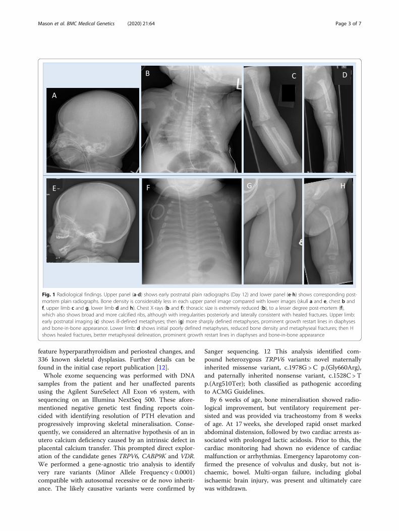
Fig. 1 Radiological findings. Upper panel (a-d) shows early postnatal plain radiographs (Day 12) and lower panel (e-h) shows corresponding post-mortem plain radiographs. Bone density is considerably less in each upper panel image compared with lower images (skull a and e, chest b andf, upper limb c and g, lower limb d and h). Chest X-rays (b and f): thoracic size is extremely reduced (b), to a lesser degree post-mortem (f),which also shows broad and more calcified ribs, although with irregularities posteriorly and laterally consistent with healed fractures. Upper limb:early postnatal imaging (c) shows ill-defined metaphyses; then (g) more sharply defined metaphyses, prominent growth restart lines in diaphysesand bone-in-bone appearance. Lower limb: d shows initial poorly defined metaphyses, reduced bone density and metaphyseal fractures; then Hshows healed fractures, better metaphyseal delineation, prominent growth restart lines in diaphyses and bone-in-bone appearance
Mason et al. BMC Medical Genetics (2020) 21:64 Page 3 of 7
Post-mortem skeletal survey radiographs were under-taken (Fig. 1). They demonstrated considerably im-proved bone density compared to early postnatalimaging, metaphyseal regions had been ill-defined onearly postnatal imaging, but were far more sharply de-fined, prominent growth restart lines were present inlong bone diaphyses and ossification was in keeping withchronological age. The ribs remained extremely short,were irregular posteriorly and laterally suggesting healedfractures, and the thoracic volume was markedlyreduced.A limited post-mortem autopsy was performed (chest,
abdomen, ribs and long bones only). The infant waswell-grown: body weight 6370 g, crown-heel length 62cm, crown-rump length 45 cm and head circumference43 cm (all within normal range for 4 months of age) withno dysmorphic features, no abnormalities of skin or hair.A narrow chest was noted.There was evidence of abnormal rib and femur bone
architecture on macroscopic and microscopic examin-ation. The bones were pale, unusually pliable and easilycut away from the costo-cartilaginous junction with ascalpel. Microscopically, the rib and femur metaphysesshowed growth plates with the expected three zones(proliferative, hypertrophic and calcified cartilage), butthe chondrocytes were relatively enlarged and activatedin keeping with increased chondroid matrix production.Towards the diaphyses, the trabeculae were of normalstructure but significantly reduced in number (Fig. 2).The bone marrow was mildly autolysed but showed nor-mal cellularity and a normal trilineage haematopoiesis.Borderline lung hypoplasia was present (lung to body
weight ratio 0.017, abnormal < 0.015). Pulmonary micro-scopic findings were in keeping with multi-organ failure,with abundant alveolar macrophages, parenchymal andintra-alveolar haemorrhage. No histological features ofinfantile respiratory distress syndrome were seen. Onmacroscopic examination, the heart was mildly enlarged(weight 30.9 g, expected 23 g for crown-rump length),but structurally normal with no pericardial effusion.Microscopic examination showed mild hypertrophy anddisarray of the cardiomyocytes with a normal conductionsystem. No significant areas of myocardial infarctionwere noted.Abdominal examination showed normal macroscopic
and microscopic appearance of the intestines with nofull thickness necrosis of the bowel or evidence of mal-rotation. The caecum was mobile and there was a largehaemorrhage in the mesentery of the sigmoid colon withapproximately 20 ml of free blood in the abdominal cav-ity in keeping with surgical intervention during emer-gency laparotomy. The liver showed patchy hepatocytenecrosis and prominent bilirubinostasis. The pancreasappeared macroscopically normal and showed no
significant histological abnormality of endocrine andexocrine glands. Immunohistochemical studies were notpossible due to the extent of autolysis.Multisystem histological features consistent with in-
tensive care treatment where present but the exact aeti-ology leading to cardiac arrest and subsequent multi-organ failure could not be established.There was no significant placental abnormality
(trimmed weight 400 g, 10-25th percentile for gestation),including no focal changes on macroscopic or micro-scopic examination on routine staining. There was noevidence of viral infection. Immunohistochemical stain-ing for TRPV6 was attempted using three antibodies(AB20C6(C-term), AB429(C-term) and AB1271(N-term)) on formalin-fixed, paraffin-embedded placentaltissue. Unfortunately, reliable staining could not beachieved. The antibodies were developed for use onfresh frozen tissue and it appears likely that the placentalTRPV6 antigen did not survive formalin fixation, al-though this has not been formally tested.In summary, post-mortem pathological examination
identified ongoing skeletal abnormality with pale, pliableand short ribs. Post-mortem radiology illustrated consid-erable improvement in bone density and metaphysealgrowth, supported by histological evidence of growth re-covery with reactive chondrocytes and adequate minera-lised normal trabeculae in the metaphyses, yet trabeculaewere sparse but normally structured in the diaphyses.
Discussion and conclusionsWe report the findings at post-mortem of an infant withantenatal-onset skeletal dysplasia caused by compoundheterozygous TRPV6 variants. TRPV6, a calcium-selective channel, plays an important role in maternal-foetal calcium transport via the placenta. Variants inTRPV6 may lead to insufficient calcium transport andabnormal skeletal development in utero. This leads topostnatal skeletal and biochemical abnormalities whichprogressively improve in the ex utero environment, asdescribed in seven recent cases.12,13 Skeletal findings im-proved postnatally in all cases, although in our case [12]the phenotype was extremely severe. In particular, theabnormally small rib cage led to life-threatening respira-tory insufficiency. Her ventilator-dependency had beenforecast to continue until approximately 18 months ofage (her unexpected death aged 4 months meant thisduration was ultimately not known). A further differencein this case, is that the skeletal dysplasia was evidentmuch earlier antenatally, whereas in the Suzuki casesskeletal abnormalities were only evident from the thirdtrimester onwards (earliest 28 weeks). Althoughmaternal-foetal calcium transfer begins at 12 weeks ges-tation, the majority occurs during the third trimester soit had been hypothesised that foetal bone abnormalities
Mason et al. BMC Medical Genetics (2020) 21:64 Page 4 of 7
would not be evident earlier [14]. We suspect that thesignificant skeletal dysplasia evident by only 20 weeks’gestation in this case reflects the severity of the com-promise to calcium transfer resulting from severely im-paired TRPV6 function. In support of this, the variantsin TRPV6 identified in our patient were predicted, by insilico protein modelling, to result in unstable or mis-folded protein which might lead to channel loss or im-paired activity.The post-mortem findings further support the concept
of significant under-mineralisation due to lack of cal-cium, especially the paleness, soft texture and flexibilityof the bones. Microscopically, the reduced bony trabecu-lae persisting in the diaphyses may reflect earlier absentor reduced calcium availability in utero, whereas the
reactive chondrocytes in the growth plate suggest bonerecovery and catch up in the normal calcium environ-ment after birth. Significant bone remodelling occursduring growth, so the reduction in trabecular numbermight progressively normalise during childhood. Opti-mism arises from other recent cases suggesting normalpostnatal growth, although duration is currently shortwith minimal linear growth details. Yet recent Trpv6mouse model studies by Fecher-Trost give caution ascortical bone architecture remained abnormal in thick-ness with reduced femoral length [15].We consider it likely that the small lung volume was a
consequence of the skeletal dysplasia rather than due toa primary pulmonary hypoplasia. This is supported bythe short ribs and markedly reduced thoracic volume.
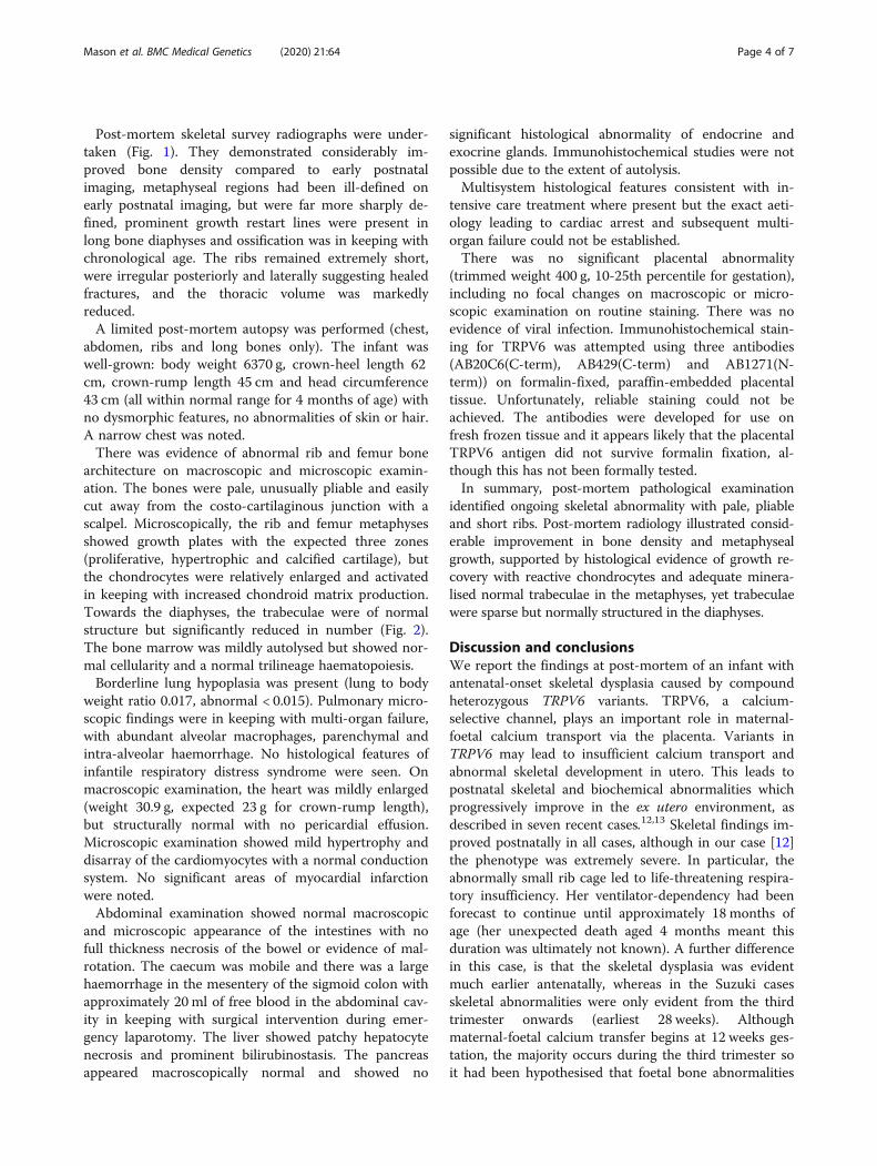
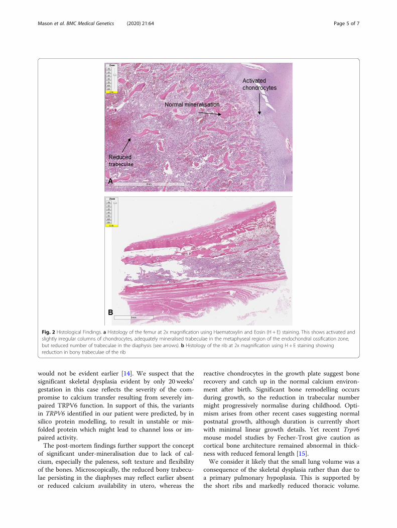
Fig. 2 Histological Findings. a Histology of the femur at 2x magnification using Haematoxylin and Eosin (H + E) staining. This shows activated andslightly irregular columns of chondrocytes, adequately mineralised trabeculae in the metaphyseal region of the endochondral ossification zone,but reduced number of trabeculae in the diaphysis (see arrows). b Histology of the rib at 2x magnification using H + E staining showingreduction in bony trabeculae of the rib
Mason et al. BMC Medical Genetics (2020) 21:64 Page 5 of 7
Moreover, the lungs showed a normal pattern of lobardevelopment and furthermore there was no histologicalremodelling of the lung tissue due to the inflammatorychanges typically seen in infantile respiratory distresssyndrome and, despite evidence of multi-organ failure,there were no other significant pulmonary findings.All organs were exposed to reduced calcium levels
during development, which could conceivably disruptphysiological processes other than skeletal development.The cardiac histology was examined in detail, as calciumis essential for the conduction system and contractilityof the cardiomyocytes. The post-mortem findings of anormal conduction system, along with the absence ofidentified arrhythmias during her first 4 months of life,provide no indication of a primary cardiac abnormality.The mild cardiac hypertrophy and disarray of the cardio-myocytes are non-specific findings and are post-mortemfindings not uncommonly seen in post-resuscitation,consistent with recurrent adrenaline and/or corticoster-oid administration. This correlates with the intensivecare treatment provided in this case rather than a resultof an abnormal calcium metabolism. Cardiac arrythmiaswere not mentioned in the other cases. Our histologicalfindings do not raise abnormalities suggesting the needfor cardiac surveillance in surviving infants and children.Clinically, there was no evidence of wider disorders of
calcium homeostasis. There was no pre-eclampsia andno maternal factors that could account for in utero cal-cium deficiency. Postnatally, oral calcium supplementa-tion at physiological doses was given briefly tocompensate for an expected persistent hypocalcaemiadue to the skeletal calcium deficiency. However, normo-calcaemia was maintained by a normal milk diet only,indicating normal intestinal calcium absorption in thepostnatal period. The exocrine pancreas is another keysite for TRPV6 expression in humans, although signifi-cance of its role is unclear. Unfortunately, histology inthis case was non-contributory due to autolysis. But an-temortem, there had been no clinical evidence of pan-creatic exocrine gland dysfunction, suggesting its rolemay be minimal.As more roles for TRPV6 in human disease are identi-
fied, further studies are vital to improve our understand-ing of TRPV6 function and identify potential pathwaysto prevent, modify or treat these disorders. Its upregula-tion in many malignancies already makes it a promisingtarget for cancer therapy. Several groups have now de-veloped systemic Trpv6 knock-out mice. The phenotypesare variable between different mouse lines but includemale hypofertility, elevated PTH, growth retardation,poor bone mineralisation and dermatitis [16–18]. How-ever, clinical correlation to human disease is not straightforward as organ-specific expression of TRPV6 variesbetween species. Studying the foetal development of
Trpv6 knock-out mice will be extremely important inadvancing our understanding of antenatal calciumhomeostasis and identifying potential therapeutic inter-vention in disorders such as pre-eclampsia and TRPV6under-mineralising skeletal dysplasia. While intra-amniotic calcium infusions could theoretically preventor correct bone abnormalities, improving thoracicgrowth and optimising future lung function, their riskprofile of pregnancy loss means they are not a realisticapproach. It has been proposed that small molecularchaperones, such as those used in cystic fibrosis, couldbe used antenatally to correct the conformation ofTRPV6 variants, although this remains hypothetical atpresent [13].In summary, we report the first post-mortem histo-
logical and radiological findings of a patient withTRPV6-associated skeletal dysplasia. The histology sug-gests that metaphyses had been compromised due to se-vere lack of calcium substrate, but then functionednormally, in fact possibly more active, in the postnatalenvironment. Bone development through collagen for-mation is intrinsically abnormal in the majority of skel-etal dysplasias. TRPV6-associated skeletal dysplasiadiffers where the main feature appears to be compro-mised development of bone quantity resulting from ex-treme calcium lack during crucial in utero development.The post-mortem radiology and histology in our casesuggested postnatal catch-up and recovery. Although, aneffect might persist as the trabeculae remained reducedin number in the diaphyses. Whether bone remodellingthroughout longer childhood growth can completelynormalise bone length and density is currently unknownand will require close monitoring in other cases.
AbbreviationsALP: Alkaline phosphatase; PTH: Parathyroid hormone; TRPV6: Transientreceptor potential cation channel subfamily V member 6; UPD: Uniparentaldisomy
AcknowledgementsDr. M Ashworth, Dr. F Maggiani and Dr. B Mozayani provided secondopinions on histology specimens. Dr. D Holmes, Dr. R Caswell, Professor SEllard helped with molecular genetic aspects of the report. We are gratefulto Dr. C Fecher-Trost who provided the three TRPV6 antibodies for placentaltesting.
Authors’ contributionsAM wrote and revised the manuscript. EG and CPB developed the conceptfor the manuscript. CPB provided clinical expertise in diagnosis andmanagement, revised the manuscript and figures. DG interpreted theradiology images and SFS provided genetic expertise during the casemanagement and revised the manuscript. EG performed the post-mortemand histological analysis. All authors read and approved the final manuscript.
FundingThis research received no specific grant from any funding agency in thepublic, commercial, or not-for-profit sectors.
Availability of data and materialsDNA sequences are available in ClinVar. Accession number SCV001167185:https://www.ncbi.nlm.nih.gov/clinvar/variation/818220/
Mason et al. BMC Medical Genetics (2020) 21:64 Page 6 of 7
Ethics approval and consent to participateThe parents provided approval and written, informed consent to participatein this study and for a limited post-mortem (chest, abdomen, ribs and longbones). We confirm that the post-mortem was undertaken in accordancewith the policies and procedures of North Bristol NHS Trust, which includesconsent for research procedures, approved by the North Bristol NHS TrustResearch and Ethics Committee.
Consent for publicationThe parents of the patient provided written, informed consent forpublication of patient information and images.
Competing interestsThe authors declare that they have no competing interests.
Author details1Bristol Medical School Translational Health Sciences, University of Bristol,Bristol, UK. 2Department of Radiology, Bristol Royal Hospital for Children,University Hospitals Bristol NHS Foundation Trust, Bristol, UK. 3Department ofClinical Genetics, St Michaels Hospital, University Hospitals Bristol NHSFoundation Trust, Bristol, UK. 4Department of Paediatric Endocrinology,Bristol Royal Hospital for Children, University Hospitals Bristol NHSFoundation Trust, Bristol, UK. 5Severn Pathology, Paediatric and PerinatalPathology, Southmead Hospital, North Bristol NHS Trust, Bristol, UK. 6Dr.Senckenberg. Institut für Pathologie, Universitätsklinikum Frankfurt/Main,Theodor-Stern-Kai 7, 60590 Frankfurt, Germany.
Received: 21 July 2019 Accepted: 20 March 2020
References1. Kovacs CS, Kronenberg HM. Maternal-fetal calcium and bone metabolism
during pregnancy, puerperium, and lactation. Endocr Rev. 1997;18(6):832–72.
2. Fecher-Trost C, Wissenbach U, Weissgerber P. TRPV6: from identification tofunction. Cell Calcium. 2017;67:116–22.
3. Moreau R, Daoud G, Bernatchez R, Simoneau L, Masse A, Lafond J. Calciumuptake and calcium transporter expression by trophoblast cells from humanterm placenta. Biochim Biophys Acta. 2002;1564(2):325–32.
4. Fixemer T, Wissenbach U, Flockerzi V, Bonkhoff H. Expression of the Ca2+−selective cation channel TRPV6 in human prostate cancer: a novelprognostic marker for tumor progression. Oncogene. 2003;22(49):7858–61.
5. Bolanz KA, Hediger MA, Landowski CP. The role of TRPV6 in breastcarcinogenesis. Mol Cancer Ther. 2008;7(2):271–9.
6. Zhuang L, Peng JB, Tou L, Takanaga H, Adam RM, Hediger MA, et al.Calcium-selective ion channel, CaT1, is apically localized in gastrointestinaltract epithelia and is aberrantly expressed in human malignancies. LabInvestig. 2002;82(12):1755–64.
7. Haché S, Takser L, LeBellego F, Weiler H, Leduc L, Forest JC, et al. Alterationof calcium homeostasis in primary preeclamptic syncytiotrophoblasts: effecton calcium exchange in placenta. J Cell Mol Med. 2011;15(3):654–67.
8. Yang SS, Lo YF, Yu IS, Lin SW, Chang TH, Hsu YJ, et al. Generation andanalysis of the thiazide-sensitive Na+ −cl- cotransporter (Ncc/Slc12a3)Ser707X knockin mouse as a model of Gitelman syndrome. Hum Mutat.2010;31(12):1304–15.
9. Yang SS, Hsu YJ, Chiga M, Rai T, Sasaki S, Uchida S, et al. Mechanisms forhypercalciuria in pseudohypoaldosteronism type II-causing WNK4 knock-inmice. Endocrinology. 2010;151(4):1829–36.
10. Nakaya K, Harbidge DG, Wangemann P, Schultz BD, Green ED, Wall SM,et al. Lack of pendrin HCO3- transport elevates vestibular endolymphatic[Ca2+] by inhibition of acid-sensitive TRPV5 and TRPV6 channels. Am JPhysiol Renal Physiol. 2007;292(5):F1314–21.
11. Wu G, Zhang W, Na T, Jing H, Wu H, Peng JB. Suppression of intestinalcalcium entry channel TRPV6 by OCRL, a lipid phosphatase associated withLowe syndrome and dent disease. Am J Physiol Cell Physiol. 2012;302(10):C1479–91.
12. Burren CP, Caswell R, Castle B, Welch CR, Hilliard TN, Smithson SF, et al.TRPV6 compound heterozygous variants result in impaired placentalcalcium transport and severe undermineralization and dysplasia of the fetalskeleton. Am J Med Genet Part A. 2018;176A:1950–5.
13. Suzuki Y, Chitayat D, Sawada H, Deardorff MA, McLaughlin HM, Begtrup A,et al. TRPV6 variants interfere with maternal-fetal calcium transport throughthe placenta and cause transient neonatal hyperparathyroidism. Am J HumGenet. 2018;102(6):1104–14.
14. Hacker AN, Fung EB, King JC. Role of calcium during pregnancy: maternaland fetal needs. Nutr Rev. 2012;70(7):397–409.
15. Fecher-Trost C, Lux F, Busch K, Raza A, Winter M, Hielscher F, et al. Maternaltransient receptor potential Vanilloid 6 (Trpv6) is involved in offspring bonedevelopment. J Bone Miner Res. 2019;20:e3646.
16. Chen F, Ni B, Yang YO, Ye T, Chen A. Knockout of TRPV6 causes osteopeniain mice by increasing osteoclastic differentiation and activity. Cell PhysiolBiochem. 2014;33:796–809.
17. Weissgerber P, Kriebs U, Tsvilovskyy J, Olausson J, Kretz O, Stoerger C,Mannebach S, et al. Excision of the Trpv6 gene leads to severe defects inepididymal Ca2+ absorption and male infertility much alike the singleD541A pore mutation. J Biol Chem. 2012;287(22):17930–41.
18. Bianco SD, Peng JB, Takanaga H, Suzuki Y, Crescenzi A, Kos CH, et al. Markeddisturbance of calcium homeostasis in mice with targeted disruption of theTrpv6 calcium channel gene. J Bone Miner Res. 2007;22(2):274–85.
Publisher’s NoteSpringer Nature remains neutral with regard to jurisdictional claims inpublished maps and institutional affiliations.
Mason et al. BMC Medical Genetics (2020) 21:64 Page 7 of 7