Original Article MicroRNA-9500 induces cell apoptosis of hepatoma...
8
Int J Clin Exp Med 2018;11(4):3749-3756 www.ijcem.com /ISSN:1940-5901/IJCEM0056853 Original Article MicroRNA-9500 induces cell apoptosis of hepatoma through targeting AKT Haosen Hao, Jiandong Zhao, Youri Zhang, Wen Wu, Jiangtao Hu, Yanjun An Department of Endoscopy Center, Shanxi Traditional Chinese Medicine Institute, No. 46 Bingzhou West Street, Taiyuan 030012, Shanxi, China Received May 5, 2017; Accepted November 14, 2017; Epub April 15, 2018; Published April 30, 2018 Abstract: Background: HCC is one of the most usual human carcinoma, while also the second commonest reason of cancer deaths among human. But its mechanism at post-transcriptional levels is not fully understood. The pur- pose of this study was to elucidate the post-transcriptional factors regulating the apoptosis process of HepG2 and Huh7. Methods: To confirm the predicted miR-9500 was matched with AKT-1, 3’ UTR luciferase activity of AKT-1 was used to assess. Then, HepG2 and Huh7 were cultured in the presence or absence of miR-9500 mimics or inhibi- tors. The AKT signaling pathway involved in apoptosis process of HepG2 and Huh7 was investigated, respectively. Results: Our work indicated that miR-9500 over-expression induced reduction of luciferase activity on AKT-1 3’-UTR, as well as the protein expression of AKT-1 in HepG2 and Huh7 through luciferase system reporter gene analysis. Therefore, AKT-1 is direct targets of miR-9500. miR-9500 suppressed expression of AKT-1. Furthermore, miR-9500 substantially accenuated the apoptosis process of HepG2 and Huh7. But transfection with inhibitors of miR-9500, antagomiR-9500, overtly attentuated expression level of AKT-1 in HepG2 and Huh7. MiR-9500 exerted effects on cell proliferation or viability, and accentuated its caspase-dependent apoptosis of HepG2 and Huh7. Conclusions: We identified miR-9500, which specifically bind to AKT-1 mRNA 3’-UTR. MiR-9500 is a crucial mediator of regulating expression of AKT-1 at post-transcriptional levels, protecting against proliferation of hepatoma cells through inhibit- ing the AKT signaling pathway. Moreover, it exerts effects on controlling viability and cell apoptosis of HepG2 and Huh7. Our findings suggest that targeting miR-9500, being involved in pathogenesis of HCC, is a promising strategy for the treatment and prevention of HCC. Keywords: MicroRNA, hepatocellular carcinoma (HCC), AKT, apoptosis Introduction HCC is one of the most usual human carcinoma and ranked fifth, while it is also the second commonest reason of cancer deaths among human. HCC was almost come from inflamma- tion process and liver cirrhosis induced by afla- toxins, ethanol, and chronic infection of hepati- tis B or C [1]. The prognosis of HCC is so poor [2] that only tiny minority patients could be cured by surgical resection or liver transplanta- tion. Consequently, the emergency necessity is effective medicines or therapeutic methods for HCC. For ages, different genetic alterations were thought to the origin of hepatocarcinogenesis, and induce malignant transformation [3]. Con- temporarily, development of cancer is no only considered to result from genomic or genetic alterations, but also metabolism of lipid [4]. The miRNAs (MicroRNAs) are a group of tiny molecules and noncoding RNA, 22 to 25 nucle- otides in length that function on regulation of gene expression at post-transcriptional level [5]. MicroRNAs (miRNAs) control gene expres- sion by pairing with incompletely matching aim sites of the 3’ untranslated regions (UTRs) of mRNA, and cause translational repression and/ or mRNA destabilization, thereby down-regulat- ing the expression of the targeted gene. The growing literatures hold up the vital function of miRNAs on expression regulation at post-tran- scriptional level. Moreover, the regulated ex- pression of genes involved in numerous biologi- cal progressions, especially for the different pathogenesis disorders (including inflammatory
Transcript of Original Article MicroRNA-9500 induces cell apoptosis of hepatoma...

Int J Clin Exp Med 2018;11(4):3749-3756www.ijcem.com /ISSN:1940-5901/IJCEM0056853
Original ArticleMicroRNA-9500 induces cell apoptosis of hepatoma through targeting AKT
Haosen Hao, Jiandong Zhao, Youri Zhang, Wen Wu, Jiangtao Hu, Yanjun An
Department of Endoscopy Center, Shanxi Traditional Chinese Medicine Institute, No. 46 Bingzhou West Street, Taiyuan 030012, Shanxi, China
Received May 5, 2017; Accepted November 14, 2017; Epub April 15, 2018; Published April 30, 2018
Abstract: Background: HCC is one of the most usual human carcinoma, while also the second commonest reason of cancer deaths among human. But its mechanism at post-transcriptional levels is not fully understood. The pur-pose of this study was to elucidate the post-transcriptional factors regulating the apoptosis process of HepG2 and Huh7. Methods: To confirm the predicted miR-9500 was matched with AKT-1, 3’ UTR luciferase activity of AKT-1 was used to assess. Then, HepG2 and Huh7 were cultured in the presence or absence of miR-9500 mimics or inhibi-tors. The AKT signaling pathway involved in apoptosis process of HepG2 and Huh7 was investigated, respectively. Results: Our work indicated that miR-9500 over-expression induced reduction of luciferase activity on AKT-1 3’-UTR, as well as the protein expression of AKT-1 in HepG2 and Huh7 through luciferase system reporter gene analysis. Therefore, AKT-1 is direct targets of miR-9500. miR-9500 suppressed expression of AKT-1. Furthermore, miR-9500 substantially accenuated the apoptosis process of HepG2 and Huh7. But transfection with inhibitors of miR-9500, antagomiR-9500, overtly attentuated expression level of AKT-1 in HepG2 and Huh7. MiR-9500 exerted effects on cell proliferation or viability, and accentuated its caspase-dependent apoptosis of HepG2 and Huh7. Conclusions: We identified miR-9500, which specifically bind to AKT-1 mRNA 3’-UTR. MiR-9500 is a crucial mediator of regulating expression of AKT-1 at post-transcriptional levels, protecting against proliferation of hepatoma cells through inhibit-ing the AKT signaling pathway. Moreover, it exerts effects on controlling viability and cell apoptosis of HepG2 and Huh7. Our findings suggest that targeting miR-9500, being involved in pathogenesis of HCC, is a promising strategy for the treatment and prevention of HCC.
Keywords: MicroRNA, hepatocellular carcinoma (HCC), AKT, apoptosis
Introduction
HCC is one of the most usual human carcinoma and ranked fifth, while it is also the second commonest reason of cancer deaths among human. HCC was almost come from inflamma-tion process and liver cirrhosis induced by afla-toxins, ethanol, and chronic infection of hepati-tis B or C [1]. The prognosis of HCC is so poor [2] that only tiny minority patients could be cured by surgical resection or liver transplanta-tion. Consequently, the emergency necessity is effective medicines or therapeutic methods for HCC.
For ages, different genetic alterations were thought to the origin of hepatocarcinogenesis, and induce malignant transformation [3]. Con- temporarily, development of cancer is no only
considered to result from genomic or genetic alterations, but also metabolism of lipid [4].
The miRNAs (MicroRNAs) are a group of tiny molecules and noncoding RNA, 22 to 25 nucle-otides in length that function on regulation of gene expression at post-transcriptional level [5]. MicroRNAs (miRNAs) control gene expres-sion by pairing with incompletely matching aim sites of the 3’ untranslated regions (UTRs) of mRNA, and cause translational repression and/or mRNA destabilization, thereby down-regulat-ing the expression of the targeted gene. The growing literatures hold up the vital function of miRNAs on expression regulation at post-tran-scriptional level. Moreover, the regulated ex- pression of genes involved in numerous biologi-cal progressions, especially for the different pathogenesis disorders (including inflammatory

MicroRNA-9500 induces apoptosis of hepatoma cell via targeting AKT
3750 Int J Clin Exp Med 2018;11(4):3749-3756
disorders) [6]. A variety of miRNAs are regarded as symbolize of a new category of inflammation mediators, too [7-9].
To assess the role of miR-9500 in hepatoma cells, we first identify whether miR-9500 se- quences is in the AKT-1 mRNA, and then evalu-ated the levels expression levels of miR-9500 in HepG2 and Huh7. Our work firstly demon-strated that AKT-1 is one of the targets of miR-9500 in HepG2 and Huh7. It revealed that miR-9500 could control survival of cell viability by regulating expression of AKT-1 and its pathway. We, therefore, investigated the effect of miR-9500 on regulating apoptosis of hepatoma cell lines. In particular, the cell behavior effects of miR-9500 have been associated with the regu-lation of AKT-1 and its signaling pathway.
Materials and methods
Culture of cell
HepG2 and Huh7 cell line was purchased from American type culture collection (ATCC). HepG2 and Huh7 were cultured with DMEM with 10% FBS. HepG2 and Huh7 cells were grown in a 5% CO2 incubator at 37°C. HiPerFect Transfection Reagent (Qiagen) was used to transfect the HepG2 and Huh7 cells according to the manu-facturer’s instructions as previously described [10].
Reporter gene assays
cDNAs encoding the entire 3’ UTR of AKT-1 (300 kb) mRNAs were predicted and amplified from total RNA of HepG2 and Huh7 using Xho I and Not I linker/primers, and then cloned into the vector pGL4 (the luciferase reporter vector) included the gene could expressed firefly and renilla luciferase. AKT-1 3’ UTRs was cloned in reverse orientation as controls lacking the miRNA target sequence [11]. Additionally, the complementary region to the seed region of miR-9500 sequence in position 955-961 and 1284-1290 of human AKT-1 3’ UTR, TCTTGCC, was mixed up with CTGCCTT. These constructs were all identified with COS-7 cells (gift of Dr. Feng Liu, Chinese Academy of Sciences), which were transfected with the reporter construct and the indicated miRNA mimics or its negative control sequences by Lipofectamine 3000 (Invitrogen, Carlsbad, CA). The activity of Renilla luciferase was normalized with the correspond-
ing control of Dual-Glo Luciferase Assay System. The mutant construct of the AKT-1-3’-UTR was created with Site-Mutation kit (Pro- mega, Madison, WI, USA). Then, either NC or miR-9500 and the plasmid were co-transfected into cells. The pGL4-control vector was used for normalization as a control. The analysis of luc- iferase activity of cells was detected with ana-lyzer VICTOR (PerkinElmer, Foster City, CA, USA) by Dual-Luciferase Reporter Assay System kit of Promega. AKT-1 protein (active, AB79792) was purchased from Abcam company (MA, USA).
Extract total RNA and clone novel miRNA
Total RNA of cells was obtained using method with TRIzol (Sigma, CA, USA), based on the pro-tocol for manufacturer. Total RNAs were isolat-ed with mirVana RNA isolation kit then get rid of the RNA smaller than 200 nt. The miR-9500 was cloned into the open code frame of vector with DynaExpress miRNA Cloning Kit based on its instructions for manufacturer, and then modified it.
The mimics of miR-9500 and transfection
Transfection with mimics of miR-9500 were performed using Lipofectamine 3000 (Sigma, CA, USA), according to the protocol of manufac-turer. The mimics of miR-9500 were showed as follows: sense 5’-AAGGGAAGAUGGUGACCAC- UU-3’ and antisense 5’-AAGUGGUCACCAUCU- UCCCUU-3’. The inhibitors of miR-9500 were showed as follow: 5’-GUGGUCACCAUCUUCCC- UU-3’. Moreover, the negative control (NC) was showed as follows: sense 5’-ACGUGACACGU- UCGGAGAAUU-3’ and antisense 5’-AAUUCUCC- GAACGUGUCACGU-3’, which was not homolo-gous with the genome sequences of human. The qRT-PCR was used to identify the dose effect of miR-9500.
The qRT-PCR analysis
We had identified the expression pattern of miR-9500 in HepG2 and Huh7 according to the TaqMan miRNA assays with its specific primers. The 2-ΔΔCt method was used to analyze the data. The qRT-PCR was performed with SYBR Green kit (Sigma, USA) and the Real-Time PCR De- tection System (Bio-Rad, Berkeley, CA, USA). The primer sequences were showed as follows: AKT-1 upstream 5’-TCCCGAGGCCAAGTCCTT-3’

MicroRNA-9500 induces apoptosis of hepatoma cell via targeting AKT
3751 Int J Clin Exp Med 2018;11(4):3749-3756
and downstream 5’-AAGGACTTGGCCTCGGGA- 3’ and Glyceraldehyde-3-phosphate dehydro- genase (GAPDH) upstream 5’-CTCATGACCAC- AGTCCATGCC-3’ and downstream 5’-GGCATG- GACTGTGGTCATGAG-3’.
Western blotting
Cells were lysed, and then total proteins were extracted with RIPA lysis buffer. Total proteins
were analyzed with electrophoresis method using Sodium Dodecyl Sulfate-polyacrylamide (SDS-PAGE) gel, and then transferred to a poly-vinylidene fluoride (PVDF) membrane (Roche, NJ, USA), which was blocked at room tempera-ture with 5% skim milk and washed with TBST for three times, probed with primary antibod-ies: p-AKT, AKT-1 (1:3000 dilution, Cell Sig- naling, Danvers, MA, USA), PI3K (1:3000 dilu-
Figure 1. MiR-9500 Inhibit Expression of AKT in hepatoma cell lines. In HepG2 and Huh7, to study if miRNAs regu-late the expression of AKT-1, the miRNAs target AKT-1 were predicted at first using online software DIANA microT v4.0. The miR-9500 may be the potentially target on AKT-1. To further identify whether miR-9500 directly ties to the 3’ UTRs of AKT-1 mRNAs, we conducted luciferase reporter assay. These results demonstrated that the relative 3’ UTR luciferase activities of AKT-1 reduced remarkably in HepG2 transfected with miR-9500 compared to control group (A) (P=0.001). But, the relative activities of 3’ UTR mut-AKT-1 (mutation of AKT-1) luciferase were similar to miR-9500 treated HepG2 compared to control group (B) (P>0.05). These data identified that mRNAs of AKT were the straight aim of miR-9500. To confirm if miR-9500 regulate the expression of AKT-1 in HepG2, we performed Western blot analyses (C). Moreover, we found that the relative activities of 3’ UTR AKT-1 luciferase were obviously reduced in miR-9500 treated Huh7 compared to control group (D) (P=0.001). But, the relative activities of 3’ UTR mut-AKT-1 (mutation of AKT-1) luciferase were similar to miR-9500 treated Huh7 compared to control group (E) (P>0.05). To identify whether the miRNAs regulate the expression of AKT-1 in Huh7, we performed Western blotting trials (F). The results indicated that, in HepG2, miR-9500 could suppress the AKT-1 expression (F). The data are presented as means ± SD from three independent experiments. *P<0.05, **P<0.01.

MicroRNA-9500 induces apoptosis of hepatoma cell via targeting AKT
3752 Int J Clin Exp Med 2018;11(4):3749-3756
tion, Santa Cruz, CA, USA), or β-actin (1:3000 dilution, Santa Cruz, CA, USA) overnight at 4°C. Then there were incubated at room tempera-ture with proper secondary antibodies (1:5000 dilutions, Santa Cruz, CA, USA).
BrdU assays and mitotic index analysis
Cells were synchronized through block method with double thymidine. After synchronizing for 16 hrs, cells were released, and then collected or fixed for assays. DNA synthesis or mitotic entry analysis were performed by BrdU labeling method. Cells were counted and cells with BrdU positive were scored with immunofluorescence microscope.
Mitotic events were determined with DNA stain-ing and time-lapse videomicroscopy. After syn-chronizing, the real-time images of cells were obtained at each 10 min. Mitotic events in cells were detected through change of morphology. Mitotic cells were counted based on DNA con-densation and nuclear morphology using DNA dye (Hoechst 33258).
Detection of apoptosis cells
Using Annexin V-FITC Apoptosis Detection Kit (Becton Dickinson, San Jose, CA, USA), the fluo-rescence-activated staining cells was sorting analyzed at 12, 24, 36 and 48 hrs after treated with inhibitors or mimics of miR-9500, respec-tively. These tests were performed with ACScan flow cytometer, the data analyse was done with Flowjo software.
Statistical analyses
Data analysis was carried out using SPSS 18.0 software (Inc. Chicago, SPSS, IL). Statistical dif-ferences between groups were analyzed with two-tailed paired Student t-test. Data of qRT-PCR and luciferase reporter assays were ex- pressed relative to the control in each experi-ment, and 95% confidence intervals were cal-culated. P<0.05 was considered with statisti-cally significant.
Results
MiR-9500 inhibit expression of AKT-1 in hepa-toma cell lines
To identify the miRNAs to target AKT-1 in HepG2 and Huh7, we first used the online software
(DIANA microT v4.0) to predict the miRNAs targeting AKT. MiR-9500, may be possibly to aimed the 3’ UTRs of AKT-1 mRNAs. Then, we investigated whether miR-9500 straight ties to the 3’ UTRs of AKT-1 mRNAs, and found that the relative activities of 3’ UTR AKT-1 luciferase were obviously reduced in miR-9500 treated HepG2 compared to control group (Figure 1A) (P=0.001). But, the relative activities of 3’ UTR mut-AKT-1 (mutation of AKT-1) luciferase were similar to miR-9500 treated HepG2 compared to control group (Figure 1B) (P>0.05). These data demonstrated that AKT-1 mRNAs straight object to miR-9500. To identify whether the miRNAs regulate the expression of AKT-1 in HepG2, we performed Western blotting trials (Figure 1C). The results indicated that, in HepG2, miR-9500 could suppress the AKT-1 expression (Figure 1C).
Moreover, we found that the relative activities of 3’ UTR AKT-1 luciferase were obviously re- duced in miR-9500 treated Huh7 compared to control group (Figure 1D) (P=0.001). But, the relative activities of 3’ UTR mut-AKT-1 (muta-tion of AKT-1) luciferase were similar to miR-9500 treated Huh7 compared to control group (Figure 1E) (P>0.05). To identify whether the miRNAs regulate the expression of AKT-1 in Huh7, we performed Western blotting trials (Figure 1F). The results indicated that, in HepG2, miR-9500 could suppress the AKT-1 expression (Figure 1F).
MiR-9500 suppress mitosis entry and prolif-eration of hepatoma cells
We further investigated the role of miR-9500 mimics and inhibitors on functions of host cells with BrdU. FCM (flow cytometry) was used to study the apoptosis progression induced by miR-9500 mimics and inhibitors. The results indicated that miR-9500 could significantly suppress proliferation (Figure 2A) and mitosis entry (Figure 2B) of HepG2 cells. Moreover, in HepG2 cells, inhibitors of miR-9500 could pro-mote cell proliferation (Figure 2A) and mitosis entry (Figure 2B) in time dependent manner.
Moreover, miR-9500 could significantly suppr- ess proliferation (Figure 2C) and mitosis entry (Figure 2D) of Huh7 cells. The inhibitors of miR-9500 could promote cell proliferation (Figure 2C) and mitosis entry (Figure 2D) in time depen-dent manner.

MicroRNA-9500 induces apoptosis of hepatoma cell via targeting AKT
3753 Int J Clin Exp Med 2018;11(4):3749-3756
MiR-9500 promote apoptosis of HepG2
The mimics of miR-9500 co- uld induced obviously cell ap- optosis of HepG2 in a time-dependent manner (Figure 3). Moreover, inhibitors of miR-9500 could repress apopto- sis of HepG2 (Figure 3A) and Huh7 (Figure 3B), respective-ly. Both of those outcomes demonstrated miR-9500 sup-press survival of hepatoma cells in time dependent man- ner.
AKT-1 rescue the apopotosis hepatoma cells induced by miR-9500
It had been reported in prior that AKT-1 was the key regu-late factor for cell proliferation [18], we further identified that through using of AKT-1 active protein in HepG2 and Huh7 cell model transfected by miR-9500. Moreover, the apopoto-sis cells of HepG2 and Huh7 were detected. These data confirmed that AKT-1 active protein could suppress sig- nificantly cell apopotosis of HepG2 (Figure 3C) and Huh7 (Figure 3D). Furthermore, the decreased of apopotosis hep-atoma cells induced by miR-9500 may obviously suppress by AKT-1 active protein (Figure 3C and 3D).
Figure 2. MiR-9500 inhibited mitosis entry and proliferation of hepatoma cells. Moreover, the characterized effect induced by miR-9500 on host cells was investigated with BrdU to analyze cell viability progression. The results showed that miR-9500 could suppress proliferation (A) and mitosis entry (B) of HepG2 cells effectively. Moreover, in HepG2 cells, the inhibitors of
miR-9500 could promote its pro-liferation (A) and mitosis entry (B) in a time-dependent manner. Moreover, miR-9500 could sig-nificantly suppress proliferation (C) and mitosis entry (D) of Huh7 cells. The inhibitors of miR-9500 could promote cell proliferation (C) and mitosis entry (D) in time dependent manner. The data are presented as means ± SD from three independent experiments. *P<0.05, **P<0.01.

MicroRNA-9500 induces apoptosis of hepatoma cell via targeting AKT
3754 Int J Clin Exp Med 2018;11(4):3749-3756
Discussion
HCC is major reason of dea- ths in patients with cirrhosis, which primarily come from alcohol abuse or chronic viral hepatitis. HCC is almost come from inflammation process and liver cirrhosis induced by aflatoxins, ethanol, and chron-ic infection of hepatitis B or C. Moreover, it is the most usual human carcinoma and ranked fifth. While it is also an vital health problem and the se- cond commonest reason of cancer deaths among human [12]. Although it is well known that several different mecha-nisms of HCC, it is still unclear about how miRNA modulate the pathogenesis process of HCC. In the present work, we
Figure 3. AKT-1 active protein rescue the apopotosis hepatoma cells induced by miR-9500. Fur-thermore, the characterized ef-fect on cell apoptosis progression caused by miR-9500 was ana-lyzed with flow cytometry (FCM). The results showed that the mim-ics of miR-9500 could induced a remarkably increase of apoptotic cells in a time-dependent man-ner. Moreover, inhibitors of miR-9500 could repress apoptosis of HepG2 (A) and Huh7 (B) in a time-dependent manner. It had been reported in prior that AKT-1 was the key regulate factor for cell proliferation [18], we further iden-tified that through using of AKT-1 active protein in HepG2 and Huh7 cell model transfected by miR-9500. Moreover, the apopotosis cells of HepG2 and Huh7 were detected. These data confirmed that AKT-1 active protein could suppress significantly cell apopo-tosis of HepG2 (C) and Huh7 (D). Furthermore, the decreased of apopotosis hepatoma cells induced by miR-9500 may obvi-ously suppress by AKT-1 active protein (C and D). The data are presented as means ± SD from three independent experiments. *P<0.05, **P<0.01.
MicroRNA-9500 induces apoptosis of hepatoma cell via targeting AKT
3755 Int J Clin Exp Med 2018;11(4):3749-3756
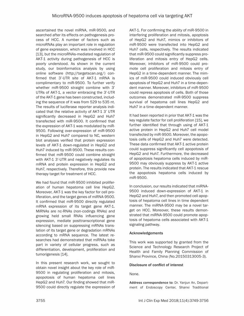
ascertained the novel miRNA, miR-9500, and searched after its effects on pathogenesis pro-cess of HCC. A number of factors such as microRNAs play an important role in regulation of gene expression, which was involved in HCC [13], but the microRNAs-mediated regulation of AKT-1 activity during pathogenesis of HCC is poorly understood. As shown in the current study, our bioinformatics analysis by using online software (http://targetscan.org/) con-firmed that 3’-UTR site of AKT-1 mRNA is complimentary to miR-9500. To further verify whether miR-9500 straight combine with 3’ UTRs of AKT-1, a vector embracing the 3’-UTR of the AKT-1 gene has been constructed, includ-ing the sequence of it was from 529 to 535 nt. The results of luciferase reporter analysis indi-cated that the relative activity of AKT-1 3’ UTR significantly decreased in HepG2 and Huh7 transfected with miR-9500. It confirmed that the expression of AKT-1 was modulated by miR-9500. Following over-expression of miR-9500 in HepG2 and Huh7 compared to NC, western blot analyses verified that protein expression levels of AKT-1 down-regulated in HepG2 and Huh7 induced by miR-9500. These results con-firmed that miR-9500 could combine straight with AKT-1 3’-UTR and negatively regulates its mRNA and protein expression in HepG2 and Huh7, respectively. Therefore, this provide new therapy target for treatment of HCC.
We had found that miR-9500 inhibited prolifer-ation of human hepatoma cell line HepG2. Moreover, AKT-1 was the key factor for cell pro-liferation, and the target genes of miRNA-9500. It confirmed that miR-9500 directly regulated mRNA expression of its target gene AKT-1. MiRNAs are nc-RNAs (non-codings RNAs) and growing held small RNAs influencing gene expression, mediate posttranscriptional gene silencing based on suppressing mRNAs trans-lation of its target gene or degradation mRNAs according to mRNA sequence. The latest re- searches had demonstrated that miRNAs toke part in variety of cellular progress, such as differentiation, development, proliferation and tumorigenesis [14].
In this present research work, we sought to obtain novel insight about the key role of miR-9500 in regulating proliferation and mitosis, apopotosis of human hepatoma cell lines HepG2 and Huh7. Our finding showed that miR-9500 could directly regulate the expression of
AKT-1. For confirming the ability of miR-9500 in interfering proliferation and mitosis, apoptosis of HepG2 and Huh7, mimics or inhibitors of miR-9500 were transfected into HepG2 and Huh7 cells, respectively. The results indicated that miR-9500 could significantly suppress pro-liferation and mitosis entry of HepG2 cells. Moreover, inhibitors of miR-9500 could pro-mote cell proliferation and mitosis entry of HepG2 in a time-dependent manner. The mim-ics of miR-9500 could induced obviously cell apoptosis of HepG2 and Huh7 in a time-depen-dent manner. Moreover, inhibitors of miR-9500 could repress apoptosis of cells. Both of those outcomes demonstrated miR-9500 suppress survival of hepatoma cell lines HepG2 and Huh7 in a time-dependent manner.
It had been reported in prior that AKT-1 was the key regulate factor for cell proliferation [15], we further identified that through using of AKT-1 active protein in HepG2 and Huh7 cell model transfected by miR-9500. Moreover, the apopo-tosis cells of HepG2 and Huh7 were detected. These data confirmed that AKT-1 active protein could suppress significantly cell apopotosis of HepG2 and Huh7. Furthermore, the decreased of apopotosis hepatoma cells induced by miR-9500 may obviously suppress by AKT-1 active protein. The results indicated that AKT-1 rescue the apopotosis hepatoma cells induced by miR-9500.
In conclusion, our results indicated that miRNA-9500 induced down-expression of AKT-1 in HepG2 and Huh7, and then promoted cell apop-tosis of hepatoma cell lines in time dependent manner. The miRNA-9500 may be a novel tar-get on HCC. Moreover, these results demon-strated that miRNA-9500 could promote apop-tosis of hepatoma cells associated with AKT-1 signaling pathway.
Acknowledgements
This work was supported by granted from the Science and Technology Research Project of Health and Family Planning Commission of Shanxi Province, China (No.20150313005-3).
Disclosure of conflict of interest
None.
Address correspondence to: Dr. Yanjun An, Depart- ment of Endoscopy Center, Shanxi Traditional

MicroRNA-9500 induces apoptosis of hepatoma cell via targeting AKT
3756 Int J Clin Exp Med 2018;11(4):3749-3756
Ch-inese Medicine Institute, No. 46 Bingzhou West Street, Taiyuan 030012, Shanxi, China. Tel: 86- 0351-4668111; Fax: 86-0351-4668111; E-mail: [email protected]
References
[1] Herold C, Reck T, Fischler P, Ott R, Radespiel-Troeger M, Ganslmayer M, Hohenberger W, Hahn EG and Schuppan D. Prognosis of a large cohort of patients with hepatocellular carcino-ma in a single European centre. Liver 2002; 22: 23-28.
[2] Okuda K. Hepatocellular carcinoma. J Hepatol 2000; 32: 225-237.
[3] Bhalla KN. Epigenetic and chromatin modifiers as targeted therapy of hematologic malignan-cies. J Clin Oncol 2005; 23: 3971-3993.
[4] Dowman JK, Hopkins LJ, Reynolds GM, Niko-laou N, Armstrong MJ, Shaw JC, Houlihan DD, Lalor PF, Tomlinson JW, Hübscher SG and New-some PN. Development of hepatocellular car- cinoma in a murine model of nonalcoholic steatohepatitis induced by use of a high-fat/fructose diet and sedentary lifestyle. Am J Pathol 2014; 184: 1550-1561.
[5] Ambros V. The functions of animal microRNAs. Nature 2004; 431: 350-355.
[6] Bartel DP. MicroRNAs: genomics, biogenesis, mechanism, and function. Cell 2004; 116: 281-297.
[7] Lewis BP, Shih IH, Jones-Rhoades MW, Bartel DP and Burge CB. Prediction of mammalian microRNA targets. Cell 2003; 115: 787-798.
[8] He L and Hannon GJ. MicroRNAs: small RNAs with a big role in gene regulation. Nat Rev Gen-et 2004; 5: 522-531.
[9] Esquela-Kerscher A and Slack FJ. Oncomirs-microRNAs with a role in cancer. Nat Rev Can-cer 2006; 6: 259-269.
[10] Shewade YM, Umrani M and Bhonde RR. Large-scale isolation of islets by tissue cul- ture of adult mouse pancreas. Transplant Proc 1999; 31: 1721-1723.
[11] O’Connell RM, Taganov KD, Boldin MP, Cheng G and Baltimore D. MicroRNA-155 is induced during the macrophage inflammatory respo- nse. Proc Natl Acad Sci U S A 2007; 104: 1604-1609.
[12] Sonia Pascual, Iván Herrera and Javier Irurzun. New advances in hepatocellular carcinoma. World J Hepatol 2016; 8: 421-438.
[13] Sumadi Lukman Anwar and Ulrich Lehmann. DNA methylation, microRNAs, and their cross-talk as potential biomarkers in hepatocellular carcinoma. World J Gastroenterol 2014; 20: 7894-7913.
[14] Filipowicz W, Bhattacharyya SN and Sonen-berg N. Mechanisms of post-transcriptional regulation by microRNAs: are the answers in sight? Nature Reviews Genetics 2008; 9: 102-114.
[15] Liu L, Lin Y, Liu L, Bian Y, Zhang L, Gao X, Li Q. 14-3-3γ regulates lipopolysaccharide-induced inflammatory responses and lactation in dairy cow mammary epithelial cells by inhibiting NF-κB and MAPKs and up-regulating mTOR signal-ing. Int J Mol Sci 2015; 16: 16622-16641.