Original Article microRNA-454 shows anti-angiogenic … shows anti-angiogenic and anti-metastatic...
9
Am J Cancer Res 2017;7(1):139-147 www.ajcr.us /ISSN:2156-6976/ajcr0045422 Original Article microRNA-454 shows anti-angiogenic and anti-metastatic activity in pancreatic ductal adenocarcinoma by targeting LRP6 Yue Fan 1 , Chenye Shi 2 , Tianyu Li 1 , Tiantao Kuang 2 Departments of 1 Integrated TCM & Western Medicine, 2 General Surgery, Zhongshan Hospital, Fudan University, Shanghai 200032, China Received November 14, 2016; Accepted December 5, 2016; Epub January 1, 2017; Published January 15, 2017 Abstract: Our previous work has shown that microRNA-454 (miR-454) can inhibit the growth of pancreatic ductal adenocarcinoma (PDAC) by blocking the recruitment of bone marrow-derived macrophages. In the present study, we aimed to explore its role in the proliferation, invasion, and pro-angiogenic activity of PDAC cells in vitro and lung metastasis in vivo. PANC-1 and MiaPaCa-2 cells were transfected with a miR-454-expressing plasmid and tested for cell proliferation, colony formation, cell cycle distribution, invasion, and pro-angiogenic activity. The target gene(s) that mediated the action of miR-454 was identified. The effect of miR-454 overexpression on lung metastasis of PDAC was evaluated in nude mice. Of note, overexpression of miR-454 significantly inhibited PDAC cell prolifera- tion and colony formation and arrests PDAC cells at the G2/M phase. Decreased invasiveness was observed in miR-454-overexpressing PDAC cells. Conditioned media from miR-454-overexpressing PANC-1 cells contained lower levels of vascular endothelial growth factor and had reduced capacity to induce endothelial cell tube-like structure formation. Mechanistically, miR-454 was found to target the mRNA of LRP6 and inhibit the activation of Wnt/β- catenin signaling in PDAC cells. Ectopic expression of LRP6 significantly reversed the suppressive effects of miR- 454 on PDAC cells. In vivo studies confirmed that miR-454-overexpressing PANC-1 cells formed significantly less lung metastases than control cells. Altogether, miR-454 functions as a suppressor in tumor growth, angiogenesis, and metastasis in PDAC, likely through downregulation of LRP6. Keywords: Angiogenesis, growth, LRP6, metastasis, microRNA-454, pancreatic cancer Introduction Pancreatic ductal adenocarcinoma (PDAC) is one of the most aggressive and lethal malig- nancies, with an overall 5-year survival rate of less than 5% [1]. Angiogenesis plays a critical role in tumor growth and metastasis, which is responsible for supplying nutrients and oxygen [2]. Vascular endothelial growth factor (VEGF), a well-defined inducer of angiogenesis, has been reported to be abundantly expressed in PDAC specimens [3]. Preclinical studies dem- onstrated that targeting VEGF signaling leads to reduced tumor growth and angiogenesis in subcutaneous tumor xenografts of human PDAC [4, 5]. Better understanding the mecha- nisms that govern tumor angiogenesis and metastasis is of importance to develop effec- tive therapies for PDAC. microRNAs (miRNAs) are a family of small non- coding RNAs that can negatively regulate a large number of genes by binding to the 3’-untranslated region (UTR) of target mRNAs [6]. Dysregulation of miRNAs commonly occurs in PDAC and has been linked to tumor progres- sion [7, 8]. For example, it was reported that miR-323-3p is underexpressed in PDAC tissues and cell lines and inhibits invasion and metas- tasis of PDAC cells [7]. miR-454 acts as an oncogene in several human cancers such as uveal melanoma [9] and non-small cell lung cancer [10]. Our previous work provides evi- dence that miR-454 is downregulated in PDAC and shows inhibitory effects on the growth of PDAC by targeting stromal cell derived factor-1 (SDF-1) [11]. Furthermore, we showed that miR- 454 overexpression impairs the ability of PDACs to recruit bone marrow-derived macrophages in
-
Upload
hoangkhuong -
Category
Documents
-
view
219 -
download
2
Transcript of Original Article microRNA-454 shows anti-angiogenic … shows anti-angiogenic and anti-metastatic...

Am J Cancer Res 2017;7(1):139-147www.ajcr.us /ISSN:2156-6976/ajcr0045422
Original Article microRNA-454 shows anti-angiogenic and anti-metastatic activity in pancreatic ductal adenocarcinoma by targeting LRP6
Yue Fan1, Chenye Shi2, Tianyu Li1, Tiantao Kuang2
Departments of 1Integrated TCM & Western Medicine, 2General Surgery, Zhongshan Hospital, Fudan University, Shanghai 200032, China
Received November 14, 2016; Accepted December 5, 2016; Epub January 1, 2017; Published January 15, 2017
Abstract: Our previous work has shown that microRNA-454 (miR-454) can inhibit the growth of pancreatic ductal adenocarcinoma (PDAC) by blocking the recruitment of bone marrow-derived macrophages. In the present study, we aimed to explore its role in the proliferation, invasion, and pro-angiogenic activity of PDAC cells in vitro and lung metastasis in vivo. PANC-1 and MiaPaCa-2 cells were transfected with a miR-454-expressing plasmid and tested for cell proliferation, colony formation, cell cycle distribution, invasion, and pro-angiogenic activity. The target gene(s) that mediated the action of miR-454 was identified. The effect of miR-454 overexpression on lung metastasis of PDAC was evaluated in nude mice. Of note, overexpression of miR-454 significantly inhibited PDAC cell prolifera-tion and colony formation and arrests PDAC cells at the G2/M phase. Decreased invasiveness was observed in miR-454-overexpressing PDAC cells. Conditioned media from miR-454-overexpressing PANC-1 cells contained lower levels of vascular endothelial growth factor and had reduced capacity to induce endothelial cell tube-like structure formation. Mechanistically, miR-454 was found to target the mRNA of LRP6 and inhibit the activation of Wnt/β-catenin signaling in PDAC cells. Ectopic expression of LRP6 significantly reversed the suppressive effects of miR-454 on PDAC cells. In vivo studies confirmed that miR-454-overexpressing PANC-1 cells formed significantly less lung metastases than control cells. Altogether, miR-454 functions as a suppressor in tumor growth, angiogenesis, and metastasis in PDAC, likely through downregulation of LRP6.
Keywords: Angiogenesis, growth, LRP6, metastasis, microRNA-454, pancreatic cancer
Introduction
Pancreatic ductal adenocarcinoma (PDAC) is one of the most aggressive and lethal malig-nancies, with an overall 5-year survival rate of less than 5% [1]. Angiogenesis plays a critical role in tumor growth and metastasis, which is responsible for supplying nutrients and oxygen [2]. Vascular endothelial growth factor (VEGF), a well-defined inducer of angiogenesis, has been reported to be abundantly expressed in PDAC specimens [3]. Preclinical studies dem-onstrated that targeting VEGF signaling leads to reduced tumor growth and angiogenesis in subcutaneous tumor xenografts of human PDAC [4, 5]. Better understanding the mecha-nisms that govern tumor angiogenesis and metastasis is of importance to develop effec-tive therapies for PDAC.
microRNAs (miRNAs) are a family of small non-coding RNAs that can negatively regulate a large number of genes by binding to the 3’-untranslated region (UTR) of target mRNAs [6]. Dysregulation of miRNAs commonly occurs in PDAC and has been linked to tumor progres-sion [7, 8]. For example, it was reported that miR-323-3p is underexpressed in PDAC tissues and cell lines and inhibits invasion and metas-tasis of PDAC cells [7]. miR-454 acts as an oncogene in several human cancers such as uveal melanoma [9] and non-small cell lung cancer [10]. Our previous work provides evi-dence that miR-454 is downregulated in PDAC and shows inhibitory effects on the growth of PDAC by targeting stromal cell derived factor-1 (SDF-1) [11]. Furthermore, we showed that miR-454 overexpression impairs the ability of PDACs to recruit bone marrow-derived macrophages in
miR-454 inhibits PDAC metastasis
140 Am J Cancer Res 2017;7(1):139-147
transwell migration assays. However, the role of miR-454 in tumor angiogenesis and metas-tasis in PDAC is still unclear.
In the present study, we determined the effects of overexpression of miR-454 on the growth, cell cycle distribution, invasion, and pro-angio-genic activity of PDAC cells in vitro and lung metastasis in vivo. The target gene(s) that mediates the action of miR-454 in PDAC were also investigated.
Materials and methods
Cell lines
Human PDAC cell lines PANC-1 and MiaPaCa-2 were obtained from the American Type Culture Collection (ATCC, Rockville, MD, USA). Cells were maintained in Dulbecco’s modified Eagle’s medium (DMEM) containing 10% fetal bovine serum (FBS), 100 U/mL penicillin, and 100 μg/mL streptomycin (Sigma-Aldrich, St. Louis, MO, USA). Primary human umbilical vein endothelial cells (HUVECs) were purchased from ScienCell Research Laboratories (Carlsbad, CA, USA) and cultured in Endothelial Cell Medium (ScienCell Research Laboratories) supplemented with 10% FBS at 37°C in a humidified atmosphere with 5% CO2. HUVECs at passages 3-8 were used in this study.
Plasmid construction and transfection
A fragment containing human miR-454 precur-sor was amplified from genomic DNA by PCR and cloned into pSuper vector (OligoEngine, Seattle, WA, USA). The complete coding region of LRP6 gene was amplified by PCR using human LRP6 cDNA (OriGene, Rockville, MD, USA) as a template and inserted into pcDNA3.1 (+) vector (Thermo Fisher Scientific, Carlsbad, CA, USA). The full-length LRP6 3’-UTR was amplified by PCR and inserted into the pGL3 plasmid (Promega, Madison, WI, USA). The mutated LRP6 3’-UTR reporter construct was generated by site-directed mutagenesis (Quik- Change, Stratagene, La Jolla, CA, USA). All con-structs were confirmed by sequencing.
PANC-1 and MiaPaCa-2 cells were seeded 16 h before transfection and transiently transfected with pSuper-miR-454 or empty vector, in some cases together with pcDNA3.1-LRP6, using Lipofectamine 2000 (Invitrogen, Carlsbad, CA, USA). At 24 h posttransfection, transfected
cells were tested for cell proliferation, invasion, and pro-angiogenic activity. For generation of stable clones, PANC-1 cells transfected with pSuper-miR-454 or empty vector were selected in the presence of puromycin (2 μg/mL; Sigma-Aldrich) for 5 days.
Quantitative real-time PCR (qRT-PCR) analysis
Total RNA was extracted from cells using Trizol reagent (Invitrogen) and reverse transcribed using the TaqMan miRNA Reverse Transcription Kit (Applied Biosystems, Foster City, CA, USA). Mature miR-454 and U6 (an internal control) levels were quantified by qRT-PCR using the TaqMan MicroRNA Assay Kit (Applied Biosys- tems). For detection of VEGF mRNA, real-time PCR was carried out using the iScript One-Step RT-PCR Kit with SYBR Green following the man-ufacturer’s instructions (Bio-Rad Laboratories, Hercules, CA, USA). PCR primers are as follows [12]: VEGF forward 5’-TCTACCTCCACCATGCCA- AGT-3’ and VEGF reverse 5’-GATGATTCTGCCC- TCCTCCTT-3’; β-actin forward 5’-TCAAGATCATT- GCTCCTCCTG-3’ and β-actin reverse 5’-CTG- CTTGCTGATCCACATCTG-3’. The VEGF mRNA level was normalized to that of β-actin.
MTT assay
Cells transfected with indicated constructs were plated onto 96-well plates at 5 × 10³ cells/well and cultured for 48 or 72 h. The 3-(4,5-dimethylthiazol-2-yl)-2,5-diphenyltetra-zolium bromide (MTT) solution (0.5 mg/mL; Sigma-Aldrich) was added to each well. After incubation for 4 h at 37°C, dimethyl sulfoxide (Sigma-Aldrich) was added. Absorbance was recorded at 570 nm.
Colony formation assay
Cells transfected with indicated constructs were seeded onto 6-well plates at 600 cells/well and allowed to grow for 10 days to form colonies. Colonies were stained with 0.1% crys-tal violet (Jiancheng, Nanjing, China) for counting.
Cell cycle analysis
Cells were harvested, washed, and fixed with 70% ethanol overnight at 4°C. Afterwards, the cells were incubated with staining solution con-taining 50 μg/ml propidium iodide (PI) and 100 μg/ml of RNase A (Sigma-Aldrich) for 30 min in

miR-454 inhibits PDAC metastasis
141 Am J Cancer Res 2017;7(1):139-147
the dark. Cell cycle distribution was analyzed using a flow cytometer.
In vitro invasion assay
Transwell invasion chambers pre-coated with Matrigel (BD Biosciences, San Jose, CA, USA) were used. Cells transfected with indicated constructs were seeded onto the top chamber 24 h after transfection at a density of 4 × 10⁴ cells/well in 24-well plates. The lower chamber was filled with fresh medium containing 10% FBS. The cells were allowed to invade through the Matrigel membrane for 24 h. Invaded cells were stained with 0.1% crystal violet and count-ed in 5 random microscopic fields for each well.
Conditioned medium from tumor cells
PANC-1 cells stably transfected with the miR-454-expressing plasmid or vector were seeded onto 6-well plates (1 × 10⁶ cells/well) and cul-tured in fresh DMEM containing 10% FBS to reach confluence. Afterwards, the medium was replaced by serum-free medium and incubated for 24 h. The conditioned medium was centri-fuged for 10 min at 3,000 g at 4°C. The super-natant was filtered through a 0.22-µm mem-brane and stored at -20°C until use.
In vitro capillary tube formation assay
In vitro HUVEC tube formation assay was per-formed as described previously [13]. Briefly, HUVECs (5 × 10⁴ cells/well) were plated onto Matrigel-coated 24-well plates and cultured in fresh DMEM or conditioned medium. After 24 h of incubation, capillary-like structures were photographed. The cumulative tube length was quantified from 5 random microscopic fields.
Enzyme-linked immunosorbent assay (ELISA)
The concentration of VEGF protein in tumor cell conditioned medium was quantified using a VEGF ELISA Kit, according to the manufactur-er’s protocol (R&D Systems, Minneapolis, MN, USA).
Western blot analysis
Cells were lysed in lysis buffer (50 mmol/l Tris-HCl, pH 7.4; 150 mmol/l NaCl; 1% NP-40 and 0.5% sodium deoxycholate) with proteinase inhibitors (Beyotime, Haimen, China). Protein concentration was determined using the BCA
Protein Assay Kit (Beyotime). Protein samples (40 μg per lane) were separated by SDS-PAGE and transferred onto nitrocellulose mem-branes. The membranes were probed with rab-bit anti-non-phospho (active) β-catenin mono-clonal antibody (#8814; Cell Signaling Tech- nology, Danvers, MA, USA), rabbit anti-total β-catenin polyclonal antibody (#9562; Cell Signaling Technology), rabbit anti-LRP6 mono-clonal antibody (#3395; Cell Signaling Technology), and rabbit anti-β-actin monoclo-nal antibody (#4970; Cell Signaling Technology) at 4°C overnight. These primary antibodies were diluted at 1:400. The membranes were then incubated for 1 h at room temperature with horseradish peroxidase-conjugated goat anti-rabbit IgG (1:2000 dilution; Santa Cruz Biotechnology, Santa Cruz, CA, USA). Protein signals were developed using the Amersham ECL Western Blotting Detection Reagent (GE Healthcare Life Sciences, Pittsburgh, PA, USA). Densitometric analysis was conducted using Quantity One software (version 4-2; Bio-Rad Laboratories).
Luciferase reporter assays
PANC-1 cells were seeded onto 24-well plates at 5 × 10⁴ cells/well and transfected with pGL3-LRP6-3’-UTR (wild-type or mutated; 200 ng) and pSuper-miR-454 or empty vector (500 ng). The pRL-TK plasmid with constitutive expression of Renilla luciferase (Promega; 20 ng) was co-transfected to control for transfec-tion efficiency. To assess β-catenin/TCF-driven transcriptional activity, miR-454-overexpress-ing PANC-1 cells were transfected with 300 ng pGL3-TopFlash or pGL3-FopFlash (Promega) and pRL-TK (10 ng) using Lipofectamine 2000. Relative luciferase activity was measured 24 h after transfection using the Dual-luciferase Reporter Assay Kit (Promega) according to the manufacturer’s instructions.
Animal studies
Five-week-old male Balb/c nude mice were pur-chased from Shanghai Laboratory Animal Center (Shanghai, China) and randomly assigned to 2 groups (n = 10): control group and miR-454 group. For metastasis study, PANC-1 cells stably transfected with the miR-454-expressing plasmid or vector were inject-ed via tail vein (2 × 10⁶ cells/mouse), as described previously [14]. Animals were killed
miR-454 inhibits PDAC metastasis
142 Am J Cancer Res 2017;7(1):139-147
at 8 weeks after cell inoculation. Lung tissues were removed and examined macroscopically for metastatic lesions. For histological analysis, lung tissue samples were fixed with 4% parafor-maldehyde, embedded in paraffin, and sec-tioned. Tissue sections were stained with hematoxylin and eosin (H&E). All animal experi-ments were performed in accordance with institutional guidelines for the care and use of animals and approved by the Institutional Animal Care and Use Committee of Fudan University (Shanghai, China).
Statistical analysis
Data are presented as a mean ± standard devi-ation and were analyzed by the Student’s t test or one-way analysis of variance (ANOVA) with the Tukey test. The incidence of lung metasta-sis was compared using the Fisher’s exact test.
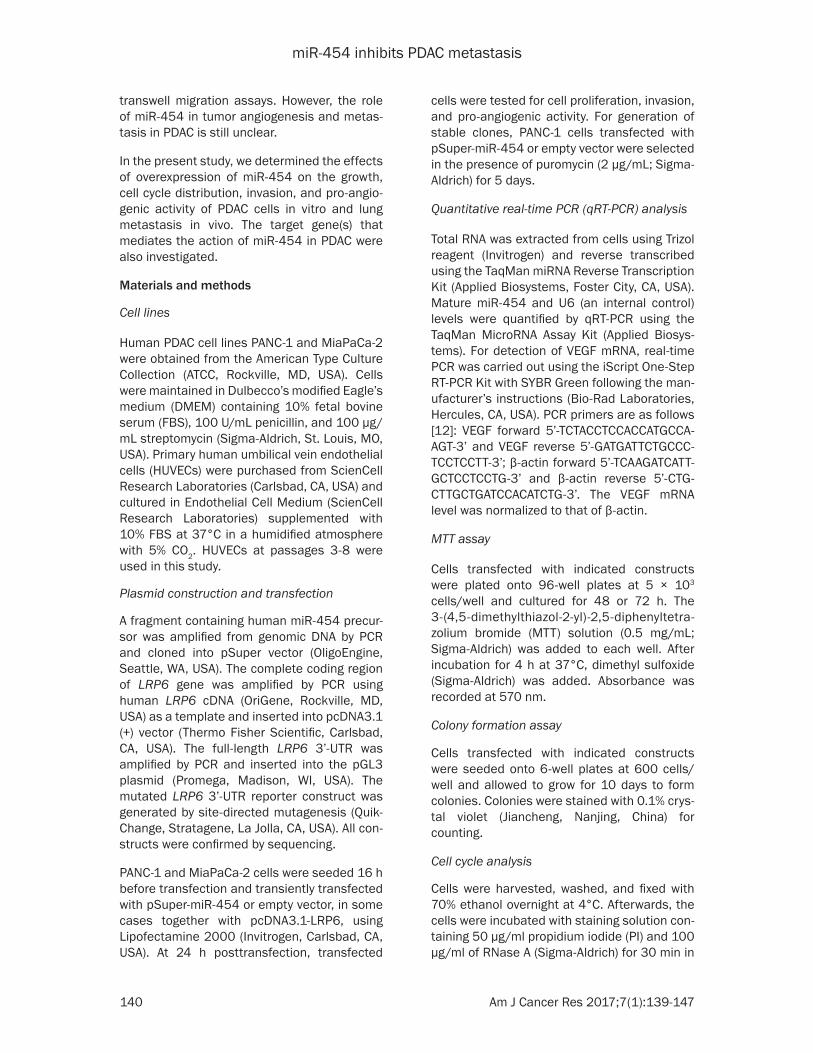
at the G0/G1 phase was significantly decreas- ed in miR-454-overexpressing cells, indicating a G2/M cell cycle arrest.
Overexpression of miR-454 suppresses the invasive and pro-angiogenic potential of PDAC cells
As determined by Matrigel-coated invasion assays, overexpression of miR-454 significant-ly decreased the number of PANC-1 invading cells through Matrigel membranes by 63% (P < 0.05 vs. vector-transfected cells; Figure 2A). Similarly, miR-454-overexpressing MiaPaCa-2 cells showed significantly less invasiveness than control cells (P < 0.05; Figure 2A). To assess the pro-angiogenic activity of miR-454-overexpressing PDAC cells, conditioned media from transfected PANC-1 cells were col-lected and used to stimulate HUVECs to form
Figure 1. miR-454 exerts growth-suppressive effects on PDAC cells. A. qRT-PCR analysis of miR-454 levels in PDAC cells transfected with the miR-454-expressing plasmid or vector. B. PDAC cells transfected with the miR-454-expressing plasmid or vector were cultured for 48 or 72 h and tested for viability by MTT assays. C. Colony formation assay. Transfected cells were plated at a low density and allowed to form colonies for 10 days. Ectopic expression of miR-454 significantly inhibited colony formation of PDAC cells. Top, representative wells of the colonies. D. Analysis of cell cycle distribu-tion by flow cytometry after PI staining. miR-454 overexpression led to an accumulation of cells at the G2/M phase in PANC-1 cells. Top, representa-tive flow cytometry histograms showing cell cycle distribution. *P < 0.05 vs. vector-transfected cells.
P-value < 0.05 indicates sta-tistically significant.
Results
miR-454 exerts growth-suppressive effects on PDAC cells
qRT-PCR analysis confirmed the restoration of miR-454 in PDAC cells transfected with the miR-454-expressing plas-mid (Figure 1A). MTT assays showed that overexpression of miR-454 significantly inhib-ited the proliferation of PANC-1 and MiaPaCa-2 cells, com-pared to vector-transfected cells (P < 0.05; Figure 1B). Colony formation assays fur-ther demonstrated that ecto-pic expression of miR-454 led to 52% and 70% reduction in colony number in PANC-1 and MiaPaCa-2 cells, respectively, after culturing for 10 days (Figure 1C). Analysis of cell cycle distribution by flow cytometry revealed that miR-454-overexpressing PANC-1 cells displayed an accumula-tion of cells at the G2/M phase (Figure 1D). In con-trast, the percentage of cells

miR-454 inhibits PDAC metastasis
143 Am J Cancer Res 2017;7(1):139-147
capillary tube-like structures. Notably, we found that addition of conditioned media from the
the TCF/LEF luciferase reporter in PANC-1 cells (Figure 3D). Taken together, miR-454 can tar-
Figure 2. Overexpression of miR-454 suppresses the invasive and pro-angio-genic potential of PDAC cells. A. Matrigel-coated invasion assays. miR-454 overexpression significantly inhibited the invasiveness of PDAC cells. Left, representative images of transwell invasion assays. B. Capillary tube-like structure formation. Conditioned media from transfected PANC-1 cells were incubated with HUVECs and tube-like structures were examined. Left, repre-sentative photomicrographs of tube-like structures. C. qRT-PCR analysis of VEGF mRNA levels in PANC-1 cells transfected with the miR-454-expressing plasmid or vector. D. Measurement of VEGF protein levels in conditioned me-dia from transfected PANC-1 cells by ELISA. *P < 0.05 vs. vector-transfected cells.
miR-454 group significantly inhibited the formation of tubes (Figure 2B). We further examined the effect of miR-454 overexpression on VEGF production. As expected, the transcription and secretion of VEGF was significantly sup-pressed in miR-454-overex-pressing PANC-1 cells (Figure 2C and 2D).
miR-454 inhibits Wnt/β-catenin signaling in PDAC cells by targeting LRP6
Bioinformatic analysis using the TargetScan algorithm (http://www.targetscan.org/vert_71/) suggested that LRP6 was a potential target gene of miR-454 (Figure 3A). Luciferase reporter assays confirmed that co-transfec-tion with the miR-454-ex-pressing plasmid resulted in a significant repression of the reporter containing the 3’-UTR of LRP6 mRNA, but not the mutated LRP6 3’-UTR (Figure 3B). Consistently, We- stern blot analysis revealed a marked downregulation of endogenous LRP6 in miR-454-overexpressing PANC-1 and MiaPaCa-2 cells (Figure 3C). Since LRP6 is an impor-tant regulator of Wnt/β-cat- enin signaling [15], we tested whether miR-454 overexpres-sion affected the activation of Wnt/β-catenin signaling in PDAC cells. As shown in Figure 3C, the levels of hypo-phosphorylated, active β-ca- tenin were remarkably low- er in miR-454-overexpressing PDAC cells than in control counterparts. Moreover, over-expression of miR-454 signifi-cantly inhibited the activity of
Figure 3. miR-454 inhibits Wnt/β-catenin signaling in PDAC cells by targeting LRP6. A. Bioinformatic analysis using the TargetScan algorithm (http://www.targetscan.org/vert_71/) suggested that miR-454 could target the 3’-UTR of LRP6 mRNA. B. Luciferase reporter assays. Delivery of miR-454 could inhibit the expression of the reporter containing the 3’-UTR of LRP6 mRNA, but not the mutated LRP6 3’-UTR. C. Western blot analysis of indicated proteins in PDAC cells transfected with the miR-454-expressing plasmid or vector. D. TCF/LEF luciferase reporter assays. Overexpression of miR-454 significantly suppressed the activity of the TCF/LEF luciferase reporter in PANC-1 cells. *P < 0.05 vs. vector-transfected cells.

miR-454 inhibits PDAC metastasis
144 Am J Cancer Res 2017;7(1):139-147
get LRP6 to suppress Wnt/β-catenin activation in PDAC cells.
Enforced expression of LRP6 reverses the sup-pressive effects of miR-454 on PDAC cells
To test the hypothesis that downregulation of LRP6 is required for the action of miR-454 in PDAC cells, we overexpressed a miRNA-resis-tant variant of LRP6 in PANC-1 cells with over-expression of miR-454 (Figure 4A). Of note, miR-454-expressing PANC-1 cells co-transfect-ed with the LRP6 construct exhibited signifi-cantly higher proliferation (Figure 4B) and inva-sion (Figure 4C) than those co-transfected with empty vector. Moreover, the pro-angiogenic activity of miR-454-overexpressing PANC-1 cells was partially restored upon ectopic expression of LRP6 (Figure 4D). These data indicate that the tumor-suppressive activity of
AC growth by interfering with the recruitment of macrophages [11]. Tumor-associated macro-phages contribute to the establishment of a tumor-favorable microenvironment [16, 17]. Therefore, it was suggested that miR-454 has the ability to modulate the tumor microenviron-ment in PDAC. Since this miRNA has been doc-umented to promote cancer cell proliferation and invasion in several cancer types [9, 10], in the present study we tested the hypothesis that miR-454 might affect multiple aspects of the biology of PDAC. In support of this hypoth-esis, we noted that restoration of miR-454 sig-nificantly inhibited the proliferation and colony formation of PDAC cells. Consistently, miR-454 overexpression was found to arrest PDAC cells at the G2/M phase. Apart from inhibition of cell growth, overexpression of miR-454 also im- paired the invasion and pro-angiogenic poten-
Figure 4. Enforced expression of LRP6 reverses the suppressive effects of miR-454 on PDAC cells. A. Western blot analysis of LRP6 in PANC-1 cells transfected with indicated constructs. B. MTT assays were performed to de-termine the viability of PANC-1 cells transfected with indicated constructs after culturing for 48 or 72 h. C. Matrigel-coated invasion assays. PANC-1 cells were transfected with indicated constructs and examined for the in-vasiveness. D. Capillary tube-like structure formation. Conditioned media from transfected PANC-1 cells were incubated with HUVECs and the length of tube-like structures was measured. *P < 0.05.
miR-454 is associated with repression of LRP6.
miR-454 inhibit tumor metas-tasis to the lung in vivo
Finally, we checked whether miR-454 could inhibit PDAC metastasis in vivo. To this end, PANC-1 cells stably transfected with the miR-454-expressing plasmid or vector were injected to the tail veins of nude mice. Lung metastasis was detected in 7 out of 10 mice injected with vector-transfected cells and 2 out of 10 mice injected with miR-454-overexpressing cells (P = 0.0349; Figure 5A and 5B). H&E staining of lung tis-sue sections (Figure 5C and 5D) confirmed that the num-ber of lung micrometastatic lesions was significantly lower in the miR-454 group than in the control group (6.2 ± 1.5 vs. 28.4 ± 3.6; P < 0.05). These results indicate the anti-metastatic potential of miR-454 in PDAC.
Discussion
Our previous work showed that miR-454 can inhibit PD-

miR-454 inhibits PDAC metastasis
145 Am J Cancer Res 2017;7(1):139-147
tial of PDAC cells. Angiogenesis is a fundamen-tal process for tumor growth and progression [2]. Tumor cells produce a number of growth factors and cytokines, which can modulate angiogenesis of endothelial cells [18, 19]. In this study, we showed that enforced expression of miR-454 resulted in a significant decline in the expression and secretion of VEGF in PDAC cells, which provides an explanation for the reduced pro-angiogenic activity of miR-454- overexpressing PDAC cells. Taken together, miR-454 functions as a tumor suppressor in PDAC. However, this miRNA exerts tumor-pro-moting effects on several other cancers such as uveal melanoma [9] and non-small cell lung
ulation of LRP6 contributes to the suppressive activity of miR-454 in the growth, invasion, and angiogenesis in PDAC. It should be mentioned that miR-454 can also downregulate target genes other than LRP6, e.g. c-Met [23]. A previ-ous study reported that miR-454 can restrict the growth and invasion of osteosarcoma cells by targeting c-Met [23]. Therefore, it is possible that other undefined target genes may also mediate the activity of miR-454 in PDAC.
Distant metastasis is regarded as a major cause of cancer-related deaths [24]. To com-plete our previous report showing the growth-suppressive activity of miR-454 [11], the pres-
Figure 5. miR-454 inhibit tumor metastasis to the lung in vivo. A. Represen-tative photographs of the metastatic lesions (indicated by arrowhead) in the lung. B. Comparison of the incidences of lung metastasis between the vector and miR-454 groups by the Fisher’s exact test. C. H&E staining of lung tissue sections revealed micrometastatic lesions (indicated by arrow). Scale bar = 150 μm. D. Bar graphs showing quantification of lung micrometastatic le-sions. *P < 0.05 vs. the vector group.
cancer [10], suggesting its context-dependent functions.
To determine the mechanism by which miR-454 exerts its suppressive activity in PDAC, we performed bioinformatic analysis and luciferase repor- ter assays and identified LRP6 as a direct target gene of miR-454. Overexpression of miR-454 led to a signific- ant inhibition of endogenous LRP6 expression in PDAC cells. Interestingly, LRP6 has the capacity to promote tumor growth and metastasis via the Wnt/β-catenin pathway [15, 20]. It has been reported that miR-126 suppresses the growth of papillary thyroid carcinoma by targeting LRP6 [21]. Similarly, biochemical inhibition of LRP6 was shown to inhibit PDAC growth, which was accompanied by inactiva-tion of Wnt/β-catenin signal-ing [22]. These studies en- courage us to hypothesize that miR-454 might exert its suppressive activity by inhibit-ing Wnt/β-catenin signaling via downregulation of LRP6. In support of this hypothesis, reduced β-catenin-dependent transcriptional activity was noted in miR-454-overex-pressing PDAC cells. Rescue experiments provided addi-tional evidence that downreg-

miR-454 inhibits PDAC metastasis
146 Am J Cancer Res 2017;7(1):139-147
ent study demonstrated that miR-454 over- expression hampered the metastatic growth of PDAC cells in the lung in mouse models. The in vivo findings are also consistent with the Transwell invasion experiments. Restoration of miR-454 may thus represent a potential therapeutic strategy for controlling PDAC metastasis.
In conclusion, miR-454 exhibits suppressive activity in growth, metastasis, and angiogene-sis in PDAC. Targeting LRP6 and subsequent inactivation of Wnt/β-catenin signaling repre-sents a novel mechanism for the action of miR-454. These observations provide a rationale to investigate the therapeutic benefits of delivery of miR-454 in the treatment of PDAC.
Disclosure of conflict of interest
None.
Address correspondence to: Tiantao Kuang, De- partment of General Surgery, Zhongshan Hospital, Fudan University, Shanghai 200032, China. E-mail: [email protected]
References
[1] Michl P, Gress TM. Current concepts and novel targets in advanced pancreatic cancer. Gut 2013; 62: 317-326.
[2] Popescu AM, Purcaru SO, Alexandru O, Dricu A. New perspectives in glioblastoma antiangio-genic therapy. Contemp Oncol (Pozn) 2016; 20: 109-118.
[3] Georgiadou D, Sergentanis TN, Sakellariou S, Filippakis GM, Zagouri F, Vlachodimitropoulos D, Psaltopoulou T, Lazaris AC, Patsouris E, Zo-grafos GC. VEGF and Id-1 in pancreatic adeno-carcinoma: prognostic significance and impact on angiogenesis. Eur J Surg Oncol 2014; 40: 1331-1337.
[4] Solorzano CC, Baker CH, Bruns CJ, Killion JJ, Ellis LM, Wood J, Fidler IJ. Inhibition of growth and metastasis of human pancreatic cancer growing in nude mice by PTK 787/ZK222584, an inhibitor of the vascular endothelial growth factor receptor tyrosine kinases. Cancer Bioth-er Radiopharm 2001; 16: 359-370.
[5] Fukasawa M, Korc M. Vascular endothelial growth factor-trap suppresses tumorigenicity of multiple pancreatic cancer cell lines. Clin Cancer Res 2004; 10: 3327-3332.
[6] Srivastava S, Tsongalis GJ, Kaur P. Role of mi-croRNAs in regulation of the TNF/TNFR gene superfamily in chronic lymphocytic leukemia. Clin Biochem 2016; 49: 1307-1310.
[7] Wang C, Liu P, Wu H, Cui P, Li Y, Liu Y, Liu Z, Gou S. MicroRNA-323-3p inhibits cell invasion and metastasis in pancreatic ductal adenocar-cinoma via direct suppression of SMAD2 and SMAD3. Oncotarget 2016; 7: 14912-14924.
[8] Fan P, Liu L, Yin Y, Zhao Z, Zhang Y, Amponsah PS, Xiao X, Bauer N, Abukiwan A, Nwaeburu CC, Gladkich J, Gao C, Schemmer P, Gross W, Herr I. MicroRNA-101-3p reverses gemcitabine resistance by inhibition of ribonucleotide re-ductase M1 in pancreatic cancer. Cancer Lett 2016; 373: 130-137.
[9] Sun L, Wang Q, Gao X, Shi D, Mi S, Han Q. Mi-croRNA-454 functions as an oncogene by reg-ulating PTEN in uveal melanoma. FEBS Lett 2015; 589: 2791-2796.
[10] Zhu DY, Li XN, Qi Y, Liu DL, Yang Y, Zhao J, Zhang CY, Wu K, Zhao S. MiR-454 promotes the progression of human non-small cell lung cancer and directly targets PTEN. Biomed Pharmacother 2016; 81: 79-85.
[11] Fan Y, Xu LL, Shi CY, Wei W, Wang DS, Cai DF. MicroRNA-454 regulates stromal cell derived factor-1 in the control of the growth of pancre-atic ductal adenocarcinoma. Sci Rep 2016; 6: 22793.
[12] Tang XD, Zhou X, Zhou KY. Dauricine inhibits insulin-like growth factor-I-induced hypoxia in-ducible factor 1alpha protein accumulation and vascular endothelial growth factor expres-sion in human breast cancer cells. Acta Phar-macol Sin 2009; 30: 605-616.
[13] Duan Y, Wu X, Zhao Q, Gao J, Huo D, Liu X, Ye Z, Dong X, Fu Z, Shang Y, Xuan C. DOT1L pro-motes angiogenesis through cooperative regu-lation of VEGFR2 with ETS-1. Oncotarget 2016; 7: 69674-69687.
[14] Anderson M, Marayati R, Moffitt R, Yeh JJ. Hexokinase 2 promotes tumor growth and me-tastasis by regulating lactate production in pancreatic cancer. Oncotarget 2016; [Epub ahead of print].
[15] Schmitt M, Metzger M, Gradl D, Davidson G, Orian-Rousseau V. CD44 functions in Wnt sig-naling by regulating LRP6 localization and acti-vation. Cell Death Differ 2015; 22: 677-689.
[16] Nielsen SR, Quaranta V, Linford A, Emeagi P, Rainer C, Santos A, Ireland L, Sakai T, Sakai K, Kim YS, Engle D, Campbell F, Palmer D, Ko JH, Tuveson DA, Hirsch E, Mielgo A, Schmid MC. Macrophage-secreted granulin supports pan-creatic cancer metastasis by inducing liver fi-brosis. Nat Cell Biol 2016; 18: 549-560.
[17] Miyake M, Hori S, Morizawa Y, Tatsumi Y, Nakai Y, Anai S, Torimoto K, Aoki K, Tanaka N, Shi-mada K, Konishi N, Toritsuka M, Kishimoto T, Rosser CJ, Fujimoto K. CXCL1-mediated inter-action of cancer cells with tumor-associated macrophages and cancer-associated fibro-
miR-454 inhibits PDAC metastasis
147 Am J Cancer Res 2017;7(1):139-147
blasts promotes tumor progression in human bladder cancer. Neoplasia 2016; 18: 636-646.
[18] He T, Qi F, Jia L, Wang S, Wang C, Song N, Fu Y, Li L, Luo Y. Tumor cell-secreted angiogenin in-duces angiogenic activity of endothelial cells by suppressing miR-542-3p. Cancer Lett 2015; 368: 115-125.
[19] Cao Z, Ding BS, Guo P, Lee SB, Butler JM, Casey SC, Simons M, Tam W, Felsher DW, Shi-do K, Rafii A, Scandura JM, Rafii S. Angiocrine factors deployed by tumor vascular niche in-duce B cell lymphoma invasiveness and che-moresistance. Cancer Cell 2014; 25: 350-365.
[20] Ren DN, Chen J, Li Z, Yan H, Yin Y, Wo D, Zhang J, Ao L, Chen B, Ito TK, Chen Y, Liu Z, Li Y, Yang J, Lu X, Peng Y, Pan L, Zhao Y, Liu S, Zhu W. LRP5/6 directly bind to frizzled and prevent frizzled-regulated tumour metastasis. Nat Commun 2015; 6: 6906.
[21] Wen Q, Zhao J, Bai L, Wang T, Zhang H, Ma Q. miR-126 inhibits papillary thyroid carcinoma growth by targeting LRP6. Oncol Rep 2015; 34: 2202-2210.
[22] Arensman MD, Nguyen P, Kershaw KM, Lay AR, Ostertag-Hill CA, Sherman MH, Downes M, Liddle C, Evans RM, Dawson DW. Calcipotriol targets LRP6 to inhibit Wnt signaling in pancre-atic cancer. Mol Cancer Res 2015; 13: 1509-1519
[23] Niu G, Li B, Sun J, Sun L. miR-454 is down-reg-ulated in osteosarcomas and suppresses cell proliferation and invasion by directly targeting c-Met. Cell Prolif 2015; 48: 348-355.
[24] Wang L, Bai YY, Yang Y, Hu F, Wang Y, Yu Z, Cheng Z. Diabetes mellitus stimulates pancre-atic cancer growth and epithelial-mesenchy-mal transition-mediated metastasis via a p38 MAPK pathway. Oncotarget 2016; 7: 38539-38550.