
CLADE Review 2003-2008 Nancy Wilkins-Diehr [email protected] CLADE 2008, June 23, 2008.
ORIGINAL ARTICLE
Discovery of a Diverse Clade of Gregarine Apicomplexans(Apicomplexa: Eugregarinorida) from Pacific Eunicid andOnuphid Polychaetes, Including Descriptions of Paralecudinan. gen., Trichotokara japonica n. sp., and T. eunicae n. sp.Sonja Rueckerta,b, Kevin C. Wakemanc & Brian S. Leanderc
a School of Life, Sport and Social Sciences, Edinburgh Napier University, Sighthill Campus, Sighthill Court, Edinburgh EH11 4BN, United Kingdom
b Shimoda Marine Research Center, University of Tsukuba, 5-10-1, Shimoda, Shizuoka 415-0025, Japan
c Department of Zoology, University of British Columbia, #3529–6270 University Boulevard, Vancouver, BC V6T 1Z4, Canada
Keywords
Eugregarines; parasite; phylogeny;
taxonomy.
Correspondence
S. Rueckert, School of Life, Sport and Social
Sciences, Edinburgh Napier University,
Sighthill Campus, Sighthill Court, Edinburgh
EH11 4BN, United Kingdom
Telephone number: +44 131 455 2490;
FAX number: +44 131 455 2291;
e-mail: [email protected]
Received: 15 June 2012; revised 13 August
2012; accepted September 25, 2012.
doi:10.1111/jeu.12015
ABSTRACT
Marine gregarines are poorly understood apicomplexan parasites with large
trophozoites that inhabit the body cavities of marine invertebrates. Two novel
species of gregarines were discovered in polychaete hosts collected in Canada
and Japan. The trophozoites of Trichotokara japonica n. sp. were oval to
rhomboidal shaped, and covered with longitudinal epicytic folds with a density
of six to eight folds/micron. The nucleus was situated in the middle of the cell,
and the mucron was elongated and covered with hair-like projections; antler-
like projections also extended from the anterior tip of the mucron. The distinc-
tively large trophozoites of Trichotokara eunicae n. sp. lacked an elongated
mucron and had a tadpole-like cell shape consisting of a bulbous anterior
region and a tapered tail-like posterior region. The cell surface was covered
with longitudinal epicytic folds with a density of three to five folds/micron.
Small subunit (SSU) rDNA sequences of both species were very divergent and
formed a strongly supported clade with the recently described species Tricho-
tokara nothriae and an environmental sequence (AB275074). This phylogenetic
context combined with the morphological features of T. eunicae n. sp. required
us to amend the description for Trichotokara. The sister clade to the Trichoto-
kara clade consisted of environmental sequences and Lecudina polymorpha,
which also possesses densely packed epicyctic folds (3–5 folds/micron) and a
prominently elongated mucron. This improved morphological and molecular
phylogenetic context justified the establishment of Paralecudina (ex. Lecudina)
polymorpha n. gen. et comb.
GREGARINES are unicellular parasites of terrestrial, fresh-
water, and marine invertebrates that infect the digestive
tract, coelomic spaces, and reproductive vesicles of their
hosts. The vast majority of described gregarine species
belong to so-called “eugregarines” (Grass�e 1953; Leander
2008; Perkins et al. 2002), which possess an extracellular
feeding stage, the trophozoite, that are conspicuously dif-
ferent in morphology and motility from the infective sporo-
zoite stage. Most eugregarines possess dense arrays of
longitudinal epicytic folds facilitating surface mediated
nutrition (Leander 2008). Because of the large number of
folds, the cells are relatively stiff and move using an actin/
myosin-based gliding mechanism (Heintzelman 2004;
Leander 2008). The trophozoites of eugregarines are also
either septate or aseptate depending on whether or not
the cell is partitioned into two visible compartments (pro-
tomerite and deutomerite). The anterior end of the troph-
ozoites is modified for attachment to host tissues and is
considered an epimerite in septate species and a mucron
in aspetate species. Mucrons and epimerites can range
from being streamlined and inconspicuous to prominent
and elongated, sometimes bearing multiple hair-like exten-
sions.
The haploid life histories of eugregarines apparently lack
an asexual proliferation phase called “merogony”,
whereby the trophozoites are able to divide into many
genetically identical individuals (Levine 1977). In general,
eugregarines have (monoxenous) life cycles involving only
© 2012 The Author(s) Journal of Eukaryotic Microbiology © 2012 International Society of Protistologists
Journal of Eukaryotic Microbiology 2013, 60, 121–136 121
Journal of Eukaryotic Microbiology ISSN 1066-5234
Published bythe International Society of ProtistologistsEukaryotic Microbiology
The Journal of
one host species. The relatively large trophozoites within
a host pair up in a process called “syzygy” and become
gamonts. A cyst forms around the pair of gamonts, form-
ing a gametocyst, and each gamont divides into numerous
gametes (Levine 1977). The pair-wise fusion of gametes
derived from each gamont forms a zygote that is then sur-
rounded by an oocyst wall. Within the oocyst, meiosis
occurs to yield four (or more, with subsequent rounds of
mitosis) spindle-shaped sporozoites (Kuriyama et al. 2005).
Hundreds of oocysts accumulate within each gametocyst,
and are usually released via host faeces or via host death
and remain in the environment until a new host ingests
them. Once ingested, the sporozoites hatch from the
oocysts and penetrate the host cells. The sporozoites
enlarge to become trophozoites that emerge from the cell
and start feeding.
Lecudina Mingazzini, 1899 (Levine, 1988) and the Lecu-
dinidae Kamm, 1922 (26 genera) are essentially “catch-all”
taxa for marine eugregarines that infect mainly polychae-
tes (Rueckert and Leander 2010). Emerging molecular
phylogenetic data combined with ultrastructural data, how-
ever, have improved our understanding of lecudinid inter-
relationships (Leander 2008; Leander et al. 2003b; Rueckert
and Leander 2009, 2010; Simdyanov 2009). For instance,
Difficilina (Rueckert et al. 2010; Simdyanov 2009) and Tri-
chotokara (Rueckert and Leander 2010) are recently estab-
lished genera within the Lecudinidae that more accurately
characterize the diversity of marine eugregarines. The
combination of molecular phylogenetic data, comparative
morphology, host affinity, and biogeography suggest that
many more genera are warranted and that some known
species of Lecudina need to be re-evaluated within the
context of available data (compare Levine 1977, 1979; Per-
kins et al. 2002; Landers and Leander 2005; Rueckert and
Leander 2008, 2009; Simdyanov 2009). The generation of
molecular phylogenetic data from characterized gregarine
species has also played a huge role in the interpretation of
environmental sequences generated from PCR surveys of
organismal diversity in specific habitats (e.g. Rueckert
et al. 2011).
In this vein, we discovered two novel species of marine
eugregarines in eunicid and onuphid polychaetes and char-
acterized their trophozoites with light microscopy (LM),
scanning electron microscopy (SEM) and small subunit
(SSU) rDNA sequences. Molecular phylogenetic analyses
of the new sequences enabled us to (1) determine the
emerging composition of a diverse Trichotokara clade, (2)
establish the cellular identities of nine environmental
sequences collected from several different habitats, and
(3) re-evaluate and revise the taxonomy of “Lecudina”
polymorpha within Paralecudina n. gen. et comb.
MATERIALS AND METHODS
Collection and isolation of organisms
Repeated dredge hauls were conducted in the Sagami-nada
Sea (34°38′43″N, 138°56′16″E) in November 2010 and Feb-
ruary 2011 at a depth of ~ 45 m during collecting trips on
the research vessel R/V Tsukuba based at the Shimoda
Marine Research Center, University of Tsukuba, Shimoda,
Shizuoka, Japan. Onuphid tubeworms Nothria cf. otsuchi-
ensis (Imajima, 1986) were collected from these samples.
The polychaete Eunice valens (Chamberlin, 1919) was col-
lected at a depth of 7–10 m while SCUBA diving at Ogden
Point (48°24′48″N, 123°23′37″W), in August 2010, in Victo-
ria, British Columbia, Canada. The intestines of the host ani-
mals were dissected with fine-tipped forceps under a low
magnification stereomicroscope (Olympus SZ61, Olympus
Corp. Tokyo, Japan/Leica MZ6, Wetzlar, Germany) to
extract the trophozoites of Trichotokara japonica n. sp. and
Trichotokara eunicae n. sp. Gut contents containing tropho-
zoites were examined using an inverted compound micro-
scope (Olympus CKX31, Olympus Corp./Zeiss Axiovert 200,
Carl-Zeiss, Goettingen, Germany, or Leica DM IL, Wetzlar,
Germany), and individual trophozoites were isolated by
micromanipulation. Before being prepared for microscopy
and DNA extraction, individual trophozoites were washed
three times in filtered and autoclaved seawater.
Light, scanning, and transmission electron microscopy
Differential interference contrast (DIC) light micrographs of
the trophozoites of T. japonica n. sp. were taken using a
system microscope (Olympus BX50, Olympus Corp.) con-
nected to a digital camera (Olympus DP70, Olympus
Corp.). The DIC light micrographs of the trophozoites of
T. eunicae n. sp. were taken with a compound microscope
(Zeiss Axioplan 2, Carl-Zeiss) connected to a colour digital
camera (Leica DC500). Individual trophozoites of T. japon-
ica n. sp. (n = 55) and T. eunicae n. sp. (n = 60) were pre-
pared for scanning electron microscopy (SEM) using the
OsO4 vapour protocol described previously (Rueckert and
Leander 2008, 2009). Isolated cells were deposited
directly into the threaded hole of a Swinnex filter holder,
containing a 5 lm polycarbonate membrane filter
(Millipore Corp., Billerica, MA), that was submerged in
10 ml of seawater within a small canister (2 cm diameter
and 3.5 cm tall). A piece of Whatman filter paper was
mounted on the inside base of a beaker (4 cm diameter
and 5 cm tall) that was slightly larger than the canister.
The Whatman filter paper was saturated with 4% OsO4
and the beaker was turned over the canister. The para-
sites were fixed by OsO4 vapours for 30 min. Ten drops
of 4% OsO4 were added directly to the seawater and the
parasites were fixed for an additional 30 min on ice. A 10-
ml syringe filled with distilled water was screwed to the
Swinnex filter holder and the entire apparatus was
removed from the canister containing seawater and fixa-
tive. The parasites were washed, then dehydrated with a
graded series of ethyl alcohol. Prepared specimens of
T. japonica n. sp. were freeze-dried with t-butanol and
specimens of T. eunicae n. sp. were critical-point dried
with CO2. Filters were mounted on stubs, sputter coated
with 5 nm gold, and viewed under a scanning electron
microscope (JEOL NeoScope JCM5000, JEOL Ltd.,
Tokyo, Japan/Hitachi S4700, Nissei Sangyo America, Ltd.,
Pleasanton, CA).
© 2012 The Author(s) Journal of Eukaryotic Microbiology © 2012 International Society of Protistologists
Journal of Eukaryotic Microbiology 2013, 60, 121–136122
Diversity of Marine Gregarine Apicomplexans Rueckert et al.
Fifty cells of T. eunicae n. sp. were manually isolated
from the gut of E. valens, washed in filtered seawater,
and placed in a 1.5-ml microfuge tube filled with 2% glu-
taraldehyde in seawater, and chilled on ice for 30 min.
Trophozoites were postfixed with 1% OsO4 in 0.2 M
sodium cacodylate buffer (SCB) (pH 7.2) for 1 h on ice,
washed three times (15 min each) with 0.2 M SCB, then
dehydrated with a graded series of ethanol washes
(30%, 50%, 75%, 85%, 90%, 95%, and 100%) at room
temperature. The sample was placed in a 1:1 acetone/
ethanol mixture for 30 min, and changed into acetone
thereafter. Cells were placed in 1:1 acetone/resin (Eppen
812) two times (6 h each) at room temperature, changed
into resin, and held at room temperature for 10–12 h,
before being polymerized overnight at 70 °C. Ultrathin
sections were cut on a diamond knife, using a Leica EM
UC6 microtome (Leica). Sections were placed on form-
var-coated grids, post-stained with uranyl acetate and
lead acetate, and viewed under a transmission electron
microscope (Hitachi H7600, Nissei Sangyo America, Ltd.).
Some data were presented on a grey or black back-
ground using Adobe Photoshop 6.0 (Adobe Systems, San
Jose, CA).
DNA isolation, PCR amplification, cloning, andsequencing
DNA from T. japonica n. sp. was extracted from two dif-
ferent isolates of trophozoites collected at different times.
Seventeen individual trophozoites (isolate 1) and 18 indi-
vidual trophozoites (isolate 2) were manually isolated from
dissected hosts. DNA from T. eunicae n. sp. was
extracted from 20 individual trophozoites after isolation
from their hosts. They were washed three times in filtered
and autoclaved seawater, and deposited into a 1.5-ml
microfuge tube.
Genomic DNA was extracted from the cells using the
MasterPure complete DNA and RNA purification Kit
(EPICENTRE, Madison, WI). Small subunit rDNA sequences
were PCR amplified using puReTaq Ready-to-go PCR beads
(GE Healthcare, Quebec, Canada) and the following
eukaryotic PCR primers for T. japonica n. sp.: F1 5′-GCGCTACCTGGTTGATCCTGCC-3′ and R1 5′-GATCCTTCTGCAGGTTCACCTAC-3′ (Leander et al. 2003a). The following
internal primers, designed to match existing eukaryotic
SSU sequences, were used for nested PCR: F2 5′-AAGTCTGGTGCCAGCAGCC-3′, F3 5′-TGCGCTACCTGGTTGATCC-3′ and R2 5′-GCCTYGCGACCATACTCC-3′. The
following primer pairs were used for the amplification of
the SSU rDNA of T. eunicae n. sp.: F4 5′ TGC GCT ACC TGG
TTG ATG ATC C 3′, R1 5′ GGG CGG TGT GTA CCA RGR G 3′,F5 5′ CGG TAA TTC CAG CTC C 3′, R3 5′ GAT CCT TCT GCA
GGT TCA CCT CA 3′. PCR products of T. japonica n. sp. corre-
sponding to the expected size (~ 1806 bp) were gel isolated
and cloned into the pSC-A-amp/kan vector using the Strata-
Clone PCR cloning kit (Agilent Technologies, Santa Clara, CA).
PCR products of T. eunicae n. sp. were gel isolated and
cloned into the pCR 2.1 vector using the TOPO TA cloning kit
(Invitrogen, Frederick, MD). Eight cloned plasmids, for each
PCR product, were digested with EcoRI, and inserts were
screened for size using gel electrophoresis. Two identical
clones were sequenced with ABI Big-dye reaction mix
using vector primers and internal primers oriented in both
directions. The SSU rDNA sequences were identified by
BLAST analysis and molecular phylogenetic analyses (Gen-
Bank Accession numbers: JX426617 T. japonica n. sp.;
JX426618, T. eunicae n. sp.).
Molecular phylogenetic analysis
The two new SSU rDNA sequences from T. japonica n.
sp. and T. eunicae n. sp. were incorporated into a 94-
sequence alignment representing the diversity of grega-
rines, some other important apicomplexan groups as well
as dinoflagellates (outgroup) using MacClade 4 (Maddi-
son and Maddison 2000) and visual fine-tuning. The pro-
gram PhyML (Guindon and Gascuel 2003; Guindon et al.
2005) was used to analyse the 96-sequence alignment
(994 unambiguously aligned positions; gaps excluded)
with maximum-likelihood (ML) using a general-time
reversible (GTR) model (Posada and Crandall 1998)
incorporating the fraction of invariable sites and a
discrete gamma distribution with four rate categories
(GTR + I + Γ + 4 model: a = 0.724 and I = 0.193 for the
96-sequence alignment). The GTR model was selected
using the program MrAIC 1.4.3 with PhyML (http://
www.abc.se/� nylander/mraic/mraic.html). ML bootstrap
(MLB) analyses were performed on 100 re-sampled data-
sets using the same program and the same
GTR + I + Γ + 4 model.
We performed two additional analyses (1) including the
three Trichotokara species, the closely related environ-
mental sequence and two environmental sequences from
the Paralecudina clade as an outgroup (GTR + I + Γ + 8
model: �ln L = 5043.79542, a = 0.696 and I = 0.139 for
this 6-sequence alignment) and (2) including the three Tri-
chotokara species, the closely related environmental
sequence and two Selenidium species as an outgroup
(GTR + I + Γ + 8 model: �ln L = 4245.65172, a = 2.777
and I = 0.438 for this 6-sequence alignment). The analyses
were performed to further evaluate the relationship
between the Trichotokara species. As the resulting phylog-
enies were similar to the ones from the 94-sequence
alignment, the data are not presented here.
Bayesian analysis of the 94-sequence alignment was
performed using the program MrBayes 3.0 (Huelsenbeck
and Ronquist 2001). The program was set to operate with
GTR, a gamma-distribution, and four Monte Carlo Markov
chains (MCMC; default temperature = 0.2). A total of
2,000,000 generations were calculated with trees sampled
every 50 generations and with a prior burn-in of 100,000
generations (2000 sampled trees were discarded; burn-in/
convergence was checked manually). A majority rule con-
sensus tree was constructed from 38,001 post-burn-in
trees. Posterior probabilities (BPP) correspond to the fre-
quency at which a given node was found in the post-burn-
in trees. Independent Bayesian runs on each alignment
yielded the same results.
© 2012 The Author(s) Journal of Eukaryotic Microbiology © 2012 International Society of Protistologists
Journal of Eukaryotic Microbiology 2013, 60, 121–136 123
Rueckert et al. Diversity of Marine Gregarine Apicomplexans
GenBank accession numbers
(AF494059) Adelina bambarooniae, (FJ459737) Amoebogrega-
rina nigra, (AJ415519) Amoebophrya sp. ex. Prorocentrum mi-
cans, (DQ462459) Ascogregarina armigerei, (DQ462456)
Ascogregarina culicis, (DQ462455) Ascogregarina taiwanensis,
(AY603402) Babesia bigemina, (HQ891113) Cephaloidophora
cf. communis from B. balanus, (HQ876008) Cephaloidophora
cf. communis from B. glandula, (L19068) Cryptosporidium
baileyi, (AF093489) Cryptosporidium parvum, (AF093502)
Cryptosporidium serpentis, (AF39993) Cytauxzoon felis,
(FJ832159) Difficilina paranemertis, (FJ832160) Difficilina
tubulani, (U67121) Eimeria tenella, (AB191437, AB252765,
AB275006, AB275008, AB275074, AB275068, AB275069,
AB275070, AB275071, AB275073, AB275103, AF372767,
AF372768, AF372769, AF372770, AF372771, AF372779,
AF372780, AF372821, AF290084,AY179975, AY179976,
AY179977, AY179988, EU050982) Environmental sequences,
(FJ832163) Filipodium phascolosomae, (FJ976721) Ganyme-
des themistos, (FJ459741) Gregarina blattarum, (FJ459743)
Gregarina coronata, (FJ459746) Gregarina kingi, (AF129882)
Gregarina niphandrodes, (FJ459748) Gregarina polymorpha,
(AF022194) Gymnodinium fuscum, (HQ876007) Heliospora ca-
prellae, (HQ891114) Heliospora cf. longissima from E. verruco-
sus, (HQ891115) Heliospora cf. longissima from E. vittatus,
(AF286023) Hematodinium sp., (AF130361) Hepatozoon cates-
bianae, (FJ459750) Hoplorhynchus acanthatholius, (DQ093796)
Lankesteria abbotti, (EU670240) Lankesteria chelyosomae,
(EU670241) Lankesteria cystodytae, (AF080611) Lankesterella
minima, (FJ832157) Lecudina longissima, (FJ832156) Lecudina
phyllochaetopteri, (AF457128) Lecudina tuzetae, (FJ459753)
Leidyana haasi, (AF457130) Leidyana migrator, (DQ093795)
Lithocystis sp., (AB000912) Marine parasite from Tridacna
crocea, (AY334568) Mattesia geminata, (AF457127) Monocys-
tis agilis, (AJ271354) Neospora caninum, (AF129883) Ophryo-
cystis elektroscirrha, (AY196706) Paralecudina (ex. Lecudina)
polymorphamorphotype 1, (AY196707) Paralecudina (ex. Lecu-
dina) polymorpha morphotype 2, (FJ459755) Paraschneideria
metamorphosa, (AY196708) Platyproteum vivax, (FJ459756)
Prismatospora evansi, (FJ459757) Protomagalhaensia granulo-
sae, (DQ093794) Pterospora floridiensis, (DQ093793) Pteros-
pora schizosoma, (GQ149767) Rhytidocystis cyamus,
(DQ273988) Rhytidocystis polygordiae, (M64244) Sarcocystis
muris, (JN857968) Selenidium boccardiella, (JN857967) Seleni-
dium idanthyrsae, (JN857966) Selenidium cf. mesnili,
(FJ832161) Selenidium orientale, (FJ832162) Selenidium pisin-
nus, (DQ683562) Selenidium serpulae, (AY196709) Selenidium
terebellae, (DQ176427) Syncystis mirabilis, (AF013418) Theile-
ria parva, (HQ876006) Thiriotia pugettiae, (M97703) Toxo-
plasma gondii, (JX426618) Trichotokara eunicae n. sp.,
(JX426617) Trichotokara japonica n. sp., (GU592817) Trichoto-
kara nothriae, (JN857969) Veloxidium leptosynaptae
RESULTS
Morphology of Trichotokara japonica n. sp. (Fig. 1–11)
The trophozoites of T. japonica n. sp. were approximately
123 lm long (63–201 lm, n = 12). Trophozoites were rigid
and capable of gliding motility. The trophozoites were
divided into the cell body proper and an elongated mucron
(Fig. 1, 7). The cell body proper was oval to slender
rhomboidal measuring 98 lm in length (50–168 lm,
n = 12) and 25 lm in width (13–42 lm, n = 12). From the
widest point of the cell body proper, the cell was quite
symmetrical. The posterior end of the cell was rounded
(Fig. 1, 4, 7), while the anterior end of the cell narrowed
into a neck-like structure at the base of the elongated mu-
cron. The mucron was shorter than the cell body proper
and measured 13.4 lm in length (7.5–33.0 lm, n = 7) and
7.1 lm in width (5.9–8.0 lm, n = 7). There was no sep-
tum visible at the junction between the cell body proper
and the mucron (Fig. 1–3). Instead, there was another
structure situated at the base of the mucron, namely a
flap-like protrusion (Fig. 1, 2, 4, 5, 8–11). Some trophozo-
ites lacked the mucron completely and there was no visi-
ble damage to the cell (Fig. 9); other trophozoites lacked
the mucron, and it seemed to have broken off (Fig. 11). A
spherical nucleus measured 17.2 (11.4–20.8) lm in diame-
ter (n = 6) and was mostly situated in the middle of the
cell body proper or slightly shifted to the anterior end
(Fig. 1, 4, 5). A nucleolus that measured 10.5 lm in diam-
eter was observed in one trophozoite. The cytoplasm had
a granular appearance (Fig. 1, 2). Gamonts without elon-
gated mucrons were observed in side-by-side intertwined
syzygy (Fig. 6). Neither sporozoites nor oocysts were
observed in our samples.
Scanning electron micrographs showed that the tropho-
zoite body surface was inscribed by densely packed longi-
tudinal epicytic folds (6–8 folds per micron) (Fig. 7). The
flap-like protrusion at the base of the mucron was also
covered with epicytic folds that were continuous with the
rest of the cell body proper (Fig. 8, 11). Epicytic folds
merged together near the posterior and anterior ends
(Fig. 10). The epicytic folds on the mucron were replaced
by hair-like projections with a mean length of 1.7 lm (1.3–2.4 lm, n = 14) and a mean width of 0.3 lm (0.25–0.34 lm, n = 13) (Fig. 7, 8, 10). The hair-like projections
were protrusions from the underlying cortex (Fig. 10). Ant-
ler-like projections at the tip of the mucron were only visi-
ble in the scanning electron micrographs (Fig. 7, 8). The
base of the antler-like projections was 1.2 (1.1–1.4) 9 1.2
(1.1–1.3) lm (Fig. 8). In most cases, the antler-like projec-
tions consisted of three branches, but in one observed
case, it consisted of four branches (Fig. 8). The branches
were 5.7 lm (3.9–8.2 lm, n = 14) long and 0.6 lm (0.43–0.73 lm, n = 12) wide.
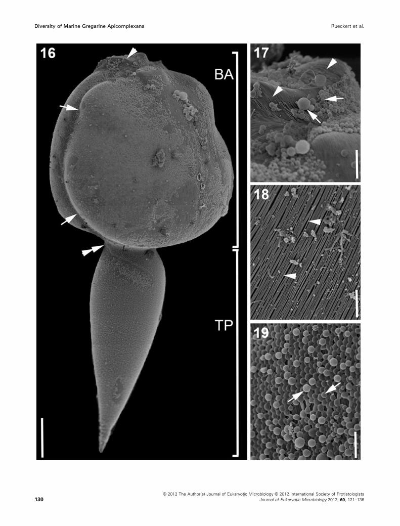
Morphology of Trichotokara eunicae n. sp. (Fig. 12–19)
The trophozoites of T. eunicae n. sp. were very long with
an approximate length of 581 lm (531–658 lm, n = 11)
(Fig. 12, 14). The shape of the cell was reminiscent of a
tadpole with a bulbous anterior region and a slender tail-
like posterior region (Fig. 12, 14, 16). There was no evi-
dence of an elongated mucron covered in hair-like projec-
tions. Trophozoites were brown in colour due to
accumulations of amylopectin in the cytoplasm. Cells were
© 2012 The Author(s) Journal of Eukaryotic Microbiology © 2012 International Society of Protistologists
Journal of Eukaryotic Microbiology 2013, 60, 121–136124
Diversity of Marine Gregarine Apicomplexans Rueckert et al.
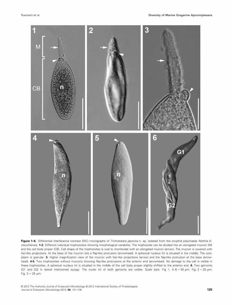
Figure 1–6. Differential interference contrast (DIC) micrographs of Trichotokara japonica n. sp. isolated from the onuphid polychaete Nothria cf.
otsuchiensis. 1-2. Different individual trophozoites showing morphological variability. The trophozoite can be divided into an elongated mucron (M)
and the cell body proper (CB). Cell shape of the trophozoites is oval to rhomboidal with an elongated mucron (arrow). The mucron is covered with
hair-like projections. At the base of the mucron sits a flap-like protrusion (arrowhead). A spherical nucleus (n) is situated in the middle. The cyto-
plasm is granular. 3. Higher magnification view of the mucron with hair-like projections (arrow) and the flap-like protrusion at the base (arrow-
head). 4-5. Two trophozoites without mucrons showing flap-like protrusions at the anterior end (arrowhead). No damage to the cell is visible in
these trophozoites. A spherical nucleus (n) is situated in the middle of the cell body proper slightly shifted to the anterior end. 6. Two gamonts
(G1 and G2) in lateral intertwined syzygy. The nuclei (n) of both gamonts are visible. Scale bars: Fig. 1, 4–6 = 50 lm; Fig. 2 = 20 lm;
Fig. 3 = 25 lm.
© 2012 The Author(s) Journal of Eukaryotic Microbiology © 2012 International Society of Protistologists
Journal of Eukaryotic Microbiology 2013, 60, 121–136 125
Rueckert et al. Diversity of Marine Gregarine Apicomplexans
© 2012 The Author(s) Journal of Eukaryotic Microbiology © 2012 International Society of Protistologists
Journal of Eukaryotic Microbiology 2013, 60, 121–136126
Diversity of Marine Gregarine Apicomplexans Rueckert et al.
rigid and capable of gliding motility. The broad anterior part
of the cell was dorso-ventrally flattened (Fig. 16) and mea-
sured 289 lm in length (244–302 lm, n = 11) and 267 lmin width (254–303 lm, n = 11). The spherical nucleus was
about 50 lm in diameter (48–53 9 47–51 lm, n = 11) and
was situated slightly off centre in the middle of the ante-
rior bulge (Fig. 12, 14). The bulbous anterior region was
encircled by a lateral groove that was also visible along
the mucron at the anterior end (Fig. 16, 17). The mucron
was free of amylopectin granules and slightly protruded
beyond the anterior end of the cell (Fig. 12, 13). The tail-
like posterior region tapered along its length into a pointed
tip with a length of 303 lm (294–317 lm, n = 11) and a
width of 94 lm (87–106 lm, n = 11). There was a slight
indentation at the junction between the anterior and pos-
terior regions of the cell, but no septum was visible in
either light (Inset Fig. 12) or scanning electron micro-
graphs (Fig. 16). The entire trophozoite was covered with
densely packed epicytic folds at a density of three to five
folds/micron (Fig. 14, 15, 17, 18). The folds were also cov-
ered with a conspicuous layer of mucilaginous material in
the form of tiny round bodies (Fig. 16, 19). Neither spor-
ozoites, gamonts in syzygy, nor oocysts were observed in
our samples.
Transmission electron microscopy showed nicely the
epicytic folds of the cortex and the trilayered membrane
complex (Fig. 14, 15).
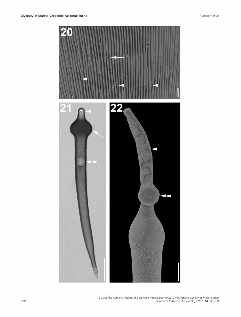
Morphology of Paralecudina polymorpha n. gen. etcomb. (ex. Lecudina polymorpha) (Fig. 20–22)
Two distinct morphotypes of P. polymorpha n. gen. et
comb. (ex. L. polymorpha) are known from the polychaete
L. japonica (compare Leander et al. 2003b; Rueckert et al.
2010). The cortex of both morphotypes was inscribed by
numerous folds, an example of which is shown in a high
magnification SEM of morphotype 1 (Fig. 20). Morphotype
1 refers to trophozoites that were 175–300 lm long and
35–50 lm wide. The anterior end was rounded with a mu-
cron free of epicytic folds, while the posterior end was
pointed. The density of folds was around three folds/
micron (compare Leander et al. 2003b). Morphotype 2
refers to skinnier trophozoites that were 475–575 lm long
and 35–60 lm wide with a density of folds of five folds/
micron. The trophozoites of morphotype 2 often pos-
sessed a prominent bulge right behind the mucron
(Fig. 21). The mucron of some trophozoites was elongated
and covered in epicytic folds reaching a length of
75–100 lm and a width of 15 lm (Fig. 22). The posterior
end was pointed. Both morphotypes were rigid and capa-
ble of gliding motility.
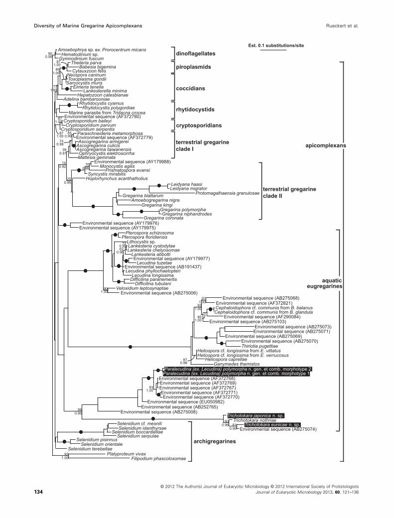
Molecular phylogenetic analyses of small subunitrDNA sequences (Fig. 23)
Phylogenetic analyses of the 96-taxon data set resulted
in a strongly supported clade (90 MLB, 0.99 BPP) of di-
noflagellates (outgroup) and a poorly resolved backbone
for the apicomplexan ingroup (Fig. 23). The apicomplexan
backbone gave rise to: (1) a clade consisting of a para-
phyletic group of coccidians and a strongly supported (99
MLB, 1.00 BPP) subclade of piroplasmids; (2) a rhytido-
cystid clade; (3) a cryptosporidian clade; (4) a weakly sup-
ported “terrestrial gregarine clade 1” (39 MLB, 0.91
BPP), consisting of neogregarines, eugregarines from
insects and an environmental sequence, as well as (5) a
stronger supported “terrestrial gregarine clade 2” (20
MLB, 0.95 BPP), consisting of monocystid eugregarines
and several eugregarines from insects. Two environmen-
tal sequences formed the sister clade to the two terres-
trial gregarine clades. The sequences from marine
archigregarines formed three different lineages (Selenidi-
um species, Veloxidium, Platyproteum and Filipodium)
that branched independently from the apicomplexan
backbone (Fig. 23). Mainly marine eugregarines formed
three sister clades: (1) A strongly supported clade (100
MLB, 1.00 BPP) consisting of urosporids (Pterospora and
Lithocystis) and lecudinids (Difficilina, Lankesteria and
Lecudina); (2) a strongly supported clade (100 MLB, 1.00
BPP) comprising gregarines (Cephaloidophora, Ganyme-
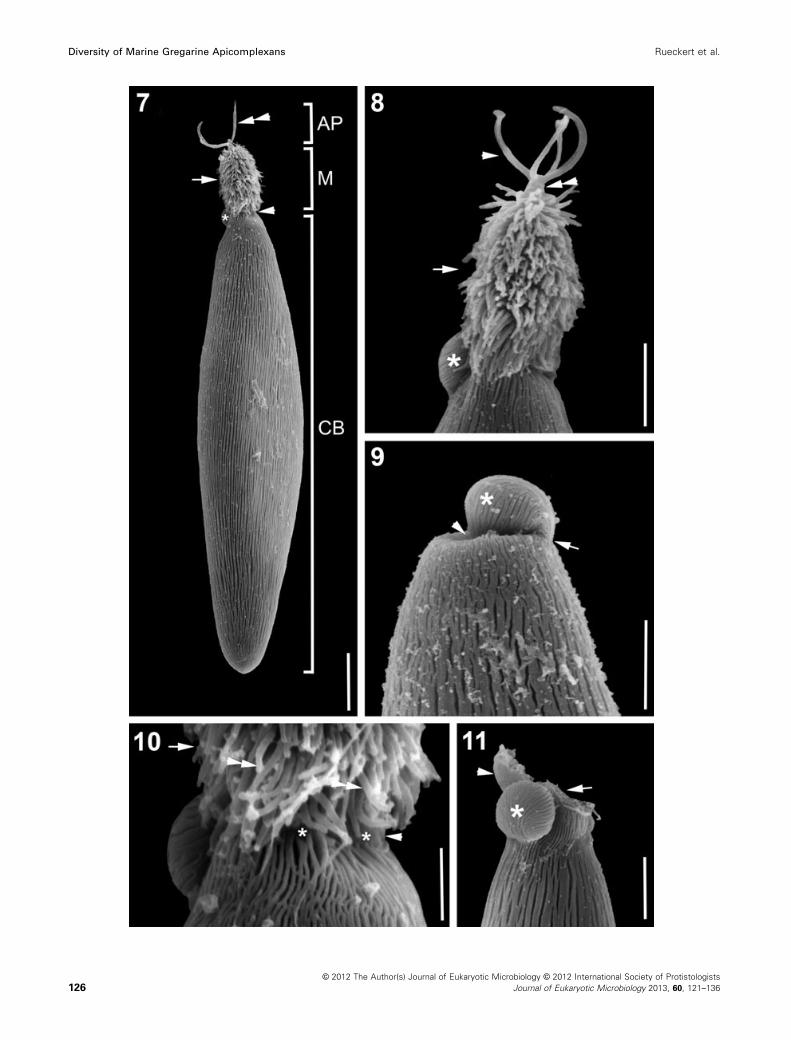
Figure 7–11. Scanning electron micrographs (SEM) showing the general morphology and surface ultrastructure of Trichotokara japonica n.
sp. isolated from the onuphid polychaete Nothria cf. otsuchiensis. 7. SEM showing a trophozoite with an elongated mucron (M) covered in hair-
like projections (arrow) and an antler-like projection (AP) at the anterior tip (double arrowhead). The cell body proper is slender rhomboidal with a
rounded posterior end. Densely packed longitudinal epicytic folds inscribe the cell body proper (CB). There seems to be a constriction between
the mucron and the rest of the cell (arrowhead), but the vision is restricted in this micrograph. In the back of the cell at the base of the mucron
lies the flap-like protrusion (asterisk). 8. High magnification SEM of the mucron (arrow) with hair-like projections protruding from the cell surface.
Situated at the anterior end of the mucron is an antler-like projection with four branches (arrowhead). The branches unite at a base that stems
from the cell cortex (double arrowhead). The flap-like protrusion (asterisk) shows epicytic folds. 9. High magnification SEM of the junction
between mucron and cell body proper. There is no mucron (arrowhead) and the flap-like protrusion (asterisk) is slightly bent towards the centre,
where the mucron would be otherwise. There is no cell damage visible (arrowhead). The flap-like protrusion shows epicytic folds (arrow) that are
continuous with the ones on the cell body proper. 10. High magnification SEM of the junction between the cell body proper and the elongated
mucron (arrow). The epicytic folds merge together at the anterior end and stop (asterisks) at the base of the mucron (arrow) leaving a small area
of smooth cortex (arrowhead) before the hair-like projections (double arrowheads) start. 11. High magnification SEM of the junction between mu-
cron and cell body proper. In this case there is visible cell damage (arrow) and the mucron seems to have broken off. There is still smooth cortex
(arrowhead) visible on the left side of the damaged cell and the flap-like protrusion (asterisk) seems to be in its original position. Scale bars:
Fig. 7 = 10 lm; Fig. 8–9, 11 = 5 lm; Fig. 10 = 2 lm.
© 2012 The Author(s) Journal of Eukaryotic Microbiology © 2012 International Society of Protistologists
Journal of Eukaryotic Microbiology 2013, 60, 121–136 127
Rueckert et al. Diversity of Marine Gregarine Apicomplexans

des, Heliospora and Thiriotia) infesting crustaceans; and
(3) a moderately supported clade (59 MLB, 0.99 BPP)
consisting of Trichotokara eunicae n. sp., T. japonica n.
sp., T. nothriae, two morphotypes of P. (ex. L.)
polymorpha n. gen. et comb. and several environmental
sequences.
Figure 12–15. Differential interference contrast (DIC) micrographs and transmission electron micrographs (TEM) of Trichotokara eunicae n. sp.
isolated from the eunicid polychaete Eunice valens. 12, 13. Different individual trophozoites showing a slight morphological variability. The tropho-
zoite can be divided into a bulbous anterior part (BA) and a slender tale-like posterior part (TP). Cell shape of the trophozoites is reminiscent of
that of a tadpole larva. There seems to be a marginal constriction (arrow) at the junction between the anterior and posterior part of the cell. The
cells are brownish in colour due to amylopectin granules in the cytoplasm. The mucron (double arrowhead) is free of amylopectin and slightly pro-
truded. A spherical nucleus (n) is situated in the middle, slightly off centre. The inset shows a close up of the junction (arrow). There is no septum
visible. 14, 15. Transmission electron micrographs (TEM) of the cortex showing the trilayered membrane complex (arrowhead) and the epicytic
folds (arrow). Scale bars: Fig. 12 = 300 lm; Inset = 75 lm; Fig. 13 = 240 lm; Fig. 14 = 2 lm, Fig. 15 = 1 lm.
© 2012 The Author(s) Journal of Eukaryotic Microbiology © 2012 International Society of Protistologists
Journal of Eukaryotic Microbiology 2013, 60, 121–136128
Diversity of Marine Gregarine Apicomplexans Rueckert et al.
Analyses of two small 6-sequence alignments (with
more sites included) were performed to test the topology
of the Trichotokara species; these analyses resulted in the
same phylogenetic relationships as the analyses of the
96-sequence alignment.
A total of 1,695 bp (excluding all indels) were compared
between the three species T. nothriae, T. japonica n. sp.
and T. eunicae n. sp., by calculating the pair-wise dis-
tances based on the Kimura 2-parameter model (Kimura
1980). This resulted in a 10.1–15.3% sequence divergence
between the three species. The sequence divergence
between the Trichotokara species T. nothriae and T. euni-
cae was lower (10.1%) than the sequence divergence
between T. nothriae and T. eunicae n. sp. (13.8%) and
between T. japonica n. sp. and T. eunicae n. sp. (15.3%).
DISCUSSION
Comparative morphology
According to WoRMS (2012), the family Lecudinidae cur-
rently consists of 25 genera. Most of the described spe-
cies fall within the genus Lecudina (Levine 1976). There
are four genera within the Lecudinidae that possess hair-
like projections: Pontesia, Cochleomeritus, Diplauxis
(Levine 1977), and the recently described genus Trichoto-
kara (Rueckert and Leander 2010). We described another
species with this feature, namely T. japonica n. sp. This
new species was isolated from a host (Nothria cf. otsu-
tchiensis) that is closely related to the host of the type
species of the genus, namely T. nothriae (Nothria conchy-
lega). Both species share the features of an elongated mu-
cron that is covered in hair-like projections and the
densely packed epicytic folds, but T. japonica n. sp. pos-
sesses more folds per micron (6–8 folds/micron) than
T. nothriae does (~ 5 folds/micron). The cell size and
shape of the two species is different and only T. japonica
has antler-like projections at the anterior tip of the mucron
(Table 1). Projections like these have not been described
before for any aseptate eugregarine species; comparable
structures are only known from the epimerites of some
septate (terrestrial) eugregarines (compare Perkins et al.
2002). The gamonts of T. nothriae undergo end-to-end syz-
ygy (Rueckert and Leander 2010), while the gamonts of
the newly described species T. japonica n. sp. undergo
side by side intertwined syzygy. Before the gamonts
undergo syzygy, they shed the mucron. Valigurov�a et al.
(2009) discussed that in the septate gregarine species,
Gregarina polymorpha retraction of the epimerite into the
protomerite is more likely than its separation during
detachment from the host epithelium. Figure 9 and 11
show both mechanisms of mucron removal. The mucron
of the trophozoite in Fig. 11 broke off leaving the cell dam-
aged. In contrast, there is no visible damage in the tropho-
zoite shown in Fig. 9, presumably due to the retraction of
the mucron.
Most gregarines within the Lecudinidae are in a size
range of 100–300 lm long, with some exceptions (Perkins
et al. 2002). One of the two new species we describe in
this study is up to 650 lm long, namely T. eunicae n. sp.
This gregarine, isolated from the polychaete E. valens, has
another feature that has not been previously described: a
very bulbous, dorso-laterally flattened anterior part of the
trophozoite cell encircled by a lateral groove. Five grega-
rine species have been described from polychaetes
belonging to the genus Eunice so far, but none for E. va-
lens (compare Levine 1976, 1977). One of these species
Deuteromera cleava Bhatia and Setna, 1938 belongs to a
group of septate gregarines. The remaining four species all
belong to the Lecudinidae: Lecudina eunicae (Lankester,
1866) Levine, 1976 from E. harassii; Lecudina bhatiai (Bha-
tia and Setna, 1938) Levine, 1976; Contortiocorpa prashadi
Bhatia and Setna, 1938; and Ulivina eunicae Bhatia and
Setna, 1938. The latter three species were isolated from
E. siciliensis. The tadpole-like shape of the trophozoites in
T. eunicae n. sp. differs considerably from three of the
four gregarine species previously described from Eunice
hosts. The species L. eunicae has a similar shape to
T. eunicae n. sp. with a pointed wedge-shaped body
posteriorly, but is significantly smaller with a length
of around 254 lm (Lankester 1866). The trophozoite of
L. bathiai is elongated oval with the widest part in the
middle, narrowing at both ends (Bhatia and Setna 1938).
The one character that sets C. prashadi apart from the
other gregarines isolated from E. siciliensisis is that the
trophozoite cell is spirally twisted upon itself (Bhatia and
Setna 1938). Bhatia and Setna (1938) describe U. eunicae
as having a neck-like protomerite with a needle-like
epimerite. This contrasts greatly to the bulbous anterior
region of T. eunicae n. sp. The trophozoite morphology of
T. eunicae n. sp. differs significantly from the descriptions
provided for all other genera and species isolated from
Eunice hosts so far.
Molecular phylogenetic data from marine gregarinesand environmental sequences
The two new sequences from the isolates described here
nested within a highly supported clade consisting of
T. nothriae and an environmental sequence (AB275074).
A comparison of all three known SSU rDNA sequences
(1,695 bases) resulted in a 10.1% sequence divergence
between the two T. nothriae and T. japonica n. sp., a
13.8% sequence divergence between T. eunicae n. sp.
and T. nothriae, and a 15.3% sequence divergence
between T. eunicae n. sp. and T. japonica n. sp. These
results reflect the extraordinary divergence of these
sequences compared with sequences of other species in
the Lecudinidae (e.g. Lankesteria; Rueckert and Leander
2008). The internal topology within the Trichotokara clade
showed that the two species with trophozoites possessing
an elongated mucron covered with hair-like projections,
namely T. nothriae and T. japonica n. sp., are paraphyletic
to a subclade consisting of environmental sequence
AB275074 and T. eunicae n. sp. It was possible that this
internal topology was an artefact associated with the very
long branches for all of the sequences in this clade. There-
fore, we tested this topology by analysing smaller align-
© 2012 The Author(s) Journal of Eukaryotic Microbiology © 2012 International Society of Protistologists
Journal of Eukaryotic Microbiology 2013, 60, 121–136 129
Rueckert et al. Diversity of Marine Gregarine Apicomplexans
© 2012 The Author(s) Journal of Eukaryotic Microbiology © 2012 International Society of Protistologists
Journal of Eukaryotic Microbiology 2013, 60, 121–136130
Diversity of Marine Gregarine Apicomplexans Rueckert et al.
ments consisting of many more unambiguously aligned
sites between the four ingroup taxa and a few outgroup
taxa. The internal topology shown in Fig. 23 was consis-
tently recovered in these analyses and suggests that troph-
ozoites with an elongated mucron covered with hair-like
projections is a synapomorphy for Trichotokara species
that was subsequently lost in T. eunicae n. sp. Therefore,
we chose to retain this genus name for the entire clade
and amend the genus description for Trichotokara accord-
ingly.
The Trichotokara clade also enabled us to establish the
cellular identity for environmental sequence DSGM-74
(AB275074), which was previously unknown undoubtedly
because of its extraordinarily long branch length (Takishita
et al. 2007). This sequence was obtained from deep-sea
methane cold-seep sediments (Takishita et al. 2007) and
presumably stems from gametocysts or oocysts that were
released from their polychaete hosts living in this environ-
ment. Environmental PCR surveys have been routinely
used to approximate organismal diversity in specific habi-
tats (Berney et al. 2004; L�opez-Garcia et al. 2001; Stoeck
et al. 2007; Takishita et al. 2007). Interpretations of these
data are stifled by the absence of molecular information
from many different lineages of eukaryotes (e.g. Dawson
and Pace 2002; Stoeck and Epstein 2003). With regard to
molecular data, marine gregarine apicomplexans are one
of the most underrepresented groups of eukaryotes, and
our recent findings have demonstrated that many of the
environmental sequences that were interpreted as “novel
kingdoms” or “early branching eukaryotes” were in fact
marine gregarines (Rueckert et al. 2011). It is clear that
interpretations of environmental sequences will become
much more powerful as more sequences of previously
undescribed gregarines become available.
The molecular phylogenetic analyses also showed that
the Trichotokara clade forms the nearest sister group to a
large clade consisting of L. polymorpha (also possessing
trophozoites with an elongated mucron) and eight environ-
mental sequences, albeit with moderate statistical support
(59 MLB, 0.99 BPP) (Fig. 23). This more inclusive clade was
very distinct from two other major clades of marine grega-
rines:(1) a clade consisting of Pterospora, Lithocystis, Lan-
kesteria, Difficilina, Lecudina, and the recently described
marine gregarine, Veloxidium and (2) a clade consisting of
gregarines that infect crustacean hosts (e.g. Cephaloido-
phora, Ganymedes, Heliospora, Thiriotia) (Fig. 23). The phy-
logenetic position of L. cf. tuzetae is nested within a main
marine gregarine clade that contains Veloxidium, a marine
gregarine with morphological characteristics most similar to
the archigregarine morphotype, at its base. (Wakeman and
Leander 2012). However, L. cf. tuzetae has trophozoites
with a basic morphology that is very similar to that of the
type species of the genus Lecudina, namely L. pellucida
(Leander et al. 2003b; Schr�evel 1969; Vivier 1968). There-fore, the molecular phylogenetic context and morphological
comparisons between the trophozoites of L. polymorpha
and Trichotokara demonstrate that L. polymorpha is clearly
not a member of the genus Lecudina. As we alluded to in
an earlier contribution (Rueckert and Leander 2010), this
evidence makes it compelling to remove L. polymorpha
from Lecudina and re-classify this species within a different
genus. Accordingly, we have established Paralecudina to
accommodate P. (ex. Lecudina) polymorpha n. gen. et
comb.
Current data suggest that the molecular phylogenetic
relationships of marine gregarines will become more
robust and informative with the inclusion of additional
sequences from newly discovered species. In this vein, it
is also necessary to revisit the type species for major gen-
era that have only been described at the morphological
level, such as L. pellucida, to validate and modify previous
inferences. Molecular phylogenetic data suggest that there
are three previously unrecognized major clades of aquatic
eugregarines: (1) a clade consisting of Pterospora, Litho-
cystis, Lankesteria, Difficilina and Lecudina; (2) a clade con-
sisting of gregarines that infect crustaceans; and (3) the
clade identified in this study consisting of Trichotokara and
Paralecudina n. gen. We anticipate that more distinctive
clades will emerge as more species of marine gregarines
are characterized at the molecular level, and that the pair-
ing of morphological and molecular data will further support
our insights as we explore the diversity and evolutionary
history of this unique group of marine apicomplexans.
TAXONOMIC SUMMARY
Phylum Myzozoa Cavalier-Smith and Chao, 2004
Subphylum Apicomplexa Levine, 1970
Class Conoidasida Levine, 1988
Subclass Gregarinasina Dufour, 1828
Order Eugregarinorida L�eger, 1900Family Lecudinidae Kamm, 1922
Genus Trichotokara amend. (Rueckert and Leander 2010)
Rueckert et al. this article
Figure 16–19. Scanning electron micrographs (SEM) showing the general morphology and surface ultrastructure of Trichotokara eunicae n. gen.
et sp. isolated from the eunicid polychaete Eunice valens. 16. SEM showing a trophozoite with a bulbous anterior part (BA) and a slender tale-like
posterior part (TP) with a pointed tip. The bulbous anterior part is dorso-ventrally flattened and encircled by a lateral groove (arrows). The cell is
somewhat constricted at the junction between the anterior and posterior part of the cell (double arrowhead). At the anterior tip of the cell the mu-
cron is slightly protruded and flattened (arrowhead). 17. High magnification SEM of the mucron area showing that even the mucron is inscribed
by epicytic folds (arrowheads). Round bodies (arrows) of different size cover part of the mucron. These are most likely a type of mucilaginous
material. 18. High magnification SEM of the densely packed longitudinal epicytic folds (arrowheads). 19. High magnification SEM of a network of
generally round bodies (arrows) that covered the epicytic folds in most parts of the cell. Scale bars: Fig. 16 = 50 lm; Fig. 17 = 10 lm; Fig. 18–
19 = 5 lm.
© 2012 The Author(s) Journal of Eukaryotic Microbiology © 2012 International Society of Protistologists
Journal of Eukaryotic Microbiology 2013, 60, 121–136 131
Rueckert et al. Diversity of Marine Gregarine Apicomplexans
© 2012 The Author(s) Journal of Eukaryotic Microbiology © 2012 International Society of Protistologists
Journal of Eukaryotic Microbiology 2013, 60, 121–136132
Diversity of Marine Gregarine Apicomplexans Rueckert et al.
Diagnosis. Trophozoites with a dense distribution of
longitudinal epicytic folds (about 5 folds/micron); mucron
usually elongated and covered in hair-like projections
(sometimes absent); cell body proper not or slightly dorso-
ventrally flattened; posterior end rounded or tapering to a
point; non-motile or gliding motility; in the intestines of
eunicid and onuphid polychaetes.
Type species. Trichotokara nothriae
Remarks. The genus Trichotokara was amended to
accommodate the newly described species T. eunicae
due to the phylogenetic findings in this study. Future stud-
ies might bring evidence for a separation of this species
from the genus Trichotokara, but at this point there are
not enough reassuring data to verify this.
Trichotokara japonica n. sp. Rueckert et al. this article
Diagnosis. Trophozoites about 123 lm long (63–201 lm,
n = 12) with an oval to slender rhomboidal shape. Cells
not flattened but round in transverse section, measuring
25 lm in width (13–42 lm, n = 12). Cells rigid with gliding
motility. Trophozoite surface inscribed by around
6–8 longitudinal epicytic folds/micron. Mucron free of
folds, covered in hair-like projections that protrude from
the cell surface. Posterior end of cell body rounded to
pointed. Mucron 13.4 lm long (7.5–33 lm, n = 7) and
7.1 lm (5.9–8.0 lm, n = 7) wide, covered with hair-like
projections 1.7 lm (1.3–2.4 lm, n = 14) long and 0.3 lm(0.25–0.34 lm, n = 13) wide at the base. Antler-like pro-
jections at the tip of the mucron, consisting of 3–4branches with a length of 5.7 lm (3.9–8.2 lm, n = 14)
and a width of 0.6 lm (0.43–0.73 lm, n = 12). Mucron
can be absent. A flap-like protrusion situated at the base
of the mucron. Spherical nucleus 17.2 (11.4–20.8) lm in
diameter (n = 6) situated in the middle of cell body proper
or slightly shifted to the anterior end. Cytoplasm granular,
brownish in colour. Gamonts with side-by-side intertwined
syzygy, without mucron. Neither sporozoites nor oocysts
were observed.
Gene sequence. The partial SSU rDNA sequence of the
gregarine, T. japonica has been deposited in the GenBank
database under Accession number JX426617.
Type locality. Sagami-nada Sea (34°38′43″N, 138°56′16″E) near by the Shimoda Marine Research Center, Uni-
versity of Tsukuba, Shimoda, Shizuoka, Japan.
Type habitat. Marine; shelly gravel sediment at a depth
of about 45 m.
Type host. Nothria cf. otsuchiensis Imajima, 1986
(Metazoa, Annelida, Polychaeta, Onuphidae).
Location in host. Intestinal lumen.
Holotype. Figure 7. Image taken from the holotype
fixed on a gold sputter-coated SEM stub. The stub has
been deposited in the Beaty Biodiversity Museum (Marine
Invertebrate Collection) at the University of British Colum-
bia, Vancouver, Canada.
Paratype. Figure 1–2Etymology. The species name japonica refers to the
country (Japan) where the host organisms were collected.
Remarks. The overall morphology resembles that of the
previously described Trichotokara type species T. nothriae,
but the possession of antler-like projections at the tip of
the mucron, a flap-like protrusion at the base of the mu-
cron, differences in size and around six to eight epicytic
folds/micro, as well as the SSU rDNA sequence distin-
guish T. japonica n. sp. from T. nothriae.
Trichotokara eunicae n. sp. Rueckert et al. this article
Diagnosis. Trophozoites about 581 lm long (531–685 lm, n = 11) with a bulbous anterior region and a tail-
like posterior region. The bulbous anterior region is slightly
dorso-ventrally flattened, 289 lm long (244–302 lm,
n = 11) and 267 lm wide (254–303 lm, n = 11), and
encircled by a lateral groove. The tail-like posterior region
tapers to a pointed tip and is 303 lm long (294–317 lm,
n = 11) and 94 lm wide (87–106 lm, n = 11). Cells rigid
and motile. Trophozoite surface inscribed by 3–5 epicytic
folds/micron. Mucron forms a slight protrusion at the
anterior end of the cell. Spherical nucleus ~ 50 lm in
diameter (48–53 9 47–51 lm, n = 11) situated slightly off
centre in the middle of the anterior bulbous region.
Cytoplasm granular, brownish in colour; granules absent in
mucron.
Gene sequence. The partial SSU rDNA sequence of the
gregarine, T. eunicae n. sp. has been deposited in the
GenBank database under Accession number JX426618.
Type locality. Ogden Point (48°24′48″N, 123°23′37″W)
near Victoria, Vancouver Island, Canada.
Type habitat. Marine; rocky habitat at a depth of 10 m.
Type host. Eunice valens (Chamberlin, 1919) (Metazoa,
Annelida, Polychaeta, Eunicidae).
Location in host. Intestinal lumen.
Holotype. Figure 16. Image taken from the holotype
fixed on a gold sputter-coated SEM stub. The stub has
been deposited in the Beaty Biodiversity Museum (Marine
Invertebrate Collection) at the University of British Colum-
bia, Vancouver, Canada.
Paratype. Figure 12–13Etymology. The species name eunicae refers to the
genus of the polychaete type host Eunice valens (Cham-
berlin, 1919).
Remarks. The phylogenetic position and the dense
rows of longitudinal folds of the trophozoites places this
species in the genus Trichotokara. The absence of an
elongated, hairy mucron; differences in the size and shape
of the trophozoites; and the SSU rDNA sequence distin-
Figure 20–22. Light micrograph (LM) and scanning electron micrographs (SEM) showing general morphology and surface ultrastructure of Para-
lecudina (ex. Lecudina) polymorpha n. gen. et comb. 20. High magnification SEM of the densely packed longitudinal epicytic folds of morphotype
1 (arrow). The arrowheads indicate terminating epicytic folds. 21. LM of an elongated trophozoite (morphotype 2) with a pointed posterior end.
The trophozoite has a prominent bulge (arrow) at the anterior end right behind the mucron (arrowhead). The oval nucleus (double arrowhead) lies
in the anterior half of the cell. 22. SEM of the elongated mucron (arrowhead) of a trophozoite (morphotype 2). The double arrowhead indicates
material that was extruded from the trophozoite. Scale bars: Fig. 20 = 1 lm; Fig. 21 = 60 lm; Fig. 22 = 20 lm.
© 2012 The Author(s) Journal of Eukaryotic Microbiology © 2012 International Society of Protistologists
Journal of Eukaryotic Microbiology 2013, 60, 121–136 133
Rueckert et al. Diversity of Marine Gregarine Apicomplexans
Environmental sequence (AF372767)
Amoebophrya sp. ex. Prorocentrum micansHematodinium sp.
Gymnodinium fuscumTheileria parva
Babesia bigeminaCytauxzoon felis
Neospora caninumToxoplasma gondii
Sarcocystis murisEimeria tenella
Lankesterella minimaHepatozoon catesbianae
Adelina bambarooniaeRhytidocystis cyamus
Rhytidocystis polygordiaeMarine parasite from Tridacna crocea
Environmental sequence (AF372780)Cryptosporidium baileyi Cryptosporidium parvum
Cryptosporidium serpentis Paraschneideria metamorphosaEnvironmental sequence (AF372779)Ascogregarina armigerei
Ascogregarina culicisAscogregarina taiwanensis
Ophryocystis elektroscirrhaMattesia geminata
Environmental sequence (AY179988)Monocystis agilis
Prismatospora evansiSyncystis mirabilis
Hoplorhynchus acanthatholiusLeidyana haasiLeidyana migrator
Protomagalhaensia granulosaeGregarina blattarumAmoebogregarina nigra
Gregarina kingiGregarina polymorphaGregarina niphandrodes
Gregarina coronataEnvironmental sequence (AY179976)
Environmental sequence (AY179975)Pterospora schizosoma
Pterospora floridiensisLithocystis sp.
Lankesteria chelyosomaeLankesteria cystodytae
Lankesteria abbottiEnvironmental sequence (AY179977)
Lecudina tuzetaeEnvironmental sequence (AB191437)Lecudina phyllochaetopteri
Lecudina longissimaDifficilina paranemertis
Difficilina tubulani
Environmental sequence (AB275006)Environmental sequence (AB275068)
Environmental sequence (AF372821)Cephaloidophora cf. communis from B. balanusCephaloidophora cf. communis from B. glandula
Environmental sequence (AF290084)Environmental sequence (AB275103)
Environmental sequence (AB275073)Environmental sequence (AB275071)
Environmental sequence (AB275069)Environmental sequence (AB275070)Thiriotia pugettiae
Heliospora cf. longissima from E. vittatusHeliospora cf. longissima from E. verrucosus
Heliospora caprellae Ganymedes themistos
Paralecudina (ex. Lecudina) polymorpha n. gen. et comb. morphotype 2Paralecudina (ex. Lecudina) polymorpha n. gen. et comb. morphotype 1
Environmental sequence (AB252765)Environmental sequence (AB275008)
Environmental sequence (AF372768)Environmental sequence (AF372769)
Environmental sequence (AF372771)
Environmental sequence (EU050982)Environmental sequence (AF372770)
Trichotokara japonica n. sp.Trichotokara nothriae
Environmental sequence (AB275074)
Filipodium phascolosomaePlatyproteum vivax
Selenidium terebellae Selenidium orientale
Selenidium pisinnusSelenidium serpulae
dinoflagellates
piroplasmids
coccidians
rhytidocystids
terrestrial gregarineclade I
terrestrial gregarineclade II
aquaticeugregarines
archigregarines
cryptosporidians
apicomplexans
Selenidium cf. mesniliSelenidium idanthyrsae
Selenidium boccardiellae
Veloxidium leptosynaptae
900.99
871.00
891.00
771.00
871.00
871.00
650.99
740.98
700.99
140.92
200.95
720.9962
0.98
911.00
931.00
801.00
870.98
931.00
590.99
Trichotokara eunicae n. sp.60
0.96 810.99
921.00
Est. 0.1 substitutions/site
901.00
390.91
© 2012 The Author(s) Journal of Eukaryotic Microbiology © 2012 International Society of Protistologists
Journal of Eukaryotic Microbiology 2013, 60, 121–136134
Diversity of Marine Gregarine Apicomplexans Rueckert et al.
guish Trichotokara eunicae n. sp. from T. japonica n. sp.
and T. nothriae.
Phylum Myzozoa Cavalier-Smith and Chao, 2004
Subphylum Apicomplexa Levine, 1970
Class Conoidasida Levine, 1988
Subclass Gregarinasina Dufour, 1828
Order Eugregarinorida L�eger, 1900Family Lecudinidae Kamm, 1922
Genus Paralecudina n. gen. Rueckert et al. this article
Diagnosis. Trophozoites with a dense distribution of
longitudinal epicytic folds (3–5 folds/micron); distinct elon-
gation of mucron present in some morphotypes; mucron
with epicytic folds; gliding motility; in the intestines of
polychaetes.
Type species. Paralecudina polymorpha n. comb. (basi-
onym L. polymorpha Schr�evel, 1963)Etymology. The generic name Paralecudina stems
from the former genus name Lecudina and the greek
word para, which means beside or near. The name
refers to the close morphological resemblance of the two
genera.
Remarks. The new genus is established for the for-
merly described gregarine L. polymorpha Schr�evel, 1963
found in the intestine of L. latreilli and L. japonica. The
phylogenetic position, the highly divergent SSU rDNA
sequence (AY196706, AY196707), and the distinctiveness
of the mucron in this lineage justify the establishment of
the new genus.
ACKNOWLEDGMENTS
The authors thank the crew of R/V Tsukuba from the Shi-
moda Marine Research Center, University of Tsukuba for
their help in collecting the specimens of Nothria cf. otsu-
chiensis, and Peter Lamont (SAMS, Oban, Scotland, U.K.)
for his identification. This work was supported by grants
from the Tula Foundation (Centre for Microbial Diversity
and Evolution), the National Science and Engineering
Research Council of Canada (NSERC 283091-09), the
Canadian Institute for Advanced Research, Program in
Integrated Microbial Biodiversity and the University of
Tsukuba, Japan.
Figure 23. Maximum-likelihood tree of apicomplexans and dinoflagellates (outgroup) as inferred using the GTR model of nucleotide substitutions,
a gamma-distribution and invariable sites on an alignment of 96 SSU rDNA sequences and 994 unambiguously aligned sites (�ln
L = 19682.90607, a = 0.724, fraction of invariable sites = 0.193, four rate categories). Numbers at the branches denote maximum likelihood (ML)
bootstrap percentages (top) and Bayesian posterior probabilities (bottom). Black dots on branches denote Bayesian posterior probabilities of 0.95
or higher and ML bootstrap percentages of 95% or higher. The sequences addressed in this study are highlighted in the black box.
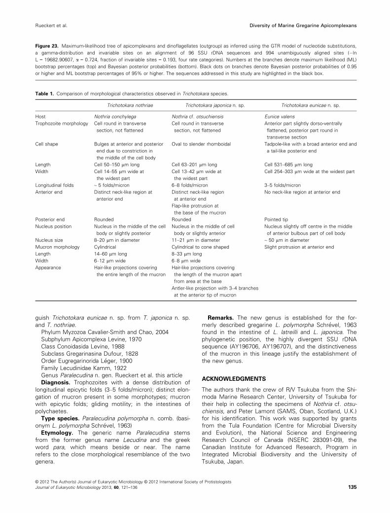
Table 1. Comparison of morphological characteristics observed in Trichotokara species.
Trichotokara nothriae Trichotokara japonica n. sp. Trichotokara eunicae n. sp.
Host Nothria conchylega Nothria cf. otsuchiensis Eunice valens
Trophozoite morphology Cell round in transverse
section, not flattened
Cell round in transverse
section, not flattened
Anterior part slightly dorso-ventrally
flattened, posterior part round in
transverse section
Cell shape Bulges at anterior and posterior
end due to constriction in
the middle of the cell body
Oval to slender rhomboidal Tadpole-like with a broad anterior end and
a tail-like posterior end
Length Cell 50–150 lm long Cell 63–201 lm long Cell 531–685 lm long
Width Cell 14–55 lm wide at
the widest part
Cell 13–42 lm wide at
the widest part
Cell 254–303 lm wide at the widest part
Longitudinal folds ~ 5 folds/micron 6–8 folds/micron 3–5 folds/micron
Anterior end Distinct neck-like region at
anterior end
Distinct neck-like region
at anterior end
Flap-like protrusion at
the base of the mucron
No neck-like region at anterior end
Posterior end Rounded Rounded Pointed tip
Nucleus position Nucleus in the middle of the cell
body or slightly posterior
Nucleus in the middle of cell
body or slightly anterior
Nucleus slightly off centre in the middle
of anterior bulbous part of cell body
Nucleus size 8–20 lm in diameter 11–21 lm in diameter ~ 50 lm in diameter
Mucron morphology Cylindrical Cylindrical to cone shaped Slight protrusion at anterior end
Length 14–60 lm long 8–33 lm long
Width 6–12 lm wide 6–8 lm wide
Appearance Hair-like projections covering
the entire length of the mucron
Hair-like projections covering
the length of the mucron apart
from area at the base
Antler-like projection with 3–4 branches
at the anterior tip of mucron
© 2012 The Author(s) Journal of Eukaryotic Microbiology © 2012 International Society of Protistologists
Journal of Eukaryotic Microbiology 2013, 60, 121–136 135
Rueckert et al. Diversity of Marine Gregarine Apicomplexans
LITERATURE CITED
Berney, C., Fahrni, J. & Pawlowski, J. 2004. How many novel
eukaryotic ‘kingdoms’? Pitfalls and limitations of environmental
DNA surveys. BMC Biol., 2:13.
Bhatia, B. L. & Setna, S. B. 1938. On some gregarine parasites
from certain polychaete worms from the Andaman Islands.
Proc. Indian Acad. Sci., 8B:231–242.Dawson, S. C. & Pace, N. R. 2002. Novel kingdom-level eukary-
otic diversity in anoxic environments. Proc. Natl. Acad. Sci.
USA, 99:8324–8329.Grass�e, P.-P. 1953. Classe des gr�egarinomorphes (Gregarinomor-
pha, N. nov., Gregarinae Haeckel, 1866; gregarinidea Lankester,
1885; gr�egarines des auteurs). In: Grass�e, P.-P. (ed.), Trait�e de
Zoologie. Masson, Paris. p. 590–690.Guindon, S. & Gascuel, O. 2003. A simple, fast, and accurate
algorithm to estimate large phylogenies by maximum likelihood.
Syst. Biol., 52:696–704.Guindon, S., Lethiec, F., Duroux, P. & Gascuel, O. 2005. PHYML
Online-a web server for fast maximum likelihood-based phylo-
genetic inference. Nucleic Acids Res., 1:33.
Heintzelman, M. B. 2004. Actin and myosin in Gregarina polymor-
pha. Cell Motil. Cytoskeleton, 58:83–95.Huelsenbeck, J. P. & Ronquist, F. 2001. MrBayes: Bayesian infer-
ence of phylogenetic trees. Bioinformatics, 17:754–755.Kimura, M. 1980. A simple method for estimating evolutionary
rates of base substitutions through comparative studies of
nucleotide sequences. J. Mol. Evol., 16:111–120.Kuriyama, R., Besse, C., G�eze, M., Omoto, C. K. & Schr�evel, J.
2005. Dynamic organization of microtubules and microtubule-
organizing centers during the sexual phase of a parasitic proto-
zoan, Lecudina tuzetae (Gregarine, Apicomplexa). Cell Motil.
Cytoskeleton, 62:195–209.Landers, S. C. & Leander, B. S. 2005. Comparative surface mor-
phology of marine coelomic gregarines (Apicomplexa, Urospori-
dae): Pterospora floridiensis and Pterospora schizosoma.
J. Eukaryot. Microbiol., 52:23–30.Lankester, E. R. 1866. Notes on the Gregarinida. Trans. Am.
Microsc. Soc., 14:23–28.Leander, B. S. 2008. Marine gregarines - evolutionary prelude to
the apicomplexan radiation? Trends Parasitol., 24:60–67.Leander, B. S., Clopton, R. E. & Keeling, P. J. 2003a. Phylogeny
of gregarines (Apicomplexa) as inferred from small-subunit
rDNA and beta-tubulin. Int. J. Syst. Evol. Microbiol., 53:345–354.
Leander, B. S., Harper, J. T. & Keeling, P. J. 2003b. Molecular
phylogeny and surface morphology of marine aseptate grega-
rines (Apicomplexa): Selenidium and Lecudina. J. Parasitol.,
89:1191–1205.Levine, N. D. 1976. Revision and checklist of the species of the
aseptate gregarine genus Lecudina. Trans. Am. Microsc. Soc.,
95:695–702.Levine, N. D. 1977. Revision and checklist of the species (other
than Lecudina) of the aseptate gregarine family Lecudinidae.
J. Protozool., 24:41–52.Levine, N. D. 1979. New genera and higher taxa of septate grega-
rines (Protozoa, Apicomplexa). J. Protozool., 26:532–536.L�opez-Garcia, P., Rodriguez-Valera, F., Pedros Alio, C. & Moreira,
D. 2001. Unexpected diversity of small eukaryotes in deep-sea
Antarctic plankton. Nature, 409:603–607.Maddison, D. R. & Maddison, W. P. 2000. MacClade 4. Sinauer
Associates, Sunderland.
Perkins, F. O., Barta, J. R., Clopton, R. E., Pierce, M. A. & Upton,
S. J. 2002. Phylum Apicomplexa. In: Lee, J. J., Leedale, G. F. &
Bradbury, P. (eds.), The Illustrated Guide to the Protozoa. Allen
Press, Inc., Lawrence. p. 190–304.Posada, D. & Crandall, K. A. 1998. MODELTEST: testing the
mode l of DNA substitution. Bioinformatics, 14:817–818.Rueckert, S. & Leander, B. S. 2008. Morphology and molecular
phylogeny of Haplozoon praxillellae n. sp. (Dinoflagellata): a
novel intestinal parasite of the maldanid polychaete Praxillella
Pacifica Berkeley. Eur. J. Protistol., 44:299–307.Rueckert, S. & Leander, B. S. 2009. Molecular phylogeny and sur-
face morphology of marine “archigregarines” (Apicomplexa) -
Selenidium spp., Filipodium phascolosomae n. sp. and Platy-
proteum n. gen. et comb. - from North-eastern Pacific peanut
worms (Sipuncula). J. Eukaryot. Microbiol., 56:428–439.Rueckert, S. & Leander, B. S. 2010. Description of Trichotokara
nothriae n. gen. et sp. (Apicomplexa, Lecudinidae) - an intestinal
gregarine of Nothria conchylega (Polychaeta, Onuphidae).
J. Invertebr. Pathol., 104:172–179.Rueckert, S., Chantangsi, C. & Leander, B. S. 2010. Molecular
systematics of marine gregarines (Apicomplexa) from North-
eastern Pacific polychaetes and nemerteans, with descriptions
of three new species: Lecudina phyllochaetopteri sp. nov., Dif-
ficilina tubulani sp. nov., and Difficilina paranemertis sp. nov.
Int. J. Syst. Evol. Microbiol., 60:2681–2690.Rueckert, S., Simdyanov, T. G., Aleshin, V. V. & Leander, B. S.
2011. Identity of a divergent environmental DNA sequence
clade using the phylogeny of gregarine parasites (Apicomplexa)
from crustacean hosts. PLoS ONE, 6:e18163.
Schr�evel, J. 1969. Recherches sur le cycle des Lecudinidae
gr�egarines parasites d’Ann�elides Polych�etes. Protistologica,
5:561–588.Simdyanov, T. G. 2009. Difficilina cerebratuli gen. n., sp. n.
(Eugregarinida: Lecudinidae) - a new gregarine species from the
nemertean Cerebratulus barentsi B€urger, 1895 (Nemertini: Cer-
ebratulidae). Parazitologiya, 43:273–287.Stoeck, T. & Epstein, S. 2003. Novel eukaryotic lineages inferred
from small-subunit rRNA analyses of oxygen depleted marine
environments. Appl. Environ. Microbiol., 69:2657–2663.Stoeck, T., Kasper, J., Bunge, J., Leslin, C., Ilyin, V. & Epstein, S.
2007. Protistan diversity in the Arctic: a case of paleoclimate
shaping modern biodiversity? PLoS ONE, 8:e728.
Takishita, K., Yubuki, N., Kakizoe, N., Inagaki, Y. & Maruyama, T.
2007. Diversity of microbial eukaryotes in sediment at a deep-
sea methane cold seep: surveys of ribosomal DNA libraries
from raw sediment samples and two enrichment cultures.
Extremophiles, 11:563–576.Valigurov�a, A., Michalkov�a, V. & Koudela, B. 2009. Eugregarine
trophozoite detachment from the host epithelium via epimerite
retraction: fiction or fact? Int. J. Parasitol., 39:1235–1242.Vivier, E. 1968. L’organisation ultrastructurale corticale de la greg-
arine Lecudina pellucida; ses rapports avec l’alimentation et la
locomotion. J. Protozool., 15:230–245.Wakeman, K. C. & Leander, B. S. 2012. Molecular Phylogeny of
Pacific Archigregarines (Apicomplexa), Including descriptions of
Veloxidium leptosynaptae n. gen., n. sp., from the Sea Cucum-
ber Leptosynapta clarki (Echinodermata), and two new species
of Selenidium. J. Eukaryot. Microbiol., 59:232–245.WoRMS (2012). Lecudinidae. Accessed through: World Register
of Marine Species. Available at: http://www.marinespecies.org/
aphia.php?p=taxdetails&id=562693 [accessed on 25 December
2012].
© 2012 The Author(s) Journal of Eukaryotic Microbiology © 2012 International Society of Protistologists
Journal of Eukaryotic Microbiology 2013, 60, 121–136136
Diversity of Marine Gregarine Apicomplexans Rueckert et al.


