Oral mucosa - mu-sofia
143
Oral mucosa
Transcript of Oral mucosa - mu-sofia

Oral mucosa


This diagram shows across section through the lip and tooth, showing some of the main features of these structures.

Normal anatomy of the oral mucosa
Oral mucosa covers the entire oral cavity;
Starts from the lips and is adjacent to the skin –Vermillion;
Lined vestibule, alveolar bone, hard and soft palate above, tongue and floor of the mouth below.


Oral mucosa is a part of an oral biological system
mo, food,
air
mo, food, air, saliva
Salivary
glands
Saliva, air, food
tooth
Оral mucosa
МО
IgA-S-
Планктонни клетки
а
МОМОmo
general immunity
local immunityand tolerance
gingival fluid
planktoniccells
gingiva
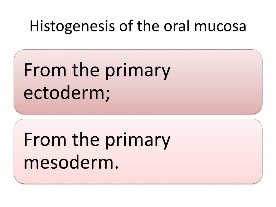
Histogenesis of the oral mucosa
From the primary ectoderm;
From the primary mesoderm.

The oral mucosa is developed around primary mouth
• Oral pit - 4 mo in uteri;
• Oral pocket - between the forebrain and the heart;
• Oropharyngeal membrane - the deepest part of the pocket;
• Rupture of the membrane - 5 mo;
• Primary mouth;
• The ectoderm covers oral cavity into the pharynx and down is covered by endoderma.

Vestibular lamina
• It is separated jaws from the soft tissue;
• Oral ectoderm covers all internal oral structures;
• Begins cell differentiation;
• Basal columnar cells are prepared for constant renewal;
• Folding of a basal layer;
• Strengthening the basal lamina;
• Specialization of the mucosa in the different areas of the mouth.

Oral ectoderm
• With the development of teeth the oral ectoderm is separated from the dental;
• With eruption of the teeth is performed reunion of these two layers;
• It becomes a fusion of the reduced enamel epithelium and oral epithelium;
• Gingival collar is formed of these two layers;• It is formed:• The gingival sulcus;• The epithelial atachman or junctional epithelium

Scheme of an oral mucosa
• multilayered epithelium;
• Basal lamina;
• Papillary folds;
• Lamina propria:
– Lamina lucida;
– Lamina densa;
• Submucosa

Structure of oral mucosa
The oral cavity is lined with stratified epithelium, which is divided into three types:
• Lining mucosa – cover the floor of the mouth and the cheeks, lips, and soft palate;
• Masticatory mucosa – covers the hard palate and alveolar ridges;
• Specialized mucosa – which covers the surfaces of the tongue.

All of the oral mucosa is made up of a thick stratified squamous epithelium, supported by a lamina propria. The epithelium is thick because the epithelial lining of the oral cavity is subject to a lot of wear and tear.

Oral mucosa
• In mobile areas, such as the soft palate, underside of the tongue, floor of the mouth, and mucosal surfaces of the
cheeks and lips, the epithelium is not keratinised.
• In other areas, such as the gums (gingivae), hard palate, and most of the upper surface of the tongue, the epithelium is keratinised.
• Underneath the oral mucosa, there is a tough collagenoussubmucosal layer, with accessory salivary glands, except where the oral mucosa lies over bone, where the submucosa is thin.

Common features of the mucosa
Lamina propria – the connenctive tissue layer immeiately below the epithelium;
It is composed of the:
• Papillary layer – connective tissue extends into pockets in the epithelium –this increases the surface of the epithelium for contact with vascular supply and nerves;
• And deeper reticular layer – contains the deeper plexus of vessels and nerves;
Beneath this zone is the submucosa.

Different types of mucosa
прикрепенаattached
Специализирана -по езика
Free
Specialized

Each type of tissue has structural differences
• The lining mucosa is:
– Soft;
– Pliable;
– Nonkeratinized.

Masticatory mucosa:
• Is keratinized;
• Indicative of the attrition, that take place during mastication.
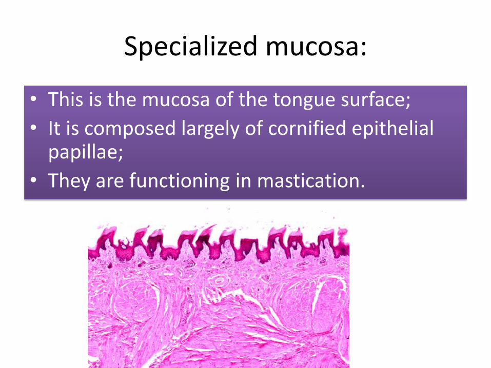
Specialized mucosa:
• This is the mucosa of the tongue surface;
• It is composed largely of cornified epithelial papillae;
• They are functioning in mastication.


Layers of epithelium
• Stratum corneum
• Stratum granulosum
• Stratum spinosum
• Stratum basale (germinativum)

Keratanized epithelium
There is a pronounced papillary layer;
It lacks submucosa;
You can see the 4 epithelial layers.


Basal layer
Their nuclei are irregulary oval and exhibit numerous mitotic figures as they undergo constant cell division;
This basal cells gradually migrate to the surface of the mucosa.

Cells of the squamous epithelium
The basal cells gradually migrate to the surface of the mucosa and undergoes changes.

Cell recovery
The cells of the oral epithelium are recovering for 1 to 2 weeks:
The cells of the attached epithelium - for 1 week;
Cells of the marginal gingiva - 2 weeks;
They contain within itself proliferative department implementing rapid division;
New cells are packed with tonofilaments which are attached to the basal lamina.

It is so named because the cells contain many keratohyalingrannules;

This soft keratin may be compared with hard keratin of the nails and hair;
Keratin is tough, nonliving material that is resistant to friction and impervious to bacterial invasion.


Cell sizes in the different layers of the gingival epithelium

• To permit cell movement and loss of individual cells along the surface, the superficial layers have surface interdigitations rather than desmosomes;
• This cells are continually becoming lost and replaced by cells of the underlying layers.

Movement of the epithelial cells
• As each cell moves to the surface of the epithelium, it does so by means of cells attachments to neighboring cells that hold until the cell has reached a specific stage of development;
• When that stage occurs, the cell attachment releases, which allows that cell to move to a higher level where it reattaches.

Basal lamina
• Basal cells interface with a membrane separating the epithelium and connective tissue;
• This membrane is called the basal lamina;
• The basal cells are attached to the basal lamina by minute disks termed hemidesmosomes;
• These thickenings of the cell membrane are supported by filaments from within the cells, anchoring fibrils that attach the basal lamina and the cells to the collagen fibers of the lamina propria.
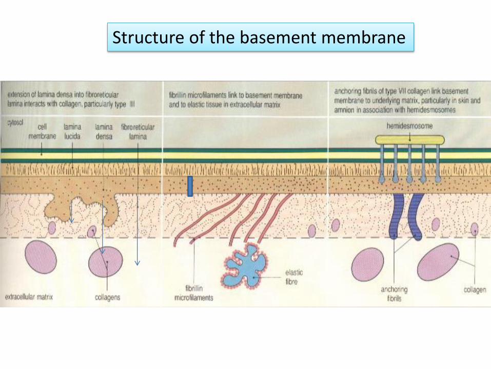
Structure of the basement membrane

Basal lamina

The intracellular binding of the epithelial cells
Cohesion - by desmosoms;
Adhesion – by hemidesmosomes;
Focal contacts - by transmembrane proteins;
Gap-links - produce holes between cells;
Functional elements of a certain type of relationship is called connecting complex

Cohesion - by desmosoms

Adhesion – by hemidesmosomes

Focal contacts - by transmembraneproteins

Gap-links - produce holes between cells


Mucosal connective tissue
Connective tissue supports other tissues and organs;
It provides a blood supply, innervation and immune protection of the oral mucosa;
Cells;
It is composed of:
Cells;Intercellular substance - glykoaminoglikans;
Fibers ;
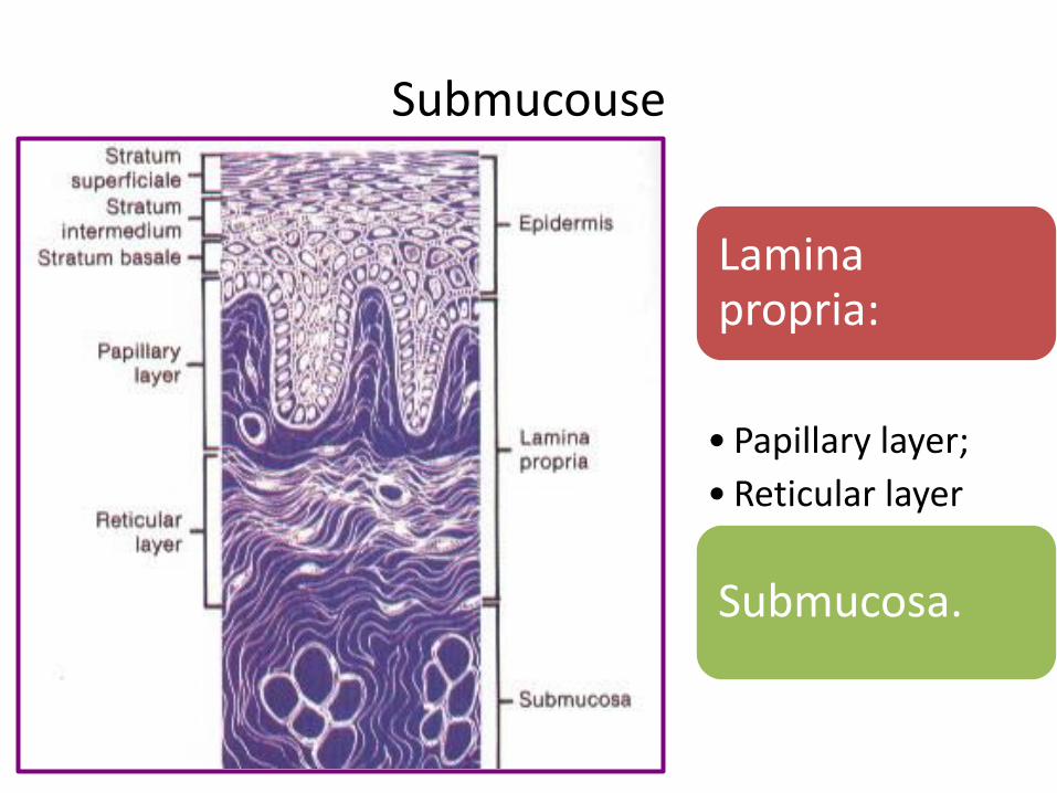
Submucouse
Laminapropria:
• Papillary layer;
• Reticular layer
Submucosa.

collagen fibers
consist of fibrils (0.2 to 0.5 μm);
They are built of parallel microfiber = (50nm);
Molecular structure of microfibres is tropokolagen;Composed of 3 polypeptide chains, which are dominated by the amino acids glycine, proline, hydroxyproline and hydroxylysine.

Collagen fibers

reticular fibers
• They are argirofil fibers which are consisting of:
– 5% collagen and 15% lipids;
• The main ingredient in them is reticulin;
• They are connecting with endothelium and provide a static position of blood vessels.

elastic fibers
Very thin - 0.1-0.2 μm;
Forming anastomoses with each other and have a frilly edge;
They are built of elastin, which is not destroyed by proteolytic enzymes;
In the free mucosa elastic fibers are few.

Masticatory mucosa
• It covers:– The gingiva;
– The hard palate;
• It is adapted to chewing forces;
• It is keratinized;
• There are sharp relief;
• It possesses strongly folded connective tissues papillae;
• It has a small amount of submucosa.

Masticatory mucosa
• This mucosa is ticker than the nonkeratinizedsurface of flat, hornified cells offering resistance to attrition;
• The basal and intermediate stratum layers (spinosum) are the same as those of nonkeratinized epithelium.

Degree of keratinization
nonkeratinized
keratinized
Specialized with areas
with a different
keratinization

Gingiva and epithelial attachment
• The gingiva surrounded the necks of the teeth and extends apically to the mucogingivaljunction;
• The gingiva develops as a coalescence of the oral and reduced enamel organ epithelium when the tooth first emerges into oral cavity.

• The reduced enamel organ epithelium makes contact with the undersurface of the oral epithelium, and the two fuse;
• Then the tooth penetrates this combined layer to enter the mouth and produces the gingiva as the epithelium continues to separate from enamel surface until of the teeth is reached;
• At this point, the gingiva covers only the cervical area of the enamel where it is attached.

Free and attached gingiva
• The free gingiva is bound on its inner margin by the gingival sulcus, which separates it from the tooth;
• On its outer margin by the oral cavity;
• And apically at its free surface by the free gingival groove.

Free and attached gingiva

• Free gingiva (or marginal gingiva) - It is thatpart of the oral mucosa that surrounds thenecks of the teeth and forms the free margin of the gingival tissue;
• It is differentiated apically from the attached gingival by the free gingival groove.
• The inner side of it forms the gingival sulcus.
• The free gingival mucosa is composed of stratified squamous epithelium that may be keratinized, parakeratinazed or sometimes nonkeratonized.


Mucogingival junction
• The attached gingiva lies adjacent to the free gingiva and is separated from the alveolar mucosa by the mucogingival junction;
• The free and attached gingiva are keratinized, but the alveolar mucosa is not;
• The attached gingiva is stippled, but the free gingiva has a smooth surface;
• In some instances, the free gingiva may be covered with parakeratinized mucosa (presence of nuclei in the cells of the surface layer.


Junctionalepithelium
• It provides attachment for the gingiva to the tooth in the cervical area and forms the epithelium-lined floor of the gingival sulcus;

Junctional epithelium
• Junctional epithelium forms the seal of the gingival epithelium and the tooth.
• It forms the floor of the gingival sulcus andextends apically to the enamel of the tooth.
• Disturbances of epithelial attachment results in deepening of the sulcus which is a sign of gingival ot periodontal disease.

Cells of the attached epithelium
• They are cytologically different from the gingival epithelium;
• They have fewer desmosomes;
• This indicated a higher rate of turnover then occurs in the other gingival epithelial cells;
• This cells are turn over in approximately 6 days.
Sulcus epithelium
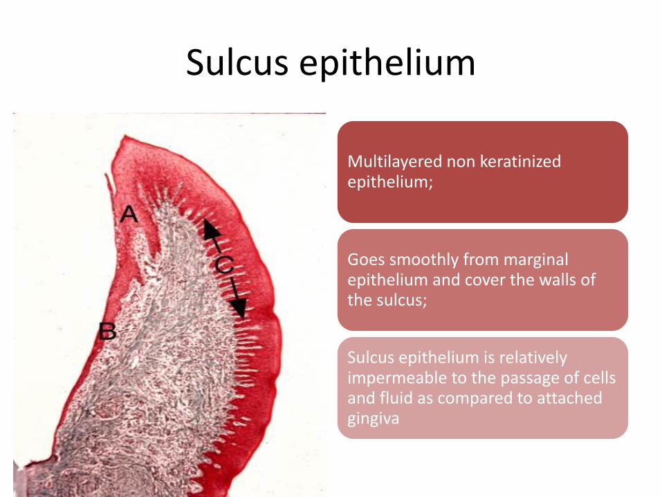
Multilayered non keratinized epithelium;
Goes smoothly from marginal epithelium and cover the walls of the sulcus;
Sulcus epithelium is relatively impermeable to the passage of cells and fluid as compared to attached gingiva

• Stratum basale cells also contain hemidesmosomes, the mechanism for the attachment of cells to salivary protein layer, which covers the cervical area of the enamel;
• Disturbance of this attachment results in a deepening of the gingiva.


Junctional epithelium
Outside is in contact with the tooth, and the inside – with connective tissue of the gingiva;
Apically it reaches the bottom of the sulcus;
Formed epithelial cuff around the tooth - the fat in the bottom of the sulcus -10 - 30 cells;
Apically is thinning to a few cells.

marginal gingiva


Attached gingiva
• The attached gingiva lies between the free gingival groove and the alveolar mucosa;
• The junction of the attached gingiva and the alveolar mucosa is called mucogingival junction;
• In healthy mouth attached gingiva shows stippling (orange-peel appearance) which is a characteristic of this type of mucosa.
Mucogingival junction


Interdental papilla and col
• Gingiva located between the teeth and extending high on the interproximal area of the crowns on the labial and kingual surfaces is known as the interdental papilla;


Interdental papilla
• Interdental papillae are those pars of gingival tissue that appear in between teeth apical to the contact points.
• Interdental grooves extend vertically between the interdental papilla corresponding to the depression in the interdental papilla called COL;
• The depression lies in the facial and lingual plane.

Positional relationship of the col in the health and disease1. The col is accentuated in inflammation;
2. The col is found to be pointed in anterior teeth and flat or concave posteriorly;
3. The contact point on each crown is represented by an oval above the col.

The col
• The junctional epithelium of this zone is known as the col;
• The col is characterized as a thin, nonkeratinizedepithelium;
• The col is more inclined in a peak between anterior teeth and more flattened or concave between posterior teeth;
• When the interproximal gingiva becomes inflamed, the col is exaggerated and is positioned higher on the neck of the tooth.
Hard palate
• The roof of the mouth is hard palate;
• It is covered with keratinazed sratified squamousepithelium;
• This epithelium is similar to that of the gingiva in the middling area, where there is no submucosa;
• The midline is known as the median raphe;
• Anteriorly an incisive papilla can be seen;
• On each side of the median raphe are ridges of tissue called rugae.


Palatal mucosa
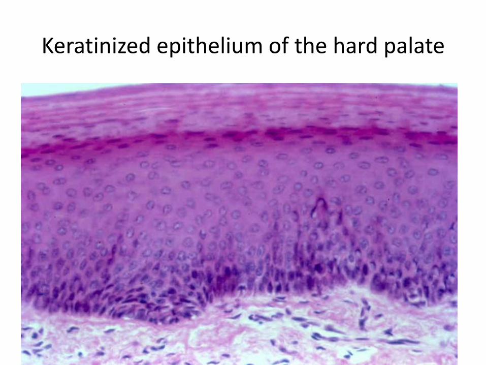
Keratinized epithelium of the hard palate

Stratum granulosum of the hard palate

Sloughed epithelial cells
The surface of the epithelium is a subject to constant renewal


Lining mucosa
• It is composed of a thin layer of epithelium and an underlying lamina propria;
• The epithelium is composed of:
– A basal layer – stratum basale;
– Stratum spinosum or str. Intermedium;
– Stratum superficial with flattened cells and many containing small oval nuclei;
– form the non keratinized epithelium.

Non keratinized epithelium
It is situated:
• At the floor of the mouth
• Transitional fold;
• Gingival sulcus;
• The lower surface of the tongue;
Surface epithelial layer is denser

Types of lining mucosa
• Subdivided into mucosa of the:
– lips;
– Soft palate;
– Cheeks;
– Floor of the mouth
– Ventral surface of the tongue;
Lips
• The inner oral surface of the lips is lined with moist surface, stratified squamous cells, and non keratinized epithelium;
• It is associated with small, round seromucousglands of the lamina propria;
• They are part of the minor salivary glands found throughout the oral cavity;
• Beneath the lamina propria is submucosa with the fibers of the orbicularis oris muscle.

Inner oral surface of the lips is lined with non keratinized epithelium
• It is associated with small, round seromucous glands of the lamina propria;
• Beneath the lamina propria is submucosa, in which fibers of the orbicularis oris muscle are located.

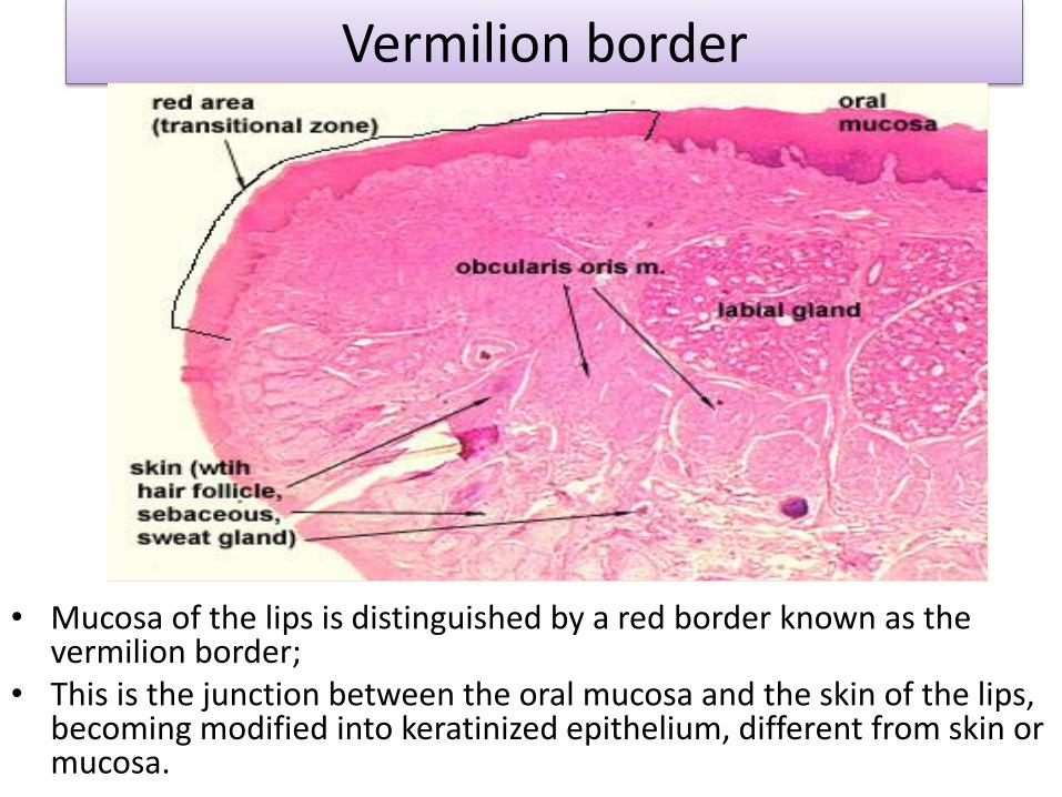
Vermilion border
• Mucosa of the lips is distinguished by a red border known as the vermilion border;
• This is the junction between the oral mucosa and the skin of the lips, becoming modified into keratinized epithelium, different from skin or mucosa.
There are tree reasons that the vermilion is red:
• The epithelium is thin;
• This epithelium contains eleidin, which is transparent;
• The blood vesels are near the surface of the papillary layer, revealing the red blood cells` color.

Floor of the mouth
• Non keratinized mucous membrane covers the floor of the mouth and appears attached to the lamina propria;
• The mucosa is firmly attached floor of the mouth are minor salivary glands;
• And the right and left major mucous glands – the sublingual glands.


Soft palate
• Lining mucosa of the soft palat is highly vascularized and more pink than the mucosa of the keratinized epithelium of the hard palate;
• Lamina propria contains many small blood vessels;
• Beneath the connective tissue of lamina propria is the submucosa, which contains muscles and mucous glands.
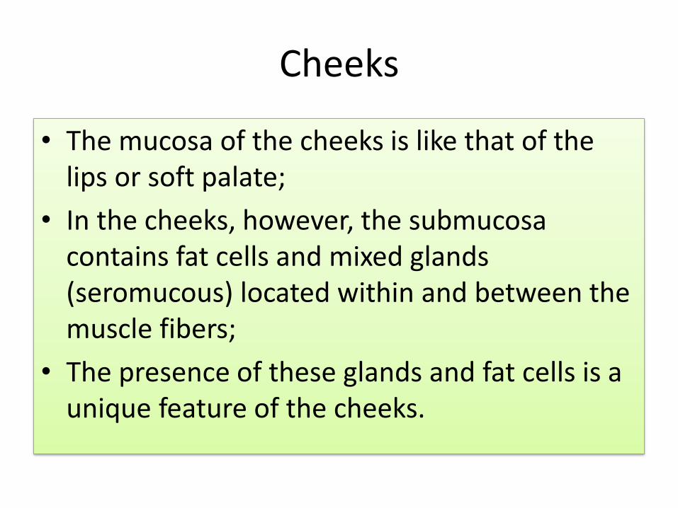
Cheeks
• The mucosa of the cheeks is like that of the lips or soft palate;
• In the cheeks, however, the submucosacontains fat cells and mixed glands (seromucous) located within and between the muscle fibers;
• The presence of these glands and fat cells is a unique feature of the cheeks.


The tongue

The tongue
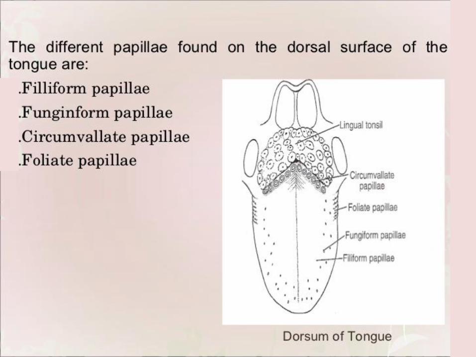
Dorsal surface of the tongue;
Lower surface of the tongue
The 2/3 of the front of the tongue - mobile part of the tongue.
It is situated in front of the line of papillae circumvallate
The root of the tongue – last 1/3.


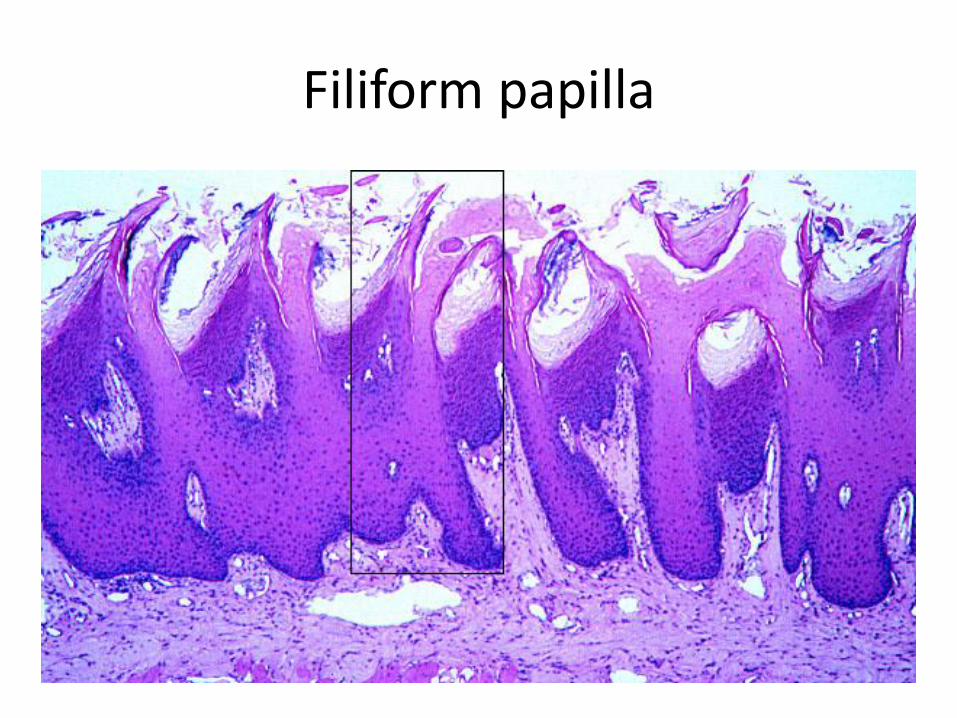
Filiform papilla
They are covering the entire front surface of the tongue;
They create conditions for retention of food and microorganisms;
Involved in the fragmentation of the food in pressure on the palate.


Filiform papilla
Filiform papilla

Papillae foliate, muscles, blood vessels

Fungiform papillae
Red, raised structures;
Covered with nonkeratinized epithelium;
Are scattered between filliform papilla;
They have taste buds at the surface.


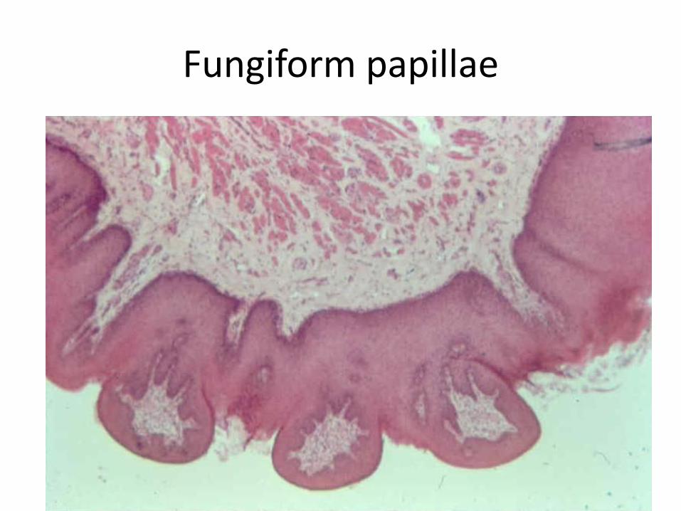
Fungiform papillae


Cirumvallate papillae
V-shaped

Circumvallate papilla with the taste buds

Pappilae circumvallate
They are in front of the terminal sulcus
They are 8 to 12 major papilla
Each papilla is surrounded by a deep groove
Inside of the sulcus is poured ductusof small serous salivary glands;
Lateral walls of the papillae have taste buds.

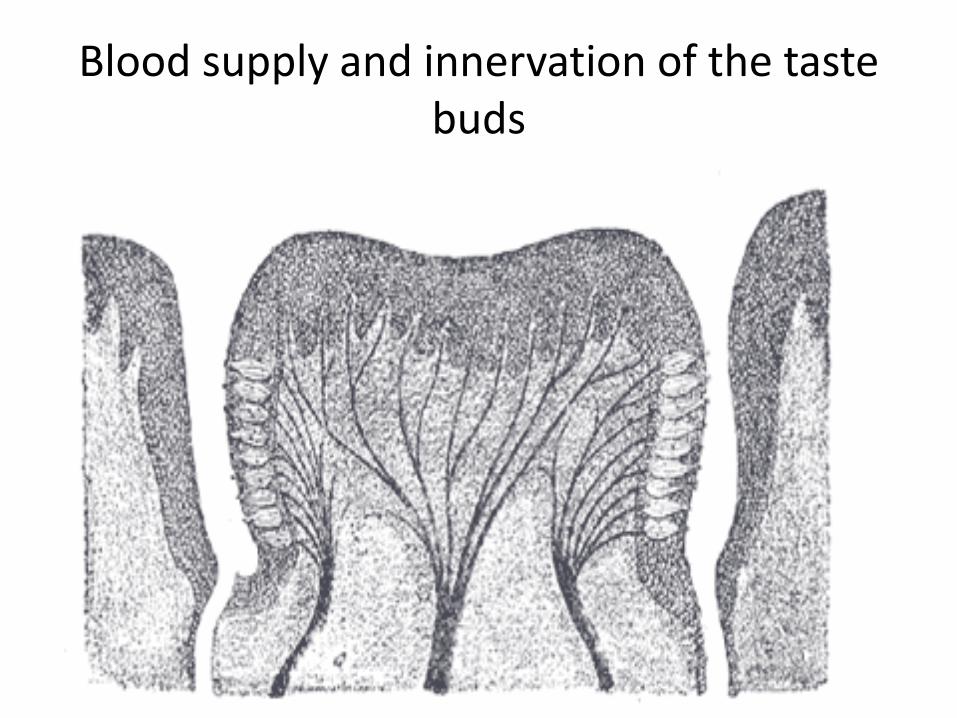
Blood supply and innervation of the taste buds

Taste buds in circumvallate papilla

Pores of taste buds


Foliate papiillae
Represent 4 to 11 parallel ridge;
Separated by deep grooves;
They are situated at along side edge of the back of the tongue;
They have a taste buds.

Foliate papillae
At the back of the tongue
Behind circumvallatepapilla.

lingual tonsils
Behind circumvallatpapillae;
They are part of Valder`soropharyngeal ring;
They are oval and convex, coupled with lingual crypts;
They are covered with nonkeratinized epithelium

Lingual tonsils


Plica fimbriata
A linear projection of the ventral surface of the tongue;
Sometimes is brown.

The tongue with a deep fissures
The fissures on the back of the tongue are normal structure;
There are individual differences in the number, location and their depth.

Features of the child mucosa
• There is a thin layer;
• A bright red because of a thin epithelium;
• Greater mitotic activity;
• Less keratinized;
• More fragile;
• With better regenerative capabilities;
• Immature child immunity affects the local and general, specific and nonspecific immune mechanisms.