Oral infusion of pomegranate fruit extract inhibits - Carcinogenesis
8
Carcinogenesis vol.33 no.3 pp.644–651, 2012 doi:10.1093/carcin/bgr308 Advance Access publication December 22, 2011 Oral infusion of pomegranate fruit extract inhibits prostate carcinogenesis in the TRAMP model Vaqar Mustafa Adhami y , Imtiaz Ahmad Siddiqui y , Deeba N.Syed, Rahul Kumar Lall and Hasan Mukhtar Department of Dermatology, School of Medicine and Public Health, University of Wisconsin, Madison, WI 53706, USA To whom correspondence should be addressed. Department of Dermatology, School of Medicine and Public Health, University of Wisconsin, Medical Sciences Center, 4385, 1300 University Avenue, Madison, WI 53706, USA. Tel: þ1 608 263 3927; Fax: þ1 608 263 5223; Email: [email protected] We earlier provided evidence that oral consumption of pomegran- ate fruit extract (PFE) inhibits prostate cancer (PCa) cell growth in nude mice. To ascertain convincing evidence of chemopreven- tive effects of PFE against PCa, its efficacy requires to be evalu- ated in animal models that closely emulate human disease. Here, we provide evidence of remarkable tumor growth inhibitory effects of PFE using the TRAMP model. Mice received 0.1 and 0.2% PFE, equivalent to 250 and 500 ml of pomegranate juice, in drinking water, starting at 6 weeks and examined at 12, 20 and 34 weeks of age. In water-fed group, 100% mice developed palpable tumors by 20 weeks compared with only 30 and 20% in the 0.1 and 0.2% PFE-supplemented groups, respectively. At 34 weeks, palpable tumors were observed in 70 of 0.1% and only 50 of 0.2% PFE-supplemented mice. Compared with median survival of 43 weeks in water-fed mice, 0.1 and 0.2% PFE- supplemented mice exhibited median life expectancy of 73 and 92 weeks, respectively. Compared with respective water-fed groups, none of the mice in PFE-supplemented groups exhibited metastases to any of the distant organs at 20 weeks and only 20% mice exhibited metastasis at 34 weeks of age. Many of the PFE- supplemented animals had multiple foci of well-differentiated carcinoma but no evidence of poorly differentiated carcinoma. PFE supplementation resulted in simultaneous and significant inhibition of IGF-I/Akt/mTOR pathways in the prostate tissues and tumors. We suggest that pomegranate juice be evaluated in clinical trials in patients at high risk for developing PCa. Introduction According to recent estimates, prostate cancer (PCa) continues to remain the most common cancer and the second leading cause of cancer-related deaths in American males (1) with similar trends in many other western countries. In the year 2011, a total of 240 890 men are estimated to be diagnosed with PCa and 33 720 PCa-related deaths are predicted in the USA alone (1). The success of the PCa prevention trials strongly suggests that the disease can be prevented by the use of non-toxic pharmacological agents (2,3). In fact, in recent years, there is great interest in developing chemopreventive strategies against PCa and more and more people are turning to the use of complementary and alternative approaches for prevention of this disease (4,5). Because of its age association and long latency, PCa represents an ideal candidate disease for chemoprevention and any modest delay achieved through pharmacological intervention could result in sub- stantial reduction in the incidence of clinically detectable disease (6). Pomegranate (Punica granatum), grown mainly in the Mediterra- nean region for its edible fruit, possesses many medicinal properties, such as being antioxidant and anti-inflammatory (6–9). The unique biochemical composition of the pomegranate fruit, being rich in an- tioxidant tannins and flavonoids, has drawn attention of many inves- tigators to study its exceptional healing qualities (10). The antioxidant activity of flavonoids obtained from pomegranate juice was observed to be close to that of green tea and significantly greater than red wine (11,12). Recent research has shown that pomegranate extracts selec- tively inhibit the growth of breast, prostate, colon, leukemia and lung cancer cells in culture (13–25). In preclinical animal studies, oral consumption of pomegranate extract inhibited growth of lung, skin, colon and prostate tumors (26–36). An initial phase II clinical trial of pomegranate juice in patients with PCa reported significant prolon- gation of prostate-specific antigen doubling time (37). Pomegranate juice ingredients have been found to be bioavailable in human serum also found to localize in prostate tissues in mice (19). This observation could suggest that many of the effects of pomegranate juice could directly affect pathways within the prostate. We have previously demonstrated that pomegranate fruit extract (PFE) possesses antiproliferative and proapoptotic properties against human PCa cells (35). We also demonstrated that oral administration of PFE to athymic nude mice implanted with androgen-sensitive CWR22Rm1 cells resulted in a significant inhibition in tumor growth with concomitant decrease in serum prostate-specific antigen levels (35). For a comprehensive evaluation of agents’ cancer chemopreven- tive properties, it is essential that the compound be tested in more than one preclinical model before recommending its evaluation in humans. Therefore, in the present study, we tested the effects of PFE against PCa development using a classical chemoprevention protocol in the transgenic TRAMP mouse model. We observed significant inhibition of PCa growth and progression. Oral supplementation of PFE inhibited metastasis and increased overall survival possibly through inhibition of multiple pathways most notably the IGF-I/Akt/mTOR pathways. Materials and methods Animals Male C57BL/6 and female heterozygous C57BL/TGN TRAMP mice, Line PB-Tag 8247NG, were purchased as breeding pairs from The Jackson Labo- ratory (Ann Arbor, MI). Transgenic males for these studies were routinely obtained as [TRAMP C57BL/6] F 1 or as [TRAMP C57BL/6] F 2 offspring. Identity of transgenic mice was established by the PCR-based DNA screening as described previously (38). The animals were bred and maintained at the AAALAC-accredited Animal Resource Facility of University of Wisconsin. Housing and care of the animals was approved by the University’s Research Animal Resource Committee in accordance with the NIH Guidelines for the Care and Use of Laboratory Animals. Study design and PFE supplementation Six-week-old TRAMP mice were randomly divided into three groups. The first group of animals received normal drinking water and served as controls. The animals of groups 2 and 3 received the same drinking water supplemented with 0.1 and 0.2% PFE (wt/vol), respectively. As described previously (33), fresh pomegranates were peeled, and the edible portion was squeezed in 70% acetone/30% distilled water (1:20, wt/vol). The red extract was filtered through Whatman no. 1 filter paper, and the filtrate was condensed, freeze-dried and stored at 4°C. The extract as analyzed by matrix-assisted laser desorption/ ionization-time of flight mass spectrometry was found to contain anthocyanins, various ellagitannins and hydrolyzable tannins (33). Water bottles were changed every other day. The 0.1 and 0.2% doses of PFE selected were based on the assumption that a typical healthy individual (70 kg) may be persuaded to drink 250 or 500 ml of pomegranate juice extracted from one or two fruits, respectively (35). Body weight, diet and water consumption were recorded weekly throughout the study. PFE-supplemented water was provided to mice beginning at 6 weeks of age and was continued until the animals were 34 weeks old, at which time the experiment was terminated. Throughout the experiment, the animals had access to chow diet ad libitum. For each experiment, 30 male Abbreviations: AMPK, AMP-activated protein kinase; GU, genitourinary; PCa, prostate cancer; PFE, pomegranate fruit extract; PI3K, phosphoinositide 3-kinase. y These authors contributed equally to this work. Ó The Author 2011. Published by Oxford University Press. All rights reserved. For Permissions, please email: [email protected] 644 Downloaded from https://academic.oup.com/carcin/article/33/3/644/2463958 by guest on 05 December 2021
Transcript of Oral infusion of pomegranate fruit extract inhibits - Carcinogenesis
Carcinogenesis vol.33 no.3 pp.644–651, 2012doi:10.1093/carcin/bgr308Advance Access publication December 22, 2011
Oral infusion of pomegranate fruit extract inhibits prostate carcinogenesis in theTRAMP model
Vaqar Mustafa Adhamiy, Imtiaz Ahmad Siddiquiy,Deeba N.Syed, Rahul Kumar Lall and Hasan Mukhtar�
Department of Dermatology, School of Medicine and Public Health,University of Wisconsin, Madison, WI 53706, USA
�To whom correspondence should be addressed. Department of Dermatology,School of Medicine and Public Health, University of Wisconsin, MedicalSciences Center, 4385, 1300 University Avenue, Madison, WI 53706, USA.Tel: þ1 608 263 3927; Fax: þ1 608 263 5223;Email: [email protected]
We earlier provided evidence that oral consumption of pomegran-ate fruit extract (PFE) inhibits prostate cancer (PCa) cell growthin nude mice. To ascertain convincing evidence of chemopreven-tive effects of PFE against PCa, its efficacy requires to be evalu-ated in animal models that closely emulate human disease. Here,we provide evidence of remarkable tumor growth inhibitoryeffects of PFE using the TRAMP model. Mice received 0.1 and0.2% PFE, equivalent to 250 and 500 ml of pomegranate juice, indrinking water, starting at 6 weeks and examined at 12, 20 and34 weeks of age. In water-fed group, 100% mice developedpalpable tumors by 20 weeks compared with only 30 and 20%in the 0.1 and 0.2% PFE-supplemented groups, respectively. At34 weeks, palpable tumors were observed in 70 of 0.1% and only50 of 0.2% PFE-supplemented mice. Compared with mediansurvival of 43 weeks in water-fed mice, 0.1 and 0.2% PFE-supplemented mice exhibited median life expectancy of 73 and92 weeks, respectively. Compared with respective water-fedgroups, none of the mice in PFE-supplemented groups exhibitedmetastases to any of the distant organs at 20 weeks and only 20%mice exhibited metastasis at 34 weeks of age. Many of the PFE-supplemented animals had multiple foci of well-differentiatedcarcinoma but no evidence of poorly differentiated carcinoma.PFE supplementation resulted in simultaneous and significantinhibition of IGF-I/Akt/mTOR pathways in the prostate tissuesand tumors. We suggest that pomegranate juice be evaluated inclinical trials in patients at high risk for developing PCa.
Introduction
According to recent estimates, prostate cancer (PCa) continues toremain the most common cancer and the second leading cause ofcancer-related deaths in American males (1) with similar trends inmany other western countries. In the year 2011, a total of 240 890 menare estimated to be diagnosed with PCa and 33 720 PCa-related deathsare predicted in the USA alone (1). The success of the PCa preventiontrials strongly suggests that the disease can be prevented by the use ofnon-toxic pharmacological agents (2,3). In fact, in recent years, thereis great interest in developing chemopreventive strategies against PCaand more and more people are turning to the use of complementaryand alternative approaches for prevention of this disease (4,5).Because of its age association and long latency, PCa represents anideal candidate disease for chemoprevention and any modest delayachieved through pharmacological intervention could result in sub-stantial reduction in the incidence of clinically detectable disease (6).
Pomegranate (Punica granatum), grown mainly in the Mediterra-nean region for its edible fruit, possesses many medicinal properties,
such as being antioxidant and anti-inflammatory (6–9). The uniquebiochemical composition of the pomegranate fruit, being rich in an-tioxidant tannins and flavonoids, has drawn attention of many inves-tigators to study its exceptional healing qualities (10). The antioxidantactivity of flavonoids obtained from pomegranate juice was observedto be close to that of green tea and significantly greater than red wine(11,12). Recent research has shown that pomegranate extracts selec-tively inhibit the growth of breast, prostate, colon, leukemia and lungcancer cells in culture (13–25). In preclinical animal studies, oralconsumption of pomegranate extract inhibited growth of lung, skin,colon and prostate tumors (26–36). An initial phase II clinical trial ofpomegranate juice in patients with PCa reported significant prolon-gation of prostate-specific antigen doubling time (37). Pomegranatejuice ingredients have been found to be bioavailable in human serumalso found to localize in prostate tissues in mice (19). This observationcould suggest that many of the effects of pomegranate juice coulddirectly affect pathways within the prostate.
We have previously demonstrated that pomegranate fruit extract(PFE) possesses antiproliferative and proapoptotic properties againsthuman PCa cells (35). We also demonstrated that oral administrationof PFE to athymic nude mice implanted with androgen-sensitiveCWR22Rm1 cells resulted in a significant inhibition in tumor growthwith concomitant decrease in serum prostate-specific antigen levels(35). For a comprehensive evaluation of agents’ cancer chemopreven-tive properties, it is essential that the compound be tested in more thanone preclinical model before recommending its evaluation in humans.Therefore, in the present study, we tested the effects of PFE againstPCa development using a classical chemoprevention protocol in thetransgenic TRAMP mouse model. We observed significant inhibitionof PCa growth and progression. Oral supplementation of PFE inhibitedmetastasis and increased overall survival possibly through inhibitionof multiple pathways most notably the IGF-I/Akt/mTOR pathways.
Materials and methods
Animals
Male C57BL/6 and female heterozygous C57BL/TGN TRAMP mice, LinePB-Tag 8247NG, were purchased as breeding pairs from The Jackson Labo-ratory (Ann Arbor, MI). Transgenic males for these studies were routinelyobtained as [TRAMP � C57BL/6] F1 or as [TRAMP � C57BL/6] F2 offspring.Identity of transgenic mice was established by the PCR-based DNA screeningas described previously (38). The animals were bred and maintained at theAAALAC-accredited Animal Resource Facility of University of Wisconsin.Housing and care of the animals was approved by the University’s ResearchAnimal Resource Committee in accordance with the NIH Guidelines for theCare and Use of Laboratory Animals.
Study design and PFE supplementation
Six-week-old TRAMP mice were randomly divided into three groups. The firstgroup of animals received normal drinking water and served as controls. Theanimals of groups 2 and 3 received the same drinking water supplementedwith 0.1 and 0.2% PFE (wt/vol), respectively. As described previously (33),fresh pomegranates were peeled, and the edible portion was squeezed in 70%acetone/30% distilled water (1:20, wt/vol). The red extract was filtered throughWhatman no. 1 filter paper, and the filtrate was condensed, freeze-dried andstored at 4�C. The extract as analyzed by matrix-assisted laser desorption/ionization-time of flight mass spectrometry was found to contain anthocyanins,various ellagitannins and hydrolyzable tannins (33). Water bottles werechanged every other day. The 0.1 and 0.2% doses of PFE selected were basedon the assumption that a typical healthy individual (70 kg) may be persuaded todrink 250 or 500 ml of pomegranate juice extracted from one or two fruits,respectively (35). Body weight, diet and water consumption were recordedweekly throughout the study. PFE-supplemented water was provided to micebeginning at 6 weeks of age and was continued until the animals were 34 weeksold, at which time the experiment was terminated. Throughout the experiment,the animals had access to chow diet ad libitum. For each experiment, 30 male
Abbreviations: AMPK, AMP-activated protein kinase; GU, genitourinary;PCa, prostate cancer; PFE, pomegranate fruit extract; PI3K, phosphoinositide3-kinase.
yThese authors contributed equally to this work.
� The Author 2011. Published by Oxford University Press. All rights reserved. For Permissions, please email: [email protected] 644
Dow
nloaded from https://academ
ic.oup.com/carcin/article/33/3/644/2463958 by guest on 05 D
ecember 2021
TRAMP mice of 6 weeks of age were divided into three equal groups of 10mice. At the termination of the experiment, blood was collected from the retro-orbital plexus under anesthesia from both experimental and control groups.The genitourinary (GU) apparatus consisting of bladder, urethra, seminalvesicles, ampullary gland and the prostate was excised, removed and weighed.The prostate gland was then separately excised using a dissecting microscope.The wet weight of GU apparatus was recorded to the nearest 0.01 g. In a sep-arate experiment, to investigate the effect of PFE supplementation on growth ofpalpable tumors and overall survival, 36 male TRAMP mice 6 weeks of agewere equally divided into three groups. The control group of animals wasprovided with drinking water, whereas animals in the experimental group re-ceived PFE-supplemented water throughout the protocol. Animals in all groupswere observed weekly for body weight, tumor progression by abdominal pal-pation and survival. For survival analyses, animals were allowed to remain inthe protocol until they died or were moribund. Moribund animals were hu-manely killed and considered censored for statistical purposes.
Magnetic resonance imaging
Five animals each from control and experimental groups were randomly se-lected and monitored for tumor growth and volume by magnetic resonanceimaging at 12, 20 and 34 weeks of age. Imaging in these animals was per-formed by using a whole-body Varain 4.5 Tesla imager (Varian, Inc. MagneticResonance Systems, Palo Alto, CA) horizontal bore imaging/spectroscopysystem (7.0 cm ID) equipped with isoflurane gas anesthesia system. To identifyprostate gland, urinary bladder was used as the reference point since the glandis located anatomically inferior and posterior to the bladder, encircling urethra(39). Tumor volumes were determined by image segmentation by manuallyoutlining tumor boundaries in parallel slices separated by 50 lM in the two-dimensional images. The areas of the outlined contours were summed andmultiplied by the interslice distance to compute tumor volume.
Ultrasound imaging
Ultrasound imaging was performed using the Vevo 770 microimaging system(Visualsonics, Ontario, Canada) with a 30 MHz transducer for three-dimensionalimage visualization and advanced analysis software. Mice were anesthetized byinhalation of isoflurane with 1–3% oxygen and ventral hair was removed usinga mild depilatory cream. Mice were placed on a thermostatically controlledheating pad to help maintain mouse body temperature. Orientation of the prostategland on ultrasound was accomplished by visualizing the bladder, which is lessechogenic and appears as a dark area.
Metastasis examination
Metastasis to lymph nodes, lungs and the liver was observed under the micro-scope. The India ink method was used to examine cancer metastasis to lungs.Animals from control and treated groups were killed and the respiratory systemwas excised. India ink was injected through the trachea into the lungs untilcompletely filled. The trachea was then tied with a thread. The ink fills normalrespiratory structures and metastases stand out as unstained regions.
Histology and immunostaining
The dorsal and lateral prostates were excised, weighed and a small portion wasfixed overnight in (10%) zinc-buffered formalin and then transferred to 70%ethanol. The sections were stained with hematoxylin and eosin. Histologicalsections were reviewed by light microscopy for the presence of PCa andclassified as prostate intraepithelial neoplasia, PIN (epithelial stratificationwith occasional mitotic figures or cribriform pattern), well differentiated(multiple epithelial mitotic figures and apoptotic bodies, invasive glands withstromal hypercellularity), moderately differentiated (many acini completelyfilled with tumor yet still forming some glandular structures) or poorly differ-entiated (sheets of malignant cells with little or no gland formation) PCaor atrophic glands only (no identifiable tumor). For immunostaining, sections(4–6 lm) were cut from paraffin-embedded tissue and mounted on slides.Immunostaining was performed using specific antibodies with appropriatedilutions and was replaced with either normal host serum or block for negativecontrols, followed by staining with appropriate biotin-conjugated secondaryantibodies followed by incubation with avidin–biotin conjugate. The slideswere developed in diaminobenzidine and counter stained with a weak solutionof hematoxylin stain. The stained slides were dehydrated and mounted inpermount and visualized on a Zeiss-Axiophot DM HT microscope (Zeiss-Axiophot, Germany). Images were captured with an attached camera linkedto a computer.
Immunoblot analysis
The dorsolateral prostate was removed from both treated and control groups,homogenized in lysis buffer to prepare cell lysates. The protein concentrationwas determined by BCA method and following the manufacturer’s protocol(Pierce, Rockford, IL). Appropriate amount of protein (25–50 lg) was resolved
over 8–14% Tris-glycine polyacrylamide gel and then transferred onto thenitrocellulose membrane. The blots were blocked using 5% non-fat dry milkand probed using appropriate primary antibodies: T-antigen (Santa Cruz Bio-technology, Santa Cruz, CA), Akt, pAkt (Ser473 and Thr308), phosphoinositide3-kinase (PI3K) (p85), p-mTOR (Ser2448), p-p70S6K, p-AMPKa (Thr172),IGF-I, rictor and raptor obtained from Cell Signaling Technology (Danvers,MA). Anti-mouse and anti-rabbit horseradish peroxidase-conjugated second-ary antibodies were obtained from Cell Signaling Technology. Followingincubation in the secondary antibody, blots were detected using chemilumi-nescence ECL kit (Amersham Life Sciences Inc., Arlington Heights, IL).Equal loading of protein was confirmed by stripping the membrane and re-probing it with b-actin antibody (Cell Signaling Technology). Protein expres-sion was analyzed in five samples from each group. Because considerablevariation in expression was observed, we selected two samples from eachgroup for final immunoblotting that is presented in this study. The two samplesselected from each group represent the variation observed.
IGF-I and IGFBP-3 assay
Serum was separated from the whole blood obtained from the retro-orbitalvenous plexus with heparinized capillary tubes, and IGF-I and IGFBP-3 levelswere determined by commercially available ELISA kits (R&D Systems Inc.,Minneapolis, MN) following the manufacturer’s protocol.
Statistical analysis
Five tumors from each group were randomly selected for all histology andimmunochemical analyses. For measurement of GU weight, Kruskal–Wallistest, a non-parametric test based on Wilcoxon scores was used to examinethe difference between two groups. For other continuous measurements, thedifference between two groups was examined by the Student’s t-test afterchecking normality. Overall survival was estimated using Kaplan–Meiermethod and difference between groups was examined using log-rank test. Dataare expressed as mean values with 95% confidence intervals for five mice. Allstatistical tests were two-sided, and P , 0.05 was considered statisticallysignificant.
Results
PFE supplementation inhibits prostate tumorigenesis and metastasis
To investigate the effects of PFE supplementation on PCa growth andprogression in TRAMP mice, 0.1 and 0.2% PFE was supplied to theanimals in drinking water starting at the age of 6 weeks. Animals wereexamined at 12, 20 and 34 weeks of age for prostate tumor growth.PFE supplementation for 28 weeks to TRAMP mice did not result inany adverse side effects. No affect was observed on the body weightgain and water intake in mice infused with PFE when compared withthe water-fed controls. Effect of PFE supplementation on prostatecarcinogenesis and metastasis from two experiments are summarizedin Table 1. As expected, 100% mice in the water-fed group developedpalpable tumors by 20 weeks of age compared with only 30 and 20%in the 0.1 and 0.2% PFE-supplemented groups, respectively, whichwas assessed by abdominal pelvic palpation. Development of palpa-ble tumors was further significantly delayed by oral supplementationof PFE. At 34 weeks of age, palpable tumors were observed inonly 50 of 0.2% PFE-supplemented mice and in 70 of 0.1% PFE-supplemented TRAMP mice. Furthermore, we studied the effect ofPFE supplementation on the metastases to the lungs, liver and thelymph nodes. While most of the metastatic lesions were observed inthe lymph nodes and the lungs, there were some cases of liver metas-tasis in advanced stages. The cumulative data at 20 and 34 weeks ofage from 10 animals, in the water-fed group showed 30 and 90%invasive tumors, which had metastasized to the lungs and the liver,respectively. In sharp contrast, in the 0.1 and 0.2% PFE-infused group,none of the 20 mice exhibited metastases to any of the distant organsstudied at 20 weeks of age and only 20% mice exhibited distant sitemetastasis at 34 weeks of age (Table 1).
Furthermore, to determine the effect of PFE supplementation onPCa in TRAMP mice, gross biological indices (wet weights) wereused to assess the tumorigenicity. An important observation in thisexperiment was that PFE supplementation resulted in a significantdecrease in GU weight (.50% in both groups) compared with thewater-fed TRAMP group at 20 weeks (Figure 1A). At 34 weeks,however, the GU wet weights were �69% lower in the 0.1%
Pomegranate juice inhibits prostate carcinogenesis
645
Dow
nloaded from https://academ
ic.oup.com/carcin/article/33/3/644/2463958 by guest on 05 D
ecember 2021
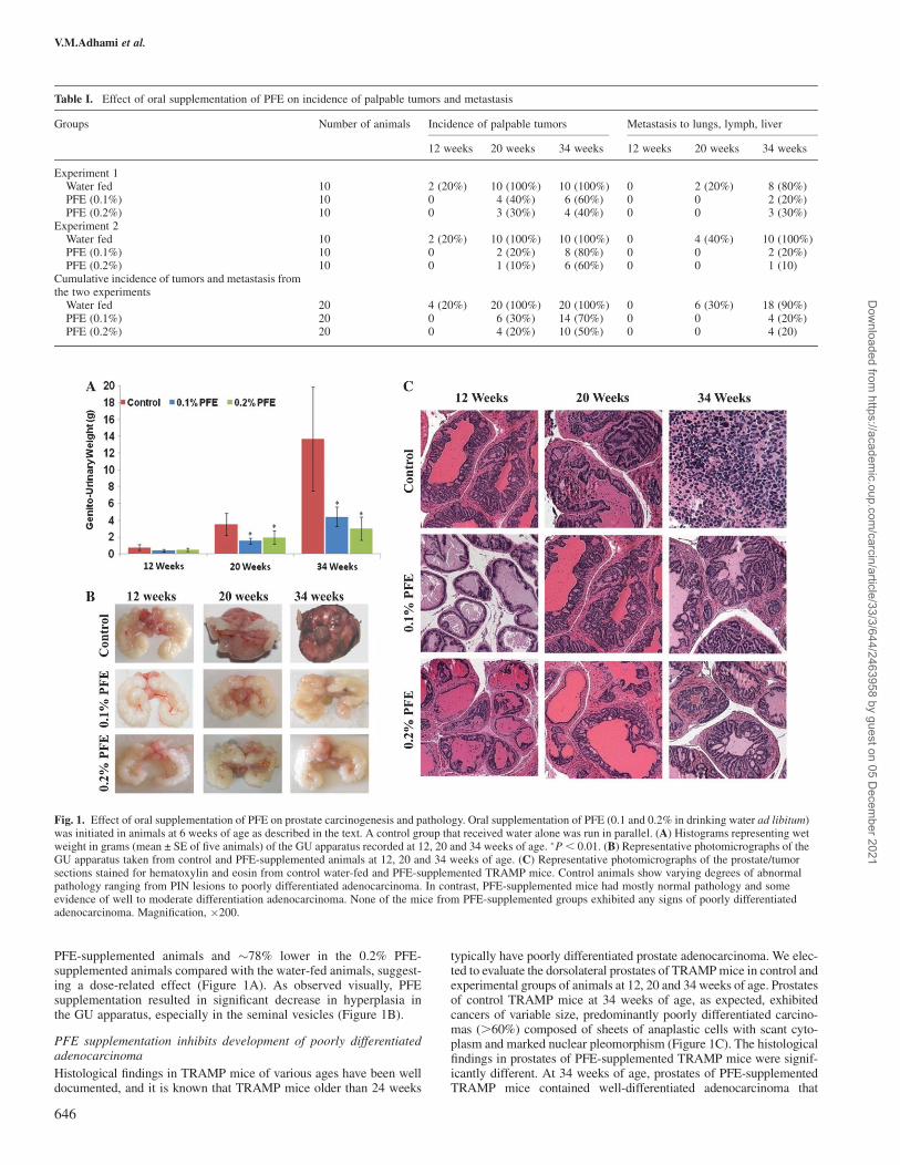
PFE-supplemented animals and �78% lower in the 0.2% PFE-supplemented animals compared with the water-fed animals, suggest-ing a dose-related effect (Figure 1A). As observed visually, PFEsupplementation resulted in significant decrease in hyperplasia inthe GU apparatus, especially in the seminal vesicles (Figure 1B).
PFE supplementation inhibits development of poorly differentiatedadenocarcinoma
Histological findings in TRAMP mice of various ages have been welldocumented, and it is known that TRAMP mice older than 24 weeks
typically have poorly differentiated prostate adenocarcinoma. We elec-ted to evaluate the dorsolateral prostates of TRAMP mice in control andexperimental groups of animals at 12, 20 and 34 weeks of age. Prostatesof control TRAMP mice at 34 weeks of age, as expected, exhibitedcancers of variable size, predominantly poorly differentiated carcino-mas (.60%) composed of sheets of anaplastic cells with scant cyto-plasm and marked nuclear pleomorphism (Figure 1C). The histologicalfindings in prostates of PFE-supplemented TRAMP mice were signif-icantly different. At 34 weeks of age, prostates of PFE-supplementedTRAMP mice contained well-differentiated adenocarcinoma that
Table I. Effect of oral supplementation of PFE on incidence of palpable tumors and metastasis
Groups Number of animals Incidence of palpable tumors Metastasis to lungs, lymph, liver
12 weeks 20 weeks 34 weeks 12 weeks 20 weeks 34 weeks
Experiment 1Water fed 10 2 (20%) 10 (100%) 10 (100%) 0 2 (20%) 8 (80%)PFE (0.1%) 10 0 4 (40%) 6 (60%) 0 0 2 (20%)PFE (0.2%) 10 0 3 (30%) 4 (40%) 0 0 3 (30%)
Experiment 2Water fed 10 2 (20%) 10 (100%) 10 (100%) 0 4 (40%) 10 (100%)PFE (0.1%) 10 0 2 (20%) 8 (80%) 0 0 2 (20%)PFE (0.2%) 10 0 1 (10%) 6 (60%) 0 0 1 (10)
Cumulative incidence of tumors and metastasis fromthe two experiments
Water fed 20 4 (20%) 20 (100%) 20 (100%) 0 6 (30%) 18 (90%)PFE (0.1%) 20 0 6 (30%) 14 (70%) 0 0 4 (20%)PFE (0.2%) 20 0 4 (20%) 10 (50%) 0 0 4 (20)
Fig. 1. Effect of oral supplementation of PFE on prostate carcinogenesis and pathology. Oral supplementation of PFE (0.1 and 0.2% in drinking water ad libitum)was initiated in animals at 6 weeks of age as described in the text. A control group that received water alone was run in parallel. (A) Histograms representing wetweight in grams (mean ± SE of five animals) of the GU apparatus recorded at 12, 20 and 34 weeks of age. �P, 0.01. (B) Representative photomicrographs of theGU apparatus taken from control and PFE-supplemented animals at 12, 20 and 34 weeks of age. (C) Representative photomicrographs of the prostate/tumorsections stained for hematoxylin and eosin from control water-fed and PFE-supplemented TRAMP mice. Control animals show varying degrees of abnormalpathology ranging from PIN lesions to poorly differentiated adenocarcinoma. In contrast, PFE-supplemented mice had mostly normal pathology and someevidence of well to moderate differentiation adenocarcinoma. None of the mice from PFE-supplemented groups exhibited any signs of poorly differentiatedadenocarcinoma. Magnification, �200.
V.M.Adhami et al.
646
Dow
nloaded from https://academ
ic.oup.com/carcin/article/33/3/644/2463958 by guest on 05 D
ecember 2021
occupied �20% at surface area, admixed with lesser amounts of mod-erately differentiated adenocarcinoma (,3% of surface area), histologythat was comparable with controls at 12 weeks of age. Many of thePFE-supplemented animals had multiple foci of well-differentiatedcarcinoma (7–15% of surface area) and some had moderately differen-tiated carcinoma (Figure 1C). Significantly, none of the mice in eithergroup of PFE-supplemented animals exhibited any signs of poorlydifferentiated adenocarcinoma.
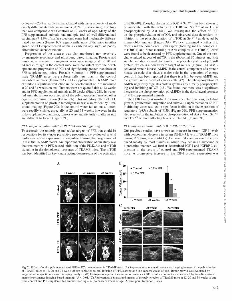
Progression of the disease was also monitored non-invasivelyby magnetic resonance and ultrasound based imaging. Prostate andtumor sizes assessed by magnetic resonance imaging at 12, 20 and34 weeks of age in the control mice were consistent with the devel-opment and progression of PCa and significantly greater than those ofPFE-supplemented mice. Prostate volumes in PFE-supplementedmale TRAMP mice were substantially less than in the controlwater-fed animals (Figure 2A). PFE-supplemented TRAMP miceexhibited a significant reduction in the development of PCa measuredat 20 and 34 weeks on test. Tumors were not quantifiable at 12 weeksand in PFE-supplemented animals at 20 weeks (Figure 2B). In water-fed animals, tumors occupied all of the pelvic space and masked otherorgans from visualization (Figure 2A). This inhibitory effect of PFEsupplementation on prostate tumorigenesis was also evident by ultra-sound imaging (Figure 2C). In the control water-fed animals, tumorswere readily visible, especially at 20 and 34 weeks; however, in thePFE-supplemented animals, tumors were significantly smaller in sizeand difficult to locate (Figure 2C).
PFE supplementation inhibits PI3K/Akt/mTOR signaling
To ascertain the underlying molecular targets of PFE that could beresponsible for its cancer preventive properties, we evaluated severalmolecules whose expression is deregulated during the progression ofPCa in the TRAMP model. An important observation of our study wasthat treatment with PFE caused inhibition of the PI3K/Akt and mTORsignaling in the dorsolateral prostates of TRAMP mice. The mTORhas been identified as key kinase acting downstream of the activation
of PI3K (40). Phosphorylation of mTOR at Ser2448 has been shown tobe associated with the activity of mTOR and Ser2448 of mTOR isphosphorylated by Akt (41). We investigated the effect of PFEon the phosphorylation of mTOR and observed dose-dependent in-hibition in the phosphorylation of mTOR at Ser2448 as detected byimmunoblot analysis (Figure 3A). We next examined whether PFEaffects mTOR complexes. Both raptor (forming mTOR complex 1,mTORC1) and rictor (forming mTOR complex 2, mTORC2) levelswere found to be decreased by PFE supplementation. One of the best-characterized targets of mTOR is the ribosomal S6 kinases and PFEsupplementation caused decrease in the phosphorylation of p70S6Kprotein, which is a downstream target of mTOR (Figure 3A). AMP-activated protein kinase (AMPK) is the central component of a proteinkinase cascade that plays a major role in the regulation of energycontrol. It has been reported that there is a link between AMPK andthe growth and survival of cancer cells (42). The phosphorylation ofAMPK negatively regulates protein synthesis by directly phosphorylat-ing and inhibiting mTOR (43). We found that there was a significantincrease in the phosphorylation of AMPKa in the dorsolateral prostatesof PFE-supplemented animals.
The PI3K family is involved in various cellular functions, includinggrowth, proliferation, migration and survival. Supplementation of PFEin drinking water resulted in significant inhibition in the expression ofregulatory (p85) subunit of PI3K (Figure 3B). PFE supplementationalso resulted in the inhibition of phosphorylation of Akt at both Ser473
and Thr308 without affecting levels of total Akt (Figure 3B).
PFE supplementation inhibits IGF-I/IGFBP-3 ratio
Our previous studies have shown an increase in serum IGF-I levelswith concomitant decrease in serum IGFBP-3 levels in TRAMP miceduring PCa progression (44,45). Because IGFs are known to be pro-duced locally by most tissues in which they act in an autocrine ora paracrine manner, we further determined IGF-I and IGFBP-3 ex-pression in the serum of control and PFE-supplemented TRAMPmice. A progressive increase in the IGF-I protein expression was
Fig. 2. Effect of oral supplementation of PFE on PCa development in TRAMP mice. (A) Representative magnetic resonance imaging images of the pelvic regionof TRAMP mice at 12, 20 and 34 weeks of age subjected to oral infusion of PFE starting at 6 (no cancer) weeks of age. Tumor growth was evaluated bylongitudinal magnetic resonance imaging analysis. (B) Histograms represent mean tumor volumes ± SE in cubic centimeter as evaluated by two-dimensionalmagnetic resonance imaging-based imaging. �P, 0.01. (C) Representative ultrasound images of the pelvic region of TRAMP mice at 12, 20 and 34 weeks of agefrom control and PFE-supplemented animals starting at 6 (no cancer) weeks of age. Arrows point to tumor tissues.
Pomegranate juice inhibits prostate carcinogenesis
647
Dow
nloaded from https://academ
ic.oup.com/carcin/article/33/3/644/2463958 by guest on 05 D
ecember 2021
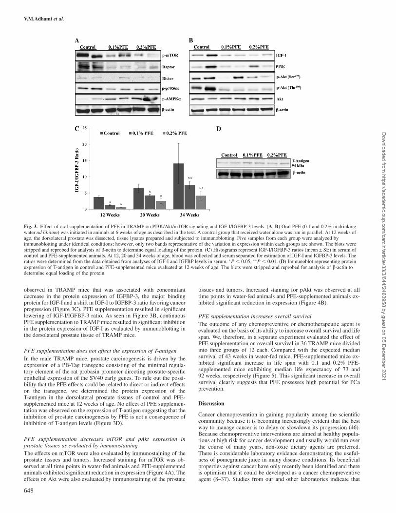
observed in TRAMP mice that was associated with concomitantdecrease in the protein expression of IGFBP-3, the major bindingprotein for IGF-I and a shift in IGF-I to IGFBP-3 ratio favoring cancerprogression (Figure 3C). PFE supplementation resulted in significantlowering of IGF-I/IGFBP-3 ratio. As seen in Figure 3B, continuousPFE supplementation to TRAMP mice resulted in significant inhibitionin the protein expression of IGF-I as evaluated by immunoblotting inthe dorsolateral prostate tissue of TRAMP mice.
PFE supplementation does not affect the expression of T-antigen
In the male TRAMP mice, prostate carcinogenesis is driven by theexpression of a PB-Tag transgene consisting of the minimal regula-tory element of the rat probasin promoter directing prostate-specificepithelial expression of the SV40 early genes. To rule out the possi-bility that the PFE effects could be related to direct or indirect effectson the transgene, we determined the protein expression of theT-antigen in the dorsolateral prostate tissues of control and PFE-supplemented mice at 12 weeks of age. No effect of PFE supplemen-tation was observed on the expression of T-antigen suggesting that theinhibition of prostate carcinogenesis by PFE is not a consequence ofinhibition of T-antigen levels (Figure 3D).
PFE supplementation decreases mTOR and pAkt expression inprostate tissues as evaluated by immunostaining
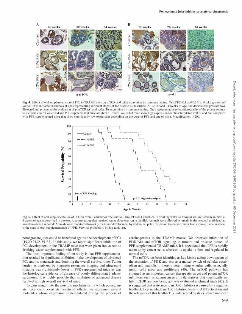
The effects on mTOR were also evaluated by immunostaining of theprostate tissues and tumors. Increased staining for mTOR was ob-served at all time points in water-fed animals and PFE-supplementedanimals exhibited significant reduction in expression (Figure 4A). Theeffects on Akt were also evaluated by immunostaining of the prostate
tissues and tumors. Increased staining for pAkt was observed at alltime points in water-fed animals and PFE-supplemented animals ex-hibited significant reduction in expression (Figure 4B).
PFE supplementation increases overall survival
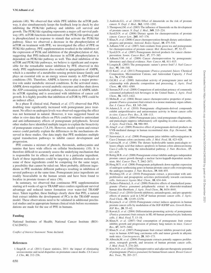
The outcome of any chemopreventive or chemotherapeutic agent isevaluated on the basis of its ability to increase overall survival and lifespan. We, therefore, in a separate experiment evaluated the effect ofPFE supplementation on overall survival in 36 TRAMP mice dividedinto three groups of 12 each. Compared with the expected mediansurvival of 43 weeks in water-fed mice, PFE-supplemented mice ex-hibited significant increase in life span with 0.1 and 0.2% PFE-supplemented mice exhibiting median life expectancy of 73 and92 weeks, respectively (Figure 5). This significant increase in overallsurvival clearly suggests that PFE possesses high potential for PCaprevention.
Discussion
Cancer chemoprevention in gaining popularity among the scientificcommunity because it is becoming increasingly evident that the bestway to manage cancer is to delay or slowdown its progression (46).Because chemopreventive interventions are aimed at healthy popula-tions at high risk for cancer development and usually would run overthe course of many years, non-toxic dietary agents are preferred.There is considerable laboratory evidence demonstrating the useful-ness of pomegranate juice in many disease conditions. Its beneficialproperties against cancer have only recently been identified and thereis optimism that it could be developed as a cancer chemopreventiveagent (8–37). Studies from our and other laboratories indicate that
Fig. 3. Effect of oral supplementation of PFE in TRAMP on PI3K/Akt/mTOR signaling and IGF-I/IGFBP-3 levels. (A, B) Oral PFE (0.1 and 0.2% in drinkingwater ad libitum) was initiated in animals at 6 weeks of age as described in the text. A control group that received water alone was run in parallel. At 12 weeks ofage, the dorsolateral prostate was dissected, tissue lysates prepared and subjected to immunoblotting. Five samples from each group were analyzed byimmunoblotting under identical conditions; however, only two bands representative of the variation in expression within each groups are shown. The blots werestripped and reprobed for analysis of b-actin to determine equal loading of the protein. (C) Histograms represent IGF-I/IGFBP-3 ratios (mean ± SE) in serum ofcontrol and PFE-supplemented animals. At 12, 20 and 34 weeks of age, blood was collected and serum separated for estimation of IGF-I and IGFBP-3 levels. Theratios were determined from the data obtained from analyses of IGF-I and IGFBP levels in serum. �P , 0.05, ��P , 0.01. (D) Immunoblot representing proteinexpression of T-antigen in control and PFE-supplemented mice evaluated at 12 weeks of age. The blots were stripped and reprobed for analysis of b-actin todetermine equal loading of the protein.
V.M.Adhami et al.
648
Dow
nloaded from https://academ
ic.oup.com/carcin/article/33/3/644/2463958 by guest on 05 D
ecember 2021
pomegranate juice could be beneficial against the development of PCa(19,20,24,28,35–37). In this study, we report significant inhibition ofPCa development in the TRAMP mice that were given free access todrinking water supplemented with PFE.
The most important finding of our study is that PFE supplementa-tion resulted in significant inhibition in the development of advancedPCa and its metastasis and doubling the overall survival time. Tumorburden as analyzed by magnetic resonance imaging and ultrasoundimaging was significantly lower in PFE-supplemented mice as wasthe histological evidence of absence of poorly differentiated adeno-carcinoma. It is highly possible that inhibition of advanced diseaseresulted in high overall survival of mice.
To gain insight into the possible mechanisms by which pomegran-ate juice could exert its beneficial effects, we examined severalmolecules whose expression is deregulated during the process of
carcinogenesis in the TRAMP mouse. We observed inhibition ofPI3K/Akt and mTOR signaling in tumors and prostate tissues ofPFE-supplemented TRAMP mice. It is speculated that PFE is rapidlytaken up by cancer cells, whereas its uptake is slow and regulated innormal cells.
The mTOR has been identified as key kinase acting downstream ofthe activation of PI3K and acts as a master switch of cellular catab-olism and anabolism, thereby determining whether cells, especiallytumor cells grow and proliferate (40). The mTOR pathway hasemerged as an important cancer therapeutic target and potent mTORinhibitors such as rapamycin and its derivatives that specifically in-hibit mTOR are now being actively evaluated in clinical trials (47). Itis suggested that resistance to mTOR inhibitors is caused by a negativefeedback loop in which mTOR inhibition leads to AKT activation andthe relevance of this feedback is underscored by its existence in cancer
Fig. 4. Effect of oral supplementation of PFE to TRAMP mice on mTOR and pAkt expression by immunostaining. Oral PFE (0.1 and 0.2% in drinking water adlibitum) was initiated in animals at ages representing different stages of the disease as described. At 12, 20 and 34 weeks of age, the dorsolateral prostate wasdissected and processed for evaluation of p-mTOR (A) and pAkt (B) expression by immunostaining. Only representative photomicrographs of the prostate/tumortissue from control water-fed and PFE-supplemented mice are shown. Control water-fed mice show high expression for phosphorylated mTOR and Akt comparedwith PFE-supplemented mice that show significantly low expression depending on the dose of PFE and age of mice. Magnification, �200.
Fig. 5. Effect of oral supplementation of PFE on overall and tumor free survival. Oral PFE (0.1 and 0.2% in drinking water ad libitum) was initiated in animals at6 weeks of age as described in the text. A control group that received water alone was run in parallel. Animals were allowed to remain in the protocol until death toascertain overall survival. Animals were monitored biweekly for tumor development by abdominal pelvic palpation to analyze tumor free survival. Time in weeksis the start of oral supplementation of PFE. Survival probability by log-rank test.
Pomegranate juice inhibits prostate carcinogenesis
649
Dow
nloaded from https://academ
ic.oup.com/carcin/article/33/3/644/2463958 by guest on 05 D
ecember 2021
patients (48). We observed that while PFE inhibits the mTOR path-way, it also simultaneously keeps the feedback loop in check by alsoinhibiting the PI3K/Akt pathway and inhibits cell survival andgrowth. The PI3K/Akt signaling represents a major cell survival path-way (49). mTOR functions downstream of the PI3K/Akt pathway andis phosphorylated in response to stimuli that activate the PI3K/Aktpathway. Since we observed a decrease in the phosphorylation ofmTOR on treatment with PFE, we investigated the effect of PFE onPI3K/Akt pathway. PFE supplementation resulted in the inhibition ofthe expression of PI3K and inhibition of the phosphorylation of Akt,suggesting that PFE-induced decrease in mTOR phosphorylation isdependent on PI3K/Akt pathway as well. This dual inhibition of themTOR and PI3K/Akt pathways, we believe is significant and respon-sible for the remarkable tumor inhibition by PFE observed in thisstudy. PFE supplementation also resulted in upregulation of AMPK,which is a member of a metabolite-sensing protein kinase family andplays an essential role as an energy sensor mainly in ATP-deprivedconditions (50). Therefore, AMPK is known to play a major protec-tive role under metabolic stressed conditions. In the activated states,AMPK down-regulates several anabolic enzymes and thus shuts downthe ATP-consuming metabolic pathways. Activation of AMPK inhib-its mTOR signaling and is associated with inhibition of cancer cellgrowth. It is highly possible that inhibition of mTOR by PFE could bean indirect effect.
In a phase II clinical trial, Pantuck et al. (37) observed that PSAdoubling time significantly increased with pomegranate juice treat-ment. No effect on androgen levels was observed suggesting the effectwas not hormonal in natural. However, it was suggested based onother in vitro data that effects on PSA could be related to antioxidantand anti-inflammatory effects of pomegranate polyphenols. Severalother studies have identified multiple targets to explain the bioactivityof pomegranate extracts (7,8,19). However, model system and extractsource could partially explain the differences in the mechanisms ob-served in these studies. Our data imply that PFE modulates multiplesignal transduction pathways to inhibit cancer development andprogression.
PFE contains a mixture of phenols, flavonoids, anthocyanins andtannins that have wide effects on cellular biochemistry (10). It istherefore difficult to accurately assess the underlying mechanism thatis responsible for its effects and relate it to a particular ingredient.Each of these ingredients could be targeting a different molecule orsome of these ingredients could be competing for the same target,a possibility that cannot be ruled out. Most probably, different ingre-dients in PFE modulate different pathways resulting in inhibition ofseveral pathways at the same time. Pomegranate juice ingredients arereadily bioavailable in the human serum and have been found tolocalize in prostate tissues of mice (36).
In summary, we observed that continuous PFE supplementationstarting at 6 weeks of age to TRAMP mice confers significant survivaladvantage and reduced tumor formation over water-fed TRAMPmice. Taken together, these findings clearly show that PFE, a naturalfruit extract inhibits the development of PCa in the TRAMP mousemodel. These observations need to be validated in additional preclin-ical studies and in appropriate human clinical trials before recommen-dations are made for the use of PFE in humans.
Funding
National Institutes of Health; National Cancer Institute (RO1-CA120451).
Conflict of Interest Statement: None declared.
References
1.Siegel,R. et al. (2011) Cancer statistics, 2011: the impact of eliminatingsocioeconomic and racial disparities on premature cancer deaths. CACancerJ. Clin., 61, 212–236.
2.Andriole,G.L. et al. (2010) Effect of dutasteride on the risk of prostatecancer. N. Engl. J. Med., 362, 1192–1202.
3.Thompson,I.M. et al. (2003) The influence of finasteride on the developmentof prostate cancer. N. Engl. J. Med., 349, 215–224.
4.Syed,D.N. et al. (2008) Dietary agents for chemoprevention of prostatecancer. Cancer Lett., 265, 167–176.
5.Khan,N. et al. (2008) Cancer chemoprevention through dietary antioxidants:progress and promise. Antioxid. Redox Signal., 10, 475–510.
6.Adhami,V.M. et al. (2007) Anti-oxidants from green tea and pomegranatefor chemoprevention of prostate cancer. Mol. Biotechnol., 37, 52–57.
7.Syed,D.N. et al. (2007) Pomegranate derived products for cancer chemo-prevention. Semin. Cancer Biol., 17, 377–385.
8.Adhami,V.M. et al. (2009) Cancer chemoprevention by pomegranate:laboratory and clinical evidence. Nutr. Cancer, 61, 811–815.
9.Longtin,R. (2003) The pomegranate: nature’s power fruit? J. Natl CancerInst., 95, 346–348.
10.El Kar,C. et al. (2011) Pomegranate (Punica granatum) Juices: ChemicalComposition, Micronutrient Cations, and Antioxidant Capacity. J FoodSci., 76, C795–C800.
11.Gil,M.I. et al. (2000) Antioxidant activity of pomegranate juice and itsrelationship with phenolic composition and processing. J. Agric. FoodChem., 48, 4581–4589.
12.Seeram,N.P. et al. (2008) Comparison of antioxidant potency of commonlyconsumed polyphenol-rich beverages in the United States. J. Agric. FoodChem., 56, 1415–1422.
13.Mehta,R. et al. (2004) Breast cancer chemopreventive properties of pome-granate (Punica granatum) fruit extracts in a mouse mammary organ culture.Eur. J. Cancer Prev., 13, 345–348.
14.Adams,L.S. et al. (2010) Pomegranate ellagitannin-derived compoundsexhibit antiproliferative and antiaromatase activity in breast cancer cellsin vitro. Cancer Prev. Res., 3, 108–113.
15.Adams,L.S. et al. (2006) Pomegranate juice, total pomegranate ellagitannins,and punicalagin suppress inflammatory cell signaling in colon cancer cells.J. Agric. Food Chem., 54, 980–985.
16.Afaq,F. et al. (2009) Protective effect of pomegranate-derived products onUVB-mediated damage in human reconstituted skin. Exp. Dermatol., 18,553–561.
17.Saruwatari,A. et al. (2008) Pomegranate juice inhibits sulfoconjugation inCaco-2 human colon carcinoma cells. J. Med. Food, 11, 623–628.
18.Larrosa,M. et al. (2006) The dietary hydrolysable tannin punicalagin re-leases ellagic acid that induces apoptosis in human colon adenocarcinomaCaco-2 cells by using the mitochondrial pathway. J. Nutr. Biochem., 17,611–625.
19.Rettig,M.B. et al. (2008) Pomegranate extract inhibits androgen-independentprostate cancer growth through a nuclear factor-kappaB-dependent mecha-nism. Mol. Cancer Ther., 7, 2662–2671.
20.Hong,M.Y. et al. (2008) Pomegranate polyphenols down-regulate expressionof androgen-synthesizing genes in human prostate cancer cells overexpressingthe androgen receptor. J. Nutr. Biochem., 19, 848–855.
21.Weisburg,J.H. et al. (2010) Pomegranate extract, a prooxidant with anti-proliferative and proapoptotic activities preferentially towards carcinomacells. Anticancer Agents Med. Chem., 10, 634–644.
22.Pacheco-Palencia,L.A. et al. (2008) Protective effects of standardized pome-granate (Punica granatum) polyphenolic extract in ultraviolet-irradiatedhuman skin fibroblasts. J. Agric. Food Chem., 56, 8434–8441.
23.Gasmi,J. et al. (2010) Growth inhibitory, antiandrogenic, and pro-apoptoticeffects of punicic acid in LNCaP human prostate cancer cells. J. Agric.Food Chem., 58, 12149–12156.
24.Koyama,S. et al. (2010) Pomegranate extract induces apoptosis in humanprostate cancer cells by modulation of the IGF-IGFBP axis. Growth Horm.IGF Res., 20, 55–62.
25.Kawaii,S. et al. (2004) Differentiation-promoting activity of pomegranate(Punica granatum) fruit extracts in HL-60 human promyelocytic leukemiacells. J. Med. Food, 7, 13–18.
26.Khan,N. et al. (2007) Oral consumption of pomegranate fruit extractinhibits growth and progression of primary lung tumors in mice. CancerRes., 67, 3475–3482.
27.Khan,N. et al. (2007) Pomegranate fruit extract inhibits prosurvival path-ways in human A549 lung carcinoma cells and tumor growth in athymicnude mice. Carcinogenesis, 28, 163–173.
28.Albrecht,M. et al. (2004) Pomegranate extracts potently suppress prolifer-ation, xenograft growth, and invasion of human prostate cancer cells.J. Med. Food, 7, 274–283.
29.Kim,N.D. et al. (2002) Chemopreventive and adjuvant therapeutic potentialof pomegranate (Punica granatum) for human breast cancer. Breast CancerRes. Treat., 71, 203–217.
V.M.Adhami et al.
650
Dow
nloaded from https://academ
ic.oup.com/carcin/article/33/3/644/2463958 by guest on 05 D
ecember 2021
30.Hora,J.J. et al. (2003) Chemopreventive effects of pomegranate seed oil onskin tumor development in CD1 mice. J. Med. Food, 6, 157–161.
31.Toi,M. et al. (2003) Preliminary studies on the anti-angiogenic potential ofpomegranate fractions in vitro and in vivo. Angiogenesis, 6, 121–128.
32.Kohno,H. et al. (2004) Pomegranate seed oil rich in conjugated linolenicacid suppresses chemically induced colon carcinogenesis in rats. CancerSci., 95, 481–486.
33.Afaq,F. et al. (2005) Anthocyanin- and hydrolyzable tannin-rich pomegran-ate fruit extract modulates MAPK and NF-kappaB pathways and inhibitsskin tumorigenesis in CD-1 mice. Int. J. Cancer, 113, 423–433.
34.Afaq,F. et al. (2005) Pomegranate fruit extract modulates UV-B-mediatedphosphorylation of mitogen-activated protein kinases and activation ofnuclear factor kappa B in normal human epidermal keratinocytes paragraphsign. Photochem. Photobiol., 81, 38–45.
35.Malik,A. et al. (2005) Pomegranate fruit juice for chemoprevention and che-motherapy of prostate cancer. Proc. Natl Acad. Sci. USA, 102, 14813–14818.
36.Seeram,N.P. et al. (2007) Pomegranate ellagitannin-derived metabolitesinhibit prostate cancer growth and localize to the mouse prostate gland.J. Agric. Food Chem., 55, 7732–7737.
37.Pantuck,A.J. et al. (2006) Phase II study of pomegranate juice for men withrising prostate-specific antigen following surgery or radiation for prostatecancer. Clin. Cancer Res., 12, 4018–4026.
38.Greenberg,N.M. et al. (1995) Prostate cancer in a transgenic mouse. Proc.Natl Acad. Sci. USA, 92, 3439–3443.
39.Rad,A.M. et al. (2008) Imaging mouse prostate gland by 3 Tesla clinicalMRI system. Open Magn. Reson. Rev., 1, 60–63.
40.Hay,N. (2005) The Akt-mTOR tango and its relevance to cancer. CancerCell, 8, 179–183.
41.Sekulic,A. et al. (2000) A direct linkage between the phosphoinositide 3-kinase-AKT signaling pathway and the mammalian target of rapamycin inmitogen-stimulated and transformed cells. Cancer Res., 60, 3504–3513.
42.Xiang,X. et al. (2004) AMP-activated protein kinase activators can inhibitthe growth of prostate cancer cells by multiple mechanisms. Biochem.Biophys. Res. Commun., 321, 161–167.
43.Fryer,L.G. et al. (2002) The anti-diabetic drugs rosiglitazone and metforminstimulate AMP-activated protein kinase through distinct signaling pathways.J. Biol. Chem., 277, 25226–25232.
44.Gupta,S. et al. (2001) Inhibition of prostate carcinogenesis in TRAMP miceby oral infusion of green tea polyphenols. Proc. Natl Acad. Sci. USA, 98,10350–10355.
45.Adhami,V.M. et al. (2004) Oral consumption of green tea polyphenolsinhibits insulin-like growth factor-I-induced signaling in an autochthonousmouse model of prostate cancer. Cancer Res., 64, 8715–8722.
46.Brody,H. (2011) Cancer prevention. Nature, 471, (suppl.), S1–S24.47.Faivre,S. et al. (2006) Current development of mTOR inhibitors as anticancer
agents. Nat. Rev. Drug Discov., 5, 671–688.48.O’Reilly,K.E. et al. (2006) mTOR inhibition induces upstream receptor
tyrosine kinase signaling and activates Akt. Cancer Res., 66, 1500–1508.49.Vivanco,I. et al. (2002) The phosphatidylinositol 3-kinase AKT pathway in
human cancer. Nat. Rev. Cancer, 2, 489–501.50.Hardie,D.G. et al. (1998) The AMP-activated/SNF1 protein kinase sub-
family: metabolic sensors of the eukaryotic cell? Annu. Rev. Biochem.,67, 821–855.
Received September 27, 2011; revised December 8, 2011;accepted December 15, 2011
Pomegranate juice inhibits prostate carcinogenesis
651
Dow
nloaded from https://academ
ic.oup.com/carcin/article/33/3/644/2463958 by guest on 05 D
ecember 2021