ORAL CAVITY 1. Teeth and Supporting...
22
Robbins and Cotran: Pathologic Basis of Disease, 7th Edition ORAL CAVITY The oral cavity is a fearsome orifice guarded by ranks of upper and lower "horns" (lamentably, quite subject to erosion), demanding constant gratification, and teeming with microorganisms, some of which are potentially harmful. Among the many disorders that affect its various parts, only the more important or frequent conditions involving the teeth and supporting structures, oral mucous membranes, lips, and tongue are considered. 1. Teeth and Supporting Structures Figure 16-1 Schematic representation of the normal dental anatomy and surrounding supporting tissues. Teeth contribute to a number of important functions, including mastication and proper speech. It is useful to briefly review normal dental anatomy before we delve into the common pathologic conditions affecting teeth. As is well known, teeth are firmly implanted in the jaw and are surrounded by the gingival mucosa (Fig. 16-1). The anatomic crown of the tooth projects into the mouth and is covered by enamel, a hard, inert, acellular tissue-the most highly mineralized tissue in the body. The enamel rests upon dentin, which is a specialized form of connective tissue that makes up the remainder of the hard tissue portion of teeth. Unlike enamel, dentin is cellular and contains numerous dentinal tubules, which contain the cytoplasmic extensions of odontoblasts. These cells are present within the pulp and continually produce new (secondary) dentin within the interior of the tooth. The pulp chamber itself is surrounded by the dentin and consists of connective tissue stroma, nerve bundles, lymphatics, and capillaries. To perform mastication, teeth must not only be composed of hard tissue, but they must also be firmly attached to the bones of the jaw. If this attachment were excessively firm, chewing would impose sufficient physical stress on the teeth to cause their loss or fracturing. Therefore, in mammals, teeth are attached to the alveolar ridge of the jaws by the periodontal ligament, which provides a strong yet flexible attachment that can withstand the forces of mastication. The periodontal ligament attaches to the alveolar bone of the jaw on one side and to cementum, present on the roots of the teeth, which acts as a "cement" to anchor the periodontal ligament to the tooth. 1
Transcript of ORAL CAVITY 1. Teeth and Supporting...

Robbins and Cotran: Pathologic Basis of Disease, 7th Edition
ORAL CAVITY
The oral cavity is a fearsome orifice guarded by ranks of upper and lower "horns" (lamentably, quite subject to erosion), demanding constant gratification, and teeming with microorganisms, some of which are potentially harmful. Among the many disorders that affect its various parts, only the more important or frequent conditions involving the teeth and supporting structures, oral mucous membranes, lips, and tongue are considered. 1. Teeth and Supporting Structures
Figure 16-1 Schematic representation of the normal dental anatomy and surrounding supporting tissues.
Teeth contribute to a
number of important functions, including mastication and proper speech. It is useful to briefly review normal dental anatomy before we delve into the common pathologic conditions affecting teeth. As is well known, teeth are firmly implanted in the jaw and are surrounded by the gingival mucosa (Fig. 16-1). The anatomic crown of the tooth projects into the mouth and is covered by enamel, a hard, inert, acellular tissue-the most highly
mineralized tissue in the body. The enamel rests upon dentin, which is a specialized form of connective tissue that makes up the remainder of the hard tissue portion of teeth. Unlike enamel, dentin is cellular and contains numerous dentinal tubules, which contain the cytoplasmic extensions of odontoblasts. These cells are present within the pulp and continually produce new (secondary) dentin within the interior of the tooth. The pulp chamber itself is surrounded by the dentin and consists of connective tissue stroma, nerve bundles, lymphatics, and capillaries.
To perform mastication, teeth must not only be composed of hard tissue, but they must also be firmly attached to the bones of the jaw. If this attachment were excessively firm, chewing would impose sufficient physical stress on the teeth to cause their loss or fracturing. Therefore, in mammals, teeth are attached to the alveolar ridge of the jaws by the periodontal ligament, which provides a strong yet flexible attachment that can withstand the forces of mastication. The periodontal ligament attaches to the alveolar bone of the jaw on one side and to cementum, present on the roots of the teeth, which acts as a "cement" to anchor the periodontal ligament to the tooth.
1

Robbins and Cotran: Pathologic Basis of Disease, 7th Edition
CARIES (TOOTH DECAY)
Dental caries, caused by focal degradation of the tooth structure, is one of the most common diseases throughout the world and is the most common cause of tooth loss before age 35. Carious lesions are the result of mineral dissolution of tooth structure by acid metabolic end products from bacteria that are present in the oral cavity and are capable of fermenting sugars. Traditionally, the rate of caries has been higher in industrialized countries, where there is ready access to processed foods containing large amounts of carbohydrates. However, global trends may change these demographics. First of all, the rate of caries has dramatically dropped in countries such as the United States, where improved oral hygiene and fluoridation of the drinking water has become a standard practice. Fluoride incorporates into the crystalline structure of enamel, forming fluoroapatite, and contributes to resistance to degradation by bacterial acids. Secondly, with globalization of the world's economy, increased amounts of processed foods with high carbohydrate content are being imported into developing nations. With these trends, one can expect the rate of caries to dramatically increase in the less developed world over the next several decades. GINGIVITIS
Gingivitis is a condition in which there is inflammation of the soft tissues that surround the teeth. Typically, the development of gingivitis is the result of a lack of proper oral hygiene, leading to an accumulation of dental plaque and calculus. Dental plaque is a complex mass of microorganisms from the normal oral flora, proteins from the saliva, and desquamated epithelial cells. Calculus consists of mineralized bacterial plaque and can form extensive deposits around the teeth. Chronic gingivitis is characterized by gingival redness, edema, bleeding, changes in contour, and loss of soft tissue adaptation to the teeth. Gingivitis can occur at any age but is most prevalent and severe in adolescence (ranging from 40% to 60%), after which the incidence tends to taper off. It is a reversible disease; therapy is primarily aimed at the reduction of the accumulation of plaque and calculus via brushing, flossing, and regular hygiene visits.1 This allows the gingival tissues to heal. PERIODONTITIS
Periodontitis refers to an inflammatory process that affects the supporting structures of the teeth: periodontal ligaments, alveolar bone, and cementum. With progression, periodontitis can lead to serious sequelae, including the loss of attachment caused by complete destruction of the periodontal ligament and alveolar bone. Loosening and eventual loss of teeth are possible. The pathogenesis of periodontal inflammation is not entirely clear. Until the 1960s, it was believed that longstanding gingivitis uniformly progressed to periodontal disease. However, this is no longer thought to be the case. Rather, the development of periodontal disease is now considered to be an independent process, which, for reasons that are still unclear, is associated with a dramatic shift in the types and proportions of bacteria along the gums.2,3 This shift, along with other environmental conditions such as poor oral hygiene, is believed to be important in the pathogenesis of periodontitis. This view is supported by significant differences in the content of dental plaque in areas of healthy and diseased periodontium. For the most part, facultative gram-positive organisms colonize healthy sites, while plaque within areas of active periodontitis contains anaerobic and microaerophilic gram-negative flora. Although 300 types of bacteria reside in the oral cavity, adult periodontitis is associated primarily with Actinobacillus actinomycetemcomitans, Porphyromonas gingivalis, and Prevotella intermedia.
While it typically presents without any associated disorders, periodontal disease can also be a component of a number of different systemic diseases, including acquired
2

Robbins and Cotran: Pathologic Basis of Disease, 7th Edition
immunodeficiency syndrome (AIDS), leukemia, Crohn disease, diabetes mellitus, Down syndrome, sarcoidosis, and syndromes associated with polymorphonuclear defects (Chédiak-Higashi syndrome, agranulocytosis, and cyclic neutropenia). In addition to being a component of certain systemic diseases, periodontal infections can also be etiologic factors in several important systemic diseases. These include, for example, infective endocarditis, pulmonary and brain abscesses, and increased adverse pregnancy outcomes.4 2 Inflammatory/Reactive Lesions
A number of soft tissue lesions of the oral cavity, which present as tumor masses or ulcerations, are indeed reactive in nature and represent inflammations induced by irritation or by unknown mechanisms. All suspicious lesions, however, should be examined by biopsy. Reactive nodules of the oral cavity are fairly common and are a diverse group. The most common fibrous proliferative lesions of the oral cavity are fibroma (61%), peripheral ossifying fibroma (22%), pyogenic granuloma (12%), and peripheral giant cell granuloma (5%).5 The most common inflammatory/reactive ulcerations of the oral cavity are traumatic and aphthous ulcers. FIBROUS PROLIFERATIVE LESIONS
The so-called irritation fibroma (Fig. 16-2) primarily occurs in the buccal mucosa along the bite line or at the gingivodental margin. It consists of a nodular mass of fibrous tissue, with few inflammatory cells, covered by squamous mucosa. Treatment is complete surgical excision.
The pyogenic granuloma (Fig. 16-3) is a highly vascular peduncular lesion, usually occurring in the gingiva of children, young adults, and, commonly, pregnant women (pregnancy tumor). The surface of the lesion is typically ulcerated and can be red to purple in color. In some cases, growth is alarmingly rapid, raising the fear of a malignant neoplasm. Histologically, these lesions demonstrate a highly vascular proliferation that is similar to
granulation tissue. Because of this histologic picture, pyogenic granulomas can also be considered a form of capillary hemangioma (Chapter 11). They either regress, particularly after pregnancy, or undergo fibrous maturation, and they may develop into a peripheral ossifying fibroma. Treatment is complete surgical excision.
Figure 16-2 Fibroma. Smooth, pink, exophytic nodule on the buccal mucosa.
3

Robbins and Cotran: Pathologic Basis of Disease, 7th Edition
Figure 16-3 Pyogenic granuloma. Erythematous, hemorrhagic, and exophytic mass arising from the gingival mucosa.
The peripheral ossifying fibroma is a relatively common growth of the gingiva that is considered to be reactive in nature rather than neoplastic. However, tspecific etiology of the lesion is unknown. Some may arise as a result of the maturation of a long-standing pyogenic granuloma,
while others do not. With a peak incidence in young and teenage females, peripheral ossifying fibromas appear as red, ulcerated, and nodular lesions of the gingiva. They are often mistaken clinically for pyogenic granulomas. Complete surgical excision down to the periosteum is the treatment of choice, as these lesions have a recurrence rate of 15% to 20%.
he
The peripheral giant cell granuloma (giant cell epulis), a relatively common lesion of the oral cavity, characteristically protrudes from the gingiva at some site of chronic inflammation. This lesion is generally covered by intact gingival mucosa, but it may be ulcerated. The clinical appearance of peripheral giant cell granuloma can be similar to that of pyogenic granuloma but, in general, this lesion is more bluish-purple in color while the pyogenic granuloma is more bright red. Histologically, however, these lesions are distinct. Peripheral giant cell granuloma is made up of a striking aggregation of multinucleate, foreign body-like giant cells separated by a fibroangiomatous stroma. Although not encapsulated, these lesions are usually well delimited and readily excised. They should be differentiated from central giant cell granulomas found within the maxilla or the mandible and from the histologically similar but frequently multiple reparative giant cell "brown tumors" seen in hyperparathyroidism (Chapter 24). APHTHOUS ULCERS (CANKER SORES)
These extremely common superficial ulcerations of the oral mucosa affect up to 40% of the population in the United States.6 They are more common in the first two decades of life, are painful and often recurrent, and tend to be prevalent within certain families. The lesions appear as single or multiple, shallow, hyperemic ulcerations covered by a thin exudate and rimmed by a narrow zone of erythema (Fig. 16-4). The underlying inflammatory infiltrate is at first largely mononuclear, but secondary bacterial infection introduces numerous neutrophils. The lesions may spontaneously resolve in 7 to 10 days or be stubbornly persistent for weeks. The causation of these lesions is obscure. Most ulcers are more painful than serious and require only symptomatic treatment.
4

Robbins and Cotran: Pathologic Basis of Disease, 7th Edition
GLOSSITIS Figure 16-4 Aphthous ulcer. Single ulceration with an erythematous halo surrounding a yellowish fibrinopurulent membrane.
Although the designation glossitis implies inflammation of the tongue, it is sometimes applied to the beefy-red tongue ein certain deficiency states; this change results from atrophy of tpapillae of the tongue and thinof the mucosa, exposing the underlying vasculature. In
instances, the atrophic changes do indeed lead to inflammation and even shallow ulcerations. Such changes may be encountered in deficiencies of vitamin B12 (pernicious anemia), riboflavin, niacinView drug information, or pyridoxine. Similar alterations are sometimencountered with sprue and iron-deficiency anemia, possibly complicated by one of the vitamin B deficiencies. The combination of iron-deficiency anemia, glossitis, and esophageal dysphagia usually related to webs is known as the Plummer-Vinson or Paterson-Kelly syndrome. Glossitis, characterized by ulcerative lesions (sometimes along the lateral borders of the tongue), may also be seen with jagged carious teeth, ill-fitting dentures, and, rarelwith syphilis, inhalation burns, or ingestion of corrosive chemicals.
ncountered
he ning
some
es
y,
3. Infections
The oral mucosa is highly resistant to its indigenous flora, having many defenses, including the competitive suppression of potential pathogens by organisms of low virulence, the elaboration of secretory immunoglobulin A and other immunoglobulins by submucosal collections of lymphocytes and plasma cells, the antibacterial effects of saliva, and the diluting and irrigating effects of food and drink. Nonetheless, any lowering of these defenses, for example, with immunodeficiency or disruption of the microbiologic balance by antibacterial therapy, sets the stage for oral infection. Most of these infections are discussed in Chapter 8, and here we only briefly recapitulate the principal features of the oral lesions. HERPES SIMPLEX VIRUS INFECTIONS
Most orofacial herpetic infections are caused by herpes simplex virus type 1 (HSV-1). However, owing to changes in sexual habits, an increase in HSV-2 (genital herpes) has been observed in the oral cavity. Primary HSV infection typically occurs in children age 2 to 4 years, is often asymptomatic, and does not cause significant morbidity. Approximately 10% to 20% of the time, primary infection presents as acute herpetic gingivostomatitis, in which there is an abrupt onset of vesicles and ulcerations throughout the oral cavity, especially in the gingiva. These lesions are also accompanied by lymphadenopathy, fever, anorexia, and irritability.
Morphology. The vesicles range from lesions of a few millimeters to large bullae and are at first filled with a clear, serous fluid, but they often rupture to yield extremely painful, red-rimmed, and shallow ulcerations. On microscopic examination, there is intracellular and
5

Robbins and Cotran: Pathologic Basis of Disease, 7th Edition
intercellular edema (acantholysis), yielding clefts that may become transformed into macroscopic vesicles. Individual epidermal cells in the margins of the vesicle or lying free within the fluid sometimes develop eosinophilic intranuclear viral inclusions, or several cells may fuse to produce giant cells (multinucleate polykaryons), changes that are demonstrated by the diagnostic Tzanck test, based on microscopic examination of the vesicle fluid. The vesicles and shallow ulcers usually spontaneously clear within 3 to 4 weeks, but the virus treks along the regional nerves and eventually becomes dormant in the local ganglia (e.g., the trigeminal).
The great preponderance of adults harbor latent HSV-1, but in some individuals, usually young adults, the virus becomes reactivated to produce the common but usually mild cold sore. The influences predisposing to activation are poorly understood but are thought to include trauma, allergies, exposure to ultraviolet light, upper respiratory tract infections, pregnancy, menstruation, immunosuppression, and excessive exposure to heat or cold. Recurrent herpetic stomatitis (in contrast to acute gingivostomatitis) occurs either at the site of primary inoculation or in adjacent mucosal areas that are associated with the same ganglion; it takes the form of groups of small (1 to 3 mm) vesicles. The lips (herpes labialis), nasal orifices, buccal mucosa, gingiva, and hard palate are the most common locations for recurrent lesions. They resemble those already described in the primary infections but are much more limited in duration, are milder, usually dry up in 4 to 6 days, and heal within a week to 10 days. OTHER VIRAL INFECTIONS
Additional viral infections that can be seen in the oral cavity as well as the head and neck region include herpes zoster, Epstein-Barr virus (EBV; mononucleosis), cytomegalovirus, enterovirus (herpangina, hand-foot-and-mouth disease, acute lymphonodular pharyngitis), and rubeola (measles).
ORAL CANDIDIASIS (THRUSH)
The many localizations of candidal infection are fully described in Chapter 8, and so this discussion is limited to presentations in the oral cavity. Candidiasis is by far the most common fungal infection in the oral cavity. As is well known, Candida albicans is a normal component of the oral flora in approximately 50% of the population. As such, three factors appear to influence the likelihood of a clinical infection: (1) immune status of the individual; (2) the strain of C. albicans present; and (3) the composition of an individual's oral flora. There are three major clinical forms of oral candidiasis, including pseudomembranous (thrush), erythematous, and hyperplastic, with a number of different variations within these groups. Only the pseudomembranous form, the most common of these, is discussed here. Also known as "thrush," pseudomembranous candidiasis typically takes the form of a superficial, curdy, gray to white inflammatory membrane composed of matted organisms enmeshed in a fibrinosuppurative exudate that can be readily scraped off to reveal an underlying erythematous inflammatory base. This fungus is a normal inhabitant of the oral cavity and causes mischief only in individuals who have some form of immunosuppression, as occurs in patients with diabetes mellitus, organ or bone marrow transplant recipients, those with neutropenia, chemotherapy-induced immunosuppression, or AIDS. In addition, broad-spectrum antibiotics that eliminate or alter the normal bacterial flora of the mouth can result in the development of oral candidiasis.
6

Robbins and Cotran: Pathologic Basis of Disease, 7th Edition
DEEP FUNGAL INFECTIONS
In addition to their more common sites of infection, certain deep fungal infections have a rather significant predilection for the oral cavity and head and neck region. Such fungi include histoplasmosis, blastomycosis, coccidioidomycosis, cryptococcosis, zygomycosis, and aspergillosis. With an increasing number of patients who are immunocompromised as a result of diseases such as AIDS or therapies for cancer and organ transplantation, the prevalence of fungal infections of the oral cavity has also increased in recent years.
4. Oral Manifestations of Systemic Disease As oral clinicians are at pains to emphasize, the mouth is a part of the body, and not
merely a gateway for delicacies. Not surprisingly, then, many systemic diseases are associated with oral lesions. In fact, it is not uncommon for oral lesions to be the first sign of some underlying systemic condition. Some of the more common are cited in Table 16-1, with a few words about the associated oral changes. Only one-hairy leukoplakia-is characterized in more detail.
Scarlet fever Fiery red tongue with prominent papillae (raspberry tongue);
white coated tongue through which hyperemic papillae project (strawberry tongue)
Measles A spotty enanthema in the oral cavity often precedes the rash; ulcerations on the buccal mucosa about Stensen duct produce Koplik spots
Infectious mononucleosis An acute pharyngitis and tonsillitis that may cause coating with a gray-white exudative membrane; enlargement of lymph nodes in the neck
Diphtheria A characteristic dirty white, fibrinosuppurative, tough, inflammatory membrane over the tonsils and retropharynx
Human immunodeficiency virus infection; AIDS
Predisposition to opportunistic oral infections, particularly with herpesvirus, Candida, and other fungi; sometimes oral lesions of Kaposi sarcoma and hairy leukoplakia (described in text)
Dermatologic Conditions* Lichen planus Reticulate, lacelike, white keratotic lesions that rarely become
bullous and ulcerated; seen in more than 50% of patients with cutaneous lichen planus; rarely, is the sole manifestation
Pemphigus Usually vulgaris; vesicles and bullae prone to rupture, leaving hyperemic erosions covered with exudate
Bullous pemphigoid Oral lesions resemble macroscopically those of pemphigus but can be differentiated histologically
Erythema multiforme A maculopapular, vesiculobullous eruption that sometimes follows an infection elsewhere, ingestion of drugs, development of cancer, or a collagen vascular disease; when it involves the lips and oral mucosa, it is referred to as Stevens-Johnson syndrome
Hematologic Disorders Pancytopenia (agranulocytosis, aplastic anemia)
Severe oral infections in the form of gingivitis, pharyngitis, tonsillitis; may extend to cellulitis of the neck (Ludwig angina)
Leukemia With depletion of functioning neutrophils, oral lesions may
7

Robbins and Cotran: Pathologic Basis of Disease, 7th Edition
appear like those in pancytopenia Monocytic leukemia Leukemic infiltration and enlargement of the gingivae, often with
accompanying periodontitis Miscellaneous Melanotic pigmentation May appear in Addison disease, hemochromatosis, fibrous
dysplasia of bone (Albright syndrome), and Peutz-Jegher syndrome (gastrointestinal polyposis)
Phenytoin (Dilantin) ingestion
Striking fibrous enlargement of the gingivae
Pregnancy A friable, red, pyogenic granuloma protruding from the gingiva ("pregnancy tumor")
Rendu-Osler-Weber syndrome
Autosomal dominant disorder with multiple aneurysmal telangiectasias from birth beneath the skin or mucosal surfaces of the oral cavity, lips, gastrointestinal tract, respiratory tract, and urinary tract as well as in internal viscera
HAIRY LEUKOPLAKIA
Hairy leukoplakia is a distinctive oral lesion that is seen in immunocompromised patients. Approximately 80% of patients with hairy leukoplakia have been infected with the human immunodeficiency virus (HIV); the presence of this lesion sometimes calls attention to the existence of the infection. However, 20% of lesions are seen in patients who are immunocompromised for other reasons, such as cancer therapy or transplant immunosuppression. Hairy leukoplakia takes the form of white, confluent patches of fluffy ("hairy"), hyperkeratotic thickenings, almost always situated on the lateral border of the tongue. The distinctive microscopic appearance consists of hyperparakeratosis and acanthosis with "balloon cells" in the upper spinous layer. Sometimes there is koilocytosis of the superficial, nucleated epidermal cells, suggestive of human papillomavirus (HPV) infection, and HPV transcripts have occasionally been found. However, EBV is present in most cells and is now accepted as the cause of the condition.7 Sometimes there is superimposed candidal infection on the surface of the lesions, adding to the "hairiness." When the hairy leukoplakia is a harbinger of HIV infection, manifestations of AIDS generally appear within 2 or 3 years.
5. Tumors and Precancerous Lesions
A number of epithelial and soft tissue neoplasms can arise in the oral cavity. Many of these tumors (e.g., papillomas, hemangiomas, lymphomas) also occur elsewhere in the body and are described adequately in other chapters. Therefore, this discussion will consider only oral squamous cell carcinoma and its associated precancerous lesions. LEUKOPLAKIA AND ERYTHROPLAKIA
As is discussed in more detail below, oral cancers are common worldwide, with a fairly high mortality. Screening and early detection in populations at risk have been proposed to decrease both the morbidity and mortality associated with oral cancer.8,9 However, the visual detection of definitive premalignant oral lesions is problematic. This is in stark contrast to skin lesions, where visual screening for melanomas of the skin has been shown to have sensitivity and specificity rates of 93% and 98%.10,11 One explanation for this discrepancy is that the early lesions frequently do not demonstrate any of the clinical characteristics observed in advanced oral cancer: ulceration, induration, pain, or associated cervical
8

Robbins and Cotran: Pathologic Basis of Disease, 7th Edition
lymphadenopathy.12 In addition, the clinical presentation of potentially premalignant lesions in the oral cavity is highly heterogeneous. We begin our discussion with two premalignant lesions-leukoplakia and erythroplakia.
The term leukoplakia is defined by the World Health Organization as "a white patch or plaque that cannot be scraped off and cannot be characterized clinically or pathologically as any other disease." Simply put, if a white lesion in the oral cavity can be given a specific diagnosis it is not a leukoplakia. This clinical term is reserved for lesions that are present in the oral cavity for no apparent reason. As such, white patches caused by entities such as lichen planus and candidiasis are not leukoplakias. Approximately 3% of the world's population have leukoplakic lesions, and somewhere between 5% and 25% of these lesions are premalignant.13 Thus, until it is proved otherwise via histologic evaluation, all leukoplakias must be considered precancerous.
Related to leukoplakia, but much less common and much more ominous, is erythroplakia. It represents a red, velvety, possibly eroded area within the oral cavity that usually remains level with or may be slightly depressed in relation to the surrounding mucosa (Fig. 16-5). The epithelium in such lesions tends to be markedly atypical, incurring a much higher risk of malignant transformation than that seen with leukoplakia. Intermediate forms are occasionally encountered that have the characteristics of both leukoplakia and erythroplakia, termed speckled leukoerythroplakia.
Both leukoplakia and erythroplakia may be seen in adults at any age, but they are usually found between ages 40 and 70, with a 2:1 male preponderance. Although these lesions have multifactorial origins, the use of tobacco (cigarettes, pipes, cigars, and chewing tobacco) is the most common antecedent.
Figure 16-5 Erythroplakia. A, Lesion of the maxillary gingiva. B, Red lesion of the mandibular alveolar ridge. Biopsy of both lesions revealed carcinoma in situ.
9

Robbins and Cotran: Pathologic Basis of Disease, 7th Edition
Figure 16-6 Leukoplakia. Clinical appearance of leuksmooth and thin with well-demarcated borders. B,D, diffuse and corrugated. (Courtesy of Drs. Neville, Pathology, Philadelphia, WB Saunders, 2002.)
Morphology. Leukoplakias ma
are buccal mucosa, floor of the mouth, ventraThey appear as solitary or multiple white pademarcated borders. They may be slightly thicthey may appear as raised, sometimes corrugahistologic examination, they present a spectrumhyperkeratosis overlying a thickened, acantho
atory
e surrounding margins. An intense subepit e of
ast 95% of cancers of the head and neck are squamous cell carcinomas (HNSCC), ity. The remainder includes adenocarcinomas (of
rious carcinomas, and other rarities. Biologically, squamo
ncer al
neoplasm in the world today. At current rates, approximately 40,000 cases in the United
oplakias is highly variable and can range from A, diffuse and thick. C, irregular with a granular surface.
Damm, Allen, Bouquot [eds], Oral & Maxillofacial
y occur anywhere in the oral cavity (favored locations l surface of the tongue, palate, and gingiva).
tches or plaques with indistinct or sharply kened and smooth or wrinkled and fissured, or ted, verrucous plaques (Fig. 16-6A-D). On
of epithelial changes ranging from tic but orderly mucosal epithelium (Fig. 16-7) to
lesions with markedly dysplastic changes sometimes merging into carcinoma in situ. The more dysplastic or anaplastic the lesion, the more likely it is that a subjacent inflamminfiltrate of lymphocytes and macrophages is present.
The histologic changes in erythroplakia only rarely consist of orderly epidermal thickening; virtually all (approximately 90%) disclose superficial erosions with dysplasia, carcinoma in situ, or already developed carcinoma in th
helial inflammatory reaction with vascular dilation accounts for the red appearancthe lesion.
SQUAMOUS CELL CARCINOMA
t leAarising most commonly in the oral cavsalivary gland origin), melanomas, va
us cell carcinomas in the oral cavity are fairly similar to those elsewhere in the head and neck, hence they are described together here. Features that apply to squamous cell caat specific sites in the head and neck are mentioned in the following discussion. Laryngesquamous cell cancers are described later.
HNSCC is an aggressive epithelial malignancy that is the sixth most common
10

Robbins and Cotran: Pathologic Basis of Disease, 7th Edition
States and more than 500,000 cases worldwide will be diagnosed each year.14 Despite numerous advances in treatment utilizing the most recent protocols for surgery, radiation, and chemot
rvival
arkedly
ary
rably se
long-
creased cigarette usage, the incidence of oral cancer in women
e eck
he
are
as
l
cells. A number of genetic alterations, some definitively identified and some inferred ot
of
herapy, the long-term survival has remained at less than 50% for the past 50 years.15,16 This dismal outlook is due to a number of factors. For example, oral cancer isoften diagnosed when the disease has already reached an advanced stage. The 5-year surate of early-stage oral cancer is approximately 80%, while survival drops to 19% for late-stage disease.17 In addition, the frequent development of multiple primary tumors mdecreases survival. The rate of second primary tumors in these patients has been reported tobe 3% to 7% per year, which is higher than for any other malignancy.18,19 This observation has led to the concept of "field cancerization." It is postulated that multiple individual primtumors develop independently in the upper aerodigestive tract as a result of years of chronic exposure of the mucosa to carcinogens.20,21 Because of such field cancerization, an individual who is fortunate to live 5 years after the initial primary tumor has up to a 35% chance of developing at least one new primary tumor within that period of time. The occurrence of new primary tumors can be particularly devastating for individuals whose initial lesions are small. The 5-year survival rate for the first primary tumor is considebetter than 50%, but in such individuals, second primary tumors are the most common cauof death.22 Therefore, the early detection of all premalignant lesions is critical for theterm survival of these patients.
Pathogenesis. The pathogenesis of squamous cell carcinoma is multifactorial. WithinNorth America and Europe, it has classically been considered to be a disease of middle-aged men who have been chronic abusers of smoked tobacco and alcohol. Not unexpectedly, therefore, and concurrent with in
is on the rise. In addition, it is now known that at least 50% of oropharyngeal cancers,particularly those involving the tonsils and the base of tongue, harbor oncogenic variants of HPV.23 (Interestingly, these patients have a better overall survival than do HPV-negativpatients.) There is increasing epidemiologic evidence that a family history of head and ncancer is a risk factor for the disease, and it is postulated that inherited genomic instability may make individuals more susceptible to developing cancer.27 Finally, actinic radiation (sunlight) and, particularly, pipe smoking are known predisposing influences to cancer of tlower lip. Outside of North America and Europe, a major regional predisposing influence is the chewing of betel quid and paan in India and parts of Asia. The betel quid is a "witches brew" that contains various ingredients such as areca nut, slaked lime, and tobacco, which wrapped in a betel leaf. While protracted irritation from ill-fitting dentures, jagged teeth, or chronic infections is no longer thought to be an important direct antecedent to oral cancer, chronic irritation of the mucosa could act as a "promoter" of cancer in much the same way alcohol does. The incidence of oral cancer in individuals under age 40 who have no known risk factors has been on the rise for the past several years.24-26 The basis of this is not understood.
Molecular Biology of Squamous Cell Carcinoma. Like all epithelial neoplasms, the development of squamous cell carcinoma is thought to be a multi-step process involving thesequential activation of oncogenes and inactivation of tumor suppressor genes in a clonapopulation of
from tumor-specific chromosomal alterations, have been found in HNSCC. While nall of the specific mutations required for progression have been delineated, a working molecular model has been established (Fig. 16-7). The first reproducible change is the losschromosomal regions of 3p and 9p21.28 Loss of heterozygosity (LOH) in conjunction with promoter hypermethylation at this locus results in the inactivation of the p16 gene, an inhibitor of cyclin-dependent kinase (Chapter 7). This alteration is associated with the transition from normal to hyperplasia/hyperkeratosis and occurs prior to the development of
11

Robbins and Cotran: Pathologic Basis of Disease, 7th Edition
histologic atypia, thus underscoring the histologic limitations for early diagnosis. SubsequenLOH at 17p with mutation of the p53 tumor suppressor gene is associated with progresdysplasia.29 Recently, it has been demonstrated that gross genomic alterations as well adeletions on 4q, 6p, 8p, 11q, 13q, and 14q may act as predictors of progression to frank malignancy.30 Ultimately, amplification and overexpression of the Cyclin D1 gene (located on chromosome 11q13), which constitutively activates cell cycle progression, is a common late event. Data suggest that alterations of this gene confer the ability to invade in certain clones.31,32
Figure 16-7 Clinprogression of or
t sion to s
ical, histologic, and molecular progression of oral cancer. A, The typical clinical al cancer. B, The histologic progression of squamous epithelium from normal, to
hyperkeratosis, to mild/moderate dysplasia, to severe dysplasia, to cancer. C, The sites of the most alterations identified as important for cancer development. (Clinical photographs lverman, M.D., from the text Silverman S: Oral Cancer. Hamilton, Ontario, BD Dekker,
olved
D1), many of the specific genes are still unknown. Second, this model does not take into ac
n the is a
e
the malignant lesions that can be very heterogeneous in presentation (see above).
protruding masses that have irregular, firm, and indurated (rolled) borders.
common genetic courtesy of Sol Si2003.)
However, while this model is a good working draft of the molecular changes invin development of HNSCC, it is incomplete. First, while some of the gross genomic alterations correlate with genes known to be important in HNSCC (such as p16, p53, and Cyclin
count alterations to genes such as the epidermal growth factor receptor (EGFR), which is overexpressed in a high percentage of HNSCC and has been successfully targeted itreatsment of this disease. Finally, as indicated above, it is increasingly clear that HNSCC heterogeneous disease in terms of etiology and therefore its molecular mechanisms of development.
Morphology. Squamous cell carcinoma may arise anywhere in the oral cavity, but thfavored locations are the ventral surface of the tongue, floor of the mouth, lower lip, soft palate, and gingiva (Fig. 16-8). The malignancies themselves are typically preceded bypresence of pre
In the early stages, cancers of the oral cavity appear either as raised, firm, pearly plaques or as irregular, roughened, or verrucous areas of mucosal thickening, possibly mistaken for leukoplakia. Either pattern may be superimposed on a background of apparent leukoplakia or erythroplakia. As these lesions enlarge, they typically create ulcerated and
12

Robbins and Cotran: Pathologic Basis of Disease, 7th Edition
On histologic examination, these cancers begin as dysplastic lesions, which may omay not progress to full-thickness dysplasia (carcinoma in situ) prior to invading the underlying connective tissue stroma. This difference in progression should be contrasted witcervical cancer (Chapter 22), in which, typically, full-thickness dysplasia, representing carcinoma in situ, develops prior to invasion. Squamous cell carcinomas ran
r
h
ge from well-differen m
s
routes al
s
6. Odontogenic Cysts and Tumors contrast to the rest of the skeleton, epithelial-lined cysts are quite common in the
jaws. The overwhelming majority of these cysts are derived from remnants of odontogenic epithelium present within the jaws. In general, these cysts are subclassified as either inflammatory or developmental (Table 16-2). Only the most common of these lesions are escribed below.
he dentigerous cyst is defined as a cyst that originates around the crown of an lt of a separation of the dental follicle.
s and are most often associated with impacted third m
t since
an a squamous cell carcinoma.
ther age but
n
tiated keratinizing neoplasms to anaplastic, sometimes sarcomatoid, tumors, and froslowly to rapidly growing lesions. However, the degree of histologic differentiation, adetermined by the relative degree of keratinization, is not correlated with behavior. As a group, these tumors tend, to infiltrate locally before they metastasize to other sites. The of extension depend on the primary site. The favored sites of local metastasis are the cerviclymph nodes, while the most common sites of distant metastasis are mediastinal lymph nodes,
lungs, liver, and bones. Unfortunately, such distant metastases are often occult at the time of discovery of the primary lesion.
Figure 16-8 Schematic representation of the sites of origin of squamoucell carcinoma of the oral cavity, in numerical order of frequency.
In
dT
unerupted tooth and is thought to be the resuRadiographically, they are unilocular lesion
olar (wisdom) teeth. Histologically, they are lined by a thin layer of stratified squamous epithelium. Often, there is a very dense chronic inflammatory cell infiltrate in theconnective tissue stroma. Complete removal of the lesion is curative. This is importanincomplete excision may result in recurrence or, very rarely, neoplastic transformation intoameloblastoma or
The odontogenic keratocyst (OKC) is an important entity to differentiate from oodontogenic cysts because of its potential to be aggressive. OKCs can be seen at anyare most often diagnosed in patients between ages 10 and 40. They occur most commonly imales within the posterior mandible. Radiographically, OKCs present as well-definedunilocular or multilocular radiolucencies. Histologically, the cyst lining consists of a thin
13

Robbins and Cotran: Pathologic Basis of Disease, 7th Edition
layer of parakeratinized or orthokeratinized stratified squamous epithelium with a prominent basal cell layer and a corrugated appearance of the epithelial surface. Treatment requires aggressive and complete removal of the lesion, as recurrence rates for inadequately removed lesions
a. Dentigerous cyst
c. Gingival cyst of newborn l cyst of adult
ntal cyst ogenic cyst
g epithelial odontogenic cyst (Gorlin cyst)
The eriapical granuloma), in contrast to the two lesions described above, is inflammatory in origin. These are extremely common lesions found at the apex of teeth. They develop as a result of longstanding pulpitis, which may be caused by advanced carious lesions or by trauma to the tooth in question. The inflammatory process may result in necrosis of the pulpal tissue, which can traverse the length of the root and exit the apex of the tooth into the surrounding alveolar bone giving rise to a periapical abscess. Over time, like a on tissue (with or without an epithelial lining) may develop. While the term granuloma is not the most appropriate termino y,
ich arises from odontogenic epithelium and shows no ectomesenchymal differentiation. It is commonly cystic, slow growing, and locally invasive but has
lium but
ue neoplasms and are cured by local excision.
can reach 60%. Multiple OKCs may occur; these patients should be evaluated for nevoid basal cell carcinoma syndrome (Gorlin syndrome), which, as we shall see, is related tomutations in the tumor suppressor gene PTCH (Chapter 25).
Table 16-2. Histologic Classification of Odontogenic Cysts
1. Inflammatory a. Periapical cyst b. Residual cyst c. Paradental cyst
2. Developmental
b. Odontogenic keratocyst
d. Gingivae. Eruption cyst f. Lateral periodog. Glandular odonth. Calcifyin
periapical cyst (also called p
ny chronic inflammatory process, a lesion with granulati
logy (as the lesion does not show true granulomatous inflammation), old terminologlike bad habits, is difficult to shed. Periapical inflammatory lesions persist as a result of the continued presence of bacteria or other offensive agents in the area. Successful treatment therefore necessitates the complete removal of offending material and appropriate restoration of the tooth or extraction.
Odontogenic tumors are a complex group of lesions with diverse histology and clinical behavior.33 Some are true neoplasms (both benign and malignant), while others are morelikely hamartomas. Odontogenic tumors are derived from odontogenic epithelium, ectomesenchyme, or both (Table 16-3). The two most common and clinically significant tumors are:
Ameloblastoma, wh
a benign course in most cases.
Odontoma, the most common type of odontogenic tumor, arises from epitheshows extensive depositions of enamel and dentin. Odontomas are probably hamartomas rather than tr
14

Robbins and Cotran: Pathologic Basis of Disease, 7th Edition
Table 16-3. Histologic Classification of Odontogenic Tumors
1. Tumors of Odontogenic Epithelium
b. Calcifying epithelial odontogenic tumor (Pindborg tumor)
d. astic carcinoma e. Malignant ameloblastoma
genic carcinoma 2. Tum
b. Odontogenic myxoma c. stoma
3. Tumors of Odontogenic Epithelium and Ectomesenchyme
ntoma ma
togenic tumor
f. Complex odontoma g. nd odontoma
SALIVA
Benign
a. Ameloblastoma
c. Squamous odontogenic tumor
Malignant
Amelobl
f. Clear cell odontoors of Odontogenic Ectomesenchyme
a. Odontogenic fibroma
Cementobla
Benign
a. Ameloblastic fibroma b. Ameloblastic fibro-odoc. Ameloblastic fibrosarcod. Adenomatoid odone. Odontoameloblastoma
Compou
Malignant
h. Ameloblastic fibrosarcoma i.
RY GLANDS
There are three major salivary glands-parotid, submandibular, and sublingual-as well as innumerable ary glands distributed throughout the mucosa of the oral cavity. All these glands are subject to inflammation or to the development of neoplasms.
1. Xerostomia
ry mouth; it is a major feature of the autoimmune disorder Sjögren syndrome, in which it is usually accompanied by dry eyes (Chapter 6). A lack of salivary
f the salivary glands.
minor saliv
Xerostomia refers to d
secretions may also be a complication of radiation therapy. The oral cavity may merely reveal dry mucosa or atrophy of the papillae of the tongue, with fissuring and ulcerations, or in Sjögren syndrome, concomitant inflammatory enlargement o
15
Robbins and Cotran: Pathologic Basis of Disease, 7th Edition
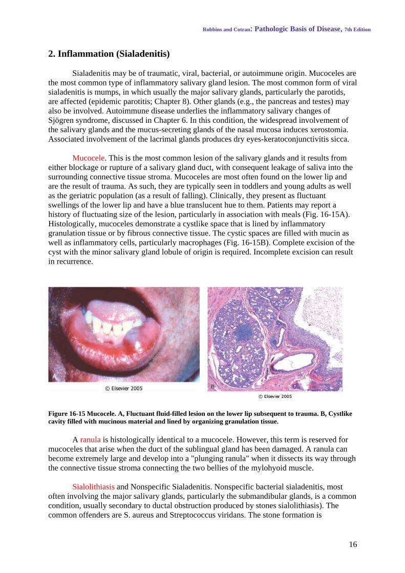
2. Inflammation (Sialadenitis)
s are viral s,
) may
syndrome, discussed in Chapter 6. In this condition, the widespread involvement of the salivary glands and the mucus-secreting glands of the nasal mucosa induces xerostomia.
l glands produces dry eyes-keratoconjunctivitis sicca.
granulation tissue or by fibrous connective tissue. The cystic spaces are filled with mucin as well as e
ystlike
n ugh
uscle.
ialolithiasis and Nonspecific Sialadenitis. Nonspecific bacterial sialadenitis, most
ialolithiasis). The common offenders are S. aureus and Streptococcus viridans. The stone formation is
Sialadenitis may be of traumatic, viral, bacterial, or autoimmune origin. Mucocelethe most common type of inflammatory salivary gland lesion. The most common form of sialadenitis is mumps, in which usually the major salivary glands, particularly the parotidare affected (epidemic parotitis; Chapter 8). Other glands (e.g., the pancreas and testesalso be involved. Autoimmune disease underlies the inflammatory salivary changes of Sjögren
Associated involvement of the lacrima
Mucocele. This is the most common lesion of the salivary glands and it results from either blockage or rupture of a salivary gland duct, with consequent leakage of saliva into the surrounding connective tissue stroma. Mucoceles are most often found on the lower lip and are the result of trauma. As such, they are typically seen in toddlers and young adults as well as the geriatric population (as a result of falling). Clinically, they present as fluctuant swellings of the lower lip and have a blue translucent hue to them. Patients may report a history of fluctuating size of the lesion, particularly in association with meals (Fig. 16-15A). Histologically, mucoceles demonstrate a cystlike space that is lined by inflammatory
inflammatory cells, particularly macrophages (Fig. 16-15B). Complete excision of thcyst with the minor salivary gland lobule of origin is required. Incomplete excision can result in recurrence.
Figure 16-15 Mucocele. A, Fluctuant fluid-filled lesion on the lower lip subsequent to trauma. B, Ccavity filled with mucinous material and lined by organizing granulation tissue.
A ranula is histologically identical to a mucocele. However, this term is reserved for mucoceles that arise when the duct of the sublingual gland has been damaged. A ranula cabecome extremely large and develop into a "plunging ranula" when it dissects its way throthe connective tissue stroma connecting the two bellies of the mylohyoid m
Soften involving the major salivary glands, particularly the submandibular glands, is a commoncondition, usually secondary to ductal obstruction produced by stones s
16
Robbins and Cotran: Pathologic Basis of Disease, 7th Edition
sometim s
suppress salivary secretion. Perhaps dehydration with decreased secretion explains the development of bacteri
ds give
le 16-
ndicated in Table 16-4, a small number of neoplasms makes up more than 90% of salivary gland tumors, and so our consideration is restricted to these. Overall, these neoplasms
ommon and represent less than 2% of tumors in humans. About 65% to 80% arise within the parotid, 10% in the submandibular gland, and the remainder in the minor salivary
% in
al to the size of the gland.
Benign Malignant
es related to obstruction of the orifices of the salivary glands by impacted food debrior by edema about the orifice after some injury. Frequently, the stones are of obscure origin. Dehydration with decreased secretory function may also predispose to secondary bacterial invasion, as sometimes occurs in patients receiving long-term phenothiazines that
al suppurative parotitis in elderly patients with a recent history of major thoracic or abdominal surgery.
Whatever the origin, the obstructive process and bacterial invasion lead to a nonspecific inflammation of the affected glands that may be largely interstitial or, when induced by staphylococcal or other pyogens, may be associated with overt suppurative necrosis and abscess formation. Unilateral involvement of a single gland is the rule. The inflammatory involvement causes painful enlargement and sometimes a purulent ductal discharge.
3. Neoplasms
Despite their relatively undistinguished normal morphology, the salivary glanrise to no fewer than 30 histologically distinct benign and malignant tumors.49-51 A classification and the relative incidence of benign and malignant tumors is shown in Tab4; not included are the rare benign and malignant mesenchymal tumors.
As i
are relatively unc
glands, including the sublingual glands. Fifteen percent to 30% of tumors in the parotid glands are malignant, in contrast to about 40% in the submandibular glands, 50the minor salivary glands, and 70% to 90% of sublingual tumors. The likelihood, then, of a salivary gland tumor being malignant is more or less inversely proportion
Pleomorphic adenoma (50%) (mixed tumor) Mucoepidermoid carcinoma (15%) Warthin tumor (5%-10%) Adenocarcinoma (NOS) (10%) Oncocytoma (1%) Acinic cell carcinoma (5%) Other adenomas (5%-10%) Adenoid cystic carcinoma (5%) Basal cell adenoma Malignant mixed tumor (3%-5%) Canalicular adenoma Squamous cell carcinoma (1%) Ductal papillomas Other carcinomas (2%)
NOS, not otherwise specified. Data from Ellis GL, Auclair PL: Tumors of the Salivary Glands. Atlas of Tumor Pathology, Third Series. Washington, DC, Armed Forces Institute of Pathology, 1996.
These tumors usually occur in adults, with a slight female predominance, but about 5% occur in children younger than age 16 years. For unknown reasons, Warthin tumors occur much more often in males than in females. The benign tumors most often appear in the fifth to seventh decades of life. The malignant ones tend, on average, to appear somewhat later. Whatever the histologic pattern, neoplasms in the parotid glands produce distinctive swellings in front of and below the ear. In general, when they are first diagnosed, both benign and
17

Robbins and Cotran: Pathologic Basis of Disease, 7th Edition
malign
, the
Because of their remarkable histologic diversity, these neoplasms have also been ixed tumors. They represent about 60% the parotid, are less common in
are re ved from a mixture o elial cells,
how both epithelial and mes y also reveal throughout a matri yxoid,
artilaginous), and even oss ithelial in others, they are presen
igins of these neoplasm
mesenchymal, are of either myoepithelial or ductal reserve cell origin (hence the designation pleomo
ing
re typically dispersed within a mesenchyme-like background of loose myxoid tissue containing islands of chondroid and,
Sometimes the epithelial cells form well-developed ducts lined by cuboidal to columnar cells with an underlying layer of deeply chromatic, small myoep ls.
r
ant lesions range from 4 to 6 cm in diameter and are mobile on palpation except in the case of neglected malignant tumors. Although benign tumors are known to have been present usually for many months to several years before coming to clinical attention, cancers seem to demand attention more promptly, probably because of their more rapid growth. Ultimatelyhowever, there are no reliable criteria to differentiate, on clinical grounds, the benign frommalignant lesions, and morphologic evaluation is necessary.
PLEOMORPHIC ADENOMA
called m of tumors inthe submandibular glands, and are relatively r in the minor salivary glands. They abenign tumors that are deri f ductal (epithelial) and myoepithand therefore they s enchymal differentiation. Theepithelial elements dispersed x along with varying degrees of mhyaline, chondroid (c eous tissue. In some tumors, the epelements predominate; t only in widely dispersed foci.
Little is known about the or s except that radiation exposure increases the risk. Equally uncertain is the histogenesis of the various components. A currently popular view is that all neoplastic elements, including those that appear
rphic adenoma).
Morphology. Most pleomorphic adenomas present as rounded, well-demarcated masses rarely exceeding 6 cm in greatest dimension (Fig. 16-16). Although they are encapsulated, in some locations (particularly the palate) the capsule is not fully developed, and expansile growth produces tonguelike protrusions into the surrounding gland, renderenucleation of the tumor hazardous. The cut surface is gray-white with myxoid and blue translucent areas of chondroid.
The dominant histologic feature is the great heterogeneity mentioned. The epithelial elements resembling ductal cells or myoepithelial cells are disposed in duct formations, acini, irregular tubules, strands, or sheets of cells. These elements a
rarely, foci of bone (Fig. 16-17).
ithelial cells. In other instances, there may be strands or sheets of myoepithelial celIslands of well-differentiated squamous epithelium may also occur. In most cases, there is no epithelial dysplasia or evident mitotic activity. There is no difference in biologic behaviobetween the tumors composed largely of epithelial elements and those composed only of seemingly mesenchymal elements.
18

Robbins and Cotran: Pathologic Basis of Disease, 7th Edition
arply circumscribed, yellow-white tumor can be seen surrounded by normal salivary gland tissue.
Figure 16-16 Pleomorphic adenoma. A, Slowly enlarging neoplasm in the parotid gland of many years duration. B, The bisected, sh
Figure 16-17 Pleomorphic adenoma. A, Low-power view showing a well-demarcated tumor with adjacentnormal salivary gland parenchyma. B, High-power view showing epithelial cells as well as myoepithelial cells found within a chondroid matrix material.
Clinical Features. These tumors present as painless, slow-growing, mobile discretmasses within the parotid or subman
e dibular areas or in the buccal cavity. The recurrence rate
(perhaps months to years later) with adequate parotidectomy is about 4% but, with attempted enucleation, approaches 25% because of failure to recognize at surgery minute protrusions from the main mass.
carcinoma arising in a pleomorphic adenoma is referred to variously as a carcinoma ex pleomorphic adenoma or a malignant mixed tumor. The incidence of malignant transformation increases with the duration of the tumor, being about 2% for tumors present less than 5 years and almost 10% for those of more than 15 years' duration. The cancer usually takes the form of an adenocarcinoma or undifferentiated carcinoma, and often it virtually completely overgrows the last vestiges of the pre-existing pleomorphic adenoma; but to substantiate the diagnosis of carcinoma ex pleomorphic adenoma, recognizable traces of
A
19

Robbins and Cotran: Pathologic Basis of Disease, 7th Edition
the latter must be found. Regrettably, these cancers, when they appear, are among the most aggressive of all salivary gland malignant neoplasms, accounting for 30% to 50% mortality in 5 years WARTHIN TUMOR (PAPILLARY CYSTADENOMA LYMPHOMATOSUM)
his curious benign neoplasm with its intimidating histologic name is the second most common salivary gland neoplasm. It arises almost always in the parotid gland (the only tumor virtually restricted to the parotid) and occurs more commonly in males than in females, usually in the fifth to seventh decades of life. About 10% are multifocal and 10% bilateral. Smokers have eight times the risk of nonsmokers for developing these tumors.
Morphology. Most Warthin tumors are round to oval, encapsulated masses, 2 to 5 cm in diam ter, arising in most cases in the superficial parotid gland, where they are readily palpable. Transection reveals a pale gray surface punctuated by narrow cystic or cleftlike spaces filled with a mucinous or serous secretion. On microscopic examination, these spaces are lined by a double layer of neoplastic epithelial cells resting on a dense lymphoid stroma
urface palisade of columnar cells having an abundant, finely granular, eosinophilic cytoplasm, imparting an oncocytic appearance, resting on a layer of cuboidal to polygonal cells. Oncocytes are epithelial cells stuffed with mitochondria that impart the granular appearance to the cytoplasm. Secretory cells are dispersed in the columnar cell layer, accounting for the secretion within the lumens. On occasion, there are foci of squamous metaplasia.
The histogenesis of these tumors has long been disputed. The occasional finding of small salivary gland rests in lymph nodes in the neck suggests that these tumors arise from the aberrant incorporation of similar inclusion-bearing lymphoid tissue in the parotids. Indeed, rarely, Warthin tumors have arisen within cervical lymph nodes, a finding that should not be misconstrued to imply a metastasis. These neoplasms are benign, with recurrence rates of only 2% after resection.
troma.
.
T
e
sometimes bearing germinal centers (Fig. 16-18). The spaces are frequently narrowed by polypoid projections of the lymphoepithelial elements. The double layer of lining cells is distinct ve, with a si
Figure 16-18 Warthin tumor. A, Low-power view showing epithelial and lymphoid elements. Note the follicular germinal center beneath the epithelium. B, Cystic spaces separate lobules of neoplastic epithelium consisting of a double layer of eosinophilic epithelial cells based on a reactive lymphoid s
20

Robbins and Cotran: Pathologic Basis of Disease, 7th Edition
21
These neoplasms are composed of variable mixtures of squamous cells, mucus-nd tumors,
nd while they occur mainly (60% to 70%) in the parotids, they account for a large fraction of salivary
veal small, mucin-
ve
h clear cells
ontaining mucin. B, Mucicarmine stains the mucin reddish-pink. (Courtesy of Dr. James Gulizia, righam and Women's Hospital, Boston.
The clinical course and prognosis depend on the grade of the neoplasm. Low-grade mors may invade locally and recur in about 15% of cases, but only rarely do they etastasize and so yield a 5-year survival rate of more than 90%. By contrast, high-grade
eoplasms and, to a somewhat lesser extent, intermediate-grade tumors are invasive and ifficult to excise and so recur in about 25% to 30% of cases and, in 30% of cases, isseminate to distant sites. The 5-year survival rate of these tumors is only 50%.
Two less common neoplasms merit brief description: adenoid cystic carcinoma and cinic cell tumor.
Adenoid cystic carcinoma is a relatively uncommon tumor, which in approximately 0% of cases is found in the minor salivary glands (in particular the palate). Among the major
MUCOEPIDERMOID CARCINOMA
secreting cells, and intermediate cells. They represent about 15% of all salivary glaa
gland neoplasms in the other glands, particularly the minor salivary glands. Overall, they are the most common form of primary malignant tumor of the salivary glands.
Morphology. Mucoepidermoid carcinomas can grow up to 8 cm in diameter and although they are apparently circumscribed, they lack well-defined capsules and are often infiltrative at the margins. Pale and gray-white on transection, they frequently re
containing cysts. The basic histologic pattern is that of cords, sheets, or cystic configurations of squamous, mucous, or intermediate cells. The hybrid cell types often hasquamous features, with small to large mucus-filled vacuoles, best seen when highlighted with mucin stains (Fig. 16-19A,B). The tumor cells may be regular and benign appearing or, alternatively, highly anaplastic and unmistakably malignant. Accordingly, mucoepidermoid carcinomas are subclassified into low, intermediate, or high grade.
Figure 16-19 A, Mucoepidermoid carcinoma s owing islands having squamous cells as well ascB
tumndd
OTHER SALIVARY GLAND TUMORS
a
5

Robbins and Cotran: Pathologic Basis of Disease, 7th Edition
salivary glands, the parotid and submandibular glands are the most common locations. Similar neoplas
spaces betwee ss
ndency
es
s tids.
gl
created a cribriform pattern enclosing secretions. B, Pe
of salivary gland
mors. Most arise in the parotids; the small remainder arise in the submandibular glands.
al a
ls are s usually
eomorphic. Several histologic patterns of growth (solid, microcystic, papillary-cystic, and follicular) are
ms is somewhat dependent on the level of pleomo
etastasize to lymph nodes. The survival rate is in the range of 90% at 5 years a
ms have been reported in the nose, sinuses, and upper airways and elsewhere. Morphology. In gross appearance, they are generally small, poorly encapsulated, infiltrative, gray-pink lesions. On histologic evaluation, they are composed of small cells having dark, compact nuclei and scant cytoplasm. These cells tend to be disposed in tubular, solid, or cribriform patterns reminiscent of cylindromas arising in the adnexa of the skin. The
n the tumor cells are often filled with a hyaline material thought to represent excebasement membrane (Fig. 16-20A). Other less common histologic patterns have been designated as tubular and solid variants.
Although slow growing, these are relentless and unpredictable tumors with a teto invade perineural spaces (Fig. 16-20B), and they are stubbornly recurrent. Eventually, 50%or more disseminate widely to distant sites such as bone, liver, and brain, sometimes decadafter attempted removal. Thus, although the 5-year survival rate is about 60% to 70%, it dropsto about 30% at 10 years and 15% at 15 years. Neoplasms arising in the minor salivary glandhave, on average, a poorer prognosis than those primary in the paro
and. A, Low-power view. The tumor cells have rineural invasion by tumor cells.
Figure 16-20 Adenoid cystic carcinoma in a salivary
The acinic cell tumor is composed of cells resembling the normal serous acinar cells
salivary glands. They are relatively uncommon, representing only 2% to 3% oftuThey rarely involve the minor glands, which normally have only a scant number of serous cells. Like Warthin tumor, they are sometimes bilateral or multicentric. They are generally small, discrete lesions that may appear encapsulated. On histologic examination, they revevariable architecture and cell morphology. Most characteristically, the cells have apparent cleared cytoplasm, but the cells are sometimes solid or at other times vacuolated. The celdisposed in sheets or microcystic, glandular, follicular, or papillary patterns. There ilittle anaplasia and few mitoses, but some tumors are occasionally slightly more pl
recognized. The clinical course of these neoplasrphism. Overall, recurrence after resection is uncommon, but about 10% to 15% of
these neoplasms mnd 60% at 20 years.
22