Opinion on the Environmental and Health Risks Posed by...
41
1 Scientific Committee on Health and Environmental Risks SCHER Opinion on the Environmental and Health Risks Posed by Depleted Uranium The SCHER adopted this opinion at its 7th plenary on 18 May 2010 after public consultation
-
Upload
dangkhuong -
Category
Documents
-
view
214 -
download
0
Transcript of Opinion on the Environmental and Health Risks Posed by...

1
Scientific Committee on Health and Environmental Risks
SCHER
Opinion on the
Environmental and Health Risks
Posed by Depleted Uranium
The SCHER adopted this opinion at its 7th plenary on 18 May 2010 after public consultation
2
About the Scientific Committees
Three independent non-food Scientific Committees provide the Commission with the scientific advice it needs when preparing policy and proposals relating to consumer safety, public health and the environment. The Committees also draw the Commission's attention to the new or emerging problems which may pose an actual or potential threat.
They are: the Scientific Committee on Consumer Safety (SCCS), the Scientific Committee on Health and Environmental Risks (SCHER) and the Scientific Committee on Emerging and Newly Identified Health Risks (SCENIHR) and are made up of external experts.
In addition, the Commission relies upon the work of the European Food Safety Authority (EFSA), the European Medicines Evaluation Agency (EMEA), the European Centre for Disease prevention and Control (ECDC) and the European Chemicals Agency (ECHA).
SCHER
Opinions on risks related to pollutants in the environmental media and other biological and physical factors or changing physical conditions which may have a negative impact on health and the environment, for example in relation to air quality, waters, waste and soils, as well as on life cycle environmental assessment. It shall also address health and safety issues related to the toxicity and eco-toxicity of biocides.
It may also address questions relating to examination of the toxicity and eco-toxicity of chemical, biochemical and biological compounds whose use may have harmful consequences for human health and the environment. In addition, the Committee will address questions relating to methodological aspect of the assessment of health and environmental risks of chemicals, including mixtures of chemicals, as necessary for providing sound and consistent advice in its own areas of competence as well as in order to contribute to the relevant issues in close cooperation with other European agencies.
Scientific Committee members
Ursula Ackermann-Liebrich, Herman Autrup, Denis Bard, Peter Calow, Stella Canna Michaelidou, John Davison, Wolfgang Dekant, Pim de Voogt, Arielle Gard, Helmut Greim, Ari Hirvonen, Colin Janssen, Jan Linders, Borut Peterlin, Jose Tarazona, Emanuela Testai, Marco Vighi
Contact:
European Commission DG Health & Consumers Directorate C: Public Health and Risk Assessment Unit C7 - Risk Assessment Office: B232 B-1049 Brussels
© European Union, 2010 ISSN 1831-4775 ISBN 978-92-79-12754-0 doi:10.2772/3253 ND-AR-09-005-EN-N
The opinions of the Scientific Committees present the views of the independent scientists who are members of the committees. They do not necessarily reflect the views of the European Commission. The opinions are published by the European Commission in their original language only.
http://ec.europa.eu/health/ph_risk/risk_en.htm
3
ACKNOWLEDGMENTS
Members of the working group are acknowledged for their valuable contribution to this opinion. The members of the working group are:
Prof. Wolfgang Dekant (Chair and Rapporteur)
Prof. Pim De Voogt
Prof. Borut Peterlin
Prof. Marco Vighi
External Experts:
Prof. Pier Roberto Danesi Ionizing Radiation Technology- University Institute for Advanced Studies (IUSS), Pavia- Italy
Prof. Victor Meineke Bundeswehr Institute of Radiobiology (University of Ulm), Munich - Germany
Prof. Ana Proykova Scientific Committee on Emerging and Newly Identified Health Risks (SCENIHR)
All Declarations of working group members are available at the following webpage:
http://ec.europa.eu/health/scientific_committees/environmental_risks/members_wg/index_en.htm
Keywords:
depleted uranium, uranium, human toxicity, environmental effects, biomonitoring, radiation
Opinion to be cited as: SCHER (Scientific Committee on Health and Environmental Risks), Opinion on the environmental and health risks posed by depleted uranium, 18 May 2010
4
TABLE OF CONTENTS
ACKNOWLEDGMENTS ...........................................................................................3 TABLE OF CONTENTS............................................................................................4 1. BACKGROUND .............................................................................................5 2. TERMS OF REFERENCE..................................................................................5 3. SCIENTIFIC RATIONALE................................................................................6
3.1. Depleted uranium - properties and usage...................................................6 3.2. Hazard assessment.................................................................................6
3.2.1. Radiological properties.......................................................................6 3.2.2. Radiation mediated effects of DU.........................................................7 3.2.3. Toxicology of uranium and depleted uranium ........................................8
3.3. Environmental toxicology of uranium....................................................... 10 3.4. Exposure assessment............................................................................ 12 3.5. Risk assessment................................................................................... 16
3.5.1. Human health risks ......................................................................... 16 3.5.2. Environmental health risks ............................................................... 17
4. RESPONSE TO TERMS OF REFERENCE........................................................... 18 4.1. Question 1........................................................................................... 18 4.2. Question 2........................................................................................... 18 4.3. Question 3........................................................................................... 19
5. REFERENCES............................................................................................. 19 Annex 1: Health effects of radiation ...................................................................... 29 Annex 2: Environmental chemistry of natural uranium............................................. 36
5
1. BACKGROUND Depleted uranium (DU) is a by-product of uranium enrichment. It is only slightly radioactive, and its extreme density and ready availability make it suitable for a number of applications, both civilian and military.
Public concern about the toxic effects of DU on humans and the environment focuses on exposure of humans and the environment to DU following military use of DU, where DU ordnance is used primarily for armour piercing purposes.
Widespread public concern over the detrimental health effects of DU started at the time of the first Gulf War (1990-91) regarding its possible links to the “Gulf War Syndrome” (widespread reports of symptoms including immune system defects, chronic pain, fatigue and memory loss by ex-combatants) and to an alleged high level of birth defects affecting ex-combatants’ children born after the conflict.
Subsequent widespread use of DU munitions was reported in the course of NATO operations in the former Yugoslavia from 1996 and the second Gulf War in 2003.
Studies by WHO1, IAEA2 and the Article 31 Committee established under the Euratom Treaty3 failed to find any conclusive evidence linking the use of depleted uranium weapons with significant risks to the health of the civilian population in former combat areas or to that of former combatants4.
The International Coalition to ban Uranium Weapons (ICBUW) disputes the radiation-exposure and dose estimation model underlying the Art 31 Committee study arguing that DU weapons present an entirely new source of environmental contamination which may directly or indirectly affect humans and the environment itself. They argue that the health effects that may be caused by DU following military uses of DU containing weapons require additional comprehensive scientific assessments5.
In May 2008, the European Parliament passed a resolution on DU weapons which called on the Commission and others inter alias:
• to commission scientific studies into the use of DU
• to establish an environmental inventory of DU contaminated areas and to provide support for projects that could assist victims and their relatives as well as for clean-up operations in the affected areas, should a negative effect on human health and the environment be confirmed.
2. TERMS OF REFERENCE 1) The SCHER is asked for an opinion building on an evaluation of available reports,
including but not restricted to those referenced above, as to the environmental and health risks posed by DU.
2) In particular SCHER is asked to assess those risks that may arise from exposure to DU in contaminated areas following military activities with DU containing weapons.
3) SCHER is asked to take into account both the chemical and radiological toxicities of DU and, if appropriate, their possible synergistic relations.
1 http://www.who.int/ionizing_radiation/env/du/en/index.html 2 http://www.iaea.org/NewsCenter/News/2003/13-571089.shtml 3 http://ec.europa.eu/energy/nuclear/radioprotection/doc/art31/opinion_en.pdf 4 It should be noted that the two latter studies concentrate exclusively on radiological and not
chemical toxic effects of exposure to DU. 5 See letter from ICBUW and annexed bibliography.
6
3. SCIENTIFIC RATIONALE
3.1. Depleted uranium - properties and usage Uranium (U) is a heavy metal. Uranium is easily oxidized in air and U metal is therefore coated with a layer of U oxides, U is therefore present mainly as oxides in the environment. Natural U is weakly radioactive and contains the radioisotopes U-234, U-235 and U-238. All U isotopes have a very long half-live and decay to many other radioisotopes, called progeny. The decay of U finally results in stable isotopes of lead (Burkhart, 1991; Choppin et al., 1966).
Depleted uranium (DU) is a by-product of uranium enrichment. Depleted uranium is less radioactive then U (see below), but retains the chemical properties of natural U. Depleted uranium has a variety of applications because of its high density and its pyrophoric properties. It has been used as counterbalance weight in aircraft, missiles, forklifts and sailboat keels. It was also used in medical radiotherapy as a radiation shield and in dental porcelain crowns (until 1982). In weapons technology, DU is used in armour plates in heavy tanks and in armour-piercing ammunition. DU-containing ammunition was first used in the 1991 Gulf War and has again been used in Serbia, in Kosovo, and in the 2003 Gulf war. Due to their high kinetic energy and the pyrophoric properties of U, DU ammunitions are used solely for the purpose of armor-piercing and have little use against other targets (Bleise et al., 2003).
3.2. Hazard assessment
3.2.1. Radiological properties
Uranium is the heaviest naturally occurring element and all isotopes of U are radioactive (Table 1). In order to produce fuel for nuclear reactors and material for nuclear weapons, U has to be "enriched" in the U-235 isotope, which is responsible for nuclear fission. During the enrichment process, the fraction of U-235 is increased from 0.72 % present in natural U to a content of U-235 between 2% and 94%. After removal of the enriched U, the remaining U has significantly reduced concentrations of U-235 and U-234, which is called DU (Table 1). Therefore, only the composition of isotopes is changed in DU as compared to U with the natural isotope composition. DU is defined as U with a percentage fraction by weight of U-235 of less then 0.711%. Typical concentrations of U-235 in DU are 0.2 to 0.3 weight-%, which represents approximately 30 - 40% of its concentration in natural U (Table 1). The specific activities of natural U (after removal of highly radioactive decay products) and DU (0.2 %) are compared in table 1.
Table 1. Relative isotopic abundance and radioactivity of chemically purified natural U and DU (0.2 %) (Benedict et al., 1981; Bleise et al., 2003; Glastone and Sesonske, 1981; Larsen, 2000). The specific activities (in Bq/mg) of uranium isotopes are 12.44 (U-238), 80 (U-235), and 2.31x10⁵(U-234)
Isotope Natural U DU
Abundance Radioactivity/mg (Bq)
Abundance Radioactivity/mg (Bq)
U-238 99.28 % 12.40 99.8 % 12.40
U-235 0.72 % 0.57 0.2 % 0.16
U-234 0.0057 % 12.40 0.001 % 2.26
Total 25.28 14.80
The radioactivity of freshly prepared DU is only about 60% the radioactivity of natural uranium as DU has less of the more radioactive isotopes U-234 and U-235 per mass unit
7
then natural U (Table 2) (Bleise et al., 2003).
All natural U isotopes emit alpha particles (table 2), i.e. positively charged ions composed of two protons and two neutrons. Both beta (high-energy electrons) and gamma (very high energy photons) activity of relevant U isotopes are low. Due to their relatively large size and charge, alpha particles have little penetrating power. The penetration range of a 5 MeV alpha particle is approximately 4 cm in air and 50 micrometers in soft tissue. Therefore, alpha particles do not penetrate the keratin layer of intact human skin. As a result, U represents a radiation hazard only after inhalation or ingestion.
Table 2. Average energy emission per transformation of the U isotopes U-238, U-235 and U-234 (Burkhart, 1991).
Average energy per transformation (MeV/Bq) Isotope
Alpha Beta Gamma
U-238 4.26 0.01 0.001
U-235 4.47 0.048 0.154
U-234 4.84 0.0013 0.002
DU penetrators collected in Kosovo contained traces of U-236, Pu-239 and Pu-240 (IAEA, 2003; UNEP, 2001). Trace amounts of Am, Np, and 99Tc were also detected (DAF-OO-ALC, 1997; Diehl, 2001). The traces of U-236 (<0.003%) may result from cross-contamination due to the use of the same equipment for handling both non-irradiated and irradiated U (TACOM, 2000). However, the increase in radiation dose due to the trace amounts of these elements and isotopes is less than 1% (WHO, 2001).
3.2.2. Radiation mediated effects of DU
In general, radiation may induce both deterministic and stochastic health effects (Hall and Giacca, 2006). Deterministic effects of radiation include the acute health effects observed after high “radiation doses”, sometimes referred to as general “radiation sickness” which is characterized by effects of radiation on rapidly proliferating cells. Depending on the amount of the deposited energy within the tissues (often simplified as “radiation dose”) these health effects might result in the hematopoetic, the gastrointestinal, the neurovascular or the cutaneous “radiation syndrome”, or a combination of these syndromes. Deterministic effects per definition only occur above a threshold radiation dose. Examples for deterministic radiation effects are “unwanted effects” observed after radiotherapies for malignant diseases, effects seen after industrial radiation accidents (IAEA, 1996), or those observed in the Hiroshima and Nagasaki victims after the attack with nuclear weapons in World War II (Kondo, 1993; Preston et al., 2003). Exposure to DU by all conceivable exposure pathways is not expected to result in deterministic effects (“radiation sickness”) in humans.
Stochastic effects are represented by the induction of mutations by radiation, which may result in cancer. Regarding stochastic effects, a linear no-threshold (LNT) dose-response hypothesis in the low dose range is assumed. For more details on radiation doses, assessment of radiation health risks, and radiation carcinogenicity, see Annex I.
Although radiation exposure is generally assumed to be carcinogenic at all dose levels, no correlation between tumour incidence and radiation has been established at low “doses” in the range of natural radiation background. This is attributable to two factors: (1) it is difficult to obtain meaningful data from epidemiological studies where exposure is near background exposure levels, and (2) the results of such studies usually do not give statistically significant differences between exposed and unexposed groups to substantiate a health impact (Hall et al., 2009). The same problems have to be faced when trying to transfer basic principles of radiation damage mechanisms such as the so-
8
called bystander effect (damage of non-irradiated cells by irradiated cells or mediators at very low doses) from in vitro to in vivo and estimating the real role for radiation carcinogenesis (Little, 2006; Williams, 2008). However, the low-dose linearity concept is still the accepted standard for radiation protection policies (Puskin, 2008). Recently, reviews of carcinogenicity and exposure to chemicals and radiation have questioned the non-threshold assumption (Averbeck, 2009; Clark, 1999; EU-SCHER, 2009) since there is increasing biological evidence for a potential threshold in radiation- and chemically-induced carcinogenicity.
The available information on radioactivity and its effects shows that high dose alpha radiation can cause a variety of effects in humans. The nature and the severity of these effects depend on several factors, including physicochemical form and solubility of the alpha-emitting isotope, route of entry, distribution, biological retention, and specific alpha-energy emitted. Since the specific alpha-emissions of both natural U and DU are low and the potential for internal exposures to U and DU in humans is very limited, there is no conclusive evidence on biological effects in humans by alpha-radiation from U (UNEP/UNCHS, 1999).
Potential radiological effects due to the intake of U and DU both by inhalation and by ingestion have been assessed by a variety of international expert groups in peer-reviewed reports. WHO had made a detailed assessment of potential radiation-mediated effects of both U and DU. This assessment included modelling of inhalation exposures and considered specific biokinetics of insoluble U-oxides and mixed U/Fe-oxides. This assessment concluded that potential exposures to DU will add only a negligible contribution to total U-intake. Any DU-derived radiation will remain below an effective radiation dose < 1 mSv and thus well below accepted dose-rate limits derived for radiation protection. This conclusion was confirmed by other international expert groups (Durante and Pugliese, 2002; EU-EURATOM, 2001; EURATOM, 2009; IAEA, 2003, 2009; Li et al., 2009; UNEP, 2001, 2002, 2003, 2007; UNEP/UNCHS, 1999; UNSCEAR, 1993, 2000b, a; WHO, 2001, 2003b) and SCHER agrees with this conclusion.
3.2.3. Toxicology of uranium and depleted uranium
Since all isotopes of an element have the same chemical properties, they also have an identical chemical toxicity; therefore, the chemical toxicity of DU is identical to that of natural U. Thus, the toxicity data on natural U can be applied to assess potential human health risks from DU exposures. Since DU has a much lower radioactivity as compared to natural U and U-containing ores, it is generally agreed that the chemical toxicity of U is the major hazard descriptor regarding assessment of health risk due to potential exposures to DU. The higher radioactivity may result in a higher toxic potency of natural U as compared to that of DU regarding potential radiation-mediated effects (ATSDR, 1999; Bleise et al., 2003; Konietzka et al., 2005; McDiarmid, 2001; WHO, 2001, 2003b). This was confirmed by the observation that chromosomal damage from U is more pronounced then that of DU when applying identical concentrations to cultured cells (Miller et al., 2002b)
Depending on the solubility of the U salt administered, systemic absorption of U from the gastrointestinal tracts is from 0.02 to 6 %. Respirable U particles in air may be deposited in the respiratory tract. Approximately 95% of inhaled particles with aerodynamic equivalent diameter (AED) larger than 10 micrometers deposit in the upper respiratory tract, most of these clear to the pharynx and thus to the GI tract. Particles <10 micrometers can reach deeper pulmonary regions (bronchioles and alveoli) and stay there for considerable time (Bleise et al., 2003). The extent of systemic availability of U particles inhaled also depends on particle characteristics such as specific surface area (Chazel et al., 1998), elemental composition, and U oxidation states.
Most (> 98 %) of the U introduced into the gastrointestinal tract is excreted with faeces (Leggett and Harrison, 1995; Tracy et al., 1992). Absorbed U is distributed to the bone and to the kidney and accumulates there. Elimination half-lives for U from the different
9
compartments in the organism vary widely with a half-life of up to 6 days for renal excretion and predicted half-lives of up to 500 days for elimination from bone (ATSDR, 1999; WHO, 2001, 2003b).
The toxicity of U is comparatively well studied. Toxicity of U salts is highly depending on solubility in water and tissues; many U oxides are of low solubility and thus also have a low potential for toxicity. As with other heavy metals, the major target organ for the toxicity of soluble U salts is the kidney. Long-term administration of U causes damage to the glomeruli and the proximal tubuli (Gilman et al., 1998a; Gilman et al., 1998b; Gilman et al., 1998c; McDonald-Taylor et al., 1992; McDonald-Taylor et al., 1997) with Lowest-Observed-Effect-Levels (LOAELs) of 0.06 mg/kg bw/day (Table 3). High concentrations of natural U given to mice during pregnancy have shown decreased fertility, toxicity to the fetus, some neurobehavioral effects, and an increased incidence of developmental variations with an overall LOAEL of 2.8 mg/kg bw/day (Albina et al., 2005; Arfsten et al., 2009; Belles et al., 2005; Domingo, 2001). As many other metals (Figgitt et al., 2010; Tsaousi et al., 2010), both U and DU have been reported to cause genotoxic effects in short term in vitro test often applied to assess genotoxicity (ATSDR, 1999; Coryell and Stearns, 2006; Hartsock et al., 2007; Knobel et al., 2006; Miller et al., 2005; Miller et al., 2004; Miller et al., 2001; Miller et al., 2002a; Wise et al., 2007; Xie et al., 2010). However, the positive in vitro tests with U are not predictive of carcinogenicity in vivo since carcinogenic effects have not been observed in animals ingesting soluble or insoluble U compounds (ATSDR, 1999). There is also no evidence for a carcinogenicity of natural U from studies of workers in U mines. The higher cancer incidence in these cohorts is likely due to inhalation exposure to radon and its decay products and not due to U particle inhalation (ATSDR, 1999; Harley, 2001; Kreuzer et al., 2009; NRC, 1991).
Both in rodents and in rabbits, repeated administration of U with drinking water gave No-Observed-Adverse-Effect-Levels (NOAELs) or LOAELs of 60 µg/kg bw per day based on subtle histopathological changes in the kidney (Table 3). These NOAELs/LOAELs have been transformed in tolerable daily intakes for natural U with an uncertainty factor of 100 to give a Tolerable-Daily-Intake (TDI) of 0.6 µg/kg bw per day. Some studies also suggest small functional changes in the kidney when humans are exposed to high (natural) U doses with drinking water at doses of 20 to 200 µg U/day (ATSDR, 1999; Zamora et al., 1998; Zamora et al., 2009). Since DU shows an identical toxicity as natural U, the TDI for natural U is also applicable to DU.
Table 3. Assessment of the chemical toxicity of U. TDI, tolerable daily intake; LOAEL, Lowest observed adverse effect level; NOAEL, No observed adverse effect level; WHO, World Health Organisation; UBA, Umweltbundesamt (Germany); BfR, Bundesinstitut für Riskikobewertung (Germany)
Agency Data base for derivation L/NOAEL [µg/kg x d]
TDI [µg/kg x d]
(WHO, 1998) rats 60; LOAEL 0.60 (EPA, 2000) rats 60; LOAEL 0.60 (UBA, 2000) rabbits < 60; NOAEL < 0.60 (WHO, 2003a) rats 60; LOAEL 0.60 (BfR, 2004) rats 60; LOAEL 0.60 (UBA, 2004) Rat and human data 50; NOAEL 0.2
A large number of recent studies have specifically addressed DU toxicity (Arnault et al., 2008; Berradi et al., 2008; Briner and Murray, 2005; Bussy et al., 2006; Coryell and Stearns, 2006; Dublineau et al., 2007; Feugier et al., 2008; Fukuda et al., 2006; Goldman et al., 2006; Grignard et al., 2008; Gueguen et al., 2007; Gueguen et al., 2006; Hahn et al., 2002; Hartsock et al., 2007; Hu and Zhu, 1990; Kalinich et al., 2002; Kundt et al., 2009; Kurttio et al., 2005; Lestaevel et al., 2005; Lestaevel et al., 2009; Miller et al., 2002a; Monleau et al., 2006a; Monleau et al., 2006b; Monleau et al., 2006c;
10
Periyakaruppan et al., 2007; Periyakaruppan et al., 2009; Pourahmad et al., 2006; Racine et al., 2009; Souidi et al., 2005; Stearns et al., 2005; Thiebault et al., 2007; Tissandie et al., 2007; Tissandie et al., 2006; Wan et al., 2006; Wise et al., 2007; Xie et al., 2010; Zhu et al., 2009). Many studies confirm that DU toxicity is identical to that of U. Some of the other studies have focused on U and DU effects after administration of single or repeated high doses, used a short time frame of observation, or focused on selected biochemical changes without characterizing functional or pathologic consequences. Other studies used inappropriate routes of administration such as intraperitoneal injection. Studies useful for risk assessment should apply the chemical of interest by a route of exposure relevant to humans for a significant part of the life-span of an experimental animal such as the studies used to derive the tolerable intakes. Therefore, all these studies do not add new relevant information to be used in risk assessment of human exposures to U and DU.
3.3. Environmental toxicology of uranium Limited data on the ecotoxicity of U are available. In the US EPA ECOTOX database, only 46 records are available for U toxicity to aquatic species. LC50 values range from 21-32,700 µg/L in crustaceans, 36,300 for an algal species, 4,000 – 100,000 µg/L in fish and 2,900-3,900 µg/L in an invertebrate species (H. viridissima). No data are recorded in the ECOTOX database for U toxicity in terrestrial species. For U oxide, four records are available in the US EPA ECOTOX database, all for the water flea C.dubia. The reported NOEC level is 30 µg/L and the LC50 is 50 µg/L (US-EPA, 2009).
Uranium in the aqueous environment generally occurs as the uranyl ion (UO22+). In
freshwater at a pH > 6, the uranyl ion forms complexes with carbonate ions (Poston et al., 1984).
The ECOTOX database contains data for uranyl sulfate (55 records, 9 species) and uranyl nitrate (105 records, 14 species). For uranyl nitrate, a (90-120 d) NOEC of 2,000 µg/L was recorded in alga. The 48 h EC50 in D.magna ranges between 4,000 and 74,000 µg/L. The 48 h LC50 in C.dubia ranges between 60-89 µg/L, whereas the (7 d) NOEC ranges between 1.5 and 8 µg/L. In fish, the 96 h LC50 values are above 3 mg/L. In duckweed, a NOEC of 500 µg/L was recorded. No data are available for uranyl nitrate in terrestrial organisms. For uranyl sulfate, the lowest (5 d) NOEC value reported was in the daphnid M. macleayi at 10 µg/L. The lowest reported LC50 in fish is 2.5 mg/L, and the lowest (4 d) NOEC is 560 µg/L. In the invertebrate H. viridissima, a (5 d) NOEC of 150 µg/L is reported. For the terrestrial environment, the (0.5 h) LOEL in reindeer lichen is 0.1 M.
The Dutch RIVM has summarized information on the occurrence and toxicity of U in the environment (Van de Plassche et al., 1999). On the basis of aquatic and terrestrial ecotoxicity data reviewed, a maximum permissible addition to background levels of 1.0 µg U per L in both seawater, freshwater and groundwater was proposed. For soil, a background concentration of 2.9 mg/kg was derived and a maximum permissible concentration of 28.3 mg U/kg of soil was proposed.
These risk limit values were proposed based on toxicity data taken from the literature. Several, but not all of the studies corresponded to the ones used in the US-EPA ECOTOX database. Chronic toxicity of U to freshwater crustaceans ranged from 10 – 1,290 µg/L (NOEC, 2 species). Acute toxicities in crustaceans ranged from 400 to 30,000 µg/L, whereas, in fish, the LC50 ranges from 730 to more than 100,000 µg/L. For the terrestrial environment, the RIVM study (Van de Plassche et al., 1999) quoted Sheppard et al. 1992 who reported a NOEC for plants of 254 mg U per kg dw of soil, and a LC50 for the earthworm L. terrestris of more than 1000 mg/kg (Sheppard et al., 1992).
Sheppard and collaborators (2005) later reviewed the chemical toxicity of U and proposed a suite of ecotoxicity thresholds for U (Table 4). The most sensitive organisms in this evaluation appeared to be the freshwater invertebrates and freshwater plants, for both of which a PNEC of 5 µg/L was proposed (Sheppard et al., 2005). They also concluded that in human risk assessments the chemical toxicity of U is the focus, and that the same is expected for non-human biota.
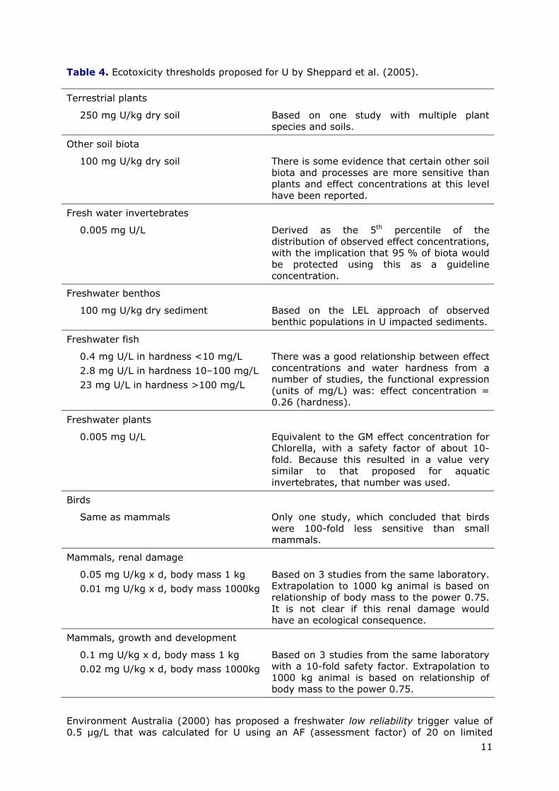
11
Table 4. Ecotoxicity thresholds proposed for U by Sheppard et al. (2005).
Terrestrial plants
250 mg U/kg dry soil Based on one study with multiple plant species and soils.
Other soil biota
100 mg U/kg dry soil There is some evidence that certain other soil biota and processes are more sensitive than plants and effect concentrations at this level have been reported.
Fresh water invertebrates
0.005 mg U/L Derived as the 5th percentile of the distribution of observed effect concentrations, with the implication that 95 % of biota would be protected using this as a guideline concentration.
Freshwater benthos
100 mg U/kg dry sediment Based on the LEL approach of observed benthic populations in U impacted sediments.
Freshwater fish
0.4 mg U/L in hardness <10 mg/L 2.8 mg U/L in hardness 10–100 mg/L 23 mg U/L in hardness >100 mg/L
There was a good relationship between effect concentrations and water hardness from a number of studies, the functional expression (units of mg/L) was: effect concentration = 0.26 (hardness).
Freshwater plants
0.005 mg U/L Equivalent to the GM effect concentration for Chlorella, with a safety factor of about 10-fold. Because this resulted in a value very similar to that proposed for aquatic invertebrates, that number was used.
Birds
Same as mammals Only one study, which concluded that birds were 100-fold less sensitive than small mammals.
Mammals, renal damage
0.05 mg U/kg x d, body mass 1 kg 0.01 mg U/kg x d, body mass 1000kg
Based on 3 studies from the same laboratory. Extrapolation to 1000 kg animal is based on relationship of body mass to the power 0.75. It is not clear if this renal damage would have an ecological consequence.
Mammals, growth and development
0.1 mg U/kg x d, body mass 1 kg 0.02 mg U/kg x d, body mass 1000kg
Based on 3 studies from the same laboratory with a 10-fold safety factor. Extrapolation to 1000 kg animal is based on relationship of body mass to the power 0.75.
Environment Australia (2000) has proposed a freshwater low reliability trigger value of 0.5 µg/L that was calculated for U using an AF (assessment factor) of 20 on limited
12
chronic data. No marine data were available to calculate a guideline value. This value should only be used as an indicative interim working level (Environment-Australia, 2000).
The OEHHA (California Office of Environmental Health Hazard Assessment) has withdrawn the previously established PHG for U of 2 picocuries per L of water, and announced to develop and adopt a new PHG in accordance to Health and Safety Code, Section 116365. Based on the current review of the new information, it can be concluded that relatively few data are available for the ecotoxicity of U and that hardly any such data are available for the terrestrial environment (OEHHA, 1998).
3.4. Exposure assessment
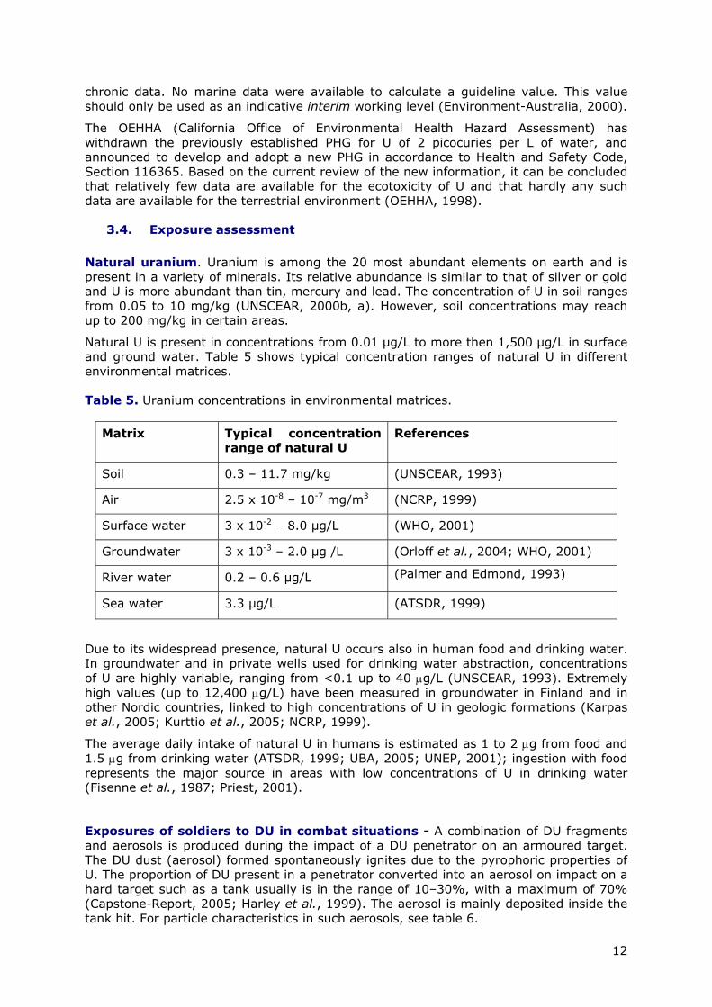
Natural uranium. Uranium is among the 20 most abundant elements on earth and is present in a variety of minerals. Its relative abundance is similar to that of silver or gold and U is more abundant than tin, mercury and lead. The concentration of U in soil ranges from 0.05 to 10 mg/kg (UNSCEAR, 2000b, a). However, soil concentrations may reach up to 200 mg/kg in certain areas.
Natural U is present in concentrations from 0.01 µg/L to more then 1,500 µg/L in surface and ground water. Table 5 shows typical concentration ranges of natural U in different environmental matrices.
Table 5. Uranium concentrations in environmental matrices.
Matrix Typical concentration range of natural U
References
Soil 0.3 – 11.7 mg/kg (UNSCEAR, 1993)
Air 2.5 x 10⁻⁸ – 10-7 mg/m3 (NCRP, 1999)
Surface water 3 x 10⁻² – 8.0 µg/L (WHO, 2001)
Groundwater 3 x 10⁻³ – 2.0 µg /L (Orloff et al., 2004; WHO, 2001)
River water 0.2 – 0.6 µg/L (Palmer and Edmond, 1993)
Sea water 3.3 µg/L (ATSDR, 1999)
Due to its widespread presence, natural U occurs also in human food and drinking water. In groundwater and in private wells used for drinking water abstraction, concentrations of U are highly variable, ranging from <0.1 up to 40 µg/L (UNSCEAR, 1993). Extremely high values (up to 12,400 µg/L) have been measured in groundwater in Finland and in other Nordic countries, linked to high concentrations of U in geologic formations (Karpas et al., 2005; Kurttio et al., 2005; NCRP, 1999).
The average daily intake of natural U in humans is estimated as 1 to 2 µg from food and 1.5 µg from drinking water (ATSDR, 1999; UBA, 2005; UNEP, 2001); ingestion with food represents the major source in areas with low concentrations of U in drinking water (Fisenne et al., 1987; Priest, 2001).
Exposures of soldiers to DU in combat situations - A combination of DU fragments and aerosols is produced during the impact of a DU penetrator on an armoured target. The DU dust (aerosol) formed spontaneously ignites due to the pyrophoric properties of U. The proportion of DU present in a penetrator converted into an aerosol on impact on a hard target such as a tank usually is in the range of 10–30%, with a maximum of 70% (Capstone-Report, 2005; Harley et al., 1999). The aerosol is mainly deposited inside the tank hit. For particle characteristics in such aerosols, see table 6.
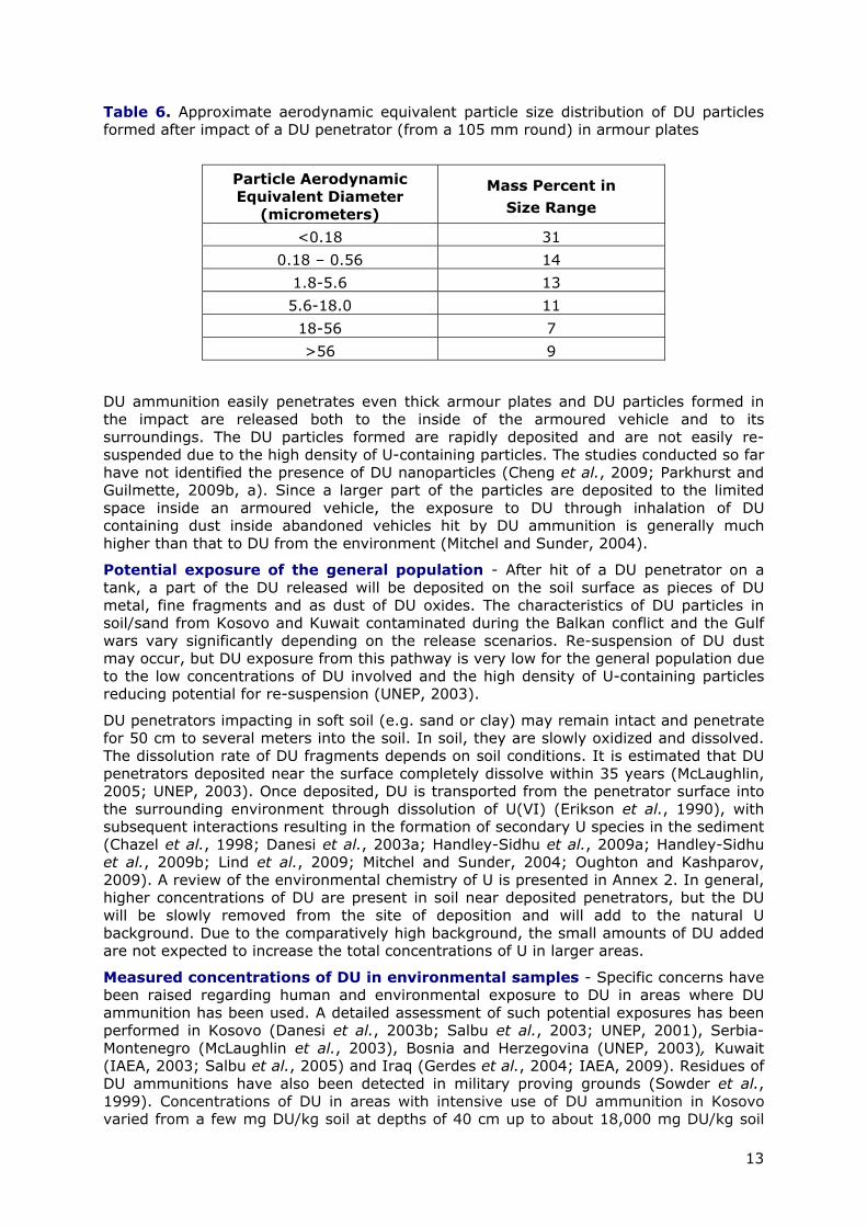
13
Table 6. Approximate aerodynamic equivalent particle size distribution of DU particles formed after impact of a DU penetrator (from a 105 mm round) in armour plates
Particle Aerodynamic Equivalent Diameter
(micrometers)
Mass Percent in Size Range
<0.18 31 0.18 – 0.56 14
1.8-5.6 13 5.6-18.0 11 18-56 7 >56 9
DU ammunition easily penetrates even thick armour plates and DU particles formed in the impact are released both to the inside of the armoured vehicle and to its surroundings. The DU particles formed are rapidly deposited and are not easily re-suspended due to the high density of U-containing particles. The studies conducted so far have not identified the presence of DU nanoparticles (Cheng et al., 2009; Parkhurst and Guilmette, 2009b, a). Since a larger part of the particles are deposited to the limited space inside an armoured vehicle, the exposure to DU through inhalation of DU containing dust inside abandoned vehicles hit by DU ammunition is generally much higher than that to DU from the environment (Mitchel and Sunder, 2004).
Potential exposure of the general population - After hit of a DU penetrator on a tank, a part of the DU released will be deposited on the soil surface as pieces of DU metal, fine fragments and as dust of DU oxides. The characteristics of DU particles in soil/sand from Kosovo and Kuwait contaminated during the Balkan conflict and the Gulf wars vary significantly depending on the release scenarios. Re-suspension of DU dust may occur, but DU exposure from this pathway is very low for the general population due to the low concentrations of DU involved and the high density of U-containing particles reducing potential for re-suspension (UNEP, 2003).
DU penetrators impacting in soft soil (e.g. sand or clay) may remain intact and penetrate for 50 cm to several meters into the soil. In soil, they are slowly oxidized and dissolved. The dissolution rate of DU fragments depends on soil conditions. It is estimated that DU penetrators deposited near the surface completely dissolve within 35 years (McLaughlin, 2005; UNEP, 2003). Once deposited, DU is transported from the penetrator surface into the surrounding environment through dissolution of U(VI) (Erikson et al., 1990), with subsequent interactions resulting in the formation of secondary U species in the sediment (Chazel et al., 1998; Danesi et al., 2003a; Handley-Sidhu et al., 2009a; Handley-Sidhu et al., 2009b; Lind et al., 2009; Mitchel and Sunder, 2004; Oughton and Kashparov, 2009). A review of the environmental chemistry of U is presented in Annex 2. In general, higher concentrations of DU are present in soil near deposited penetrators, but the DU will be slowly removed from the site of deposition and will add to the natural U background. Due to the comparatively high background, the small amounts of DU added are not expected to increase the total concentrations of U in larger areas.
Measured concentrations of DU in environmental samples - Specific concerns have been raised regarding human and environmental exposure to DU in areas where DU ammunition has been used. A detailed assessment of such potential exposures has been performed in Kosovo (Danesi et al., 2003b; Salbu et al., 2003; UNEP, 2001), Serbia-Montenegro (McLaughlin et al., 2003), Bosnia and Herzegovina (UNEP, 2003), Kuwait (IAEA, 2003; Salbu et al., 2005) and Iraq (Gerdes et al., 2004; IAEA, 2009). Residues of DU ammunitions have also been detected in military proving grounds (Sowder et al., 1999). Concentrations of DU in areas with intensive use of DU ammunition in Kosovo varied from a few mg DU/kg soil at depths of 40 cm up to about 18,000 mg DU/kg soil
14
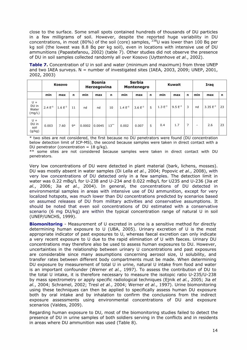
close to the surface. Some small spots contained hundreds of thousands of DU particles in a few milligrams of soil. However, despite the reported huge variability in DU concentrations, in most (80%) of the soil (core) samples, 238U was lower than 100 Bq per kg soil (the lowest was 8.8 Bq per kg soil), even in locations with intensive use of DU ammunitions (Papastefanou, 2002) (table 7). Other studies did not observe the presence of DU in soil samples collected randomly all over Kosovo (Uyttenhove et al., 2002).
Table 7. Concentration of U in soil and water (minimum and maximum) from three UNEP and two IAEA surveys. N = number of investigated sites (IAEA, 2003, 2009; UNEP, 2001, 2002, 2003)
Kosovo Bosnia Herzegovina
Serbia Montenegro
Kuwait Iraq
min max n min max n min max n min max n min max n
U + DU in Water (mg/L)
2.4 E-5 1.6 E-3 11 nd nd 10 1.4 E-5 3.6 E-3 5 1.3 E-3 9.5 E-3 3 nd 3.35 E-3 23
U + DU in soil
(g/kg)
0.003 7.60 9* 0.0002 0.0045 13** 0.002 0.007 5 0.4 1.7 7 - 2.6 23
* two sites are not considered, the first because no DU penetrators were found (DU concentration below detection limit of ICP-MS), the second because samples were taken in direct contact with a DU penetrator (concentration = 18 g/kg). ** some sites are not considered because samples were taken in direct contact with DU penetrators. Very low concentrations of DU were detected in plant material (bark, lichens, mosses). DU was mostly absent in water samples (Di Lella et al., 2004; Popovic et al., 2008), with very low concentrations of DU detected only in a few samples. The detection limit in water was 0.22 mBq/L for U-238 and U-234 and 0.022 mBq/L for U235 and U-236 (Jia et al., 2006; Jia et al., 2004). In general, the concentrations of DU detected in environmental samples in areas with intensive use of DU ammunition, except for very localized hotspots, was much lower than DU concentrations predicted by scenarios based on assumed releases of DU from military activities and conservative assumptions. It should be noted that even soil concentrations of DU estimated with a conservative scenario (6 mg DU/kg) are within the typical concentration range of natural U in soil (UNEP/UNCHS, 1999).
Biomonitoring - Measurement of U excreted in urine is a sensitive method for directly determining human exposure to U (UBA, 2005). Urinary excretion of U is the most appropriate indicator of past exposures to U, whereas faecal excretion can only indicate a very recent exposure to U due to the rapid elimination of U with faeces. Urinary DU concentrations may therefore also be used to assess human exposures to DU. However, uncertainties in the relationship between urinary U concentrations and past exposures are considerable since many assumptions concerning aerosol size, U solubility, and transfer rates between different body compartments must be made. When determining DU exposure by measurement of total U in urine, natural U intake from food and water is an important confounder (Werner et al., 1997). To assess the contribution of DU to the total U intake, it is therefore necessary to measure the isotopic ratio U-235/U-238 by mass spectrometry or apply specific radiological techniques (Ejnik et al., 2005; Jia et al., 2004; Schramel, 2002; Tresl et al., 2004; Werner et al., 1997). Urine biomonitoring using these techniques can then be applied to specifically assess human DU exposure both by oral intake and by inhalation to confirm the conclusions from the indirect exposure assessments using environmental concentrations of DU and exposure scenarios (Valdes, 2009).
Regarding human exposure to DU, most of the biomonitoring studies failed to detect the presence of DU in urine samples of both soldiers serving in the conflicts and in residents in areas where DU ammunition was used (Table 8).

15
Table 8. Concentration of U in urine samples of residents from different regions and soldiers engaged in combat or peacekeeping missions in areas where DU ammunition was used. Presence of DU can be determined by the ratio of ratio of 235U/238U which is 0.002001 for DU and 0.007253 for natural U). Detection limit for DU depends on total U concentrations present and instrument precision; usually, deviation of isotope ratio by > 0.3 % indicates presence of DU. When total daily excretion of U was given, adjustment to a urine concentration of U/DU was performed based a urine output of 1. 5 L/day. ("na", not applicable since only total urinary U was determined)
Urinary concentration [ng/L] Region Sample
type Year Range Mean
235U/238U As determined
by ICPMS Reference
Germany, n = 1500 24 h urine
2001 -2003
6.5 – 21 11.5 na (UBA, 2005)
USA, n = 2464 Spot urine
46 (95th)
8 (GM)
na (NHANES, 2005)
Jordan, n = 60 given in microg/day
18 – 2647 135 (GM)
na (Al-Jundi et al., 2004)
Italy, n = 38 Spot urine
1999 3 – 26 10 na (Galletti et al., 2003)
Finland, n = 205 2647 (95th)
64 (GM)
na (Karpas et al., 2005)
German peacekeepers in Kosovo (n = 1228) samples analyzed within one year after return to Germany
24 h urine
1999 – 2006
0.6 – 171.5 12.82 (GM) 0.007253 + 0.3 %
(Oeh et al., 2007a)
Kosovo residents living in area where DU was used, after conflict
24 h urine
2001-2002
2.92 – 266.81 Not given 0.007253 + 0.3 %
(Oeh et al., 2007b)
UK, n = 199, combat veterans from Gulf war
Spot urine
3.9 – 4.6 (95th)
3.9 0.0072358 (Bland et al., 2007)
UK, n = 24, involved in clean-up in Iraq
Spot urine
2.0 – 3.6 (95% CI)
2.7 0.0072463 (Bland et al., 2007)
UK, n = 22, medics deployed to Iraq
Spot urine
2.9 – 5.9 (95% CI)
4.2 0.0072411 (Bland et al., 2007)
UK, non-combat n = 96
Spot urine
3.4 – 4.6 (95% CI)
3.9 0.0072359 (Bland et al., 2007)
US, 1 700 US soldiers from Gulf war and after gulf war
24 h urine
2003 - 2008
10 + 1 based on a creatinine
concentration of 0.9 g/L
Three samples gave isotopic signatures
indicative of traces of DU
(Dorsey et al., 2009)
US, workers in plant producing DU, n = 5
Spot urine
79.6 0.00461 (Parrish et al., 2008)
US, residents near plant producing DU, n = 17
Spot urine
2.64 0.00720 (Parrish et al., 2008)
US, 28 soldiers involved in friendly fire incidents with DU-ammunition; 12 reference soldiers from 1992 Gulf war
Spot urine
1997-1999
16 – 180 in those exposed
to DU in friendly fire; 11 – 79 in reference
group
59 in those exposed to DU in friendly fire incidents, 15 in reference
group, (medians)
Change of isotopic
signature in samples from 10 of the 28
soldiers exposed to DU, and in
one in reference
group
(Gwiazda et al., 2004)
France, 154 soldiers serving in Gulf region and 54 in the Balkans
Spot urine
1999-2003
Not detected, detection limit < 10 mBq/L per isotope
(Cazoulat et al., 2008)
16
Due to the long half-life of U, spot urine samples could be used for exposure assessment.
The biomonitoring results show that the incorporation of DU in soldiers serving in Kosovo and Iraq and in residents of Kosovo is very low (Table 8). The ICP-MS method is very sensitive and can easily detect exposures to DU based on the ratio of the U isotopes 235U/238U. Even the presence of a low percentage of DU in the total U excretion can be detected. The sensitivity of the method is demonstrated by a significantly changed isotope ratio in workers in a DU-plant and also in some residents living in the vicinity of the plant (Parrish et al., 2008) either exposed through releases of DU from the plant into drinking water or in the air (Table 8) despite total U concentrations in urine in the normal range. The detection of DU in soldiers in friendly fire incidents during the 1st Gulf war, but without retained DU-shrapnel, also indicates that inhaled DU-aerosols formed after impact are taken up from the lung and that biomonitoring is appropriate to confirm past inhalation exposures to DU-aerosols.
In summary, general contamination with DU, even in areas of heavy fighting with documented intensive use of DU ammunition, is low or could not be demonstrated. This confirms the reliability of the exposure scenarios and the assessment based on environmental monitoring.
3.5. Risk assessment
3.5.1. Human health risks
Health risk assessment determines whether a chemical (including radioactive material) may cause adverse health effects, the probability that these effects will occur, and at what level and frequency of exposure they may occur. Toxicology focuses on the identification and quantization of potential hazards by using animal studies as surrogates for humans.
Several terms frequently used and misused in risk assessment and its perception require clarification. In a discussion of the health effects of potentially toxic chemicals, the terms “hazard” and “risk” are often used with an identical meaning, although they are clearly different. Hazard defines the intrinsic toxicity of a chemical and is not identical to risk. Risk is the estimated or measured probability of injury or death resulting from exposure to a specific chemical. Risk may be described either in semi-quantitative terms such as high or low risk or in quantitative terms.
The health risks due to contact with potentially toxic chemicals are dependent on the conditions of exposure, since not only the intrinsic toxicity of a chemical determines the magnitude of the adverse effect but also the dose. The magnitude of toxic effects is the product of the intrinsic toxicity of a chemical multiplied by the dose taken up by exposed animals or humans; thus, all toxic effects are dose-dependent and even very toxic chemicals may not cause toxic effects when the dose is low. If the dose is zero, despite a very high intrinsic toxicity of a specific chemical, the toxic effect and the risk of adverse health effects will be zero. On the other hand, chemicals with low intrinsic toxicity may induce toxic effects when the dose is high and may thus pose a significant risk. In toxicological terms, risk is therefore the product of the intrinsic toxicity of a chemical and the exposure characteristics.
The US National Research Council stated that ingesting U in food and water at the naturally occurring levels will not cause cancer or other health problems in people (ATSDR, 1999; NRC, 1991), In addition, in U miners, there was “no association between exposures to uranium and lung cancer at cumulative internal dose levels lower than 200 mSv” (ATSDR, 1999; NRC, 1991). Especially for the U miners it is accepted that radon exposure is the main cancer risk factor and that smoking is the most important confounder in these studies (Harley, 2001). Based on the radiological profile of natural U and DU, radiological health hazards are also not expected. Since exposures to DU both in soldiers and in residents in areas with military use of DU could not be detected or is very low, and exposures are thus well below thresholds for chemical toxicity or accepted limits
17
for radiological protection of the general population, health risks due to the chemical and radiological toxicity of DU are not expected. The conclusion is supported by all expert panels that were tasked with risk assessment for DU uses regarding the general population (EU-EURATOM, 2001; EURATOM, 2009; IAEA, 2003, 2009; UNEP, 2001, 2002, 2003, 2007; UNEP/UNCHS, 1999; UNSCEAR, 1993, 2000b, a; WHO, 2001).
An increased frequency of malformations in offspring from combat veterans deployed in areas where DU ammunitions were used was claimed, but could not be substantiated (McDiarmid et al., 2009; Sumanovic-Glamuzina et al., 2003). Reports on an increase in malformations in southern Iraq and/or Kuwait were not located in the scientific literature.
3.5.2. Environmental health risks
Risk for the terrestrial environment - A precise quantitative characterisation of the risk for the soil ecosystem is not simple due to the difficulty of calculating a Predicted Environmental Concentration (PEC) and to the lack of toxicological data on U and DU required for calculating a Predicted No Effect Concentration (PNEC). However, some general conclusions can be made.
The concentrations of DU measured in soil in all investigated sites (see table 7), even in locations with intensive use of DU ammunitions, are within the typical concentration range of U in soil (see table 5), with the exception of samples taken in the immediate vicinity of DU penetrators. Therefore, soil concentrations in impacted areas are of the same order of background levels of U in natural soils. As indicated above, a risk limit value of 28 mg/kg was derived by RIVM (Van de Plassche et al., 1999) for soil. It follows that potential risk to the environment is likely to occur in very limited areas, only directly in contact with DU.
Risk for the aquatic environment - As for soil, similar difficulties are encountered for characterizing the risk for the aquatic environment, though some toxicological data are available for aquatic organisms.
The lowest chronic toxicity values reported for U are in the 1.0 to 10 µg/L range (see section ecotoxicity). This would mean that if an assessment factor of 10 would be applied for calculating a PNEC, a value of 0.1 to 1 µg/L would result. However, as mentioned in previous opinions of the SCHER – see for example the SCHER Opinion on Copper (EU-SCHER, 2009), the standard TGD procedure for calculating a PNEC should be applied with caution to natural elements such as U, in particular if one considers that calculated values are within the range of background concentrations of U in water. The RIVM proposal for a maximum permissible addition to background levels of 1.0 µg U/L is also difficult to apply because it is not clear whether concentrations measured in the impacted areas (see table 7) represent the natural background concentrations or values modified by DU emissions.
However, it must be noted that most data reported as concentrations measured in surface water of impacted areas, except for Kuwait data, are below 1 µg U/L. Therefore, it can be concluded that a risk for the aquatic environment is unlikely to occur.
Risk for secondary poisoning - Uranium has been measured in plants and animals (earthworms). However, transfer factors in plants and animals are low and related to environmental concentrations. For example, in the US EPA ECOTOX Database (US-EPA, 2009), for rainbow trout, a bioconcentration factor of 37 and a BCF value of 4.2 for molluscs has been recorded. Therefore, the potential for secondary poisoning due to DU in impacted areas is low and limited to very restricted sites close to or directly in contact with ammunitions.
18
4. RESPONSE TO TERMS OF REFERENCE
4.1. Question 1
The SCHER is asked for an opinion building on an evaluation of available reports, including but not restricted to those referenced above, as to the environmental and health risks posed by DU.
Since DU has a much lower radioactivity as compared to natural U and U-containing ores, it is generally agreed that the chemical toxicity of U is the major hazard descriptor regarding assessment of health risk due to potential exposures to DU (UNEP, 2001, 2002, 2003, 2007; UNEP/UNCHS, 1999; WHO, 2001, 2003b). SCHER agrees with this concept. Therefore, the toxicity data on natural U can be applied to assess DU since the chemistry and the chemical toxicology of isotopes are identical. Human health risk due to chemical toxicity and radiation from U and DU only occur when the uranium is ingested or inhaled.
The human toxicity of U is comparatively well studied; the major target organ for soluble U salts is the kidney. Both in rodents and in rabbits, repeated administration of U with drinking water gave NOAELs or LOAELs of 60 µg/kg bw/day based on subtle histopathological changes in the kidney. These NOAELs/LOAELs have been transformed in tolerable daily intake for natural U with an uncertainty factor of 100 to give a TDI of 0.6 µg/kg bw per day. Since DU shows an identical toxicity to that of natural U, this TDI is also applicable to DU.
As alpha particles emitted from DU have a very limited range in tissue, DU is not a significant external radiation hazard. Therefore, health effects expected from external radiation caused by DU are limited to unrealistic direct skin contact scenarios. Intake of DU from the environment after use of DU ammunition could not be demonstrated and environmental concentrations of DU, except very close to deposited penetrators and tanks hit, are very low. SCHER therefore agrees with the conclusion of UNEP, IAEA and others that environmental and human health risks due to a potential widespread distribution of DU are not expected due to the very limited exposure to DU as compared to background exposures to natural U (EU-EURATOM, 2001; UNEP, 2001, 2002, 2003, 2007; UNEP/UNCHS, 1999; WHO, 2001, 2003b). Higher exposures to DU dust will only occur when entering vehicles hit by DU ammunition shortly after the hit, and in combat situations when in close proximity to a tank hit by DU ammunition. Therefore, vehicles hit by DU should be made inaccessible to the general public and be properly disposed. Used DU ammunition should also be collected and properly disposed.
4.2. Question 2 In particular, SCHER is asked to assess those risks that may arise from exposure to DU in contaminated areas following military activities with weapons containing DU.
Internal exposure to DU can occur through inhalation, ingestion, and embedded fragments or contaminated wounds (mainly for soldiers). Inhalation of dust is considered as one of the major pathways of DU exposure in combat situations and may also occur from re-suspended particles. Detailed assessments of such exposures have been performed. UNEP, IAEA, several State Governments and research organisations quantified environmental exposures to DU in the Balkans, Kuwait and in Iraq. Presence of DU and natural U can be assessed with high sensitivity by quantifying U isotopes by ICP-MS or by specific radiological techniques.
The many available measurements show that DU, after military use in combat, will mainly be located inside of military vehicles hit by DU ammunition and in their close vicinity. DU ammunition in soil will slowly corrode and hotspots with high local concentrations of DU may remain locally close to the impact site. Based on the available data, only a very small part of the DU released after the impact on a hard target will be more widely distributed in the environment.

19
DU intake with food and drinking water in areas with use of DU ammunition is well below tolerable exposure levels regarding chemical and radiological toxicity of U and DU. In summary, these studies have shown that general contamination with DU, even in areas of heavy fighting with documented or presumed intensive use of DU ammunition, is very low; in many cases, presence of DU could not be detected despite the use of highly sensitive methods like ICP-MS and alpha-spectrometry.
In the opinion of SCHER, the environmental monitoring, which included soil, drinking water and biota, was adequate to conclude that, except in areas very close to destroyed vehicles and penetrators, DU contamination in the war zones is not widespread and is generally low. Due to the low exposures, possible risks for terrestrial and aquatic ecosystems are considered very low.
Besides environmental measurements, biomonitoring for the presence of DU has been performed in military personnel and long-term Kosovo residents. Most of these studies have failed to find increased concentrations of DU in the sampled population. Therefore, SCHER agrees with the conclusions of UNEP and other reports that exposures of the general population to DU from environmental sources after military uses are very low. Due to the very low exposures, which do not significantly increase the body burden of U isotopes, additional health risks are not expected.
Further support for an absence of health effects of lower DU exposures can be derived from the medical monitoring of Gulf War veterans with embedded DU shrapnels and health monitoring of other veterans. Individuals with embedded DU shrapnel have much higher concentrations of total U in blood and urine as compared to the general population and to soldiers without direct DU exposure (Gwiazda et al. 2004). Sub-clinical effects have been observed in high-level DU concentrations (McDiarmid et al., 2009), but overt health effects due to the release of DU from the embedded shrapnel were not observed (McDiarmid et al., 2009) by health monitoring for more than 16 years.
4.3. Question 3
SCHER is asked to take into account both the chemical and radiological toxicities of DU and, if appropriate, their possible synergistic relations
Since all U isotopes are radioactive and have an identical chemical toxicity, the available information on health effects of U always represents a combination of radiological effects and chemical toxicity. It is therefore impossible to study chemical and radiological effects of U separately. Health effects based on this combination serve as a basis for deriving tolerable exposures. A potential combination of radioactivity and chemical toxicity is therefore covered. Any synergy between chemical toxicity and radioactivity is less pronounced with DU as compared to natural U due to the lower radioactivity of DU.
5. REFERENCES Al-Jundi, J., Werner, E., Roth, P., Hollriegl, V., Wendler, I., and Schramel, P. (2004). Thorium and uranium contents in human urine: influence of age and residential area. J Environ Radioact 71, 61-70.
Albina, M. L., Belles, M., Linares, V., Sanchez, D. J., and Domingo, J. L. (2005). Restraint stress does not enhance the uranium-induced developmental and behavioral effects in the offspring of uranium-exposed male rats. Toxicology 215, 69-79.
Arfsten, D. P., Still, K. R., Wilfong, E. R., Johnson, E. W., McInturf, S. M., Eggers, J. S., Schaeffer, D. J., and Bekkedal, M. Y. (2009). Two-generation reproductive toxicity study of implanted depleted uranium (DU) in CD rats. J Toxicol Environ Health A 72, 410-427.
Arnault, E., Doussau, M., Pesty, A., Gouget, B., Van der Meeren, A., Fouchet, P., and Lefevre, B. (2008). Natural uranium disturbs mouse folliculogenesis in vivo and oocyte meiosis in vitro. Toxicology 247, 80-87.
20
ATSDR (1999). Toxicological profile for uranium. Agency for Toxic Substances and Disease Registry. US Public Health Service. Department of Health & Human Services, Atlanta GA. .
Averbeck, D. (2009). Does scientific evidence support a change from the LNT model for low-dose radiation risk extrapolation? Health Phys 97, 493-504.
Belles, M., Albina, M. L., Linares, V., Gomez, M., Sanchez, D. J., and Domingo, J. L. (2005). Combined action of uranium and stress in the rat. I. Behavioral effects. Toxicol Lett 158, 176-185.
Benedict, M., Pigford, T. H., and Levi, H. W. (1981). Nuclear Chemical Engineering. McGraw-Hill.
Berradi, H., Bertho, J. M., Dudoignon, N., Mazur, A., Grandcolas, L., Baudelin, C., Grison, S., Voisin, P., Gourmelon, P., and Dublineau, I. (2008). Renal anemia induced by chronic ingestion of depleted uranium in rats. Toxicol Sci 103, 397-408.
BfR (2004). Uran in natürlichen Mineral- und anderen, zum Verzehr bestimmten Wässern. Stellungnahme des Bundesinstituts für Risikobewertung vom 3. März 2004.
Bland, D. J., Rona, R. J., Coggon, D., Anderson, J., Greenberg, N., Hull, L., and Wessely, S. (2007). Urinary isotopic analysis in the UK Armed Forces: No evidence of depleted uranium absorption in combat and other personnel in Iraq. Occup Environ Med.
Bleise, A., Danesi, P. R., and Burkart, W. (2003). Properties, use and health effects of depleted uranium (DU): a general overview. J Environ Radioact 64, 93-112.
Briner, W., and Murray, J. (2005). Effects of short-term and long-term depleted uranium exposure on open-field behavior and brain lipid oxidation in rats. Neurotoxicol Teratol 27, 135-144.
Burkhart, W. (1991). Uranium, thorium and decacy products. In Metals and their compounds in the environment, occurence. Analysis and biological relevance (E. Merian, Ed.), pp. 1275-1287. VCH Verlagsgesellschaft, Waldheim.
Bussy, C., Lestaevel, P., Dhieux, B., Amourette, C., Paquet, F., Gourmelon, P., and Houpert, P. (2006). Chronic ingestion of uranyl nitrate perturbs acetylcholinesterase activity and monoamine metabolism in male rat brain. Neurotoxicology 27, 245-252.
Capstone-Report (2005). U.S. Army Capstone Depleted Uranium Aerosols Study & Human Health Risk Assessment. For service members and their families.
Cazoulat, A., Lecompte, Y., Bohand, S., Castagnet, X., and Laroche, P. (2008). [Urinary uranium analysis results on Gulf war or Balkans conflict veterans]. Pathol Biol (Paris) 56, 77-83.
Chazel, V., Houpert, P., and Ansoborlo, E. (1998). Effect of U3O8 Specific Surface Area on In Vitro Dissolution, Biokinetics, and Dose Coefficients Radiat Prot Dosimetry 79, 39-42.
Cheng, Y. S., Kenoyer, J. L., Guilmette, R. A., and Parkhurst, M. A. (2009). Physicochemical characterization of Capstone depleted uranium aerosols II: particle size distributions as a function of time. Health Phys 96, 266-275.
Choppin, G., Lilienzin, J. O., and Rydberg, J. (1966). Radiochemistry and nuclear chemistry. Butterworth-Heinemann, Oxford.
Clark, R. (1999). Control of low-level radiation exposure: time for a change? J Radiol Prot 19, 107-115.
Coryell, V. H., and Stearns, D. M. (2006). Molecular analysis of hprt mutations generated in Chinese hamster ovary EM9 cells by uranyl acetate, by hydrogen peroxide, and spontaneously. Mol Carcinog 45, 60-72.
DAF-OO-ALC (1997). Gulf War depleted uranium munition expenditure, April 30,1997. Department of Air Force - Memorandum from Headquarters - Ogden Air Logistic Center.
21
Danesi, P. R., Bleise, A., Burkart, W., Cabianca, T., Campbell, M. J., Makarewicz, M., Moreno, J., Tuniz, C., and Hotchkis, M. (2003a). Isotopic composition and origin of uranium and plutonium in selected soil samples collected in Kosovo. J Environ Radioact 64, 121-131.
Danesi, P. R., Markowicz, A., Chinea-Cano, E., Burkart, W., Salbu, B., Donohue, D., Ruedenauer, F., Hedberg, M., Vogt, S., Zahradnik, P., and Ciurapinski, A. (2003b). Depleted uranium particles in selected Kosovo samples. J Environ Radioact 64, 143-154.
Di Lella, L. A., Frati, L., Loppi, S., Protano, G., and Riccobono, F. (2004). Environmental distribution of uranium and other trace elements at selected Kosovo sites. Chemosphere 56, 861-865.
Diehl, P. (2001). WISE uranium project, fact sheets.
Domingo, J. L. (2001). Reproductive and developmental toxicity of natural and depleted uranium: a review. Reprod Toxicol 15, 603-609.
Dorsey, C. D., Engelhardt, S. M., Squibb, K. S., and McDiarmid, M. A. (2009). Biological monitoring for depleted uranium exposure in U.S. Veterans. Environ Health Perspect 117, 953-956.
Dublineau, I., Grandcolas, L., Grison, S., Baudelin, C., Paquet, F., Voisin, P., Aigueperse, J., and Gourmelon, P. (2007). Modifications of inflammatory pathways in rat intestine following chronic ingestion of depleted uranium. Toxicol Sci 98, 458-468.
Durante, M., and Pugliese, M. (2002). Estimates of radiological risk from depleted uranium weapons in war scenarios. Health Phys 82, 14-20.
Ejnik, J. W., Todorov, T. I., Mullick, F. G., Squibb, K., McDiarmid, M. A., and Centeno, J. A. (2005). Uranium analysis in urine by inductively coupled plasma dynamic reaction cell mass spectrometry. Anal Bioanal Chem 382, 73-79.
Environment-Australia (2000). The National Water Quality Management Strategy: Australian and New Zealand guidelines for fresh and marine water quality. Volume 2, Aquatic ecosystems.
EPA (2000). Drinking water criteria document for uranium. U.S. Environmental Protection Agency, Washington, DC.
Erikson, R. L., Hostetler, C. J., Divine, J. R., and Price, K. R. (1990). A review of the environmental behavior of uranium derived from depleted uranium alloy penetrators,Technical Report PNL-7213, Pacific Northwest Lab., Richland, WA (USA).
EU-EURATOM (2001). Depleted uranium - Opinion of the Group of Experts established according to article 31 of the EURATOM Treaty.
EU-SCHER (2009). Scientific Committee on Health and Environmental Risks opinion on: Voluntary Risk Assessment Report on Copper and its compounds, Environmental Part Adopted by the SCHER by written procedure on 12 February 2009.
EURATOM (2009). Depleted uranium - Opinion of the Group of Experts established according to article 31 of the EURATOM Treaty. http://ec.europa.eu/energy/nuclear/radiation_protection/doc/art31/2009_06_report.pdf.
Feugier, A., Frelon, S., Gourmelon, P., and Claraz, M. (2008). Alteration of mouse oocyte quality after a subchronic exposure to depleted Uranium. Reprod Toxicol 26, 273-277.
Figgitt, M., Newson, R., Leslie, I. J., Fisher, J., Ingham, E., and Case, C. P. (2010). The genotoxicity of physiological concentrations of chromium (Cr(III) and Cr(VI)) and cobalt (Co(II)): An in vitro study. Mutat Res.
Fisenne, I. M., Perry, P. M., Decker, K. M., and Keller, H. W. (1987). The daily intake of 234,235,238U, 228,230,232Th and 226,228Ra by New York City residents. Health Phys 53, 357-363.
Fukuda, S., Ikeda, M., Chiba, M., and Kaneko, K. (2006). Clinical diagnostic indicators of
22
renal and bone damage in rats intramuscularly injected with depleted uranium. Radiat Prot Dosimetry 118, 307-314.
Galletti, M., D'Annibale, L., Pinto, V., and Cremisini, C. (2003). Uranium daily intake and urinary excretion: a preliminary study in Italy. Health Phys 85, 228-235.
Gerdes, A., Weyer, S., Brey, G., and Durakovic, A. (2004). Monitoring depleted uranium contamination in the biosphere of Iraq using MC-ICP-MS. Geochimica et Cosmochimica Acta, A506.
Gilman, A. P., Moss, M. A., Villeneuve, D. C., Secours, V. E., Yagminas, A. P., Tracy, B. L., Quinn, J. M., Long, G., and Valli, V. E. (1998a). Uranyl nitrate: 91-day exposure and recovery studies in the male New Zealand white rabbit. Toxicol Sci 41, 138-151.
Gilman, A. P., Villeneuve, D. C., Secours, V. E., Yagminas, A. P., Tracy, B. L., Quinn, J. M., Valli, V. E., and Moss, M. A. (1998b). Uranyl nitrate: 91-day toxicity studies in the New Zealand white rabbit. Toxicol Sci 41, 129-137.
Gilman, A. P., Villeneuve, D. C., Secours, V. E., Yagminas, A. P., Tracy, B. L., Quinn, J. M., Valli, V. E., Willes, R. J., and Moss, M. A. (1998c). Uranyl nitrate: 28-day and 91-day toxicity studies in the Sprague-Dawley rat. Toxicol Sci 41, 117-128.
Glastone, S., and Sesonske, A. (1981). Nuclear Reactor Engineering. Van Nostrand Reinhold.
Goldman, M., Yaari, A., Doshnitzki, Z., Cohen-Luria, R., and Moran, A. (2006). Nephrotoxicity of uranyl acetate: effect on rat kidney brush border membrane vesicles. Arch Toxicol 80, 387-393.
Grignard, E., Gueguen, Y., Grison, S., Lobaccaro, J. M., Gourmelon, P., and Souidi, M. (2008). Contamination with depleted or enriched uranium differently affects steroidogenesis metabolism in rat. Int J Toxicol 27, 323-328.
Gueguen, Y., Souidi, M., Baudelin, C., Dudoignon, N., Grison, S., Dublineau, I., Marquette, C., Voisin, P., Gourmelon, P., and Aigueperse, J. (2006). Short-term hepatic effects of depleted uranium on xenobiotic and bile acid metabolizing cytochrome P450 enzymes in the rat. Arch Toxicol 80, 187-195.
Gueguen, Y., Grandcolas, L., Baudelin, C., Grison, S., Tissandie, E., Jourdain, J. R., Paquet, F., Voisin, P., Aigueperse, J., Gourmelon, P., and Souidi, M. (2007). Effect of acetaminophen administration to rats chronically exposed to depleted uranium. Toxicology 229, 62-72.
Gwiazda, R. H., Squibb, K., McDiarmid, M., and Smith, D. (2004). Detection of depleted uranium in urine of veterans from the 1991 Gulf War. Health Phys 86, 12-18.
Hahn, F. F., Guilmette, R. A., and Hoover, M. D. (2002). Implanted depleted uranium fragments cause soft tissue sarcomas in the muscles of rats. Environ Health Perspect 110, 51-59.
Hall, E. J., and Giacca, A. J. (2006). Radiobiology for the Radiologist. Lippincott Williams & Wilkins, Philadelphia, USA.
Hall, E. J., Metting, N., Puskin, J., and Ron, E. (2009). Low dose radiation epidemiology: what can it tell us? Radiat Res 172, 134-138.
Handley-Sidhu, S., Worsfold, P. J., Boothman, C., Lloyd, J. R., Alvarez, R., Livens, F. R., Vaughan, D. J., and Keith-Roach, M. J. (2009a). Corrosion and fate of depleted uranium penetrators under progressively anaerobic conditions in estuarine sediment. Environ Sci Technol 43, 350-355.
Handley-Sidhu, S., Worsfold, P. J., Livens, F. R., Vaughan, D. J., Lloyd, J. R., Boothman, C., Sajih, M., Alvarez, R., and Keith-Roach, M. J. (2009b). Biogeochemical controls on the corrosion of depleted uranium alloy in subsurface soils. Environ Sci Technol 43, 6177-6182.
Harley, N. H., Foulkes, E. C., Hilborne, L. H., Hudson, A., and Anthony, C., R. (1999).

23
Depleted uranium, a review of the scientific literature as it pertains to gulf war illness. . RAND (Research and Development), Santa Monica.
Harley, N. H. (2001). Toxic effects of radiation and radioactive materials. In Cassarett and Doull's toxicology. The baseic science of poisons (C. D. Klaassen, Ed.). McGraw-Hill, New York.
Hartsock, W. J., Cohen, J. D., and Segal, D. J. (2007). Uranyl acetate as a direct inhibitor of DNA-binding proteins. Chem Res Toxicol 20, 784-789.
Hu, Q. Y., and Zhu, S. P. (1990). Induction of chromosomal aberrations in male mouse germ cells by uranyl fluoride containing enriched uranium. Mutat Res 244, 209-214.
IAEA (1996). Proceedings of International Conference ONE DECADE AFTER CHERNOBYL, Vienna, Austria, 8-12 April 1996. International Atomic Energy Agency, Vienna - Radiological Assessment Report Series.
IAEA (2003). Radiological conditions in areas of Kuwait with residues of depleted uranium. International Atomic Energy Agency, Vienna - Radiological Assessment Report Series.
IAEA (2009). Radiological conditions in selected areas of Southern Iraq with residues of depleted uranium. International Atomic Energy Agency, Vienna - Radiological Assessment Report Series.
Jia, G., Belli, M., Sansone, U., Rosamilla, S., and Gaudino, S. (2004). Concentration, distribution and characteristics of depleted uranium (DU) in the Kosovo ecosystem: A comparison with the uranium behavior in the environment uncontaminated by DU. Journal of Radioanalytical and Nuclear Chemistry 260, 481-494.
Jia, G., Belli, M., Sansone, U., Rosamilia, S., and Gaudino, S. (2006). Concentration and characteristics of depleted uranium in biological and water samples collected in Bosnia and Herzegovina. J Environ Radioact 89, 172-187.
Kalinich, J. F., Ramakrishnan, N., Villa, V., and McClain, D. E. (2002). Depleted uranium-uranyl chloride induces apoptosis in mouse J774 macrophages. Toxicology 179, 105-114.
Karpas, Z., Paz-Tal, O., Lorber, A., Salonen, L., Komulainen, H., Auvinen, A., Saha, H., and Kurttio, P. (2005). Urine, hair, and nails as indicators for ingestion of uranium in drinking water. Health Phys 88, 229-242.
Knobel, Y., Glei, M., Weise, A., Osswald, K., Schaferhenrich, A., Richter, K. K., Claussen, U., and Pool-Zobel, B. L. (2006). Uranyl nitrilotriacetate, a stabilized salt of uranium, is genotoxic in nontransformed human colon cells and in the human colon adenoma cell line LT97. Toxicol Sci 93, 286-297.
Kondo, S. (1993). Comparison of the acute effects of the Hiroshima-Nagasaki atomic bombings and of the Chernobyl reactor accident. In Health effects of low-level radiation. Kinki University press, Medical Physics Publishing, Osaka Japan, Madison WI USA.
Konietzka, R., Dieter, H., and Voss, J.-U. (2005). Vorschlag für einen gesundheitlichen Leitwert für Uran in Trinkwasser. Umweltmed Forsch Prax 10, 133-143.
Kreuzer, M., Grosche, B., Schnelzer, M., Tschense, A., Dufey, F., and Walsh, L. (2009). Radon and risk of death from cancer and cardiovascular diseases in the German uranium miners cohort study: follow-up 1946-2003. Radiat Environ Biophys.
Kundt, M. S., Martinez-Taibo, C., Muhlmann, M. C., and Furnari, J. C. (2009). Uranium in drinking water: effects on mouse oocyte quality. Health Phys 96, 568-574.
Kurttio, P., Komulainen, H., Leino, A., Salonen, L., Auvinen, A., and Saha, H. (2005). Bone as a possible target of chemical toxicity of natural uranium in drinking water. Environ Health Perspect 113, 68-72.
Larsen, I. L. (2000). Some notes and comments regarding natural and processed uranium isotopes. Radioactivity & Radiochemistry 11, 6-11.
24
Leggett, R. W., and Harrison, J. D. (1995). Fractional absorption of ingested uranium in humans. Health Phys 68, 484-498.
Lestaevel, P., Houpert, P., Bussy, C., Dhieux, B., Gourmelon, P., and Paquet, F. (2005). The brain is a target organ after acute exposure to depleted uranium. Toxicology 212, 219-226.
Lestaevel, P., Romero, E., Dhieux, B., Ben Soussan, H., Berradi, H., Dublineau, I., Voisin, P., and Gourmelon, P. (2009). Different pattern of brain pro-/anti-oxidant activity between depleted and enriched uranium in chronically exposed rats. Toxicology 258, 1-9.
Li, W. B., Gerstmann, U. C., Hollriegl, V., Szymczak, W., Roth, P., Hoeschen, C., and Oeh, U. (2009). Radiation dose assessment of exposure to depleted uranium. J Expo Sci Environ Epidemiol 19, 502-514.
Lind, O. C., Salbu, B., Janssens, K., Proost, K., and Danesi, P. R. (2009). Characterisation of DU particles from Kosovo and Kuwait. In Radioactive particles in the environment (D. H. Oughton, and V. Kshparov, Eds.). Springer Science+Business Media B.V.
Little, J. B. (2006). Cellular radiation effects and the bystander response. Mutat Res 597, 113-118.
McDiarmid, M. A. (2001). Depleted uranium and public health. BMJ 322, 123-124.
McDiarmid, M. A., Engelhardt, S. M., Dorsey, C. D., Oliver, M., Gucer, P., Wilson, P. D., Kane, R., Cernich, A., Kaup, B., Anderson, L., Hoover, D., Brown, L., Albertini, R., Gudi, R., and Squibb, K. S. (2009). Surveillance results of depleted uranium-exposed Gulf War I veterans: sixteen years of follow-up. J Toxicol Environ Health A 72, 14-29.
McDonald-Taylor, C. K., Bhatnagar, M. K., Gilman, A., Yagminas, A., and Singh, A. (1992). Uranyl nitrate-induced glomerular basement membrane alterations in rabbits: a quantitative analysis. Bull Environ Contam Toxicol 48, 367-373.
McDonald-Taylor, C. K., Singh, A., and Gilman, A. (1997). Uranyl nitrate-induced proximal tubule alterations in rabbits: a quantitative analysis. Toxicol Pathol 25, 381-389.
McLaughlin, J. P., Vintro, L. L., Smith, K. J., Mitchell, P. J., and Zunic, Z. S. (2003). Actinide analysis of a depleted uranium penetrator from a 1999 target site in southern Serbia. J Environ Radioact 64, 155-165.
McLaughlin, J. P. (2005). Public health and environmental aspects of DU International Congress Series 1276, 137-140.
Miller, A. C., Mog, S., McKinney, L., Luo, L., Allen, J., Xu, J., and Page, N. (2001). Neoplastic transformation of human osteoblast cells to the tumorigenic phenotype by heavy metal-tungsten alloy particles: induction of genotoxic effects. Carcinogenesis 22, 115-125.
Miller, A. C., Stewart, M., Brooks, K., Shi, L., and Page, N. (2002a). Depleted uranium-catalyzed oxidative DNA damage: absence of significant alpha particle decay. J Inorg Biochem 91, 246-252.
Miller, A. C., Xu, J., Stewart, M., Brooks, K., Hodge, S., Shi, L., Page, N., and McClain, D. (2002b). Observation of radiation-specific damage in human cells exposed to depleted uranium: dicentric frequency and neoplastic transformation as endpoints. Radiat Prot Dosimetry 99, 275-278.
Miller, A. C., Brooks, K., Smith, J., and Page, N. (2004). Effect of the militarily-relevant heavy metals, depleted uranium and heavy metal tungsten-alloy on gene expression in human liver carcinoma cells (HepG2). Mol Cell Biochem 255, 247-256.
Miller, A. C., Bonait-Pellie, C., Merlot, R. F., Michel, J., Stewart, M., and Lison, P. D. (2005). Leukemic transformation of hematopoietic cells in mice internally exposed to depleted uranium. Mol Cell Biochem 279, 97-104.
25
Miller, A. C., Stewart, M., Rivas, R., Marino, S., Randers-Pehrson, G., and Shi, L. (2007). Observation of radiation-specific damage in cells exposed to depleted uranium: hprt gene mutation frequency. Radiation Measurements 42, 1029-1032.
Mitchel, R. E., and Sunder, S. (2004). Depleted uranium dust from fired munitions: physical, chemical and biological properties. Health Phys 87, 57-67.
Monleau, M., Blanchardon, E., Claraz, M., Paquet, F., and Chazel, V. (2006a). The effect of repeated inhalation on the distribution of uranium in rats. J Toxicol Environ Health A 69, 1629-1649.
Monleau, M., De Meo, M., Frelon, S., Paquet, F., Donnadieu-Claraz, M., Dumenil, G., and Chazel, V. (2006b). Distribution and genotoxic effects after successive exposure to different uranium oxide particles inhaled by rats. Inhal Toxicol 18, 885-894.
Monleau, M., De Meo, M., Paquet, F., Chazel, V., Dumenil, G., and Donnadieu-Claraz, M. (2006c). Genotoxic and inflammatory effects of depleted uranium particles inhaled by rats. Toxicol Sci 89, 287-295.
NCRP (1999). National Council on Radiation Protection and Measurements - Report No. 130 - Biological effects and exposure limits for "hot particles".
NHANES (2005). Third national report on human exposure to environmental chemicals. Centers for Disease Control and Prevention, National Health and Nutrition Examination Survey, Atlanta, GA.
NRC (1991). National Research Council. Comparative dosimetry of radon in mines and homes. National Academy Press, Washington, DC.
Oeh, U., Li, W. B., Hollriegl, V., Giussani, A., Schramel, P., Roth, P., and Paretzke, H. G. (2007a). Daily uranium excretion in German peacekeeping personnel serving on the Balkans compared to ICRP model prediction. Radiat Prot Dosimetry 127, 329-332.
Oeh, U., Priest, N. D., Roth, P., Ragnarsdottir, K. V., Li, W. B., Hollriegl, V., Thirlwall, M. F., Michalke, B., Giussani, A., Schramel, P., and Paretzke, H. G. (2007b). Measurements of daily urinary uranium excretion in German peacekeeping personnel and residents of the Kosovo region to assess potential intakes of depleted uranium (DU). Sci Total Environ 381, 77-87.
OEHHA (1998). Office of Environmental Health Hazard Assessment: Announcement of the Withdrawal of the Previously Established Public Health Goal for Uranium in Drinking Water [07/10/98] http://www.oehha.ca.gov/water/phg/urn_crnr.html (accessed Dec. 2009).
Orloff, K. G., Mistry, K., Charp, P., Metcalf, S., Marino, R., Shelly, T., Melaro, E., Donohoe, A. M., and Jones, R. L. (2004). Human exposure to uranium in groundwater. Environ Res 94, 319-326.
Oughton, D. H., and Kashparov, V. (Eds.) (2009). Radioactive particles in the environment. Springer Science & Business Media.
Palmer, M. R., and Edmond, J. M. (1993). Uranium in river water. Geochim Cosmochim Acta 93, 4947-4955.
Papastefanou, C. (2002). Depleted uranium in military conflicts and the impact on the environment. Health Phys 83, 280-282.
Parkhurst, M. A., and Guilmette, R. A. (2009a). Overview of the Capstone depleted uranium study of aerosols from impact with armored vehicles: test setup and aerosol generation, characterization, and application in assessing dose and risk. Health Phys 96, 207-220.
Parkhurst, M. A., and Guilmette, R. A. (2009b). Conclusions of the Capstone depleted uranium aerosol characterization and risk assessment study. Health Phys 96, 393-409.
Parrish, R. R., Horstwood, M., Arnason, J. G., Chenery, S., Brewer, T., Lloyd, N. S., and Carpenter, D. O. (2008). Depleted uranium contamination by inhalation exposure and its
26
detection after approximately 20 years: implications for human health assessment. Sci Total Environ 390, 58-68.
Periyakaruppan, A., Kumar, F., Sarkar, S., Sharma, C. S., and Ramesh, G. T. (2007). Uranium induces oxidative stress in lung epithelial cells. Arch Toxicol 81, 389-395.
Periyakaruppan, A., Sarkar, S., Ravichandran, P., Sadanandan, B., Sharma, C. S., Ramesh, V., Hall, J. C., Thomas, R., Wilson, B. L., and Ramesh, G. T. (2009). Uranium induces apoptosis in lung epithelial cells. Arch Toxicol 83, 595-600.
Popovic, D., Todorovic, D., Frontasyeva, M., Ajtic, J., Tasic, M., and Rajsic, S. (2008). Radionuclides and heavy metals in Borovac, Southern Serbia. Environ Sci Pollut Res Int 15, 509-520.
Poston, P. M., Hanf, J. R. W., and Simmons, m. A. (1984). Toxicity of uranium to Daphnia magna. Water, Air, & Soil Pollution 22, 289-298.
Pourahmad, J., Ghashang, M., Ettehadi, H. A., and Ghalandari, R. (2006). A search for cellular and molecular mechanisms involved in depleted uranium (DU) toxicity. Environ Toxicol 21, 349-354.
Preston, D. L., Shimizu, Y., Pierce, D. A., Suyama, A., and Mabuchi, K. (2003). Studies of mortality of atomic bomb survivors. Report 13: Solid cancer and noncancer disease mortality: 1950-1997. Radiat Res 160, 381-407.
Priest, N. D. (2001). Toxicity of depleted uranium. Lancet 357, 244-246.
Puskin, J. S. (2008). What can epidemiology tell us about risks at low doses? Radiat Res 169, 122-124.
Racine, R., Gueguen, Y., Gourmelon, P., Veyssiere, G., and Souidi, M. (2009). Modifications of the expression of genes involved in cerebral cholesterol metabolism in the rat following chronic ingestion of depleted uranium. J Mol Neurosci 38, 159-165.
Salbu, B., Janssens, K., Lind, O. C., Proost, K., and Danesi, P. R. (2003). Oxidation states of uranium in DU particles from Kosovo. J Environ Radioact 64, 167-173.
Salbu, B., Janssens, K., Lind, O. C., Proost, K., Gijsels, L., and Danesi, P. R. (2005). Oxidation states of uranium in depleted uranium particles from Kuwait. J Environ Radioact 78, 125-135.
Schramel, P. (2002). Determination of 235U and 238U in urine samples using sector field inductively coupled plasma mass spectrometry. J Chromatogr B Analyt Technol Biomed Life Sci 778, 275-278.
Sheppard, S. C., Evenden, W. G., and Anderson, A. J. (1992). Multiple assays of uranium toxicity in soil. Environmental Toxicology and Water Quality 7, 275-294.
Sheppard, S. C., Sheppard, M. I., Gallerand, M. O., and Sanipelli, B. (2005). Derivation of ecotoxicity thresholds for uranium. J Environ Radioact 79, 55-83.
Souidi, M., Gueguen, Y., Linard, C., Dudoignon, N., Grison, S., Baudelin, C., Marquette, C., Gourmelon, P., Aigueperse, J., and Dublineau, I. (2005). In vivo effects of chronic contamination with depleted uranium on CYP3A and associated nuclear receptors PXR and CAR in the rat. Toxicology 214, 113-122.
Sowder, A. G., Clark, S. B., and Fjeld, R. A. (1999). The Transformation of Uranyl Oxide Hydrates: The Effect of Dehydration on Synthetic Metaschoepite and Its Alteration to Becquerelite. Environ Sci Technol 33, 3552-3557.
Stearns, D. M., Yazzie, M., Bradley, A. S., Coryell, V. H., Shelley, J. T., Ashby, A., Asplund, C. S., and Lantz, R. C. (2005). Uranyl acetate induces hprt mutations and uranium-DNA adducts in Chinese hamster ovary EM9 cells. Mutagenesis 20, 417-423.
Sumanovic-Glamuzina, D., Saraga-Karacic, V., Roncevic, Z., Milanov, A., Bozic, T., and Boranic, M. (2003). Incidence of major congenital malformations in a region of Bosnia and Herzegovina allegedly polluted with depleted uranium. Croat Med J 44, 579-584.
27
TACOM (2000). Review of transuranics (TRU) in depleted uranium armor. Tank-Automotive and Armaments Command (TACOM) Memorandum ATTN:AMCSF.
Thiebault, C., Carriere, M., Milgram, S., Simon, A., Avoscan, L., and Gouget, B. (2007). Uranium induces apoptosis and is genotoxic to normal rat kidney (NRK-52E) proximal cells. Toxicol Sci 98, 479-487.
Tissandie, E., Gueguen, Y., Lobaccaro, J. M., Paquet, F., Aigueperse, J., and Souidi, M. (2006). Effects of depleted uranium after short-term exposure on vitamin D metabolism in rat. Arch Toxicol 80, 473-480.
Tissandie, E., Gueguen, Y., Lobaccaro, J. M., Grandcolas, L., Voisin, P., Aigueperse, J., Gourmelon, P., and Souidi, M. (2007). In vivo effects of chronic contamination with depleted uranium on vitamin D3 metabolism in rat. Biochim Biophys Acta 1770, 266-272.
Tracy, B. L., Quinn, J. M., Lahey, J., Gilman, A. P., Mancuso, K., Yagminas, A. P., and Villeneuve, D. C. (1992). Absorption and retention of uranium from drinking water by rats and rabbits. Health Phys 62, 65-73.
Tresl, I., De Wannemacker, G., Quetel, C. R., Petrov, I., Vanhaecke, F., Moens, L., and Taylor, P. D. (2004). Validated measurements of the uranium isotopic signature in human urine samples using magnetic sector-field inductively coupled plasma mass spectrometry. Environ Sci Technol 38, 581-586.
Tsaousi, A., Jones, E., and Case, C. P. (2010). The in vitro genotoxicity of orthopaedic ceramic (Al2O3) and metal (CoCr alloy) particles. Mutat Res 697, 1-9.
UBA (2000). Stellungnahme zur Toxizität von Uran für das Land Niedersachsen. Umweltbundesamt, UBA-AZ.: II.4.7-A352/00 vom 18. Juli 2000 7, 822-827.
UBA (2004). Fachgespräch "Uran im Roh- und Trinkwasser". Berlin, am 15.96.2004.
UBA (2005). Uran und Human-Biomonitoring - Stellungnahme der Kommission Human-Biomonitoring des Umweltbundesamtes. Bundesgesundheitsbl-Gesundheitsforsch-Gesundheitsschutz 7, 822-827.
UNEP (2001). United Nations Environment Programme - Balkans Task Forsce: Depleted Uranium in Kosovo. Post-Conflict Environmental Assessment.
UNEP (2002). United Nations Environment Programme: Depleted Uranium in Serbia and Montenegro. UNEP Post-Conflict Environmental Assessment in the Federal Republic of Yugoslavia.
UNEP (2003). United Nations Environment Programme: Depleted Uranium in Bosnia and Herzegovina. Post-Conflict Environmental Assessment.
UNEP (2007). United Nations Environment Programme: Technical Report on capacity-building for the assessment of depleted uranium in Iraq.
UNEP/UNCHS (1999). UNEP/UNCHS Balkans Task Force (BTF): The potential effects on human health and the environment arising from possible use of depleted uranium during the 1999 Kosovo conflict. A preliminary assessment.
UNSCEAR (1993). United Nations Scientific Committee on the Effects of Atomic Radiation - Sources and effects of ionizing radiation. UNSCEAR Report to the General Assembly, with Scientific Annexes.
UNSCEAR (2000a). United Nations Scientific Committee on the Effects of Atomic Radiation - Sources and effects of ionizing radiation, vol. II Effects. UNSCEAR Report to the General Assembly, with Scientific Annexes.
UNSCEAR (2000b). United Nations Scientific Committee on the Effects of Atomic Radiation - Sources and effects of ionizing radiation, vol. I Sources. UNSCEAR Report to the General Assembly, with Scientific Annexes.
US-EPA (2009). United States Environmental Protection Agency ECOTOX Database. http://cfpub.epa.gov/ecotox/ (accessed Dec 2009).

28
Uyttenhove, J., Lemmens, M., and Zizi, M. (2002). Depleted uranium in Kosovo: results of a survey by gamma spectrometry on soil samples. Health Phys 83, 543-548.
Valdes, M. (2009). Estimating the lung burden from exposure to aerosols of depleted uranium. Radiat Prot Dosimetry 134, 23-29.
Van de Plassche, E., van de Hoop, M., Posthumus, R., and Crommentuyn, T. (1999). Risk limits for B, Ag, Ti, Te, U, and organosilicon compounds in the framework of EU directive 76/464/EEC. RIVM 601501005, Bilthoven, The Netherlands.
Wan, B., Fleming, J. T., Schultz, T. W., and Sayler, G. S. (2006). In vitro immune toxicity of depleted uranium: effects on murine macrophages, CD4+ T cells, and gene expression profiles. Environ Health Perspect 114, 85-91.
Werner, E., Roth, P., Wendler, I., Schramel, P., Hellmann, H., and kratzel, U. (1997). Feasibility of ICP-MS for the assessment of uranium excretion in urine. J Radioanal Nuclear Chem 226, 201-203.
WHO (1998). Guidelines for drinking water quality, Geneva.
WHO (2001). Depleted uranium - Sources, exposures and health effects. Department of Protection of the Human Environment, World Health Organization, Geneva.
WHO (2003a). Guidelines for drinking water quality, Geneva.
WHO (2003b). Depleted uranium - World Health Organisation Fact sheet No. 257.
Williams, D. (2008). Radiation carcinogenesis: lessons from Chernobyl. Oncogene 27 Suppl 2, S9-18.
Wise, S. S., Thompson, W. D., Aboueissa, A. M., Mason, M. D., and Wise, J. P., Sr. (2007). Particulate depleted uranium is cytotoxic and clastogenic to human lung cells. Chem Res Toxicol 20, 815-820.
Xie, H., LaCerte, C., Thompson, W. D., and Wise, J. P., Sr. (2010). Depleted uranium induces neoplastic transformation in human lung epithelial cells. Chem Res Toxicol 23, 373-378.
Zamora, M. L., Tracy, B. L., Zielinski, J. M., Meyerhof, D. P., and Moss, M. A. (1998). Chronic ingestion of uranium in drinking water: a study of kidney bioeffects in humans. Toxicol Sci 43, 68-77.
Zamora, M. L., Zielinski, J. M., Moodie, G. B., Falcomer, R. A., Hunt, W. C., and Capello, K. (2009). Uranium in drinking water: renal effects of long-term ingestion by an aboriginal community. Arch Environ Occup Health 64, 228-241.
Zhu, G., Xiang, X., Chen, X., Wang, L., Hu, H., and Weng, S. (2009). Renal dysfunction induced by long-term exposure to depleted uranium in rats. Arch Toxicol 83, 37-46.
29
ANNEX 1: HEALTH EFFECTS OF RADIATION
Radiation Energy - Radioactive materials emit energy in the form of alpha particles, beta particles and gamma rays, X rays, and neutrons.
Gamma and X rays consist of photons that behave like high energy particles. However, large numbers of photons behave, as a whole, like light waves. The shorter the wavelength of the gamma or X ray, the higher the energy of the individual photons. Beta particles are emitted with a range of energies, which is a characteristic of each radionuclide. The higher energy beta particles move faster and their range and penetrating properties are greater. Alpha particles travel more slowly than beta particles, but they are heavier and consequently they usually have a higher kinetic energy.
The energy of these particles is expressed using the unit electronvolt (eV). Typically, particles released from radioactive elements have initial energies measured in thousands of electronvolts (kilo electron-volt, keV) and millions of electron-volts (mega electron-volt, MeV).
How radiation travels through human tissue - As radiation travels through human tissue, it interacts with the atoms and molecules present. In a single interaction, the radiation will generally transfer only a small part of its energy by causing ionisation. Thus, radiation leaves a trail of ionized atoms and molecules. The density of ions in the trail is an indication of the amount of energy deposited, the linear energy transfer (LET). Radiation may be described as high or low LET.
Alpha particles are high LET. After successive collisions, an alpha particle loses all of its energy and creates a short, dense trail of ions. Depending upon their initial energy, beta particles can travel several meters in air and about a centimetre in tissue. Gamma and X rays (high energy photons) have a range of many meters in air and many centimetres in tissue.
Biological basis for radiation effects - Ionizing radiation may have a direct action on molecules (for example DNA) within the cell by breaking the bonds between the atoms. Ionization of other molecules such as water may produce free radicals, which may damage DNA and disrupt cellular chemistry and function. Mechanisms are capable of identifying and repairing limited damage to improve cell survival. However, incorrect or incomplete repair may cause late effects of radiation such as cancer. Functional cells are less radiosensitive than the mitotic cells. When an organ is irradiated, the greater damage to the mitotic cells may cause them to fail to reproduce and reduction in their number usually takes time to progress through the cell renewal system. A characteristic latent period occurs until the normal loss of functional cells results in observable effects. The radiosensitivity may be increased or decreased by factors such as diet, oxygen concentration and temperature.
Radiation dose - Radiation is absorbed by all material. Each kilogram (kg) of material absorbs some energy (joule or J). That unit, the J/kg, is used for the measurement of the absorbed dose. In radiation protection, that unit is expressed in Gray (Gy).
The absorbed dose does not give an indication of possible biological effects. The biological risk caused by different types of radiation can be calculated by multiplying the absorbed radiation dose (Gy) by a radiation weight factor (WR). The lowest is 1 for gamma radiation and the highest 20 for alpha radiation. When an absorbed dose is multiplied by the appropriate radiation weight factor, the resulting quantity is the equivalent dose measured in Sieverts (Sv) (Table 1). The radiation dose expressed in Sv represents the amount of radiation energy deposited in tissue. As Sv is a fairly large unit of measurement, the milli-Sievert (mSv) is frequently used and the average human dose from background radiation is about 0.002 Sv or 2 mSv per year. Radon gas in homes on average causes additional doses of 1 to 3 mSv per year. An X-ray examination most often causes exposures between 0.2 and 5 mSv.
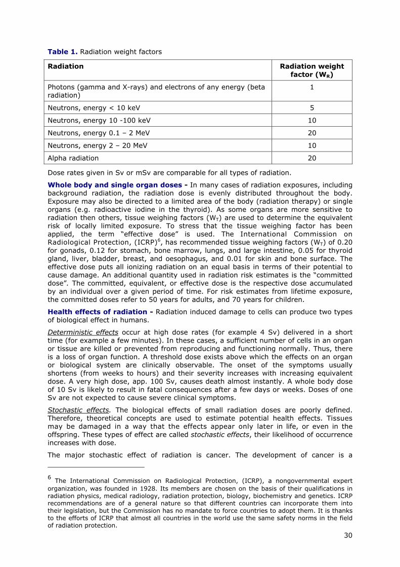
30
Table 1. Radiation weight factors
Radiation Radiation weight factor (WR)
Photons (gamma and X-rays) and electrons of any energy (beta radiation)
1
Neutrons, energy < 10 keV 5
Neutrons, energy 10 -100 keV 10
Neutrons, energy 0.1 – 2 MeV 20
Neutrons, energy 2 – 20 MeV 10
Alpha radiation 20
Dose rates given in Sv or mSv are comparable for all types of radiation.
Whole body and single organ doses - In many cases of radiation exposures, including background radiation, the radiation dose is evenly distributed throughout the body. Exposure may also be directed to a limited area of the body (radiation therapy) or single organs (e.g. radioactive iodine in the thyroid). As some organs are more sensitive to radiation then others, tissue weighing factors (WT) are used to determine the equivalent risk of locally limited exposure. To stress that the tissue weighing factor has been applied, the term “effective dose” is used. The International Commission on Radiological Protection, (ICRP)6, has recommended tissue weighing factors (WT) of 0.20 for gonads, 0.12 for stomach, bone marrow, lungs, and large intestine, 0.05 for thyroid gland, liver, bladder, breast, and oesophagus, and 0.01 for skin and bone surface. The effective dose puts all ionizing radiation on an equal basis in terms of their potential to cause damage. An additional quantity used in radiation risk estimates is the “committed dose”. The committed, equivalent, or effective dose is the respective dose accumulated by an individual over a given period of time. For risk estimates from lifetime exposure, the committed doses refer to 50 years for adults, and 70 years for children.
Health effects of radiation - Radiation induced damage to cells can produce two types of biological effect in humans.
Deterministic effects occur at high dose rates (for example 4 Sv) delivered in a short time (for example a few minutes). In these cases, a sufficient number of cells in an organ or tissue are killed or prevented from reproducing and functioning normally. Thus, there is a loss of organ function. A threshold dose exists above which the effects on an organ or biological system are clinically observable. The onset of the symptoms usually shortens (from weeks to hours) and their severity increases with increasing equivalent dose. A very high dose, app. 100 Sv, causes death almost instantly. A whole body dose of 10 Sv is likely to result in fatal consequences after a few days or weeks. Doses of one Sv are not expected to cause severe clinical symptoms.
Stochastic effects. The biological effects of small radiation doses are poorly defined. Therefore, theoretical concepts are used to estimate potential health effects. Tissues may be damaged in a way that the effects appear only later in life, or even in the offspring. These types of effect are called stochastic effects, their likelihood of occurrence increases with dose.
The major stochastic effect of radiation is cancer. The development of cancer is a
6 The International Commission on Radiological Protection, (ICRP), a nongovernmental expert organization, was founded in 1928. Its members are chosen on the basis of their qualifications in radiation physics, medical radiology, radiation protection, biology, biochemistry and genetics. ICRP recommendations are of a general nature so that different countries can incorporate them into their legislation, but the Commission has no mandate to force countries to adopt them. It is thanks to the efforts of ICRP that almost all countries in the world use the same safety norms in the field of radiation protection.
31
complex, multistage process involving initiation promotion, and progression of the tumour, a process that usually takes many years. Radiation appears to act principally at the initiation stage by introducing mutations in DNA. Stochastic effects occur at all dose levels. A modified cell can give rise to a clone of cells that may eventually result in cancer. A modified stem cell in the reproductive organs (gonads) that transmits genetic code to the descendants of the irradiated person may provide incorrect hereditary information and cause effects in offspring.
By observing the occurrence of cancer in the irradiated group and comparing with the number of cancers expected in an otherwise similar but non-irradiated group, the raised risk of cancer per unit dose can be estimated. This is commonly called a risk factor. Especially in case of radiation related cancers it is most important to include data for large groups of people to minimize the statistical uncertainties and take account of confounders that affect the spontaneous development of cancer.
Additional information on the deterministic and stochastic (probability) effects of radiation - On August 6, 1945, part of the population in the city of Hiroshima in Japan received radiation doses of all magnitudes. When trying to exhaust the graphite fire at the nuclear reactor in Chernobyl, 48 men received whole body doses exceeding 4 Sv with largest whole body doses between 12 and 16 Sv. A radiation dose of more than 3 to 4 Sv damages the bone marrow, the intestine, the neurovascular system and the skin, and is fatal. Lower whole body doses in the range of 2 Sv are not life threatening, but some symptoms of radiation sickness such as tiredness, vomiting, and lack of appetite may occur.
The stochastic effects (mainly cancer) after exposure to radiation have been observed in a few cohorts receiving high radiation doses. For instance, an increased cancer incidence of 6% above background was observed among the 100,000 atomic bomb survivors who received the highest radiation doses, but no increased cancer incidence was observed at doses below 300 mSv.
Risk factors for cancers - The main sources of information on the additional risk of cancer following exposure of the whole body to gamma radiation are studies of the survivors of the atomic bombs dropped on Hiroshima and Nagasaki in 1945.
Other risk estimates for the exposure of various tissues and organs to X rays and gamma rays come from people exposed to external radiation for the treatment of non-malignant or malignant conditions and for diagnostic purposes, and from people in the Marshall Islands exposed to severe fallout from atmospheric nuclear weapons tests. Information on the effects of alpha-emitting radionuclides comes from miners exposed to Radon and its decay products, from workers exposed to Radium-226 in luminous paint, from some patients treated with Radium-224 for bone disease, and from other patients given an X ray contrast medium containing Thorium oxide (CBEIR, 1990; IAEA, 1996; ICRP, 1984, 1991; UNSCEAR, 1993, 2000a, b).
The Linear No-Threshold (LNT) hypothesis - It must be taken into consideration that most of the atomic bomb survivors and other exposed groups received high doses over short periods of time. Observations of the cancer incidence in these groups, along with estimates of the doses they received indicate a linear relationship between dose and risk at high dose rates.
However, most radiation exposures involve low doses delivered over long periods of time. At these low levels of exposure, studies of cancer incidence in the exposed population do not provide direct evidence about the relationship between dose and risk, because the number of extra cancers that might be expected to result from the radiation exposure is too small (compared to the total number of cancer cases in the population, especially in case of rare cancers) to detect. It is, therefore, necessary to consider other information about the effects of radiation to judge the most likely form of the dose-risk relationship. For many years, a linear the relationship at low doses has been accepted (known as the 'linear-no threshold' or LNT hypothesis), i.e. that any radiation dose has a detrimental effect, which, however, may be infinitesimally small.

32
Nevertheless, several radiobiological experiments have suggested that low doses of radiation have no detrimental effect, because the body can successfully repair all of the damage caused by the radiation or low doses of radiation may stimulate the repair mechanisms in cells. On the other hand, other experiments were the basis for theories that low doses of radiation are more harmful (per unit of dose) than high doses, or that hereditary effects of radiation could worsen from generation to generation.
For some types of highly ionizing radiation, such as alpha particles, the risk factor is the same at low doses as at high doses. However, for weakly ionizing radiation, such as gamma rays, there is considerable radiobiological evidence that dose-response is more complicated. For these types of radiation, a linear relationship is a good approximation of dose response for both the low dose and high dose regions, but the risk per unit dose (the slope of the linear relationship) is less at low doses and dose rates. ICRP has estimated the risk factors for fatal cancers from low doses and dose rates in this way using a judicious reduction factor of two.
After a major review of biological effects at low doses, ICRP concluded in 2000 (UNSCEAR, 2000a, b) that for the time being, the Linear No-Threshold or LNT hypothesis is consistent with present knowledge and remains accordingly, the most scientifically defensible approximation of low dose response.
Biological effects observed over the past two decades in various in vivo and in vitro cell culture experiments interpreted that low dose/low flux ionizing radiation have significantly different biological responses as compared to high-dose radiation. Exposure of cell populations to very low fluxes of alpha-particles or incorporated radionuclides resulted in biological effects in both the irradiated and in non-irradiated cells in the population and cells exposed to medium from irradiated cultures indirectly responded to the radiation exposure. This phenomenon, termed the 'bystander response', has been postulated to influence both the estimation of risks of exposure to ionizing radiation and radiotherapy. However, an amplification of radiation-induced cytotoxic and genotoxic effects by the “bystander effect” is in contrast to the observations of adaptive responses, which are generally induced following exposure to low dose, low linear energy transfer radiation and tend to attenuate radiation-induced damage.
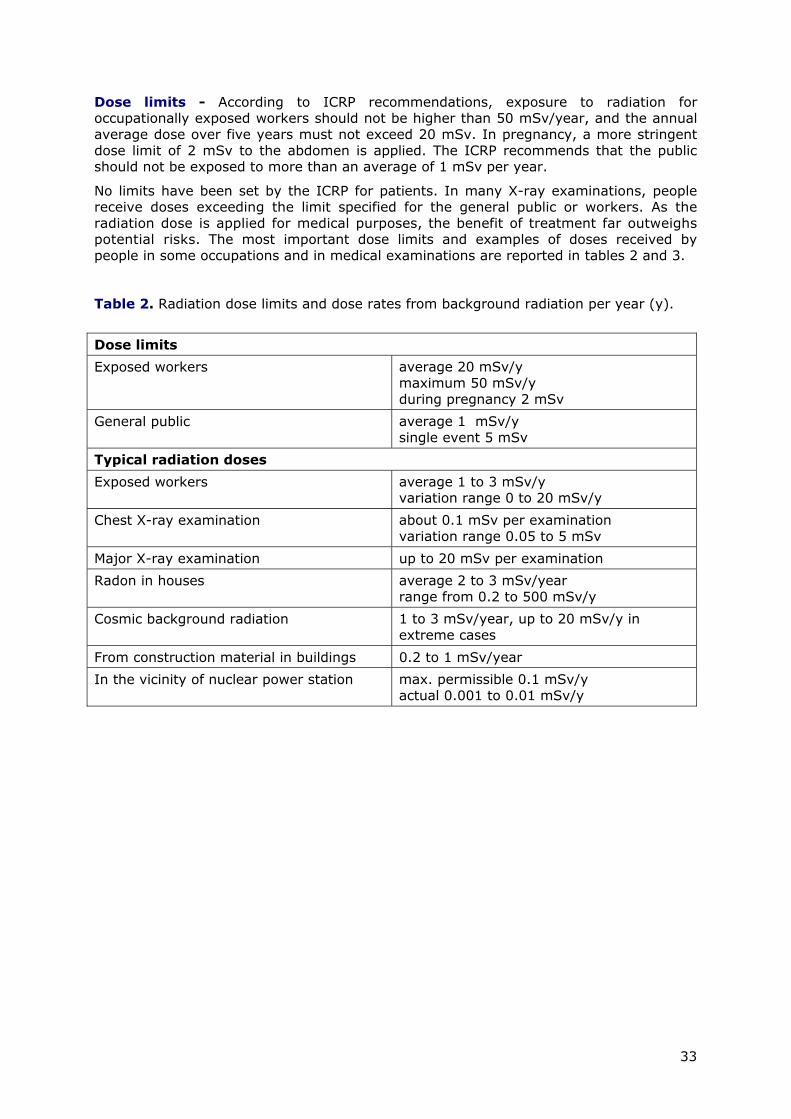
33
Dose limits - According to ICRP recommendations, exposure to radiation for occupationally exposed workers should not be higher than 50 mSv/year, and the annual average dose over five years must not exceed 20 mSv. In pregnancy, a more stringent dose limit of 2 mSv to the abdomen is applied. The ICRP recommends that the public should not be exposed to more than an average of 1 mSv per year.
No limits have been set by the ICRP for patients. In many X-ray examinations, people receive doses exceeding the limit specified for the general public or workers. As the radiation dose is applied for medical purposes, the benefit of treatment far outweighs potential risks. The most important dose limits and examples of doses received by people in some occupations and in medical examinations are reported in tables 2 and 3.
Table 2. Radiation dose limits and dose rates from background radiation per year (y).
Dose limits
Exposed workers average 20 mSv/y maximum 50 mSv/y during pregnancy 2 mSv
General public average 1 mSv/y single event 5 mSv
Typical radiation doses
Exposed workers average 1 to 3 mSv/y variation range 0 to 20 mSv/y
Chest X-ray examination about 0.1 mSv per examination variation range 0.05 to 5 mSv
Major X-ray examination up to 20 mSv per examination
Radon in houses average 2 to 3 mSv/year range from 0.2 to 500 mSv/y
Cosmic background radiation 1 to 3 mSv/year, up to 20 mSv/y in extreme cases
From construction material in buildings 0.2 to 1 mSv/year
In the vicinity of nuclear power station max. permissible 0.1 mSv/y actual 0.001 to 0.01 mSv/y

34
Table 3. Examples of radiation doses received.
a(ICRP, 1995), b(IAEA, 2004), c(Foikanos et al., 2007), d(EHC, 1997), e(ANS, 2000)
References
ANS (2000). American Nuclear Society. Estimate your personal annual radiation dose. www.ohiou.edu/ehs/docs/10_persnl_rad_dose.pdf.
CBEIR (1990). Committee on the Biological Effects of Ionizing Radiations: Health effects of exposure to low levels of ionizing radiation: BEIR V (http://www.nap.edu/openbook.php?record_id=1224&page=R1). National Academic Press, Washington DC.
EHC (1997). Environmental Health Centre. Radiation Basics. EHP, Washington DC.
Foikanos, K., Sarroui, I., and Pashalidis, I. (2007). Increased radiation exposure by granite used as natural tiling rock in cypriot houses. Radiation Measurements 42, 446-448.
IAEA (1996). International basic safety standards for protection against ionizing radiation and for the safety or radiation sources. International Atomic Energy Agency, Vienna - Safety Series 115, www-pub.iaea.org/MTCD/publications/PDF/SS-115-Web/Start.pdf.
IAEA (2004). Radiation, people and the environment. International Atomic Energy Agency, Vienna - IAEA/PUA.75/04-00391.
ICRP (1984). Nonstochastic Effects of Ionizing Radiation International Commission on Radiological Protection (ICRP) publication No. 41. Annals of the ICRP 14, 1-33.
ICRP (1991). Recommendations of the International Commission on Radiological Protection 1990 (ICRP) Publication No. 60. Annals of the ICRP 21, 1-201.
ICRP (1995). Age dependent doses to members of the public fromintake ofradionuclides: Part 4, Inhalation dose coefficients, International Commission on Radiological Protection
Type of exposure Yearly dose
Natural average world background a 2.400 mSv/y
Cosmic raysd [26] 0.310 mSv/y
Naturally occurring radioactive elementsd [26] 0.390 mSv/y
Additional annual dose received by people living in a room with granite floor-tilesc
up to 1.0 mSv/y
Average annual effective dose in different occupations Yearly dose
Civil aircrewb 3 mSv/y
Coal minersb 0.70 mSv/y
Radiotherapyb 0.60 mSv/y
Radiologyb 0.50 mSv/y
Dentistryb 0.060 mSv/y
Watching TV regularly (2-3 hours per day)e 0.010 mSv/y
Type of exposure (one exposure) Dose
One computer tomography (CT) to bodyb 1.100 mSv
One mammogramd 0.100 mSv
One dental X-rayb 0.080 mSv
One chest X-rayb 0.100 mSv
One crossing of the USA by jet aircraftd 0.050 mSv
35
(ICRP) publication No. 71. Annals of ICRP 25, i-xiv.
UNSCEAR (1993). United Nations Scientific Committee on the Effects of Atomic Radiation - Sources and effects of ionizing radiation. UNSCEAR Report to the General Assembly, with Scientific Annexes.
UNSCEAR (2000a). United Nations Scientific Committee on the Effects of Atomic Radiation - Sources and effects of ionizing radiation, vol. I Sources. UNSCEAR Report to the General Assembly, with Scientific Annexes.
UNSCEAR (2000b). United Nations Scientific Committee on the Effects of Atomic Radiation - Sources and effects of ionizing radiation, vol. II Effects. UNSCEAR Report to the General Assembly, with Scientific Annexes.
Publications recommended for further reading on the subject
Averbeck, D. Non-targeted effects as a paradigm breaking evidence. Mutat Res 687, 7-12.
Goodhead, D. T. (2010). New radiobiological, radiation risk and radiation protection paradigms. Mutat Res 687, 13-16.
Cohen, B.L. The cancer risk from low-level radiation, Health Physics 39 (1980) 659.
Coggle, J.E. Biological Effects of Radiation, Taylor & Francis Ltd, 1983.
Yalov, R. Biological effects of low-level radiation, Ch. 12 in M. E. BURNS (Ed), Low Level Radioactive Waste Regulation: Science, Politics, and Fear, Lewis Publishers, 1988.
NATIONAL RESEARCH COUNCIL, Health effects of exposure to low levels of ionizing radiation. BEIR V, National Academy Press, 1990.
Nias, A.H.W. An Introduction to Radiobiology, J Wiley & Sons, 1990.
United Nations Scientific Committee on the Effects of Atomic Radiation (UNSCEAR): Genetic and somatic effects of ionizing radiation, United Nations, 1993.
Bacq, Z.M., Alexander, P. Fundamentals of Radiobiology, Pergamon Press, Oxford, 1961.
Cadet, J. et al., Radiation-induced damage to DNA. Nucl Instr. Meth. Phys. Res. B151, 1 (1999).
Cohen, B.L. Validity of the linear no-threshold theory of radiation carcinogenesis at low doses, Nucl. Energy 38, 157 (1999).
Feinendegen, L.E., Bond, V.P., Sondhaus, C.A. in: Biological effects of low dose radiation. Yamada, T. et al., Editors. Elsevier (2000).
Feinendegen, L.E., Bond, V.P., Sondhaus, C.A., Altman, K.I. Cell protection by low doses of ionizing radiation challenges the concept of linearity; in Recent Aspects of Cellular and Applied Radiobiology, p. 3, Schneeweiss, F.H.A., Sharan, R.N. Editors. Research Centre, Uelich,, Germany (1999).
Jeggo, P.A. The fidelity of repair of radiation damage, Radiat. Protect. Dosim. 99,117 (2002).
Shimizu, Y., Kato, H., Schull, W.J. Studies of the mortality of A-bomb survivors, Part 9. Radiat. Research 121, 120 (1990).
Volpe, P. et al., Influence of low doses of radiation on the DNA double helix, gene expression and membrane state, Int. J. Radiat. Medicine 1, 78 (1999),
36
ANNEX 2: ENVIRONMENTAL CHEMISTRY OF NATURAL URANIUM
In the environment uranium can be present in the forms of U(IV) and U(VI). Uranium (VI) is more mobile than U(IV) because of aqueous complexation reactions involving ligands commonly found in natural waters; carbonate and phosphate are considered most important. Dissolved U(VI) exists in solution as the uranyl ion (UO2 2+), and form complexes with OH-, CO3
2-, F-, PO43-, SO4
2- and organic ligands.
Uranium that is leached from fragments and dust particles of DU will be transported in the soil or bedrock as UO2
2+ ions in precipitating water. Under oxidizing conditions, most of the uranium is in the form of soluble ions. Under reducing conditions, most uranium is present in insoluble forms.
Plants have a limited uptake of uranium. In a study of the effects on plants growing at a test site for DU, the observed concentration ratios were 0.02-0.13. However, the uranium levels in roots were much higher (ratios: 0.28-5.26). This variability was probably caused by small particles of soil and presumably, uranium adhering to roots. Furthermore, uranium colloids may have been absorbed on the root surfaces. The group of Oliver (Oliver et al., 2007) showed that DU deposited in soil at military training sites was indeed assimilated into plant and earthworm tissues. Plant tissue U concentrations were related to soil concentrations, the relationship was not linear which is in agreement with numerous studies investigating plant assimilation of U. However, the presence of DU in plants can also be a result of uptake from airborne DU (Jia et al., 2006; Zunic et al., 2008).
Direct ingestion of contaminated soil by cattle and sheep could also be considered as a pathway to food chain transfer. However, the transfer factor in animals is relatively low, comparable to the one for transfer to plants.
U(IV) is sparingly soluble but U chemistry predicts that particle weathering rate increases with the oxidation state for U and should be higher for U particles with average oxidation state +4.6 and +5.3 than for UO2 (Kashparov et al., 1999).
More information on the fate of DU can be derived from studies of DU deposition in military training sites were large amounts of DU ammunitions have been used in small areas. In soils of military training sites (Oliver et al., 2007, 2008a; Oliver et al., 2008b) U can become associated with exchangeable, organic, carbonate, Fe/Mn oxide and silicate mineral components of soil. As a consequence, DU could potentially move through soil in the form of dissolved species or as a variety of colloidal entities transported by porewater. Soil pH and soil porewater pH have an important influence on U sorption and mobility. Soil organic matter can play a key role in both solid-phase binding of U and in its aqueous phase mobilization, via formation of dissolved and/or colloidal U species (Crancon and Van der Lee, 2003; Mibus et al., 2007; Vandenhove et al., 2007). Environment modelling indicates that in a desert DU movement is minimal (Johnson et al., 2006). Free-living and plant symbiotic (mycorrhizal) fungi can colonize DU surfaces and transform metallic DU into uranyl phosphate minerals (Fomina et al., 2008). The potential for the migration of uranium depends on soil properties (Eh, pH, presence of complexing ligands, etc.). In a column-leaching study with DU amended soils found that DU was more readily mobilized than the U naturally present in the test soil and that the rate of mobilization/leaching increased with time (Vandenhove et al., 2007). The soil water distribution coefficient (Kd) of uranium for various soil pH values are given in Table 1. The reported values should be taken as indicative because the organic carbon content of a soil also strongly influences the Kd for uranium (soils high in organic carbon having a larger Kd).

37
Table 1. Values of Kd for uranium at various soil pHs
Soil pH Kd ml/g
3 <1
4 0.4
8 25
6 100
7 63
8 0.4
9 <1
10 <1
The transport of DU can also be affected by attenuation reactions that reduce uranium concentration in ground water and surface waters. These reactions include ion exchange and specific adsorption of uranium on organic matter, clay minerals, and ferric oxides and oxyhydroxides commonly present in soil. Under aerobic conditions, iron can play a key role in controlling the movement through soil. Uranium will bind to many iron minerals and adsorbs to humic matter in the soil. Uptake (complexation) by organic compounds will slow the migration of uranium through soil by several orders of magnitude, so that it becomes essentially immobile.
Radiological properties of uranium
All isotopes of uranium are radioactive. Naturally occurring uranium contains three isotopes, namely 238U, 235U, and 234U. 235U and 238U are the origin of two different decay chains. All uranium isotopes have the same chemical properties because they all have the same number of protons (92), but different radiological properties. The radioactivity of isotopes is dependent on the half-life. The most abundant naturally occurring uranium isotope, 238U, has the longest half-life and, consequently the lowest specific activity. Because of its higher specific activity 234U contributes as much as 238U
to the radioactivity of natural uranium although the weight percentage of this isotope is extremely small (0.006%).
Uranium isotopes decay to other radioactive elements that eventually decay to stable lead isotopes. In the decay process, alpha, beta and gamma radiation is emitted.
In nature, uranium is in secular equilibrium with the daughters of the decay chain. The short half-lives, chemical nature and decay mode of many of the decay products of 238U and 235U significantly increase the radiological hazard of natural uranium mineralization and uranium ore and natural uranium together with its daughters yield more decays per second as pure uranium. These radioactive progeny are removed during the uranium ore purification, making the uranium significantly less radiologically toxic than equivalent amounts of the original ore.
When uranium is separated from its ores, the decay chain is broken. Only thorium (234Th - beta type of decay) and protactinium (234Pa - beta type of decay), reach equilibrium with 238U within about 1 year and contribute to the radioactivity of the purified uranium. The remaining members of the decay chain following 234U take thousands of years to reach equilibrium and can be neglected. 235U follows the same pattern and only thorium (231Th - beta type of decay), reaches equilibrium rapidly. The decay products of 238U (234Th and 234Pa) and 234U (231Th) are responsible for the presence of beta and some gamma radiation in purified uranium. Both uranium and DU and their immediate decay products (e.g. 234Th, 234Pa and 231Th) emit alpha and beta particles with a very small amount of gamma radiation. Alpha and beta radiations are not very penetrating and are easily absorbed in the air and the skin.

38
Uranium is a metal of high density (18.9 g/cm3). The earth's crust contains an average of about 3 ppm (= 3 g/t) uranium, and seawater approximately 3 ppb (= 3 mg/t).
Naturally occurring uranium consists of three isotopes, all of which are radioactive: U-238, U-235, and U-234. U-238 and U-235 are the parent nuclides of two independent decay series, while U-234 is a decay product of the U-238 series.
Properties of the Natural Uranium Isotopes
U-234 U-235 U-238
half-life 244,500 years 703.8·106 years 4.468·109 years
specific activity 231.3 MBq/g 80,011 Bq/g 12,445 Bq/g
Isotopic Composition of Natural Uranium
U-234 U-235 U-238 Total
atom % 0.0054% 0.72% 99.275% 100%
weight % 0.0053% 0.711% 99.284% 100%
activity % 48.9% 2.2% 48.9% 100%
activity in 1 g Unat 12,356 Bq 568 Bq 12,356 Bq 25,280 Bq
Uranium-238 Decay Series
Nuclide Half-Life Radiation *
U-238 4.468 · 109 years alpha
Th-234 24.1 days beta
Pa-234m 1.17 minutes beta
U-234 244,500 years alpha
Th-230 77,000 years alpha
Ra-226 1,600 years alpha
Rn-222 3.8235 days alpha
Po-218 3.05 minutes alpha
Pb-214 26.8 minutes beta
Bi-214 19.9 minutes beta
Po-214 63.7 microseconds alpha
Pb-210 22.26 years beta
Bi-210 5.013 days beta
Po-210 138.378 days alpha
Pb-206 stable -
only major decays shown in addition, all decays emit gamma radiation
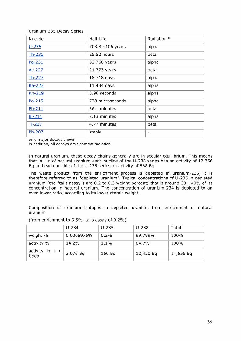
39
Uranium-235 Decay Series
Nuclide Half-Life Radiation *
U-235 703.8 · 106 years alpha
Th-231 25.52 hours beta
Pa-231 32,760 years alpha
Ac-227 21.773 years beta
Th-227 18.718 days alpha
Ra-223 11.434 days alpha
Rn-219 3.96 seconds alpha
Po-215 778 microseconds alpha
Pb-211 36.1 minutes beta
Bi-211 2.13 minutes alpha
Tl-207 4.77 minutes beta
Pb-207 stable -
only major decays shown in addition, all decays emit gamma radiation
In natural uranium, these decay chains generally are in secular equilibrium. This means that in 1 g of natural uranium each nuclide of the U-238 series has an activity of 12,356 Bq and each nuclide of the U-235 series an activity of 568 Bq.
The waste product from the enrichment process is depleted in uranium-235, it is therefore referred to as "depleted uranium". Typical concentrations of U-235 in depleted uranium (the "tails assay") are 0.2 to 0.3 weight-percent; that is around 30 - 40% of its concentration in natural uranium. The concentration of uranium-234 is depleted to an even lower ratio, according to its lower atomic weight.
Composition of uranium isotopes in depleted uranium from enrichment of natural uranium
(from enrichment to 3.5%, tails assay of 0.2%)
U-234 U-235 U-238 Total
weight % 0.0008976% 0.2% 99.799% 100%
activity % 14.2% 1.1% 84.7% 100%
activity in 1 g Udep 2,076 Bq 160 Bq 12,420 Bq 14,656 Bq

40
Natural Uranium Activity (U-238 series) Depleted Uranium Activity (U-238 series)
Natural Uranium Activity (U-238 series)
Initially, it only contains the uranium isotopes. Within a few days, Th-231 (U-235 series), and within a few months, Th-234 and Pa-234m (U-238 series) grow in. The activity then remains stable for more than 10,000 years. After this time, Th-230 and all other decay products of the U-238 series, and Pa-231 and all other decay products of the U-235 series grow in. This could, however, only occur with residual ore concentrate not consumed for nuclear fuel production.
Depleted Uranium Activity (U-238 series)
Within a few months, the isotopes of Th-234 and Pa-234m grow in to the value given by the activity of the U-238. The total activity in the depleted uranium then remains constant for around 10,000 years. Then, Th-230 with all its decay products starts growing in. After around 100,000 years, U-234 grows in to the activity level given by the U-238, further promoting the ingrowth of Th-230 and decay products. After around 2 million years, all nuclides are in secular equilibrium, and the total activity reaches a maximum and remains at this level for a billion years.
From residual U-235, Th-231 grows in within a few days. After around 10,000 years, Pa-231 and all other decay products of the U-235 series start growing in.
References
Crancon, P., and Van der Lee, J. (2003). Speciation and mobility of uranium(VI) in humic-containing soils. Radiochim Acta 91, 673-679.
Fomina, M., Charnock, J. M., Hillier, S., Alvarez, R., Livens, F., and Gadd, G. M. (2008). Role of fungi in the biogeochemical fate of depleted uranium. Curr Biol 18, R375-377.
Jia, G., Belli, M., Sansone, U., Rosamilia, S., and Gaudino, S. (2006). Concentration and characteristics of depleted uranium in biological and water samples collected in Bosnia and Herzegovina. J Environ Radioact 89, 172-187.
Johnson, B. E., Medina, V. F., and Cunniff, D. (2006). Numerical simulation of depleted uranium transport in a desert environment using a distributed parameter watershed model. Pract. Periodical Haz., Toxic and Radioactive Waste Mgmt. 10, 179-189.
Kashparov, V. A., Oughton, D. H., Zvarich, S. I., Protsak, V. P., and Levchuk, S. E. (1999). Kinetics of fuel particle weathering and 90Sr mobility in the Chernobyl 30-km
41
exclusion zone. Health Phys 76, 251-259.
Mibus, J., Sachs, S., Pfingsten, W., Nebelung, C., and Bernhard, G. (2007). Migration of uranium(IV)/(VI) in the presence of humic acids in quartz sand: a laboratory column study. J Contam Hydrol 89, 199-217.
Oliver, I. W., Graham, M. C., MacKenzie, A. B., Ellam, R. M., and Farmer, J. G. (2007). Assessing depleted uranium (DU) contamination of soil, plants and earthworms at UK weapons testing sites. J Environ Monit 9, 740-748.
Oliver, I. W., Graham, M. C., MacKenzie, A. B., Ellam, R. M., and Farmer, J. G. (2008a). Depleted uranium mobility across a weapons testing site: isotopic investigation of porewater, earthworms, and soils. Environ Sci Technol 42, 9158-9164.
Oliver, I. W., Graham, M. C., Mackenzie, A. B., Ellam, R. M., and Farmer, J. G. (2008b). Distribution and partitioning of depleted uranium (DU) in soils at weapons test ranges - investigations combining the BCR extraction scheme and isotopic analysis. Chemosphere 72, 932-939.
Vandenhove, H., Van Hees, M., Wannijn, J., Wouters, K., and Wang, L. (2007). Can we predict uranium bioavailability based on soil parameters? Part 2: soil solution uranium concentration is not a good bioavailability index. Environ Pollut 145, 577-586.
Zunic, Z. S., Mietelski, J. W., Blazej, S., Gaca, P., Tomankiewicz, E., Ujic, P., Celikovic, I., Cuknic, O., and Demajo, M. (2008). Traces of DU in samples of environmental bio-monitors (non-flowering plants, fungi) and soil from target sites of the Western Balkan region. J Environ Radioact 99, 1324-1328.