Open Access Full Text Article Management of tinea capitis ... · Kerion Celsi and favus have the...
10
© 2010 Bennassar and Grimalt, publisher and licensee Dove Medical Press Ltd. This is an Open Access article which permits unrestricted noncommercial use, provided the original work is properly cited. Clinical, Cosmetic and Investigational Dermatology 2010:3 89–98 Clinical, Cosmetic and Investigational Dermatology Dovepress submit your manuscript | www.dovepress.com Dovepress 89 REVIEW open access to scientific and medical research Open Access Full Text Article 7992 Management of tinea capitis in childhood Antoni Bennassar Ramon Grimalt Dept of Dermatology, Hospital Clinic, University of Barcelona, Barcelona, Spain Correspondence: Ramon Grimalt Hospital Clinic,Villarroel 170, 08036 Barcelona, Spain Fax +34 937 800011 Email [email protected] Abstract: Tinea capitis (TC) is a common dermatophyte infection affecting primarily prepubertal children. The causative pathogens belong to only two genera: Trichophyton and Microsporum. Although there is a great local variation in the epidemiology of TC worldwide, T. tonsurans is currently the most common cause of TC with M. canis second. Even though there is an emerg- ing number of anthropophilic scalp infections, M. canis remains the predominant causative organism in many countries of the Mediterranean basin, the most important dermatophyte car- riers being stray cats and dogs as well as pet puppies, kittens and rabbits. TC always requires systemic treatment because topical antifungal agents do not penetrate down to the deepest part of the hair follicle. Since the late 1950s, griseofulvin has been the gold standard for systemic therapy of TC. It is active against dermatophytes and has a long-term safety profile. The main disadvantage of griseofulvin is the long duration of treatment required which may lead to reduced compliance. The newer oral antifungal agents including terbinafine, itraconazole, ketokonazole, and fluconazole appear to have efficacy rates and potential adverse effects similar to those of griseofulvin in children with TC caused by Trichophyton species, while requiring a much shorter duration of treatment. They may, however, be more expensive. Keywords: tinea capitis, children, fungal infection, greseofulvin, terbinafine, itraconazole, fluconazole, treatment, pediatric infection Introduction Tinea capitis (TC) is a dermatophyte infection of the scalp hair follicles and interven- ing skin. 1,2 It affects primarily prepubertal children. The reported prevalence in Europe is around 1.5%. The causative pathogens belong to only two genera: Trichophyton and Microspo- rum. Although there is a great local variation in the epidemiology of TC worldwide, T. tonsurans is currently the most common cause of TC with M. canis the second. Even though there is an emerging number of anthropophilic scalp infections, M. canis remains the predominant causative organism in many countries of the Mediterranean basin, the most important dermatophyte carriers being stray cats and dogs as well as pet puppies, kittens and rabbits. Dermatophyte classification and pathogenesis of TC Dermatophytes are keratinophilic fungi which belong to three genera: Trichophyton, Microsporum, and Epidermophyton. On the basis of host preference and natural habitat, dermatophytes are classified as anthropophilic, zoophilic, and geophilic.
Transcript of Open Access Full Text Article Management of tinea capitis ... · Kerion Celsi and favus have the...
© 2010 Bennassar and Grimalt, publisher and licensee Dove Medical Press Ltd. This is an Open Access article which permits unrestricted noncommercial use, provided the original work is properly cited.
Clinical, Cosmetic and Investigational Dermatology 2010:3 89–98
Clinical, Cosmetic and Investigational Dermatology Dovepress
submit your manuscript | www.dovepress.com
Dovepress 89
R e v I e w
open access to scientific and medical research
Open Access Full Text Article
7992
Management of tinea capitis in childhood
Antoni BennassarRamon GrimaltDept of Dermatology, Hospital Clinic, University of Barcelona, Barcelona, Spain
Correspondence: Ramon Grimalt Hospital Clinic, villarroel 170, 08036 Barcelona, Spain Fax +34 937 800011 email [email protected]
Abstract: Tinea capitis (TC) is a common dermatophyte infection affecting primarily prepubertal
children. The causative pathogens belong to only two genera: Trichophyton and Microsporum.
Although there is a great local variation in the epidemiology of TC worldwide, T. tonsurans is
currently the most common cause of TC with M. canis second. Even though there is an emerg-
ing number of anthropophilic scalp infections, M. canis remains the predominant causative
organism in many countries of the Mediterranean basin, the most important dermatophyte car-
riers being stray cats and dogs as well as pet puppies, kittens and rabbits. TC always requires
systemic treatment because topical antifungal agents do not penetrate down to the deepest part
of the hair follicle. Since the late 1950s, griseofulvin has been the gold standard for systemic
therapy of TC. It is active against dermatophytes and has a long-term safety profile. The main
disadvantage of griseofulvin is the long duration of treatment required which may lead to reduced
compliance. The newer oral antifungal agents including terbinafine, itraconazole, ketokonazole,
and fluconazole appear to have efficacy rates and potential adverse effects similar to those of
griseofulvin in children with TC caused by Trichophyton species, while requiring a much shorter
duration of treatment. They may, however, be more expensive.
Keywords: tinea capitis, children, fungal infection, greseofulvin, terbinafine, itraconazole,
fluconazole, treatment, pediatric infection
IntroductionTinea capitis (TC) is a dermatophyte infection of the scalp hair follicles and interven-
ing skin.1,2 It affects primarily prepubertal children. The reported prevalence in Europe
is around 1.5%.
The causative pathogens belong to only two genera: Trichophyton and Microspo-
rum. Although there is a great local variation in the epidemiology of TC worldwide,
T. tonsurans is currently the most common cause of TC with M. canis the second.
Even though there is an emerging number of anthropophilic scalp infections,
M. canis remains the predominant causative organism in many countries of the
Mediterranean basin, the most important dermatophyte carriers being stray cats and
dogs as well as pet puppies, kittens and rabbits.
Dermatophyte classification and pathogenesis of TCDermatophytes are keratinophilic fungi which belong to three genera: Trichophyton,
Microsporum, and Epidermophyton. On the basis of host preference and natural habitat,
dermatophytes are classified as anthropophilic, zoophilic, and geophilic.

Clinical, Cosmetic and Investigational Dermatology 2010:3submit your manuscript | www.dovepress.com
Dovepress
Dovepress
90
Bennassar and Grimalt
TC is mainly caused by anthropophilic and zoophilic
species of the genera Trichophyton and Microsporum.1,2
On the basis of the type of hair invasion, dermatophytes
are also classified as endothrix, ectothrix or favus.
In endothrix infection the fungus grows completely within
the hair shaft, the hyphae are converted to arthroconidia
(spores) within the hair while the cuticle surface of the hair
remains intact.
In ectothrix infection hair invasion develops in a man-
ner similar to endothrix except that the hyphae destroy the
hair cuticle and grow around the exterior of the hair shaft.
Arthroconidia may develop both within and outside the hair
shaft. Elongated hyphae, parallel to the long axis of the hair,
persist within the hair.
Favus is characterized by production of hyphae, which are
parallel to the long axis of the hair shaft. When the hyphae
degenerate, long tunnels are left within the hair shaft.1,2
Ectothrix anthropophilic infections potentially spread
rapidly whereas endothrix and favic infections are less
contagious.3
EpidemiologyTC is a common dermatophyte infection affecting primarily
prepubertal children. Adults are infrequently affected.2,4,5
The reported prevalence in Europe ranges between 0.23%
and 2.6%.6,7
The causative pathogens belong to only two genera:
Trichophyton and Microsporum. Although there is a great
local variation in the epidemiology of TC worldwide,
T. Tonsurans is currently the most common cause of TC and
M. Canis the second one (Table 1).8
Even though there is an emerging number of anthropo-
philic scalp infections, M. canis remains the predominant
causative organism in many countries of the Mediterranean
basin, being stray cats and dogs as well as pet puppies, kittens
and rabbits the most important dermatophyte carriers.8
On the other hand, anthropophilic TC has been mainly
reported in children of Afro-Caribbean descent living in
urban areas. These dermatophytosis are most frequently
incurred by contact with an infected child, either directly
or via fomites.8,9 It has been recently reported that an
asymptomatic adult carrier may provide a source for contin-
ued reinfection in children.
T. schoenleinii causes a chronic form of TC that is usu-
ally acquired before adolescence and extending into adult-
hood. Fortunately, it has nearly disappeared from developed
countries.10
Clinical presentationFour clinical infection patterns have been reported. Different
clinical presentation may arise depending on the causative
organism, the type of hair invasion, and the specific host
T-lymphocyte inflammatory response.4
Noninflammatory black dot pattern is clinically char-
acterized by well-demarcated areas of hair loss. Fungal
arthrospores proliferate inside the hair shafts, weakening
them. Hairs break off at or below the scalp surface, giving the
characteristic appearance of black dots on the alopecic patch.
Cell-mediated immunity to fungal antigen skin test is usually
negative and adenopathy is often absent. (Figure 1).
Noninflammatory seborrheic dermatitis type is a diffuse
or patchy, fine, white, adherent scale affecting the scalp.
This is the most difficult to diagnose because it resembles
dandruff and only one third of patients have a positive potas-
sium hydroxide examination.
In inflammatory tinea capitis (Kerion), there are one or
multiple tender, inflamed, alopecic nodules with pustules on
their surface. Fever, occipital adenopathy, leukocytosis, and
even a diffuse, morbilliform rash may occur. Most patients
have a positive skin test to fungal antigen, suggesting that
the patient’s immune response may account for the intense
inflammation.
Table 1 worldwide etiological agents causing TC
Species Types
Trichophyton tonsurans AnthropophilicMicrosporum canis ZoophilicMicrosporum audouinii AnthropophilicTrichophyton soudanense AnthropophilicTrichophyton violaceum Anthropophilic
Figure 1 Tinea capitis endotrix.
Clinical, Cosmetic and Investigational Dermatology 2010:3 submit your manuscript | www.dovepress.com
Dovepress
Dovepress
91
Management of tinea capitis in childhood
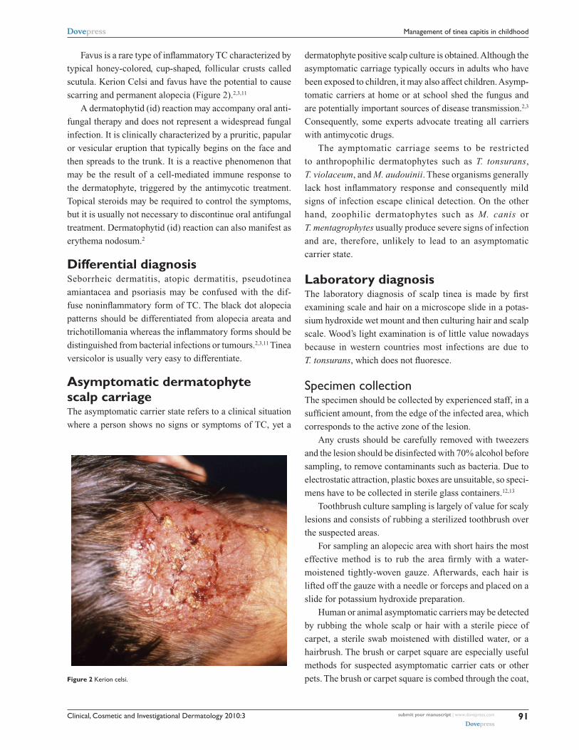
Favus is a rare type of inflammatory TC characterized by
typical honey-colored, cup-shaped, follicular crusts called
scutula. Kerion Celsi and favus have the potential to cause
scarring and permanent alopecia (Figure 2).2,3,11
A dermatophytid (id) reaction may accompany oral anti-
fungal therapy and does not represent a widespread fungal
infection. It is clinically characterized by a pruritic, papular
or vesicular eruption that typically begins on the face and
then spreads to the trunk. It is a reactive phenomenon that
may be the result of a cell-mediated immune response to
the dermatophyte, triggered by the antimycotic treatment.
Topical steroids may be required to control the symptoms,
but it is usually not necessary to discontinue oral antifungal
treatment. Dermatophytid (id) reaction can also manifest as
erythema nodosum.2
Differential diagnosisSeborrheic dermatitis, atopic dermatitis, pseudotinea
amiantacea and psoriasis may be confused with the dif-
fuse noninflammatory form of TC. The black dot alopecia
patterns should be differentiated from alopecia areata and
trichotillomania whereas the inflammatory forms should be
distinguished from bacterial infections or tumours.2,3,11 Tinea
versicolor is usually very easy to differentiate.
Asymptomatic dermatophyte scalp carriageThe asymptomatic carrier state refers to a clinical situation
where a person shows no signs or symptoms of TC, yet a
dermatophyte positive scalp culture is obtained. Although the
asymptomatic carriage typically occurs in adults who have
been exposed to children, it may also affect children. Asymp-
tomatic carriers at home or at school shed the fungus and
are potentially important sources of disease transmission.2,3
Consequently, some experts advocate treating all carriers
with antimycotic drugs.
The aymptomatic carriage seems to be restricted
to anthropophilic dermatophytes such as T. tonsurans,
T. violaceum, and M. audouinii. These organisms generally
lack host inflammatory response and consequently mild
signs of infection escape clinical detection. On the other
hand, zoophilic dermatophytes such as M. canis or
T. mentagrophytes usually produce severe signs of infection
and are, therefore, unlikely to lead to an asymptomatic
carrier state.
Laboratory diagnosisThe laboratory diagnosis of scalp tinea is made by first
examining scale and hair on a microscope slide in a potas-
sium hydroxide wet mount and then culturing hair and scalp
scale. Wood’s light examination is of little value nowadays
because in western countries most infections are due to
T. tonsurans, which does not fluoresce.
Specimen collectionThe specimen should be collected by experienced staff, in a
sufficient amount, from the edge of the infected area, which
corresponds to the active zone of the lesion.
Any crusts should be carefully removed with tweezers
and the lesion should be disinfected with 70% alcohol before
sampling, to remove contaminants such as bacteria. Due to
electrostatic attraction, plastic boxes are unsuitable, so speci-
mens have to be collected in sterile glass containers.12,13
Toothbrush culture sampling is largely of value for scaly
lesions and consists of rubbing a sterilized toothbrush over
the suspected areas.
For sampling an alopecic area with short hairs the most
effective method is to rub the area firmly with a water-
moistened tightly-woven gauze. Afterwards, each hair is
lifted off the gauze with a needle or forceps and placed on a
slide for potassium hydroxide preparation.
Human or animal asymptomatic carriers may be detected
by rubbing the whole scalp or hair with a sterile piece of
carpet, a sterile swab moistened with distilled water, or a
hairbrush. The brush or carpet square are especially useful
methods for suspected asymptomatic carrier cats or other
pets. The brush or carpet square is combed through the coat, Figure 2 Kerion celsi.
Clinical, Cosmetic and Investigational Dermatology 2010:3submit your manuscript | www.dovepress.com
Dovepress
Dovepress
92
Bennassar and Grimalt
trapping fungal spores with hair and debris, and then pressed
onto the surface of the culture medium.13
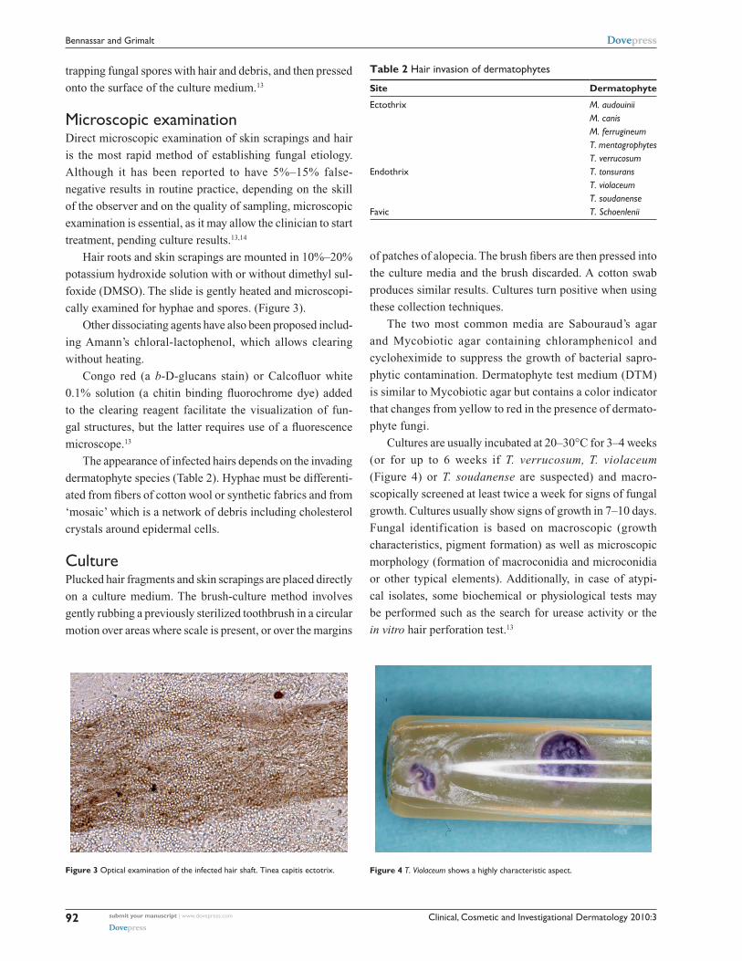
Microscopic examinationDirect microscopic examination of skin scrapings and hair
is the most rapid method of establishing fungal etiology.
Although it has been reported to have 5%–15% false-
negative results in routine practice, depending on the skill
of the observer and on the quality of sampling, microscopic
examination is essential, as it may allow the clinician to start
treatment, pending culture results.13,14
Hair roots and skin scrapings are mounted in 10%–20%
potassium hydroxide solution with or without dimethyl sul-
foxide (DMSO). The slide is gently heated and microscopi-
cally examined for hyphae and spores. (Figure 3).
Other dissociating agents have also been proposed includ-
ing Amann’s chloral-lactophenol, which allows clearing
without heating.
Congo red (a b-D-glucans stain) or Calcofluor white
0.1% solution (a chitin binding fluorochrome dye) added
to the clearing reagent facilitate the visualization of fun-
gal structures, but the latter requires use of a fluorescence
microscope.13
The appearance of infected hairs depends on the invading
dermatophyte species (Table 2). Hyphae must be differenti-
ated from fibers of cotton wool or synthetic fabrics and from
‘mosaic’ which is a network of debris including cholesterol
crystals around epidermal cells.
CulturePlucked hair fragments and skin scrapings are placed directly
on a culture medium. The brush-culture method involves
gently rubbing a previously sterilized toothbrush in a circular
motion over areas where scale is present, or over the margins
of patches of alopecia. The brush fibers are then pressed into
the culture media and the brush discarded. A cotton swab
produces similar results. Cultures turn positive when using
these collection techniques.
The two most common media are Sabouraud’s agar
and Mycobiotic agar containing chloramphenicol and
cycloheximide to suppress the growth of bacterial sapro-
phytic contamination. Dermatophyte test medium (DTM)
is similar to Mycobiotic agar but contains a color indicator
that changes from yellow to red in the presence of dermato-
phyte fungi.
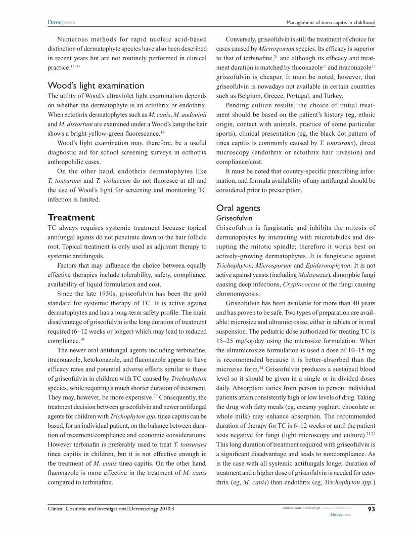
Cultures are usually incubated at 20–30°C for 3–4 weeks
(or for up to 6 weeks if T. verrucosum, T. violaceum
(Figure 4) or T. soudanense are suspected) and macro-
scopically screened at least twice a week for signs of fungal
growth. Cultures usually show signs of growth in 7–10 days.
Fungal identification is based on macroscopic (growth
characteristics, pigment formation) as well as microscopic
morphology (formation of macroconidia and microconidia
or other typical elements). Additionally, in case of atypi-
cal isolates, some biochemical or physiological tests may
be performed such as the search for urease activity or the
in vitro hair perforation test.13
Figure 3 Optical examination of the infected hair shaft. Tinea capitis ectotrix.
Table 2 Hair invasion of dermatophytes
Site Dermatophyte
ectothrix M. audouiniiM. canisM. ferrugineumT. mentagrophytesT. verrucosum
endothrix T. tonsuransT. violaceumT. soudanense
Favic T. Schoenlenii
Figure 4 T. Violaceum shows a highly characteristic aspect.
Clinical, Cosmetic and Investigational Dermatology 2010:3 submit your manuscript | www.dovepress.com
Dovepress
Dovepress
93
Management of tinea capitis in childhood
Numerous methods for rapid nucleic acid-based
distinction of dermatophyte species have also been described
in recent years but are not routinely performed in clinical
practice.15–17
wood’s light examinationThe utility of Wood’s ultraviolet light examination depends
on whether the dermatophyte is an ectothrix or endothrix.
When ectothrix dermatophytes such as M. canis, M. audouinii
and M. distortum are examined under a Wood’s lamp the hair
shows a bright yellow-green fluorescence.18
Wood’s light examination may, therefore, be a useful
diagnostic aid for school screening surveys in ecthotrix
anthropohilic cases.
On the other hand, endothrix dermatophytes like
T. tonsurans and T. violaceum do not fluoresce at all and
the use of Wood’s light for screening and monitoring TC
infection is limited.
TreatmentTC always requires systemic treatment because topical
antifungal agents do not penetrate down to the hair follicle
root. Topical treatment is only used as adjuvant therapy to
systemic antifungals.
Factors that may influence the choice between equally
effective therapies include tolerability, safety, compliance,
availability of liquid formulation and cost.
Since the late 1950s, griseofulvin has been the gold
standard for systemic therapy of TC. It is active against
dermatophytes and has a long-term safety profile. The main
disadvantage of griseofulvin is the long duration of treatment
required (6–12 weeks or longer) which may lead to reduced
compliance.19
The newer oral antifungal agents including terbinafine,
itraconazole, ketokonazole, and fluconazole appear to have
efficacy rates and potential adverse effects similar to those
of griseofulvin in children with TC caused by Trichophyton
species, while requiring a much shorter duration of treatment.
They may, however, be more expensive.20 Consequently, the
treatment decision between griseofulvin and newer antifungal
agents for children with Trichophyton spp. tinea capitis can be
based, for an individual patient, on the balance between dura-
tion of treatment/compliance and economic considerations.
However terbinafin is preferably used to treat T. tonsurans
tinea capitis in children, but it is not effective enough in
the treatment of M. canis tinea capitis. On the other hand,
fluconazole is more effective in the treatment of M. canis
compared to terbinafine.
Conversely, griseofulvin is still the treatment of choice for
cases caused by Microsporum species. Its efficacy is superior
to that of terbinafine,21 and although its efficacy and treat-
ment duration is matched by fluconazole22 and itraconazole23
griseofulvin is cheaper. It must be noted, however, that
griseofulvin is nowadays not available in certain countries
such as Belgium, Greece, Portugal, and Turkey.
Pending culture results, the choice of initial treat-
ment should be based on the patient’s history (eg, ethnic
origin, contact with animals, practice of some particular
sports), clinical presentation (eg, the black dot pattern of
tinea capitis is commonly caused by T. tonsurans), direct
microscopy (endothrix or ectothrix hair invasion) and
compliance/cost.
It must be noted that country-specific prescribing infor-
mation, and formula availability of any antifungal should be
considered prior to prescription.
Oral agentsGriseofulvinGriseofulvin is fungistatic and inhibits the mitosis of
dermatophytes by interacting with microtubules and dis-
rupting the mitotic spindle; therefore it works best on
actively-growing dermatophytes. It is fungistatic against
Trichophyton, Microsporum and Epidermophyton. It is not
active against yeasts (including Malassezia), dimorphic fungi
causing deep infections, Cryptococcus or the fungi causing
chromomycosis.
Griseofulvin has been available for more than 40 years
and has proven to be safe. Two types of preparation are avail-
able: microsize and ultramicrosize, either in tablets or in oral
suspension. The pediatric dose authorized for treating TC is
15–25 mg/kg/day using the microsize formulation. When
the ultramicrosize formulation is used a dose of 10–15 mg
is recommended because it is better-absorbed than the
microzise form.24 Griseofulvin produces a sustained blood
level so it should be given in a single or in divided doses
daily. Absorption varies from person to person: individual
patients attain consistently high or low levels of drug. Taking
the drug with fatty meals (eg, creamy yoghurt, chocolate or
whole milk) may enhance absorption. The recommended
duration of therapy for TC is 6–12 weeks or until the patient
tests negative for fungi (light microscopy and culture).23,24
This long duration of treatment required with griseofulvin is
a significant disadvantage and leads to noncompliance. As
is the case with all systemic antifungals longer duration of
treatment and a higher dose of griseofulvin is needed for ecto-
thrix (eg, M. canis) than endothrix (eg, Trichophyton spp.)
Clinical, Cosmetic and Investigational Dermatology 2010:3submit your manuscript | www.dovepress.com
Dovepress
Dovepress
94
Bennassar and Grimalt
infections. Mycological cure and efficacy rates are generally
high, being in the range of 80%–96%.25
Treatment failures can be observed due to poor compli-
ance, fungal resistance, drug interactions, or side effects.
Griseofulvin is a safe drug. Headaches and gastroin-
testinal disturbances are the most common side effects.
The dosage can be temporarily lowered to see if symptoms
clear, but sometimes the drug must be discontinued. Severe
allergic reactions, hepatic toxicity and leucopenia rarely
occur; therefore routine blood studies are not necessary
unless treatment is to last for many months or the dosage is
exceptionally high (Table 3).
It is contraindicated in children with porphyria, lupus
erythematosus, or severe liver disease.26
Drug interactions can occur with warfarin, phenobarbital
and cyclosporine since griseofulvin is a potent inducer of
microsomal cytochrome P-450 enzymes.24,25 The main disad-
vantage of griseofulvin is the long duration of treatment.25–28
TerbinafineTerbinafine belongs to the allyamine class of drugs, a new
generation of antifungal agents. It is fungicidal to dermato-
phytes since it inhibits squalene epoxidase, a membrane-
bound enzyme in the biosynthetic pathway of sterol synthesis
of the fungal cell membrane. It is well absorbed and binds
strongly and nonspecifically to plasma proteins.
The absorption characteristics are not altered when
terbinafine is taken with food. Its clearance in children is 40%
higher than in adults. Since terbinafine is highly lipophilic
and keratophilic, it is distributed throughout adipose tissue,
dermis, epidermis, nails, and hair and persists in these tissues
for weeks. Persistence of the drug in plasma is of concern
when side effects are experienced. Terbinafine is delivered
to the stratum corneum via the sebum and, to a lesser extent,
through incorporation into the basal keratinocytes and dif-
fusion through the dermis-epidermis. Terbinafine is not
found in eccrine sweat. It remains in skin at concentrations
above the mean inhibitory concentration (MIC) for most
dermatophytes for 2 to 3 weeks after discontinuation of
long-term oral therapy. After 6 and 12 weeks of oral therapy,
terbinafine has been detected in the nail plate for 30 and
36 weeks, respectively, at a concentration well above the
MIC for most dermatophytes. Terbinafine is metabolized in
the liver, and dose adjustments may be needed in patients
with liver or renal dysfunction.
It is available as 250 mg tablets. The standard pediatric
single daily dose is 62.5 mg (10–20 kg); 125 mg (20–40 kg)
and 250 mg ($40 kg). Some suggest a weight-based dose
of 4 to 5 mg/kg per day as an alternative.29 Terbinafine is
concentrated in the hair and may remain present at fungicidal
concentrations for several weeks after a course of treatment
has been completed.30 The duration of treatment is generally
4 weeks, although shorter durations (2 weeks) have also been
reported to be effective.28,31,32
Higher dosages (10–25 kg: 125 mg/day; .25 kg:
250 mg/day or 12.5 mg/kg/day) or longer duration of treatment
(8–12 weeks) may be required for M. canis infection.33–36
Side effects of terbinafine are rare and include gastrointes-
tinal symptoms, rashes and headache. Liver enzyme abnor-
malities and drug reactions are occasionally seen. Plasma
concentrations are reduced by rifampicin and increased by
cimetidine.30
ItraconazoleItraconazole is a triazole antifungal agent against Trichophy-
ton and Microsporum spp. It exhibits both fungistatic and
fungicidal activity depending on its concentration in the
tissues, though its primary mode of action is fungistatic
by inhibiting the cytochrome P-450-dependent enzymes,
blocking the synthesis of ergosterol, the principal compo-
nent of fungal cell membranes. Itraconazole is lipophilic
and has a high affinity for keratinizing tissues. It adheres to
the lipophilic cytoplasm of keratinocytes in the nail plate,
allowing progressive buildup and persistence in the nail
Table 3 Dosing pediatric regimens for the treatment of tinea capitis
Antifungal agent
Dosage Duration of treatment
Griseofulvin Microsize Ultramicrosize
20–25 mg/kg/day 10–15 mg/kg/day
6–12 weeks or longer until fungal cultures are negative
Terbinafine 10–20 kg: 62.5 mg/day 20–40 kg: 125 mg/day .40 kg: 250 mg/day Or 4–5 mg/kg/day
Trichophyton spp.: 2–4 weeks Microsporum spp.: 8–12 weeks
Itraconazole Capsules: 5 mg/kg/day Oral solution: 3 mg/kg/day
Daily dosing: 2–6 weeks Pulse regimen (1 week with 2 weeks off between the first 2 pulses and 3 weeks between the 2nd and 3rd): 2–3 pulses (range: 1–5)
Fluconazole Daily dosing: 5–6 mg/kg/day weekly dosing: 8 mg/kg once weekly
3–6 weeks 8–12 weeks

Clinical, Cosmetic and Investigational Dermatology 2010:3 submit your manuscript | www.dovepress.com
Dovepress
Dovepress
95
Management of tinea capitis in childhood
plate. The drug reaches high levels in the nails that persist
for at least 6 months after discontinuation of 3 months of
therapy and during pulsed cycles. The concentration in the
stratum corneum remains detectable for 4 weeks after therapy.
Itraconazole levels in sebum are 5 times higher than those in
plasma and remain high for as long as 1 week after therapy.
This fact suggests that secretion in sebum may account for
the high concentrations found in skin. The drug has an affinity
for mammalian cytochrome P-450 enzymes, as well as for
fungal P-450-dependent enzyme, and thus has the potential
for clinically-important interactions with astemizole, rifam-
picin, oral contraceptives, H2 receptor antagonists, warfarin
and cyclosporine.
It is available as capsules or an oral solution. Itracon-
azole capsule formulation should be ingested with a meal
whereas the oral solution should be taken in the fasting state
for optimum bioavailability. The response to therapy does
not appear to depend upon the formulation administered
(capsules versus suspension).
The recommended pediatric dose is 5 mg/kg/day given
continuously or by repeat pulsing. Where the oral solution
is used, dosage is reduced to 3 mg/kg/day.37
Using the continuous regimen, the duration of treat-
ment for Trichophyton spp.38 and Microsporum spp. tinea
capitis39 is 2 and 6 weeks with cure rates of 85.7% and 88%
respectively. It must be noted that the 6-week regimen of
itraconazole is of comparable efficacy to griseofulvin, in
cases of Microsporum-TC.19
In the pulse regimen (one pulse of 5 mg/kg/day for 1 week
with 2 weeks off between the first two pulses and 3 weeks
between the second and third), the number of pulses required
for the treatment depends in part on the severity of the TC.40
In this way it may be possible to individualize the number of
pulses administered according to the clinical response.
Side effects of itraconazole include headache, gastro-
intestinal complaints, rash and occasionally liver enzyme
abnormalities. Less common is peripheral edema especially
when taken with calcium channel blockers.
Itraconazole may increase plasma concentration of
cyclosporine, certain benzodiazepines (midazolam, tri-
azolam, alprazolam, and estazolam), digoxin, and cisapride.
Concomitant use of H2-receptor antagonists, phenytoin,
isoniazid, and rifampin may reduce the plasma concentration
of itraconazole. Its use is strongly discouraged for patients
with elevated or abnormal liver enzymes, active liver disease
or who have experienced liver toxicity with other antifungal
azole drugs. It is contraindicated in patients with evidence of
ventricular dysfunction such as congestive heart failure.
FluconazoleFluconazole is primarily a fungistatic triazole, preventing the
conversion of lanosterol to ergosterol, an essential component
of the fungal cytoplasmic membrane. It is distinguished from
the other azoles by its water solubility that results in excellent
bioavailability by the oral route. Because fluconazole is highly
soluble in water it is transported to the skin through sweat and
concentrated by evaporation. It achieves high concentrations
in the epidermis and nails and persists up to 3 months.41
It is available as a tablet or oral suspension. Doses of
5–6 mg/kg/ per day for 4–6 weeks can effectively treat TC.22
Once-weekly 8 mg/kg pulse dosing for 8–12 weeks is an
alternative regimen.42
Evidence suggest that in respect to Trichophyton species
TC, a 2–4-week regimen of fluconazole has similar cure rates
to a 6-week regimen of griseofulvin.20
Two studies that included 140 children found similar
cure rates of 2–4 weeks of fluconazole when compared with
6 weeks of griseofulvin (RR 0.92; 95% CI 0.80 to 1.05).
Side effects of fluconazole are similar to other azole
derivatives. Hematologic and hepatic toxicity may occasion-
ally occur.
Drug interaction: terfenadine, cisapride (risk of serious
cardiac arrhythmias)
Contraindications: severe liver disease. Use with caution
in patients sensitive to other azoles.
Topical agentsAdjunctive topical therapies such as Selenium sulphide,43
zinc pyrithione, povidoneiodide or ketoconazole44 shampoos
as well as fungicidal creams or lotions45 have been shown
to decrease the carriage of viable spores responsible for
the disease contagion and reinfection and may shorten the
cure rate with oral antifungal. A terbinafine solution 0.01%
completely killed arthroconidia of five Trichophyton species
after an exposure time of 15–30 min.46
The topical fungicidal cream/lotion should be applied to
the lesions once daily for a week.45
The shampoo should be applied to the scalp and hair for
5 minutes twice-weekly for 2–4 weeks39,47 or three times
weekly until the patient is clinically and mycologically
cured.19 The authors recommend the latter in conjunction with
one week of topical fungicidal cream or lotion application.
Additional measuresSchool attendanceKeeping children out of school after starting therapy is
controversial. Several experts suggest that once treatment
Clinical, Cosmetic and Investigational Dermatology 2010:3submit your manuscript | www.dovepress.com
Dovepress
Dovepress
96
Bennassar and Grimalt
has been initiated with oral and topical agents, the children
should, for practical reasons, be allowed back to school or day
care although there is still a risk of infecting fellow students.26
On the other hand, other experts recommend exemption
from school/kindergarten attendance, regardless of the type
of dermatophyte, for approximately 2 weeks after initiation
of treatment, a period necessary for significant decline of
infection load in the hair follicle.45
Patient education is, therefore, of utmost importance in
eradicating TC.
It must be stressed that the degree of disease transmis-
sion depends on the type of the dermatophyte isolated, the
most contagious being the ectothrix anthropophilic. The
latter potentially spreads rapidly and often causes epidem-
ics in schools.48 Additionally, topical fungicidal treatment,
nowadays, can kill arthroconidia rapidly.46
Therefore the following are recommended: If the caus-
ative agent is an anthropophilic ectothrix the child should
usually be allowed school/kindergarten attendance one week
after initiation of treatment. Wood’s light is useful to monitor
the disappearance of contaminating spores. In all other cases
the child should be allowed to attend school/kindergarten as
soon as the treatment has been initiated.
When the child is back at school, he/she should be
strongly advised not to share items such as combs, hair-
brushes, scarves, and hats, as fomites may play a role in
transmission. School staff may help in enforcing this.
So, in all cases with TC caused by anthropophilic der-
matophytes the school authorities should be notified.48
Sports that lead to prolonged close physical contact (eg,
wrestling) should be prohibited until the risk of infection no
longer exists.
Plucking the involved hair, as practiced in many coun-
tries, may help in the rapid resolution of the infection as it
physically removes large amounts of yeasts.
Sources of infectionUpon positive microscopy and pending culture clinical
examination of family members is urgently recommended.
Appropriate mycological samples should be taken initially
only from those with signs of infection.
Zoophilic organisms such as M. canis cause an inflam-
matory response in nearly all those infected. Conversely,
anthropophilic organisms, usually either T. tonsurans or
T. violaceum cause a mild or noninflammatory response, thus
making them good candidates for asymptomatic carriage.2
Subsequently, if an anthropophilic organism is finally identi-
fied by culture in the index case, then appropriate quantitative
culturing should be performed on all family members/close
contacts even in the absence of clinical signs (brush method).
‘Close contacts’ include playmates in close physical contact,
and additionally, in very young children (kindergarten through
second grade) their schoolmates, since these children are more
susceptible and have a greater risk of disease transmission.
It remains unclear whether carriers should be treated with
topical antifungal shampoos or oral antifungal, with both,
or with neither. In those with moderate or heavy growth of
culture, oral therapy may be justified as these individuals are
particularly likely to develop an overt clinical infection; they
are a reservoir of transmission, and are unlikely to respond
to topical treatment alone.
For those with low spore counts on culture, twice-weekly
selenium sulfide or 2% ketoconazole shampoo for up to
12 weeks is probably adequate.3
Pets (eg, dogs, cats, guinea pigs, hamsters) should also
be examined and treated as necessary.
It must be noted, however, that stray cats or dogs fre-
quently infect children living in developing countries.
Viable fungal spores have been isolated from the floor,
backs of chairs, clothing, beds, pillows, curtains, brushes,
combs, scissors, and other shared facilities in the household.
Consequently the washable items (eg, bedding and textiles)
should be laundered, carpets should be vacuum-cleaned, and
floors mopped with a strong disinfectant. Brushes and combs
as well as other hair accessories should be disinfected after
use or discarded.3 The 2007 German-Speaking Mycological
Society Guideline on TC noted that for items that can be
boiled, eg, combs or possibly hairbrushes, 5 min in boiling
water is sufficient. Scissors may be placed in an instrument
disinfectant eg, 5 min in a Mucocit-B drill bath (this alcohol-
based product is designed for disinfecting dental drills).45
Steroids/antibiotics/antihistaminesCurrent data indicate that the use of steroids for Kerion
Celsi may reduce scaling and itching but does not reduce
the clearance time compared with griseofulvin alone.49,50
Prednisolone may be used as oral treatment at 1 mg/kg per
day for 7 days though this is not recommended as part of
routine care for kerion.51
Also, there are no studies that support the routine use of
antibiotics in patients with kerion because kerion Celsi is
rarely subject to secondary bacterial infection.52 Incision or
excision of kerion nodules is not recommended.53
In patients with pruritus, systemic antihistamines can
reduce discomfort and may prevent distribution of spores
via finger scratching.
Clinical, Cosmetic and Investigational Dermatology 2010:3 submit your manuscript | www.dovepress.com
Dovepress
Dovepress
97
Management of tinea capitis in childhood
Follow-upClinical and mycological examinations of affected children
should be conducted at regular intervals (2–4 weeks). The
treatment may be stopped after the culture becomes negative
or when hair regrowth is clinically evident: consequently
the duration of treatment can be individualized according
to the response.
The causes of treatment failure include suboptimal
absorption of the medication, relative insensitivity of the
organism, reinfection and lack of compliance with the long
courses of treatment.
If at the end of the standard treatment period fungi can
still be isolated from the lesional skin, but clinical signs have
improved, the recommendation is to continue the original
regimen for another month. If there has been no clinical
improvement then, the original regimen can again be extended
for a further month though in these cases it is also reasonable
to switch to an alternative antifungal. Periodic monitoring of
hepatic enzymes and complete blood count is recommended
in children during prolonged therapy with itraconazole or
terbinafine (.4 and 6 weeks, respectively).19 Additionally
renal function should be monitored when the child is receiving
prolonged treatment with griseofulvin or fluconazole.
ComplicationsSome complications have already been cited in the text but
we should also consider the possibility of scarring, cicatricial
alopecia, superinfections by bacteria (impetigo) and changes
in skin color.
If the treatment is adequate, in general, the prognosis
is good.
ConclusionWe believe that most studies indicate that there is enough
evidence to support the use of griseofulvin to treat tinea
capitis in children, which is caused by T. tonsurans, M. canis,
T. mentagrophytes, and T. violaceum.
Overall, griseofulvin is considered to be safe in
children.
Terbinafine, when compared with griseofulvin, produces
good results in a shorter time of treatment, making participant
compliance less of a problem.
One potential disadvantage, however, is that terbinafine is
only available in tablet form. While tablets may be preferred
by some children (aged five years and older, perhaps), they
may not allow for dosage individualization.
We believe that although griseofulvin will continue
to remain the antifungal drug of choice in tinea capitis,
terbinafine may constitute an alternative drug which is well
tolerated and has few side effects. It would be interesting
to see more comparisons between the newer and r elatively
expensive antifungals for tinea capitis in children. There
are currently a limited number of trials involving differ-
ent doses, and further information is needed on treat-
ment doses and frequency for all antifungals including
griseofulvin.
AcknowledgmentsImages 1 to 4 are courtesy of Dr M Lecha and Dr V Lecha.
DisclosureThe authors report no conflicts of interest in this work.
References 1. Gupta AK, Summerbell RC. Tinea capitis. Med Mycol. 2000;
38:255–287. 2. Elewski B. Tinea capitis: a current perspective. J Am Acad Dermatol.
2000;42:1–20. 3. Ilkit M, Demirhindi H. Asymptomatic dermatophyte scalp carriage:
laboratory diagnosis, epidemiology and management. Mycopathologia. 2008;165:61–71.
4. Razzaq Adel AA, Sultan AO, Basmiah AM, Aftab A, Nabel N. Prevalence of tinea capitis in southern Kuwait. Mycoses. 2007;50:317–320.
5. Romano C, Gianni C, Papini M. Tinea capitis in infants less than 1 year of age. Pediatr Dermatol. 2001;18:465–468.
6. Trivino-Duran L, Torres-Rondriguez JM, Martinez-Roig A, et al. Prevalence of tinea capitis and tinea pedis in Barcelona schoolchildren. Pediatr Infect Dis J. 2005;24:137–141.
7. Hay RJ, Clayton YM, De Silva N, Midgley G, Rossor E. Tinea capitis in south-east London: a new pattern of infection with public health implications. Br J Dermatol. 1996;135:955–958.
8. Hay RJ, Robles W, Midgley G, Moore MK. European Confederation of Medical Mycology Working Party on Tinea Capitis. Tinea capitis in Europe: new perspective on an old problem. J Eur Acad Dermatol Venereol. 2001;15:229–233.
9. Ginter-Hanselmayer G, Weger W, Ilkit M, Smolle J. Epidemiology of tinea capitis in Europe: current state and changing patterns. Mycoses. 2007;50 Suppl 2:6–13.
10. Niczyporuk W, Krajewska-Kuzak E, Zukaszuk C. Tinea capitis favosa in Poland. Mycoses. 2004;47:257–260.
11. Fuller LC, Child FJ, Midgley G, Higgins EM. Diagnosis and manage-ment of scalp ringworm. BMJ. 2003;326:539–541.
12. Boralevi F, Léauté-Labrèze C, Roul S, Couprie B, Taïeb A. Lupus-erythematosus-like eruption induced by Trichophyton mentagrophytes infection. Dermatology. 2003;206:303–306.
13. Robert R, Pihet M. Conventional methods for the diagnosis of dermato-phytes. Mycopathologia. 2008;166:295–306.
14. Panasiti V, Borroni RG, Devirgiliis V, et al. Comparison of diagnostic methods in the diagnosis of dermatomycoses and onychomycoses. Mycoses. 2006;49:26–29.
15. Liu D, Coloe S, Baird R, Pedersen J. Application of PCR to the iden-tification of dermatophyte fungi. J Med Microbiol. 2000;49:493–497.
16. Kac G. Molecular approaches to the study of dermatophytes. Med Mycol. 2000;38:329–336.
17. Kanbe T, Suzuki Y, Kamiya A, et al. Species identification of dermato-phytes Trichophyton, Microsporum and Epidermophyton by PCR and PCR-RFLP targeting of the DNA topoisomerase II genes. J Dermatol Sci. 2003;33:41–54.
Clinical, Cosmetic and Investigational Dermatology
Publish your work in this journal
Submit your manuscript here: http://www.dovepress.com/clinical-cosmetic-and-investigational-dermatology-journal
Clinical, Cosmetic and Investigational Dermatology is an interna-tional, peer-reviewed, open access, online journal that focuses on the latest clinical and experimental research in all aspects of skin disease and cosmetic interventions. All areas of dermatology will be covered; contributions will be welcomed from all clinicians and
basic science researchers globally. This journal is indexed on CAS. The manuscript management system is completely online and includes a very quick and fair peer-review system, which is all easy to use. Visit http://www.dovepress.com/testimonials.php to read real quotes from published authors.
Clinical, Cosmetic and Investigational Dermatology 2010:3submit your manuscript | www.dovepress.com
Dovepress
Dovepress
Dovepress
98
Bennassar and Grimalt
18. Kefalidou S, Odia S, Gruseck E, Schmidt T, Ring J, Abeck D. Wood’s light in Microsporum canis positive patients. Mycoses. 1997;40:461–463.
19. Elewski BE. Treatment of tinea capitis: beyond griseofulvin. J Am Acad Dermatol. 1999;40(6 Pt 2):S27–S30.
20. Gonzalez U, Seaton T, Bergus G, Jacobson J, Martinez-Monzon C. Systemic antifungal therapy for tinea capitis in children. Cochrane Database Sys Rev. 2007;17:CD004685.
21. Elewski BE, Cáceres HW, DeLeon L, et al. Terbinafine hydrochloride oral granules versus griseofulvin suspension in children with tinea capitis: results of two randomized, investigator-blinded, multicenter, international, controlled trials. J Am Acad Dermatol. 2008;59:41–54.
22. Foster KW, Friedlander SF, Panzer H. A randomized controlled trial assessing the efficacy of fluconazole in treatment of pediatric tinea capitis. J Am Acad Dermatol. 2005;53:798–809.
23. López-Gómez S, Del Palacio A, Van Cutsem J, Soledad Cuétara M, Iglesias L, Rodriguez-Noriega A. Itraconazole versus griseofulvin in the treatment of tinea capitis: a double-blind randomized study in children. Int J Dermatol. 1994;33:743–747.
24. Roberts BJ, Friedlander SF. Tinea capitis: a treatment update. Pediatr Ann. 2005;34:191–200.
25. Bennett ML, Fleisher AB, Loveless JW, Feldman SR. Oral griseofulvin remains the treatment of choice for tinea capitis in children. Pediatr Dermatol. 2000;17:304–309.
26. Higgins EM, Fuller LC, Smith CH. Guidelines for the management of tinea capitis. British Association of Dermatologists. Br J Dermatol. 2000;143:53–58.
27. Gupta AK, Cooper EA, Bowen JE. Meta-analysis: griseofulvin efficacy in the treatment of tinea capitis. J Drugs Dermatol. 2008;7:369–372.
28. Gupta KA, Adam P, Dlova N, et al. Therapeutic options for the treatment of tinea capitis caused by trychophyton species: Griseofulvin versus the new oral antifungal agants, terbinafine, itraconazole and fluconazole. Pediatr Dermatol. 2001;18:433–438.
29. Gupta AK, Adamiak A, Cooper EA. The efficacy and safety of terbi-nafine in children. J Eur Acad Dermatol Venereol. 2003;17:627–640.
30. Gupta AK, Cooper EA, Lynde CW. The efficacy and safety of terbinafine in children. Dermatol Clinics. 2003;21:511–520.
31. Friedlander SF, Aly R, Krafchik B, et al. A randomized, double-blind, parallel group duration-finding study of oral terbinafine in children with tinea capitis due to Trichophyton species. Pediatr. 2002;109:602–607.
32. Haroon TS, Hussain I, Aman S, et al. A randomized double-blind comparative study of terbinafine for 1, 2 and 4 weeks in tinea capitis. Br J Dermatol. 1996;135:86–88.
33. Lipozencic J, Skerlev M, Orofino-Costa R, et al. A randomized, double-blind, parallel-group, duration-finding study of oral terbinafine and open-label, high-dose griseofulvin in children with tinea capitis due to Microsporum species. Br J Dermatol. 2002;146:816–823.
34. Koumantaki E, Kakourou T, Rallis E, Riga P, Georgalla S. Doubled dose of oral terbinafine is required for Microsporum canis tinea capitis. Pediatr Dermatol. 2001;4:339–342.
35. Friedlander SF, Aly R, Krafchik B, et al. Terbinafine in the treatment of Trichophyton tinea capitis: a randomized, double-blind, parallel-group, duration-finding study. Pediatr. 2002;109:602–607.
36. Devliotou-Panagiotidou D, Koussidou-Eremondi TH. Efficacy and tolerability of 8 weeks’ treatment with terbinafine in children with tinea capitis caused by Microsporum canis: a comparison of three doses. J Eur Acad Dermatol Venereol. 2004;18:155–159.
37. Gupta AK, Solomon RS, Adam P. Itraconazole oral solution for the treatment of tinea capitis. Br J Dermatol. 1998;139:104–106.
38. Jahangir M, Hussain I, Ul Hasan M, Haroon TS. A double-blind, randomized, comparative trial of itraconazole versus terbinafine for 2 weeks in tinea capitis. Br J Dermatol. 1998;139:672–674.
39. Ginter-Henselmayer G, Smolle J, Gupta A. Itraconazole in the treatment of tinea capitis caused by Microsporum canis: Experience in a large cohort. Pediatr Dermatol. 2004;21:499–502.
40. Gupta AK, Hofstader SL, Summerbell RC, et al. Treatment of tinea capitis with itraconazole capsule pulse therapy. J Am Acad Dermatol. 1998;39(2 Pt 1):216–219.
41. Dastghaib L, Azizzadeh M, Jafari P. Therapeutic options for the treat-ment of tinea capitis: Griseofulvin versus fluconazole. J Dermatol Treat. 2005;16:43–46.
42. Gupta AK, Dlova N, Taborda P, et al. Once-weekly fluconazole is effective in children in the treatment of tinea capitis: a prospective, multicentre study. Br J Dermatol. 2000;142:965–968.
43. Allen HB, Honig PJ, Leyden JJ, McGinley KJ. Selenium sulfide: adjunctive therapy for tinea capitis. Pediatrics. 1982;69:81–83.
44. Greer DL. Successful treatment of tinea capitis with 2% ketoconazole shampoo. Int J Dermatol. 2000;39:302–304.
45. Seebacher C, Abeck D, Brasch J, et al. Tinea capitis: ringworm of the scalp. Mycoses. 2007;50:218–226.
46. Gupta AK, Ahmad I, Summerbell RC. Comparative efficacies of com-monly used disinfectants and antifungal pharmaceutical spray prepara-tions against dermatophytic fungi. Med Mycol. 2001;39:321–328.
47. Fuller LC, Smith CH, Cerio R, et al. A randomised comparison of four weeks of terbinafine versus eight weeks of griseofulvin for the treatment of tinea capitis – advantages of a shorter treatment schedule. Br J Dermatol. 2001;144:321–327.
48. Weill FX, Bernier V, Maleville J, et al. Epidémie de teignes du cuir chevelu à microsporum audouinii var. langeronii dans un groupe scholaire. Bordelais J Mycol Méd. 1999;9:52–56.
49. Honig PJ, Caputo GL, Leyden JJ, McGinley K, Selbst SM, McGravey AR. Treatment of kerions. Pediatr Dermatol. 1994;11:69–71.
50. Hussain I, Muzaffar F, Rashid T, Ahmad TJ, Jahangir M, Haroon TS. A randomized, comparative trial of treatment of kerion celsi with griseofulvin plus oral prednisolone vs griseofulvin alone. Med Mycol. 1999;37:97–99.
51. Ali S, Graham TA, Forgie SE. The assessment and management of tinea capitis in children. Pediatr Emerg Care. 2007;23:662–665.
52. Thoma-Greber E, Zenker S, Röcken M, Wolff H, Korting HC. Surgical treatment of tinea capitis in childhood. Mycoses. 2003;46:351–354.
53. von Laer Tschudin L, Laffitte E, Baudraz-Rosselet F, Dushi G, Hohlfeld J, de Buys Roessingh AS. Tinea capitis: no incision nor exci-sion. J Pediatr Surg. 2007;42:E33–E36.