downloads.hindawi.comdownloads.hindawi.com/journals/acp/2004/319580.pdfCellular Oncology 26 (2004)...
100
Cellular Oncology 26 (2004) 171–269 171 IOS Press HUMAN BREAST ADENOCARCINOMA: DNA CONTENT, CHROMOSOMES, GENE EXPRESSION AND PROGNOSIS Gert Auer, Ulrike Kronenwett, Uwe Roblick, Bo Franzén, Jens Habermann, Roland Sennerstam and Thomas Ried Department of Oncology and Pathology, Karolinska Institute and Hospital, Stockholm, Sweden (E-mail: [email protected]) Previous results from 409 patients with primary breast adenocarcinomas demonstrated a strong rela- tionship between nuclear DNA content of breast can- cer cells and prognosis. Tumors exhibiting DNA val- ues within the limits of normal tissues (DNA euploidy) were found to be correlated with a favorable progno- sis. In contrast, tumors with increased and non-modal DNA content values (DNA aneuploidy) were found in- dicative of poor prognosis. This was observed to be the case regardless of whether the percentage of cells above 2.5c or 5c, DNA index/modal value, or the his- togram typing according to Auer et al. (type I–IV) were utilized to discriminate low-grade from high-grade ma- lignant cases [1,2]. Multivariate Cox regression analy- sis showed that histogram typing provided significant (p< 0.001) prognostic information, independent of any other histopathological typing, and in cases of duc- tal carcinomas, histological grading. This prognostic significance was also independent of established sur- vival determinants, such as tumor size and nodal sta- tus. Nevertheless, postoperative tumor size (p = 0.04) and nodal status (p = 0.003) showed also predictive significance, while histological typing and grading did not. Apart from this, an expected trend in relative risk rates for the various malignancy grades of ductal car- cinomas could be observed (p< 0.002), however it did not prove to be independent of histogram typing of DNA profiles. Conflicting results have been obtained concerning the relation between axillary nodal status and the ploidy level of breast tumors. Our data from 980 patients [3,4] show no clear correlation between DNA histogram type and axillary node status, which is important and suggests that these two factors are inde- pendent prognostic variables. Thus, patients with his- togram type I and node-negative tumors were found to have an excellent prognosis with 95% probability of 10-year survival. In contrast, patients with DNA his- togram IV and node-positive tumors were shown to have an extremely bad prognosis with only 31% prob- ability of 10-year survival [3]. Chromosome analysis in tissues from benign breast lesions, histogram type I and histogram type IV breast carcinomas, showed pronounced differences in chro- mosomal aberrations [5]. By means of comparative genomic hybridization (CGH) analysis, we observed a clear difference in the frequency of copy number changes, when benign tissue samples were compared with carcinomas. No copy number changes were ob- served in benign tissue, whereas varying frequencies of chromosomal aberrations were found in all carci- nomas. When histogram type I tumors were compared with histogram type IV ones, the differences in both the frequency as well as the chromosomal distribution of numerical aberrations were obvious. Type I tumors revealed few copy number changes that involved virtu- ally exclusively the gain or loss of entire chromosomes or chromosomal arms. Noteworthy is the observation that the cytogenetic correlate of the poor prognosis of patients suffering from type IV carcinomas was a sig- nificantly higher number of chromosomal aberrations, that also involve subchromosomal, regional, low and high copy number increases (amplifications). In line with these findings, southern blot hybridisation analy- sis showed an amplification of one or more oncogenes studied (c-erb-2, cyc-D1, int-2, c-myc, MDM 2) in 43 out of 98 (44%) histogram type IV carcinomas, but in only 1 out of 17 (6%) histogram type I tumors [6]. Our DNA content studies also show that simple de- termination of the stemline position is not the optimal DNA measure of intrinsic tumor malignancy poten- tial. The fraction of cells scattered outside the modal peaks of the histograms are of utmost importance for adequate cytochemical malignancy grading in breast carcinomas. Thus, based on image cytometrical DNA content data we could clearly distinguish two subtypes of aneuploidy, strongly associated with high, respec- tively low clinical malignancy. These aneuploid sub- types could be defined by the percentage of non-modal DNA values as measured by the “Stemline Scatter In- dex” (SSI), which is defined as the sum of the per- centage of cells in the S-phase region, the G2 exceed- ing rate and the coefficient of variation (CV) of the tumor stemline. Logistic regression analysis showed S-phase (p = 5.3E-04) as contributing most to the discriminative strength of the SSI, followed by CV and G2 Exc (p = 0.003 and p = 0.03 respectively), whereas none of the three summands was found to be selective on its own [7]. Using logistic regression, we could determine the cut-off value of SSI = 8.8% (p = 0.03), which enabled us to also subdivide diploid and tetraploid tumors into clinically low (SSI 8.8%) and high (SSI > 8.8%) malignant variants. 1570-5870/04/$17.00 2004 – IOS Press and the authors. All rights reserved
Transcript of downloads.hindawi.comdownloads.hindawi.com/journals/acp/2004/319580.pdfCellular Oncology 26 (2004)...

Cellular Oncology 26 (2004) 171–269 171IOS Press
HUMAN BREAST ADENOCARCINOMA:DNA CONTENT, CHROMOSOMES, GENEEXPRESSION AND PROGNOSIS
Gert Auer, Ulrike Kronenwett, Uwe Roblick,Bo Franzén, Jens Habermann, Roland Sennerstamand Thomas RiedDepartment of Oncology and Pathology, KarolinskaInstitute and Hospital, Stockholm, Sweden (E-mail:[email protected])
Previous results from 409 patients with primarybreast adenocarcinomas demonstrated a strong rela-tionship between nuclear DNA content of breast can-cer cells and prognosis. Tumors exhibiting DNA val-ues within the limits of normal tissues (DNA euploidy)were found to be correlated with a favorable progno-sis. In contrast, tumors with increased and non-modalDNA content values (DNA aneuploidy) were found in-dicative of poor prognosis. This was observed to bethe case regardless of whether the percentage of cellsabove 2.5c or 5c, DNA index/modal value, or the his-togram typing according to Auer et al. (type I–IV) wereutilized to discriminate low-grade from high-grade ma-lignant cases [1,2]. Multivariate Cox regression analy-sis showed that histogram typing provided significant(p < 0.001) prognostic information, independent ofany other histopathological typing, and in cases of duc-tal carcinomas, histological grading. This prognosticsignificance was also independent of established sur-vival determinants, such as tumor size and nodal sta-tus. Nevertheless, postoperative tumor size (p = 0.04)and nodal status (p = 0.003) showed also predictivesignificance, while histological typing and grading didnot. Apart from this, an expected trend in relative riskrates for the various malignancy grades of ductal car-cinomas could be observed (p < 0.002), however itdid not prove to be independent of histogram typing ofDNA profiles. Conflicting results have been obtainedconcerning the relation between axillary nodal statusand the ploidy level of breast tumors. Our data from980 patients [3,4] show no clear correlation betweenDNA histogram type and axillary node status, which isimportant and suggests that these two factors are inde-pendent prognostic variables. Thus, patients with his-togram type I and node-negative tumors were found tohave an excellent prognosis with 95% probability of10-year survival. In contrast, patients with DNA his-togram IV and node-positive tumors were shown tohave an extremely bad prognosis with only 31% prob-ability of 10-year survival [3].
Chromosome analysis in tissues from benign breastlesions, histogram type I and histogram type IV breastcarcinomas, showed pronounced differences in chro-mosomal aberrations [5]. By means of comparativegenomic hybridization (CGH) analysis, we observeda clear difference in the frequency of copy numberchanges, when benign tissue samples were comparedwith carcinomas. No copy number changes were ob-served in benign tissue, whereas varying frequenciesof chromosomal aberrations were found in all carci-nomas. When histogram type I tumors were comparedwith histogram type IV ones, the differences in boththe frequency as well as the chromosomal distributionof numerical aberrations were obvious. Type I tumorsrevealed few copy number changes that involved virtu-ally exclusively the gain or loss of entire chromosomesor chromosomal arms. Noteworthy is the observationthat the cytogenetic correlate of the poor prognosis ofpatients suffering from type IV carcinomas was a sig-nificantly higher number of chromosomal aberrations,that also involve subchromosomal, regional, low andhigh copy number increases (amplifications). In linewith these findings, southern blot hybridisation analy-sis showed an amplification of one or more oncogenesstudied (c-erb-2, cyc-D1, int-2, c-myc, MDM 2) in 43out of 98 (44%) histogram type IV carcinomas, but inonly 1 out of 17 (6%) histogram type I tumors [6].
Our DNA content studies also show that simple de-termination of the stemline position is not the optimalDNA measure of intrinsic tumor malignancy poten-tial. The fraction of cells scattered outside the modalpeaks of the histograms are of utmost importance foradequate cytochemical malignancy grading in breastcarcinomas. Thus, based on image cytometrical DNAcontent data we could clearly distinguish two subtypesof aneuploidy, strongly associated with high, respec-tively low clinical malignancy. These aneuploid sub-types could be defined by the percentage of non-modalDNA values as measured by the “Stemline Scatter In-dex” (SSI), which is defined as the sum of the per-centage of cells in the S-phase region, the G2 exceed-ing rate and the coefficient of variation (CV) of thetumor stemline. Logistic regression analysis showedS-phase (p = 5.3E-04) as contributing most to thediscriminative strength of the SSI, followed by CVand G2 Exc (p = 0.003 and p = 0.03 respectively),whereas none of the three summands was found tobe selective on its own [7]. Using logistic regression,we could determine the cut-off value of SSI = 8.8%(p = 0.03), which enabled us to also subdivide diploidand tetraploid tumors into clinically low (SSI � 8.8%)and high (SSI > 8.8%) malignant variants.
1570-5870/04/$17.00 2004 – IOS Press and the authors. All rights reserved

172 Abstracts
One possible reason for stemline scattering is im-paired distribution of chromosomes at mitosis, causedby numerical or structural centrosome aberrations. Cy-clin A and E have been demonstrated to be involvedin centrosome duplication. Real time quantitative PCRmeasurements of cyclin A and E transcript levelsshowed statistically significantly increased values inthe tumors with a high SSI, compared to those with alow SSI. In addition centrosomal aberrations were ob-served in an average of 9.6% of the measured cells inaneuploid carcinomas with high SSI values and in anaverage of 2.5% of the cells in aneuploid and diploidtumors with a low SSI. CGH analysis indicated a cleardifference of chromosomal aberrations between e.g.the two categories of aneuploid carcinomas. Highlyscattered aneuploid variants were found to be charac-terized by increased numbers of chromosomal aberra-tions, especially subchromosomal, regional amplifica-tions compared to aneuploid or diploid tumors withlow SSI values.
Protein expression analysis by means of high reso-lution two-dimensional gelelectrophoresis (2-DE) ex-posed significant expression differences not only be-tween benign breast tissue and cancer tissue, but alsobetween tumors with high and low SSI. Thus, e.g. thehigh molecular weight tropomysins TM1, TM2 andTM3 were found to be highly expressed in ductal hy-perplasia and fibroadenomas, but absent in all carcino-mas [8]. Similarly, the levels of cytokeratins such asCK7, CK8, CK15 and CK18 were significantly lowerin carcinomas compared to fibroadenomas [9,10]. Incontrast, members of e.g. the stress protein family(pHSP60, calreticulin), oncoprotein 18 variant, elonga-tion factor, glutathione S-transferase, superoxide dis-mutase, etc., were found to be upregulated in carci-nomas. High levels of e.g. β-tubulin, vimentin, andHSP90 were observed in carcinomas with high SSI,but were weakly expressed in carcinomas with low SSIvalues.
In summary our data show that the DNA contentdistribution pattern of a given malignant epithelial cellpopulation is closely related to the degree of centroso-mal, chromosomal and gene aberrations, and in turn toaltered gene expression patterns, both at the RNA andprotein level, including specific posttranslational mod-ifications. Our studies also clarify the superiority of
the non-modal proportion of a tumor’s DNA histogramover the modal DNA content value, in prediction of tu-mor aggressiveness.
References
[1] G. Auer, T. Caspersson and A. Wallgren, DNA-content and sur-vival in mammary carcinoma, Analytical and Quantitative Cy-tology Journal 2(2) (1980), 161–165.
[2] A.G. Fallenius, G.U. Auer and J.M. Carstensen, Prognostic sig-nificance of DNA measurements in 409 consecutive breast can-cer patients, Cancer 62 (1988), 331–341.
[3] A.G. Fallenius, S.A. Franzén and G.U. Auer, Predictive valueof nuclear DNA content in breast cancer in relation to clinicaland morphologic factors. A retrospective study of 227 consec-utive cases, Cancer 62 (1988), 521–530.
[4] M. Aubele, G. Auer, A. Voss, U. Falkmer, L.-E. Rutquist andH. Höfler, Different risk groups in node-negative breast cancer:Prognostic vulue of cytophotometrically assessed DNA, mor-phometry and texture, Int. J. Cancer 63 (1995), 7–12.
[5] T. Ried, K.E. Just, H. Holtgreve-Grez, S. du Manoir, M.R. Spe-icher, E. Schröck, C. Latham, H. Blegen, A. Zetterberg, T. Cre-mer and G. Auer, Comparative genomic hybridization of for-malin fixed, paraffin embedded breast tumors reveals differentpatterns of chromosomal gains and losses in fibroadenomas anddiploid and aneuploid carcinomas, Cancer Research 55 (1995),5415–5423.
[6] C. Latham, S. Månér, H. Blegen, E. Eriksson, P. Zickert,G. Auer and A. Zetterberg, Relationship between oncogeneamplification, aneuploidy and altered expression of p53 inbreast cancer, International Journal of Oncology 8 (1996),359–365.
[7] U. Kronenwett, S. Huwendiek, C. Östring, N. Portwood,U.J. Roblick, Y. Pawitan, A. Alaiya, R. Sennerstam, A. Zetter-berg and G. Auer, Improved grading of breast adenocarcino-mas based on genomic instability, Cancer Research (submit-ted) (2003).
[8] B. Franzén, S. Linder, K. Uryu, A.A. Alaiya, T. Hirano, H. Katoand G. Auer, Expression of tropomyosin isoforms in benignand malignant human breast lesions, Br. J. Cancer 73 (1996),909–913.
[9] B. Franzén, S. Linder, A.A. Alaiya, E. Eriksson, K. Uryu, T. Hi-rano, Okuzawa, H. Kato and G.U. Auer, Analysis of polypep-tide expression in benign and malignant human breast lesions:down-regulation of cytokeratins, British Journal of Cancer 73(1996), 1632–1638.
[10] B. Franzén, S. Linder, A.A. Alaiya, E. Eriksson, K. Fujioka,A.-C. Bergman, H. Jörnvall and G. Auer, Analysis of polypep-tide expression in benign and malignant human breast lesions,Electrophoresis 18 (1997), 582–587.

Abstracts 173
CHROMOSOMES, PLOIDY AND GENETICIMBALANCES OF LUNG CANCER
Iver PetersenInstitute of Pathology, Charité Medical School, Hum-boldt University, Berlin, Germany
Lung cancer is a heterogeneous and highly aggres-sive disease which is reflected by a wealth of geneticalterations on the DNA and RNA level. In an attemptto understand this apparent chaos we used screen-ing methods like Comparative Genomic Hybridization(CGH), Suppression Subtractive Hybridization (SSH)and cDNA microarrays. In addition, cytogenetic infor-mation on lung tumors were retrieved from the Mitel-man Database of Chromosome Aberration and ana-lyzed for chromosome numbers and alterations.
From the Mitelman database, in total 660 lung tu-mors were identified which 446 were histologicallytyped. The analysis was then mainly restricted to 160adenocarcinomas (ADC), 145 squamous cell carcino-mas (SCC), 14 ADC-SCC, 38 large cell lung car-cinomas (LCLC) and 49 small cell lung carcinomas(SCLC). All showed at least subtle chromosomal ab-normalities indicative of aneuploidy. About 30% and22% of the near diploid ADC and SCC, respectively,carried only single chromosome change, in particu-lar loss of chromosome Y and gain of chromosome7, in contrast to only 8% of LCLC being generallyhighly aneuploid and carrying the highest chromo-some numbers of all lung cancer subtypes. Exceptfor 1 case (2%), all SCLC were highly aneuploid al-though 27% carried a near diploid chromosome num-ber. DNA measurement of primary SCLC may indicatean even higher percentage of pseudodiploid cases in upto 90% of cases. Except for the near diploid cases, SCCwere almost invariably hyperdiploid. In contrast, hy-podiploid tumors were present in the ADC and SCLCsubgroups, both being associated with a high degreeof aneuploidy. Beside the near diploid cases, the his-togram of the lung carcinomas according to their chro-mosome numbers showed a second peak in the neartriploid range.
CGH revealed typical patterns of chromosomal im-balances in each lung cancer subtype and also specificalterations that were significantly associated with tu-mor progression and differentiation [1–6]. Amazingly,there were even chromosomal imbalances detectablethat correlated with organ specific metastasis to thebrain [5]. The highest prevalence of alterations wereobserved in SCLC. The data confirms that aneuploidy
is a key factor in lung carcinogenesis being early de-tectable and also associated with tumor progression.Different chromosome numbers and imbalances are as-sociated with lung cancer subtypes, their variation bychromosomal instability may cause transition in tumormorphology and differentiation.
The expression analysis is able to translate the ge-netic imbalances into disregulations of specific genesthus carrying the potential to identify candidates for di-agnostic and therapeutic purposes [7–10]. Our cDNAmicroarray study [8] using a 24,000-element chip rep-resenting more than 17,000 unique genes on 67 lungcancer specimens including five SCLC from 56 pa-tients showed that the major subtypes, i.e. squamous,adeno-, large cell and small cell carcinomas clusteredinto individual subgroups apart from normal lung. Forthe clustering a subset of 918 cDNA clones was cho-sen that discriminated best between the tumors formdifferent patients (compared to tumor pairs from oneindividual). Doing so, the above mentioned tumor sub-groups were associated with the up- or downregulationof a cluster of genes being most characteristic each tu-mor type. Squamous cell carcinoma (SCC) of the lungshowed characteristics of a “true” squamous epithe-lium with expression of genes like p63, and cytoker-atins 5, 13, and 17. Large cell carcinomas showed ex-pression of genes involved in tissue remodeling. Ade-nocarcinomas, the largest subcollective, separated intothree subgroups that were significantly different in sur-vival. In group 3 adenocarcinomas with bad survival,genes involved in lung differentiation like TTF1 weredownregulated. Together with large cell carcinomasthe gene expression pattern suggested an epithelial–mesenchymal transition.
In summary, CGH and expression profiling are pow-erful tools for lung cancer characterisation and theidentification of new diagnostic and therapeutic can-didate genes. Microarrays may be used to supplementthe conventional classification, however, the analysisof specific genes by RT-PCR or immunohistochemistrymay prove to be an easier and cheaper alternative inthis respect. Finally, our studies inspired two modelsfor lung cancer progression, one being associated withsmall cell (neuroendocrine) dedifferentiation [11], theother with large cell (mesenchymal) dedifferentiation.
References
[1] T. Ried, I. Petersen, H. Holtgreve-Grez, M.R. Speicher,E. Schrock, S. du Manoir and T. Cremer, Mapping of multiple

174 Abstracts
DNA gains and losses in primary small cell lung carcinomas bycomparative genomic hybridization, Cancer Res. 54(7) (1994),1801–1806.
[2] I. Petersen, H. Langreck, G. Wolf, A. Schwendel, R. Psille,P. Vogt, M.B. Reichel, T. Ried and M. Dietel, Small cell lungcancer is characterized by a high incidence of deletions onchromosomes 3p, 4q, 5q, 10q, 13q and 17p, Br. J. Cancer 75(1997), 79–85.
[3] A. Goeze, K. Schluns, G. Wolf, Z. Thasler, S. Petersen andI. Petersen, Chromosomal imbalances of primary and metasta-tic lung adenocarcinomas, J. Pathol. 196 (2002), 8–16.
[4] S. Petersen, M. Aninat-Meyer, K. Schluns, K. Gellert, M. Di-etel and I. Petersen, Chromosomal alterations in the clonal evo-lution to the metastatic stage of squamous cell carcinomas ofthe lung, Br. J. Cancer 82 (2000), 65–73.
[5] I. Petersen, A. Hidalgo, S. Petersen, K. Schluns, C. Schewe,M. Pacyna-Gengelbach, A. Goeze, B. Krebber, T. Knosel,O. Kaufmann, J. Szymas and A. von Deimling, Chromosomalimbalances in brain metastases of solid tumors, Brain Pathol.10 (2000), 395–401.
[6] CGH online tumor database at http://amba.charite.de/cgh/index.html.
[7] S. Petersen, C. Heckert, J. Rudolf, K. Schlüns, O.I. Tchernitsa,R. Schäfer and I. Petersen, Gene expression profiling of ad-vanced lung cancer, Int. J. Cancer 86 (2000), 512–517.
[8] M.E. Garber, O.G. Troyanskaya, K. Schluens, S. Petersen,Z. Thaesler, M. Pacyna-Gengelbach, M. van de Rijn, G.D.Rosen, C.M. Perou, R.I. Whyte, R.B. Altman, P.O. Brown,D. Botstein and I. Petersen, Diversity of gene expression in ade-nocarcinoma of the lung, Proc. Natl. Acad. Sci. USA 98 (2001),13784–13789.
[9] Q. An, M. Pacyna-Gengelbach, K. Schluns, N. Deutschmann,S. Guo, Y. Gao, J. Zhang, S. Cheng and I. Petersen, Identifica-tion of differentially expressed genes in immortalized humanbronchial epithelial cell line as a model for in vitro study oflung carcinogenesis, Int. J. Cancer 103 (2003), 194–204.
[10] S. Difilippantonio, Y. Chen, A. Pietas, K. Schlüns, M. Pacyna-Gengelbach, N. Deutschmann, H.M. Padilla-Nash, T. Ried andI. Petersen, Gene expression profiles in human non-small andsmall cell lung cancer, Eur. J. Cancer 39 (2003), 1936–1947.
[11] I. Petersen and S. Petersen, Towards a genetic-based classifi-cation of human lung cancer, Anal. Cell. Pathol. 22(3) (2001),111–121.

Abstracts 175
DNA-ANEUPLOIDY AS A MARKER OFPROGRESSION IN ATYPICAL SQUAMOUSCELLS OF UNKNOWN SIGNIFICANCE(ASCUS), AND LOW GRADE SQUAMOUSINTRAEPITHELIAL LESIONS (LSIL),CONTAINING CERVICAL SMEARS
A. Böcking1, V.Q.H. Nguyen2 and H.J. Grote1
1Institute of Cytopathology, Universitiy Clinics, Düs-seldorf, Germany, 2Department of Obstetrics and Gy-necology, Hue University Medical School, Vietnam
Objectives: To compare positive (PPV) and negativepredictive values (NPV) of conventional cervical cy-tology and of DNA-image cytometry (DNA-ICM) us-ing DNA-aneuploidy as a marker for the prediction ofprogressive behaviour of cervical smears with AtypicalSquamous Cells of Undetermined Significance (AS-CUS) or Low-Grade Squamous Intraepithelial Lesions(LSIL) in a routine setting. Additionally interobserverreproducibility of DNA-ICM was tested remeasuringthe slides by a second observer (V.Q.H.N.).
Study design: 197 patients with Pap smears, di-agnosed as ASCUS or LSIL were included into aprospective cohort study. Slides were classified accord-ing to the Bethesda system. DNA-ICM was performedusing an AUTOCYTE QUIC DNA-workstation afterrestaining the smears according to Feulgen, consider-ing the four consensus reports of the European Soci-ety of Analytical Cellular Pathology (ESACP) on stan-dardized diagnostic DNA-image-cytometry [4–7]. Themean interval between the initial cytological/DNA-cytometric diagnoses on routine smears and their his-tological verification was one month. Minimum cy-tological follow up interval was six months. DNA-aneuploidy was defined as either atypical stemlines ou-side 2c, 4c or 8c ±10% or the detection of cells >9c(9c Exceeding Events, [3]).
Results: Using �CIN II as an output criterion, PPVof cytology was 34.9% and that of DNA-ICM 64.3%.NPV of DNA-euploidy for non-progression within sixmonths was 85%. Using �CIN III as an output cri-terion PPV of cytology was 21.6% and of DNA-ICM42.9%. NPV of DNA-euploidy for the prediction ofnon-progression within six months was 93.3%. Differ-ences in PPVs between cytology and DNA-ICM werehighly significant (p < 0.001). The overall proportionof agreement between two observers in DNA-ICM was94.1%, κ = 0.87, CI = 0.74–0.99.
Discussion: We could demonstrate the high prog-nostic validity of DNA-ICM using stemline- or single-cell-aneuploidy as a marker for the differentiation be-tween progressive and non-progressive, cytologicallydoubtful cervical lesions. Whereas euploid ASCUS orLSIL-lesions can be controlled cytologically after sixmonths, aneuploid lesions should immediately be con-trolled histologically or removed. Our study addition-ally showed a good interobserver reproducibility of di-agnostic DNA-ICM on cervical smears with ASCUSor LSILs in a routine setting.
References
[1] A. Böcking, C.P. Adler, H.D. Common, M. Hilgarth,B. Granzen and W. Auffermann, Algorithm for a DNA cy-tophotometric diagnosis and grading of malignancy, Anal.Quant. Cytol. Histol. 6 (1984), 1–7.
[2] A. Böcking, M. Hilgarth, W. Auffermann, C. Hack-Werdier,D. Fischer-Becker and G. von Kalkreuth, DNA-cytometric di-agnosis of prospective malignancy in borderline lesions of theuterine cervix, Acta Cytol. 30 (1986), 608–615.
[3] R. Chatelain, T. Schunck, E.M. Schindler, A.E. Schindler andA. Böcking, Diagnosis of prospective malignancy in koilocyticdysplasia of the cervix with DNA cytometry, J. Reprod. Med.34 (1989), 505–510.
[4] A. Böcking, F. Giroud and A. Reith, Consensus report of theESACP task force on standardisation of diagnostic DNA imagecytometry, Anal. Cell. Pathol. 8 (1995), 67–74.
[5] G. Haroske, F. Giroud, A. Reith et al., 1997 ESACP consen-sus report on diagnostic DNA image cytometry. Part I: Basicconsiderations and recommendations for preparation, measure-ment and interpretation, Anal. Cell. Pathol. 17 (1998), 189–200.
[6] F. Giroud, G. Haroske, A. Reith and A. Böcking, 1997 ESACPconsensus report on diagnostic DNA image cytometry. Part II:Specific recommendations for quality assurance, Anal. Cell.Pathol. 17 (1998), 201–207.
[7] G. Haroske, J.P.A. Baak, H. Danielsen et al., Fourth uptatedESACP consensus report on diagnostic DNA image cytometry,Anal. Cell. Pathol. 23 (2001), 89–95.
[8] A. Böcking and V.Q.H. Nguyen, Diagnostic and prognostic useof DNA-image-cytometry in cervical squamous intraepitheliallesions and invasive carcinoma, Cancer Cytopathol. (2004) (inpress).
[9] V.Q.H. Nguyen, H.J. Grote, N. Pomjanski, K. Knops andA. Böcking, Interobserver reproducibility of DNA-image-cytometry in ASCUS or higher cervical cytology, Cancer Cy-topathol. 26(3) (2004), 143–150.

176 Abstracts
INDUCTION OF ANEUPLOIDY DURINGCERVICAL CARCINOGENESIS: EVIDENCEFOR AN UNSTABLE TETRAPLOIDINTERMEDIATE
Andrew J. Olaharski1, Rita Sotelo2, Patricia Guzman3,Gilberto Luna-Solorza2, Maria Gonsebatt3 andDavid A. Eastmond1
1Environmental Toxicology Graduate Program, Uni-versity of California, Riverside, CA 92521, USA (Tel.:+1 909 787-4497, Fax: +1 909 787-3087, E-mail:[email protected]), 2Department of Cytopa-thology, Mexican National Cancer Institute, MexicoCity, Mexico, 3Department of Genomic Medicine andEnvironmental Toxicology, UNAM, Mexico City, Mex-ico
The mechanisms responsible for the developmentof aneuploidy during cervical carcinogenesis have notbeen clearly defined. We hypothesize that the major-ity of the observed numerical chromosomal aberrationsfollow a sequential pattern where the aneuploid cer-vical lesions characteristic of the advanced stages ofthe disease form via chromosomal loss from a transienttetraploid intermediate. To substantiate this mecha-nism, we are currently conducting a molecular epi-demiological study to track the evolution of chromo-somal alterations during the progression of cervicalcarcinogenesis. We have screened 1000 cervical cellsfrom each of 128 different women exhibiting variousstages of normal, dysplastic, and cancerous cells us-ing multiple probe fluorescence in situ hybridization
(FISH) for the presence of chromosome alterations af-fecting chromosomes 3 & 17. Nuclei containing fourhybridization regions for both chromosomes 3 and 17were considered to represent tetraploid cells whereasnuclei containing three hybridization regions for eitherchromosome were scored as aneuploid (hyperdiploid)cells. Significant increases in both tetraploid and ane-uploid cells were seen with disease progression. Theproportion of women exhibiting elevated frequenciesof tetraploidy and aneuploidy increased from 1/26 and0/26 among women with normal Pap smears to 20/39and 22/39 for women with high-grade cervical lesions(HGSIL). Tetraploid cervical cells were often observedin the absence of aneuploid cells whereas the major-ity of aneuploid cells appeared to be near-tetraploidin chromosome number. Interestingly, in 39 of the 40cases exhibiting elevated frequencies of near-tetraploidaneuploid cells, a preferential loss of 17 was seen. Inonly one case was the loss of chromosome 3 morecommon. Micronuclei, a biomarker of genomic insta-bility, were found at increased frequencies in the cer-vical cells and were significantly associated with thepresence of tetraploid cells. By using a pancentromericDNA probe to identify the mechanism through whichmicronuclei were formed, we observed that there weresignificant increases in micronucleated cells formedthrough both chromosome loss and breakage. These re-sults indicate that aneuploidy as well as genomic in-stability (as manifested by loss of entire chromosomesand chromosome fragments) often develops in cervicalcells from a transient and unstable tetraploid interme-diate.

Abstracts 177
ANEUPLOIDY IN LEUKEMIADEVELOPMENT
Luoping Zhang and Martyn T. SmithMolecular Epidemiology and Toxicology Laboratory,School of Public Health, 140 Warren Hall, Universityof California, Berkeley, CA 94720-7360, USA (Tel.:+1 510 643-5189, Fax: +1 510 642-0427, E-mail:[email protected])
Aneuploidy is a common event in the developmentof leukemia. In childhood leukemia, hyperdiploidy isthe most common type of cytogenetic abnormality.Approximately 30% of acute lymphoblastic leukemias(ALL) in children are hyperdiploid, and contain morethan 50 chromosomes per cell. For example, in a studyof childhood leukemia here in Northern California weand our collaborators found that 29% of the ALL caseswere hyperdiploid [1]. The most common chromo-some affected is 21 and to a slightly lesser extent X,4, 6, 14 and 18 [2,3]. Interestingly, hyperdiploid child-hood leukemias have recently been shown to arise inutero [4]. The clonal hyperdiploid cells are present atbirth and can be detected in newborn blood spots calledGuthrie cards. The hyperdiploid chromosomes comefrom both the mother and father and a recent studyshows there is no evidence of imprinting [5]. Our lab-oratory and others have also shown that other forms ofchildhood leukemia arise in utero [6–9].
In acute myeloid leukemias in children the sameclonal chromosomal changes are observed as in adults.The loss of chromosomes 5 and 7 (monosomy) and thegain of chromosome 8 (trisomy) are common clonalchromosomal abnormalities [10]. There is even a raremonosomy 7 syndrome in which many of the chil-dren develop a myelodysplastic syndrome that in manycases goes on to acute leukemia [11]. The currentthinking as to how these selective aneuploidies arise isthat random damage occurs to the DNA or the spindleapparatus and that selective advantage causes clonesharboring these abnormalities to grow faster than sur-rounding cells. The initial damage that leads to the lossor gain of the chromosome, whether spontaneous orcaused by chemicals, radiation or a virus, is consideredto be random rather than selective to the specific chro-mosomes. Recently, we have tested an alternate idea,that metabolites of the leukemogenic chemical, ben-zene cause a higher rate of chromosome gain and losson the chromosomes involved in leukemogenesis andthat, as such are selective in their effects.
Occupational exposure to benzene has been shownto induce both numerical (aneuploidy) and structuralchromosome aberrations in circulating blood lympho-cytes [12]. Early studies showed that the loss or gainof C-group chromosomes (6–12, X) was often ob-served in benzene-associated leukemia patients. Morerecently, we have reported that the loss of chromo-somes 5 and 7 (monosomy 5 and 7) and the gain ofchromosomes 8 and 21 (trisomy 8 and 21), are signif-icantly increased in benzene-exposed workers in com-parison with controls [13,14]. We have expanded onthese studies using a new OctoChrome device thatwas originally conceived in our laboratory and is cur-rently manufactured by CytoCell (Banbury, UK). Us-ing this method one can detect numerical and structuralchanges in all 24 chromosomes on a single slide.
The 8-square OctoChrome FISH technique wastested in a pilot study of 11 subjects (6 exposed and5 matched controls) [15]. The long-term goal of thiswork is to determine if the damaging effects of ben-zene are greater in some chromosomes than in others.Initial analysis of this small group of 11 workers in-dicates that benzene exposure (>5 ppm TWA) causedincreases in loss (monosomy) of some chromosomesbut not others. The effects of benzene on each chromo-some were assessed as the incidence rate ratio (IRR)from a Poisson regression model with the strongest ef-fects being reflected by the highest IRR values. Mono-somy of chromosomes 5, 6, 7 and 10 had the high-est IRRs and statistical significance in this preliminarystudy (IRR > 2.5, p < 0.005). On the other hand, themonosomy levels of seven other chromosomes (1, 4,9, 11, 15, 22 and Y) were unchanged in the exposedworkers with IRRs close to 1.0, suggesting that ben-zene has the capability of producing selective effectson certain chromosomes. Similar selective effects werealso observed on the induction of trisomy (gain of achromosome). We are expanding these studies to a to-tal of 88 subjects (31 controls, 31 exposed to <10 ppmand 26 to �10 ppm of benzene) in order to defini-tively test of our hypothesis that the damaging effectsof benzene are greater in some chromosomes than inothers.
In order to produce the chromosome-damaging ef-fects described above, benzene must be metabolizedto one or more genotoxic metabolites [16]. The mostlikely candidate toxic metabolites are 1,4-benzoqui-none and 2-hydroxy-1,4-benzoquinone derived fromthe polyphenolic metabolites hydroquinone (HQ) and1,2,4-benenetriol (BT), respectively. HQ and BT havepreviously been shown to induce micronuclei in hu-
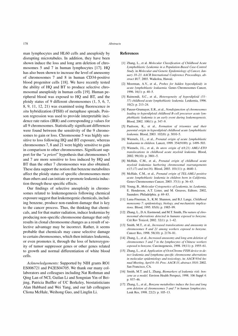
178 Abstracts
man lymphocytes and HL60 cells and aneuploidy bydisrupting microtubules. In addition, they have beenshown induce the loss and long arm deletion of chro-mosomes 5 and 7 in human lymphocytes [17]. HQhas also been shown to increase the level of aneusomyof chromosomes 7 and 8 in human CD34-positiveblood progenitor cells [18]. We have recently testedthe ability of HQ and BT to produce selective chro-mosomal aneuploidy in human cells [19]. Human pe-ripheral blood was exposed to HQ and BT, and theploidy status of 9 different chromosomes (1, 5, 6, 7,8, 9, 11, 12, 21) was examined using fluorescence insitu hybridization (FISH) of metaphase spreads. Pois-son regression was used to provide interpretable inci-dence rate ratios (IRR) and corresponding p values forall 9 chromosomes. Statistically significant differenceswere found between the sensitivity of the 9 chromo-somes to gain or loss. Chromosome 5 was highly sen-sitive to loss following HQ and BT exposure, whereaschromosomes 7, 8 and 21 were highly sensitive to gainin comparison to other chromosomes. Significant sup-port for the “a priori” hypothesis that chromosomes 5and 7 are more sensitive to loss induced by HQ andBT than the other 7 chromosomes was also obtained.These data support the notion that benzene metabolitesaffect the ploidy status of specific chromosomes morethan others and can initiate or promote leukemia induc-tion through these specific effects.
Our findings of selective aneuploidy in chromo-somes related to leukemogenesis following chemicalexposure suggest that leukemogenic chemicals, includ-ing benzene, produce non-random damage that is keyto the cancer process. Thus, the thinking that chemi-cals, and for that matter radiation, induce leukemias byproducing non-specific chromosome damage that onlyresults in clonal chromosome changes as a result of se-lective advantage may be incorrect. Rather, it seemsprobable that chemicals may cause selective damageto certain chromosomes, which then initiates leukemia,or even promotes it, through the loss of heterozygos-ity of tumor suppressor genes or other genes relatedto growth and normal differentiation of white bloodcells.
Acknowledgements: Supported by NIH grants RO1ES006721 and P42ES04705. We thank our many col-laborators and colleagues including Nat Rothman andQing Lan of NCI, Guilan Li and Songnian Yin of Bei-jing, Patricia Buffler of UC Berkeley, biostatisticiansAlan Hubbard and Wei Yang, and our lab colleaguesCliona McHale, Weihong Guo, and Linglin Li.
References
[1] Zhang, L., et al. Molecular Classification of Childhood AcuteLymphoblastic Leukemia in a Population-Based Case-ControlStudy. in Molecular and Genetic Epidemiology of Cancer, Jan-uary 18–23. AACR International Conference Proceedings, ab-stract B17. 2003. Waikoloa, Hawaii.
[2] Moorman, A.V., et al., Probes for hidden hyperdiploidy inacute lymphoblastic leukaemia. Genes Chromosomes Cancer,1996. 16(1): p. 40–5.
[3] Raimondi, S.C., et al., Heterogeneity of hyperdiploid (51–57) childhood acute lymphoblastic leukemia. Leukemia, 1996.10(2): p. 213–24.
[4] Panzer-Grumayer, E.R., et al., Nondisjunction of chromosomesleading to hyperdiploid childhood B-cell precursor acute lym-phoblastic leukemia is an early event during leukemogenesis.Blood, 2002. 100(1): p. 347–9.
[5] Paulsson, K., et al., Formation of trisomies and theirparental origin in hyperdiploid childhood acute lymphoblasticleukemia. Blood, 2003. 102(8): p. 3010–5.
[6] Wiemels, J.L., et al., Prenatal origin of acute lymphoblasticleukaemia in children. Lancet, 1999. 354(9189): p. 1499–503.
[7] Wiemels, J.L., et al., In utero origin of t(8;21) AML1-ETOtranslocations in childhood acute myeloid leukemia. Blood,2002. 99(10): p. 3801–5.
[8] McHale, C.M., et al., Prenatal origin of childhood acutemyeloid leukemias harboring chromosomal rearrangementst(15;17) and inv(16). Blood, 2003. 101(11): p. 4640–1.
[9] McHale, C.M., et al., Prenatal origin of TEL-AML1-positiveacute lymphoblastic leukemia in children born in California.Genes Chromosomes Cancer, 2003. 37(1): p. 36–43.
[10] Young, B., Molecular Cytogenetics of Leukemia, in: Leukemia,E. Henderson, A.T. Lister, and M. Greaves, Editors. 2002,Saunders: Philadelphia. p. 69–83.
[11] Luna-Fineman, S., K.M. Shannon, and B.J. Lange, Childhoodmonosomy 7: epidemiology, biology, and mechanistic implica-tions. Blood, 1995. 85(8): p. 1985–99.
[12] Zhang, L., D.A. Eastmond, and M.T. Smith, The nature of chro-mosomal aberrations detected in humans exposed to benzene.Crit Rev Toxicol, 2002. 32(1): p. 1–42.
[13] Smith, M.T., et al., Increased translocations and aneusomy inchromosomes 8 and 21 among workers exposed to benzene.Cancer Res, 1998. 58(10): p. 2176–81.
[14] Zhang, L., et al., Increased aneusomy and long arm deletion ofchromosomes 5 and 7 in the lymphocytes of Chinese workersexposed to benzene. Carcinogenesis, 1998. 19(11): p. 1955–61.
[15] Zhang, L., et al. Application of OctoChrome FISH device to de-tect leukemia and lymphoma specific chromosome aberrationsin molecular epidemiology and toxicology, in: AACR 93rd An-nual Meeting, April 6–10. Proc. AACR 33, abstract 3810. 2002.San Francisco, CA.
[16] Smith, M.T. and L. Zhang, Biomarkers of leukemia risk: ben-zene as a model. Environ Health Perspect, 1998. 106 Suppl 4:p. 937–46.
[17] Zhang, L., et al., Benzene metabolites induce the loss and longarm deletion of chromosomes 5 and 7 in human lymphocytes.Leuk Res, 1998. 22(2): p. 105–13.

Abstracts 179
[18] Smith, M.T., et al., Hydroquinone, a benzene metabolite, in-creases the level of aneusomy of chromosomes 7 and 8 inhuman CD34-positive blood progenitor cells. Carcinogenesis,2000. 21(8): p. 1485–90.
[19] Zhang, L., et al., Is chromosomal aneuploidy induced by ben-zene metabolites random or selective? Environ Mol Mutagen.39, Suppl 33, abstract 230, 2002.

180 Abstracts
USE OF DNA PLOIDY MEASUREMENTS INSCREENING FOR EARLY CANCER ANDPRE-NEOPLASTIC LESIONS OF THEUTERINE CERVIX
Xiao Rong Sun1,2, David Garner1 and Branko Palcic1
1Cancer Imaging Department, BC Cancer Agency,200-601 West Broadway, Vancouver, BC, Canada V5Z4C2 (Tel.: +1 604 877 6000 ext. 3037, Fax: +1 604877 6063, E-mail: [email protected]), 2LandingMedical High Technology Ltd and Wuhan Family Plan-ning Committee, Wuhan, Hubei, China
Introduction: Population based screening of womenfrom the onset of sexual activity based on regular in-tervals of 2–3 years has proven to reduce several foldthe incidence of invasive cervical cancer as well as themortality due to this disease [1]. However, such an un-dertaking requires significant resources as well as avery large number highly skilled cytotechnologists andcytopathologists. Therefore, to implement such pro-grams in the conventional way may not be practical oreven possible in many countries.
Materials and methods: We have developed a meth-od to screen for early cancer and pre-neoplastic le-sions (marked atypia) of uterine cervix that is basedon the measurements of DNA ploidy of exfoliated cer-vical smears. The method is based on a simple, liq-uid based preparation whereby cells from the cervicalbrush are first suspended in a liquid fixative and thencyto-centrifuged onto a microscope slide [2]. The nu-clei of the cells are then stained by a DNA specificand stoichiometric stain [3]. A fully automated highresolution image cytometer [4,5] is then used to mea-sure the size and the DNA content of the cell nucleion the slide. On average, 2,000 with the range of 1,000to 8,000, cell nuclei are measured per slide consum-ing in less than 10 minutes (range 6–15 minutes) ofthe system’s time. All samples with cells containingabnormal amount of DNA are examined by cytotech-nologists (any cell with atypical amount of DNA couldbe brought under crosshair of the microscope for man-
ual observation) for the conventional diagnosis. Sam-ples without cells having abnormal DNA content aredeclared normal and no further observations are madeby cytotechnologists.
Results: Using this approach we examined 6,000women in rural area around Wuhan (Hubei Province,China) and compared this approach with conventional(manual) method using the best trained cytopatholo-gists. The conventional method found 4 cancers and 7cases of marked atypia, while the new method found 8cancers and 27 marked atypia cases. All cancers foundby the conventional method were also found by thenew method.
Discussion: This approach is now being furthertested on a much larger scale with the goal to imple-ment it as the keystone for the population based screen-ing for cervical cancer in Hubei Province and laterin other provinces in China and elsewhere. The newmethod has a higher sensitivity (reduced false nega-tive rate) at the same specificity, requires fewer skilledtechnologists and is more cost-effective than conven-tional Pap screening.
References
[1] American Cancer Society: Cancer Facts and Figures 1993. At-lanta, G.A., The American Cancer Society, Inc, 1993, pp. 13–14.
[2] Sun X.R., Che D.Y., Yan X.C.: New Liquid Based SamplePreparation Method for Gynecologic Cytology. Chinese Cytol-ogy and Pathology Society 2004, p. 45.
[3] Tezcan H., MacAulay C., Palcic B.: Analysis of various samplepreparations and staining methods for computer assisted im-age cytometry device. 13th International Meeting of AnalyticalCytology, Breckenridge, CO, Sept. 4–9, 1988; p. 46.
[4] Garner D., MacAulay C., Palcic B.: Cytology automation up-date: Xillix automated cervical cell screening system. ASCTNews 1993; 14: 35–38.
[5] Garner D., Ferguson D., Palcic B.: The Cyto-Savant System.In The Automation of cercival cancer screening. Edited byH.K. Grohs, O.A.N. Husain. Hong Kong, Igaku-Shoin Med-ical, 1994, pp. 305–317.

Abstracts 181
CARCINOGENESIS BY ANEUPLOIDIZATION
Peter Duesberg1, Ruhong Li1, David Rasnick1,Alice Fabarius2 and Ruediger Hehlmann2
1Department of Molecular and Cell Biology, DonnerLab, University of California Berkeley, Berkeley, CA94720, USA, 2III Medizinische Klinik of the Universityof Heidelberg at Mannheim, 68305 Mannheim, Ger-many
Despite over 100 years of cancer research, the causeof cancer is still a matter of debate [1,2]. Indeed, can-cer has been hiding the secret of its genetic origin, likea magician hides the secrets of its trade. It has kept thissecret by supporting many competing theories simul-taneously with bits of evidence from its large reper-toire of exotic genotypes and phenotypes. In additioncancer has kept researchers guessing on what is in thatblack box of the exceedingly long and phenotype-lesslag periods from initiation of cancer with carcinogensto carcinogenesis [3].
Because carcinogenesis is irreversible, there is aconsensus that cancer is caused by some kind of mu-tation, but there is no consensus on what kind of mu-tation it actually is. A majority of scientists currentlythink that specific gene mutations are the cause of can-cer, but others think that specific rearrangements of thenormal chromosome balance, alias, specific aneuploi-dies are the cause, and yet others think that mutationscause cancer via aneuploidy [1,2].
Here we try to determine, which of these theoriesprovides a coherent explanation for all of the many oddfeatures of cancer and carcinogenesis, focusing primar-ily on the prevailing mutation and the competing aneu-ploidy theories. For this purpose we first briefly definethese theories.
1. The mutation-cancer theory
The mutation theory holds that cancer is the resultof 4 to 7 gene mutations [4,5], which either generatedominant oncogenes or inactivate recessive tumor sup-pressor genes or both [6–10].
However, the mutation theory suffers from 3 unre-solved problems:
(1) There are no consistent correlations between anyparticular gene mutations and cancer [11–13].
(2) Despite intensive efforts of over 2 decades thereis as yet no functional proof that one or any combina-
tion of mutant genes from cancer cells can transformnormal diploid cells into cancer cells [12,14–17]. In-deed, artificially mutated mice with mutant oncogenesor without tumor suppressor genes or even with com-binations of both in their germ line are surprisinglyprocreative [16,18–22], and their cancer risk is “strain-dependent” [20] but within the known range of labo-ratory mice [3,23,24]. Although some of these stud-ies point out that mutant mice have higher cancer risksthan unaltered controls, e.g. Donehower et al. [20], thecellular cancer risk of these artificially mutated miceis extremely low. Since cancers originate from singlecells [25–27] (see below) and mice consist of about5 × 1010 cells, the cellular cancer risk of mice withouttumor suppressor genes is only 5 × 10⁻¹⁰. It is thusscarcely an argument for a role of such genes in car-cinogenesis.
(3) Based on the normal, spontaneous gene mutationrate of about 10−6 per mitosis [28] only 1 in 1024 to1042 human cells would ever become cancer cells [18,29–31]. This number would be even lower, if the muta-tion rates of the recessive cancer genes, as for examplethe hypothetical tumor suppressor genes, are squared.Since humans consist of 1014 cells, only 1 in 1010 to1028 humans would ever get cancer. In other wordscancer would hardly exist. To reconcile spontaneouscarcinogenesis with the spontaneous mutation rates,the proponents of the mutation theory have postulatedthat, prior to mutation of prospective cancer genes, an-other class of cellular genes must be mutated to muta-tor genes, which in turn would mutate prospective can-cer genes to real cancer genes [8,29,32–35].
However, genes with this potential are only rarelyfound in cancer cells [1,10,13]. Moreover, since mu-tator genes and their mutations are escalating auto-catalytically, their presence is eventually suicidal andthus hard to reconcile with the long latent periods ofcarcinogenesis and particularly with the immortalityof cancer cells [13]. Tomlinson et al. expressed thesereservations about the mutator-gene hypothesis as fol-lows, “The scenarios for a role of a raised mutation rateassume that there is no selective disadvantage to a cellin having an increased number of mutations. This maynot be the case: for example, a deleterious or lethal mu-tation may be much more likely than an advantageousmutation. More subtly, an accumulated mutational loadmight induce apoptosis” [36].
Thus there is no consistent correlative or functionalproof for the mutation theory. In addition the postu-lated mutator genes are hard to reconcile with the long,preneoplastic lag periods of carcinogenesis and evenharder with the immortality of cancer cells.

182 Abstracts
2. The aneuploidy-cancer theory
The aneuploidy theory holds that somatic evolu-tion of the karyotype of a single cell causes ran-dom and cancer-specific aneuploidies, which encodecancer-specific phenotypes. The principle of generat-ing new phenotypes from old genes – and thus inde-pendently of gene mutation – is also the basis of phy-logenesis. Phylogenesis generates new species by re-arranging old, phylogenetically conserved genes intonew sets of chromosomes [37]. Thus cancer cells arenew, semi-autonomous species of their own rather thanmutants of their precursor cells.
According to the aneuploidy theory the somatickaryotype evolution is initiated by a random aneu-ploidy, which is either induced by carcinogens or spon-taneously (see Fig. 1). Aneuploidy destabilizes chro-mosomes because it unbalances – and thus corruptsthe normal functions of – numerous highly conservedteams of proteins including those, which segregate,synthesize and repair chromosomes. Aneuploidy alsocatalyzes gene mutations by corrupting protein teamsthat repair DNA and synthesize nucleotide pools. Thusaneuploid cells undergo chromosome non-disjunctionsand gene mutations due to error-prone chromosomesegregation and error-prone DNA repair and synthe-
sis. The degrees of the resulting genomic destabiliza-tion would be proportional to the degree of aneu-ploidy [38,39].
The basis for the somatic evolution of neoplasticcells from randomly aneuploid precursors is selectionof rare chromosome combinations with advantages forabnormal growth. Thus the rate-limiting step of car-cinogenesis is the aneuploidy-catalyzed generation ofnew chromosome arrangements with neoplastic pheno-types by random karyotype variations – a process thatis also analogous to phylogenesis. However, since thegeneration of a new, autonomous species is infinitelyless likely than the generation of a parasitic cancer cell,phylogenesis is much slower than carcinogenesis.
According to the aneuploidy theory, immortality ofcancer cells derives from the inherent heterogeneity ofaneuploid cell populations. Indeed cancers are hetero-geneous, “polyphyletic” [40] cell populations, whichinclude sub-species that can survive otherwise fatalmutations, cytotoxic drugs, metastasis to heterologouslocations, transplantation to heterologous hosts, etc.via sub-species-specific karyotypes. These karyotypeseither activate alternative drug-resistant biochemicalpathways or eliminate drug-specific receptors. It is be-cause of this inherent genetic heterogeneity that popu-lations of aneuploid cells are “immortal”, although in-dividual cells are not.
Fig. 1.

Abstracts 183
In sum, the aneuploidy theory proposes that the in-herent instability of aneuploidy is sufficient to gen-erate the multilateral genomic instability of neoplas-tic and preneoplastic cells, and is thus independent of,although not necessarily free of gene mutation (seeFig. 1). The majority, if not all, of the many heteroge-neous gene mutations of cancer cells [1,2] may just beinevitable, but functionally irrelevant consequences ofaneuploidy (see also below).
3. The abilities of the mutation and aneuploidytheories to explain 9 features of cancer andcarcinogenesis
In the following we test the two genetic theories ofcancer for their abilities to explain 9 features of cancerand carcinogenesis:
(1) Cancers are clonal. Nearly all cancers originatefrom single cells based on preneoplastic and neoplas-tic genetic markers [25–27]. This feature is compatiblewith both genetic theories of cancer.
(2) Aneuploidy is ubiquitous in cancer. Cancers areaneuploid [25,41–44]. By contrast, conventional genemutations are independent of, and thus typically notassociated with karyotype alterations. It follows thataneuploidy is necessary for carcinogenesis, as is pos-tulated by the aneuploidy theory. By contrast, the mu-tation theory predicts diploid cancers.
(3) Abnormal gene expression profiles of cancercells correlate with aneuploidy. According to Rud-don’s Cancer biology, “Abnormal gene expression isthe sine qua non of cancer cells” [27]. Recent analy-ses have shown that the abnormal gene expression pro-files of cancer cells correlate very closely with the ane-uploid doses of the corresponding chromosomes [45–47]. By contrast, the gene mutation theory predicts thatthe expression of only a few genes is altered, namelythe 4 to 7 that are mutated and those that might be con-trolled by them.
(4) No cancer-specific gene mutations. About 50%of all cancers of a given kind contain various genemutations of hypothetical oncogenes and tumor sup-pressor genes [11,30,48–51]. However, consistent cor-relations between such mutations and specific kindsof cancers have not been found [16,18,30,31]. For ex-ample, Little reports “While radiation-induced cancersshow multiple unbalanced chromosomal rearrange-ments, few show specific translocations or deletions as
would be associated with the activation of known onco-genes or tumor suppressor genes” [12]. Grosovskyet al. also find, “no consistent elevation of specific lo-cus mutation rate has been reported” [52]. Further Gis-selson et al. note, “the correlation coefficient betweenbreakpoint frequency and telomere length [a poten-tial mutator] was low in both osteosarcomas and pan-creatic sarcomas” [53]. Moreover, mutations of onco-genes and tumor-suppressor genes of many clonal can-cers are non-clonal, and thus not necessary for car-cinogenesis [19]. A survey of genomic instability byLengauer et al. states in 1998 (a) “no consistent patternof defects in polymerases has been found in tumors”and (b) mismatch repair deficiencies are only in “13%of colorectal, . . . endometrial and gastric cancers . . .other types are rarely (<2%) MMR-deficient” [54]. Inthe words of a recent survey by Scientific American,“A few cancer-related genes, such as p53, do seem tobe mutated in the majority of tumors. But many othercancer genes are changed in only a small fraction ofcancer types, a minority of patients, or a sprinkling ofcells within a tumor” [2]. Since specific gene mutationsare not consistently associated with any kind of can-cer, they cannot be necessary for carcinogenesis [19].However, an abnormally high rate of gene mutations isa predictable consequence of aneuploidy.
(5) Karyotypes and gene mutations of cancers areunstable. Karyotypes of cancer cells may change froma few to 100% per mitosis, proportional to the degreeof aneuploidy [38,39,55]. It is for this reason that thekaryotypes of “clonal” cancers are heterogeneous, de-spite the clonal origin of cancers [1,13,16,25]. Like-wise gene mutations of cancers are unstable, becausethey are also heterogeneous in clonal cancers [19].However, conventional gene mutations are just as sta-ble as the corresponding wild types, e.g. spontaneouslymutating at less than 10−6 per mitosis per haploid gene(see above). By contrast, genomic instability is an in-evitable consequence of aneuploidy. Indeed, clonality,aneuploidy and genomic instability define cancer.
(6) Cancers have unique, exotic phenotypes, neverobserved in diploid biology. The exotic phenotypes ofcancers include “immortality”, a phenotype that hasnever been achieved by any diploid cell, despite 3 bil-lion years of mutations! In addition cancer cells canbecome readily resistant to cytotoxic drugs such asthose used in chemotherapy, and can metastasize fromone differentiated tissue to another [56]. None of thesephenotypes has ever been observed in diploid animals.Thus cancer cells can generate phenotypes that are not

184 Abstracts
ever generated in normal diploid cells by gene muta-tions. However, the unique ability of aneuploid cells togenerate variant sub-species by altering its karyotypeprovides a coherent explanation. Accordingly, the ex-otic phenotypes of cancers reflect variant sub-speciesthat can evade toxins, otherwise lethal mutations andnormal histologic barriers.
(7) Mutagenic and non-mutagenic carcinogens in-duce cancers. Besides mutagenic radiations and alky-lating agents numerous non-mutagenic substances arecarcinogenic including polycyclic aromatic hydrocar-bons, hormones, metal ions, butter yellow, solid bod-ies, asbestos, etc. [18,57,58]. It follows that gene mu-tation is not necessary for the induction of carcinogen-esis. By contrast, aneuploidy can be induced either bygene mutations that lead to direct or indirect chromo-some breaks and rearrangements, or can be induced byagents that physically or chemically destroy the spin-dle apparatus – independent of gene mutation.
(8) Long lag periods from carcinogen to cancer.At carcinogenic doses carcinogens, e.g. radiations orchemicals, initiate carcinogenesis immediately, be-cause no subsequent treatments are necessary [13].Cancer appears only after lag periods of years in exper-imental animals and decades in accidentally exposedhumans [13,26]. Take the late cancers after the Hi-roshima bomb as an example [26]. Since mutagens andaneuploidogens act fast, initiation is compatible withthe mutation and the aneuploidy theory. However, thelong latent periods of carcinogenesis cannot be recon-ciled with gene mutation. But induction of a randomaneuploidy followed by a somatic karyotype evolutionwith selection for autonomous growth, provides a co-herent explanation for the long lag periods of carcino-genesis.
(9) Age bias of cancer. The cancer risk of animalsand humans increases exponentially with age [4,26].By contrast, the mutation theory predicts cancer atyoung age. According to the mutation theory all butone of the 4–7 genes postulated to cause cancer shouldbe heritable. Thus newborns with all but one muta-tion missing from a carcinogenic combination shoulddevelop cancer at young age, as soon as the missingmutation has occurred in one cell of their body. But,there is virtually no cancer at young age. By contrast,the aneuploidy theory correctly predicts the age bias ofcancer for two reasons: (i) Since aneuploidy is not her-itable [59,60], carcinogenesis by aneuploidization hasto be somatically initiated and completed, a processthat appears to take decades for solid cancers in hu-mans [13,26,61]. (ii) As more and more initiated cellsare accumulated and able to multiply over a lifetimethe risk of carcinogenesis would increase exponen-tially (as is also postulated by the mutation theory).
4. Conclusions
In sum aneuploidy is the genetic alteration that pro-vides a coherent explanation for 9 of the 9 features ofcancer and carcinogenesis described above and sum-marized in Table 1, but gene mutation only explains 3.In view of this we conclude that carcinogenesis is initi-ated by random aneuploidy and completed by specificaneuploidies, which evolve from random aneuploidyautocatalytically, but inefficiently and slowly over longperiods of time. Thus carcinogenesis is analogous toevolution and cancer cells are new, albeit only semi-autonomous cell species, rather than mutants of theirprogenitor cells. According to the aneuploidy theory,
Table 1
Cancer and carcinogenesis in the light of the mutation and aneuploidy theories
Features of cancer and carcinogenesis Mutation Aneuploidy
theory theory
1 Cancers are clonal, based on neoplastic and preneoplastic markers + +
2 Aneuploidy is ubiquitous in cancer − +
3 Abnormal gene expression profiles of cancer cells correlate with aneuploidy − +
4 No cancer-specific gene mutations − +
5 Karyotypes and genotypes are unstable, and therefore heterogeneous or non-clonal − +
6 Cancers have unique, exotic phenotypes, never observed in diploid biology: immortality, resistance to cytotoxicdrugs, metastasis
− +
7 Cancers are induced by mutagenic and non-mutagenic carcinogens +/− +
8 Carcinogens initiate carcinogenesis immediately, but cancer follows only after lags of many years or many cellgenerations
+/− +
9 Age bias of cancer − +

Abstracts 185
the many non-specific mutations of cancer cells are in-evitable consequences of the inherent genomic insta-bility of aneuploidy.
If confirmed, the aneuploidy theory offers a scien-tific basis for the identification and early treatment ofpreneoplastic lesions. In addition the theory proposesthat cancer prevention will benefit from testing foodsand drugs for their abilities to induce aneuploidy.
References
[1] Marx, J. (2002) Debate surges over the origins of genomic de-fects in cancer. Science 297, 544–546.
[2] Gibbs, W.W. (2003) Untangling the roots of cancer. Sci Am289, 56–65.
[3] Foulds, L. (1975) Neoplastic Development, Vol 2 (AcademicPress, London, New York, San Francisco).
[4] Armitage, P., and Doll, R. (1954) The age distribution of cancerand a multi-stage theory of carcinogenesis. Br J Cancer 8, 1–12.
[5] Renan, M.J. (1993) How many mutations are required for tu-morigenesis? Implications from human cancer data. Mol Car-cinog 7, 139–146.
[6] Lewin, B. (1997). Genes VI (Oxford University Press, Oxford).
[7] Lodish, H., Berk, A., Zipursky, S.L., Matsudaira, P., Baltimore,D., and Darnell, J. (1999). Molecular Cell Biology, Fourth edn(W.H. Freeman & Co., New York and Basingstoke UK).
[8] Nowak, M.A., Komarova, N.L., Sengupta, A., Jallepalli, P.V.,I.-M., S., Vogelstein, B., and Lengauer, C. (2002) The role ofchromosomal instability in tumor initiation. Proc Natl Acad SciUSA 99, 16226–16231.
[9] Rajagopalan, H., Nowak, M.A., Vogelstein, B., andLengauer, C. (2003) The significance of unstable chromosomesin colorectal cancer. Nat Rev Cancer 3, 695–701.
[10] Pihan, G., and Doxsey, S.J. (2003) Mutations and aneuploidy:co-conspirators in cancer? Cancer Cell 4, 89–94.
[11] Haber, D.A., and Fearon, E.R. (1998) The promise of cancergenetics. Lancet 351, SII 1–8.
[12] Little, J.B. (2000) Radiation carcinogenesis. Carcinogenesis21, 397–404.
[13] Duesberg, P., and Li, R. (2003) Multistep carcinogenesis:a chain reaction of aneuploidizations. Cell Cycle 2, 202–210.
[14] Li, R., Sonik, A., Stindl, R., Rasnick, D., and Duesberg, P.(2000) Aneuploidy versus gene mutation hypothesis of can-cer: recent study claims mutation, but is found to support ane-uploidy. Proc Natl Acad Sci USA 97, 3236–3241.
[15] Stanbridge, E.J. (1990) Human Tumor Suppressor Genes.Annu. Rev. Genet. 24, 615–657.
[16] Harris, H. (1995). The cells of the body; a history of somaticcell genetics (Cold Spring Harbor Lab Press, Plainview, NY).
[17] Li, R., Rasnick, D., and Duesberg, P. (2002) Correspondencere: D. Zimonjic et al., Derivation of human tumor cells invitro without widespread genomic instability. Cancer Res.,61: 8838–8844, 2001. Cancer Res 62, 6345–6348; discussion6348–6349.
[18] Duesberg, P., and Rasnick, D. (2000) Aneuploidy, the somaticmutation that makes cancer a species of its own. Cell MotilCytoskeleton 47, 81–107.
[19] Duesberg, P.H. (2003) Are cancers dependent on oncogenes oron aneuploidy? Cancer Genet Cytogenet 143, 89–91.
[20] Donehower, L.A., Harvey, M., Siagle, B.L., McArthur, M.J.,Montgomery, C.A., Jr., Butel, J.S., and Bradley, A. (1992) Micedeficient for p53 are developmentally normal but susceptible tospontaneous tumors. Nature 356, 215–221.
[21] Smits, R., Kielman, M.F., Breukel, C., Zurcher, C., Neufeld, K.,Jagmohan-Changur, S., Hofland, N., van Dijk, J., White, R.,Edelmann, W., et al. (1999) Apc1638T: a mouse model delin-eating critical domains of the adenomatous polyposis coli pro-tein involved in tumorigenesis and development. Genes Dev 13,1309–1321.
[22] Steinberg, D. (2003) A cell-cycle couple loses its luster. TheScientist 17, 26–27.
[23] Heston, W.E. (1942) Inheritance of susceptibility to sponta-neous pulmonary tumors in mice. J. Natl. Cancer Inst. 3, 79–82.
[24] Heston, W.E. (1982) In Cancer, a comprehensive treatise,Becker, F.F. (Plenum Press, New York), pp. 47–71.
[25] Koller, P.C. (1972). The Role of Chromosomes in Cancer Biol-ogy (Springer-Verlag, New York).
[26] Cairns, J. (1978). Cancer: Science and Society (W.H. Freemanand Company, San Francisco).
[27] Ruddon, R.W. (1981). Cancer Biology (Oxford UniversityPress, New York, Oxford).
[28] Vogel, F., and Motulsky, A.G. (1986). Human genetics: prob-lems and approaches (Springer Verlag, Berlin, Heidelberg,New York, Tokio).
[29] Loeb, L.A. (1991) Mutator phenotype may be required for mul-tistage carcinogenesis. Cancer Res. 51, 3075–3079.
[30] Strauss, B.S. (1992) The origin of point mutations in humantumor cells. Cancer Res 52, 249–253.
[31] Duesberg, P.H., and Schwartz, J.R. (1992) Latent viruses andmutated oncogenes: no evidence for pathogenicity. Prog Nu-cleic Acid Res Mol Biol 43, 135–204.
[32] Cahill, D.P., Kinzler, K.W., Vogelstein, B., and Lengauer, C.(1999) Genetic instability and darwinian selection in tumours.Trends in Biological Sciences (TIBS) 24, M57–M60.
[33] Nowell, P.C. (1976) The clonal evolution of tumor cell popula-tions. Science 194, 23–28.
[34] Strauss, B.S. (2000) The stability of the genome and the geneticinstability of tumors. Perspect Biol Med 43, 286–300.
[35] Loeb, L.A., Loeb, K.R., and Anderson, J.P. (2003) Multiplemutations and cancer. Proc Natl Acad Sci USA 100, 776–781.
[36] Tomlinson, I.P., Novelli, M.R., and Bodmer, W.F. (1996) Themutation rate and cancer. Proc Natl Acad Sci USA 93, 14800–14803.
[37] O’Brien, S., Menotti-Raymond, M., Murphy, W., Nash, W.,Wirnberg, J., Stanyon, R., Copeland, N., Jenkins, N., Wom-ack, J., and Marshall Graves, J. (1999) The promise of compar-ative genomics in mammals. Science 286, 458–481.
[38] Duesberg, P., Rausch, C., Rasnick, D., and Hehlmann, R.(1998) Genetic instability of cancer cells is proportional to theirdegree of aneuploidy. Proc Natl Acad Sci USA 95, 13692–13697.
186 Abstracts
[39] Fabarius, A., Hehlmann, R., and Duesberg, P.H. (2003) Insta-bility of chromosome structure in cancer cells increases expo-nentially with degrees of aneuploidy. Cancer Genet Cytogenet143, 59–72.
[40] Hauschka, T., and Levan, A. (1958) Cytologic and functionalcharacterization of single cell clones isolated from the Krebs-2and Ehrlich Ascites tumors. J Natl Cancer Inst 21, 77–111.
[41] Sandberg, A.A. (1990) The chromosomes in human cancerand leukemia, Second edn (Elsevier Science Publishing, NewYork).
[42] Heim, S., and Mitelman, F. (1995) Cancer Cytogenetics, Sec-ond edn (Wiley-Liss, New York).
[43] Balaban, G.B., Herlyn, M., Clark, W.H., Jr., and Nowell, P.C.(1986) Karyotypic evolution in human malignant melanoma.Cancer Genet Cytogenet 19, 113–122.
[44] Atkin, N.B., and Baker, M.C. (1990) Are human cancers everdiploid–or often trisomic? Conflicting evidence from directpreparations and cultures. Cytogenet Cell Genet 53, 58–60.
[45] Wang, K., Gan, L., Jeffery, E., Gayle, M., Gown, A.M.,Skelly, M., Nelson, P.S., Ng, W.V., Schummer, M., Hood, L.,and Mulligan, J. (1999) Monitoring gene expression profilechanges in ovarian carcinomas using cDNA microarray. Gene229, 101–108.
[46] van ’t Veer, L.J., Dai, H., van de Vijver, M.J., He, Y.D., Hart,A.A., Mao, M., Peterse, H.L., van der Kooy, K., Marton, M.J.,Witteveen, A.T., et al. (2002) Gene expression profiling pre-dicts clinical outcome of breast cancer. Nature 415, 530–536.
[47] Pollack, J.R., Sorlie, T., Perou, C.M., Rees, C.A., Jeffrey,S.S., Lonning, P.E., Tibshirani, R., Botstein, D., Borresen-Dale,A.L., and Brown, P.O. (2002) Microarray analysis reveals amajor direct role of DNA copy number alteration in the tran-scriptional program of human breast tumors. Proc Natl AcadSci USA 99, 12963–12968.
[48] Hollstein, M., Rice, K., Greenblatt, M.S., Soussi, R., Fuchs, R.,Sorlie, T., Hovig, E., Smith-Sorensen, B., Montesano, R., andHarris, C.C. (1994) Database of p53 gene somatic mutations inhuman tumors and cell lines. Nucleic Acids Res 22, 3551–3555.
[49] Rajagopalan, H., Bardelli, A., Lengauer, C., Kinzler, K.W.,Vogelstein, B., and Velculescu, V.E. (2002) Tumorigenesis:RAF/RAS oncogenes and mismatch-repair status. Nature 418,934.
[50] Hermsen, M., Postma, C., Baak, J., Weiss, M., Rapallo, A.,Sciutto, A., Roemen, G., Arends, J.W., Williams, R., Gia-retti, W., et al. (2002) Colorectal adenoma to carcinoma pro-gression follows multiple pathways of chromosomal instability.Gastroenterology 123, 1109–1119.
[51] Lingle, W.L., Barrett, S.L., Negron, V.C., D’Assoro, A.B.,Boeneman, K., Liu, W., Whitehead, C.M., Reynolds, C., andSalisbury, J.L. (2002) Centrosome amplification drives chro-mosomal instability in breast tumor development. Proc NatlAcad Sci USA 99, 1978–1983.
[52] Grosovsky, A.J., Parks, K.K., Giver, C.R., and Nelson, S.L.(1996) Clonal analysis of delayed karyotypic abnormalities andgene mutations in radiation-induced genetic instability. MolCell Biol 16, 6252–6262.
[53] Gisselsson, D., Jonson, T., Petersen, A., Strombeck, B., DalCin, P., Hoglund, M., Mitelman, F., Mertens, F., and Man-dahl, N. (2001) Telomere dysfunction triggers extensive DNAfragmentation and evolution of complex chromosome abnor-malities in human malignant tumors. Proc Natl Acad Sci USA98, 12683–12688.
[54] Lengauer, C., Kinzler, K.W., and Vogelstein, B. (1998) Geneticinstabilities in human cancers. Nature 396, 643–649.
[55] Lengauer, C., Kinzler, K.W., and Vogelstein, B. (1997) Geneticinstability in colorectal cancers. Nature 386, 623–627.
[56] Pitot, H.C. (2002). Fundamentals of Oncology, fourth edn(Marcel Dekker, Inc., New York).
[57] Lijinsky, W. (1989) A view of the relation between carcinogen-esis and mutagenesis. Env Mol Mutagenesis 14, 78–84.
[58] Oshimura, M., and Barrett, J.C. (1986) Chemically inducedaneuploidy in mammalian cells: mechanisms and biologicalsignificance in cancer. Environ Mutagen 8, 129–159.
[59] Hernandez, D., and Fisher, E.M. (1999) Mouse autosomal tri-somy: two’s company, three’s a crowd. Trends Genet 15, 241–247.
[60] Hassold, T.J. (1986) Chromosome abnormalities in human re-productive wastage. Trends in Genetics 2, 105–110.
[61] Bauer, K.H. (1963) Das Krebsproblem, 2d edn (Springer Ver-lag, Berlin, Goettingen, Heidelberg).

Abstracts 187
THE INDUCTION OF CHROMOSOMALINSTABILITY AS AN INDIRECT RESPONSETO IONIZING RADIATION AND OTHERTOXIC AGENTS
Eric G. Wright, Philip J. Coates and Sally A. LorimoreUniversity of Dundee, Department of Molecular andCellular Pathology, Ninewells Hospital and MedicalSchool, Dundee DD1 9SY, Scotland, UK (Tel.: +4401382 632169, Fax: +44 01382 633952, E-mail:[email protected])
1. Inherited and inducible chromosome instability
The ability to maintain genome integrity in theface of endogenously and exogenously generated DNAdamage is critical for healthy survival and complexhomeostatic mechanisms have evolved to allow cellu-lar adaptation to cellular stress and injury. In recentyears there has been considerable progress in identi-fying the mechanisms by which eukaryotes respondto potentially harmful insults by initiating processesthat either enhance cell survival or lead to the reg-ulated loss of damaged or unwanted cells. Inheritedor acquired deficiencies in genome maintenance sys-tems contribute significantly to the development of ma-lignant diseases and there are well-recognized chro-mosome instability/breakage syndromes that producecomplex and often multi-system effects characterizedby a significant predisposition to malignancy.
Since the discovery of the induction of mutationsand chromosome aberrations by ionizing radiation inthe early years of the twentieth century it has been ac-cepted that these effects are due to DNA being irre-versibly changed at the time of exposure, either dur-ing the processing and enzymatic repair of the radia-tion damage or during the first round of DNA replica-tion immediately after exposure. As malignant trans-formation is generally regarded as being initiated by agene mutation or a chromosomal aberration, the initiat-ing lesion for malignant transformation has been simi-larly attributed to direct DNA damage. Accordingly, ithas been widely accepted that most of these changestake place immediately following exposure. Thus, ifthe damage were repaired, the progeny of an irradiatedcell would appear normal (Fig. 1a) but if misrepaired,the progeny would be expected to show any transmis-sible radiation-induced genetic change and all cells de-rived from such a cell would exhibit the same geneticchange, i.e. the effect would be clonal (Fig. 1b).
Fig. 1. Models of the responses of clonogenic cells to ionizing radia-tion with mutations and/or chromosomal aberrations shown as filledcircles and apparently normal cells as open circles. (a) If a cell faith-fully repairs DNA damage then its clonal descendants will appearnormal. (b) If a cell is directly mutated by radiation then all its de-scendants will express the same mutation. (c) Radiation-induced ge-nomic instability is characterized by non-clonal effects in descendantcells.
In recent years, many laboratory studies have de-monstrated non-clonal chromosome aberrations andmutations in the clonal progeny of irradiated cells.In addition, the progeny of irradiated cells have beenshown to exhibit an enhanced death rate and loss of re-productive potential that persists for many generationsand possibly indefinitely in established cell lines. Theterms lethal mutations and delayed reproductive deathare used interchangeably for this delayed death pheno-type. All the various effects in which delayed death,gene mutations and a variety of chromosomal abnor-
188 Abstracts
malities can be demonstrated in cells that are not them-selves irradiated but are the progeny of cells exposedto ionizing radiation many cell divisions previously(Fig. 1c) have been interpreted as manifestations ofa radiation-induced genomic instability [1–4]. Similareffects are now being reported for a range of chemicalexposures [5–8]. Induced instability is a genome-wideprocess and the cellular phenotype is similar to that ofthe inherited chromosome instability syndromes, char-acterized by spontaneously high levels of chromoso-mal abnormalities and mutations. Despite the appar-ent similarities, radiation-induced genomic instabilityseems to reflect epigenetic processes rather than muta-tion of genome maintenance genes [9–13] but the in-duced instability phenotype in both haemopoietic tis-sue [14] and mammary epithelium [15] is strongly in-fluenced by genetic factors with some genotypes be-ing susceptible and others relatively resistant. Clearly,any process that increases the frequency with whichgenetic changes arise will increase the probability ofpotential malignant changes in target cells and poten-tially in tumour cells although it may be difficult to de-termine how an ‘initiating event’ arose and it may notbe possible to distinguish between chromosomal insta-bility as a delayed effect of exposure and chromosomalinstability arising as a consequence of the malignantprocess.
2. Mechanisms underlying inducible instability
At present, the mechanism of induction of instabil-ity by ionizing radiation and other agents is not fullyunderstood nor is it clear whether all endpoints re-flect a common mechanism. In all the various stud-ies, the frequency of induced instability is orders ofmagnitude greater than that of conventional gene mu-tation frequencies and although in some studies us-ing established cell lines a large number of post-irradiation cell divisions before assay might have al-lowed for selection of a radiation-induced gene muta-tion that confers a mutator phenotype, overall the dataindicate that the mechanism underlying induced in-stability is epigenetic. Typically, the spontaneous fre-quency of gene mutations in mammalian cells is ofthe order of 10−6 and this increases some 10-foldto ∼10−5 (0.001% of surviving clonogenic cells) af-ter exposure to 1 Gy X-rays. However, approximately10% of surviving cells produce clones that exhibit de-layed hypoxanthine phosphoribosyl-transferase (hprt)mutations and a similar or much greater proportion ex-
hibit chromosomal instability. In a comparative studyof hprt mutations induced directly by irradiation orarising as a consequence of induced instability, 75% ofthose induced directly by X-rays (‘conventional’ muta-tions) involved partial or total gene deletions and 25%small scale or point mutations, whereas only 28% ofthe delayed mutations were associated with large dele-tions and the majority were small scale changes [16].This observation of a mutation spectrum more likethat of spontaneously arising mutations than conven-tional radiation-induced mutations is similar to the cy-togenetic investigations in which the aberrations as-sociated with radiation-induced chromosomal instabil-ity in primary cells are similar to those arising spon-taneously in the cells. Unstable aberrations commonlyresult in cell lethality and this may account for a com-ponent of the delayed reproductive death/lethal muta-tion phenotype in some cell systems [10,17]. The as-sociation of radiation-induced chromosomal instabil-ity with increased intracellular reactive oxygen species(ROS), oxidative DNA base damage [9] and an asso-ciation of increased ROS with radiation-induced de-layed death caused by ongoing apoptosis or necro-sis in CHO cells [10,18] provides a potential epige-netic mechanism for radiation-induced genomic insta-bility. However, some death may also result from sig-nal antonymy generating an apoptotic response to con-flicting simultaneous signals for proliferation and cellcycle arrest [19].
3. Radiation-induced bystander effects andclastogenic factors
The paradigm of genetic alterations being restrictedto direct DNA damage has also been challenged by ob-servations in which cells that are not exposed to ion-izing radiation exhibit responses typically associatedwith direct radiation exposure as a consequence of con-tact with irradiated cells or after receiving certain sig-nals from irradiated cells. These phenomena are collec-tively known as radiation-induced bystander effects [3,4,20]. Bystander effects may reflect at least two sep-arate mechanisms for the transfer of a damaging sig-nal from irradiated cells. One mechanism seems de-pendent on gap junction intercellular communicationstimulating a damage-signalling pathway mediated bythe tumour suppressor p53. Other studies implicate asecond mechanism in which irradiated cells secrete cy-tokines such as TGF-β or IL-8 or other factors that

Abstracts 189
act to increase intracellular levels of reactive oxygenspecies in unirradiated cells.
Prior to these recent studies of bystander effects,there are numerous reports of clastogenic factors inthe plasma of irradiated individuals that are capa-ble of inducing chromosome breaks when added tocultures of unirradiated cells but with considerableinter-individual variation in both production and re-sponse. These factors are also produced by other cel-lular stresses and in patients with a variety of chro-mosome instability syndromes and inflammatory dis-orders [21,22]. These clastogenic factors are producedvia superoxide and also induce the production of super-oxide; this may be the explanation of their persistenceover many years. The vicious circle of clastogenic fac-tor formation and action shifts the pro-oxidant/anti-oxidant balance in cells towards the pro-oxidant stateand clastogenic factors can be regarded as markers ofoxidative stress. Their clastogenic activity may be re-lated to the formation of lipid peroxidation productsinosine nucleotides and cytotoxic cytokines; all can-didates for mediating radiation-induced bystander ef-fects.
4. Links between genotype-dependentchromosomal instability radiation-inducedbystander effects, and inflammation
Investigations of radiation-induced genomic insta-bility in haemopoietic cells have revealed that chromo-somal instability may be bystander-mediated [23,24]and may also produce, bystander interactions (Fig. 2)involving inter-cellular signalling, production of cy-tokines and free radical generation [13,25,26]. Thecells responsible for bystander-mediated chromoso-mal instability in unirradiated haemopoietic cells invivo [25,26] are likely to be of the mononuclearphagocytic (monocyte/macrophage) lineage with char-acteristics in common with the activated phagocytesfound in inflammatory conditions as such cells areable to produce gene mutations, DNA base modifi-cations, DNA strand breaks and cytogenetic damagein neighbouring cells. In vivo, because of their mi-gratory properties, it is possible that activated phago-cytic cells generated as a consequence of induced in-stability may also contribute to genetic damage in non-haemopoietic cells. That instability-derived activatedphagocytes may produce genetic lesions in neighbour-ing cells has similar implications to the mechanismsproposed to explain the relationship between inflam-
Fig. 2. Models of the inter-relationship between radiation-inducedgenomic instability in haemopoietic tissues [23,26] with mutationsand/or chromosomal aberrations shown as filled circles and appar-ently normal cells as open circles and an ‘activated cell’ capable ofproducing a bystander signal as a hatched circle. (a) A bystandersignal may induce a transmissible genomic instability or a damageresponse in an unirradiated clonogenic cell (I) or in a cell that hasdescended from an unirradiated clonogenic cell (II). In this model,instability may be a consequence of bystander signalling from an‘activated cell’ and bystander effects may initiate and be responsiblefor radiation-induced genomic instability. (b) Radiation-induced ge-nomic instability transmitted from an irradiated clonogenic cells mayresult in an ‘activated cell’ capable of inducing a bystander-mediatedtransmissible genomic instability or a damage response in an unir-radiated clonogenic cell (I) or in a cell that has descended from anunirradiated clonogenic cell (II). In this model, radiation-induced ge-nomic instability produces cells that provide a bystander signal ableto induce instability; i.e. bystander effects can be both a cause (a)and/or a consequence (b) of inducible instability.
mation and carcinogenesis. Studies in vitro of cells,other than haemopoietic cells, that implicate solu-ble factors and processes involving reactive oxygenspecies in non-targeted effects would be consistentwith free radical/cytokine-mediated mechanisms com-parable to an inflammatory reaction. It is of particu-lar interest that a persistent sub-clinical inflammationamong Japanese A-bomb survivors has recently been

190 Abstracts
Fig. 3. (a) A schematic representation of how genetic modifiers influencing the p53 response pathway would reduce or reinforce the apoptoticresponse in a genotype-dependent manner. (b) The implications of genetic modification of responses in the p53 pathway for the expression ofradiation-induced genomic instability and bystander effects in haemopoietic cells are shown schematically as a differential sectoring of responsein genetic strains, susceptible to radiation-induced chromosomal instability and expressing low levels of delayed death and those relativelyresistant to the expression of chromosomal instability expressing high levels of delayed death.
reported and it is suggested that radiation-induced en-hancement of inflammatory reactions might contributeas an epigenetic and/or bystander effect to the devel-opment of several radiation-induced disorders, includ-ing the non-malignant conditions now reported in theseindividuals [27,28].
Taken together, a number of studies indicate thatradiation-induced genomic instability and untargetedbystander effects may reflect inter-related aspects ofinflammatory-type responses to radiation-inducedstress and injury and contribute to the variety ofthe pathological consequences of radiation exposures.However, it is clear that whether a damaging sig-nal is a consequence of direct radiation or arises asa consequence of an untargeted process, there aregenotype-dependent and cell-type specific modifiers ofsignalling that influence the efficiency with which adamaged cell initiates an apoptotic or growth arrestresponse [13,29–31]. These genetically modified sig-nalling processes (Fig. 3) may contribute to the under-lying mechanisms for the probability of tumour devel-opment and the type of tumour induced by exposureto a given genotoxic agent being strongly dependenton genetic background. The genetic background thatproduces the more effective apoptotic response and
phagocytic clearance would be less predisposed to ad-verse consequences of exposure due to a more effectiveelimination of unstable and potentially malignant cells.Thus, both the degree of inducible genomic instabilityand the potential consequences of this phenotype ap-pear to represent a balance between the production ofgenotoxic/clastogenic factors and the response of thecell to such damaging agents. Both signal productionand signal response may be significantly influenced bygenetic and cell-type specific factors.
References
[1] E.G. Wright, Radiation-induced genomic instability in haemo-poietic cells, International Journal of Radiation Biology 74(1998) 681–687.
[2] J.B. Little, Radiation-induced genomic instability, Interna-tional Journal of Radiation Biology 74 (1998) 663–671.
[3] W.F. Morgan, Non-targeted and Delayed Effects of Exposure toIonizing Radiation: II. Radiation-Induced Genomic Instabilityand Bystander Effects In Vivo, Clastogenic Factors and Trans-generational Effects, Radiation Research 159 (2003) 581–596.
[4] W.F. Morgan, Non-targeted and delayed effects of exposureto ionizing radiation: I. Radiation-induced genomic instabilityand bystander effects in vitro, Radiation Research 159 (2003)567–580.
Abstracts 191
[5] C.L. Limoli, M.I. Kaplan, J.W. Phillips, G.M. Adair, andW.F. Morgan, Differential induction of chromosomal instabil-ity by DNA strand-breaking agents, Cancer Research 57 (1997)4048–4056.
[6] N. Coen, C. Mothersill, M. Kadhim, and E.G. Wright, Heavymetals of relevance to human health induce genomic instability,Journal of Pathology 195 (2001) 293–299.
[7] G. Sciandrello, R. Barbaro, F. Caradonna, and G. Barbata,Early induction of genetic instability and apoptosis by arsenicin cultured Chinese hamster cells, Mutagenesis 17 (2002) 99–103.
[8] R.P. Phillipson, S.E. Tobi, J.A. Morris, and T.J. McMillan,UV-A induces persistent genomic instability in human ker-atinocytes through an oxidative stress mechanism, Free RadicBiol Med 32 (2002) 474–480.
[9] S.M. Clutton, K.M. Townsend, C. Walker, J.D. Ansell, andE.G. Wright, Radiation-induced genomic instability and per-sisting oxidative stress in primary bone marrow cultures, Car-cinogenesis 17 (1996) 1633–1639.
[10] C.L. Limoli, A. Hartmann, L. Shephard, C.R. Yang,D.A. Boothman, J. Bartholomew, and W.F. Morgan, Apopto-sis, reproductive failure, and oxidative stress in Chinese ham-ster ovary cells with compromised genomic integrity, CancerResearch 58 (1998) 3712–3718.
[11] C.L. Limoli, and E. Giedzinski, Induction of chromosomal in-stability by chronic oxidative stress, Neoplasia 5 (2003) 339–346.
[12] C.L. Limoli, E. Giedzinski, W.F. Morgan, S.G. Swarts, G.D.Jones, and W. Hyun, Persistent oxidative stress in chromoso-mally unstable cells, Cancer Res 63 (2003) 3107–3111.
[13] S.A. Lorimore, P.J. Coates, and E.G. Wright, Radiation-induced genomic instability and bystander effects: inter-relatednontargeted effects of exposure to ionizing radiation, Oncogene22 (2003) 7058–7069.
[14] G.E. Watson, S.A. Lorimore, S.M. Clutton, M.A. Kadhim,and E.G. Wright, Genetic factors influencing alpha-particle-induced chromosomal instability, International Journal of Ra-diation Biology 71 (1997) 497–503.
[15] B. Ponnaiya, M.N. Cornforth, and R.L. Ullrich, Radiation-induced chromosomal instability in BALB/c and C57BL/6mice: the difference is as clear as black and white, RadiationResearch 147 (1997) 121–125.
[16] J.B. Little, H. Nagasawa, T. Pfenning, and H. Vetrovs,Radiation-induced genomic instability: delayed mutagenic andcytogenetic effects of X rays and alpha particles, Radiation Re-search 148 (1997) 299–307.
[17] C. Mothersill, M.A. Kadhim, S. O’Reilly, D. Papworth, S.J.Marsden, C.B. Seymour, and E.G. Wright, Dose- and time-response relationships for lethal mutations and chromosomalinstability induced by ionizing radiation in an immortalized hu-man keratinocyte cell line, International Journal of RadiationBiology 76 (2000) 799–806.
[18] C.L. Limoli, M.I. Kaplan, E. Giedzinski, and W.F. Morgan, At-tenuation of radiation-induced genomic instability by free rad-
ical scavengers and cellular proliferation, Free Radicals in Bi-ology and Medicine 31 (2001) 10–19.
[19] U. Hibner, and A. Coutinho, Signal antonymy: a mechanism forapoptosis induction, Cell Death and Differentiation 1 (1994)33–37.
[20] C. Mothersill, and C. Seymour, Radiation-induced bystandereffects: past history and future directions, Radiation Research155 (2001) 759–767.
[21] I. Emerit, Clastogenic factors: detection and assay, Methods inEnzymology 186 (1990) 555–564.
[22] I. Emerit, Reactive oxygen species, chromosome mutation, andcancer: possible role of clastogenic factors in carcinogenesis,Free Radicals in Biology and Medicine 16 (1994) 99–109.
[23] S.A. Lorimore, M.A. Kadhim, D.A. Pocock, D. Papworth, D.L.Stevens, D.T. Goodhead, and E.G. Wright, Chromosomal in-stability in the descendants of unirradiated surviving cells afteralpha-particle irradiation, Proceedings of the National Acad-emy of Sciences USA 95 (1998) 5730–5733.
[24] G.E. Watson, S.A. Lorimore, D.A. Macdonald, and E.G.Wright, Chromosomal instability in unirradiated cells inducedin vivo by a bystander effect of ionizing radiation, Cancer Re-search 60 (2000) 5608–5611.
[25] S.A. Lorimore, P.J. Coates, G.E. Scobie, G. Milne, and E.G.Wright, Inflammatory-type responses after exposure to ioniz-ing radiation in vivo: a mechanism for radiation-induced by-stander effects?, Oncogene 20 (2001) 7085–7095.
[26] S.A. Lorimore, and E.G. Wright, Radiation-induced genomicinstability and bystander effects: related inflammatory-type re-sponses to radiation-induced stress and injury? A review, Inter-national Journal of Radiation Biology 79 (2003) 15–25.
[27] T. Hayashi, Y. Kusunoki, M. Hakoda, Y. Morishita, Y. Kubo,M. Maki, F. Kasagi, K. Kodama, D.G. Macphee, and S. Ky-oizumi, Radiation dose-dependent increases in inflammatoryresponse markers in A-bomb survivors, International Journal ofRadiation Biology 79 (2003) 129–136.
[28] K. Neriishi, E. Nakashima, and R.R. Delongchamp, Persistentsubclinical inflammation among A-bomb survivors, Interna-tional Journal of Radiation Biology 77 (2001) 475–482.
[29] C.E. Mothersill, K.J. O’Malley, D.M. Murphy, C.B. Seymour,S.A. Lorimore, and E.G. Wright, Identification and characteri-zation of three subtypes of radiation response in normal humanurothelial cultures exposed to ionizing radiation, Carcinogene-sis 20 (1999) 2273–2278.
[30] M. Wallace, P.J. Coates, E.G. Wright, and K.L. Ball, Differ-ential post-translational modification of the tumour suppressorproteins Rb and p53 modulate the rates of radiation-inducedapoptosis in vivo, Oncogene 20 (2001) 3597–3608.
[31] P.J. Coates, S.A. Lorimore, K. Lindsay, and E.G. Wright,Tissue-specific p53 responses to ionizing radiation and theirgenetic modification: the key to tissue-specific tumour suscep-tibility?, Journal of Pathology, in press (2003).

192 Abstracts
GROSS GENOMIC ABERRATIONS,ANEUPLOIDY – A CAUSE RATHER THANA CONSEQUENCE OF MALIGNANTTRANSFORMATION
A. Reith and J. SudboDepartment of DNA Diagnostics at St. Joseph’s Hos-pital and Medical Center, Phoenix, AZ 85013, USA(E-mail: [email protected])
Numerical or gross genomic aberrations, also re-ferred to as aneuploidy, are common features in early,precancerous lesions as well as cancerous lesions. Thehypothesis that aneuploidy, a consequence of unequalchromosome segregation caused by abnormal centro-somes, plays a decisive role in the neoplastic process,was proposed a century ago by Boveri, and has recentlymade an impressive comeback. New evidence pointsto aneuploidy as a cause rather than a consequence ofmalignant transformation. Altered fidelity of chromo-some segregation and centrosome alterations causinggenomic instability, has been implicated in this processat an early stage.
We have recently demonstrated that patients withoral leukoplakia, a premalignacy of the oral mucosa,are at risk of developing cancer. Among the 150 pa-tients with verified epithelial dysplasia, a carcinomadeveloped in 36 (24%). Of the 150 cases investigated,103 (69%) were classified as diploid (normal), 20(13%) tetraploid (intermediate) and 27 (18%) aneu-ploid (abnormal). Only 3 of 103 (3%) diploid cases,as opposed to 23 of 27 (84%) aneuploid cases, de-veloped a carcinoma during a mean duration of 103months follow-up. From the intermediate group of 20tetraploid cases, 12 (60%) later developed a carci-noma. The malignant transformation rate of patientswith DNA aneuploid oral leukoplakia was 70% within3 years [1].
Among 45 patients with non-dysplastic oral leuko-plakias, 4 of the 5 patients with aneuploid lesions de-veloped an oral cell carcinoma [2].
Compared to those with leukoplakia, patients witherythroplakias have a higher tendency of malignanttransformation. Of 25 patients with aneuploid lesions,23 (92%) developed an oral carcinoma, compared tonone of the patients with diploid lesions (0%). Further-more, the survival rate for patients with diploid ery-thoplakia was 100%, whereas the mortality rate for pa-tients with aneuploid lesions was quite high [3].
Recent evidence implies that the isoform of cy-clooxygenase, (COX-2 may be involved in several im-portant events throughout the tumorigenic process, andits overexpression has been related to genetic instabil-ity, apoptosis, angiogenesis and invasiveness. In oralcarcinomas, COX-2 was expressed in 26 of 29 patients(88%) and aneuploidy was observed in 25 cases (94%).Notably, of the 22 patients with dysplastic leukoplakialesions, COX-2 was exclusively expressed in a sub-group of nine patients (41%) identified to be at highrisk of developing cancer by the aneuploidy of theirlesions [4].
This studies clearly demonstrates that a subset of pa-tients with oral premalignancies, as defined by the ge-nomic instability marker aneuploidy (a global molecu-lar marker), are at high risk of developing carcinomas.Thus, aneuploid leukoplakias and aneuploid erytho-plakias are tantamount to carcinoma and patients withthese lesions should be viewed and treated accordingly.Conceptually, these studies place gross genomic aber-rations or aneuploidy as the cause of the malignanttransformation at the beginning of this process, that is,at a time where chemopreventive measures are indi-cated [5].
References
[1] Sudbø J., Kildal W., Risberg B., Koppang H.S., DanielsenH.E. and Reith A. DNA content as a prognostic marker inpatients with oral leukoplakia, The New England Journal ofMedicine, 2001, 344(17):1270–78. Journal of Pathology, 2001,194(4):303–10.
[2] Sudbø J., Ried T., Bryne M., Kildal W., Danielsen H.E. andReith A. Abnormal DNA content predicts the occurrence ofcarcinomas in non-dysplastic oral white patches, Oral Oncol-ogy, 2001, 18(1):11–18.
[3] Sudbø J., Kildal W., Johannessen A.C., Koppang H.S.,Sudbø A., Danielsen H.E., Risberg B. and Reith A. Gross ge-nomic aberrations in precancer: clinical implications of a long-term follow-up study in oral erythroplakias, Journal of ClinicalOncology, 2002 (2)20:456–62.
[4] Sudbø J., Ristimaki A., Sondresen J.E., Kildal W., Boy-sen M., Koppang H.S., Reith A., Risberg B., Nesland J.M., andBryne M. Cyclooxygenase-2 (COX-2) expression in high-riskpremalignant oral lesions, Oral Oncol, 2003, 39:497–505.
[5] Reith A. and Sudbø J. Impact of genomic instability in risk as-sessment and chemoprevention of oral premalignancies, Inter-national Journal of Cancer, 101(3):205–209.
Abstracts 193
INSTABILITY OF CHROMOSOMESTRUCTURE IN CANCER CELLS INCREASESEXPONENTIALLY WITH DEGREES OFANEUPLOIDY
Alice Fabarius1, Ruediger Hehlmann1 andPeter H. Duesberg2
1III. Medizinische Klinik Mannheim of the Universityof Heidelberg at Mannheim, Wiesbadener Str. 7-11,68305 Mannheim, Germany (Tel.: +49 621-4211, Fax:+49 621-4248, E-mail: [email protected]), 2Dept.Mol. and Cell Biol., Donner Laboratory, UC Berke-ley, Berkeley, CA 94720, USA (Tel.: +1 510 642-6549,Fax: +1 642 643-6455, E-mail: [email protected])
Structurally altered or “marker” chromosomes arethe cytogenetic hallmarks of cancer cells, but theirorigins are still debated. Here we propose that aneu-ploidy, an abnormal combination of chromosomes thatis ubiquitous in cancer, catalyzes structural alterationof chromosomes via DNA breaks. Aneuploidy causessuch breaks by unbalancing cooperative enzymes syn-thesizing and maintaining DNA and nucleotide pools,and even histones via the corresponding genes. DNAbreaks then initiate deletions, amplifications and intra-and inter-chromosomal translocations. Our hypothesis
predicts that the rate at which chromosomes are al-tered is proportional to the degree of aneuploidy: themore abnormal the number and balance of chromo-somes the higher the rate of structural alterations. Totest this prediction we have determined the rates atwhich clonal cultures of diploid and aneuploid Chinesehamster cells generate new, and thus non-clonal, struc-turally altered chromosomes per generation. Based onanalyses of about 20 metaphases the number of new,structurally altered chromosomes were 0 per diploid,0–0.23 per near-diploid/aneuploid, 0.2–1.4 per hypo-triploid, 3.25–4.8 per hyper-triploid and 0.4 per near-tetraploid cells. Thus instability of chromosome struc-ture increased exponentially as aneuploidy deviatedfrom the normal diploid and tetraploid chromosomebalance. But, the particular chromosomes engaged inaneuploidy also affected the rates of alteration, par-ticularly at low aneuploidy indices. We conclude thataneuploidy is sufficient to cause structural instabilityof chromosomes. We also provide evidence that ane-uploidy is sufficient to cause numerical instability ofchromosomes and the many genomic mutations of can-cer cells that have been attributed to various mutatorgenes. Further, we suggest that certain structurally al-tered chromosomes encode cancer-specific phenotypesthat cannot be generated by abnormal combinations ofintact chromosomes.
194 Abstracts
ANEUPLOIDY THEORY PROVIDES THE“ALTERNATIVE PLAUSIBLE” EXPLANATIONOF CANCER
David RasnickDept. Molecular & Cell Biology, University of Ca-lifornia at Berkeley, Berkeley, CA 94720, USA (Tel.:+1 408 857-3505, Fax: +1 510 588-4596, E-mail:[email protected])
Abstract. The hundred-year-old aneuploidy theoryprovides a more comprehensive, productive, and sat-isfying explanation of carcinogenesis than gene mu-tation. Theodor Boveri was the first to point out thatthe normal cell is a product of the particular inter-action of the balanced complement of chromosomeswhile malignant cells, on the other hand, resulted fromaneuploidy, an uneven distribution of the qualitativelydifferent chromosomes. The uneven and variable dis-tribution of chromosomes produced a myriad of irre-versible morphologically and metabolically abnormalphenotypes. Due to intrinsic error propagation, chro-mosomal imbalance was irreversible and progressiveonce generated, eventually leading to what Boveri de-scribed as tumors. Thus, a century ago Boveri under-stand that the autocatalyzed progression of aneuploidyis carcinogenesis. Modern experimental and analyticaltechniques such as phenotypic transformation analysishave confirmed Boveri’s original aneuploidy theory ofcancer. Independent of gene mutation, aneuploidy ex-plains the gross biochemical abnormalities, abnormalcellular size and morphology of cancer cells, as wellas the appearance of tumor-associated antigens, highlevels of secreted proteins responsible for invasivenessand loss of contact inhibition, tumor formation, geneticinstability leading to rapid appearance of drug resis-tance, the time-course of human cancers, carcinogen-induced tumors in mice, the absence of immune sur-veillance, and the failure of chemotherapy.
Keywords: aneuploidy, cancer, gene, mutation, chro-mosome, Boveri.
1. Introduction
The origin and nature of cancer has been one ofthe great enigmas since the time of the Egyptians andGreeks. The central paradox is that tumors are us andyet not us. The hundreds of different types of cancerare distinguishable in their details yet they all displaythe global or macroscopic characteristics that readily
identify them as cancer. In an attempt to understandand explain carcinogenesis, cancer researchers haveconstructed elaborate roadmaps of metabolic pathwaysdecorated with putative cancer genes. A striking fea-ture of such maps is that they are static. They say noth-ing about the dynamics of the traffic along the path-ways.
Of the numerous hallmarks of cancer, subtlety isnot among them. To this day pathologist use the large,highly variable morphology of nuclei to diagnose can-cer cells. The 60–90 chromosomes typically found incancer cells is a much more satisfying explanation ofthese features than specific gene mutation [42]. A re-markable observation in virtually every type of maturesolid cancer is the convergence of the average DNAindex to around 1.7 [37]. In the ever-growing lists ofpathways and cancer genes, where does one find theexplanation of this phenomenon?
Another problem, often ignored, is the profoundlack of functional evidence for the so-called cancergenes. In a recent census of cancer genes, Futreal et al.acknowledged that a mutant gene is typically convictedof causing cancer on little more than guilt by asso-ciation. The authors state that, “Most cancer geneshave been identified and initially reported on the ba-sis of genetic evidence (that is, the presence of somaticor germline mutations) and without biological infor-mation supporting the oncogenic effects of the muta-tions. The underlying rationale for interpreting a mu-tated gene as causal in cancer development is that thenumber and pattern of mutations in the gene are highlyunlikely to be attributable to chance. So, in the ab-sence of alternative plausible explanations, the muta-tions are likely to have been selected because they con-fer a growth advantage on the cell population fromwhich the cancer has developed” [19].
However, according to Cairns one of the problemswith gene mutation causing cancer is that most muta-tions lead to loss of functions, rather than creation ofnew function [6]. Indeed, it is well documented thatdirect evidence for mutation in one or several genestransforming normal human cells into cancer is scant tonon-existent [11,15–17,29]. Therefore, in the absenceof functional evidence, it is not at all likely that muta-tions in cancer genes “have been selected because theyconfer a growth advantage”. A more likely explanationfor the mutations cataloged in the Futreal et al. censusof 291 cancer genes is that they are innocuous, hencemore readily accommodated as aneuploid cancer cellscompete for survival in competition with normal, eu-ploid cells.

Abstracts 195
In short, the most that can be said after a quarter-century of effort is that, “in the absence of alternativeplausible explanations”, gene mutation may confer agrowth advantage on cells from which cancer has de-veloped. Until convincing functional evidence is pro-duced that one or more mutations can transform nor-mal diploid human cells into cancer, then the 291 “can-cer genes” remain hypothetical.
2. Aneuploidy theory provides the “alternativeplausible” explanation of cancer
The hundred-year-old aneuploidy theory providesa more comprehensive, productive, and satisfying ex-planation of carcinogenesis than gene mutation. Overa century ago David Hansemann observed asymmet-ric mitoses in all of the epithelial cancers he exam-ined [22]. This led him to the hypothesis that, “The cellof the malignant tumor is a cell with a certain abnor-mal chromatin content”. Theodor Boveri agreed withHansemann. Boveri said that while the normal cell isa product of the particular interaction of the balancedcomplement of chromosomes, malignant cells resultedfrom the uneven distribution of the qualitatively differ-ent chromosomes. The essence of cancer, he said, wasnot the abnormal mitosis itself, but rather “a certainabnormal chromatin constitution, the way in which itoriginates having no significance. Each process whichbrings about this chromatin constitution, would resultin the origin of a malignant tumor” [5,33,34,43].
Boveri used non-mutagenic methods, such as disper-mic sea urchin eggs or mechanical agitation of cellsin culture, to produce multipolar mitoses [4] whichled to an uneven and variable distribution of chromo-somes, which in turn produced a myriad of irreversiblemorphologically and metabolically abnormal pheno-types. Due to intrinsic error propagation, chromoso-mal imbalance was irreversible and progressive oncegenerated, eventually leading to what he described astumors. Hansemann’s asymmetric mitoses representthe visible manifestation of the autocatalyzed progres-sion of aneuploidy during cell division [37]. Consistentwith Hansemann and Boveri, our hypothesis is simplystated: the autocatalyzed progression of aneuploidy iscarcinogenesis [10–12,30,37–39].
It is well documented that the normal human com-plement of exactly two copies each of 23 distinct chro-mosomes is very stable and does not readily becomeaneuploid in vivo or in vitro [1]. The initiation step ofcarcinogenesis is the production of near-diploid ane-
uploid cells that appear morphologically normal andalmost always go undetected. Thinking only of “gate-keeper”, “caretaker” and “checkpoint” genes [27,41],proponents of gene mutation ask: how is this aneu-ploidy initiated? Viable aneuploid cells are caused byexposure to radiation, chemical and physical (e.g. as-bestos) carcinogens, and mitotic accidents. In otherwords, carcinogens are aneuploidogens [10,11,13–15,29,30,44]. This explains why half of all carcinogensare not mutagenic – because it is the aneuploidogenicpotential of these agents that counts. In general, sub-stances and processes that interfere with normal celldivision, which unfortunately includes many types ofchemotherapy, are probably carcinogenic to some de-gree. The depressing regularity of relapse and the ap-pearance of unrelated new cancers following the com-pletion of chemotherapy and radiation are due in partto their power to cause aneuploidy [8,20,23].
As Boveri pointed out, aneuploidy interferes withnormal cell division. The asymmetry caused by ane-uploidy represents the real “check-point” of cell divi-sion by disrupting the balance of forces required foranaphase [9]. However, if enough near-diploid cellspersist, eventually some will undergo tetraploidizationby one of the mechanisms described by Oksala andTherman [36]. Tetraploidization produces the large,morphologically abnormal nuclei that pathologists candetect. Since doubling the number of chromosomespreserves the chromosomal imbalance of the near-diploid cells, the newly formed near-tetraploid cells re-tain the viability of their near-diploid precursors. How-ever, with each cell division, the genetically unstablenear-tetraploid cells lose chromosomes, undergo chro-mosomal breaks, fusions, and tend to die out in increas-ingly large numbers as the aneuploid cells chaoticallymarch towards the equilibrium DNA index of 1.7 thatis typical of mature cancer cells [38].
3. Phenotypic transformation analysis supportsthe aneuploidy theory of cancer
The commonly held view that cancer is caused bythe mutation of a few specific genes [32] derives fromthe widespread assumption that complex pathwaysmust have rate-determining, rate-controlling, or rate-limiting steps; a view that has dominated biochemicalkinetics for nearly a century. The search for oncogenesand tumor suppressor genes, then, is simply the searchfor the rate-determining molecular steps in carcinogen-esis. However, the past 25-years of applying metabolic

196 Abstracts
control analysis (MCA) to glycolysis, the tricarboxylicacid cycle, photosynthesis, and the syntheses of fattyacids, urea, nucleotides, and amino acids has conclu-sively shown that complex systems are not controlledby slow or rate-determining steps [18,24].
The results and insights of MCA make clear thatin order to understand the genesis and evolution ofcomplex phenotypes, one must go beyond simply con-structing genetic roadmaps [7]. An analysis of the dy-namics of the traffic along the pathways is essential.While mechanisms such as cooperative feedback inhi-bition are still addressed by MCA, they are given lessemphasis than in classical studies of metabolic regu-lation. One of the fundamental discoveries of MCA isthat even with a complete knowledge of the detailedproperties of specific genes and gene products it is notpossible to either predict or describe the phenotypesof complex biological systems in terms of a few indi-vidual genetic components [21,25]. This result applieswith equal force to gene mutations. Therefore, alter-ations in a handful of “gatekeeper” or “caretaker” or“checkpoint” genes are likely insufficient – if not ir-relevant – for the generation of cancer-specific pheno-types, since their numbers are too few to alter the nor-mal phenotype, and since there is as yet no independentevidence that they are exceptionally pleiotropic [39].
While molecular biology focuses on specific genes,MCA, on the other hand, analyzes the connectednessof all metabolic components. MCA has been shownto accurately describe metabolic schemes of any com-plexity. In practice, however, MCA is limited to sys-tems of 50 or so components. Phenotypic transforma-tion analysis (PTA) is an adaptation that bypasses thepractical limitations of MCA and allows for the globalanalysis of the whole cell, tissue or organ [39]. The re-sults of PTA confirm the growing awareness that it isthe fraction of the genome, φ, undergoing differentialexpression – not the magnitude, π, of the differentialexpression – that controls phenotypic transformation[3,39,40]. Furthermore, transforming the robust nor-mal phenotype into cancer requires an average 2-foldincrease in the expression of thousands of normal geneproducts [39]. Thus, the change in gene dose of largefractions of the genome caused by aneuploidy pro-duces highly non-linear (i.e. qualitative) changes in thephysiology and metabolism of cells and tissues [31].There are three general scenarios of how a change ingene dose in decreasing fractions, φ, of the genome af-fects the phenotype of a cell [39]:
(1) For φ = 1, the genome remains balanced re-gardless of changes in dose since there is a lin-
ear relationship between the total metabolic out-put, F , and the DNA content of a cell. This ac-counts for the rare occurrence of live births oftriploid and tetraploid infants, in which everycell has exactly 3 or exactly 4 copies, respec-tively, of each of the 23 distinct chromosomes.
(2) For φ < 1, the genome is aneuploid. The effectaneuploidy has on the metabolism of a cell be-comes increasingly non-linear as the imbalancebetween the metabolic output, Fa, of an aneu-ploid cell and its DNA content increases.
(3) When φ � 1, changes in gene dose have littlediscernable effect on the metabolism of a celland have no effect at all on its morphology andother physical properties. Examples are Down’ssyndrome and the 7 mutant genes thought tocause colon cancer. PTA predicts that on aver-age cells with a gain in gene dose should havea survival advantage over those experiencing aloss. This phenomenon, which has been exper-imentally confirmed [31], contributes to the au-tocatalyzed progression of aneuploidy.
The imbalance between the phenotype and genotypeof aneuploid cells is quantifiable. A measure of this im-balance is the relative aneuploid flux, Fa, divided bythe DNA index [39]. Since cancer-specific genetic in-stability has been shown to be proportional to the de-gree of genetic imbalance of aneuploid cells [12,28],the ratio of the aneuploid flux, Fa, to the DNA in-dex defines the flux stability index, SF [39]. A plotof SF versus DNA index predicts that the most ge-netically unstable cells have a DNA index exactlyhalfway between the near diploid or pseudo-diploidand near tetraploid or pseudo-tetraploid values, i.e.DNA index = 1.5. The experimentally observed orderof genetic instability for a series of colon cancer celllines has confirmed the predictions of the flux stabilityindex function [39].
To me, the most significant failure of the gene mu-tation hypothesis of cancer is its complete inability toexplain the kinetics of carcinogenesis. The differentialequation of PTA used to model the autocatalyzed pro-gression of aneuploid cells during cell division [37]was integrated in order to analyze the time–course dataof Armitage and Doll [2] for a number of cancers.The resulting sigmoidal equation produces a good fit tothe sigmoidal time–course data for all the human can-cers [37]. The 7-gene mutation hypothesis [26,35], onthe other hand, produces a curve that is parabolic up-wards and, more importantly, fails to explain the timecourse of human cancers.
Abstracts 197
4. Conclusion
Aneuploidy theory provides the “alternative plausi-ble” explanation of cancer, independent of gene mu-tation. Aneuploidy theory and phenotypic transforma-tion analysis explain the gross biochemical abnormal-ities, abnormal cellular size and morphology of can-cer cells, as well as the appearance of tumor-associatedantigens, high levels of secreted proteins responsiblefor invasiveness and loss of contact inhibition, tumorformation, genetic instability leading to rapid appear-ance of drug resistance, the Hayflick limit of culturedcells, the time-course of human cancers, carcinogen-induced tumors in mice, the absence of immune sur-veillance, and the failure of chemotherapy [11,37–39].
Acknowledgements
I am grateful to Bob Leppo for his long-time sup-port of our research on aneuploidy as the cause of can-cer. Bob provided a generous grant that made it possi-ble for us to sponsor the first conference on aneuploidyand cancer: clinical and experimental aspects, that washeld in Oakland, California, January 23–26, 2004. I amespecially thankful to Peter Duesberg for the opportu-nity of a lifetime to work with him on reviving and ex-tending Boveri’s aneuploidy theory of cancer.
References
[1] T. Akagi, K. Sasai, and H. Hanafusa, Refractory nature ofnormal human diploid fibroblasts with respect to oncogene-mediated transformation. Proc Natl Acad Sci USA, 100 (2003),13567–72.
[2] P. Armitage and R. Doll, The age distribution of cancer and themulti-stage theory of carcinogenesis. British Journal of Cancer,8 (1954), 1–12.
[3] C.R. Boland and L. Ricciardiello, How many mutations doesit take to make a tumor? Proc Natl Acad Sci USA, 96 (1999),14675–7.
[4] T. Boveri, On multipolar mitoses as a means of analysis of thecell nucleus, in Foundations of experimental embryology, B.H.Willer and J.M. Oppenheimer, Editors. (1902), Prentice Hall:Englewood Cliffs, NJ. p. 74–97.
[5] T. Boveri, Zur Frage der Entstehung maligner Tumoren.(1914), Jena: Fischer.
[6] J. Cairns, Cancer: Science and Society, in A Series of Books inBiology. (1978), W.H. Freeman and Company: San Francisco.p. 35–61.
[7] A. Cornish-Bowden, Fundamentals of enzyme kinetics. (1995),London: Portland Press, Biochemical Society.
[8] R.E. Curtis, et al., Risk of malignant mixed mullerian tumorsafter tamoxifen therapy for breast cancer. J Natl Cancer Inst, 96(2004), 70–4.
[9] H. Dewar, et al., Tension between two kinetochores suffices fortheir bi-orientation on the mitotic spindle. Nature, 428 (2004),93–7.
[10] P. Duesberg, et al., Aneuploidy precedes and segregates withchemical carcinogenesis. Cancer Genetics and Cytogenetics,119 (2000), 83–93.
[11] P. Duesberg and D. Rasnick, Aneuploidy, the somatic mutationthat makes cancer a species of its own. Cell Motility and Cy-toskeleton, 47 (2000), 81–107.
[12] P. Duesberg, et al., Genetic instability of cancer cells is pro-portional to their degree of aneuploidy. Proceedings of theNational Academy of Sciences (U.S.A.), 95 (1998), 13692–13697.
[13] P. Duesberg, R. Stindl, and R. Hehlmann, Explaining the highmutation rates of cancer cells to drug and multidrug resis-tance by chromosome reassortments that are catalyzed by ane-uploidy. Proc Natl Acad Sci USA, 97 (2000), 14295–300.
[14] P. Duesberg, R. Stindl, and R. Hehlmann, Origin of multidrugresistance in cells with and without multidrug resistance genes:Chromosome reassortments catalyzed by aneuploidy. Proc NatlAcad Sci USA, 98 (2001), 11283–8.
[15] P. Duesberg, et al., Aneuploidy versus gene mutation as causeof cancer. Current Science, 81 (2001), 490–500.
[16] P.H. Duesberg, Oncogenes and cancer. Science, 267 (1995),1407–1408.
[17] P.H. Duesberg and J.R. Schwartz, Latent viruses and mutatedoncogenes: no evidence for pathogenicity. Progress in NucleicAcid Research and Molecular Biology, 43 (1992), 135–204.
[18] D. Fell, Understanding the control of metabolism. Frontiers inMetabolism 2, ed. K. Snell. (1997), London: Portland Press.301.
[19] P.A. Futreal, et al., A census of human cancer genes. Nat RevCancer, 4 (2004), 177–83.
[20] S. Garattini and V. Bertele, Efficacy, safety, and cost of newanticancer drugs. BMJ, 325 (2002), 269–71.
[21] J.K. Habermann, et al., Pronounced chromosomal instabilityand multiple gene amplifications characterize ulcerative colitis-associated colorectal carcinomas. Cancer Genet Cytogenet,147 (2003), 9–17.
[22] D. Hansemann, Ueber asymmetrische Zelltheilung in epithelKrebsen und deren biologische Bedeutung. Virschows Arch.Pathol. Anat., 119 (1890), 299–326.
[23] C. Herens, et al., Clonal chromosome aberrations in Philadel-phia-negative cells from chronic myelocytic leukemia patientstreated with imatinib mesylate: report of two cases. CancerGenet Cytogenet, 147 (2003), 78–80.
[24] H. Kacser and J.A. Burns, The control of flux. Symposia of theSociety for Experimental Biology, 27 (1973), 65–104.
[25] H. Kacser and J.A. Burns, The molecular basis of dominance.Genetics, 97 (1981), 639–666.
[26] K. Kinzler and B. Vogelstein, Lessons from hereditary colorec-tal cancer. Cell, 87 (1996), 159–170.
[27] K.W. Kinzler and B. Vogelstein, Cancer-susceptibility genes:gatekeepers and caretakers. Nature, 386 (1997), 761, 763.

198 Abstracts
[28] C. Lengauer, K.W. Kinzler, and B. Vogelstein, Genetic insta-bility in colorectal cancers. Nature, 386 (1997), 623–627.
[29] R. Li, et al., Aneuploidy versus gene mutation hypothesis: re-cent study claims mutation, but is found to support aneuploidy.Proceedings of the National Academy of Sciences (USA), 97(2000), 3236–3241.
[30] R. Li, et al., Aneuploidy 100% correlated with chemical trans-formation of Chinese hamster cells. Proceedings of the Na-tional Academy of Sciences (U.S.A.), 94 (1997), 14506–14511.
[31] D.L. Lindsley, et al., Segmental aneuploidy and the geneticgross structure of the Drosophila genome. Genetics, 71 (1972),157–84.
[32] H. Lodish, et al., Molecular Cell Biology. (1995), New Yorkand Oxford UK: Scientific American Books, Inc. by W.H. Free-man & Co.
[33] K. Manchester, The quest by three giants of science for an un-derstanding of cancer. Endeavour, 21 (1997), 72–6.
[34] K.L. Manchester, Theodor Boveri and the origin of malignanttumours. Trends in Cell Biology, 5 (1995), 384–387.
[35] C.O. Nordling, A new theory on the cancer-inducing mecha-nism. British Journal of Cancer, 7 (1953), 68–72.
[36] T. Oksala and E. Therman, Mitotic abnormalities and cancer,in Chromosomes and cancer, J. German, Editor. (1974), JohnWiley & Sons: New York. p. 239–263.
[37] D. Rasnick, Auto-catalyzed progression of aneuploidy explainsthe Hayflick limit of cultured cells, carcinogen-induced tu-mours in mice, and the age distribution of human cancer. Bio-chemical Journal, 348 (2000), 497–506.
[38] D. Rasnick, Aneuploidy theory explains tumor formation, theabsence of immune surveillance and the failure of chemother-apy. Cancer Genetics Cytogenetics, 136 (2002), 66–72.
[39] D. Rasnick and P.H. Duesberg, How aneuploidy affectsmetabolic control and causes cancer. Biochemical Journal, 340(1999), 621–630.
[40] D.L. Stoler, et al., The onset and extent of genomic instabilityin sporadic colorectal tumor progression. Proc Natl Acad SciUSA, 96 (1999), 15121–6.
[41] I. Tomlinson and W. Bodmer, Selection, the mutation rate andcancer: ensuring that the tail does not wag the dog. NatureMedicine, 5 (1999), 11–12.
[42] L. Wilkens, et al., Induction of aneuploidy by increasing chro-mosomal instability during dedifferentiation of hepatocellularcarcinoma. Proc Natl Acad Sci USA, 101 (2004), 1309–1314.
[43] U. Wolf, Theodor Boveri and his book “On the problem ofthe origin of malignant tumors”, in Chromosomes and Cancer,J. German, Editor. (1974), John Wiley & Sons: New York. p. 3–20.
[44] E.G. Wright, Inherited and inducible chromosomal instability:a fragile bridge between genome integrity mechanisms and tu-mourigenesis. J Pathol, 187 (1999), 19–27.
Abstracts 199
CHROMOSOME ROSETTES,INTERCHROMOSOMAL TETHERS AND THEORIGIN OF ANEUPLOIDY
Robert G. Nagele, Gilbert Siu, Alison M. Rigby andMary C. KosciukDepartment of Molecular Biology, University of Medi-cine and Dentistry of New Jersey, School of Osteo-pathic Medicine, 2 Medical Center Drive, Stratford, NJ08012, USA (Tel.: +1 856 566-6083, Fax: +1 419 791-3345, E-mail: [email protected])
Abstract. Much evidence now suggests that ane-uploidy is required for the initiation and progressionof cancer. How cells acquire the additional chromo-somes to achieve aneuploidy and how this new aneu-ploid state is successfully propagated in cancer cellsin growing tumors are key questions. Previous studieshave shown that, during mitosis of normal and cancercells, all chromosomes are always incorporated intoa single, wheel-like array called a rosette, raising thepossibility that rosettes may play a role in transmis-sion of the aneuploid state to subsequent cell genera-tions through mitosis. Here, we have used time-lapsecinematography to track the movements of individualchromosomes in living cells as well as fluorescencein situ hybridization (FISH) and micromanipulation tostudy the dynamic aspects of rosette assembly. Re-sults suggest that chromosomes are tandemly attachedto one another throughout the cell cycle at the levelof their centromeres by an elastic tether. We proposethat this tether plays a major role in maintaining mi-totic fidelity in normal cells and telomerase-positive,aneuploid cancer cells. It also provides a driving forcefor the observed microtubule-independent, organizedchromosome congression as well as for the proper seg-regation of chromosomes that may not have success-fully attached to spindle fibers via their kinetochores.
Keywords: mitosis, aneuploidy, cancer, micromanip-ulation, chromosome, segregation.
1. Introduction
1.1. Cancer and aneuploidy
Cancer remains a major cause of death worldwide.Despite advancements in diagnosis and treatment, theoverall survival rate has not improved significantly inthe last two decades. An obstacle to eventually eradi-cating this disease is the fact that we understand very
little about how cancer cells originate from their nor-mal counterparts. Current thought posits that cancercells arise from the clonal evolution and natural selec-tion of genetically unstable cells that acquire increas-ingly aggressive and proliferative behaviors [13]. Re-cent work has emphasized the key (possibly requisite)role that aneuploidy plays in (1) initiating cancer and(2) progressively destabilizing the karyotype, therebyfacilitating the continued evolution of abnormal geno-types and phenotypes during cancer progression [1,2,8,10,12]. The underlying mechanisms associated withthe establishment of the initial aneuploidy, the tempo-ral association of this event with aging, and the evolu-tion and selective propagation of additional viable ane-uploid cell types remain unresolved, but it is clear thatthese events are at the heart of cancer genesis and pro-gression.
1.2. Chromosome rosettes in normal and cancer cells
For the past several years our interest in mechanismsassociated with the initiation of cancer have focused onhow cells acquire additional chromosomes to achieveaneuploidy and how this new state of aneuploidy issuccessfully propagated from one cell generation to thenext. We believe that an important clue comes from thepeculiar arrangement of chromosomes which becomesapparent during cell division. In cells from all organ-isms that have been examined thus far (human, rodent,avian, amphibian, fish, sea urchin and plants) chro-mosomes invariably arrange themselves by the end ofprometaphase into a default, nearly planar, wheel-likeconfiguration called a mitotic rosette (Fig. 1A–B) [7–10]. Within rosettes, centromeres of chromosomes aretightly packed into a circular array, forming a cen-tral “hub”, while chromosome arms project radiallyfrom this hub (Fig. 1B). In cells with smaller chro-mosomes, such as human and rodent cells, the wheel-like chromosome arrangement is particularly striking.Cells with very large chromosomes (e.g., newt lungcells) also form rosettes but the ends of these unusuallylong chromosomes are often folded or intertwined, of-ten masking their inherent arrangement into a rosette.In cultured cells, chromosome rosettes are oriented up-right (i.e., perpendicular to the plane of the substra-tum), such that they appear as a bar or plate whenviewed from above – hence the historical misnomer“metaphase plate”. Assembly of the rosette occursrapidly (within 1–2 min) following dissolution of thenuclear envelope occuring at the end of prometaphase,and this configuration persists throughout the remain-

200 Abstracts
Fig. 1. (A) Phase-contrast micrograph of a normal human diploid fibroblast cell at the end of prometaphase. The cell is still somewhat flattened onthe substratum, the nuclear envelope has recently disassembled and all chromosomes are arranged into a nearly planar, wheel-like chromosomerosette. (B) Staining of the chromosomes of the same cell shown in (A) with DAPI reveals the central hub of the rosette and the chromosomearms projecting radially from this hub. (C–D) Tracking of sequential movements of identified (color rings) centromeres within the nucleus of thesame cell from S phase through completion of rosette assembly reveals that relative chromosome positions remain essentially unchanged duringthis interval.
der of mitosis and can still be observed in daughtercells immediately following cytokinesis, prior to as-sembly of the new nuclear envelope [7–9]. In mitoticnormal diploid human cells, all chromosomes are al-ways incorporated into a single rosette, and there isa remarkable tendency for chromosome homologs tobe positioned on opposite sides of the rosettes andfor heterologs to exhibit reproducible spatial interrela-tionships [7,11]. However, this relationship can be al-
tered or even lost in some normal diploid cells in long-term cultures and in cancer cells. Analyses of diploid,triploid and tetraploid cells has shown that, regard-less of how many chromosomes a cell possesses, allof them are always incorporated into a single rosettewhich appears to be composed of tandemly linked hap-loid sets [8]. The functional significance of the chro-mosome rosette and its remarkable chromosome dis-tribution pattern is not known, but its apparent ubiq-
Abstracts 201
uity and conservation among human cells attests to itslikely importance in the fidelity of chromosome con-gression and segregation, and possibly in the mainte-nance of proper relative positioning of chromosomesat interphase, at a time when they are transcriptionallyactive [9].
Importantly, as in normal cells, all chromosomesin mitotic cancer cells are always incorporated intoa single aneuploid rosette, suggesting that the rosettemay play a pivotal role in the successful transmissionof this aneuploid state to succeeding cell generationsthrough mitosis. In the aneuploid rosettes of mitoticcancer cells, the relative spatial positioning of chromo-some homologs is often variable across the entire cellpopulation, but tends to be similar among recently di-vided daughter cells. How and when additional chro-mosomes are inserted into rosettes to form these newaneuploid configurations, how chromosomes are sofirmly held into the rosette configuration and the roleof progressive telomere shortening and chromosomeend fusion in driving this process are questions thatlie at the foundation of cancer cell evolution. In thisreport, we have employed time-lapse cinematography,fluorescence in situ hybridization (FISH) and micro-manipulation to study dynamic aspects of rosette as-sembly. Our results strongly suggest that chromosomesare tandemly attached to one another throughout thecell cycle at the level of their centromeres by a struc-ture that appears to function as an elastic tether. Wepropose that this tether plays a key, failsafe role inmaintaining mitotic fidelity of both normal cells andtelomerase-positive, aneuploid cancer cells by pro-viding the driving forces for microtubule-independentchromosome congression to the “metaphase plate” andby serving as a failsafe system for chromosome seg-regation in the event of occasional failed spindle fiberattachment to kinetochores.
2. Results
2.1. Tracking individual chromosome movementsusing time-lapse cinematography/FISH
We have carried out a correlated time-lapse cin-ematography/FISH study in an effort to reconstructthe sequence of chromosome movements associatedwith formation of the chromosome rosette. In phase-contrast images of living cells just prior to (or en-gaged in) mitosis, highly condensed regions of hu-man chromosomes (e.g., subsequently identified us-
ing FISH as centromeric subdomains) appear as phase-dense (dark) spots (Fig. 1C–D). Sequential movementsof centromeres within nuclei of living cultured humancells can be followed throughout interphase up to theend of prometaphase in successive photographs. Upondissolution of the nuclear envelope, signaling the on-set of prometaphase, centromeres begin to rapidly co-alesce into a roughly circular array (Fig. 1E) whichundergoes continued compaction to form the wheel-like chromosome rosette by the end of prometaphase(Fig. 1F). Tracking individual centromere (see coloredcircles) movements from S phase through completionof rosette assembly shows that relative chromosomepositions remain essentially unchanged during this in-terval. This argues against the idea that chromosomesmove individually and randomly during their congres-sion and documents the maintenance of a close re-lationship between relative chromosome positions atinterphase and their positions within mitotic rosettes(Figs 1C, 1F) [9,12]. The coordinated nature of thesemovements among adjacent centromeres strongly sug-gests the presence of centromeric interconnections dur-ing these movements. The fact that premitotic chro-mosome movements, as evidenced by aggregation oftheir centromeres into tightly packed linear centromerearrays, occur within nuclei prior to dissolution of thenuclear envelope, indicates that these movements aremicrotubule-independent. Indeed, none of the chro-mosome movements up to and including chromosomerosette assembly (i.e., premitotic movements of chro-mosomes prior to nuclear envelope breakdown, cen-tromere coalescence into linear centromere arrays orassembly of chromosome rosettes) are inhibited bycolcemid or nocodozole.
2.2. Micromanipulation reveals the presence ofelastic centromeric interconnections
Following earlier reports by Maniotis et al. [4], wehave used microsurgery and micromanipulation to testthe nature and physical features of the connections thathold adjacent chromosomes together within chromo-some rosettes (Fig. 2). Pulling on chromosomes sit-uated within the rosettes of living human fibroblastswith a microneedle (�) results in the sequential re-moval of all remaining chromosomes (arrows) that ap-pear to remain attached to one another in tandem by anelastic tether (Figs 2A–E). Surprisingly, once the lastchromosome is released from the cell, the elastic tetherbetween adjacent chromosomes apparently recoils andchromosomes quickly re-assemble themselves into a

202 Abstracts
Fig. 2. (A–F) A series of photographs illustrating the steps involved in removal of chromosomes from a living human fibroblast. Pulling onindividual chromosomes results in the sequential removal of all chromosomes that appear to be interconnected to one another in tandem by anelastic thread which we have called a tether. Release of the last chromosomes from the cell results in recoiling of the extracted chromsomesback into a “rosette configuration” at the tip of the microprobe. (G–H) Isolated mitotic rosettes were subjected to gentle homogenation, cytospunand exposed to unidirectional fluid shear forces. Under these conditions, ruptured chromosome rosettes formed long, linear arrays of tandemlyinterconnected chromosomes.
“rosette configuration” at the tip of the microprobe(Fig. 2F). This leads us to suggest that the drivingforces for chromosome congression to form the rosetteconfiguration may originate from the inherent elastic-ity and/or contraction of interchromosomal tethers thatinterconnect adjacent centromeric subdomains. As afurther test for the presence of tethers, mitotic rosetteswere isolated from cells and subjected to gentle ho-mogenation with the aim of breaking interchromo-somal tethers. When these rosette homogenates werecytospun and exposed to unidirectional fluid shearforces, long, linear arrays of attached chromosomes,corresponding to linearized rosettes were observed(Figs 2G–H). FISH using chromosome-specific probesconfirmed that the “beads on the string” were individ-
ual chromosomes. Together, these results strongly sug-gest that (1) chromosomes are interconnected in tan-dem at the level of their centromeres to form a rosetteconfiguration and (2) the physical basis for this asso-ciation is an elastic thread which we refer to as an in-terchromosomal tether. Studies are currently underwayto determine the structure, function and composition ofthese tethers.
3. Discussion
The functional significance of the chromosome ro-sette configuration and the observed conservation ofchromosome spatial order within them is not known,

Abstracts 203
but several obvious possibilities present themselves.First, it is conceivable that an interconnected, orderedchromosome array in the form of a rosette helps tomaintain the remarkably high degree of accuracy ofchromosome segregation during mitosis. For example,if chromosomes are interconnected at metaphase viatethers, such interconnections could conceivably serveas a mitotic failsafe device for occasions when spindlefiber–kinetochore associations are deficient in one ormore chromosomes. In this case, tethers would ensurethat a chromosome not properly connected to spindlemicrotubules would still segregate successfully. Sec-ond, relative chromosome positioning may be criticalfor establishing the proper chromosome positional re-lationships necessary for maintaining normal patternsof gene expression in interphase cells [11,12]. Third,the chromosome rosette may be of clinical signifi-cance because mis-segregation of chromosomes dur-ing mitosis can cause incorporation of extra chromo-somes into rosettes, resulting in a new state of ane-uploidy, which appears to be a common, very earlystep in the multistage process of tumorigenesis [1,2,13]. Additional indirect evidence supporting the exis-tence of tethers includes the following: (1) the consis-tency of chromosome position in rosettes and the main-tenance of this positional fidelity from one cell gen-eration to the next [3,5,7]; (2) the tandem linkage ofintact haploid sets to form single diploid, triploid andtetraploid rosettes [6–9]; (3) the close relationship be-tween chromosome positions in mitotic rosettes andin their corresponding interphase nuclei [3,9]; (4) thefact that chromosome rosettes assemble very rapidlywithin 1 minute of disolution of the nuclear envelope;(5) the persistant segregation of maternal and paternalchromosomes in rosettes during early embryogenesis[5,7,8]; and (6) the existence of organized premitoticchromosome movements prior to dissolution of the nu-clear envelope.
We have shown that, in immortalized cell lines, allchromosomes are always incorporated into a singleaneuploid rosette. This observation favors the idea that,during the evolution of cancer cells, a state of stableaneuploidy evolves through natural selection, culmi-nating in a new (but altered) chromosome spatial order.Studies described here strongly suggest that interchro-mosomal interconnections or tethers are of primary im-portance for the normal behavior of chromosomes dur-ing mitosis and, perhaps, for the creation and surpris-ingly stable propagation of abnormal karyotypes dur-ing proliferation of immortalized cancer cells. How ad-ditional chromosomes are inserted into rosettes dur-
ing the development of aneuploidy is a question thatlies at the basis of cancer cell evolution. In telomerase-negative cells approaching M2 crisis, relative chromo-some positional order within mitotic rosettes appearsto be extremely unstable, whereas it becomes muchmore stable in telomerase-positive immortalized cells.This observation favors the idea that a state of sta-ble aneuploidy gradually evolves through repeated mi-totic mis-segregations (aneuploidizations) and naturalselection of telomerase-negative cells, culminating in anew, relatively stable, chromosome positional order inaneuploid cells that have become telomerase-positiveand have managed to rejuvenate their telomeres. Re-lengthening of telomeres in the latter cells would tendto prevent further chromosome end-fusions and subse-quent chromosome mis-segregations, thus allowing amore faithful propagation of the most recent aneuploidconfiguration in the newly immortalized cancer cell.
Acknowledgement
This work was supported in part by a grant from theNational Institute on Aging (AG00925).
References
[1] Duesberg P., Rausch C., Rasnick D. and Hehlmann R. (1998)Genetic instability of cancer cells is proportional to their degreeof aneuploidy. Proc. Natl. Acad. Sci. USA 95: 13692–13697.
[2] Duesberg P. and Li R. (2003) Multistep carcinogenesis: a chainreaction of aneuploidizations. Cell Cycle 2: 202–210.
[3] Koss L.G. (1998) Characteristics of chromosomes in polarizednormal human bronchial cells provide a blueprint for nuclearorganization. Cytogent. Cell Genet. 82: 230–237.
[4] Maniotis A.J., Bojanowski K. and Ingber D.E. (1997) Mechan-ical continuity and reversible chromosome disassembly withinintact genomes from living cells. J. Cell. Biochem. 65: 114–130.
[5] Mayer W., Smith A., Fundele R. and Haaf T. (2000) Spatialseparation of parental genomes in preimplantation mouse em-bryos. J. Cell Biol. 148: 629–634.
[6] Mosgoller W., Leitch A., Brown J. and Heslop-Harrison J.(1991) Chromosome arrangements in human fibroblasts at mi-tosis. Hum. Genet. 88: 27–33.
[7] Nagele R.G., Freeman T., McMorrow L. and Lee H.(1995) Precise spatial positioning of chromosomes duringprometaphase: evidence for chromosome order. Science, 270:1831–1835.
[8] Nagele R., Freeman T., Fazekas J., Lee K., Tejera Z. and Lee H.(1998) Chromosome spatial order in human cells: evidence forearly origin and faithful propagation. Chromosoma 107: 330–338.

204 Abstracts
[9] Nagele R., Freeman T., McMorrow L., Tejera Z., Kitson-Wind K. and Lee H. (1999) Chromosomes exhibit preferentialpositioning in quiescent human cells. J. Cell Science 112: 525–535.
[10] Nagele R.G., Velasco A., Anderson W.J., McMahon D.J.,Thomson Z., Fazekas J., Wind K. and Lee H. (2001) Telomereassociations in interphase nuclei: possible role in maintenanceof interphase chromosome topology. J. Cell Science, 114: 377–388.
[11] Parada L.A., McQueen P.G. and Misteli T. (2004) Tissue-specific spatial organization of genomes. Genome Biol. 5; R44.
[12] Parada L.A., McQueen P.G., Munson P.J. and Misteli T. (2002)Conservation of relative chromosome positioning in normaland cancer cells. Curr Biol. 12: 1692–1697.
[13] Tlsty T.D. (1997) Genomic instability and its role in neoplasia.Curr Top Microbiol Immunol. 221: 37–46.

Abstracts 205
THE CONTINUUM MODEL OF THEEUKARYOTIC CELL CYCLE: ORIGINS,APPLICATIONS, AND EXPERIMENTALSUPPORTS
Stephen CooperDepartment of Microbiology and Immunology, Uni-versity of Michigan Medical School, Ann Arbor, MI48109-0620, USA (Tel.: +1 734 764-4215, Fax: +1 734764-3562, E-mail: [email protected])
The Continuum Model of the eukaryotic cell cycleproposes that the regulation of the cell cycle is deter-mined by a continuous accumulation process occurringbetween the starts of S phases. In contrast, the currentand consensus model of the cell cycle proposes thatthere are numerous sequentially arranged G1-phasecontrols that regulate passage through the cell cycle.For example, the restriction point, the decision to enterG0, decisions related to growth and division versus ar-rest and differentiation, as well as numerous G1-phasebiochemical events are proposed to occur specificallyin the G1-phase. The Continuum Model proposes thatthere are no unique G1-phase dependent controls.
The origins of the Continuum Model stems fromstudies on the bacterial cell cycle. Over three decadesago it was observed that the S and G2 phases in bac-teria (termed the C and D periods) were invariant asgrowth rate for a particular cell varied. A similar phe-nomenon was observed in eukaryotic cells where S andG2 were relatively invariant while the G1 phase var-ied as growth rate (and interdivision times) varied. Thefinding of G1-phase-less eukaryotic cells set the stagefor a unified view of the cell cycle. This unified view istermed the Continuum Model.
The Continuum Model explains a vast array of ex-perimental data. Thus, the continuum model explainsthe existence of G1-less cells [1], the variation in in-terdivision times of exponentially growing cells [2],the pattern of c-myc expression during the cell cy-cle [3], the proposed G0 phase [4–6], the inabilityof whole-culture synchronization procedures to actu-ally synchronize cells [6–11] the shortening of the G1phase by induction of cyclins [12], the pattern of yeastcell growth during the cell cycle [13], the phospho-rylation pattern of Rb protein (i.e., the finding of noG1-phase phosphorylation of Rb protein) [14,15], thefinding of cells in differentiated tissues with a G1-phase amount of DNA [16], the problems with mi-croarray analysis of gene expression during the cell cy-cle [17–20], how FACS analysis has affected our un-
derstanding of cell-cycle controls and parameters [21],and the determination of cell size as a function ofgrowth rate [22].
The Continuum Model has been the subject of anumber of reviews [6,9–11,15,18]. Many of the rele-vant papers can be read directly at www.umich.edu/∼cooper.
References
[1] Cooper S.: A unifying model for the G1 period in prokaryotesand eukaryotes. Nature 1979, 280:17–19.
[2] Cooper S.: The continuum model: statistical implications.J Theor Biol 1982, 94:783–800.
[3] Cooper S.: The continuum model and c-myc synthesis duringthe division cycle. J Theor Biol 1988, 135:393–400.
[4] Cooper S.: On G0 and cell cycle controls. Bioessays 1987,7:220–223.
[5] Cooper S.: On the proposal of a G0 phase and the restrictionpoint. FASEB J 1998, 12:367–373.
[6] Cooper S.: Reappraisal of serum starvation, the restrictionpoint, G0, and G1-phase arrest points. FASEB J 2003, 17:333–340.
[7] Cooper S.: Minimally disturbed, multi-cycle, and reproduciblesynchrony using a eukaryotic “baby machine”. Bioessays 2002,24:499–501.
[8] Cooper S.: Rethinking synchronization of mammalian cells forcell-cycle analysis. Cell Mol Life Sci 2003, 6:1099–1106.
[9] Cooper S.: The continuum model and G1-control of the mam-malian cell cycle. Prog Cell Cycle Res 2000, 4:27–39.
[10] Cooper S.: Bacterial Growth and Division. San Diego. Acad-emic Press; 1991.
[11] Cooper S.: The continuum model: application to G1-arrest andG(0), In C. Nicolini (ed.), Cell Growth. Plenum Press, NewYork 1981. p. 315–336.
[12] Cooper S.: On the interpretation of the shortening of the G1-phase by overexpression of cyclins in mammalian cells. ExpCell Res 1998, 238:110–115.
[13] Cooper S.: Length extension in growing yeast: is growth expo-nential? – yes. Microbiology 1998, 144:263–265.
[14] Cooper S., Yu C., Shayman J.A.: Phosphorylation–dephosphorylation of retinoblastoma protein not necessary forpassage through the mammalian cell division cycle. IUBMBLife 1999, 48:225–230.
[15] Cooper S., Shayman J.A.: Revisiting retinoblastoma proteinphosphorylation during the mammalian cell cycle. Cell MolLife Sci 2001, 58:580–595.
[16] Cooper S.: Revisiting the relationship of the mammalian G1phase to cell differentiation. J Theor Biol 2001, 208:399–402.
[17] Cooper S.: Cell cycle analysis and microarrays. Trends in Ge-netics 2002, 18:289–290.
[18] Cooper S., Shedden K.: Microarray analysis of gene expressionduring the cell cycle. Cell & Chromosome 2003, 2:1.

206 Abstracts
[19] Shedden K., Cooper S.: Analysis of cell-cycle-specific geneexpression in human cells as determined by microarrays anddouble-thymidine block synchronization. Proc Natl Acad SciUSA 2002, 99:4379–4384.
[20] Shedden K., Cooper S.: Analysis of cell-cycle-specific geneexresssion in Saccharomyces cerevisiae as determined by Mi-croarrays and Multiple synchronization methods. Nuc AcidsRes 2002, 30:2920–2929.
[21] Cooper S.: How the change from FLM to FACS affected ourunderstanding of the G1 phase of the cell cycle. Cell Cycle2003, 2:157–159.
[22] Cooper S.: Control of mammalian cell size and cell size main-tenance: Analysis of the model of Conlon and Raff, exponentialand linear growth, checkpoints, and control of the mammaliancell cycle. Molecular Modeling and Theoretical Biology 2004,Submitted.

Abstracts 207
GENETIC PATHWAYS AND DETERMINANTSOF CLINICAL OUTCOME IN COLORECTALCANCER
Rosa-Ana Risques1,*, Victor Moreno2, Maria Ribas3,Eugenio Marcuello4, Gabriel Capellà2 andMiguel A. Peinado3
1Dept. of Pathology, University of Washington, Box357705, HSB K.081, Seattle, WA 98195-7705, USA(Tel.: +1 206 543 5337, Fax: +1 206 616 8271,E-mail: [email protected]), 2Institute Catalàd’Oncologia, 3Institute de Recerca Oncològica, Hos-pital Duran i Reynals, Granvia km 2.7, L’Hospitalet,08907 Barcelona, Spain, 4Servei d’Oncologia, Hospi-tal de Sant Pau, Avda Sant Antoni M. Claret 167, 08025Barcelona, Spain
1. Introduction
Tumor progression is an evolutional process deter-mined by the generation of heterogeneity and the se-lection of variants most suited to survival, growth andinvasion [1]. Although colorectal cancer is one of thebest examples of the multistep nature of tumorigenesis,the underlying mechanisms that sustain the evolutionof these tumors are mostly unknown, with the excep-tion of tumors with the microsatellite mutator pheno-type (MSI) [2]. Since most colorectal cancers progresswithout MSI, alternative mechanisms have been pos-tulated based on distinctive morphological, biologicaland genetic features of tumors [3–15]. However, theheterogeneous nature of the experimental approachesused to define the subtypes of colorectal tumors andthe diversity of sample collections precludes the estab-lishment of agreed models with potencial applicability.
To add some light to the identification of the ge-netic pathways of progression in colorectal tumori-genesis, we have explored the interrelationships be-tween specific molecular alterations, genomic disrup-tion profiles, and clinical features in a series of 129 car-cinomas collected prospectively. Comprehensive ap-proaches resolving chromosomal and subchromosomalalterations have been used to ascertain the type anddegree of chromosomal instability. Multivariate corre-lates of molecular profiles have enabled us to classifycolorectal carcinomas into five groups with distinctivefeatures and determinants of clinical outcome. We alsopropose that the use of this classification may be usefulto better reveal the factors governing the clinical out-come in each group.
2. Material and methods
The genomic damage of 129 sporadic colorectaltumors was analyzed using two different techniques:flow cytometry to measure aneuploidy and AP-PCR tomeasure allelic gains and losses. Aneuploidy mostlyreveals gross numerical changes, whereas the genomicdamage measured by AP-PCR (genomic damage frac-tion, GDF) includes both chromosome copy numbervariations and simple and complex unbalanced re-arrangements [16,17]. To quantify tumor aneuploidy ina comprehensive way, we created a new index (Aneu-ploidy Index, AI) that considers both, the degree andthe extension of aneuploidy in the tumor [18]. The re-lation between GDF and AI was analyzed as well astheir prognostic value. These results together with dataregarding microsatellite instability, p53 and k-ras mu-tations, and clinicopathological characteristics of thepatients have been used to classify colorectal carcino-mas.
3. Results
39 (30.2%) tumors were diploid and 90 (69.7%)were aneuploid. Both, AI and GDF were related toleft location of the tumors; however, high values of AIwere associated with advanced Dukes’ stage whereashigh values of GDF were associated with p53 muta-tions. For aneuploid tumors, both AI and GDF hadprognostic value independent of Dukes’ stage. How-ever, there was no correlation between both parame-ters, what indicates that they are independent. There-fore, the combination of both variables was the bestpredictor of survival in aneuploid tumors. For diploidtumors it was surprising to find a subset of cases withvery bad outcome: those in Dukes’ stage C. This indi-cates that in diploid tumors the acquisition of malignityis related to the invasion of lymph nodes and suggeststhat diploid tumors may follow a pathway of progres-sion independent of aneuploid tumors.
Taking all the results together, the following fivegroups (Fig. 1) can be defined based on the type andlevel of cumulated genomic damage: (1) tumors withmicrosatellite instability, right location and good prog-nosis; (2) diploid tumors lacking p53 mutations, leftand right location, low subchromosomal damage andbad prognosis; (3) diploid tumors with p53 mutations,left location, high levels of subchromosomal damageand good prognosis; (4) high aneuploid tumors, p53mutations, left location, high levels of numerical and

208 Abstracts
Fig. 1. Scheme of the hypothesized genetic pathways of tumor progression in colorectal cancer based on a model integrating different forms ofgenomic instability. Each pathway is depicted according to the approximate percentage of cases displaying a given type of genetic damage. Themain features of each group and the proposed decision tree used to assign each case to the groups are shown.
structural chromosomal alterations and bad prognosis;and finally (5) low aneuploid tumors, no p53 muta-tions, left and right location, low levels of structuralchromosomal alterations and good prognosis.
4. Discussion
Chromosomal instability has been proposed as theagent responsible for the genomic disruption observedin the majority of colorectal cancer cells [19,20]. Al-though this is an attractive hypothesis, the nature of thisinstability (or instabilities) and the underlying mech-anism(s) are still a matter of debate. If chromosomalinstability drives tumor progression, the type and de-gree of instability are likely to leave identity marks inthe genome of the neoplastic cell. This will result inspecific profiles of chromosomal disruption. The use ofappropriate methods to detect and/or quantify chromo-somal aberrations of a heterogeneous nature is there-fore instrumental to the characterization and identifica-tion of distinct pathways of tumor progression.
In our setting, ploidy analysis by flow cytometryand detection of allelic imbalances by AP-PCR offer
simple and comprehensive approaches to the globalanalysis of genomic damage resulting in the generationof two estimates of chromosomal instability (AI andGDF). Although they cannot be completely dissectedto accurately reflect every type of chromosomal alter-ation separately and they coexist in some tumors, thestatistical analysis of our data show that both types ofgenomic damage are independent.
Our results taken together with previous investiga-tions advocate a classification of colorectal tumors ac-cording to their pattern of genomic disruption. Thesegroups are likely to follow different pathways of tu-mor progression, which would be characterized by ei-ther different types of genomic instability or the accu-mulation of certain types of genetic alterations even inthe absence of instability. Moreover, specific prognos-tic factors are revealed for each group. Although thisclassification must be refined with additional geneticanalysis, the subtypes of colorectal cancers proposedhere are likely to constitute the core of actual pathwaysof tumor progression. This indicates a need for differ-ent prognostic assessments depending on which groupthe tumor belongs to.

Abstracts 209
References
[1] Leith, J.T. and Dexter, D.L. Mammalian tumor cell heterogene-ity. Boca Raton, Florida: CRC Press, Inc., 1986.
[2] Perucho, M., Peinado, M.A., Ionov, Y., Casares, S.,Malkhosyan, S., and Stanbridge, E.J. Defects in replication fi-delity of simple repeated sequences reveal a new mutator mech-anism for oncogenesis. Cold Spring Harb Symp Quant Biol,59: 339–348, 1994.
[3] Olschwang, S., Hamelin, R., Laurent-Puig, P., Thuille, B., DeRycke, Y., Li, Y.J., Muzeau, F., Girodet, J., Salmon, R.J., andThomas, G. Alternative genetic pathways in colorectal carcino-genesis. Proc Natl Acad Sci USA, 94: 12122–12127, 1997.
[4] Delattre, O., Olschwang, S., Law, D.J., Melot, T., Rem-vikos, Y., Salmon, R.J., Sastre, X., Validire, P., Feinberg, A.P.,and Thomas, G. Multiple genetic alterations in distal and prox-imal colorectal cancer. Lancet, 2: 353–356, 1989.
[5] Dutrillaux, B. Pathways of chromosome alteration in humanepithelial cancers. Adv Cancer Res, 67: 59–82, 1995.
[6] Bardi, G., Sukhikh, T., Pandis, N., Fenger, C., Kronborg, O.,and Heim, S. Karyotypic characterization of colorectal adeno-carcinomas. Genes Chromosomes Cancer, 12: 97–109, 1995.
[7] Kinzler, K.W. and Vogelstein, B. Lessons from hereditary col-orectal cancer. Cell, 87: 159–170, 1996.
[8] Breivik, J., Lothe, R.A., Meling, G.I., Rognum, T.O., Borresen-Dale, A.L., and Gaudernack, G. Different genetic pathways toproximal and distal colorectal cancer influenced by sex-relatedfactors. Int J Cancer, 74: 664–669, 1997.
[9] Tomlinson, I., Ilyas, M., Johnson, V., Davies, A., Clark, G., Tal-bot, I., and Bodmer, W. A comparison of the genetic pathwaysinvolved in the pathogenesis of three types of colorectal cancer.J Pathol, 184: 148–152, 1998.
[10] Toyota, M., Ahuja, N., Ohe-Toyota, M., Herman, J.G., Baylin,S.B., and Issa, J.P. CpG island methylator phenotype in col-orectal cancer. Proc Natl Acad Sci USA, 96: 8681–8686, 1999.
[11] Lindblom, A. Different mechanisms in the tumorigenesis ofproximal and distal colon cancers. Curr Opin Oncol, 13: 63–69, 2001.
[12] Yashiro, M., Carethers, J.M., Laghi, L., Saito, K., Slezak, P.,Jaramillo, E., Rubio, C., Koizumi, K., Hirakawa, K., andBoland, C.R. Genetic pathways in the evolution of morpho-logically distinct colorectal neoplasms. Cancer Res, 61: 2676–2683, 2001.
[13] Jass, J.R., Whitehall, V.L., Young, J., and Leggett, B.A. Emerg-ing concepts in colorectal neoplasia. Gastroenterology, 123:862–876, 2002.
[14] Iacopetta, B. Are there two sides to colorectal cancer? Int JCancer, 101: 403–408, 2002.
[15] Goel, A., Arnold, C.N., Niedzwiecki, D., Chang, D.K., Ric-ciardiello, L., Carethers, J.M., Dowell, J.M., Wasserman, L.,Compton, C., Mayer, R.J., Bertagnolli, M.M., and Boland, C.R.Characterization of sporadic colon cancer by patterns of ge-nomic instability. Cancer Res, 63: 1608–1614, 2003.
[16] Scarpa, A., Taruscio, D., Scardoni, M., Iosi, F., Paradisi, S., En-nas, M.G., Rigaud, G., Moore, P.S., and Menestrina, F. Nonran-dom chromosomal imbalances in primary mediastinal B-celllymphoma detected by arbitrarily primed PCR fingerprinting.Genes Chromosomes Cancer, 26: 203–209, 1999.
[17] Arribas, R., Ribas, M., Risques, R.A., Masramon, L., Tor-tola, S., Marcuello, E., Aiza, G., Miro, R., Capella, G., andPeinado, M.A. Prospective assessment of allelic losses at4p14–16 in colorectal cancer: two mutational patterns and a lo-cus associated with poorer survival. Clin Cancer Res, 5: 3454–3459, 1999.
[18] Risques, R.A., Moreno, V., Marcuello, E., Petriz, J., Cancelas,J.A., Sancho, F.J., Torregrosa, A., Capella, G., and Peinado,M.A. Redefining the significance of aneuploidy in the prognos-tic assessment of colorectal cancer. Lab Invest, 81: 307–315,2001.
[19] Lengauer, C., Kinzler, K.W., and Vogelstein, B. Genetic insta-bility in colorectal cancers. Nature, 386: 623–627, 1997.
[20] Duesberg, P., Rausch, C., Rasnick, D., and Hehlmann, R. Ge-netic instability of cancer cells is proportional to their degree ofaneuploidy. Proc Natl Acad Sci USA, 95: 13692–13697, 1998.
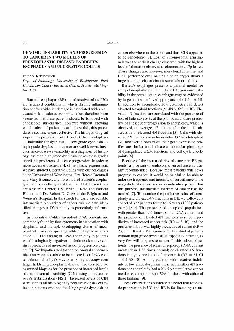
210 Abstracts
GENOMIC INSTABILITY AND PROGRESSIONTO CANCER IN TWO MODELS OFPRENEOPLASTIC DISEASE: BARRETT’SESOPHAGUS AND ULCERATIVE COLITIS
Peter S. RabinovitchDept. of Pathology, University of Washington, FredHutchinson Cancer Research Center, Seattle, Washing-ton, USA
Barrett’s esophagus (BE) and ulcerative colitis (UC)are acquired conditions in which chronic inflamma-tion and/or epithelial damage is associated with an el-evated risk of adenocarcinoma. It has therefore beensuggested that these patients should be followed withendoscopic surveillance, however without knowingwhich subset of patients is at highest risk, this proce-dure is not time or cost-effective. The histopathologicalsteps of the progression of BE and UC from metaplasia→ indefinite for dysplasia → low grade dysplasia →high grade dysplasia → cancer are well known, how-ever, inter-observer variability in a diagnosis of histol-ogy less than high grade dysplasia makes these gradesunreliable predictors of disease progression. In order tomore accurately assess risk of neoplastic progression,we have studied Ulcerative Colitis with our colleaguesat the University of Washington, Drs. Teresa Brentnalland Mary Bronner, and have studied Barrett’s esopha-gus with our colleagues at the Fred Hutchinson Can-cer Research Center, Drs. Brian J. Reid and PatriciaBlount, and Dr. Robert D. Odze at the Brigham andWomen’s Hospital. In the search for early and reliableintermediate biomarkers of cancer risk we have iden-tified changes in DNA ploidy as particularly informa-tive.
In Ulcerative Colitis aneuploid DNA contents arecommonly found by flow cytometry in association withdysplasia, and multiple overlapping clones of aneu-ploid cells may occupy large fields of the precancerouscolon [1]. The finding of DNA aneuploidy in patientswith histologically negative or indefinite ulcerative col-itis is predictive of increased risk of progression to can-cer [2]. We hypothesized that chromosomal abnormal-ities that were too subtle to be detected as a DNA con-tent abnormality by flow cytometry might occupy evenlarger fields in preneoplastic disease, and therefore weexamined biopsies for the presence of increased levelsof chromosomal instability (CIN) using fluorescencein situ hybridization (FISH). Increased levels of CINwere seen is all histologically negative biopsies exam-ined in patients who had focal high grade dysplasia or
cancer elsewhere in the colon, and thus, CIN appearedto be pancolonic [3]. Loss of chromosomal arm sig-nals was the earliest change observed, with the highestlevel of alteration observed as chromosome 17p losses.These changes are, however, non-clonal in nature, andFISH performed even on single colon crypts shows alarge heterogeneity of chromosomal abnormalities.
Barrett’s esophagus presents a parallel model forstudy of neoplastic evolution. As in UC, genomic insta-bility in the premalignant esophagus may be evidencedby large numbers of overlapping aneuploid clones [4].In addition to aneuploidy, flow cytometry can detectelevated tetraploid fractions (% 4N > 6%) in BE. Ele-vated 4N fractions are correlated with the presence ofloss of heterozygosity at the p53 locus, and are predic-tive of subsequent progression to aneuploidy, which isobserved, on average, 17 months after the initial ob-servation of elevated 4N fractions [5]. Cells with ele-vated 4N fractions may be in either G2 or a tetraploidG1, however in both cases their gene expression pro-files are similar and indicate a molecular phenotypeof dysregulated G2/M functions and cell cycle check-points [6].
Because of the increased risk of cancer in BE pa-tients, a program of endoscopic surveillance is usu-ally recommended. Because most patients will neverprogress to cancer, it would be helpful to be able totailor the frequency and intensity of surveillance to themagnitude of cancer risk in an individual patient. Forthis purpose, intermediate markers of cancer risk areneeded [7]. To examine the predictive value of aneu-ploidy and elevated 4N fractions in BE, we followed acohort of 322 patients for up to 15 years (1338 patient-years) [8,9]. The presence of aneuploid populationswith greater than 1.35 times normal DNA content andthe presence of elevated 4N fractions were both pre-dictive of increased cancer risk (RR = 8–10), and thepresence of both was highly predictive of cancer (RR =23, CI = 10–50). Management of the subset of patientswithout high grade dysplasia is especially difficult, asvery few will progress to cancer. In this subset of pa-tients, the presence of either aneuploidy (DNA contentgreater than 1.35 times normal) or elevated 4N frac-tions is highly predictive of cancer risk (RR = 25, CI= 6.5–98) [8]. Among patients with negative, indefi-nite or low grade dysplasia, those with neither 4N frac-tions nor aneuploidy had a 0% 5-yr cumulative cancerincidence, compared with 28% for those with either ofthese findings [9].
These observations reinforce the belief that neoplas-tic progression in UC and BE is facilitated by an un-

Abstracts 211
derlying process of chromosomal and genetic instabil-ity, with aneuploidy as an overt manifestation. To de-termine whether telomere shortening might be a con-tributing factor in such instability, we examined telom-ere lengths by fluorescence in situ hybridization. Inboth BE and UC, chromosomal instability and telom-ere shortening are present in large fields of histolog-ically non-dysplastic mucosa in early stages of dis-ease, and the extent of telomere shortening is corre-lated with the degree of chromosomal instability de-tected by FISH. In UC, chromosomal arm and cen-tromere losses, but not gains, are highly correlated withtelomere shortening (p � 0.001 and p = 0.001, re-spectively) [10]. Frequencies of chromosomal lossesare greater and telomeres are shorter in the nondysplas-tic mucosa from UC patients with dysplasia or can-cer elsewhere in their colon, as compared to chronicUC patients who have never had dysplasia [10]. InUC the extent of chromosomal instability and telom-ere shortening is also related to the frequency ofanaphase bridges, suggesting that telomere shorteningcontributes to chromosomal instability by promotingbridge-breakage-fusion cycles. Therefore, telomere at-trition and chromosomal instability in these diseasesmay be early events in a cascade that leads to DNAcontent abnormalities, loss of tumor suppressors, and,ultimately, cancer.
References
[1] Levine, D.S., Rabinovitch, P.S., Haggitt, R.C., Blount, P.L.,Dean, P.J., Rubin, C.E., and Reid, B.J. Distribution of aneu-ploid cell populations in ulcerative colitis with dysplasia orcancer. Gastroenterology 101:1198–1210, 1991.
[2] Rubin, C.E., Haggitt, R.C., Burmer, G.C., Brentnall, T.A.,Stevens, A.C., Levine, D.S., Dean, P.J., Kimmey, M., Perera,D.R., and Rabinovitch, P.S. DNA aneuploidy in colonic biop-sies predicts future development of dysplasia in ulcerative col-itis. Gastroenterology 103:1611–1620, 1992.
[3] Rabinovitch P.S., Dziadon S., Bronner M.P., Emond M.J.,Crispin D.A., Haggitt R.C., and Brentnall T.A. PancolonicChromosomal Instability Precedes Dysplasia and Cancer in Ul-cerative Colitis. Cancer Research 59:5148–53, 1999.
[4] Rabinovitch, P.S., Reid, B.J., Haggitt, R.C., Norwood, T.H.,and Rubin, C.E. Progression to cancer in Barrett’s esophagusis associated with genomic instability. Lab. Invest. 60:65–71,1988.
[5] Galipeau P.C., Cowan D.S., Sanchez C.A., Barrett M.T.,Emond M.J., Levine D.S., Rabinovitch P.S., and Reid B.J.17p (p53) allelic losses, 4N (G2/tetraploid) populations, andprogression to aneuploidy in Barrett’s esophagus. Proc. Natl.Acad. Sci. U.S.A. 93: 7081–7084, 1996.
[6] Barrett M.T., Pritchard D., Palanca-Wessels C., Anderson J.,Reid B.J., and Rabinovitch P.S. Molecular phenotype of spon-taneously arising 4N (G2-tetraploid) intermediates of neoplas-tic progression in Barrett’s esophagus. Cancer Res. 63:4211–7,2003.
[7] Reid B.J., Blount P.L., and Rabinovitch P.S. Biomarkers in Bar-rett’s esophagus. Gastrointest. Endosc. Clin. N. Am. 13:369–97, 2003.
[8] Rabinovitch P.S., Longton G., Blount P.L., Levine D.S., andReid, B.J. Predictors of progression in Barrett’s esophagus III:baseline flow cytometric variables. American Journal of Gas-troenterology 96:3071–3083, 2001.
[9] Reid, B.J., Levine, D.S., Longton, G., Blount, P.L., and Rabi-novitch, P.S. Predictors of Progression to cancer in Barrett’sesophagus: baseline histology and flow cytometry identify lowand high risk patient subsets. American Journal of Gastroen-terology 95:1669–1676, 2000.
[10] O’Sullivan J.N., Bronner M.P., Brentnall T.A., Finley J.C.,Shen W.-T., Emerson S., Emond M.J., Gollahon K.A.,Moskovitz A.H., Crispin D.A., Potter J.D., and RabinovitchP.S. Chromosomal instability in ulcerative colitis is related totelomere shortening. Nature Genetics, 32:280–4, 2002.

212 Abstracts
BIOLOGICAL AND CLINICALSIGNIFICANCE OF NONRANDOMANEUPLOIDY PATTERNS IN CHILDHOODACUTE LYMPHOBLASTIC LEUKEMIA (ALL)
Oskar A. HaasCCRI, St. Anna Children’s Hospital, Vienna, Austria(E-mail: [email protected])
Childhood B-cell precursor acute lymphoblastic leu-kemia (BCP-ALL) comprises a heterogeneous groupof diseases with distinct clinically and biologicallyrelevant genetic features, which were originally clas-sified according to chromosome number and ploidylevels (Table 1). The vast majority of specific recip-rocal rearrangements that were subsequently identi-fied with cytogenetic and further characterized withmolecular genetic means are typically found in the(pseudo)diploid category. The aneuploid groups, onthe other hand, are characterized by mainly numeri-cal changes and only few, less specific structural re-arrangements [1–11].
Table 1
Classification of BCP-ALL according to the ploidy level
Ploidy level Chromosome Frequency Prognostic
number relevance
Near-haploid 23–29 <1% bad
Hypodiploid 30–44 1% bad
(Pseudo)diploid 46 65–70% bad to very good*
Hyperdiploid 50–65 25–30% very good
Near-triploid 66–73 <1% good to moderate
Near-tetraploid 82–94 1% moderate to bad
*The specific type of reciprocal rearrangement determines progno-sis.
Despite their common occurrence, however, nextto nothing is known about the mode of generation,the role and the contribution of whole chromosomechanges to the neoplastic transformation process. Thisis mainly due to the lack of attractive and testable hy-potheses as regards the potential deregulative effectsof single or multiple chromosome copy number de-viations on the genome-wide gene expression levels,but also on the difficulty to approach this problem ex-perimentally. The development of novel investigativetools, such as various molecular genetic, FISH andgene array techniques, that enable the evaluation oftens of thousands of genes simultaneously, will eventu-ally help to shed some light on the pathogenetic signif-icance and biological consequences of these intriguing
karyotype patterns [12]. My contribution will thus pro-vide a brief overview about what is known so far aboutthese genetic alterations in childhood ALL.
1. Near-haploid ALL
Near haploid cases are extremely rare and are prog-nostically unfavorable [2,8,10,13]. They contain atleast the equivalent of a complete haploid set of chro-mosomes, two sex chromosomes and, in most in-stances, also two copies of chromosomes 10, 14, 18and 21. Haploid clones commonly coexist with hyper-diploid ones, which then contain the duplicated chro-mosome complement [2,8,10,13]. Whether pure hap-loid leukemia exist, must therefore remain open.
2. Hyperdiploid ALL
With an incidence of approximately 25% to 30%,hyperdiploidy is one of the most common abnormal-ities in childhood ALL [7,11,14]. By definition, thechromosome number ranges from 50 to 65 with amean peak at 55. The intriguing peculiarities of theseleukemia are the nearly exclusive presence of nonran-dom trisomies of chromosomes 4, 6, 10, 14, 17, 18, 20and X, whereas chromosomes 1, 2, 3, 12 and 16 vir-tually always remain disomic [5,7,9,11]. Chromosome21, on the other hand, is generally present in four andsometimes five copies.
Hyperdiploidy is generally considered as prognosti-cally favorable, although up to 20% of affected chil-dren still relapse [1,4,9,11]. The statistical evaluationof a large number of such hyperdiploid karyotypes re-vealed that a combination of trisomies 4, 10 and 17seems to be especially advantageous [9]. Based on theparameters age, sex, and the presence or absence of tri-somy 4 and 18, Moorman et al. were able to furthersubdivide hyperdiploid cases into three different riskgroups [11].
Occasionally, hyperdiploidy can also result from theduplication of a near-haploid clone [2,8,10,13]. Inter-estingly, these cases have a similarly poor prognos-tic outlook as their near-haploid counterparts. The twotypes of hyperdiploidy can be easily distinguished,however, because haploid and hyperdiploid clones usu-ally coexist. Moreover, the duplicated hyperdiploidclones contain two or four copies of each chromo-somes, but lack the characteristic trisomies that arepresent in the more common other form of hyper-diploidy.
Abstracts 213
3. Near-triploid and near-tetraploid ALL
With an incidence of approximately 1%, ALL casesin the near-triploid and near-tetraploid range are ex-tremely rare. They seem to result from mitotic errorsthat differ from those, which generate the above de-scribed hyperdiploidy and, therefore, represent mostlikely a distinct biological subset with a poor out-come [3,7].
4. On the formation of hyperdiploid karyotypes
The mechanism, which leads to the increased num-ber and the selection of particular chromosomes in hy-perdiploid ALL, remains largely unknown. Theoreti-cally, the following four possibilities could account forits formation [13,15–17]:
– The doubling of a near-haploid set of chromo-somes, which would result in a widespread lossof heterozygosity (LOH) due to the generation ofuniparental disomies (UPD);
– An initial tetraploidization with subsequent chro-mosome losses, which would result in LOH forapproximately one third of the chromosomes andequal dosages on tetrasomic chromosomes;
– The sequential gain of individual chromosomesduring consecutive cell divisions, which would re-sult in unequal allele dosages for two thirds of thetetrasomic chromosomes;
– And, finally, the simultaneous gain of chromo-somes in a single abnormal mitosis, which wouldresult in equal dosages of tetrasomic chromo-somes.
The currently available molecular genetic data indi-cate that hyperdiploidy usually arises from a diploidkaryotype through a simultaneous nondisjunction errorat a single abnormal cell division or, as already men-tioned above, the duplication of a near-haploid kary-otype [13,16,17]. This notion is further corroboratedby the fact that, in the vast majority of cases, tetrasomy21 results from a duplication of both homologs. Never-theless, the possibility of sequential gains can also notbe excluded with certainty so far [17].
In hyperdiploid forms of ALL, the chromosomenumber as well as their combinations may vary frompatient to patient [1,4,5,7,9,11].However, once formed,the abnormal karyotype is uniform and remarkable sta-ble in the malignant cell population of an individual pa-tient. Taking these observations into account, Heerema
and coworkers investigated in which order the chro-mosome acquisition may occur by analyzing whichchromosome are present in karyotypes with a partic-ular modal chromosomes number [14]. They foundthat chromosomes 21 and X are present in virtually allcases. Karyotypes with a modal number of 52–54 chro-mosomes gained extra copies of chromosomes 14, 6,4, 18, 17 and 10 in a consecutive order, whereas thosewith a modal number of 56–60 chromosomes had chro-mosomes 8, 5, 12 and 11 in addition. No other chromo-somes are consistently present until a modal number of68 chromosomes is reached, when nearly all chromo-somes are encountered. Chromosomes 1, 2, 3, 7, 9, 13,15, 16, 19, 20, 22 and Y become consistently trisomiconly in karyotypes with a modal chromosome numberof 68.
Based on a suggestion that imprinting phenomenamay be instrumental in the chromosomal selectionprocess [18], Paulsson et al. studied the parental ori-gin of the supernumerary chromosomes in 10 hyper-diploid cases [17]. However, except for trisomy 8,which was of paternal origin in 8 of 8 patients, and tri-somy 14, which was of maternal origin in 7 of 8 cases,they found no preferential duplication of maternally orpaternally-derived alleles [17].
5. Genome-wide epigenetic deregulation ratherthan specific gene mutations as a possiblepathogenetic factor in hyperdiploid ALL
The vast majority of childhood neoplasms, includ-ing various types of acute leukemia, are already initi-ated in utero during a period, when the delicate bal-ance between growth, development and differentiationof the early fetal organogenesis is disrupted. In linewith this notion, Wassermann et al. provided the firstcircumstantial evidence that ALL is initiated alreadyvery early during B-cell development [19]. More re-cently, it was then proven that the potentially leuke-mogenic nondisjunction event actually takes place alsoduring the same period and, therefore, must be a veryearly leukemogenic event [20].
Epigenetic disturbances, such as those that resultfrom abnormal DNA-methylation or chromosome mis-segregation, are the earliest and most ubiquitouschanges, which precede and concur with malignanttransformation. Considering the common occurrenceof premalignant neoplasms in very young children thatstill can regress or differentiate, an epigenetic first stepin these diseases is therefore a particularly appealing

214 Abstracts
one. In hyperdiploid and haploid ALL, this possibil-ity is backed up by the general lack of gross structuralchromosome abnormalities. In these cases, the tissue-specific growth, survival and differentiation controlmechanisms may thus be disturbed solely by the un-balanced distribution of particular chromosomes. Tri-somies of certain chromosomes may either enhance theproliferation capacity of early lymphoid cells througha change in dosage or relative dosage of a set of genesor in a similar process block differentiation [13,16,18].The more benign behavior of hyperdiploid ALL ver-sus the more aggressive one of haploid ALL may beexplained by the lack of a trans-acting gene expres-sion control mechanisms in the latter. In this contextit should be noted, however, that almost 70% of theover-expressed genes in hyperdiploid ALL are locatedon the chromosome X or 21 [12]. Finally, the extraor-dinary genetic make-up of hyerdiploid leukemic cellscould certainly also explain their rather unique in vitrobehavior. Since they rapidly undergo apoptosis, it isvirtually impossible to propagate them in culture [21,22]. It therefore comes also of no surprise that not asingle cell line has been yet established from such ahyperdiploid ALL.
Acknowledgements
Our work is supported by the research program“Genome Research for Health” of the Austrian Min-istry of Education, Science and Culture (GEN-AUChild, GZ 200.071/3-VI/2a/2002), by the “Fonds zurFoerderung der wissenschaftlichen Forschung” (grantP15150) and by the “Oesterreichische Kinderkreb-shilfe”.
References
[1] Jackson J.F., Boyett J., Pullen J., Brock B., Patterson R.,Land V., Borowitz M., Head D., Crist W. (1990) Favorableprognosis associated with hyperdiploidy in children with acutelymphocytic leukemia correlates with extra chromosome 6.A Pediatric Oncology Group study. Cancer 66:1183–1189.
[2] Gibbons B., MacCallum P., Watts E., Rohatiner A.Z., Webb D.,Katz F.E., Secker-Walker L.M., Temperley I.J., Harrison C.J.,Campbell R.H. (1991) Near haploid acute lymphoblasticleukemia: seven new cases and a review of the literature.Leukemia 5:738–743.
[3] Pui C.H., Carroll A.J., Head D., Raimondi S.C., Shuster J.J.,Crist W.M., Link M.P., Borowitz M.J., Behm F.G., LandV.J., et al. (1990) Near-triploid and near-tetraploid acute lym-phoblastic leukemia of childhood. Blood 76:590–596.
[4] Harris M.B., Shuster J.J., Carroll A., Look A.T., BorowitzM.J., Crist W.M., Nitschke R., Pullen J., Steuber C.P., LandV.J. (1992) Trisomy of leukemic cell chromosomes 4 and 10identifies children with B-progenitor cell acute lymphoblasticleukemia with a very low risk of treatment failure: a PediatricOncology Group study. Blood 79:3316–3324.
[5] Mertens F., Johansson B., Mitelman F. (1996) Dichotomy ofhyperdiploid acute lymphoblastic leukemia on the basis of thedistribution of gained chromosomes. Cancer Genet Cytogenet92:8–10.
[6] Raimondi S.C., Roberson P.K., Pui C.H., Behm F.G.,Rivera G.K. (1992) Hyperdiploid (47–50) acute lymphoblasticleukemia in children. Blood 79:3245–3252.
[7] Raimondi S.C., Pui C.H., Hancock M.L., Behm F.G., Fi-latov L., Rivera G.K. (1996) Heterogeneity of hyperdiploid(51–67) childhood acute lymphoblastic leukemia. Leukemia10:213–224.
[8] Ma S.K., Chan G.C., Wan T.S., Lam C.K., Ha S.Y., Lau Y.L.,Chan L.C. (1998) Near-haploid common acute lymphoblas-tic leukaemia of childhood with a second hyperdiploid line: aDNA ploidy and fluorescence in-situ hybridization study. Br JHaematol 103:750–755.
[9] Heerema N.A., Sather H.N., Sensel M.G., Zhang T., Hutchin-son R.J., Nachman J.B., Lange B.J., Steinherz P.G., BostromB.C., Reaman G.H., Gaynon P.S., Uckun F.M. (2000) Prognos-tic impact of trisomies of chromosomes 10, 17, and 5 amongchildren with acute lymphoblastic leukemia and high hyper-diploidy (>50 chromosomes). J Clin Oncol 18:1876–1887.
[10] Stark B., Jeison M., Gobuzov R., Krug H., Glaser-Gabay L.,Luria D., El Hasid R., Harush M.B., Avrahami G., Fisher S.,Stein J., Zaizov R., Yaniv I. (2001) Near haploid childhoodacute lymphoblastic leukemia masked by hyperdiploid line: de-tection by fluorescence in situ hybridization. Cancer Genet Cy-togenet 128:108–113.
[11] Moorman A.V., Richards S.M., Martineau M., Cheung K.L.,Robinson H.M., Jalali G.R., Broadfield Z.J., Harris R.L., Tay-lor K.E., Gibson B.E.S., Hann I.M., Hill F.G.H., Kinsey S.E.,Eden T.O.B., Mitchell C.D., Harrison C.J. (2003) Outcome het-erogeneity in childhood high-hyperdiploid acute lymphoblasticleukemia. Blood 102:2756–2762.
[12] Yeoh E.J., Ross M.E., Shurtleff S.A., Williams W.K., Patel D.,Mahfouz R., Behm F.G., Raimondi S.C., Relling M.V., Pa-tel A., Cheng C., Campana D., Wilkins D., Zhou X., Li J.,Liu H., Pui C.H., Evans W.E., Naeve C., Wong L., DowningJ.R. (2002) Classification, subtype discovery, and prediction ofoutcome in pediatric acute lymphoblastic leukemia by gene ex-pression profiling. Cancer Cell 1:133–143.
[13] Onodera N., McCabe N.R., Nachman J.B., Johnson F.L., LeBeau M.M., Rowley J.D., Rubin C.M. (1992) Hyperdiploidyarising from near-haploidy in childhood acute lymphoblasticleukemia. Genes Chromosomes Cancer 4:331–336.
[14] Heerema N.A., Shuster J., Biegel J., Camitta B., Cooley L.D.,Hirsch B., Magenis H.E., Patil S., Pettenati M.J., Pullen J., Rai-mondi S.C., Rao K., Schneider R.E., Roulston D., Sanger W.,Sather H.N., Sutcliff M.J., vanTuinen P., Watson M.S., Car-oll A.J. (2002) Pattern of extra chromosomes in pediatrichyperdiploid (50 to 67 chromosomes) acute lymphoblöasticleukemia (ALL) is modal number (mn) dependent. Blood100:308a.

Abstracts 215
[15] Shannon K. (1992) Genetic alterations in leukemia: Events ona grand scale. Blood 80:1–2.
[16] Onodera N., McCabe N.R., Rubin C.M. (1992) Formation ofa hyperdiploid karyotype in childhood acute lymphoblasticleukemia. Blood 80:203–208.
[17] Paulsson K., Panagopoulos I., Knuutila S., Jee K.J., Garwicz S.,Fioretos T., Mitelman F., Johansson B. (2003) Formation oftrisomies and their parental origin in hyperdiploid childhoodacute lymphoblastic leukemia. Blood 102:3010–3015.
[18] Haas O.A. (1996) Is genomic imprinting involved in thepathogenesis of hyperdiploid and haploid acute lymphoblas-tic leukemia of childhood? Acta Genet Med Gemellol 45:239–242.
[19] Wasserman R., Galili N., Ito Y., Reichard B.A., Shane S.,Rovera G. (1992) Predominance of fetal type DJH joiningin young children with B precursor lymphoblastic leukemia
as evidence for an in utero transforming event. J Exp Med176:1577–1581.
[20] Panzer-Gruemayer E.R., Fasching K., Panzer S., Hettinger K.,Schmitt K., Stoeckler-Ipsiroglu S., Haas O.A. (2002) Nondis-junction of chromosomes leading to hyperdiploid childhood B-cell precursor acute lymphoblastic leukemia is an early eventduring leukemogenesis. Blood 100:347–349.
[21] Ito C., Kumagai M., Manabe A., Coustan-Smith E., RaimondiS.C., Behm F.G., Murti K.G., Rubnitz J.E., Pui C.H., Cam-pana D. (1999) Hyperdiploid acute lymphoblastic leukemiawith 51 to 65 chromosomes: a distinct biological entity with amarked propensity to undergo apoptosis. Blood 93:315–320.
[22] Zhang Y., Lu J., van denBerghe J., Lee S.H. (2002) Increasedincidence of spontaneous apoptosis in the bone marrow ofhyperdiploid childhood acute lymphoblastic leukemia. ExpHematol 30:333–339.

216 Abstracts
INDUCTION OF ANEUPLOIDY ANDCHANGES IN REPLICATION PATTERN BYELECTROMAGNETIC FIELDS
Avital Korenstein-Ilan1, Alexander Barbul1,Alon Eliran2, Avi Gover2, Yael Eshet2, Eli Jerby2 andRafi Korenstein1
1Dept. of Physiology and Pharmacology, and 2De-partment of Electrical Engineering-Physical Electron-ics, Tel-Aviv University, Tel-Aviv, Israel (Tel.: +972-3-6406042, Fax: +972-6408982, E-mail: [email protected])
1. Introduction
Maintenance of genomic stability is essential for thesurvival of any organism. Multiple mechanisms haveevolved to insure homeostasis at the cellular level.A crucial stage in a cell’s cycle, at which errors leadingto loss of stability occur, is the S phase during whichreplication of the genetic complement happens (Lucasand Feng, 2003). In order to ensure genomic integrity,DNA replication has to occur in a very ordered man-ner at a specific portion of time during the S-phase,with the two allelic sequences replicating during thesame time domain (Lin et al., 2003). Thus, it is evi-dent that genomic stability depends on the fidelity ofDNA replication, DNA repair mechanisms and chro-mosome segregation. Malfunctions in these processeshave been reported to be connected with carcinogene-sis and developmental pathologies (Biggins and Wal-czak, 2003; Nasmyth, 2002; Duesberg and Rasnick,2001; Korenstein-Ilan et al., 2002).
The specific time interval during DNA synthesisat which a given DNA sequence is being replicatedappears to be a reliable indicator of its transcrip-tional activity. Accordingly, expressed DNA loci un-dergo early replication, while unexpressed ones repli-cate late. Epigenetic alterations involving chromatinremodeling have also been implicated in gene activitystatus as well as in tumorigenesis (methylation: Estler,2003; and acetylation: Sutherland and Costa, 2003).Thus, determining the replicating status of a sequenceopens a window into the orchestration of mechanismsof gene activation and silencing.
The processes of carcinogenesis involve many stepsin succession. Two hallmarks of the cancerous pheno-type are aneuploidy and changes in gene expression.Thus an attempt has been made to estimate the riskof human exposure to different types of non-ionizing
radiation in the environment, by following changes inaneuploidy and replication timing and asynchrony ofcentromeres following in vitro exposure of human lym-phocytes.
2. Methods
Fluorescence In Situ Hybridization (FISH) for thedetection of copy number – aneuploidy – has becomean essential tool in the diagnosis and management of avariety of solid tumors and hematologic malignanciesin the clinical setting, as well as an aid in the identifica-tion of particular genetic disorders and in prenatal di-agnosis. After establishing the FISH assay as a reliabletool for the analysis of metaphases, interphase FISHhas become the assay of choice for determining thecopy number of a sequence in a cell. It affords screen-ing of a large population of cells in a single preparationand enables to easily score various loci simultaneously.Determining aneuploidy using FISH is also amenableto automation, thus saving time as well as bias scoring.
The same cytogenetic preparations can be used todetermine the temporal order of replication, based onthe FISH replication assay, first described by Seliget al. (1992), which is by now accepted as reliablemethod for the determination of the mode of allelicreplication (reviewed in Korenstein-Ilan et al., 2002).Accordingly, the replication status of a locus is inferredfrom the shape of the hybridization signal obtained atinterphase following FISH with a locus-specific probe.This assay detects the chromatin conformation of thesequence studied. Prior to replication, each identifiedDNA locus shows a single dot like hybridization signal(singlet; S), while at the end of replication it assumesa doubled bipartite structure (doublet; D). Cells withone singlet and one doublet (SD – Fig. 1a) represent S-phase cells in which only one of the allelic sequenceshas replicated. Cells with two singlets (SS – Fig. 1b)represent those in which both sequences are unrepli-cated, and cells with two doublets (DD – Fig. 1c) rep-resent those in which both sequences have replicated.Accordingly, a high frequency of SD cells shows asyn-chrony in replication timing of the two allelic counter-parts; high frequency of DD cells indicates early repli-cation of the identified locus; and high frequency of SScells points to late replication.
Using two different exposure set ups, we exposedlymphocytes to CW non-ionizing radiation at two dif-ferent regions of the electromagnetic spectrum, in twospecially designed exposure systems. The first expo-
Abstracts 217
Fig. 1. Analysis of replication timing and asynchrony: (a) a cell in which one sequence has alredy replicated and one has not yet replicated (SDcell); (b) a cell in which neither sequence has replicated (SS cell) – indicating late replication; (c) a cell in which both sequences have alreadyreplicated (DD cell) – indicating early replication.
sure system (Fig. 2a) produced radiation of 100 GHz(THz radiation), while the second one (Fig. 2b) pro-duced radiation characteristic for cellular phones of800 MHz (RF radiation). Human lymphocyte cell cul-tures were set up and harvested according to standardcytogenetic techniques. For each exposed culture, wehad a control sample which was grown in a differentincubator and a sham exposed one, which was placedin the same incubator as the exposed sample, but on adifferent level so that it was not exposed. Cells in theTHz set-up, were exposed for one, two or 24 hours.Cells in the RF exposure system were irradiated for ei-ther 24 or 72 hours. At the termination of the exposurecultures were removed to the “control incubator” tillharvest time, after a total of 72 hours from culture setup.
We scanned slides of nuclei derived from cultureshybridized with probes specific for the centromericregions of chromosomes 11 (orange labeled; Vysis,USA) and 17 (green labeled; Vysis, USA) using theMetafer platform for semi-automatic interphase FISHscoring. Cells were scored automatically; the gallerywas then manually corrected by two independent tech-nicians. Between 800 and 1100 cells were scoredfrom each culture. The Metafer platform automaticallypresents the results obtained for the levels of chromo-somal gains and losses for each locus plus a correlationbetween the two loci. The subset of cells which hadtwo hybridization signals for both signals, were manu-ally analyzed for the pattern of replication of 600 cells,as described above.
In addition, the scanned files of the slides whichwere acquired during the search were inspected formetaphases and the number of signal for each ofthe loci was noted. The proportion of aneuploidy inmetaphases was compared with that determined for in-terphase nuclei.
3. Results
Exposure to radiations at the two different electro-magnetic spectral regions conferred increased levelsof asynchronous replication which were accompaniedby increased levels of aneuploidy in a dose dependentmanner. We determined that the THz radiation inducedincrease in both replication asynchrony and aneuploidyafter two hours and 24 hours of exposure, but no ef-fect was observed after one hour radiation. Exposureof cells for 72 hours to RF radiation revealed elevationin both replication asynchrony and aneuploidy. How-ever, exposure for a shorter time of 24 hours showedchanges in replication asynchrony and timing only,whereas no alteration in aneuploidy was observed.
4. Discussion
The results obtained demonstrate an increased ane-uploidy and elevation of asynchronous replication ofcentromers following exposure to both THz and RF ra-diations. The observed effects were dependent on thelength of the exposure time, showing a dose response

218 Abstracts
Fig. 2. Schemes of the exposure system used. (a) The exposure system used for the THz frequency and (b) the RF frequency radiation.
relationship. The increased asynchronous replicationof centromers was found to be correlated with elevatedaneuploidy. It may be suggested that coordinated repli-cation of centromers is necessary for the proper func-tion of the segregating machinery. Moreover it seems
that the epigenetic changes involved in replication pre-cede the onset of increase in aneuploidy. These find-ings suggest that prolonged exposure to non-ionizingradiation increases genomic instability, thus constitut-ing a risk factor for cancer.
Abstracts 219
Acknowledgements
This research was funded by MAFAT/IMOD and bythe Commission of the European Communities, spe-cific RTD program “Quality of Life and Managementof Living Resources”, QLK4-2000-00129.
References
[1] Biggins S. and Walczak C.E. (2003) Captivating capture: howmicrotubules attach to kinetochores. Current Biology, 13:R449–R460.
[2] Duesberg P. and Rasnick D. (2001) Aneuploidy, the somatic mu-tation that makes cancer a species of its own. Cell Motil Cy-toskeleton. 47(2):81–107.
[3] Estler M. (2003) Relevance of DNA methylation in the manage-ment of cancer. The Lancet Oncol 4:351–358.
[4] Korenstein-Ilan A., Amiel A., Lalezari S., Lishner M. and AviviL. (2002) Allele specific replication associated with aneuploidyin blood cells of patients with hematological malignancies. Can-cer Genet. and Cytogenet 139:97–103.
[5] Lin C.M., Fu H., Martinovsky M., Bouhassira E., and AladjemM.I. (2003) Dynamic alterations of replication timing in mam-malian cells. Curr Biol 13:1019–28.
[6] Lucas I. and Feng W. (2003) The essence of replication timing.Cell Cycle 2:560–563.
[7] Nasmyth K. (2002) Segregating sister genomes: the molecularbiology of chromosome separation. Science 297:559–565.
[8] Selig S., Okumura K., Ward D.C. and Cedar H. (1992) Deliana-tion of DNA replication time zones by fluorescence in situ hy-bridization. The EMBO J 11:1217–1225.
[9] Sutherland J.E. and Costa M. (2003) Epigenetics and the envi-ronment. Ann N.Y. Acad Sci 983:151–160.

220 Abstracts
CENTROSOME AMPLIFICATION,CHROMOSOME INSTABILITY, ANDKARYOTYPIC CONVERGENCE
Tatsuo Oikawa1,2, Amy Staubach2, Masaru Okuda1
and Kenji Fukasawa2
1Laboratory of Veterinary Internal Medicine, Facultyof Agriculture, Yamaguchi University, Yamaguchi 753-8515, Japan, 2Department of Cell Biology, Universityof Cincinnati College of Medicine, PO Box 670521,Cincinnati, OH 45267-0521, USA
Chromosome instability (CIN), usually associatedwith mitotic defects, plays a critical role in tumor pro-gression via promoting mutations responsible for aseries of malignant phenotypes. Abnormal amplifica-tion of centrosomes has been shown to be a majorcause of mitotic defects and CIN in cancer cells [1].The centrosome is a non-membranous organelle, com-posed of a pair of centrioles and a number of dif-ferent proteins surrounding the centriole pair (PCM)(Fig. 1A). During mitosis, two centrosomes gener-ated from semi-conservative duplication, form spindlepoles and direct formation of bipolar mitotic spindles(Fig. 1B, a), which is an essential event for accuratechromosome transmission to daughter cells. The pres-ence of more than two centrosomes (centrosome am-plification) will disrupt formation of bipolar spindles(Fig. 1B, b & c), leading to an increased frequencyof chromosome transmission errors [2]. There are sev-eral mechanisms responsible for centrosome amplifi-cation, including uncontrolled duplication of centro-somes, improper splitting of centriole pairs and cy-tokinesis block followed by progression into the nextcell cycle [2]. Genotoxic insults such as irradiationand exposure to certain chemotherapeutic agents in-duce centrosome amplification both directly by uncou-
pling centrosome duplication and cell cycle progres-sion and indirectly by inducing genetic alterations ofcertain genes whose products are involved in numeralhomeostasis of centrosomes. One of such genes is p53:loss or mutational inactivation of p53 results in cen-trosome amplification [3]. Interestingly, many of ad-vanced tumors as well as cultured tumor cells whichlack functional p53 show extensive aneuploidy, yet thealtered chromosome makeup is faithfully transmittedto daughter cells, hence they are chromosomally stable.Moreover, many of these cells contain normal numberof centrosomes. It has been hypothesized that duringtumor progression, cells acquire a CIN phenotype (i.e.,centrosome amplification) in an early stage, resultingin a karyotypically heterogeneous population. Amongthese cells, one or few acquire chromosome composi-tions that promise the best growth properties under agiven environment, which gradually dominate the pop-ulation. For this population of cells, maintenance ofthis particular karyotype becomes a primary selectionpressure, forcing the selection of the offspring whichhas acquired mutation(s) that counteracts the CIN phe-notype (i.e., restoring normal centrosome profiles). Wehave simulated this “karyotypic convergence” modelduring tumor progression in culture [4]. Embryonic ep-ithelial cells from 8-week-old male mice lacking p53(p53−/−MEEs) were passaged for 50 passages (p50).Every 5 passages, cells were examined for chromo-some number by direct counting of metaphase spreads,and centrosome profiles by immunostaining γ-tubulin(Fig. 2). In early passages, the number of chromosomesextensively deviated from the norm (4N = 80), indi-cating that chromosomes were unstable in early pas-sage p53−/− MEEs. However, at ∼p30, the distri-bution of the chromosome number per cell becamenarrower. At p50, particular populations of cells weredominant in the culture: ∼60% of cells contain 90–94
Fig. 1. Centrosome structure and abnormal mitosis caused by amplified centrosomes.

Abstracts 221
Fig. 2. Karyotypic convergence of p53−/− MEEs in culture is accompanied with restoration of normal centrosome profiles.
chromosomes. The changes in the frequency of centro-some amplification during passaging strikingly paral-leled to destabilization/stabilization of chromosomes:high frequencies of centrosome amplification was ob-served up to p30. Thereafter, the extent of centrosomeamplification gradually decreased, and at ∼p50, cen-trosomes behaved normally: cells contained either oneor two centrosomes, and the frequency of abnormal mi-tosis due to amplified centrosomes was indistinguish-able from wild-type cells (<1% of total mitotic cells).These findings strongly suggest that chromosome in-stability in p53−/− mouse cells is primarily attributedto centrosome amplification, and that centrosome am-plification initially imposed by loss of p53 was sup-pressed in the late passage cells.
Our findings put forward a model of karyotypicconvergence in p53−/− cells during prolonged cul-ture, which may be applied to karyotypic conver-gence during tumor progression (Fig. 3). In early pas-sages, p53−/− cells suffer extensive CIN due cen-trosome amplification, resulting in karyotypic hetero-geneity. At a certain time point in culture, one or fewcells within this karyotypically heterogeneous popu-lation acquire chromosome compositions that promiseadvantageous growth phenotypes (i.e., faster growth,serum-independent growth, anchorage-independent
Fig. 3. Karyotypic convergence in cultured p53−/− cells.

222 Abstracts
growth, loss of cell–cell contact growth inhibition,minimal requirement of nutrients). Such cells gradu-ally dominate the culture. For those cells, maintenanceof this particular chromosome composition becomespriority, forcing the selection of cell(s) that counter-act the cause of CIN (i.e., centrosome amplification inp53−/− cells) to maintain the chromosome composi-tion that allows the most efficient growth under a givenenvironment.
References
[1] D’Assoro A.B., Lingle W.L. and Salisbury J.L. (2002) OncogeneReviews 4:6146–6153.
[2] Fukasawa K. (2002) Oncogene Reviews 4:6140–6145.
[3] Tarapore P. and Fukasawa K. (2002) Oncogene Reviews 4:6234–6240.
[4] Chiba S., Okuda M., Mussman J.G. and Fukasawa K. (2000)Exp. Cell Res. 258:310–321.

Abstracts 223
COLORECTAL CANCER PROGRESSIONMODEL OF CHROMOSOMAL CHANGESDURING LYMPHATIC AND LIVERMETASTASIS FORMATION
Thomas Knösel1, Karsten Schlüns1, Ulrike Stein2,Holger Schwabe2, Peter Michael Schlag2,Manfred Dietel1 and Iver Petersen1
1Institute of Pathology, Charité - Campus Mitte, Berlin,Germany (Tel.: +49 (0)30450536155, Fax: +49 (0)30450536110, E-mail: [email protected]),2Department of Surgery and Oncology, RRC, CharitéCampus Buch, 13125 Berlin, Germany
Based on the analysis of colorectal cancer (CRC) bycomparative genomic hybridization (CGH) we estab-lished a progression model of this tumor type [1]. Intotal, 63 tumor specimens from 40 patients were in-vestigated comprising 30 primary tumors, 22 systemicmetastases (12 liver, 6 brain, 4 abdominal wall metas-tases) and 11 lymph node tumors. The overall patternof alterations was similar as previously described [2],tumor subgroups i.e. hematogenous metastases showedmore alterations than lymph node tumors, particularlymore deletions 1p, 3, 4, 5q, 10q, 14 and 21q21 andgains at 1q, 7p, 12qter, 13, 16 and 22q. Comparingliver metastases with their corresponding primary tu-mors particularly deletions at 2q, 5q, 8p, 9p, 10q and21q21 and gains of 1q, 11, 12qter, 17q12–q21, 19, and22q were more often observed.
Our data and the therof derived colorectal pro-gression model indicate that there are independentpathways of colorectal tumor dissemination and thatthese are associated with a nonrandom accumulationof chromosomal alterations underlying the differentcharacteristics of the metastatic phenotype. It alsohighlights two well known pathogenetic mechanisms:(1) Metastasis formation may occur immediately afterinvasion of the primary tumor. (2) Hematogenous dis-semination may occur independently from lymphatictumor spread. Obviously, there is an overlap betweenthe genetic alterations of the tumor subgroups correlat-ing with the fact that many tumors develop both lymphnode or hematogenous filiae. Lymphatic spread, how-ever, is not a prerequisite of systemic disseminationwhich may occur early after cancer initiation. Accord-ingly there is probably no stepwise acquisition, but aselection of tumor cell clones carrying the favorablemetastasis-associated lesions that are initially gener-ated randomly by the inherent chromosomal instabilityof most colorectal carcinomas.
References
[1] Knösel T., Schlüns K., Stein U., Schwabe H., Schlag P.M., DietelM., Petersen I. Chromosomal alterations during lymphatic andliver metastasis formation of colorectal cancer. Neoplasia, 2004,January/February, in press.
[2] Knösel T., Petersen S., Schwabe H., Schlüns K., Stein U., SchlagP.M., Dietel M., Petersen I. Incidence of chromosomal imbal-ances in advanced colorectal carcinomas and their metastases.Virchows Archiv 2002 Feb 440:187–194.

224 Abstracts
PATTERNS AND CONSEQUENCES OFCHROMOSOMAL ANEUPLOIDY IN CANCERCELLS
Thomas Ried, Michael J. Difilippantonio,Cristina Montagna, Kerstin Heselmeyer-Haddad,Hesed Padilla-Nash, Jens Habermann andMadhvi UpenderCenter for Cancer Research, National Cancer Insti-tute, NIH, Bethesda, MD 20892, USA
Chromosomal aberrations are key events in the ini-tiation and progression of cancer and can be detectedin virtually all tumors. In the hematological malig-nancies, such aberrations frequently result in proto-oncogene dysregulation mediated by way of chromo-somal translocations. These events can be triggered bydeficiencies in proteins involved in double strand re-pair [1]. In cancers of epithelial origin, however, bal-anced chromosomal translocations are rare. In mostinstances, chromosomal aberrations result in genomicimbalances [2,3]. This has been firmly established bythe complementary analysis of solid tumor genomesusing spectral karyotyping (SKY) and comparative ge-nomic hybridization (CGH). In addition, carcinomasreveal recurrent numerical chromosomal aberrations,which we refer to as chromosomal aneuploidies [4].Using a combination of tissue microdissection andCGH, we analyzed genomic changes in the progres-sion of cervical and colorectal neoplasia. Both tumortypes are characterized by a strictly conserved distri-bution of genomic imbalances. For instance, the tran-sition of cervical dysplasia to invasive carcinomas al-most invariably required the acquisition of extra copiesof chromosome 3q. In colorectal carcinomas, specificimbalances on chromosomes 7, 8q, 13, 18q and 20were detected. Some of these changes occur at earlystages. For instance, extra copies of chromosome 7could be detected in colon polyps before mutationalinactivation of p53. However, the acquisition of spe-cific gains of chromosome 3q in cervical tumorigen-esis was preceded by the presence of high risk HPV.We therefore conclude that compromising p53 and Rb-function via viral infection is a necessary, yet not suf-ficient, condition for cervical tumorigenesis. The aver-age number of chromosomal copy number alterations(ANCA, measured by dividing the number of genomicimbalances by the number of cases studied) increasedwith increasing stages of cellular dysplasia [4]. Ofnote, specific chromosomal aneuploidies can occur inan otherwise diploid genome [5]. We have extended
these analyses to mouse models of human carcinomas,including mammary gland adenocarcinomas inducedby either over-expressing of erbB2 or by conditional,breast epithelium specific, knockout of BRCA1 [6,7].Both cancer models revealed centrosome abnormali-ties that result in gross chromosomal and nuclear ane-uploidy. Despite this chromosomal aneuploidy and in-tratumoral heterogeneity, we detected a non-randomdistribution of chromosomal gains and losses when thetumor genomes were analyzed by CGH. It thereforeappears that the acquisition of tumor specific chromo-somal imbalances renders a selective growth advan-tage to tumor cells and is conserved across speciesboundaries. The prevalent chromosomal instability canbe interpreted as a mean to acquire these imbalances.We compared the average number of copy alterations(ANCA) in different mouse models. In general, mousetumors induced by a strong oncogenic stimulus showeda lower ANCA index than those induced by deficien-cies in tumor suppressor genes. This could indicate thata strong oncogenic promotor of cellular proliferationwould alleviate the requirement of chromosomal im-balances to be acquired and maintained. Tumor mod-els induced by conditional inactivation of tumor sup-pressor genes (such as BRCA1 in the mouse mammarygland) therefore appear to more faithfully recapitulatethe sequential genome destabilization that we observeduring human tumorigenesis. These data were corrob-orated using interphase cytogenetics with probe cock-tails specific for the aberrant chromosomes in touch-preps from cancer specimens. In order to assess theconsequences of chromosomal aneuploidies with re-spect to gene expression levels, we have conducted ex-tensive comparisons between cytogenetic aberrationsand expression profiles using cDNA arrays both in ex-perimental cancer models as well as in primary tumorspecimen.
Hypothetically, chromosomal aneuploidies couldhave two possible consequences: (i) the expression ofall or most genes located on a chromosome is affectedby chromosomal gain or loss, or (ii) the expressionof only a few genes, whose reduced or increased ex-pression is critical for tumorigenesis, is the target ofchromosomal aneuploidy during tumorigenesis. Thus,whole chromosome copy number changes may simplyreflect an economical and readily achievable mecha-nism for the cells to achieve and maintain such expres-sion changes. However, the latter would implicate si-lencing mechanisms for all other genes located on theaffected chromosome present in increased copy num-bers. Data from experimental cancer models, from nor-

Abstracts 225
mal epithelial tissues that acquired chromosomal aneu-ploidies after immortalization and transformation, andfrom experimentally induced chromosomal trisomiesafter microcell mediated chromosome transfer suggestthat only a subset of genes on those chromosomes thatare subject to significant copy number increase as aconsequence of genomic gain, however, the averageexpression values of all gene on a trisomic chromo-somes increases as well [8,9]. The respective contri-bution of these changes (strong upregulation of only afew genes or low level increase of all genes on a chro-mosome) with respect to malignant transformation andtumorigenesis remains to be elucidated.
References
[1] Difilippantonio, M., Zhu, J., Chen, H.T., Meffre, E., Nussen-zweig, M.C., Max, E., Ried, T., and Nussenzweig, A. (2000).DNA repair protein Ku80 suppresses chromosomal aberrationsand malignant transformation. Nature 404:510–514.
[2] Ghadimi, B.M., Sackett, D.L., Difilippantonio, M.J.,Schröck, E., Jauho, A., Neumann, T., Jauho, A., Auer, G.,and Ried, T. (2000). Centrosome amplification and instabilityoccurs exclusively in aneuploid, but not in diploid colorectalcancer cell lines and correlates with numerical chromosomalaberrations. Genes Chromosomes. Cancer. 27:183–190.
[3] Ghadimi, B.M., Schröck, E., Walker, R.L., Wangsa, D.,Jauho, A., Meltzer, P.S., and Ried, T. (1999). Specific chromo-somal aberrations and amplification of the AIB1 nuclear recep-tor coactivator gene in pancreatic carcinomas. Am. J. Pathol.154:525–536.
[4] Ried, T., Heselmeyer-Haddad, K., Blegen, H., Schröck, E.,and Auer, G. (1999). Genomic changes defining genesis, pro-gression, and malignancy potential in human solid tumors:A phenotype/genotype correlation. Genes Chromosomes. Can-cer 25:195–204.
[5] Heselmeyer-Haddad K., Janz V., Castle P.E., Chaudhri N.,White N., Wilber K., Morrison L.E., Auer G., Burroughs F.H.,Sherman M.E., and Ried T. Detection of genomic amplificationof the human telomerase gene (TERC) in cytologic specimensas a genetic test for the diagnosis of cervical dysplasia. Am JPathol. 2003 Oct; 163(4):1405–16.
[6] Xu, X., Weaver, Z., Linke, S., Li, C., Gotay, J., Wang, X.-W.,Harris, C.C., Ried, T., and Deng, C.-D. (1999). Centrosome am-plification and a defective G2-M cell cycle checkpoint inducegenetic instability in BRCA1 exon 11 isoform-deficient cells.Mol. Cell 3:389–395.
[7] Montagna C., Andrechek E.R., Padilla-Nash H., Muller W.J.,and Ried T. Centrosome abnormalities, recurring deletions ofchromosome 4, and genomic amplification of HER2/neu de-fine mouse mammary gland adenocarcinomas induced by mu-tant HER2/neu. Oncogene. 2002 Jan 3; 21(6):890–8.
[8] Phillips J.L., Hayward S.W., Wang Y., Vasselli J., Pavlovich C.,Padilla-Nash H., Pezullo J.R., Ghadimi B.M., Grossfeld G.D.,Rivera A., Linehan W.M., Cunha G.R., and Ried T. The con-sequences of chromosomal aneuploidy on gene expression pro-files in a cell line model for prostate carcinogenesis. Cancer Res.2001 Nov 15; 61(22):8143–9.
[9] Platzer P., Upender M.B., Wilson K., Willis J., Lutterbaugh J.,Nosrati A., Willson J.K., Mack D., Ried T., and Markowitz S.Silence of chromosomal amplifications in colon cancer. CancerRes. 2002 Feb 15; 62(4):1134–8.

226 Abstracts
SEQUENCE OF CENTROMERESEPARATION – POSSIBLE LINK WITHANEUPLOIDY AND CANCER
Baldev K. VigDepartment of Biology, University of Nevada, Reno,NV 89557-0015, USA (Tel.: +1 775 784-6544, Fax:+1 775 784-1302, E-mail: [email protected])
At meta-anaphase junction the centromeres of eu-karyotic genomes separate in non-random geneticallydetermined sequence. The process appears to be gov-erned by the composition and quantity of the pericen-tric heterochromatin. In mouse which harbors minorsatellite of uniform composition in its pericentric re-gion, the timing of separation is directly related to thequantity of the satellite DNA; the larger the quantity,the later the separation. In human, with several satellitefractions in the neighborhood of the centromere, thesituation is complex.
The sequence of centromere separation is not af-fected by the application of spindle inhibitors (e.g.,Colcemid, colchicine, vinblastine, etc.). Also, there ap-pears to be no difference between the separated and un-separated centromeres in the pattern of their phospho-rylation and acetylation. In contrast to the reports onbiochemical studies on cell cycle, our studies show thatthe separated centromeres as well as the entire chromo-some is highly phosphorylated [1] as well as acetylatedat anaphase.
The ‘inactive’ centromeres in multicentric chromo-somes separate ahead of the normal or functionalcentromeres in the entire genome. This might indi-cate that early separation of a normal centromere,ahead of its schedule, might lead to abnormal behav-ior and/or function of the centromere. More recently,it has been shown that the sequence of separation canbe influenced by some chemicals belonging to variousgroups [2]. In conformity with the above statement, thedrugs which induced alterations in the sequence of cen-tromere separation also induce aneuploidy.
Currently, we are looking into the aberrations ofcentromere separation in individuals with Down syn-drome and their parents. Our preliminary data showthat chromosome 21 in the Downs’ individuals, and ap-parently in their mothers, exhibit premature separationwith higher than normal frequency. Some previous datafrom our lab and the literature show that errors in thesequence of centromere separation are associated withbirth defects and certain cancers.
References
[1] Garcia-Orad, A., Gomez, P., and Vig, B.K. (2001) HistoneH3 phosphorylation of mammalian chromosomes. ChromosomeResearch 9:487–494.
[2] Vig, B.K. and Willcourt, M. (1998) Decondensation of pericen-tric heterochromatin alters the sequence of centromere separa-tion in mouse cells. Chromosoma 107:417–423.

Abstracts 227
ALTERATIONS IN NUCLEAR MORPHOLOGY,CENTROSOMES, AND CHROMOSOMALSTABILITY INDUCED BY EXPRESSION OFMUTANT P53
Vivian Negron, Cathleen Gibson, Kathleen Kitzmannand Wilma LingleExperimental Pathology, Mayo Clinic, 200 First St SW,Rochester, MN 55905, USA (Tel.: +1 507 538-1287,Fax: +1 507 282-1805, E-mail: [email protected])
1. Introduction
Our recent studies of p53 mutations in human breastcancer demonstrated that tumors with mutant p53 tendto have a significantly greater capacity to nucleatemicrotubules (MTs) than do tumors with wild typep53 [1]. MT nucleation is a major function of the cen-trosome during both interphase and mitosis. The inter-phase MT cytoskeleton, through interactions with theactin and intermediate filament cytoskeletons, affectscell polarity, vesicular trafficking, cell–cell adhesions,and cell migration. During mitosis, duplicated centro-somes nucleate the MTs of the bipolar mitotic spin-dle apparatus. Centrosome amplification can affect in-terphase and mitotic centrosome function by disrupt-ing cell polarity and by increasing the frequency ofmultipolar mitoses [2,3]. In breast tumors, the extentof centrosome amplification correlates with the levelof chromosomal instability (CIN), supporting the hy-pothesis that centrosome amplification can cause im-proper chromosome segregation through the formationof multipolar mitotic spindles [1]. In spite of this cor-relation between centrosome amplification and CIN,there has been no demonstration that centrosome am-plification precedes CIN. In the work presented here,we explored the timing of changes in centrosomes, nu-clear morphology, and CIN induced by expression ofmutant p53 in cultures of mammary epithelial cells.
2. Methods
To further our understanding of the relationship be-tween p53 and centrosome function, we have createdtwo adenovirus vectors, M72 and M114, containingtwo different p53 mutations. We chose the p53 mu-tations based on the characteristics of the tumors inwhich they were found. Tumor BT0072, with p53 mu-tant M72, had 3 fold greater MT nucleating capac-
ity, 10 fold greater centrosome number, and 3.4 foldgreater CIN level than normal breast tissues. BT0114,with p53 mutant M114, had 11 fold greater MT nucle-ation capacity, 3 fold greater centrosome number, and1.5 fold greater CIN than normal breast tissues. TheM72 mutant is a truncation mutation that deletes theDNA binding domain, the nuclear localization signal,and the tetramerization domain, while the M114 has apoint mutation in the DNA binding domain. We usedthe adenoviral vectors to deliver the mutant p53 to cul-tured human mammary epithelial cells previously im-mortalized with human telomerase. Infection rates ofgreater than 90% were achieved with the adenovirusvectors. We assayed infected cell cultures at days 2, 8,and 16 post-infection to determine the alterations in-duced by the 2 specific mutant p53s in nuclear mor-phology, centrosomes, MT kinetics, and chromosomalstability. Specifically, we measured the frequency oflobed, micro-, and large nuclei, centrosome character-istics, microtubule regrowth characteristics, and CINof chromosomes 3, 7, and 17.
3. Results
3.1. p53 expression
As determined by Western blotting and immunhisto-chemistry, expression of mutant p53 was maximal ondays 2 and 8, although cells continued to express mu-tant protein for more than 2 weeks. Results for proteinexpression and other parameters measured below aresummarized in Table 1.
3.2. Nuclear morphology
On days 2 and 8, cells expressing M72 had signifi-cantly higher frequencies of cells with lobed nuclei andmicronuclei than did uninfected control cells or controlcells expressing GFP-adenovirus, with the frequencypeaking on day 8. M72-expressing cells never had anincrease in large nuclei. The frequency of lobed andmicronuclei was increased in M114-expressing cellsonly on day 8. Also on day 8, cells expressing M114,but not M72, had significantly more large nuclei thancontrol cells.
3.3. Centrosome characteristics
Cells expressing M72 had amplified centrosomesmore frequently than did control cells on days 2 and 8,

228 Abstracts
Table 1
Timing of maximal effects of expression of p53 mutants
but not day 16. The frequency of centrosome amplifi-cation peaked in M114-expressing cells on day 8, andremained greater than control cells through day 16. Thefrequency of non-adjacent centrosomes was also af-fected by the expression of mutant p53. The effect wasrelatively weak in M72-expressing cells, and was ap-parent only on day 2. The effect was more pronouncedin M114-expressing cells, especially on day 8. Centro-some separation could be induced in control cells byMT depolymerization. This induction was largely mit-igated by expression of both p53 mutants, except forday 8 of M114 when separation was hyperstimulatedby MT depolymerization.
3.4. MT asters and regrowth
On days 2, 8, and 16 in MT regrowth experiments,cells expressing either p53 mutant had more frequentmultiple and disorganized asters than did control cells.The effects were maximal for multiple asters on days 8and 16 for both p53 mutants. M72 induced the greatestfrequency of disorganized asters on day 8. The num-ber of MTs was greater on all 3 days in M72- andM114-expressing cells than in control cells. However,the length of MTs was significantly greater in cells ex-pressing M114 when compared to control cells only onday 8.
3.5. Chromosomal stability
Neither p53 mutant caused increased levels of CINon day 2. The M72 mutant caused a 2 fold increase inCIN on day 8; but by day 16 CIN levels had returned tonormal levels. In contrast, M114-expression cells hadnormal levels of CIN on days 2 and 8, but by day 16had CIN levels approximately 3 fold greater than con-trol cells.
4. Discussion
As seen in the timeline presented in Table 1, M72and M114 have subtle differences in the timing of theireffects on nuclear morphology, centrosomes, MTs, andCIN. The M72 p53 mutation maximally affects cen-trosomes and the number of MTs and begins to affectnuclear morphology, centrosome separation, and mul-tiple asters as soon as day 2 post infection. The effectof M72 on CIN was apparent only transiently on day8, with no detectable effect on either day 2 or day 16.In general, the effects of the M72 mutation on all ofthe parameters measured in this study appeared soonerthan the effects of M114. M114 expression maximallyeffected nuclear morphology, centrosomes, and MTs
Abstracts 229
on day 8 with effects on centrosome separation, mul-tiple asters, and MT regrowth first appearing on day2. M114 effects on CIN were only detectable on day16, which interestingly, is after the peak of expressionof the mutant protein. This indicates that expressionof the mutant p53 can cause a long-lasting change inthe mechanisms responsible for the fidelity of chro-mosomal segregation. The differences in the effects ofthe two p53 mutants is likely due to the nature of themutations; the M72 mutant is truncated and lacks theDNA binding domain, the nuclear localization signal,and the tetramerization domain. The M114 mutant isa point mutation located in the DNA binding domain.However, both mutants increased levels of CIN onlyafter changes in centrosomes and MTs had occurred.
5. Conclusions
The experiments presented here are the first demon-stration that centrosome amplification and changes inMT growth and organization precede chromosomal in-stability. These results also demonstrate that p53 mu-tants can directly affect the MT cytoskeleton. In thecase of the truncated M72 mutant, these changes oc-cur even in the absence of transcriptional regulation via
DNA binding. Specific p53 mutations differ in the tim-ing and degree of their effects on centrosome structureand chromosomal instability; in vivo, these mutantsmay have differential effects on tumor progression andmetastasis. Identification of particular p53 mutations inthe tumors of breast cancer patients and understandingthe specific effects of these mutations on centrosomes,the cytoskeleton, and chromosomal instability may aidin developing custom tailored chemotherapeutic treat-ments.
Acknowledgement
This work was supported by the Fraternal Order ofthe Eagles’ Cancer Research Award and Departmentof Defense Breast Cancer Research Program AwardDAMD17-98-1-8122 to WL.
References
[1] Lingle et al., PNAS 99 (2002) 1978.
[2] Lingle et al., Am. J. Pathol. 155 (1999) 1941.
[3] Lingle et al., PNAS 95 (1998) 2950.

230 Abstracts
IS CENTROSOME AMPLIFICATION ANDAURORA A KINASE OVEREXPRESSION APRELUDE TO CHROMOSOME INSTABILITYAND ANEUPLOIDY?
Thea M. Goepfert, Melissa A. Munoz,Joshua M. Newton, David G. Edwards andWilliam R. BrinkleyDepartment of Cellular and Molecular Biology, Bay-lor College of Medicine, Houston, TX, USA (Tel.:+1 713 798-5262, Fax: +1 713 798-5762, E-mail:[email protected])
1. Introduction
The dual and opposing nature of centrosomes facili-tate the assembly of a bipolar spindle apparatus neededfor the orderly and equal partitioning of chromosomesduring mitosis. In addition, centrosomes function asmicrotubule organizing centers to configure and main-tain cell shape, polarity and morphogenesis. There-fore, the striking presence of centrosome anomaliesin tumor cells, including amplification and impairedcapacity to assemble and organize microtubules [1,2]has been linked to chromosome instability and aneu-ploidy in neoplastic cells. Although Boveri [3] identi-fied the centrosome as the principle instigator of ane-uploidy and cancer over a century ago, his hypothe-sis was subsequently abandoned. However, in light ofnew experimental results in our laboratory and else-where [4,5], Boveri’s hypothesis has been resurrected.In addition, centrosome anomalies and cancer havealso been linked with the discovery of a member ofthe aurora kinase gene family, STK15/aurora A, whichis localized to the centrosome, and overexpressed inmany human tumors (for review see [6]). Moreover,ectopic expression of STK15 was shown to induce cen-trosome amplification and aneuploidy in vitro [7] andwas found to correlate with enhanced clinical aggres-siveness, invasion and increased rate of metastasis inhuman bladder cancer [8].
2. Methods
We have investigated the correlation between cen-trosome amplification, aurora A expression and ane-uploidy in a variety of experimental tumor mod-els including human tumor cells in vitro and in anexperimental animal model of tumorigenesis in ratmammary glands using quantitative fluorescence mi-croscopy, Northern Analysis, and Fluorescence Recov-ery after Photobleaching (FRAP).
3. Results and discussion
To date, we have found a consistent and positivecorrelation among centrosome amplification, aurora Aoverexpression, and aneuploidy in each of the abovemodels. Moreover, aurora A has been shown to targetcentrosomes with a rapid and dynamic exchange in andout of the centrosomal domain with a t1/2 of 3 seconds,as determined in living HeLa cells using GFP-taggedaurora A and FRAP [9].
We analyzed aurora A kinase and centrosome ampli-fication during tumorigenesis in a well-characterizedrat mammary tumor model (Fig. 1) using mammaryglands from virgin and parous animals exposed tothe carcinogen methylnitrosourea (MNU). The ratmodel for mammary carcinogenesis closely approxi-mates conditions in human mammary glands and fa-cilitates studies of the sequence of molecular eventsleading to mammary tumorigenesis [10,11]. Our stud-ies demonstrate that aurora A kinase overexpression,centrosome amplification and tumor progression arelinked processes (Fig. 2). It is hypothesized that hor-mones induce a switch in the outgrowth of specificstem cell populations that result in progeny with per-sistent changes in the intracellular pathways govern-ing proliferation and response to carcinogens [11–17].Our studies indicate that these intrinsic differences be-tween the parous and virgin gland may also influencethe expression of aurora A kinase, thereby protectingmammary epithelial cells from centrosome amplifica-tion and associated events preceding tumor formationand progression. Based on these observations we con-clude that aurora A expression and centrosome ampli-fication play a key role in tumor development and pro-gression, and both could provide a powerful tool forthe assessment of prognostic factors in breast cancer.
An unusual feature of MNU-induced rat mammarytumors is that they are composed of predominantlydiploid or near-diploid cells [18]. Since we have clearlyshown that centrosome amplification and overexpres-sion of aurora A to be early events in rat mammarycarcinogenesis [10], we continue to evaluate how theseevents relate to the clonal origin and ultimate out-growth of diploid tumor cell populations. Analysisof rat mammary epithelial cells by flow cytometrydemonstrated an early manifestation of aneuploidy fol-lowing MNU exposure, as predicted by our companionexperiments. However, the aneuploid population wasfound to be transitory, dissipating within 40 days afterexposure to the carcinogen. Ultimately, the survivingcells displayed a near diploid profile. We are still eval-
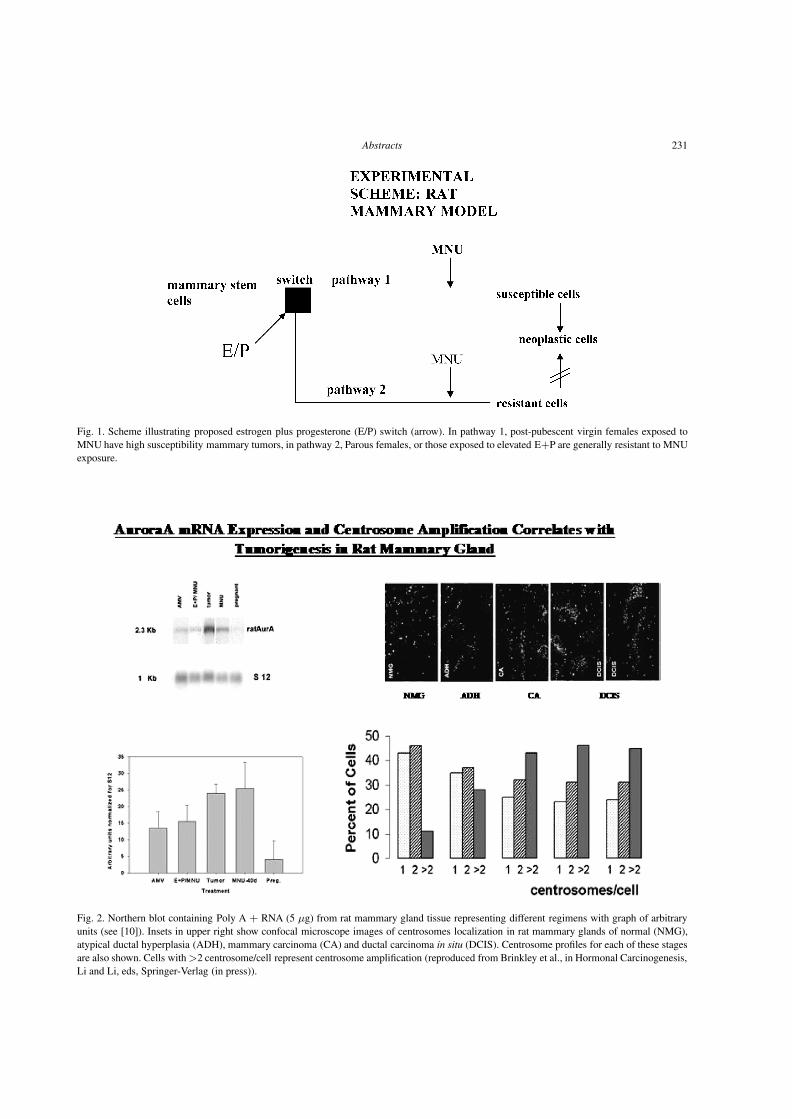
Abstracts 231
Fig. 1. Scheme illustrating proposed estrogen plus progesterone (E/P) switch (arrow). In pathway 1, post-pubescent virgin females exposed toMNU have high susceptibility mammary tumors, in pathway 2, Parous females, or those exposed to elevated E+P are generally resistant to MNUexposure.
Fig. 2. Northern blot containing Poly A + RNA (5 µg) from rat mammary gland tissue representing different regimens with graph of arbitraryunits (see [10]). Insets in upper right show confocal microscope images of centrosomes localization in rat mammary glands of normal (NMG),atypical ductal hyperplasia (ADH), mammary carcinoma (CA) and ductal carcinoma in situ (DCIS). Centrosome profiles for each of these stagesare also shown. Cells with >2 centrosome/cell represent centrosome amplification (reproduced from Brinkley et al., in Hormonal Carcinogenesis,Li and Li, eds, Springer-Verlag (in press)).
232 Abstracts
uating these results, but they raise the interesting pos-sibility that centrosome amplification, and subsequentaneuploidy, correlate with chromosome instability, butmay not always be a prelude to the clonal outgrowth oftumor cells.
4. Conclusions
Centrosome amplification and elevated expressionof the serine/threonine kinase aurora A are consistentand early events in the onset and progression of cells toaneuploidy. Although the mechanism providing a di-rect link between defects in the mitotic apparatus andaneuploidy has yet to be fully elucidated in tumor cells,there is a growing list of molecular components andregulatory processes that make the mitotic machinerya prime target for tumorigenesis In all model systemsstudied thus far, with the possible exception of MNU-induced mammary tumorigenesis, centrosome amplifi-cation appears to correlate directly with aurora A over-expression, aneuploidy and cancer.
Acknowledgements
Supported, in part, by grants from NIH CA-41424to WRB and PO1 CA-64255 to D Medina.
References
[1] Salisbury, J.L., Whitehead, C.M., Lingle, W.L., and Barrett,S.L. Centrosomes and cancer. Biol Cell, 91: 451–460, 1999.
[2] Lingle, W.L., Barrett, S.L., Negron, V.C., D’Assoro, A.B.,Boeneman, K., Liu, W., Whitehead, C.M., Reynolds, C., andSalisbury, J.L. Centrosome amplification drives chromosomalinstability in breast tumor development. Proc Natl Acad SciUSA, 99: 1978–1983, 2002.
[3] Boveri, T.H. Zur Frage der Entstehung maligner Tumoren. 1–64, 1914.
[4] Brinkley, B.R. and Goepfert, T.M. Supernumerary centrosomesand cancer: Boveri’s hypothesis resurrected. Cell Motil Cy-toskeleton, 41: 281–288, 1998.
[5] Nigg, E.A. Centrosome aberrations: cause or consequence ofcancer progression? Nat Rev Cancer, 2: 815–825, 2002.
[6] Katayama, H., Brinkley, W.R., and Sen, S. The Aurora kinases:role in cell transformation and tumorigenesis. Cancer Metasta-sis Rev, 22: 451–464, 2003.
[7] Zhou, H., Kuang, J., Zhong, L., Kuo, W.L., Gray, J.W.,Sahin, A., Brinkley, B.R., and Sen, S. Tumour amplified kinaseSTK15/BTAK induces centrosome amplification, aneuploidyand transformation. Nat Genet, 20: 189–193, 1998.
[8] Sen, S., Zhou, H., Zhang, R.D., Yoon, D.S., Vakar-Lopez, F.,Ito, S., Jiang, F., Johnston, D., Grossman, H.B., Ruifrok,A.C., Katz, R.L., Brinkley, W., and Czerniak, B. Amplifica-tion/overexpression of a mitotic kinase gene in human bladdercancer. J Natl Cancer Inst, 94: 1320–1329, 2002.
[9] Stenoien, D.L., Sen, S., Mancini, M.A., and Brinkley, B.R. Dy-namic association of a tumor amplified kinase, Aurora-A, withthe centrosome and mitotic spindle. Cell Motil Cytoskeleton,55: 134–146, 2003.
[10] Goepfert, T.M., Adigun, Y.E., Zhong, L., Gay, J., Medina, D.,and Brinkley, W.R. Centrosome amplification and overexpres-sion of aurora A are early events in rat mammary carcinogene-sis. Cancer Res, 62: 4115–4122, 2002.
[11] Sivaraman, L., Stephens, L.C., Markaverich, B.M., Clark, J.A.,Krnacik, S., Conneely, O.M., O’Malley, B.W., and Medina, D.Hormone-induced refractoriness to mammary carcinogenesisin Wistar-Furth rats. Carcinogenesis, 19: 1573–1581, 1998.
[12] Russo, J., Tay, L.K., and Russo, I.H. Differentiation of themammary gland and susceptibility to carcinogenesis. BreastCancer Res Treat, 2: 5–73, 1982.
[13] Grubbs, C.J., Farnell, D.R., Hill, D.L., and McDonough, K.C.Chemoprevention of N-nitroso-N-methylurea-induced mam-mary cancers by pretreatment with 17 beta-estradiol andprogesterone. J Natl Cancer Inst, 74: 927–931, 1985.
[14] Sinha, D.K., Pazik, J.E., and Dao, T.L. Prevention of mammarycarcinogenesis in rats by pregnancy: effect of full-term and in-terrupted pregnancy. Br J Cancer, 57: 390–394, 1988.
[15] Russo, I.H., Koszalka, M., and Russo, J. Comparative study ofthe influence of pregnancy and hormonal treatment on mam-mary carcinogenesis. Br J Cancer, 64: 481–484, 1991.
[16] Thordarson, G., Jin, E., Guzman, R.C., Swanson, S.M.,Nandi, S., and Talamantes, F. Refractoriness to mammary tu-morigenesis in parous rats: is it caused by persistent changesin the hormonal environment or permanent biochemical alter-ations in the mammary epithelia? Carcinogenesis, 16: 2847–2853, 1995.
[17] Sivaraman, L., Hilsenbeck, S.G., Zhong, L., Gay, J., Conneely,O.M., Medina, D., and O’Malley, B.W. Early exposure of therat mammary gland to estrogen and progesterone blocks co-localization of estrogen receptor expression and proliferation.J Endocrinol, 171: 75–83, 2001.
[18] Haag, J.D., Brasic, G.M., Shepel, L.A., Newton, M.A., Grubbs,C.J., Lubet, R.A., Kelloff, G.J., and Gould, M.N. A compara-tive analysis of allelic imbalance events in chemically inducedrat mammary, colon, and bladder tumors. Mol Carcinog, 24:47–56, 1999.

Abstracts 233
PATHOLOGIC TELOPHASES: SIGNIFICANTSOURCE OF INTERPHASE ANEUPLOIDY
Rüdiger G. SteinbeckDept. Oncology and Pathology, Karolinska Instituteand Hospital, 171 76 Stockholm, Sweden (Tel.: +49431-38901-11, E-mail: [email protected])
1. Introduction
The abnormal distribution of DNA values in inter-phase nuclei and the corresponding chromosome ane-uploidy was recognized being a problem in cancer re-search, when slide based microphotometry appeared.The proposal was raised that it would be of interest tomeasure precancerous lesions [1]. Indeed, such quan-titative cytochemical studies on normal and malignant,but most important also on premalignant cells fromthe human uterine cervix [2] were performed at Stock-holm’s Karolinska in 1964.
The correlation between “DNA aneuploidy” in inter-phase nuclei and cytogenetic aneuploidy on countablechromosomes was a crucial approach for further inves-tigations. Nils Atkin (1962) has pointed out that theploidy-level indicated by the modal interphase DNAvalue was in agreement with that he had obtained fromchromosome counts [3]. Furthermore, he concludedthat only those metaphases having the modal (stem-line) chromosome number regularly went on to com-plete division. Consequently, DNA measurements onanaphases and telophases are a more reliable indicatorof the stem-line than are measurements on prophasesand metaphases. The comparison of the DNA contentand chromosome number was a pivotal approach to re-alize genomic instability occurring in human tumori-genesis [4].
Here is to remember the year 1890, when DavidHansemann, then third assistant to Rudolf Virchowin Berlin, described asymmetric cell divisions in Ep-ithelkrebsen [5]. Chromatin bridges in anaphase segre-gation, lagging and sticky chromosomes caused obvi-ously unbalanced telophases. Twenty-four years later,Theodor Boveri has incorporated Hansemann’s find-ings into a general hypothesis on the origin of malig-nant tumors [6].
The focus of the present essay is to quantify the mi-totic failure in tumorigenesis. Not only the frequencyof karyokinesis is correlated to tumor pathology, butthe nuclear divisions also do not obey the equationalrule of mitosis. Instead, asymmetric anaphases andtelophases characterize dysplasia as well as cancer.
2. Methods
A variety of human tissues were diagnosed accord-ing to standards in pathology. Histological specimenswere from breast, uterine cervix, skin, oral mucosa,stomach and colon mucosa. Nuclear DNA contentswere determined by slide-based microphotometry onFeulgen stained sections of 8 µm and 15 µm for inter-phase nuclei and division figures, respectively. In anycase, the reliability of microphotometry depends on thethickness of the specimen (Figs 1, 2). We thoroughlyinvestigated the bias caused by inappropriate micro-tome adjustment [7].
Fig. 1. Feulgen DNA from interphase nuclei in 8 µm (") and 4 µm(F) sections, each compared with replica imprints. Mean ploidy of150 nuclei was recorded from 21 breast cancers.
Fig. 2. Depth requirements for mitotic divisions as seen from 8 µmand 15 µm sections. Feulgen stained specimens from oral mucosa:Epu – epulis, PG – pyo-genic granuloma, Ulc – common ulcus;P – prophase, M – metaphase, T – both halves of telophase.

234 Abstracts
3. Results
Premalignant and malignant lesions exhibit a wealthof nuclear divisions. Their morphology might be closeto normal mitoses. However, microphotometry revealsthat these figures possess abnormal DNA contents be-ing therefore pathologic entities (Fig. 3). Multipolar di-vision figures might be much more striking, but occuronly in the range of 10−3 and do not make a decidinginfluence upon tumor growth.
Tumor progression was documented in nuclear di-vision figures by their quantitative aberrations from a
regular 4.0c DNA content. True mitoses were used asinternal standard either from normal mucosae or frominflamed tissues. Table 1 shows exemplary recordsfrom oral and colon mucosae [8]. The significance ofthe results was high-lightened by the mitotic activityin fetal liver, whereas many nuclei of the breast cancercell-line MDA231 contained more than 4.0c DNA.
According to the Hansemann–Boveri hypothesis,a tumor originates from mutated nuclei that segregatetheir chromosomal aberrations into the daughter prod-ucts. The injury may occur anywhere during the cell
Fig. 3. Imprint cytology from breast cancer, HE stained. Specimens were destained and subject to Feulgen procedure for microphotometry.Prophase 6.1c (left), metaphase 7.2c, anaphase 5.2c. Bar: 10 µm.
Table 1
Frequency distribution of DNA content in nuclear divisions
Histology (n1/n2) P M T 4.5c ER 5.0c ER
(%) (%) (%) (%) (%)
Oral mucosa
Pyogenic granuloma 18/457 42.7 46.2 11.1 0.8 ± 0.5 0
LGD 14/125 30.4 56.0 13.6 4.2 ± 2.4 0
HGD 15/341 35.2 60.1 4.7 56.6 ± 7.6 40.6± 6.8
Ca 25/940 33.9 60.8 5.3 73.3 ± 4.4 57.9± 4.6
Colon mucosa
Normal mucosa 71/5 52.1 42.3 5.3 0 0
Bacterial colitis 34/322 37.3 46.6 16.1 1.4 ± 0.8 0.2± 0.2
Ulcerative colitis
LGD 93/850 51.0 36.4 12.6 23.6 ± 1.8 5.5± 0.9
HGD 22/246 44.3 39.8 15.9 43.2 ± 5.5 22.0± 6.0
Colon adenoma
LGD 14/425 46.6 39.1 14.3 7.9 ± 2.0 2.8± 1.3
HGD 12/498 44.8 42.4 13.0 25.8 ± 4.4 16.2± 4.1
Ca 16/538 52.4 39.2 8.4 62.1 ± 4.4 46.3± 7.0
Control
Fetal liver 18/456 42.6 43.6 13.8 0 0
MDA231 2/167 34.1 46.1 19.8 41.4 ± 5.6 21.6± 3.7P – prophase, M – metaphase, T – both halves of telophase. ER – exceeding rate, c – genome equiv-alent; n1 – number of cases; n2 – divisions. LGD – low-grade, HGD – high-grade dysplasia. Scatterwas standard error of the mean.
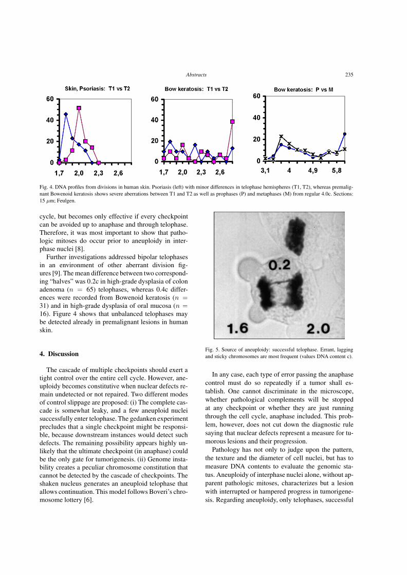
Abstracts 235
Fig. 4. DNA profiles from divisions in human skin. Psoriasis (left) with minor differences in telophase hemispheres (T1, T2), whereas premalig-nant Bowenoid keratosis shows severe aberrations between T1 and T2 as well as prophases (P) and metaphases (M) from regular 4.0c. Sections:15 µm; Feulgen.
cycle, but becomes only effective if every checkpointcan be avoided up to anaphase and through telophase.Therefore, it was most important to show that patho-logic mitoses do occur prior to aneuploidy in inter-phase nuclei [8].
Further investigations addressed bipolar telophasesin an environment of other aberrant division fig-ures [9]. The mean difference between two correspond-ing “halves” was 0.2c in high-grade dysplasia of colonadenoma (n = 65) telophases, whereas 0.4c differ-ences were recorded from Bowenoid keratosis (n =31) and in high-grade dysplasia of oral mucosa (n =16). Figure 4 shows that unbalanced telophases maybe detected already in premalignant lesions in humanskin.
4. Discussion
The cascade of multiple checkpoints should exert atight control over the entire cell cycle. However, ane-uploidy becomes constitutive when nuclear defects re-main undetected or not repaired. Two different modesof control slippage are proposed: (i) The complete cas-cade is somewhat leaky, and a few aneuploid nucleisuccessfully enter telophase. The gedanken experimentprecludes that a single checkpoint might be responsi-ble, because downstream instances would detect suchdefects. The remaining possibility appears highly un-likely that the ultimate checkpoint (in anaphase) couldbe the only gate for tumorigenesis. (ii) Genome insta-bility creates a peculiar chromosome constitution thatcannot be detected by the cascade of checkpoints. Theshaken nucleus generates an aneuploid telophase thatallows continuation. This model follows Boveri’s chro-mosome lottery [6].
Fig. 5. Source of aneuploidy: successful telophase. Errant, laggingand sticky chromosomes are most frequent (values DNA content c).
In any case, each type of error passing the anaphasecontrol must do so repeatedly if a tumor shall es-tablish. One cannot discriminate in the microscope,whether pathological complements will be stoppedat any checkpoint or whether they are just runningthrough the cell cycle, anaphase included. This prob-lem, however, does not cut down the diagnostic rulesaying that nuclear defects represent a measure for tu-morous lesions and their progression.
Pathology has not only to judge upon the pattern,the texture and the diameter of cell nuclei, but has tomeasure DNA contents to evaluate the genomic sta-tus. Aneuploidy of interphase nuclei alone, without ap-parent pathologic mitoses, characterizes but a lesionwith interrupted or hampered progress in tumorigene-sis. Regarding aneuploidy, only telophases, successful

236 Abstracts
in clonal selection, drive tumor development and theswitch to malignancy.
References
[1] Sandritter W. Methods and results in quantitative cytochemistry.In: Wied G.L., ed. Introduction to quantitative cytochemistry, vol1. New York: Academic Press, 159–182, 1966.
[2] Caspersson O.: Quantitative cytochemical studies on normal,malignant, premalignant and atypical cell populations from thehuman uterine cervix. Acta Cytol. 8: 45–60, 1964.
[3] Atkin N. The relationship between the deoxyribonucleic acidcontent and the ploidy of human tumours. Cytogenet 1: 113–122, 1962.
[4] Atkin N.B., Mattinson G., Baker M.C. A comparison of theDNA content and chromosome number of fifty human tumours.Brit J Cancer 20: 87–101, 1966.
[5] Hansemann D. Ueber asymmetrische Zelltheilung in Ep-ithelkrebsen und deren biologische Bedeutung. Arch PatholAnat Physiol Klin Med 119: 299–326, 1890.
[6] Boveri T. Zur Frage der Entstehung maligner Tumoren. G. Fis-cher, Jena, 1914.
[7] Steinbeck R.G., Auer G.U., Zetterberg AD. Reliability and sig-nificance of DNA measurements in interphase nuclei and divi-sion figures in histological sections. Eur J Cancer 35: 787–795,1999.
[8] Steinbeck R.G. The DNA content of chromosome division fig-ures and interphase nuclei classifies ulcerative colitis. Eur J Can-cer 34: 175–181, 1998.
[9] Steinbeck R.G. Imbalance of morphologically addressedtelophases reflects interphase DNA aneuploidy in tumorigenesis.Eur J Histochem 41: 243–254, 1997.

Abstracts 237
THE CLINICAL CONSEQUENCES OFMASSIVE GENOMIC IMBALANCES INCANCERS
George L. Gabor Miklos1,2 and Ryszard Maleszka3
1Chief Scientific Officer and Vice President, HumanGenetic Signatures Pty Limited (E-mail: [email protected]), 2Director, Secure Genetics PtyLimited, 81 Bynya Road, Palm Beach, Sydney, NSW,Australia, 2108 (E-mail: [email protected],Tel.: +61 2 9974 3000, Fax: +61 2 9974 3111), 3VisualSciences, Research School of Biological Sciences, Aus-tralian National University, Canberra, ACT, Australia,2600
We examined genomes of cancerous cells in termsof microarray data, DNA methylation, multidrug re-sistance and renewed attempts at generating cancercells by oncogenic transformation [1]. Most cancer-ous genomes are non-diploid, rearranged and vari-ably methylated [2–14]. The data highlight the im-portance of the fraction of any cancerous genome un-dergoing differential RNA expression [4], the impor-tance of genetic background on the cancerous pheno-type [15] and the importance of stochastic gene ex-pression in network fluxes [16–18]. The data provideinsights into, (i) cancer data transfer from mouse tohuman, (ii) methylation data transfer from cancerouscell lines to clinical samples, and (iii) the concept thatmutations in a handful of oncogenes, tumor suppres-sor genes, repair genes and cell cycle genes are theprime movers in the genesis of cancer [5,14,19]. Giventhe exceedingly poor record of new cancer drug de-velopments based on gene selection from functionalgenomics approaches, future therapeutic avenues willneed to move to methylation-based gene regulatorynetworks operating from grossly imbalanced genomes.
1. Genomic technologies
Recent technological advances are inundating uswith data [20]. At the genomic level, RepresentationalOligonucleotide Microarray Analysis is providing dataon genomic regions with altered copy number [21].At the level of the methylated genome (the methy-lome) emerging technologies report on the methylationstatus of selected CpG islands, promoters, repetitiveelements and histone modifications [9]. At the tran-scriptional level, microarrays, Serial Analysis of GeneExpression, Massively Parallel Signature Sequenc-
ing, BeadArrayTM and Mass Spectrometry-based ap-proaches generate avalanches of data. At the level ofproteomes, automated multidimensional liquid chro-matography coupled to mass spectrometry, (LC-LC-MS-MS) and 2D gel platforms interfaced with multi-compartment electrolysers provide data on protein pro-files. At the immunohistochemical level, high through-put robotic imaging technology, integrated with bioin-formatic deconvolution, is allowing insights into cellu-lar aspects of the cancerous state [22].
Genome-wide data are fed into bioinformatics pipe-lines in which an ever increasing number of algo-rithmic protocols produce Molecular Portraits of Can-cers [23,24]. Further analysis, however, quickly con-centrates on only a handful of gene products which aresignificantly increased or decreased in tumors, as theseare believed to drive the etiology and pathogenesis ofcancers [25]. The most important of these are thoughtto be oncogenes, tumor suppressors, repair genes andcell cycle genes [1,14,19,25,26], which are, withoutexception, all prioritized as potential targets for thetherapeutic pipeline.
2. Genomic status of cancer cells; cytogenetics
While genomic imbalances have always been diag-nostic for solid tumors, recent data on multiple myelo-mas extends this near ubiquity of aneuploidy andnonrandom recurrent chromosomal abnormalities tohematological neoplasms [27]. Each row of this figureis the chromosomal constitution of an individual pa-tient, and each column represents a departure from thediploid condition, with trisomies being shown in yel-low and monosomies in red. In addition, a large propor-tion of patients with hypodiploidy and near-tetraploidyalso carry a translocation involving the immunoglobu-lin heavy chain locus, IgH and these translocations arefrequently unbalanced and associated with monosomyelsewhere in the genome [28]. Thus massive changesin gene dosage are a hallmark of these hematologicneoplasms.
3. Genomic status of cancer cells; microarrays
Microarray analysis of cancerous tissue reveals mul-tiple regions of the genome with altered copy number.Primary breast tumors analyzed with a combination ofgenomic and mRNA expression microarrays allow si-multaneous detection of imbalances at both the DNA

238 Abstracts
and RNA levels. Amplifications of chromosomal re-gions are found in every breast cancer tumor and onevery human chromosome in at least one sample. Mostimportantly, a significant fraction of amplified genesare correspondingly more highly expressed [8]. Thusresidents of amplicons and deletions have their RNAoutputs determined by their nondiploid status, partlyby the altered regulatory networks of the chromosoma-lly corrupted cell and partly by the altered methylome.These RNA outputs impact upon proteomic networksin a nonlinear manner [29]. Given the genomic vari-ation between cancer cell populations and the nonlin-ear nature of transcriptional output to phenotype, it ishardly surprising that microarray studies of 300 breastcancers reveal that tumors with increased HER2 lev-els are as aggressive as those with normal HER2 lev-els [30].
4. Methylation
Single locus analyses reveal that certain regionsof cancerous genomes can be hypermethylated, hy-pomethylated or show no change [9]. The hyperme-thylation of CpG islands, for example, is believed tolead to the development and progression of all com-mon forms of human cancer, with the regulatory in-activation of tumor suppressors claimed to be an im-portant component. However, the data differ betweenmouse and human sources. There can be completelydifferent organizations of CpG island promoters be-tween homologous mouse and human genes [31] andmethylation in cell lines and primary malignancies areradically different. At least 60% of loci methylated incell lines are never methylated in primary tumors. Thuscancerous cell lines are a poor resource for identify-ing novel targets of DNA hypermethylation involved inoncogenesis [6]. The unresolved issue from the methy-lation characteristics of any genome is whether genesilencing is a causal event in the initiation of cancer, orwhether it is a by-product of the ongoing evolution ofthe tumor cell population.
5. Mouse to human
Although mice with a knockout of the catalyticgamma subunit of phosphatidylinositol-3-OH wereoriginally found to have a high incidence of colorectalcarcinomas, three subsequent independent knockoutsof the same gene, in different genetic backgrounds,
never developed colorectal carcinomas [15]. Similarlya mutant tumor suppressor (APC) in the C57BL/6strain has many polyps in the colon, whereas the samemutant tumor suppressor in the AKR strain has veryfew polyps. The universal finding is that the pheno-type depends on the background in which it is evalu-ated. In addition, knockouts of cancer-related genes inmice do not necessarily reproduce similar phenotypesto those from mutations in homologous human genes.Knockout of the mouse Wt1 gene does not predisposeto cancer whereas human WT1 does. Inherited muta-tions in mouse p53 do not predispose to breast can-cers, whereas human p53 does. Transgenic mice thatmodel Ras in humans do not give rise to the same tu-mors as in humans. Lastly, even though the Ras systemis thought to be highly conserved between differentspecies, microarray data reveal substantial differencesbetween rodents and humans in the signaling pathwaydownstream of Ras [1,32]. Conservation at the singlegene level between elements of a network is no guar-antee of conservation at the level of network fluxes andoutputs.
The fragility of classification schemes such as onco-genes and tumor suppressors is also exposed in dif-ferent organisms and in different genetic backgrounds.The prototypic oncogene, p53, was reclassified as a tu-mor suppressor after a decade of being an oncogene.Similarly, another oncogene of many years standing,Kras2, has now been shown to have tumor suppres-sor properties [33]. Mouse tumor suppressors such asSmad3, E2f1 and Mom1 have not been found to bemutated in any human cancer [26]. Even more im-portantly, however, recent data fail to replicate earlierwork on oncogenic transformation [1] and cast consid-erable doubt on the original mutational underpinningsof such transformation [34]. The notion that a smallnumber of key genes can produce permanent onco-genic transformation in normal diploid human cellsonce again rests on very insecure foundations. In con-trast, gross genomic imbalances and transformation arecorrelated in these newer data.
Finally, recent data on cyclin E and cyclin-dependentkinase 2 (CDK2) which for a decade have been held tobe pivotal in driving cancer cell proliferation, have re-vealed that knockouts and various inhibitory methodssuch as RNAi, antisense and dominant-negatives, allfail to stop mitosis in colon cancers, cervical cancersand osteosarcomas. Five recent papers totally demol-ish the long held view. McCormick, a leading cancerresearcher involved in generating some of these data,concludes that there’s no real evidence that CDK2 does

Abstracts 239
anything in somatic cells [35]. Clearly, researchers anddrug companies need to reevaluate drug developmentefforts aimed at any single cell cycle protein [35].
6. Genomic imbalances
The dominant paradigm that cancer cells arise fromnormal diploid cells through the acquisition of a se-ries of mutations in a handful of oncogenes, tumor sup-pressor genes, repair genes and genes impinging on thecell cycle, is in need of serious revision. The cleanesttest of this is afforded by the data showing that cancer-ous cells and aneuploid cell lines, but not diploid cells,rapidly become resistant to chemotherapeutic drugs.The additional finding that such cells spontaneouslyand rapidly revert their phenotype is galvanic in itsimplications [36]. Even if standard gene mutation ormethylation reprogramming were able to account forthe acquisition of multidrug resistance, either by inac-tivation of drug-metabolizing genes, or by activation ofmultidrug transporters, for example, the rapid sponta-neous reversion of such phenotypes in diploid contextsis near impossible. However understanding the rapidityof this reversion in terms of chromosomally unstableaneuploid genomes is straightforward [36].
The available data make it highly probable that theinitial event predisposing to a cancer is one that gen-erates a massive genomic imbalance involving hun-dreds of genes. In its simplest forms this is a sponta-neous nondisjunction event or a translocation. A singleautosomal nondisjunction event generates aneuploiddaughter cells and will be a huge shock to most reg-ulatory networks [2,4,7,11,13]. The resultant genomeswill now need to instantaneously cope with hundredsof extra copies of genes in either a trisomic or mono-somic condition. Similarly, a balanced translocation,while initially only effecting genes at its breakpoints,eventually becomes unbalanced [28,37], so hundredsof genes are again in a haploid state. Networks in theseaneuploid and unbalanced translocation situations areinitially pushed to either haplo- or triplo-thresholds.Compensatory nondisjunctions of nonhomologs willpartially alleviate such imbalances, a phenomenon welldescribed in yeast [38]. In addition, further mutationaland methylational changes occur in the chromosoma-lly corrupted genomes, but these are all secondary con-sequences of unbalanced systems attempting to reachsome form of stability. The clinical outcomes of ge-nomic imbalances at any given time will depend onthe initial event (intrinsic or extrinsic), that perturbed
the cellular circuitry, the order in which subsequentamplifications, deletions and translocations occur, thegenome-wide methylation profile, and the various poly-morphisms (both SNP and larger rearrangements) thatare unique to each individual and whose totality influ-ences the phenotypic outcome.
Huge changes in gene dosages involving hundredsof genes are of such enormous consequence that theyswamp most piecemeal gene-by-gene alterations. Inthe long term, aneuploid, segmentally aneuploid andunbalanced translocation-carrying cell populations be-come inexorably trapped in devastating transitionsfrom one unbalanced state to another [39]. Their ge-nomes become so fundamentally flawed in a grosschromosomal sense that there is no possibility of a re-turn to the initial diploid condition. It is self evidentthat drugs targeted against single genes will have van-ishingly low probabilities of rectifying such derangedregulatory circuitry.
The results of continuing genomic imbalances willmanifest clinically as extensive heterogeneity withinthe same tumor, heterogeneity between different tu-mors from the same patient and heterogeneity betweenpatients. Available data corroborate this. Microarraydata from three different areas of the same kidney tu-mor are instructive; the correlation among the expres-sion profiles for the three areas of the same tumor onlyexceeds 0.5 in two of the six patients sampled [40].Prostate cancer often has many distinct foci, only oneof which may be invasive and have a deleterious ef-fect on the patient [41]. The tumor variation betweenpatients requires no comment; a huge literature docu-ments the extensive between-individual variation.
These data on variation mean that no two can-cers will have exactly the same genomic imbalancesand rearrangements, nor the same methylome, even inmonozygotic twins. It is this variation that is pivotalto any cancerous cell population as it evolves alongits unique genomic trajectory, sometimes achieving ametastatic status and sometimes simply remaining be-nign. Recognizing the types of trajectories is the keydiagnostic on which therapeutic decisions can be basedfor the individual. Attempting to redirect trajectoriesfrom becoming potentially devastating to remainingbenign is a challenge in understanding networks.
In conclusion, with very few exceptions such asGleevec and hematological neoplasms, the approach ofthe last 30 years of focusing on single genes has beenmanifestly unproductive at delivering drug targets inthe solid tumors, which, after all, make up 90% of can-cers. It’s rarely the single gene that is the culprit, it’s the

240 Abstracts
imbalanced genome that is the problem, with networkperturbations changing from tumor to tumor. It wouldbe prudent for high priority therapeutic strategies to fo-cus on preventing somatic genomic imbalances and at-tempting to shunt cancerous cell populations to benigncul-de-sacs.
Three decades of cancer data reinforce one con-clusion; the prioritized single gene approach is as-suredly almost always doomed, both diagnosticallyand therapeutically. It’s legacy is continuing thera-peutic failures and unexpected complications, suchas the increase in brain cancers with women treatedwith Trastuzamab (Herceptin) for breast cancer [42].The genomic imbalance-network approach, by con-trast, faces up to the reality. The choice of approachesis stark indeed.
References
[1] Akagi, T. et al., Proc. Natl. Acad. Sci. USA 100, 13567–13572(2003).
[2] Li, R. et al., Proc. Natl. Acad. Sci. USA 94, 14506–14511(1997).
[3] Lengauer, C. et al., Nature 396, 643–649 (1998).
[4] Rasnick, D. and Duesberg, P.H. Biochem. J. 340, 621–630(1999).
[5] Hanahan, D. and Weinberg, R.A. Cell 100, 57–70 (2000).
[6] Smiraglia, D.J. et al., Hum. Mol. Genetics 10, 1413–1419(2001).
[7] Fabarius, A. et al., Proc. Natl. Acad. Sci. USA 99, 6778–6783(2002).
[8] Pollack, J.R. et al., Proc. Natl. Acad. Sci. USA 99, 12963–12968 (2002).
[9] Herman, J.G. and Baylin, S.B. N. Engl. J. Med. 349, 2042–2054 (2003).
[10] Fabarius, A. et al., Cancer Genetics and Cytogenetics 143, 59–72 (2003).
[11] Duesberg, P. and Li, R. Cell Cycle 2, A31–A39 (2003).
[12] Dai, Z. et al., Hum. Mol. Genetics 12, 791–801 (2003).
[13] Duesberg, P. et al., IUBMB (2004).
[14] Loeb, L.A. et al., Proc. Natl. Acad. Sci. USA 100, 776–781(2003).
[15] Barbier, M. et al., Nature 413, 796 (2001).
[16] Hume, D.A. BLOOD 96, 2323–2328 (2000).
[17] Cook, D.L. et al., Proc. Natl. Acad. Sci. USA 95, 15641–15646(1998).
[18] Blake, W.J. et al., Nature 422, 633–637 (2003).
[19] Vogelstein, B. et al., Nature 408, 307–310 (2000).
[20] The Chipping Forecast, Nature Genetics Supplement 32,(2002).
[21] Lucito, R., et al., Genome Research 13, 1–15 (2003)
[22] Schubert, W. et al. J. Theor. Medicine 4, 67–74 (2002).
[23] Perou, C.M. et al., Nature 406, 747–752 (2000).
[24] Chung, C.H. et al., Nature Genetics (Supplement) 32, 533–540(2002).
[25] Zhang, L. et al., Science 276, 1268–1272 (1997).
[26] Macleod, K. Curr. Opin. Genet. Dev. 10, 81–93 (2000).
[27] Fonseca, R. et al., Blood 102, 2562–2567 (2003).
[28] Fonseca, R. et al., Blood 101, 4569–4575 (2003).
[29] Miklos, G.L.G. and Maleszka, R. Proteomics 1, 30–41 (2001).
[30] Camp, R.L. et al., Cancer Research 63, 1445–1448 (2003).
[31] Cuadrado, M. et al., EMBO Reports 21, 586–592 (2001).
[32] Hamad, N.M. et al., Genes and Development 16, 2045–2057(2002).
[33] Zhang, Z. et al., Nature Genetics 29, 25–33 (2001).
[34] Zimonjic, D. et al., Cancer Research 61, 8838–8844 (2001).
[35] Steinberg, D. www.the-scientist.com/yr2003/dec/research1_0301201 (2003).
[36] Duesberg, P. et al., Proc. Natl. Acad. Sci USA 98, 11283–11288(2001).
[37] Hayman, S.R. et al., Blood 98, 2266–2268 (2001).
[38] Hughes, T.R. et al., Nature Genetics 25, 333–337 (2000).
[39] Stock, R.P. and Bialy, H. Nature Biotechnology 21, 13–14(2003).
[40] Vasselli, J.R. et al., Proc. Natl. Acad. Sci. USA 100, 6958–6963(2003).
[41] Masters, J.R.W. and Lakhani, S.R. Nature 404, 921 (2000).
[42] Bendell, J.C. et al., Cancer 97, 2972–2979 (2003).
Abstracts 241
DRUG RESISTANCE-SPECIFIC ANEUSOMIESOF HUMAN COLON AND BREAST CANCERCELLS
Ruhong Li and Peter DuesbergDepartment of Molecular and Cell Biology, DonnerLab, University of California-Berkeley, Berkeley, CA94720, USA
We have observed previously that only aneuploidand tumorigenic, but not diploid rodent cells acquireand lose drug-resistance at high rates [1,2]. In view ofthis we have proposed that specific assortments and re-arrangements of chromosomes, altering the dosage ofthousands of regulatory and structural genes, may gen-erate drug-resistance. The proposal is based on the factthat aneuploidy destabilizes the karyotype autocatalyt-ically and thus generates numerous new chromosomecombinations and reassortments, of which some mayconfer resistance to cytotoxic drugs [3]. But the pre-dicted resistance-specific aneusomies were not foundin the drug-resistant rodent cells, because the resis-tant cells were not karyotyped and because their chro-mosomes were too unstable to detect specific aneu-ploidies numerically. In the meantime we have foundthat the karyotypes of several human colon and breast
cancer cell lines are much more stable than those oequally aneuploid rodent lines, a feature that wouldfacilitate the detection of specific aneusomies. There-fore, we have now selected several puromycin-resistantclones from these human cell lines in an effort tofind the predicted drug resistance-specific aneusomies.Here we report that, indeed, 3 out of 3 puromycin-resistant clones of colon cancer lines, and that 1 outof 1 puromycin-resistant clone of a breast cancer linecontained each 2 to 3 resistance-specific aneusomies.We conclude that specific aneusomies are necessary, ifnot sufficient to generate drug-resistant phenotypes.
References
[1] Duesberg, P., Stindl, R., and Hehlmann, R. Explaining the highmutation rates of cancer cells to drug and multidrug resistanceby chromosome reassortments that are catalyzed by aneuploidy.Proc. Natl. Acad. Sci. USA, 97: 14295–14300, 2000.
[2] Duesberg, P., Stindl, R., and Hehlmann, R. Origin of multidrugresistance in cells with and without multidrug resistance genes:Chromosome reassortments catalyzed by aneuploidy. Proc. Natl.Acad. Sci. USA, 98: 11283–11288, 2001.
[3] Duesberg, P. and Li, R. Multistep carcinogenesis: a chain reac-tion of aneuploidizations. Cell Cycle, 2: 202–210, 2003.

242 Abstracts
IN SITU QUANTIFICATION OF GENOMEINSTABILITY IN BREAST TUMORS
Koei Chin1, Carlos Ortiz de Solarzano1, Sheryl Krig2,Colin Collins1, Martha Stampfer2, Paul Yaswen2,Stephen Lockett2 and Joe Gray1,2
1Comprehensive Cancer Center and Laboratory Medi-cine, University of California, San Francisco, SanFrancisco, CA 94143-0808, USA (Tel.: +1 415 476-3630, Fax: +1 415 476-8218, E-mail: [email protected]), 2Lawrence Berkeley National Laboratory, LifeSciences Division, Berkeley, CA 94720, USA
1. Introduction
Genome instability is one of key features in breastand other solid tumors, contributes to the accumula-tion and the selection needed for their progression.However, the little is known about the onset and ac-tual level of instability during cancer progression. Toaddress this, we assessed copy number variation oftwo different target loci in genome using dual-colorfluorescence in situ hybridization (FISH) and three-dimensional (3D) image analysis. We quantified thelevel of genome instability by enumerating actual copynumber of FISH signals in ∼40 µm thick sections atvarious stages of breast cancers. Our analysis exhib-ited low instability in normal skin, normal duct andusual ductal hyperplasia (UDH) in breast. However,instability was remarkably high in ductal carcinomain situ (DCIS) and somewhat lower in invasive can-cer (IC). DNA content in DCIS and IC was also in-creased by measuring nuclei in 3D images. To con-firm this process in vitro, we quantified copy numberof same loci using dual-color FISH and measured aver-age of chromosome composition by array based com-parative genomic hybridization (CGH) at several pas-sages of ZNF217-mediated immortalized human mam-mary epithelial cells (HMEC). We also assessed telom-ere events to reveal the correlation between instabilityand telomere dysfunction (crisis). The in vitro analysissuggested that instability sharply increased at the tran-sition from UDH to DCIS by passing through telom-ere crisis and was reduced in IC by telomerase reacti-vation.
2. Methods
2.1. Tissue processing and microscope imaging
Frozen tissues were obtained from UCSF Breast On-cology Tissue Repository. The tissues were evaluatedin H&E sections and cut in 20 ∼ 40 µm. Dual-colorFISH to sections were performed for the centromereof chromosome 1 (1c) and chromosome 20q13.2 (20q)with DNA counterstaining by YO-PRO-1. The probeswere labeled with Alexa568 and Cy5, respectively.Several areas per section were imaged using a con-focal microscope (Zeiss LSM410) with a 63× objec-tive. Three consecutive scans were executed per imag-ing with 488, 568 and 633nm lasers, corresponding toYO-PRO-1, Alexa568 and Cy5. The images typicallycarry 512× 512× 100 voxels with 0.2× 0.2× 0.3 µmresolution.
2.2. Image analysis and statistics
A nuclear segmentation and FISH signal detectionin 3D images were semiautomatically executed usinga custom program developed in house. Briefly, nucleiwere delineated as high intensity regions against low(background) intensity regions and visually assessedto detect clusters of nuclei. The clustered nuclei weresubdivided and the voxels comprising nucleus weresummed as DNA content. FISH signals of 1c and 20qin each segmented nucleus were detected similarly.The analytical accuracy was established by a statisti-cal model that assumed random loss of true signals andrandom detection of spurious signals in all normal ar-eas (a total of 537 nuclei). Linear regression analysiswas used to assess correlations between copy numberof FISH signals and nuclear volume.
2.3. HMEC analysis
ZNF217-mediated immortalized HMECs were usedfor genomic analysis at several passages through telom-ere crisis. The chromosome composition was assessedusing array CGH and the copy number of 1c and20q was measured using dual-color FISH as describedabove. Telomere length and telomerase activity at eachpassage were measured by Southern blot and TRAPassay analyses, respectively.

Abstracts 243
3. Results
The statistical analysis in normal tissue indicatedthat 93 ± 4% of genuine FISH signals were correctlydetected, with a 4 ± 4% probability of an spurious sig-nal being incorrectly detected. The results are similarto those previously reported for disaggregated nuclei.Therefore our segmentation is accurate enough for theanalysis of copy number changes. The level of insta-bility in normal ductal epithelium, UDH, DCIS and ICwas calculated as the standard deviation of the copynumber enumerated in several images of each tissue.The level of instability in the HMEC cells was simi-larly estimated at several passages before, during andafter immortalization. One UDH sample presented aunimodal bivariant distribution which however, a sig-nificant number of randomly distributed nuclei (22%,p < 0.001) with a single copy of chromosome. Allthree DCIS cases exhibited an extremely high level ofinstability. One DCIS case showed strong correlationbetween copy number and nuclear volume, suggest-ing concomitant polyploidy. Spatial statistical analysisof all three samples exhibited that nuclei with signif-icantly different chromosomal composition were ran-domly mixed, suggesting that the variation of genecopy was caused by genome instability, but not bystem cell like clonal expansion. Analysis of IC showedsomewhat lower level of instability in all cases withareas of both normal and abnormal ploidy. Cells wereagain randomly mixed. The analysis of HMEC usingFISH and CGH revealed increased instability duringtelomere dysfunction (crisis) at passages 21–22 fol-lowed by slightly decreased instability after telomerasereactivation.
4. Discussion
Our study showed a significant increase in the levelof instability at the transition from hyperplasia to DCIScarrying malignant phenotype. Instability slightly de-creased in IC, may suggest that some of IC are com-posed of a clonal subpopulation selected from cellsin DCIS with a proliferative advantage. HMEC analy-sis showed a similar increasing of instability duringtelomeric crisis, suggesting critical telomere shorten-ing caused genomic instability. Probably the similarprocess occurs in vivo at the transition from UDH toDCIS. IC would be formed by further selection of cellswith strong survival and/or proliferative factors and byclonal expansion of selected cells with telomerase re-activation. Therefore deranged cancer genome in ICcould be little stabilized and could retain a lethal ma-lignancy.
5. Conclusion
We have demonstrated how FISH to thick sectionscan be effectively used for in situ analysis of genomeinstability. By using our system, we were able to quan-tify the level of genome instability at the accumula-tion of chromosomal aberration in intact cells. Ourstudy suggested the mechanism of genomic instabil-ity in DCIS and slightly reduced instability in IC al-though further assessment is required to confirm these.By measuring telomere repeat and telomerase activityin vivo, we would demonstrate our hypothesis that thetransition to DCIS in fact coincides with telomere dys-function.

244 Abstracts
CENTROSOME GENES INVOLVED IN CELLCYCLE PROGRESSION, CYTOKINESIS,SPINDLE FUNCTION AND THE FIDELITY OFCHROMOSOME SEGREGATION
Stephen Doxsey, German Pihan and Aruna PurohitProgram in Molecular Medicine University of Massa-chusetts Medical School, Worcester, MA 01605, USA(Tel.: +1 508 856-1613, Fax: +1 508 856-4289,E-mail: [email protected])
Centrosomes were named by Theodor Boveri over100 years ago because they were discrete structuresfound at the center of the cell (hence the term centro-some or central body) (see [1]). Centrosomes are theonly nonmembranous organelles in vertebrate cells.They are 1–2 microns in diameter and lie in close prox-imity to the nucleus. Centrosomes are composed ofcentrioles and pericentriolar material (PCM, Fig. 1).Centrioles are barrel-shaped structures ∼0.5 micronslong and ∼0.2 microns in diameter. Nine sets of tripletmicrotubules make up the barrel. Other protein struc-tures are found both inside and outside centrioles(see [2]). Each centrosome has two centrioles that areoriented at right angles to one another. The older ma-ternal centriole has additional appendages that distin-guish it from the younger daughter centriole (see be-low).
The best-known function of the centrosome is itsability to nucleate the growth of new microtubules. Mi-crotubules are organized into astral arrays in interphasecells and form mitotic spindles in mitosis (Fig. 1, left).Microtubules nucleated from centrosomes at the polesof the mitotic spindle bind to chromosomes and po-sition them at the spindle center. As cells exit mito-sis, chromosomes move toward the spindle poles alongtracks formed by microtubules. This results in segrega-tion of chromosomes into two daughter cells followingcell division (cytokinesis).
Like chromosomes, centrosomes duplicate preciselyonce every cell cycle. The fidelity and timing of cen-trosome duplication is essential for ensuring that thisprocess is effectively coupled to other events such ascell cycle progression and DNA replication. Uncou-pling of these events can lead to excess centrosomesthat organize multipolar spindles (Fig. 1, right) or sin-gle centrosomes that organize monopolar spindles.
If centrosome number or function is perturbed, spin-dle abnormalities can result (see [2]). For example, su-pernumerary centrosomes can lead to the formation ofspindles with multiple poles (Fig. 1, right). In turn,multipolar spindles can segregate chromosomes intomore than two cell progeny (Fig. 2). Moreover, multi-directional forces exerted on a single chromosome ina multipolar spindle can create chromosome breaks(Fig. 2, bottom right). In other cases, failure to dupli-cate or separate centrosomes can lead to the forma-
Fig. 1. Centrosomes and mitotic spindle organization in normal and tumor cells. Immunofluorescent image of centrosomes in a nontumor cellthat organize a normal bipolar spindle in mitotis (left). A cell from a breast tumor showing excess centrosomes that organize a multipolar spindle(right) that induces chromosome missegregation aneuploidy. Images: Keith Mikule and Steve Doxsey.

Abstracts 245
Fig. 2. Chromosome missegregation in a human prostate tumor cell. PC-3 cell line constitutively expressing histone H2B fused to green fluores-cent protein (GFP) to label chromosomes. Cells were examined by fluorescence time lapse imaging using optical sectioning and deconvolutionto obtain three-dimensional images. Three-dimensional images are displayed as two-dimensional projections. Images: Agata Jurczyk and SteveDoxsey.
tion of monopolar spindles that are unable to segregatechromosomes resulting in cell division failure. In all ofthese scenarios, daughter cells would receive abnormalnumbers of chromosomes and become aneuploid.
Recently, we and others demonstrated that centro-somes are abnormal in nearly all human malignant car-cinomas (Figs 3, 4) [3,4]. We next addressed whethercentrosome defects were a secondary result of tumori-genesis or if they contributed to the process, perhapsthrough the induction of genetic instability. We firstasked whether centrosome defects were present in pre-cancerous lesions. To our surprise, we found that cen-trosome defects were present in 20–55% of precancer-ous lesions of the breast, cervix and prostate [5]. Ofgreat interest, was data showing that aberrant centro-somes were present in precisely the same lesions thatwere aneuploid. We also found that the centrosomeprotein pericentrin was elevated in many human carci-nomas [6].
A second approach that we used to address the roleof centrosome proteins in genetic instability was totest whether pericentrin could induce tumor-like fea-tures. We constructed cell lines stably overexpress-ing the protein and found that they showed centro-some defects, genetic instability, enhanced prolifera-
tion, growth in soft agar, loss of mitotic checkpointcontrol, changes in cell and nuclear morphology andabrogation of the mitotic checkpoint [6,7]. We recentlydemonstrated that pericentrin binds PKA, PKC, cyto-plasmic dynein and the gamma tubulin ring complex,and that specific disruption of pericentrin’s interactionwith some of these proteins induced a tumor-like phe-notype [7–11].
The third approach we took to address the role ofcentrosome proteins in genetic instability was to screenfor cancer-like features following centrosome gene si-lencing. We found that silencing of many centrosomegenes affected the fidelity of chromosome segrega-tion either through spindle dysfunction or cytokinesisfailure [12]. We unexpectedly found that silencing ofnearly all centrosome genes induced cell cycle arrest innormal cells but not in cells with abrogated checkpointfunction [12].
In our current model, altered centrosome proteinlevels cause centrosome defects that may contributeto tumorigenesis through the induction of genetic andcellular disorganization. Centrosome defects could actas epigenetic modifiers of the genome and togetherwith genetic mutations, provide a powerful drivingforce for increased genetic instability [13]. This condi-

246 Abstracts
Fig. 3. Centrosomes are abnormal in size, shape and number in human tumor cells. Abnormal centrosomes in a breast tumor cell (right, yellow)compared with a normal diploid cell (lower left). Newly nucleated microtubules (green) arise from a single centrosome in the nontumor cell(yellow, bottom left). In the tumor cell, multiple centrosomes of hetergeneous size, shape and number, nucleate microtubules (right) and contributeto the formation of multipolar spindles and chromosome missegregation (not shown). Blue, nuclei. Images: Steve Doxsey.
Fig. 4. Centrosomes are abnormal in size, shape and number in hu-man prostate tumors. Abnormal centrosomes in a prostate tumor(right, brown) compared with normal centrosomes in nontumor cells(left, arrowheads). Tissue section stained by immunoperoxidase forthe centrosome protein pericentrin and for hematoxylin and eosin.Note consistent size of centrosome in nontumor cells and abnormalcentrosome numbers, shapes and sizes in tumor tissue.
tion could accelerate accumulation of alleles carryingpro-oncogenic mutations and loss of alleles containingwild-type tumor suppressor genes, features character-istic of the most prevalent human cancers. The conse-quence of these events would be to create a larger poolof genetically altered cells from which to spawn clonalpopulations with greater survival potential. This ge-nomic plasticity could ultimately facilitate emergenceof gene dosage changes that are pro-tumorigenic [14].
References
[1] Wheatley, D.N. The Centriole. (Elsevier Biomedical Press,Amsterdam, 1982). 2.
[2] Doxsey, S.J. Re-evaluating centrosome function. Nature Re-views in Molecular Biology 2: 688–699, 2001.
[3] Pihan, G., Purohit, A., Knecht, H., Woda, B., Quesenberry, P.,and Doxsey, S.J. Centrosome defects and genetic instability inmalignant tumors. Cancer Research, 58: 3974–3985, 1998.
[4] Lingle, W.L., Lutz, W.H., Ingle, J.N., Maihle, N.J., and Sal-isbury, J.L. Centrosome hypertrophy in human breast tumors:implications for genomic stability and cell polarity. Proc. Natl.Acad. Sci., 95: 2950–2955, 1998.
[5] Pihan, G.A., Wallace, J., Zhou, Y., and Doxsey, S.J. Centro-some abnormalities and chromosome instability occur togetherin pre-invasive carcinomas. Cancer Res., 63: 1398–1404, 2003.
Abstracts 247
[6] Pihan, G.A., Purohit, A., Wallace, J., Malhotra, R., Liotta, L.,and Doxsey, S.J. Centrosome defects can account for cellularand genetic changes that characterize prostate cancer progres-sion. Cancer Res., 61: 2212–2219, 2001.
[7] Purohit, A., Tynan, S.H., Vallee, R., and Doxsey, S.J. Direct in-teraction of pericentrin with cytoplasmic dynein light interme-diate chain contributes to mitotic spindle organization. J. CellBiol., 147: 481–491, 1999.
[8] Dictenberg, J., Zimmerman, W., Sparks, C., Young, A.,Vidair, C., Zheng, Y., Carrington, W., Fay, F., and Doxsey, S.J.Pericentrin and gamma tubulin form a protein complex and areorganized into a novel lattice at the centrosome. J. Cell Biol.,141: 163–174, 1998.
[9] Young, A., Dictenberg, J., Purohit, A., Tuft, R., and Doxsey, S.Dynein-mediated assembly of pericentirn and gamma tubulinonto centrosomes. Mol. Biol. Cell, 11: 2047–2056, 2000.
[10] Diviani, D., Langeberg, L.K., Doxsey, S.J., and Scott, J.D. Peri-centrin anchors protein kinase A at the centrosome through a
newly identified RII-binding domain. Curr. Biol., 10: 417–420,2000.
[11] Chen, D., Purhit, A., Halilovic, E., Doxsey, S.J., and Newton,A.C. Centrosome anchoring of protein kinase C betaII by peri-centrin controls microtubule organization, spindle function andcytokinesis. J. Biol. Chem., in press, 2003.
[12] Gromley, A., Jurczyk, A., Sillibourne, J., Halilovic, E., Mo-gensen, M., Groisman, I., Blomberg, M., and Doxsey, S.A novel human protein of the maternal centriole is required forthe final stages of cytokinesis and entry into S phase. J. CellBiol., 161: 535–545, 2003.
[13] Pihan, G.A. and Doxsey, S.J. The mitotic machine as a sourceof genetic instability in cancer. Sem. Cancer Res., 9: 289–302,1999.
[14] Pihan, G. and Doxsey, S.J. Mutations and aneuploidy: co-conspirators in cancer? Cancer Cell, 4: 89–94, 2003.

248 Abstracts
TELOMERASE REACTIVATION ANDGENOMIC INSTABILITY DURINGIMMORTAL TRANSFORMATION OFCULTURED HUMAN MAMMARYEPITHELIAL CELL
Martha Stampfer1, James Garbe1, Koei Chin2,Collin Collins2, Joe Gray1,2, Fred Waldman2,Karen Swisshelm4, Thea Tlsty3 and Paul Yaswen1
1Lawrence Berkeley National Laboratory, Life Sci-ences Division, Berkeley, CA 94720, USA, 2CancerResearch Institute, University of California, San Fran-cisco, San Francisco, CA 94143-0808, USA, 3De-partment of Pathology, University of California, SanFrancisco, San Francisco, CA 94143-0506, USA,4Department of Pathology, University of Washington,Seattle WA 98195, USA (Tel.: +1 510 486-7273, Fax:+1 510 486-4475, E-mail: [email protected])
1. Introduction
Recent studies utilizing genetically engineered mice[1,2] and human tissues and cells [3–8] have ledto new models for the mechanisms underlying earlystage human carcinogenesis and genomic instabil-ity [1,7,9,10]. These models propose that progressivetelomere erosion with DNA replication, in the ab-sence of sufficient telomerase activity, eventually leadsto telomere dysfunction. The eroded telomeres areviewed as DNA damage, and if ligated by the non-homologous end-joining (NHEJ) pathway, can cre-ate end-to-end fusions and ongoing cycles of break-age/fusion/bridge (BFB). The resulting genomic insta-bility may inhibit or promote transformation, depend-ing upon the context (e.g., pre-existing mutations suchas in p53, cell type, and environmental conditions).This type of genomic instability can then be amelio-rated by re-expression of telomerase activity, whichmaintains telomeres at short, stable lengths, therebyconferring both an indefinite proliferative potential and“capped” telomeres no longer subject to NHEJ. Theimmortality conferred by telomerase reactivation isconsidered crucial for human carcinogenesis, since un-limited proliferative potential may be required to ac-cumulate the multiple rare errors necessary for malig-nancy and metastasis. The amelioration of genomic in-stability likely also contributes to carcinogenesis bymaintaining the viability of cells that have acquiredmalignancy-associated derangements.
Cells derived from normal somatic tissues of long-lived organisms such as humans exhibit extremelystringent repression of telomerase activity and cellu-lar senescence in vitro. In contrast, most human can-cers express telomerase activity. The errors that allowtelomerase reactivation to occur during human carcino-genesis have not been well defined. In order to un-derstand the mechanisms underlying immortal trans-formation and telomerase reactivation during humanbreast carcinogenesis, we have undertaken a long-termprogram to generate pathologically relevant models ofhuman mammary epithelial cell (HMEC) immortaliza-tion. Our in vitro studies support the model describedabove [8,11–17].
2. Methods and results
HMEC have been obtained from reduction mam-moplasty tissues showing no epithelial cell pathology.Cultured HMEC encounter two distinct proliferationbarriers that must be compromised for finite lifespanHMEC to achieve immortality. A first barrier (stasis)is RB-mediated in response to various stresses, can befrequently overcome by alterations in pathways gov-erning RB, and is not a response to critically shorttelomere lengths. Cells at stasis do not exhibit chromo-somal aberrations and artificial introduction of hTERT,the catalytic subunit of telomerase, does not provide agrowth advantage to these cell populations. In HMEC,stasis is associated with up-regulated expression ofthe cyclin-dependent kinase inhibitor p16INK4, a lowlabeling index (LI), and cell arrest in G1. HMECcultured in a serum-free medium can spontaneouslydown-regulate p16 expression, associated with methy-lation of the p16 promoter. The resulting p16(−) post-selection HMEC can grow for an additional 30–70population doublings (PD), with ongoing telomere ero-sion due to the absence of telomerase activity, beforeencountering a second proliferation barrier.
As they approach this second proliferation barrier(agonescence or crisis), post-selection HMEC showevidence of telomere dysfunction. Gross chromosomalaberrations are seen 10–20 population doublings be-fore cessation of net proliferation and include abun-dant telomeric associations, and chromosome fusionand breakage events. Cytogenetic analysis shows that100% of metaphases eventually exhibit structural ab-normalities. If p53 is functional, these aberrations in-duce a mostly viable arrest in all phases of the cell cy-cle, with a mean TRF (telomere restriction fragment)
Abstracts 249
length of ∼5 kb and a LI of ∼15%. Some mitotic fail-ures and cell death also occurs. This molecular phe-notype is distinct from stasis or crisis, prompting theuse of a new term “agonescence”, to distinguish itfrom the viable stasis arrest and the non-viable crisis.Agonescence is telomere length-dependent, and canbe efficiently circumvented by exogenous introductionof hTERT. HMEC arrested at both stasis and agones-cence exhibit a senescent morphology (large, flat,vacuolated cells) and express senescence-associatedβ-galactosidase activity, so these markers cannot beused to distinguish between these two distinct typesof senescence arrest. Crisis occurs in cells that havelost p53 function and thus p53-dependent checkpoints,and is characterized by massive cell death and a LIof ∼40%.
Spontaneous transformation to immortality has notbeen observed in cells derived from normal humanbreast tissues. We postulate that at least two alterationsare required to activate endogenous telomerase activityin cells that have overcome stasis. In cultured HMEC,the likelihood that all the necessary errors would occurin the same cell, even under conditions where wide-spread genomic errors are generated, is exceedinglysmall. However, if the telomere dysfunction barrier isapproached in cells already harboring one error predis-posing to immortality, the genomic instability resultingfrom telomere dysfunction can give rise to rare addi-tional complementary errors that allow reactivation ofendogenous telomerase activity and immortalization.We have overcome this stringent barrier and obtainedimmortally transformed HMEC lines by exposing cellscultured from normal tissues to a variety of pathologi-cally relevant immortalizing agents, e.g., chemical car-cinogens, oncogenes known to be overexpressed in hu-man breast cancers (c-Myc, ZNF217), and inhibitors ofp53 function. The rarity of immortal transformation incells exposed to these agents, and the clonal origins ofthe immortal lines, suggests that the pre-existing errorneeds to be complemented by additional error(s) gen-erated during the time of genomic instability.
Overcoming the telomere-length barrier in culturedHMEC appears to involve reactivation of telomerase,and amelioration of the prior genomic instability. How-ever, newly immortal p53(+) HMEC lines initially dis-play little or no detectable telomerase activity using aTRAP assay, due to the ability of p53 to repress telom-erase in newly immortal HMEC. When telomeres be-come extremely short (mean TRF <3 kb) an extendedprocess termed conversion ensues, wherein the p53-mediated repression of telomerase is gradually relieved
and telomerase activity gradually increases. ImmortalHMEC lacking functional p53 show initial telomeraseactivity. Fully immortal HMEC maintain telomeres atshort, stable lengths.
The genomic stability of p53(+) and p53(−) HMECtransitioning through telomere dysfunction and telom-erase reactivation has been examined by karyology,fluorescence in situ hybridization (FISH) and com-parative genomic hybridization (CGH). Instability wasmaximal during the period of telomere dysfunction,and was reduced thereafter in p53(+) lines. We haveobtained one carcinogen-treated p53(+) HMEC linethat immortalized prior to the onset of widespread ge-nomic instability (initial mean TRF >5 kb). Newly im-mortal populations of this line show continued telom-ere erosion until a mean TRF of 2–3 kb, without evi-dence of genomic instability as assessed by karyotypeanalysis and CGH. Recent studies suggest that verylow levels of telomerase activity are present in thenewly immortal cells and may preferentially maintainthe shortest telomeres. These short telomeres may con-sequently be present in a capped form not seen as dam-aged DNA, and thus not subject to NHEJ and the re-sulting BFB cycles.
3. Discussion
Our studies provide support for the model that asenescence barrier based upon telomere dysfunctiondrives chromosomal aberrations that can either restrainor promote further transformation, depending upon thecellular context. In most cells, telomere dysfunctionprovides a stringent limit to the number of replica-tions a single lineage can undergo, and thus limits theopportunity for deleterious mutations to accumulatewithin that lineage. However, rare mutations predis-posing to telomerase reactivation that do arise may becomplemented by chromosomal aberrations generatedby the telomere dysfunction occurring when telomeresbecome severely eroded. Our data suggest that over-coming telomere dysfunction involves reactivation oftelomerase, which maintains telomeric ends and pre-vents them from making telomeric associations.
The generation of widespread chromosomal aberra-tion at telomere-length dependent senescence may ac-count for some of the genomic instability seen in hu-man carcinomas, even those like breast cancer that arelargely p53(+). We hypothesize that the degree of ane-uploidy in transformed cell lines or tumors may dependupon the timing of the errors that allow telomerase re-

250 Abstracts
activation, rather than just on p53 status or other mu-tations that might specifically promote genomic insta-bility. Cells that accumulate all the errors necessary forimmortalization after the onset of telomere dysfunc-tion may harbor karyotypic derangements that get per-petuated by ongoing BFB cycles. Cells that acquire allthe necessary errors prior to the onset of telomere dys-function could avoid the resultant gross genomic in-stability and become immortal with near-diploid kary-otypes. This model is consistent with our small sampleof immortally transformed HMEC lines.
The relevance of our in vitro model to human breastcarcinogenesis is supported by recent in vivo observa-tions of early stages of cancer progression. Studies us-ing FISH to examine telomere length in human tumortissues and pre-malignant lesions, such as ductal carci-nomas in situ (DCIS), have shown that telomere short-ening is one of the earliest and most prevalent mole-cular changes in tumorigenesis [4,5,18]. Evidence ofgenomic instability is also first observed during earlystages of malignant progression of many organ types,including in breast DCIS [6,19,20]. While some fur-ther instability may occur during progression to pri-mary and metastatic disease, the major emergence ofinstability occurs at pre-malignant stages. Breast can-cers, like most epithelial-derived tumors, exhibit a highdegree of karyotypic complexity. Although the mannerin which genomic stability is compromised is presentlynot well defined, the type of instability shown, grosschromosomal rearrangements, is consistent with er-rors generated by double strand breaks, or NHEJ “re-pair” of uncapped telomeres. Assays for telomeraseactivity show that it too first becomes detected at thestage of DCIS (in ∼50%), and then increases to nearly100% of primary tumors [21–25]. These data, espe-cially their temporal order in carcinogenesis, are allconsistent with the model that short telomeres inducetelomere dysfunction, leading to genomic instability,which is then partially resolved by the reactivation oftelomerase.
Our data has been generated utilizing our uniquecollection of HMEC lines immortalized using onco-genic agents/molecular derangements associated withbreast cancer in vivo. We believe this is a critically im-portant consideration if in vitro studies are to be used asmodels for clinical interventions. Most in vitro immor-talization systems employ agents (e.g., SV40-T, HPV-E6/E7, H-ras) that have not been linked to breast can-cer pathogenesis. Additionally, determining the actualderangements that occur during carcinogenesis to re-activate telomerase and thus ameliorate genomic in-
stability can not be done in systems that bypass thisquestion by artificial introduction of hTERT. Further-more, short-lived animals do not exhibit stringent re-pression of telomerase even in normal tissues. Con-sequently, they normally express RB-mediated, non-telomere length dependent senescence, but not senes-cence associated with telomere dysfunction. We pos-tulate that overcoming telomere dysfunction may bea rate-limiting step in human carcinogenesis. Under-standing the mechanisms responsible for reactivationof endogenous telomerase during human carcinogene-sis, as well as assaying potential therapeutic agents thattarget telomerase activity, may require examination ofhuman cells in vitro and in vivo.
References
[1] Maser, R.S. and DePinho, R.A. Connecting chromosomes, cri-sis, and cancer. Science, 297: 565–569, 2002.
[2] Chang, S., Khoo, C.M., Naylor, M.L., Maser, R.S., and De-Pinho, R.A. Telomere-based crisis: functional differences be-tween telomerase activation and ALT in tumor progression.Genes & Devel, 17: 88–100, 2003.
[3] Gisselsson, D., Jonson, T., Petersen, A., Strombeck, B., DalCin, P., Hoglund, M., Mitelman, F., Mertens, F., and Man-dahl, N. Telomere dysfunction triggers extensive DNA frag-mentation and evolution of complex chromosome abnormali-ties in human malignant tumors. Proc. Natl. Acad. Sci. USA,98: 12683–12688, 2001.
[4] Meeker, A.K., Hicks, J.L., E.A., P., March, G.E., Bennett, C.J.,Delannoy, M.J., and De Marzo, A.M. Telomere shortening is anearly somatic DNA alteration in human prostate tumorigenesis.Cancer Res., 62: 6405–6409, 2002.
[5] Meeker, A.K., Beckman-Hicks, J.L., Argani, P., Iacobuzio-Donahue, C.A., Montgomery, E.A., Westra, W.H., Hansel,D.E., and De Marzo, A.M. Direct evidence of telomere short-ening and telomere length heterogeneity in human intraepitelialneoplasia (IEN). Proc. Am. Assoc. Cancer Res., 44: 1188,2003.
[6] Allred, D.C. and Mohsin, S.K. Biological features of premalig-nant disease in the human breast breast cancer. J. Mam. GlandBiol. Neoplasia, 5: 351–364, 2000.
[7] Albertson, D.G., Collins, C., McCormick, F., and Gray, J.W.Chromosome aberrations in solid tumors. Nat. Genet., 34: 369–376, 2003.
[8] Romanov, S., Kozakiewicz, K., Holst, C., Stampfer, M.R.,Haupt, L.M., and Tlsty, T. Normal human mammary epithelialcells spontaneously escape senescence and acquire genomicchanges. Nature, 409: 633–637, 2001.
[9] Desmaze, C., Soria, J.C., Freulet-Marriere, M.A., Mathieu, N.,and Sabatier, L. Telomere-driven genomic instability in cancercells. Cancer Lett., 194: 173–182, 2003.
[10] Feldser, D.M., Hackett, J.A., and Greider, C.W. Telomere dys-function and the initiation of genome instability. Nat. Rev. Can-cer, 3: 623–627, 2003.

Abstracts 251
[11] Stampfer, M.R. and Yaswen, P. Human epithelial cell immor-talization as a step in carcinogenesis. Cancer Lett., 194: 199–208, 2003.
[12] Stampfer, M.R. and Bartley, J.C. Induction of transformationand continuous cell lines from normal human mammary epithe-lial cells after exposure to benzo(a)pyrene. Proc. Natl. Acad.Sci. USA, 82: 2394–2398, 1985.
[13] Stampfer, M.R., Bodnar, A., Garbe, J., Wong, M., Pan, A.,Villeponteau, B., and Yaswen, P. Gradual phenotypic conver-sion associated with immortalization of cultured human mam-mary epithelial cells. Mol. Biol. Cell, 8: 2391–2405, 1997.
[14] Brenner, A.J., Stampfer, M.R., and Aldaz, C.M. Increasedp16INK4a expression with onset of senescence of humanmammary epithelial cells and extended growth capacity withinactivation. Oncogene, 17: 199–205, 1998.
[15] Stampfer, M., Garbe, J., Levine, G., Lichsteiner, S., Vasserot,A., and Yaswen, P. Expression of the telomerase catalytic sub-unit, hTERT, induces resistance to transforming growth factorb growth inhibition in p16INK4 (−) human mammary epithelialcells. Proc. Natl. Acad. Sci., USA, 98: 4498–4503, 2001.
[16] Nonet, G., Stampfer, M.R., Chin, K., Gray, J.W., Collins, C.C.,and Yaswen, P. The ZNF217 gene amplified in breast cancerspromotes immortalization of human mammary epithelial cells.Cancer Res., 61: 1250–1254, 2001.
[17] Stampfer, M.R., Garbe, J., Wigington, D., Nijjar, T., Swis-shelm, K., and Yaswen, P. Rapid conversion to telomerase(+)full immortality of indefinite lifespan human mammary ep-ithelial cell lines lacking functional p53. Oncogene, 22: 5238–5251, 2003.
[18] Meeker, A.K., Beckman-Hicks, J.L., Argani, P., and De Marzo,A.M. Dramatic telomere shortening occurs in breast luminalepithelial cells in subsets of non-malignant, normal-appearinglobules and small ducts: a potential early molecular mecha-nism underlying breast tumorigenesis. Proc. Am. Assoc. Can-cer Res., 44: 182, 2003.
[19] O’Connell, P., Pekkel, V., Fuqua, S.A., Osborne, C.K., Clark,G.M., and Allred, D.C. Analysis of loss of heterozygosity in399 premalignant breast lesions at 15 genetic loci. J. Natl. Can-cer Inst., 90: 697–703, 1998.
[20] Waldman, F.M., DeVries, S., Chew, K.L., Moore, D.H., 2nd,Kerlikowske, K., and Ljung, B.M. Chromosomal alterations inductal carcinomas in situ and their in situ recurrences. J. Natl.Cancer Inst., 92: 313–320, 2000.
[21] Hiyama, E., Gollahon, L., Kataoka, T., Kruoi, K.,Yokoyama, T., Gazdar, A.F., Hiyama, K., Piatyszek, M.A., andShay, J.W. Telomerase activity in human breast tumors. J. Nat.Cancer Inst., 88: 116–122, 1996.
[22] Bednarek, A., Sahin, A., Brenner, A.J., Johnston, D.A., and Al-daz, C.M. Analysis of telomerase activity in breast cancer: pos-itive detection at the in situ breast carcinoma stage. Clin. Can-cer Res., 3: 11–16, 1997.
[23] Sugino, T., Yoshida, K., Bolodeoku, J., Tahara, H., Buley,I., Manek, S., Wells, C., Goodison, S., Ide, T., Suzuki, T.,Tahara, E., and Tarin, D. Telomerase activity in human breastcancer and benign breast lesions: diagnostic applications inclinical specimens, including fine needle aspirates. Int. J. Can-cer, 69: 301–306, 1996.
[24] Shpitz, B., Zimlichman, S., Zemer, R., Bomstein, Y., Ze-havi, T., Liverant, S., Bernehim, J., Kaufman, Z., Klein, E.,Shapira, Y., and Klein, A. Telomerase activity in ductal carci-noma in situ of the breast. Breast Cancer Res. Treat., 58: 65–69, 1999.
[25] Poremba, C., Shroyer, K.R., Frost, M., Diallo, R., Fogt, F.,Schafer, K.-L., Burger, H., Shroyer, A.L., Dockhorn-Dworniczak, B., and Boecker, W. Telomerase is a highly sen-sitive and specific molecular marker in fine-needle aspirates ofbreast lesions. J. Clin. Onc., 17: 2020–2026, 1999.

252 Abstracts
MECHANISMS OF CHROMOSOMEINSTABILITY RESULTING FROMTELOMERE LOSS
Anthony W.I. Lo, Carl N. Sprung, Bijan Fouladi,Mehrdad Pedram, Laure Sabatier, Michelle Ricoul,Gloria E. Reynolds and John P. MurnaneRadiation Oncology Research Laboratory, Universityof California, San Francisco, and Laboratoire de Ra-diobiologie et Oncologie, Commissariat à l’EnergieAtomique, Fontenay-aux Roses, France
Telomere loss can result in chromosome rearrange-ments associated with tumorigenesis. Human tumorcells commonly demonstrate spontaneous telomereloss despite the expression of telomerase, suggest-ing fundamental deficiencies in the ability to prop-erly maintain telomeres. We have utilized selectablemarker genes located adjacent to a telomere to investi-gate the nature of the chromosome rearrangements re-sulting from telomere loss in both mouse embryonicstem (ES) cells (MCB 22:4836) and human tumor cells(Neoplasia 2:540; Neoplasia 4:531). Mouse ES cellshave a very low rate of spontanesous telomere loss, al-though telomere loss can be induced by transfectionof expression vectors containing the I-SceI endonu-clease, which specifically introduces a double-strandbreak at an 18-bp site located within the plasmid se-quences. In contrast, the human tumor cell line has ahigh rate of spontaneous telomere loss despite the pres-ence of telomerase activity, which is typical of manytumor cells due to their inability to properly maintaintelomeres. Telomere loss induced by the I-SceI en-donuclease in the mouse ES cells resulted primarily inthe addition of new telomeres at the site of the break,while telomere addition resulting from spontaneoustelomere loss was relatively infrequent event in the hu-man tumor cells. In both cell types, chromosomes thatdid not acquire a new telomere underwent sister chro-matid fusion and subsequent breakage/fusion/bridge(B/F/B) cycles. B/F/B cycles occur when the fusedsister chromatids break during anaphase as the twocentromeres are pulled in opposite directions, result-ing in a chromosome without a telomere in the nextcell cycle. Following DNA replication the sister chro-matids fuse again, repeating the cycle. The B/F/B cy-cles in mouse ES cells were relatively short, lasting
only a few cell cycles, while B/F/B cycles in humantumor cells continued for more than 20 cell genera-tions. In both cell types, B/F/B cycles resulted in am-plification of subtelomeric DNA sequences, and con-tinued until the chromosome acquired a new telomere.The most common mechanism for telomere acquisi-tion during B/F/B cycles in both cell types involvednonreciprocal translocations of the ends of other chro-mosomes. Although these translocations result in theacquisition of a new telomere and the stabilization ofthe marker chromosome, they transfer the instabilityto the chromosome that donated the telomere sinceit is now missing a telomere. Thus, chromosome in-stability resulting from the loss of a telomere can bepassed sequentially from one chromosome to another.Telomeres can also be acquired by duplication, pos-sibly involving break-induced replication, which un-like nonreciprocal translocation results in the net gainof a telomere and therefore results in stabilization ofthe genome. Direct addition of telomeres is a relativelyrare event during B/F/B cycles even in the mouse EScell line, and therefore direct addition appears to oc-cur primarily when a break occurs in close proximityto a telomere. These results demonstrate that telom-ere loss can result in amplification and nonreciprocaltranslocations commonly associated with human can-cer. Telomere loss can also lead to other forms of chro-mosome instability, including formation of double-minute chromosomes, complex rearrangements involv-ing other chromosomes, and aneuploidy. B/F/B cycleshave been shown to lead to the formation of double-minute chromosomes by looping out the amplified re-gions, which appear to be highly unstable, apparentlydue to the presence of the inverted repeat structures.Chromosomes involved in B/F/B cycles have were alsofound to fuse with other chromosomes, transferringthe amplified regions to these chromosomes followingbreakage of the dicentric chromosomes. Finally, B/F/Bcycles may lead to aneuploidy, since dicentric chromo-somes may cause a failure of cytokinesis resulting intetraploid cells that can progress to aneuploidy. B/F/Bcycles can therefore result in a wide variety of chro-mosome changes in cancer cells in addition to the typi-cal structures commonly expected for B/F/B cycles in-volving amplification of a gene on the end of the chro-mosome on which it was originally found.
Abstracts 253
ARE THERE DIFFERENT TYPES OF CANCERANEUPLOIDY?
D.C. Allison and A.L. NestorDepartments of Surgery, Physiology and MolecularMedicine, and the MCO Microscopy Imaging Center,Medical College of Ohio, 3035 Arlington Ave. Toledo,OH 43614-5804, USA (Tel.: +1 419 383-3970, Fax:+1 419 383-6230, E-mail: [email protected])
1. Introduction
Aneuploidy is produced by an initial (2C) to (4C)polyploidization followed by chromosome loses andrearrangements to widely varying numbers and typesof chromosomes. Aneuploidy formation has been mod-eled as a “random process” [1,2], implying that withcontinued division all cancers will eventually convergeto similar chromosome compositions [3]. Such ran-dom models, however, do not easily explain the widelyvarying clinical behavior of aneuploid human cancers:For example, aneuploid neuroblastomas often sponta-neously regress [4] while aneuploid pancreatic carci-nomas are almost invariably fatal [5]. A wide spectrumof clinical outcomes is found for other aneuploid tu-mors [6–33].
We hypothesize that this heterogeneity of clinicaloutcomes for patients with aneuploid tumors may bein part due to the existence of different types of aneu-ploidy, some of which favor destabilization of the can-cer genome and promote malignant progression andother types of aneuploidy which are more geneticallystable and produce relatively indolent tumors. In sup-port of this possibility, it has become recently apparentthat all of the steps involved in the formation of ane-uploid cells are controlled by diverse sets of gene net-works controlling DNA replication and repair and tran-sit through mitosis (reviewed in [34,35]). These genenetworks may provide a complex genetic substrate forthe production of different types of cancer aneuploidy:For example, the initial (2C) to (4C) tetraploidiza-tion step, felt to precede the formation of most ane-uploid cells, could be either due to aberrations in theDNA synthesis complete-checkpoint, the S/G2 DNA-damage checkpoint, the wait anaphase-checkpoint, thetelophase exit pathway, and other, as of yet unidenti-fied, genes responsible for mitotic transit in differentaneuploid precursor cells. The particular pathway uti-lized for producing tetraploidy in a given cancer mightexert a profound effect on its subsequent rate of chro-
mosome loss and genetic stability. The chromosomeloss or non-disjunction rate in a given tumor couldalso be influenced by various genetic defects in themechanisms responsible for chromosome attachmentto the mitotic spindle, the wait anaphase checkpoint,and so on. Finally, the number of recombinant and bro-ken chromosomes in a given aneuploid cancer may belargely a function of its specific combination of ge-netic defects in the G1/S-DNA damage checkpoint, thep53-controlled and/or other DNA-repair mechanisms,and abnormal activations, or sloppiness, of the chro-mosome recombination pathways, etc.
2. Methods
To test this hypothesis, karyotypes from the diploidCCD-34Lu fibroblast and the aneuploid A549 andSUIT-2 cancer lines underwent fluorescence in situhybridization (FISH) and DAPI-counterstaining. In-tact metaphase- (M), anaphase- (A) and telophase- (T)cells of these lines were also Feulgen-stained and in-dividual DNA contents measured by image-analysismicroscopy. The number of DAPI-stained and FISH-identified chromosomes, 1–22, X, Y, the M-, A-, andT-cell DNA measurements, and chromosome structuralabnormalities were compared using the chi-square,Mann–Whitney rank sum, and the Levene’s equality ofvariance tests.
3. Results
Virtually all of the evaluable diploid CCD-34Lukaryotypes had 46 chromosomes with two, normal ap-pearing homologs (Figs 1A, D). The aneuploid chro-mosome numbers per karyotype were highly variable,averaging 62 and 72 for the A549 and SUIT-2 lines, re-spectively (p < 0.01, Figs 1B, C). However, the A549chromosome numbers were more narrowly distrib-uted than the SUIT-2 karyotypes chromosome num-bers (p < 0.01). The averages and spreads of the in-tact M-cell DNA distributions exactly paralleled thoseof the DAPI-chromosome number distributions for thethree lines, ruling out inadvertent selection bias beingresponsible for the differences in the aneuploid chro-mosome counts. Measurement of the DNA differencesbetween intact A/T-cell chromosomal masses revealedthat the diploid CCD-34Lu cells always had balancedmitoses. However, the aneuploid A549 and SUIT-2cells often had uneven mitoses, with unequal partition-

254 Abstracts
Fig. 1. Localization of chromosome specific FISH probes in DAPI-stained karyotypes. The diploid CCD-34Lu (A), and aneuploid A549 (B)and SUIT-2 (C) cell lines were hybridized with specific centromeric and paint DNA probes specific for chromosomes (1 red, 16 green), (19red, 13 green) and (17 red, 4 green) respectively. (D) A composite karyotype of FISH-identified chromosomes of the diploid CCD-34Lu line.(E) FISH-localization’s of (a) extrachromosomal fragments (ECF’s) found in the aneuploid SUIT-2 line showing two normal centromeric local-ization’s of the FISH probes for chromosome 1 and in five ECF’s from the same karyotype. (b) Normal chromosome 19 paint localization nextto chromosome 19 breaks and refusions to another chromosomes(s) found in the same karyotype of the aneuploid A549 line. (c) Centromericlocalization of the FISH probe in a normal chromosome 2 and in several abnormally placed, telomeric regions classified as END’s in the samekaryotype of the A549 line. (d) Fusion’s found in both aneuploid cell lines with centromeric chromosome probes labeled in red (r) and green (g)from left to right: 18r/16g, 7r/10g, 20r/9g, and 18r/10g. (Reproduced, with permission from Isaka, T., Nestor, A.L., Takada, T. and Allison, D.C.Chromosomal variations within aneuploid cancer lines. J Histochem Cytochem, 51, 1343–53, 2003.)
Fig. 2. The average percentages of FISH-identified chromosome abnormalities per individual chromosome in the (A) A549 and (B) SUIT-2 lines.(Reproduced, with permission from Isaka, T., Nestor, A.L., Takada, T. and Allison, D.C. Chromosomal variations within aneuploid cancer lines.J Histochem Cytochem, 51, 1343–53, 2003.)

Abstracts 255
ing of DNA into the daughter chromosomal masses.However, uneven mitoses were more frequent and ofa greater magnitude in the SUIT-2 line than the A549line (p < 0.01). There were no FISH-identified struc-tural abnormalities in the diploid CCD-34Lu chromo-somes (Fig. 1D). Surprisingly, 25% of the A549 chro-mosomes had FISH-detected, structural abnormalities(Fig. 1E), compared to only 7% of the SUIT-2 chromo-somes (Figs 2A, B).
4. Conclusions
It is apparent that the chromosomal compositionsand mitotic abnormalities of the long term, aneuploidA549 and SUIT-2 cancer lines are widely divergent.These results suggest that differing genetic changes inthe cell cycle, DNA repair, recombination, and mitotictransit gene networks of these lines, rather than chance,govern their chromosome makeups. If this turns out tobe the general case, it may prove that varying geneticdefects in these gene networks between different tu-mors also contribute to the wide spectrum of clinicalaggressiveness of aneuploid human cancers.
References
[1] Shackney, S.E., Smith, C.A., Miller, B.W., Burholt, D.R.,Murtha, K., Giles, H.R., Ketterer, D.M., and Pollice, A.A.Model for the genetic evolution of human solid tumors. CancerResearch, 49: 3344–3354, 1989.
[2] Rasnick, D. Aneuploidy theory explains tumor formation, theabsence of immune surveillance, and the failure of chemother-apy. Cancer Genetics and Cytogenetics, 136: 66–72, 2002.
[3] Levan, A. Chromosomes in cancer tissue. Annals of the NewYork Academy of Sciences, 63: 774–794, 1956.
[4] Taylor, S.R., Blatt, J., Costantino, J.P., Roederer, M., and Mur-phy, R.F. Flow cytometric DNA analysis of neuroblastoma andganglioneuroma. A 10-year retrospective study. Cancer, 62:749–754, 1988.
[5] Allison, D.C., Piantadosi, S., Hruban, R.H., Dooley, W.C.,Fishman, E.K., Yeo, C.J., Lillemoe, K.D., Pitt, H.A., Lin, P.,and Cameron, J.L. DNA content and other factors associatedwith ten-year survival after resection of pancreatic carcinoma.Journal of Surgical Oncology, 67: 151–159, 1998.
[6] Crissman, H.A. and Tobey, R.A. Cell-cycle analysis in 20minutes. Science, 184: 1297–1298, 1974.
[7] Bedi, K.S. and Goldstein, D.J. Apparent anomalies in nuclearFeulgen-DNA contents. Role of systematic microdensitometricerrors. Journal of Cell Biology, 71: 68–88, 1976.
[8] Allison, D.C., Ridolpho, P.F., Rasch, E.M., Rasch, R.W., andJohnson, T.S. Increased accuracy of absorption cytophotomet-ric DNA values by control of stain intensity. Journal of Histo-chemistry and Cytochemistry, 29: 1219–1228, 1981.
[9] Hardie, D.C., Gregory, T.R., and Hebert, P.D.N. From pixelsto picograms: a beginners’ guide to genome quantification byFeulgen image analysis densitometry. Journal of Histochem-istry and Cytochemistry, 50: 735–749, 2002.
[10] Swift, H.F. The constancy of deoxyribose nucleic acid in plantnuclei. Proc Natl Acad Sci USA, 36: 643–654, 1950.
[11] Swift, H.F. The deoxyribose nucleic acid content of animal nu-clei. Physiology Zoology, 23: 169–198, 1950.
[12] Boveri, T. On multipolar mitoses as a means of analysis of thecell nucleus. In: B. Willer and J. Oppenheimer (eds.), Foun-dations of Experimental Embryology, pp. 74–97. EnglewoodCliffs, NJ: Prentice Hall Inc., 1902, 1964.
[13] Hsu, T. and Moorhead, P. Chromosome anomalies in humanneoplasms with special reference to the mechanisms of poly-ploidization and aneuploidization in the Hela strain. Annals ofthe New York Academy of Sciences, 63: 1083–1094, 1956.
[14] Makino, S. Further evidence favoring the concept of the stemcell in ascites tumors of rats. Annals of the New York Academyof Sciences, 63: 818–830, 1956.
[15] Levine, D.S., Sanchez, C.A., Rabinovitch, P.S., and Reid, B.J.Formation of the tetraploid intermediate is associated with thedevelopment of cells with more than four centrioles in theelastase-simian virus 40 tumor antigen transgenic mouse modelof pancreatic cancer. Proc Natl Acad Sci USA, 88: 6427–6431,1991.
[16] Joensuu, H., Klemi, P., and Eerola, E. DNA aneuploidy in fol-licular adenomas of the thyroid gland. American Journal ofPathology, 124: 373–376, 1986.
[17] Joensuu, H., Klemi, P.J., and Alanen, K.A. Co-existence oftwo aneuploid stemlines in benign adenomas. A report of threecases with stemline heterogeneity. Virchows Archiv a, Patho-logical Anatomy and Histopathology, 415: 175–180, 1989.
[18] van den Ingh, H.F., Griffioen, G., and Cornelisse, C.J. Flow cy-tometric detection of aneuploidy in colorectal adenomas. Can-cer Research, 45: 3392–3397, 1985.
[19] Anniko, M., Tribukait, B., and Wersall, J. DNA ploidy andcell phase in human pituitary tumors. Cancer, 53: 1708–1713,1984.
[20] Armitage, N.C., Robins, R.A., Evans, D.F., Turner, D.R., Bald-win, R.W., and Hardcastle, J.D. The influence of tumour cellDNA abnormalities on survival in colorectal cancer. BritishJournal of Surgery, 72: 828–830, 1985.
[21] Hedley, D.W., Rugg, C.A., and Gelber, R.D. Association ofDNA index and S-phase fraction with prognosis of nodes pos-itive early breast cancer. Cancer Research, 47: 4729–4735,1987.
[22] Scott, N.A., Wieand, H.S., Moertel, C.G., Cha, S.S., Beart,R.W., and Lieber, M.M. Colorectal cancer. Dukes’ stage, tumorsite, preoperative plasma CEA level, and patient prognosis re-lated to tumor DNA ploidy pattern. Archives of Surgery, 122:1375–1379, 1987.
[23] Hamming, J.F., Schelfhout, L.J., Cornelisse, C.J., van, d.V.C.J.,Goslings, B.M., Hermans, J., and Fleuren, G.J. Prognosticvalue of nuclear DNA content in papillary and follicular thy-roid cancer. World Journal of Surgery, 12: 503–508, 1988.
[24] Christiansen, H. and Lampert, F. Tumour karyotype discrimi-nates between good and bad prognostic outcome in neuroblas-toma. British Journal of Cancer, 57: 121–126, 1988.
256 Abstracts
[25] Jones, D.J., Moore, M., and Schofield, P.F. Prognostic signifi-cance of DNA ploidy in colorectal cancer: a prospective flowcytometric study. British Journal of Surgery, 75: 28–33, 1988.
[26] Joensuu, H. and Klemi, P.J. DNA aneuploidy in adenomas ofendocrine organs. American Journal of Pathology, 132: 145–151, 1988.
[27] Kheir, S.M., Bines, S.D., Vonroenn, J.H., Soong, S.J., Urist,M.M., and Coon, J.S. Prognostic significance of DNA aneu-ploidy in stage I cutaneous melanoma. Annals of Surgery, 207:455–461, 1988.
[28] Clark, G.M., Dressler, L.G., Owens, M.A., Pounds, G.,Oldaker, T., and McGuire, W.L. Prediction of relapse or sur-vival in patients with node-negative breast cancer by DNA flowcytometry. New England Journal of Medicine, 320: 627–633,1989.
[29] Ellis, C.N., Frey, E.S., Burnette, J.J., Akin, J.M.J., Reading, C.,Gaskin, T.A., and Blakemore, W.S. The content of tumor DNAas an indicator of prognosis in patients with T1N0M0 andT2N0M0 carcinoma of the breast. Surgery, 106: 133–138,1989.
[30] Donohue, J.H., Nagorney, D.M., Grant, C.S., Tsushima, K., Il-strup, D.M., and Adson, M.A. Carcinoma of the gallbladder.
Does radical resection improve outcome? Archives of Surgery,125: 237–241, 1990.
[31] Heimann, T.M., Miller, F., Martinelli, G., Mester, J., Kurtz,R.J., Szporn, A., and Fasy, T. Significance of DNA contentabnormalities in small rectal cancers. American Journal ofSurgery, 159: 199–202, 1990.
[32] Eskelinen, M., Lipponen, P., Collan, Y., Marin, S., Alhava, E.,and Nordling, S. Relationship between DNA ploidy and sur-vival in patients with exocrine pancreatic cancer. Pancreas, 6:90–95, 1991.
[33] Allison, D.C., Bose, K.K., Hruban, R.H., Piantadosi, S., Doo-ley, W.C., Boitnott, J.K., and Cameron, J.L. Pancreatic cancercell DNA content correlates with long-term survival after pan-creatoduodenectomy. Annals of Surgery, 214: 648–656, 1991.
[34] Allison, D.C., Nestor, A.L., and Isaka, T. Chromosomes dur-ing cell division. In: D.N. Cooper and N. Thomas (eds.), En-cyclopedia of the Human Genome. United Kingdom: NaturePublishing Group, 2003.
[35] Isaka, T., Nestor, A.L., Takada, T., and Allison, D.C. Chromo-somal variations within aneuploid cancer lines. J HistochemCytochem, 51: 1343–1353, 2003.
Abstracts 257
EXPLOITING MICROARRAYMEASUREMENTS TO UNDERSTAND THEGLOBAL EFFECTS OF ANEUPLOIDY INCANCER
Zoltan Szallasi1, Özge Alper2, Meena Augustus3 andGreg Klus4
1Children’s Hospital Informatics Program, HarvardMedical School, Boston, MA, USA (Tel.: +1 617 355-2179, E-mail: [email protected]), 2Department ofVincent T. Lombardi Cancer Center, Georgetown Uni-versity Medical School, Washington, DC 20007, USA,3Avalon Pharmaceuticals, Germantown, MD, 20876,USA, 4Uniformed Services University of the HealthSciences, Bethesda, MD, 20817, USA
Aneuploidy involves the rearrangement of the en-tire genetic information of the cell. This is expectedto translate into global changes responsible for ma-lignancies at various levels of the intracellular regula-tory network, including the level of gene expressionpatterns. Understanding this connection, however, isnot an obvious task [1]. Chromosomal gains, lossesand rearrangements will obviously have a direct effecton gene expression levels but these directly inducedchanges need to retain the self-consistent nature of theentire genetic network of the cell. In other words, theexpression level of each gene needs to be consistentwith the expression level of its regulatory inputs. In or-der to understand the effect of aneuploidy on globalgene expression patterns we need to develop our under-standing on the ploidy regulation of gene expressionand develop methods that take into account the variousfeedback loops influencing this direct effect.
Microarray technology allows us to measure theexpression levels of thousands of genes but this in-formation needs to be reassembled in a fashion thatwould produce testable biological hypotheses. We arecurrently developing computational approaches thatwould allow us to determine the relevant gene expres-sion modules in cancer and their inferred regulatorynetwork in a probabilistic fashion. We will review ourinitial results on two human cancer related models.
In our first set of experiments we aimed to cor-relate aneuploidy and gene expression patterns withthe phenotype of normal, immortal and neoplasticbreast epithelial cells. We have measured gene expres-sion patterns in normal primary breast epithelium, im-mortalized non-malignant cells, in vitro transformedcells and breast cancer cell lines. We have also an-alyzed these cells by complementary genomic hy-bridization or SKY analysis when appropriate. Im-mortalized cells harbor several chromosomal translo-cations with a (near) normal chromosomal number andwith already a significant number of differentially ex-pressed genes relative to normal cells. During malig-nant transformation the cell loses its ability to main-tain a normal “centromere-count” and a new burst ingene expression changes appear. Our initial analysiswas aimed at understanding the source of this secondwave of gene expression changes.
One of the ultimate goals of our approach is to studycancer cells as a continuously mutating genetic net-work under evolutionary pressure.
We will also present microarray based results on arecently established model system in which aneuploidywas effectively reversed in human cancer [2]. The re-versal of aneuploidy was due to the selective apopto-sis of cells with abnormal chromosome numbers. Inother words the cells regained their ability to “countcentromeres”. The more normal karyotype of the cellswas accompanied by more normal phenotypic behav-ior. Microarray measurements revealed that the expres-sion levels of significant number of genes returned toa more normal level, thus the reversal of aneuploidyshowed a very strong correlation with reversal to morenormal gene expression patterns.
References
[1] Klus, G.T., et al.. Pacific Symposium on Biocomputing, 6:42–51,2001.
[2] Pack, S.D., et al. Cancer Research 2004 (in press).
258 Abstracts
ELEVATION IN GENOMIC INSTABILITYFOLLOWING EXPOSURE OF CELLS TONON-IONIZING THZ RADIATION
Rafi Korenstein1, Alexander Barbul1, Alon Eliran2,Avi Gover2 and Avital Korenstein-Ilan1
1Dept. of Physiology and Pharmacology, and 2De-partment of Electrical Engineering-Physical Electron-ics, Tel-Aviv University, Tel-Aviv, Israel (Tel.: +972-3-6406042, Fax: +972-6408982, E-mail: [email protected])
1. Introduction
Recent technological break-through in electromag-netic radiation sources, components and devices, in theTHz region (which spans the spectral interval betweenthe microwave- and the infrared regions of the electro-magnetic spectrum) has triggered new applications inthe field of material science, biology and biomedicine.Particularly, biological applications are based on thespecific spectroscopic fingerprints of biological mat-ter in the far infrared and tera-hertz (THz) spectralregions, due to the high density of electronic, vibra-tional and rotational states. In addition, the differentvalues of the absorption coefficient and index of re-fraction of the water and the tissue carbonated proteinsat such frequencies, provide a unique contrast mech-anism for biomedical imaging applications. In orderto examine the safety of being exposed to THz radi-ation we have studied genetic and epigenetic changesinduced in lymphocytes following exposure to radia-tion at 100 GHz.
2. Methods
The exposure to 100 GHz radiation was carriedout in a specially designed exposure system, main-taining a temperature of 37◦C. Since the penetrationdepth of radiation at 100 GHz into a water suspen-sion of lymphocytes is very short (∼0.13 mm), themost efficient way to illuminate uniformly the lym-phocyte cells lying, due to gravity, at the bottom ofthe culture flasks was by illuminating them from thebottom. The power density of the 100 GHz radiationat the bottom of the flask was 0.05 mW/cm2 whichcorresponds to specific absorption rate of 3.2 mW/gr(where the international guidlines limit exposure to a
value of 1 mW/cm2 for the general population andto 5 mW/cm2 for occupational exposure). Lympho-cytes isolated from peripheral blood were irradiatedfor 1, 2 and 24 hours and were harvested by commoncytogenetic procedures 69 to 72 hours after the on-set of exposure. The genetic and epigenetic markersfor genomic instability were the increase of the lev-els of aneuploidy and replication synchrony, respec-tively. These parameters were evaluated by interphaseFISH based cytogenetics. We scanned slides of nu-clei, derived from the exposed and appropriate controlcultures, hybridized with probes specific for the cen-tromeric regions of chromosomes 11 (orange labeled;Vysis, USA) and 17 (green labeled; Vysis, USA) us-ing the Metafer platform for semi-automatic interphaseFISH scoring. Cells were scored automatically; thegallery was then manually corrected by two indepen-dent technicians. Between 800 and 1100 cells werescored from each culture. The Metafer platform auto-matically presents the results obtained for the levels ofchromosomal gains and losses for each locus plus acorrelation between the two loci. The subset of cellswhich had two hybridization signals for both signals,were manually analyzed for the pattern of replicationof 600 cells.
3. Results
The exposure of lymphocytes to THz radiation in-duced increase in both replication asynchrony andaneuploidy after two hours and 24 hours of expo-sure, but no effect was observed after one hour radi-ation. The aneuploidy level, in the exposed cultures,increased by 37% for centromere of chromosome 17(CEN17) and by 50% for centromere of chromosome11 (CEN11) compared to the control cultures. Thelevel of aneuploidy observed in the control cultureswas 9.4 ± 2.5% and 7.2 ± 2.5% for CEN17 andCEN11, respectively. After two hours of exposure thelevels of aneuploidy levels increased to 12.8 ± 3.7%and 9.5 ± 2.9%, respectively (p < 10−3). These lev-els increased slightly more after 24 hours of expo-sure to 13.1 ± 3.6% and 11.8 ± 2.5%, respectively(p < 10−2). When analyzing the frequency of asyn-chronous replication in these cultures we observed aneven more pronounced effect. While two hours of ex-posure led to an increase of about 56% in the fre-quency of asynchronous replication of each of thetwo loci tested, 24 hours of exposure led to a 108%

Abstracts 259
and 71% increase for CEN17 and CEN11, respec-tively. The levels of asynchronous replication in thecontrol cultures were 14.7 ± 3.3% and 14.4 ± 3.5%for CEN17 and CEN11, respectively. After exposurethese levels reached 24.7 ± 5.3% after two hours and28.5 ± 4.7% after 24 hours for CEN17 (p < 10−5).For CEN11, after two hours of exposure asynchro-nous replication was observed in 22.4 ± 4.9% of cellsand was 24.6 ± 5.0% after 24 hours of exposure(p < 10−4).
4. Discussion
Both genotoxic and epigenetic effects are induced inlymphocytes following exposure to CW 100 GHz radi-ation of 0.05 mW/cm2 when exposure period exceedsone hour. The induced effects seem to saturate alreadyfor short exposures and occur at power density muchlower than those set by the international guidelines forexposure limits.

260 Abstracts
REVISITING G1-PHASE ARREST ANDSYNCHRONIZATION BY LOVASTATIN USINGVIDEOGRAPHIC TIME-LAPSE ANALYSIS OFINTERDIVISION TIMES
Stephen CooperDepartment of Microbiology and Immunology, Uni-versity of Michigan Medical School, Ann Arbor, MI48109-0620, USA (Tel.: +1 734 764-4215, Fax: +1 734764-3562, E-mail: [email protected])
It has been proposed that lovastatin arrests cellsin the G1-phase of the division cycle, and that re-lease from lovastatin inhibition produces a synchro-nized culture [1]. A new method of methocel-time-lapse-videography is introduced to analyze cell di-vision patterns following lovastatin treatment. Time-lapse videomicroscopic analysis of the cell divisionpattern following release of L1210 cells from lovasta-tin inhibition indicates that the released cells are notsynchronized. Moreover, lovastatin does not appear toarrest cells with a G1-phase amount of DNA as hasbeen proposed. Analysis of previously published syn-chronization and growth-arrest experiments supportsthese experimental results. It is concluded that lovas-tatin neither synchronizes cells, nor arrests cells in theG1-phase of the division cycle [2].
A reconsideration of whole-culture approaches tosynchronization indicates that such methods, in theory,cannot synchronize cells. This is because whole cul-ture methods do not lead to a narrowing of the sizedistribution of cells. Thus, cells may be arrested witha particular DNA content (e.g., a G1-phase amount of
DNA) but this uniform DNA content does not mean thecells represent cells at a point during the division cyclebecause the cells do not have a size representative ofthe normal cell size at a particular time during the cellcycle. Consideration of the three most common meth-ods of whole-culture synchronization – arrest with aG1-phase amount of DNA, inhibition of DNA synthe-sis, and inhibition of mitosis – reveals that all of thesemethods do not work to produce cells representativeof cells at any particular point during the normal celldivision cycle [3,4].
These results are consistent with proposal that batchmethods of synchronization (i.e., those that treat allcells equally) cannot, in theory, synchronize cells.
A general analysis of cell-cycle controls have beenpresented in recent reviews [5,6].
Additional relevant results may be seen at www.umich.edu/∼cooper.
References
[1] Keyomarsi K., Sandoval L., Band V., Pardee A.B.: Synchroniza-tion of tumor and normal cells from G1 to multiple cell cyclesby lovastatin. Cancer Res 1991, 51:3602–3609.
[2] Cooper S.: Reappraisal of G1-phase arrest and synchronizationby lovastatin. Cell Biol Int 2002, 26:715–727.
[3] Cooper S.: Reappraisal of serum starvation, the restriction point,G0, and G1-phase arrest points. FASEB J 2003, 17:333–340.
[4] Cooper S.: Rethinking synchronization of mammalian cells forcell-cycle analysis. Cell Mol Life Sci 2003, 6:1099–1106.
[5] Cooper S.: The continuum model and G1-control of the mam-malian cell cycle. Prog Cell Cycle Res 2000, 4:27–39.
[6] Cooper S., Shayman J.A.: Revisiting retinoblastoma proteinphosphorylation during the mammalian cell cycle. Cell Mol LifeSci 2001, 58:580–595.

Abstracts 261
STATISTICAL ANALYSIS OF PROPOSEDHUMAN CELL-CYCLE-SPECIFIC AND YEASTCELL-CYCLE-SPECIFIC GENE EXPRESSIONPATTERNS BASED ON MICROARRAYS
Kerby Shedden1 and Stephen Cooper2
1Department of Statistics, University of Michigan, AnnArbor Michigan 48109-1285, USA (Tel.: +1 734 764-0438, Fax: +1 734 763-4676, E-mail: [email protected]), and 2Department of Microbiology and Im-munology, University of Michigan Medical School,Ann Arbor Michigan 48109-0620, USA (Tel.: +1 734764-4215, Fax: +1 734 764-3562, E-mail: [email protected])
Using a statistical approach, we re-examine the pro-posal that numerous genes in human cells are ex-pressed with a periodicity consistent with cell-cycle-specific expression [1]. The postulation of these cycli-cally expressed genes was based on microarray analy-sis of thousands of genes on Affymetrix chips. The de-gree to which periodic patterns are present in the pub-lished data is consistent with random fluctuations in theexperimental points, with no underlying biologically-based temporal periodicity [2]. There is no need to pos-tulate cell-cycle-specific expression patterns to explainthe microarray data. Among our observations is evi-dence that there is no periodic appearance of peaksover the proposed two generations of synchrony thatcannot be accounted for by random noise and exper-imental variation as randomized data exhibit periodicpatterns similar to the actual experimental data, peakamplitude does not decay in the second period as ex-pected if cells were actually synchronized, and a com-parison of two replicate experiments shows that datafor genes identified as having periodic expression is notreproducible either in peak height or phase location.We conclude that the published microarray data doesnot support the proposal that in human cells there arenumerous cell-cycle-specifically-expressed genes [2].
Microarray analysis of gene expression during theyeast division cycle has led to the proposal that asignificant number of genes in Saccharomyces cere-visiae are expressed in a cell-cycle specific manner [3].Four different methods of synchronization were usedfor cell-cycle analysis. Randomized data exhibit peri-odic patterns of lesser strength than the experimentaldata. Thus the cyclicities in the expression measure-ments in the four experiments presented do not arise
from chance fluctuations or noise in the data. How-ever, when the degree of cyclicity for genes in differ-ent experiments are compared, a large degree of non-reproducibility is found [4]. Re-examining the phasetiming of peak expression, we find that three of the ex-periments (those using α-factor, CDC28, and CDC15synchronization) show consistent patterns of phasing,but the elutriation synchrony results demonstrate a dif-ferent pattern than the other arrest-release synchroniza-tion methods. Specific genes can show a wide rangeof cyclical behavior between different experiments; agene with high cyclicity in one experiment can showessentially no cyclicity in another experiment. The elu-triation experiment, possibly being the least perturb-ing of the four synchronization methods, may give themost accurate characterization of the state of gene ex-pression during the normal, unperturbed cell cycle. Un-der this alternative explanation, the observed cyclici-ties in the other three experiments are a stress responseto synchronization, and may not reproduce in unper-turbed cells [4].
A complete review of a large number of microarrayanalyses of gene expression during the cell cycle ofdifferent eukaryotic cell types has been published [5].
Additional relevant results may be seen at www.umich.edu/∼cooper. Also, the papers listed here can beread directly at this site.
References
[1] Cho R.J., Huang M., Campbell M.J., Dong H., Steinmetz L.,Sapinoso L., Hampton G., Elledge S.J., Davis R.W., LockhartD.J.: Transcriptional regulation and function during the humancell cycle. Nat Genet 2001, 27:48–54.
[2] Shedden K., Cooper S.: Analysis of cell-cycle-specific geneexpression in human cells as determined by microarrays anddouble-thymidine block synchronization. Proc Natl Acad SciUSA 2002, 99:4379–4384.
[3] Spellman P.T., Sherlock G., Zhang M.Q., Iyer V.R., Anders K.,Eisen M.B., Brown P.O., Botstein D., Futcher B.: Comprehen-sive identification of cell cycle-regulated genes of the yeast Sac-charomyces cerevisiae by microarray hybridization. Mol BiolCell 1998, 9:3273–3297.
[4] Shedden K., Cooper S.: Analysis of cell-cycle-specific gene ex-presssion in Saccharomyces cerevisiae as determined by Mi-croarrays and Multiple synchronization methods. Nuc Acids Res2002, 30:2920–2929.
[5] Cooper S., Shedden K.: Microarray analysis of gene expressionduring the cell cycle. Cell & Chromosome 2003, 2:1.
262 Abstracts
PHOSPHORYLATION–DEPHOSPORYLATIONOF RETINOBLASTOMA PROTEIN IS NOTNECESSARY FOR PASSAGE THROUGH THEMAMMALIAN CELL DIVISION CYCLE
Stephen Cooper1, Chao Yu2 and James Shayman2
1Department of Microbiology and Immunology, Uni-versity of Michigan Medical School, Ann Arbor, MI48109-0620, USA (Tel.: +1 734 764-4215, Fax: +1 734764-3562, E-mail: [email protected]), 2NephrologyDivision, Department of Internal Medicine, Universityof Michigan Medical School, Ann Arbor, MI 48109,USA
Phosphorylation of the Retinoblastoma protein (Rb)during the G1 phase of the mammalian cell divisioncycle is currently believed to be a controlling elementregulating the passage of cells into S phase [1–8].We find, however, that the suspension-grown cell linesU937, L1210, and MOLT-4 contain exclusively hyper-phosphorylated Rb. Rb protein is phosphorylated evenin the newborn cells just produced by division. Fur-thermore, when adherent NIH3T3 cells are grown atvery low densities to avoid overgrowth and contact in-hibition, these adherent cells also contain only hyper-phosphorylated Rb. NIH3T3 cells exhibit hypophos-phorylation when the cells are grown at moderate tohigh cell densities. Non-adherent cell lines grown tohigh densities also dephosphorylate the Rb protein.We propose that cultures of adherent cells such asNIH3T3, when grown to moderate cell densities, aremade up of two populations of cells: (a) cells thatare relatively isolated and therefore growing exponen-tially without contact inhibition; these cells have phos-phorylated Rb protein, and (b) cells that are growth-inhibited by local cell density or contact inhibition;these cells have dephosphorylated Rb protein. The ob-servation in adherent cell lines that Rb is both hyper-and hypophosphorylated in the G1 phase and only hy-perphosphorylated in the S- and G2-phases, is thus ex-plained by the effects of cell density and contact in-hibition. When adherent cells are grown to moderatedensities there are two populations of cells. Growth in-hibited cells have a G1-phase amount of DNA and de-phosphorylated Rb protein while growing cells exhibita range of DNA contents (G1-, S-, and G2/M-phaseDNA contents). It thus appears that there is a phos-phorylation event occurring in the G1 phase becausetwo types of Rb protein are present in cells with a G1-phase amount of DNA while only phosphorylated Rb isfound in cells with S- and G2/M-phase DNA contents.
Phosphorylation–dephosphorylation of Rb protein dur-ing the G1 phase is not a necessary process during theNIH3T3, L1210, MOLT-4, and U937 division cycles.We propose that phosphorylation–dephosphorylationof Rb is independent of the division cycle and is pri-marily determined by growth conditions throughoutthe division cycle [1–3].
It may be argued that the cells studied in these ex-periments, non-adherent, leukemic cells of mouse andhuman origin, may be mutated in such a way as to havelost their “normal” control system for Rb phosphory-lation. In answer to this critique it should be notedthat all of the cells studied have a normal cell cy-cle, grow normally, and do not appear aberrant in anyaspect of growth. The point of the experiments per-formed on Rb protein is that this is a case of normalcell growth without any need or requirement for cyclicphosphorylation/dephosphorylation. More to the point,the experiments on the effect of growth density onNIH3T3 cells provides a simple explanation of whymany researchers have obtained results that have beeninterpreted as supporting a cycle-specific pattern of Rbphosphorylation. If cells are not grown so as to avoidall growth inhibition, one can get a mixture of cells thatare growing and that are growth arrested. The grow-ing cells would have Rb phosphorylated in all phasesof the cell cycle. The non-growing cells would havea G1-phase amount of DNA and coincidentally (i.e.,independent of the DNA content) have dephosphory-lated retinoblastoma protein. Analysis of cells by phasewould indicate two types of Rb protein, phosphory-lated and dephosphorylated in the cells with a G1-phase amount of DNA while there would be only phos-phorylated Rb in cells with S and G2/M phase amountsof DNA. Other experiments on cells “synchronized”(i.e., treated in such a way as to have a particularDNA content) by whole-culture synchronization meth-ods have been analyzed. These results are consistentwith the proposals made by the continuum model.
These results suggest that the postulation of Rbphosphorylation as an archetypal G1-phase event needsto be reevaluated.
Additional relevant results may be seen at www.umich.edu/∼cooper.
References
[1] Cooper S., Yu C., Shayman J.A.: Phosphorylation–dephos-phorylation of retinoblastoma protein not necessary for passagethrough the mammalian cell division cycle. IUBMB Life 1999,48:225–230.

Abstracts 263
[2] Cooper S., Shayman J.A.: Revisiting retinoblastoma proteinphosphorylation during the mammalian cell cycle. Cell Mol LifeSci 2001, 58:580–595.
[3] Cooper S.: The continuum model and G1-control of the mam-malian cell cycle. Prog Cell Cycle Res 2000, 4:27–39.
[4] Ewen M.E.: The cell cycle and the retinoblastoma protein fam-ily. Cancer Metastasis Rev 1994, 13:45–66.
[5] Cobrinik D., Whyte P., Peeper D.S., Jacks T., Weinberg R.A.:Cell cycle-specific association of E2F with the p130 E1A-binding protein. Genes Dev 1993, 7:2392–2404.
[6] Lundberg A.S., Weinberg R.A.: Control of the cell cycle andapoptosis. Eur J Cancer 1999, 35:1886–1894.
[7] Whyte P.: The retinoblastoma protein and its relatives. SeminCancer Biol 1995, 6:83–90.
[8] Mayol X., Grana X.: pRB, p107 and p130 as transcriptional reg-ulators: role in cell growth and differentiation. Prog Cell CycleRes 1997, 3:157–169.
264 Abstracts
SPECIFIC ANEUSOMIES IN CHINESEHAMSTER CELLS AT DIFFERENT STAGE OFNEOPLASTIC TRANSFORMATION,INITIATED BY NITROSOMETHYLUREA
Alice Fabarius1, Andreas Willer1, George Yerganian2,Ruediger Hehlmann1 and Peter Duesberg1,3
1II Medizinische Klinik Mannheim of the Universityof Heidelberg at Mannheim, Wiesbadener Str. 7-11,68305 Mannheim, Germany (Tel.: +49 621-4211, Fax:+49 621-4248, E-mail: [email protected]), 2Cyto-gen Research & Development, 89 Bellview Hill Rd.,Boston, MA 02132, and Foster Research Labora-tory, Brandeis University, Waltham, MA 02254, USA,3Dept. Mol. and Cell Biol., Stanley Hall, UC Berke-ley, Berkeley, CA 94720, USA (Tel.:+1 510 642-6549,Fax:+1 510 643-6455, E-mail: [email protected])
We have recently proposed that carcinogens ini-tiate carcinogenesis by inducing random aneuploidywhich autocatalytically evolves cancer-specific kary-otypes. Here, three predictions of this hypothesiswere tested: Prediction 1, Carcinogens generate ane-uploidy. Indeed, up to 90% of Chinese hamster em-bryo (CHE) cells were rendered aneuploid by Nitro-somethylurea (NMU) prior to neoplastic transforma-tion, although NMU is thought to cause cancer by“direct mutagenesis”. Prediction 2, Aneuploidy gen-erates neoplastic karyotypes autocatalytically, because
it renders mitosis error-prone, by unbalancing spindleproteins, and chromosomes unstable, by unbalancingcooperating repair enzymes. This evolution is accel-erated by supplemental carcinogens. Accordingly 90foci of transformed cells appeared 10 weeks after thefirst of six NMU-treatments of 2 million CHE cells.After latencies of 7 to 21 weeks these transformed cellsgenerated tumors in syngeneic hamsters. Prediction 3,Specific aneuploidies generate the multiple stages ofneoplastic transformation, e.g. transformation in vitroversus tumors in our system. Surely, 79% of the cellstransformed in vitro were trisomic for chromosome 3,and 59% were monosomic for chromosome 10, com-pared to 8% expected for random distribution of anyaneusomy among the 12 CH chromosomes. Moreover,52% shared both trisomy 3 and monosomy 10, com-pared to 0.6% expected for random distribution of anytwo aneusomies. Among the tumor cells, 65% were tri-somic for chromosome 3, 51% were trisomic for chro-mosome 5, and 30% shared both trisomies. The spe-cific aneusomies of tumors included structurally al-tered chromosomes with segmental aneuploidy. Thusthe two stages of transformation have common anddistinct, specific aneusomies. Surprisingly the sameaneusomies have been observed by others in tumori-genic CH cells, decades ago, but have been aban-doned as causes of transformation. We conclude thattransformation-specific aneuploidy is either necessary,or necessary and sufficient for neoplastic transforma-tion.

Abstracts 265
ELEVATED LEVELS OF TETRAPLOIDCERVICAL CELLS IN ASCUS HPV-POSITIVEPAP SMEARS
Andrew J. Olaharski and David A. EastmondEnvironmental Toxicology Graduate Program, Depart-ment of Cell Biology & Neuroscience, 5429 AlfredBoyce Hall, University of California, Riverside, River-side, CA 92521, USA (Tel.:+1 909 787-3146, Fax:+1 909 787-3087, E-mail: [email protected])
1. Introduction
The diagnosis of an Atypical Squamous Cells ofUndetermined Significance (ASCUS) Pap smear is bydefinition vague and ambiguous. It has no histologiccounterpart and conveys only that the patient has cyto-logic findings that may or may not be associated witha cervical lesion [1]. Because clear diagnostic infor-mation is not obtained from an ASCUS Pap smear,the proper follow-up and management of women diag-nosed with ASCUS cells is controversial [2]. A con-servative “wait and see” approach is often recom-mended as cervical cancer is a relatively slow de-veloping disease [3], and only a small proportion ofwomen diagnosed as ASCUS are determined to har-bor advanced high grade lesions upon subsequent col-poscopy or biopsy [4]. Conversely many women of-ten undergo additional costly and invasive procedures(colposcopy and biopsy) in an attempt to ensure thatthe quasi-normal, quasi-abnormal cells detected on anASCUS smear are not representative of a cancerous le-sion. The ramifications of over-treatment are enormousas approximately 2.5 million women are diagnosedwith an ASCUS Pap smear annually in the UnitedStates [1]. Recent efforts have attempted to use thepresence of Human Papilloma Virus (HPV) DNA toidentify women with elevated risks of developing cer-vical lesions. Epidemiological evidence clearly indi-cates that persistent HPV infection is the number oneetiological agent associated with the development ofcervical cancer [5]. Several studies have demonstratedthat women originally diagnosed with an ASCUS Papsmear and who are subsequently shown by colposcopyand biopsy to have a high-grade lesion are almostalways infected with HPV [6], suggesting that HPVtriage is a very sensitive method to detect high-gradelesions. However, the discrimination between womenwho have a high grade lesion and those who do notusing solely an ASCUS HPV-positive Pap smear re-
sult becomes problematic as almost 20% of all nor-mal asymptomatic women are HPV positive [7]. Somehave questioned the value of using HPV testing as asecondary diagnostic tool to the ASCUS Pap smear [8]due to its low specificity in identifying women whowill develop neoplastic lesions [9]. Considering thatmost HPV infections regress spontaneously [10], evenamong the majority of women diagnosed with equiv-ocal or abnormal cervical smears [11], the triage ofwomen based upon infection with HPV may not be thebest option. A biomarker of an additional molecularevent further along the pathway to cervical carcinogen-esis may provide more specificity in the diagnosis andtreatment of the disease [12]. Numerical chromosomalabnormalities such as tetraploidy and aneuploidy fre-quently accompany cervical cancer development andare believed to represent early and important geneticevents during cervical carcinogenesis. We have previ-ously shown that tetraploidy is a transient and geneti-cally unstable intermediate which through chromoso-mal loss can lead to the aneuploid lesions characteris-tic of neoplastic cervical lesions [13]. The identifica-tion of elevated levels of numerical chromosomal aber-rations in women diagnosed with ASCUS Pap smearsmay therefore be of prognostic value.
2. Methods
We used multiple probe fluorescence in situ hy-bridization (FISH) to simultaneously analyze chromo-somes 3 & 17 in 1000 cervical epithelial cells fromeach of 257 different women for the presence of nu-merical chromosomal aberrations. HPV status was de-termined using either Digene’s Hybrid Capture II testor using polymerase chain reaction (PCR) to simul-taneously amplify the HPV L1 and human β-globingenes. The MY09 and MY11 primers were used to am-plify HPV DNA and enable the identification of at least20 different HPV types.
3. Results
A statistically significant increase in the proportionof women diagnosed as ASCUS HPV-positive weredetermined to have elevated levels of tetraploid cer-vical cells (5/69) as compared to the Normal HPV-negative women (0/75; p-value of 0.02). A signifi-cant association also exists between the presence ofelevated levels of numerical chromosomal aberrations

266 Abstracts
(both tetraploidy and aneuploidy) and HPV infectionwhen the analysis is performed irrespective of diagnos-tic categories. 10/127 HPV-positive patients exhibitedelevated levels as compared to 1/130 HPV-negativepatients (p-value of 0.005). Similar results were alsoobserved for aneuploidy (6/127 for HPV-positive and0/130 for HPV-negative; p-value of 0.014).
4. Discussion and conclusions
Consistent with expectations, the frequencies of nu-merical chromosomal aberrations in cervical cells ob-tained from the majority of women diagnosed withan ASCUS Pap smear do not differ significantly fromthose diagnosed as Normal. A modest but statisticallysignificant proportion of women diagnosed as ASCUSHPV-positive, however, did have elevated levels oftetraploid cervical cells. The incidence of ASCUSdiagnosed women exhibiting elevated frequencies oftetraploid cells is similar to the proportion of ASCUSdiagnosed women who are believed to harbor moreserious lesions. Furthermore, our results suggest thatthese numerical chromosomal aberrations are the con-sequence of HPV infection and as such may provide amore specific diagnostic tool to identify women withelevated risks of developing cervical lesions. These re-sults are consistent with our hypothesis that tetraploidyis a transient and genetically unstable intermediate inthe development of cervical cancer that may be usedto identify women diagnosed with ASCUS Pap smearsthat have an elevated risk of developing cervical can-cer.
References
[1] Bonfiglio, T.A. (2002). Atypical squamous cells of undeter-mined significance: a continuing controversy. Cancer 96, 125–127.
[2] Davey, D.D., Nielsen, M.L., Naryshkin, S., Robb, J.A., Cohen,T., and Kline, T.S. (1996). Atypical squamous cells of undeter-mined significance. Current laboratory practices of participantsin the College of American Pathologists Interlaboratory. Com-parison Program in Cervicovaginal Cytology. Arch Pathol LabMed 120, 440–444.
[3] Cannistra, S.A., and Niloff, J.M. (1996). Cancer of the uterinecervix. N Engl J Med 334, 1030–1038.
[4] Kinney, W.K., Manos, M.M., Hurley, L.B., and Ransley, J.E.(1998). Where’s the high-grade cervical neoplasia? The impor-tance of minimally abnormal Papanicolaou diagnoses. ObstetGynecol 91, 973–976.
[5] Munoz, N. (2000). Human papillomavirus and cancer: the epi-demiological evidence. J Clin Virol 19, 1–5.
[6] Solomon, D., Schiffman, M., and Tarone, R. (2001). Compar-ison of three management strategies for patients with atypicalsquamous cells of undetermined significance: baseline resultsfrom a randomized trial. J Natl Cancer Inst 93, 293–299.
[7] Bosch, F.X., and De Sanjose, S. (2003). Chapter 1: humanpapillomavirus and cervical cancer-burden and assessment ofcausality. J Natl Cancer Inst Monogr 2003, 3–13.
[8] Kaufman, R.H. (2001). Is there a role for human papillo-mavirus testing in clinical practice? Obstet Gynecol 98, 724–725.
[9] Paraskevaidis, E., Malamou-Mitsi, V., Koliopoulos, G.,Pappa, L., Lolis, E., Georgiou, I., and Agnantis, N.J. (2001).Expanded cytological referral criteria for colposcopy in cervi-cal screening: comparison with human papillomavirus testing.Gynecol Oncol 82, 355–359.
[10] Schiffman, M., and Kjaer, S.K. (2003). Chapter 2: natural his-tory of anogenital human papillomavirus infection and neopla-sia. J Natl Cancer Inst Monogr 2003, 14–19.
[11] Nobbenhuis, M.A., Helmerhorst, T.J., van den Brule, A.J.,Rozendaal, L., Voorhorst, F.J., Bezemer, P.D., Verheijen, R.H.,and Meijer, C.J. (2001). Cytological regression and clearanceof high-risk human papillomavirus in women with an abnormalcervical smear. Lancet 358, 1782–1783.
[12] Solomon, D. (2003). Chapter 14: role of triage testing in cervi-cal cancer screening. J Natl Cancer Inst Monogr 2003, 97–101.
[13] Olaharski AJ, Sotelo-Regil, R., Gonsebatt, M.E., Guzmán, P.,and Eastmond, D.A. (2003). Induction of Aneuploidy DuringCervical Carcinogenesis: Evidence for an Unstable TetraploidIntermediate. In preparation.
Abstracts 267
CHROMOSOME TRANSFER INDUCEDANEUPLOIDY RESULTS IN COMPLEXDYSREGULATION OF THE CELLULARTRANSCRIPTOME IN NORMALIMMORTALIZED AND DIPLOID CANCERCELLS
Madhvi B. Upender, Jens K. Habermann,Lisa M. McShane, Edward L. Korn, J. Carl Barrett,Michael J. Difilippantonio and Thomas RiedGenetics Branch and Laboratory for Biosystemsand Cancer, Center for Cancer Research, and Biomet-ric Research Branch, National Cancer Institute/NIH,USA (Tel.:+1 301 402-2008, Fax:+1 301 402-1204,E-mail: [email protected])
1. Introduction
Aneuploidy is a consistent genetic alteration of thecancer genome [1–4]. When the first quantitative mea-surements of the DNA content of cancer cells were per-formed, aneuploidy was defined as a variation in nu-clear DNA content in the population of cancer cellswithin a tumor [5]. With increased resolution of cyto-genetic techniques, such as chromosome banding com-parative genomic hybridization [6], and spectral kary-otyping and m-FISH [7,8] it has become clear that inaddition to nuclear aneuploidy, specific non-randomchromosomal imbalances exist. Indeed, despite geneticinstability in cancer genomes, cancer cell populationsas a whole display a surprisingly conserved, tumorspecific pattern of genomic imbalances [4,9]. At earlysteps in the sequence of malignant transformation dur-ing human tumorigenesis, i.e. in pre-invasive polypsor dysplastic lesions, such chromosomal aneuploidiescan be the first detectable genetic aberration [1,4,10].This suggests that there is both an initial requirementfor the acquisition of specific chromosomal aneuploi-dies, and a requirement for the maintenance of theseimbalances despite genomic and chromosomal insta-bility. This would be consistent with continuous se-lective pressure to retain a specific pattern of chromo-somal copy number changes in the majority of tumorcells.
In cervical carcinomas, for instance, the gain ofchromosome 3q is present in more than 85% of allcases [10]. This specific chromosomal imbalance oc-curs already in premalignant lesions [11]. When usinginterphase cytogenetics with BAC clones for chromo-some 3q it has become clear that a considerable per-
centage of moderately and severely dysplastic lesionscontain clonal cell clusters that have acquired addi-tional copies of this chromosome. This aneuploidy canoccur in either diploid or tetraploid cells [12]. Simi-larly, one of the first genomic alterations observed incolonic polyps is trisomy of chromosome 7. In invasivecarcinomas, this aberration is maintained, however, ad-ditional copy number imbalances of chromosomes 8,13, and 20 occur [1,4].
The conservation of these tumor and tumor-stagespecific patterns of chromosomal aneuploidies sug-gests that they play a fundamental biological role in tu-morigenesis. It remains, however, unresolved how suchgenomic imbalances affect global gene expression pat-terns. One could postulate that expression levels ofall transcriptionally active genes on trisomic chromo-somes would increase in accordance with the chro-mosome copy number. Alternatively, changing the ex-pression level of only one or a few genes residing onthat chromosome through tumor specific chromoso-mal aneuploidies may be the selective advantage nec-essary for tumorigenesis. That would require the per-manent transcriptional silencing of most of the residentgenes.
2. Methods
Methodology to analyze the consequences of chro-mosomal imbalances in tumor genomes has becomeavailable through the development of microarray basedgene expression profiling, yet the few reports thatspecifically address this problem come to quite dif-ferent conclusions [13–16]. Due to the large numberof chromosomal aberrations usually found in cancercells, it is difficult, if not impossible, to identify theconsequences of specific trisomies, independent fromother co-existing genomic imbalances and gene muta-tions. In order to develop a model system that allowsdirect correlation of acquired chromosome copy num-ber alterations with transcriptional activity in geneti-cally identical cells, we have used microcell mediatedchromosome transfer. The introduction of three dif-ferent chromosomes into karyotypically diploid, mis-match repair deficient colorectal cancer cells and intoimmortalized normal breast epithelial cells allowed anassessment of the consequences of specific aneuploi-dies on global gene expression levels relative to theirdiploid parental cells.

268 Abstracts
Fig. 3. Global gene expression profiles.
3. Results
Our results show that, regardless of chromosomeor cell type, chromosomal trisomies result in a sig-nificant increase in the average transcriptional activ-ity of the trisomic chromosome. Figure 1 shows thedistribution and expression levels of all genes mappedon each of the three introduced chromosomes. In allfour clones, the expression of all genes on the trisomicchromosome is slightly increased and very few geneswere found to have very high expression ratios. Ta-ble 1 shows the expression ratio values averaged acrossall genes on a particular chromosome. The introduc-tion of chromosome 3, 7 or 13 in the DLD1 cell lineor chromosome 3 in immortalized mammary epithe-lial cell line resulted in a significant increase in aver-age gene expression on only the trisomic chromosome(Ratio.3vs.0 = 1.17, p < 0.0001; Ratio.7vs0 = 1.19,p < 0.0001; Ratio.13vs0 = 1.26, p = 0.0009;Ratio3vs.0 = 1.20, p < 0.0001, respectively). This in-crease is accompanied by an increase in the expressionof genes on other chromosomes as well, with trisomyof chromosome 7 in the diploid colorectal cancer cellhaving the most pronounced effect.
4. Conclusions
First, alterations in the copy number of whole chro-mosomes results on average in an increased expres-sion of transcriptionally active genes residing on thatchromosome. Second, chromosomes not observed tobe aneuploid in particular tumor types (i.e. chromo-some 3 in colorectal tumors) also have increased tran-scriptional activity when placed into that cellular en-vironment. Thus, their presence is not neutral with re-spect to the transcriptome, and in fact the resulting in-creased expression of certain genes residing on thosechromosomes (such as tumor suppressor genes) mayexplain why they are selected against. Third, aneu-ploidy not only affects gene expression levels on thechromosomes present in increased copy numbers, but asubstantial number of genes residing on chromosomesother than that which has been introduced are signifi-cantly increased or decreased. We therefore postulatethat the genomic imbalances observed in cancer cellsexert their affect through a complex pattern of tran-scriptional dysregulation.

Abstracts 269
Table 2
Average gene expression profiles by chromosome
Chr. Ratio.0 Ratio.7vs0 p.7vs0 Ratio.13vs0 p.13vs0 Ratio.3vs0 p.3vs0 Ratio.0 Ratio.3vs0 p.3vs0
1 0.96 1.03 0.0242 1.01 – 0.99 – 0.98 1.02 –2 0.98 1.03 – 1.00 – 1.00 – 1.00 0.98 –3 0.92 0.98 – 1.00 – 1.17 <0.0001 0.98 1.20 <0.00014 1.04 0.98 – 0.98 – 0.98 – 1.11 0.96 –5 0.93 1.00 – 0.99 – 1.02 – 0.99 0.99 –6 0.93 1.05 0.0183 1.02 – 0.99 – 0.94 0.96 –7 0.86 1.19 <0.0001 0.98 – 0.99 – 0.88 1.02 –8 0.93 1.02 – 1.02 – 1.01 – 1.00 0.98 –9 0.96 1.00 – 1.00 – 0.98 – 0.96 1.00 –
10 1.01 1.01 – 0.99 – 1.00 – 1.03 1.01 –11 0.92 1.01 – 0.97 – 1.00 – 0.95 0.99 –12 0.92 1.05 0.0147 0.99 – 1.00 – 0.93 0.99 –13 0.99 1.01 – 1.26 0.0009 1.04 – 1.11 0.99 –14 0.92 1.02 – 1.01 – 1.02 – 0.93 1.00 –15 0.90 1.03 – 1.02 – 1.01 – 0.97 1.01 –16 0.92 1.05 – 1.01 – 0.99 – 0.87 1.00 –17 1.00 1.06 – 1.02 – 0.99 – 0.98 0.98 –18 1.03 1.01 – 1.04 – 1.01 – 1.19 0.94 –19 0.94 1.02 – 0.95 – 0.99 – 0.94 1.01 –20 0.87 1.01 – 1.01 – 0.99 – 0.93 1.00 –21 0.89 1.04 – 1.03 – 0.99 – 0.87 1.00 –22 0.94 1.04 – 0.98 – 0.97 – 0.91 1.00 –X 0.79 1.03 – 0.99 – 0.99 – 0.85 1.02 –Y 1.10 1.00 – 0.99 – 0.74 0.0002 0.99 0.99 –
References
[1] Heim, S. and Mitelman, F. (1987) Cancer Cytogenetics. NewYork: Alan R. Liss.
[2] Duesberg, P., Rausch, C., Rasnick, D., and Hehlmann, R.(1998) Genetic instability of cancer cells is proportional to theirdegree of aneuploidy. Proc. Natl. Acad. Sci. USA, 95: 13692–13697.
[3] Lengauer, C., Kinzler, K.W., and Vogelstein, B. (1997) Geneticinstability in colorectal cancers. Nature, 386: 623–626.
[4] Ried, T., Heselmeyer-Haddad, K., Blegen, H., Schrock, E.,and Auer, G. (1999) Genomic changes defining the gene-sis, progression, and malignancy potential in solid human tu-mors: a phenotype/genotype correlation. Genes, Chromosomes& Cancer, 25: 195–204.
[5] Caspersson, T., Auer, G., Fallenius, A., and Kudynowski, J.(1983) Cytochemical changes in the nucleus during tumour de-velopment. Histochemical Journal, 15: 337–362.
[6] Kallioniemi, A., Kallioniemi, O-P., Sudar, D., Rutovitz, D.,Gray, J.W., Waldman, F., and Pinkel, D. (1992) Comparativegenomic hybridization for molecular cytogenetic analysis ofsolid tumors. Science, 258: 818–821.
[7] Schröck, E. et al. (1997) Spectral karyotyping refines cytoge-netic diagnostics of constitutional chromosomal abnormalities.Hum. Genet., 101: 255–262.
[8] Fauth, C. and Speicher, M.R. (2001) Classifying by colors:FISH-based genome Analysis. Cytogenet. Cell. Genet., 93: 1–10.
[9] Knuutila, S. et al. (1998) DNA copy number amplifications inhuman neoplasms. Am. J. Pathology, 152(5): 1107–1123.
[10] Heselmeyer-Haddad, K. et al. (1997) Advanced-stage cervicalcarcinomas are defined by a recurrent pattern of chromosomalaberrations revealing high genetic instability and a consistentgain of chromosome Arm 3q. Genes Chrom. Cancer, 19: 233–240.
[11] Kirchhoff, M. et al. (1999) Comparative genomic hybridizationreveals a recurrent pattern of chromosomal aberrations in se-vere dysplasia/carcinoma in situ of the cervix and in advanced-stage cervical carcinoma. Genes, Chromosomes & Cancer,24:144–150.
[12] Heselmeyer-Haddad, K. et al. (2003) Detection of genomic am-plification of the human telomerase gene (TERC) in cytologicspecimens as a genetic test for the diagnosis of cervical dyspla-sia. Am. J. Path., 163(4): 1405–1416.
[13] Hyman, E. et al. (2002) Impact of DNA amplification ongene expression patterns in breast cancer. Cancer Research, 62:6240–6245.
[14] Phillips, J.L. et al. (2001) The consequences of chromosomalaneuploidy on gene expression profiles in a cell line model forprostate carcinogenesis. Cancer Research, 61: 8143–8149.
[15] Platzer, P. et al. (2002) Silence of chromosomal amplificationsin colon cancer. Cancer Research, 62: 1134–1138.
[16] Pollack, J.R. et al. (2002) Microarray analysis reveals a ma-jor direct role of DNA copy number alteration in the transcrip-tional program of human breast tumors. PNAS, 99(20): 12963–12968.

Submit your manuscripts athttp://www.hindawi.com
Stem CellsInternational
Hindawi Publishing Corporationhttp://www.hindawi.com Volume 2014
Hindawi Publishing Corporationhttp://www.hindawi.com Volume 2014
MEDIATORSINFLAMMATION
of
Hindawi Publishing Corporationhttp://www.hindawi.com Volume 2014
Behavioural Neurology
EndocrinologyInternational Journal of
Hindawi Publishing Corporationhttp://www.hindawi.com Volume 2014
Hindawi Publishing Corporationhttp://www.hindawi.com Volume 2014
Disease Markers
Hindawi Publishing Corporationhttp://www.hindawi.com Volume 2014
BioMed Research International
OncologyJournal of
Hindawi Publishing Corporationhttp://www.hindawi.com Volume 2014
Hindawi Publishing Corporationhttp://www.hindawi.com Volume 2014
Oxidative Medicine and Cellular Longevity
Hindawi Publishing Corporationhttp://www.hindawi.com Volume 2014
PPAR Research
The Scientific World JournalHindawi Publishing Corporation http://www.hindawi.com Volume 2014
Immunology ResearchHindawi Publishing Corporationhttp://www.hindawi.com Volume 2014
Journal of
ObesityJournal of
Hindawi Publishing Corporationhttp://www.hindawi.com Volume 2014
Hindawi Publishing Corporationhttp://www.hindawi.com Volume 2014
Computational and Mathematical Methods in Medicine
OphthalmologyJournal of
Hindawi Publishing Corporationhttp://www.hindawi.com Volume 2014
Diabetes ResearchJournal of
Hindawi Publishing Corporationhttp://www.hindawi.com Volume 2014
Hindawi Publishing Corporationhttp://www.hindawi.com Volume 2014
Research and TreatmentAIDS
Hindawi Publishing Corporationhttp://www.hindawi.com Volume 2014
Gastroenterology Research and Practice
Hindawi Publishing Corporationhttp://www.hindawi.com Volume 2014
Parkinson’s Disease
Evidence-Based Complementary and Alternative Medicine
Volume 2014Hindawi Publishing Corporationhttp://www.hindawi.com