On some Queensland Trematodes, with Ana- tomical...
47
ON SOME QUEENSLAND TEEMATODES. 361 On some Queensland Trematodes, with Ana- tomical Observations and Descriptions of New Species and Genera. 1 By S. J. Johnston, B.A., D.Sc, Demonstrator of Biology, University of Sydney. With Plates 22—27. THE Trematodes comprising the subject-matter of the present work were collected by officers of the Institute and forwarded to me from time to time by the director, Dr. Anton Breinl, together with notes on the location of the parasites in their hosts and on the methods of preservation employed. The specimens were fixed for the most part in hot 70 per cent. alcohol, or in sublimate acetic, hot or cold ; and, in cases where the form of the worm seemed to require it, some were fixed under slight pressure. The specimens converted into whole mounts I have generally stained in Ehrlich's or Delafield's htematoxylin and cleared in clove oil; all the forms except two, which were represented by single specimens, were studied by means of serial sections as well as iu whole mounts. I wish to thank Dr. Breinl for giving me the opportunity to study these forms, some of which presented some very interesting features : I have also to thank Professor Haswell, in whose laboratories the work was carried out, for valuable suggestions and criticism, as well as for the loan of much of the literature from his private library. 1 A report on the Trematodes collected by the Australian Institute of Tropical Medicine during the years 1911 and 1912. VOL. 59, PAET 3.—NEW SERIES. 23
Transcript of On some Queensland Trematodes, with Ana- tomical...
ON SOME QUEENSLAND TEEMATODES. 361
On some Queensland Trematodes, with Ana-tomical Observations and Descriptions ofNew Species and Genera.1
ByS. J. Johnston, B.A., D.Sc,
Demonstrator of Biology, University of Sydney.
With Plates 22—27.
THE Trematodes comprising the subject-matter of the presentwork were collected by officers of the Inst i tute and forwardedto me from time to time by the director, Dr. Anton Breinl,together with notes on the location of the parasites in theirhosts and on the methods of preservation employed. Thespecimens were fixed for the most part in hot 70 per cent.alcohol, or in sublimate acetic, hot or cold; and, in caseswhere the form of the worm seemed to require it, some werefixed under slight pressure. The specimens converted intowhole mounts I have generally stained in Ehrlich's orDelafield's htematoxylin and cleared in clove oi l ; all the formsexcept two, which were represented by single specimens, werestudied by means of serial sections as well as iu whole mounts.
I wish to thank Dr. Breinl for giving me the opportunityto study these forms, some of which presented some veryinteresting features : I have also to thank Professor Haswell,in whose laboratories the work was carried out, for valuablesuggestions and criticism, as well as for the loan of much ofthe literature from his private library.
1 A report on the Trematodes collected by the Australian Instituteof Tropical Medicine during the years 1911 and 1912.
VOL. 59, PAET 3.—NEW SERIES. 23
362 S/ J. JOHNSTON.
The worms were obtained from mammals, birds, reptilesand fishes, and comprise the following species:
From MAMMALS :
Opisthotreina cochleare Fischer, from the oeso-phagus of Halicore dugong,
Rhabdiopoeus taylori gen. et. sp. n., from theintestine of Halicore dugong.
From BIRDS :
Echinostorna revolututn Froelich, from the intes-tine of Anas superciliosa.
Patagifer bilobus Rud., from the intestine ofPlafcalea regia.
Typhlocoelura reticulare sp. n.,1 from the intestineof Anseranas semipalmatus.
Allopyge antigones gen. et. sp. n.,1 from theintestine of Antigone anst ra las iana.
From REPTILES :Polyangium l inguatula Lss., from the intestine of
Chelone midas.Octangium sagi t ta Lss., from the intestine of
Oh el one midas.Microscaphidium aberrans Lss.,~ from the intes-
tine of Chelone midas.Diaschistorchis pandus Bra., gen. n., from the
intestine o£ Chelone midas and Chelone imbricata.From TELEOST FISHES:
Pleorchis oligovchis sp. n., from the intestine ofTetraodon hispidus.
S teringotrerna pnlchrum sp. n., from the intestineof Tetraodon hispidus.
From ELASMOBBANCH FISHES :
Petalodistomuni polycladum gen. et. sp. n., from. the body-cavity of Dasybatis kuhl i i .1 As the relatives of these two forms usually occur in the body-cavity
of birds, I made special inquiries about the location in the hosts, andam assured by the collector that they were obtained from the intestine.
2 Collected by Dr. H. L. Kesteven at Mast Head Island.
ON SOME QUEENSLAND TREMATODES. 363
Pe ta lod is tomum cyniatodes sp. n., from the body-cavity of Dasyba t i s kuh l i i .
In the new species and genera a diagnosis of each is given,together with a more extended account of the anatomy andremarks on the affinities and position in the system.
PROM: MAMMALS.
On two occasions a number of trematodes of remarkableform were collected by Mr. F. H. Taylor, of the Institute,from the intestine of dugongs (Hal icore dugong) , caughtoff the Queensland coast. From the first animal examinedsixteen of these worms were obtained; from the second, ten.As the discoverer of the form, I have associated Mr. Taylor'sname with the species. Allied in their general organisationto the monostomid family Notocotylidse, these worms exhibita number of peculiar and unique features, as set out in thefollowing description :
Rhabdiopcous1 taylor i gen. et sp. n. (Pigs. 1-4 and15-26.)
Diagnosis.—Large size, elongated, rounded at each end,convex dorsally and concave ventrally. Ventral surfacecovered with la rge hooks. Excretory pore on the dorsalsurface near the posterior end. Complex, p ro t rus ib leproboscis lies in a cavity near the posterior end of thebody. No pharynx; intestinal limbs joined at their posteriorends by a t r a n s v e r s e commissure. Genital pore closealongside the sucker ; copulatory organs very elon-ga ted and compara t ive ly narrow, the cirrus sacsurrounding only a part of the vesicula seuiinalis. Testessymmetrically placed in the hinder end of the body, outsidethe intestinal limbs. Ovary between the testes ; large shell-gland near the ovary. No Laurer's canal nor receptaculumseminis. Yolk-glands behind the tes tes and outside the
1 pufiSiov, a little rod; voutv, to make.

364 S. J. JOHNSTON.
intestinal limbs. Coils of the uterus running transversely,very numerous and close together, extending outwards alittle beyond the intestinal limbs. Eggs small, operculated,with a long filament at each end.
Host: Halicore dugong, in the intestine.Type-specimen in the Museum of the Australian Institute
of Tropical Medicine, Townsville, No. T. 33. Co-type in theAustralian Museum, No. W. 363.
These worms are large in size, up to 22 mm. long by 5 mm.broad. In shape they are elongated and lancet-like, graduallytapering to a rounded point in front, broader andmore bluntlyrounded off behind. While the body is comparatively thinanteriorly, it becomes very thick at the posterior end, wherethe gonads and the peculiar digitate organ lie. The dorsalsurface is convex; the venti'al flat or slightly concave in.front, but more deeply concave in the posterior region, wherethe lateral aud posterior edges of the bod}r are turned inventrally.
The ventral surface of the body is covered by a dense matof hooks, the bases of the thick shafts of which form atessellated pattern all over it. Each hook, sickle-shaped inlongitudinal section, consists of a stout shaft and a backwardlydirected bifid point, turned almost at right-angles to the shaft.They are 0"107 mm. long and 0"064 mm. broad at the base.These hooks are set in a very thick cuticular layer (figs. 15and 16), which is apt to peel off in large patches or to comeoff bodily from the whole surface. This may happen, notonly in the case of specimens fixed in hot sublimate acetic,but also in those specimens fixed in hot 70 per cent, alcohol.The majority of the specimens, however, preserved this layerintact.
The sucker, almost circular in form, measures 0733 mm. indiameter in specimens 22 mm. long — i. e. is almost onethirtieth the body-length. It is subterminal and directedveutrally. There is no pharynx, but the oesophagus, which is-moderately long and stout-walled, leads directly out of theoral sucker. The intestinal liinbs are very thin-walled. They
ON SOME QUEENSLAND TRKMATODES. 365
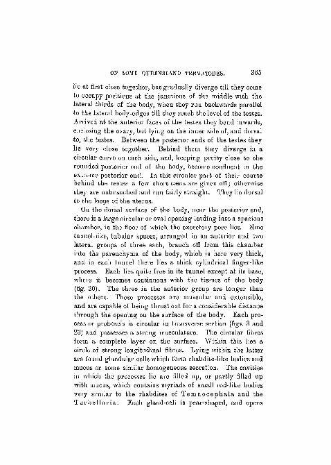
lie at first close together, but gradually diverge till they cometo occupy positions at the junctions of the middle with thelateral thirds of the body, when they run backwards parallelto the lateral body-edges till they reach the level of the testes.Arrived at the anterior faces of the testes they bend inwards,enclosing the ovary, but lying on the inner side of, and doi'salto, the testes. Between the posterior ends of the testes theylie very close together. Behind them they diverge in acircular curve on each side, and, keeping pretty close to therounded posterior end of the body, become confluent in theextreme posterior end. I n this circular part of their coursebehind the testes a few short cseca are given off; otherwisethey are unbranched and run fairly straight. They lie dorsalto the loops of the uterus.
On the dorsal surface of the body, near the posterior end,there is a large circular or oval opening leading into a spaciouschamber, in the floor of which the excretory pore lies. Ninetunnel-like, tubular spaces, arranged in an anterior and twolateral groups of three each, branch off from this chamberinto the parenchyma of the body, which is here very thick,and in each tunnel there lies a thick cylindrical finger-likeprocess. Each lies quite free in its tunnel except at its base,where it becomes continuous with the tissues of the body(fig. 20). The three in the anterior group are longer thanthe others. These processes are muscular and extensible,and are capable of being thrust out for a considerable distancethrough the opening on the surface of the body. Each pro-cess or proboscis is circular in transverse section (figs. 3 and23) and possesses a strong musculature. The circular fibresform a complete layer on the surface. Within this lies acircle of strong longitudinal fibres. Lying within the latterare found glandular cells which form rhabdite-like bodies andmucus or some similar homogeneous secretion. The cavitiesin which the processes lie are filled up, or partly filled upwith mucus, which contains myriads of small rod-like bodiesvery similar to the rhabdites of T e m n o c e p h a l a and theT u r b e l l a r i a . Each gland-cell is pear-shaped, and opens

366 S. J. JOHNSTON.
on the surface of the process through a long duct which runsbetween the longitudinal muscle-fibres. Just at the tips ofthe processes the muscular layers are much reduced, and thegland-cells seem to have converted their entire protoplasminto mucus and rhabdites (fig. 4). Lying in the middle ofthe parenchyma of the process is a large space surroundedby a very definite muscular layer, as well as several smallercavities. With the central space in each process a largebranch given off from the excretory vesicle communicates(fig. 22). Fluid forced into this channel from the excretorysystem apparently takes part in the protrusion of the process,and, together with the action of the muscular system of theprocess, would render it tense.
These processes, according to Taylor, who found them, donot seem to be used in any way for attachment. The functionof rhabdites in the flatworms seems to me to be obscure, novery satisfactory explanation having been offered for them.In this case they probably give a certain amount of stiffnessto the secretion of the processes, and may have au irritating-effect. The whole organ is most probably used for cleaningup an area of the wall of its host's intestine, so as to makea place to which the sucker can be effectively applied.
Muscular System.—The muscular layers of the body-wall are very strongly developed. A complete layer ofcircular fibres lies immediately adjacent to the cuticle ; withinthe circular is a. thick layer of longitudinal fibres, and internalto this is a layer of diagonal fibres, somewhat thicker thanthe outer circular layer. A dorso-ventri.il system of largemuscle-fibres is developed to a very marked extent (figs.15 and 16). The majority of these are fairly perpendicularto the dorsal and ventral surfaces of the body; many, however,are oblique in their direction. At each end, at the pointwhere they reach the level of the diagonal fibres of the body-wall, these dorso-ventral fibres become divided into a numberof branches, so that the area of their insertion into the cuticleis very much greater than the area of the cross-section of thefibre.

ON SO3IK QUEENSLAND TRBMATODJSS. 367
E x c r e t o r y S y s t e m . — T h e large branched vesicle opens,into the proboscis chamber. The pore is provided with astrongly developed sphincter which is capable of keeping1 itclosed when the fluid of the vesicle is being forced into thefinger-like processes of the proboscis. The vesicle divides intofour wide bays antenorly and each of these bays becomesgradually narrowed iuto one of the four main trunk vessels.Those placed laterally diverge as far as the testes, round theouter sides of which they skirt, and then run forwards parallelto the sides of the body. Towards the anterior end theygradually trend iuwards, and at a level just in front of andventral to the intestinal fork they become joined in the formof a circular arch (fig. 26). All along their course they areprovided with a number of wide side branches, regularlygiven off on the outer side and extending to the edge of thebody (fig. 1). These branches do not anastomose with oneanother. The two vessels given off from the vesicle nearerthe middle line pass the testes on the inuer side and runforwards parallel to and within the intestinal limbs. Theyalso give off a number of branches, but end blindly in frontnear the intestinal fork, without forming a transverse anas-tomosis. The four main excretory vessels and the intestinallimbs are placed approximately in the same plane, so that theyall appear together in the same horizontal (parallel to theventral surface) section (fig. 26).
N e r v o u s S y s t e m . — A pair of large cerebral ganglia areconnected by a very thick commissure which crosses dorsallyover the middle of the oesophagus (fig. 19). The principaltrunks, in the form of a pair of lateral nerve-cords, graduallydiverge outwards towards the sides of the body, at the sametime sinking ventrally so that at a point not far behind theintestinal fork they come to occupy a position immediatelyinternal to the muscular layers of the ventral body-wall (fig.15). From the lateral trunks many small nerves are givenoff and are distributed laterally, inwards and dorsally.
R e p r o d u c t i v e Organs.—The genital opening is at theanterior end, on the ventral surface, quite close to. the sucker
368 S. J. JOHNSTON.
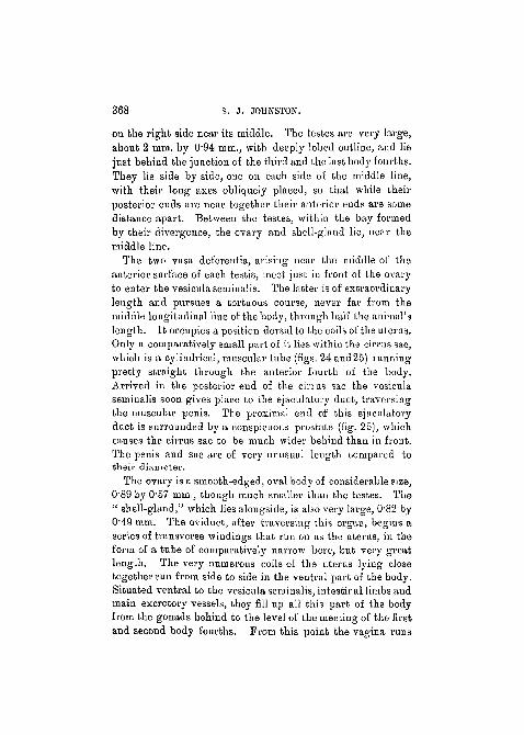
on the right side near its middle. The testes are very large,about 2 mm. by 094 mm., with deeply lobed outline, and liejust behiud the junction of the third and the last body fourths.They lie side by side, one on each side of the middle line,with their long axes obliquely placed, so that while theirposterior ends are near together their anterior ends are somedistance apart. Between the testes, within the bay formedby their divergence, the ovary and shell-gland lie, near themiddle line.
The two vasa deferentia, arising neav the middle of theanterior surface of each testis, meet just in f font of the ovaryto enter the vesicula seminalis. The latter is of extraordinarylength and pursues a tortuous course, never far from themiddle longitudinal line of the body, through half the animal'slength. It occupies a position dorsal to the coils of the uterus.Only a comparatively small part of it lies within the cirrus sac,which is a cylindrical, muscular tube (figs. 24 and 25) runningpretty straight through the anterior fourth of the body.Arrived in the posterior end of the cirrus sac the vesiculaseminalis soon gives place to the ejaculatory duct, traversingthe muscular penis. The proximal end of this ejaculatoryduct is surrounded by a conspicuous prostate (fig. 25), whichcauses the cirrus sac to be much wider behind than in front.The penis and sac are of very unusual length compared totheir diameter.
The ovary is a smooth-edged, oval body of considerable size,O89 by 0-57 rain., though much smaller than the testes. The" shell-gland," which lies alongside, is also very large, 082 byO49 mm. The oviduct, after traversing this organ, begins aseries of transverse windings that run on as the uterus, in theform of a tube of comparatively narrow bore, but very greatlength. The very numerous coils of the uterus lying closetogether run from side to side in the ventral part of the body.Situated ventral to the vesicula seminalis, intestinal limbs andmain excretory vessels, they fill up all this part of the bodyfrom the gonads behind to the level of the meeting of the firstand second body fourths. Prom this point the vagina runs
ON SOME QUEENSLAND TREMATODES. 369
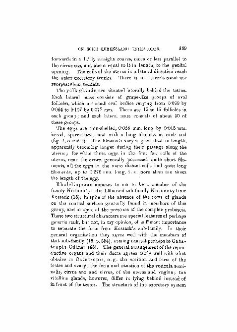
forwards in a fairly straight course, more or less parallel tothe cirrus sac, and about equal to it iu length, to the geuitalopening. The coils of the uterus in a lateral direction reachthe outer excretory trunks. There is no Laurer's canal norreceptaculum seminis.
The y o l k - g l a n d s are situated laterally behind the testes.Each lateral mass consists of grape-like groups of ovalfollicles, which are small oval bodies varying from 0"099 by0-064 to 0107 by 0'077 mm. There n,re 12 to 15 follicles ineach group; and each lateral mass consists of about 50 ofthese groups.
The eggs are thin-shelled, 0-026 mm. long by 0"015 mm.broad, operculated, and with a long filament at each end(fig. 2, a and b). The filaments vary a good deal iu length,apparently becoming longer during their passage along theuterus; for while those eggs in the first few coils of theuterus, near the ovary, generally possessed quite short fila-ments, all the effa's in the more distant coils had quite Ions'filaaients, up to 0'279 mm. long, i . e . more than ten timesthe length of the egg.
R h a b d i o p o e u s appears to me to be a member of thefamily N o t o c o t y l i d a s Liihe and sub-family N o t o c o t y l i n a sKossack (18), in spite of the absence of the rows of glaudson the ventral surface generally found in members of thisgroup, and iu spite of the presence of the complex proboscis.These two structural chai'acters are special features of perliapsgeneric rank, but not, iu my opinion, of sufficient importanceto separate the form from Kossack's sub-family. In theirgeneral organisation they agree well with the members ofthat sub-family (18, p. 554), coming nearest perhaps to C a t a -t r o p i s Odhner (48). The general arrangement of the repro-ductive organs aud their ducts agrees fairly well with whatobtains in O a t a t r o p i s , e . g . the position and form of thetestes and ovary; the form and situation of the vesicula semi-nalis, cirrus sac and cirrus, of the uterus and vagina; thevitelline glands, however, differ in lying behind instead ofin front of the testes. The structure of the excretory system

370 . S. J. JOHNSTON. -
also agrees pretty well with that of Cata t ropis . Rhabdio-pceus differs from the latter further in having the intestinallimbs fused behind, in the absence of the serial ventralglands, as well as in the presence of the complicated proboscisand the thick layer of spines on the ventral surface.
Opis tho t rema cochleare Fisch.
Eleven specimens were found in the oesophagus ofHalicore dugong, by Dr. Strangnian, at Port Darwin.They were smaller than Semper's specimens, on whichFischer worked, and were all sexually immature. Thelargest specimen measured 7 mm. long by 4 mm. broad.The majority were close to this in size, while the smallestwas only 3 mm. long. The sexual ducts were all well formed,though the gonads themselves were only represented by verysmall patches of cells. The already formed cirrus distinguishesthese specimens at once from 0. pulnionale von Linstowfound in the lungs of the same host. The position of thetestes is also different, being external to the intestinal limbsiu 0. cochleare , but internal to them in 0. pnlmonale(24). Fischer (14) mentions the spines on the ventral surfaceof the species under discussion, but makes no mention of thefact, readily seen in sections, that the anterior part of thedorsal surface bears smaller, more scattered spines.
FROM BIEDS.
Fam. EchinostomidEe.
Bchinostoma revolutum Froel.
One complete specimen, and a fragment consisting of theposterior half, were obtained from the intestine of the"black-duck," Anas superci l iosa. According to Loossthis species of worm (described undername E. echinatuniR.) exhibits considerable variation, and the description given
ON SOME QUEENSLAND TREMATOPES. 371
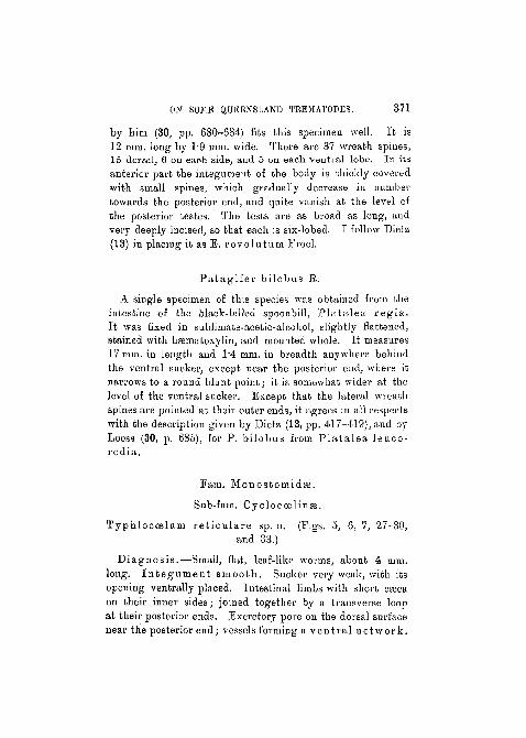
by him (30, pp. 680-684) fits this specimen well. It is12 mm. long by 1*9 mm. wide. There are 37 wreath spines,15 dorsal, 6 on each side, and 5 on each ventral lobe. In itsanterior part the integument of the body is thickly coveredwith small spines, which gradually decrease in numbertowards the posterior end, and quite vanish at the level ofthe posterior testes. The tests are as broad as long, andvery deeply incised, so that each is six-lobed. I follow Dietz(13) in placing it as E. revolutum Froel.
Pa tag i fe r bilobus R.
A single specimen of this species was obtained from theintestine of the black-billed spoonbill, P la ta lea reg ia .It was fixed in sublimafce-acetic-alcohol, slightly flattened,stained with hasmatoxylin, and mounted whole. It measures17 mm. in length and 1'4 mm. in breadth anywhere behiudthe ventral sucker, except near the posterior end, where itnarrows to a round blunt point; it is somewhat wider at thelevel of the ventral sucker. Except that the lateral wreathspines are pointed at their outer ends, it agrees in all respectswith the description given by Dietz (13, pp. 417-419), and byLooss (30, p. 685), for P. bilobus from P la t a l ea leuco-rodia.
Pam. Monostomidte.
Sub-fam. Cyclocoslinse.
Typhlocoelum re t i cu la re sp. n. (Figs. 5, 6, 7, 27-30,and 33.)
Diagnosis.—Small, flat, leaf-like worms, about 4 mm.long. I n t e g u m e n t smooth. Sucker very weak, with itsopening ventrally placed. Intestinal limbs with short cascaon their inner sides; joined together by a transverse loopat their posterior ends. .Excretory pore on the dorsal surfacenear the posterior end; vessels forming a vent ra l ne twork .

372 S. J. JOHNSTON.
Genital pore cm the ventral aspect, just beneath the oeso-phagus. Testes large, much branched bodies; copulatoryorgans moderately developed. Ovary oval, small; no Laurer'scanal nor receptaculum seininis. Uterus richly coiledin dorso-vent ra l loops, not pass ing ou twards be-yond the intestinal limbs, and filling up all the space betweenthe intestinal limbs from the gonads behind to the intestinalfork in front. Follicles of the yolk-glands minute (O022 mm.),extending from the beginning of the intestine in front, form-ing a loop behind the intestinal commissure behind.
Eggs 0-107 by 0~073 mm. (?)Host: The pied goose, Auserauas semipa lmatus , in the
intestine.Type-specimen in the Museum of the Australian Institute
of Tropical Medicine, Queensland, No. T. 34.Co-type specimen in the Australian Museum, No. W. 365.Five specimens were obtained from the intestine of the
pied goose, Anseranas semipalmatus , at Townsville.They are small leaf-like forms, oval in shape, quite flat,
aud thin dorsoventrally, 4-3 mm. long by 1*83 mm. broad.The integument is quite smooth.
The anterior sucker is so weakly developed as to be almostnon-existent. It is represented by a funnel-shaped depressionopening on the ventral surface near the anterior end; itswalls are provided with a few weak muscle-fibres, which aremore concentrated near its base. At the bottom it opensinto a very well-developed aud muscular pharynx, 0-228 mm.loug by 0'163 mm. broad.
The oesophagus is short but distinct, and divides behindinto the two intestinal limbs, which run backwards parallel tothe edge of the body and fuse together so as to complete thering at the posterior end. A few short caeca are given off onthe inner aspect of each limb.
Excre tory System.—The excretory pore is on thedorsal surface, near the posterior end. It is surrounded by adistinct sphincter, and leads into a more or less spaciouschamber situated wholly behind the intestine (n'g. 5). Into

ON SOME QUEENSLAND TBEMATODES. 373
its anterior end two main excretory vessels open. Thesecross over the posterior intestinal loop and run forwards asfar as the sides of the pharynx, where they end blindly. Intheir forward course they lie close to, and fairly parallel tothe intestinal limbs, re-crossing them near their anterior end.On their outer sides they give off a number of mostly un-divided branches; on their inner aspect they give off anumber of branches which, anastomosing freely, form anetwork in the middle of the body (figs. 6 and 33) lyingventral to the uterus. From all the branches finer tubes aregiven off which end in flame-cells. These finer tubes areapparently intra-cellular, as not more than one nucleus couldbe made out in the wall of any of those examined. The wallsof the larger vessels—the main trunks, network and branches—as well as the vesicle are composed of a nucleated syncytium.
N e r v o u s S y s t e m . — T w o large cerebral ganglia, com-posed of the usual nerve-cells and fibres, lie, one on eitherside, iu front of the pharynx (fig. 29). They are joined by athick commissure, and give off a number of fine nerves to theintegument in the front of the body. Behind, each gives offa thick nerve, the lateral nerve-cord, containing in its coursemany nerve-cells as well as fibres (fig. 7). These nerve-cordsrun backwards just outside the intestinal limbs, and in theextreme posterior end of the body, behind the excretoryvesicle, join together to complete the circuit. Branches aregiven off, both on the outer and inner aspects of these nerve-cords, and at the points where they leave the main t runkslittle heaps of nerve-cells usually occur. The internal branchesfuse with one another so as to form a network on the ventralsurface of the body, just internal to the muscular layers, andventral to the network of excretory vessels (fig. 27).
G e n i t a l S y s t e m . — T h e common genital pore is situatedon the ventral surface just beneath the oesophagus—i. e. infront of the intestinal fork. The testes are large branchedbodies lying near the posterior end, but completely within thespace bounded by the intestinal loop. They are somewhatobliquely placed one behind the other. The posterior, lying-
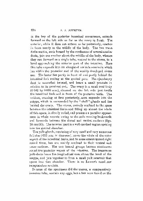
374 • S. J . JOHNSTON, -
in the bay of the posterior intestinal commissure, extendsforward on the left side as far as the ovary in front. Theanterior, while it does not extend so far posteriorly, reachesin front nearly to the middle of the body. The two vasadeferent ia , each formed by the confluence of several smallerducts, join one another about the middle of the body, whencethey run forward as a single tube, ventral to the uterus, to. alevel approaching the anterior part of the intestine. Herethis tube expands into an elongated vesicula semiualis whichlies within the posterior end of the weakly developed cirrussac. The latter lies partly in front of and partly behind theintestinal fork ending at the genital pore. The ejaculatoryduct is somewhat twisted, and bears a small prostate inrelation to its proximal end. The ovary is a small oval body(O142 by 0'086 mm.), situated ou the lefb side just insidethe intestinal limb and in front of the posterior testis. Theoviduct, running at first posteriorly, soon expands into theootype, which is surrounded by the " shell "-glands and liesbehind the ovary. The uterus, eutirely confined to the spacebetween the intestinal limbs and filling up almost the wholeof this space, is closely coiled, and presents a peculiar appear-ance in whole mounts owing to the coils running backwardsand forwards between the dorsal and ventral surfaces (figs.28 and 30). The terminal part is a well-marked vagina openinginto the genital chamber.
The yolk-glands, consisting of very small and very numerousfollicles ("022 mm. in diameter), cover the whole of the outeraspect of the intestinal limbs, and to some extent spread rightrouud them, but are mainly confined to their ventral andouter surfaces. The two lateral groups become continuousround the posterior aspect of the intestine. The transverseyolk-ducts leave the longitudinal ones about the level of theootype, and join together to form a small yolk reservoir thatopens into that chamber. There is no Laurer's canal.norreceptaculum seminis.
In none of the specimens did the uterus, a comparativelyimmense tube, contain any eggs, but a few were found on the
ON SOME QUEENSLAND TREMATODES. 375
surface of the body or in the intestine. As no other trema-todeswere found in the host, which was subjected to a carefulexamination, and the form and size of the eggs found conformpretty well with those in closely related species, it is almostbeyond doubt that these eggs belong to the animal with whichthey were found associated. They are large, oval in shape,yellow in colour, O107 mm. long by O073 mm. broad.
This worm appears to be closely related to Typhloccelumcucumerinum Rud. as described by Kossack (18, pp. 543-548), in spite of the occurrence of the small ventral sucker inthe latter. I have been able to find no trace of such a suckerin my specimens, either in the whole mounts or sections. Theoccurrence of such a sucker in worms, in all other respectsobviously associated with the Monostomidfe, must^ I think, belooked upon as an atavism—a thing of local interest, but of nogreat systematic importance.
T. r e t i cu la re differs from T. cucumerinum in size andshape; in the more elongated form of its pharynx; in thegreater extension forward of the anterior testis and the greaterlength of the separate vasa deferentia; in the arrangement ofthe yolk-glands behind the intestine; in the much'smallersize of the ovary; in the very characteristic dorso-ventralwinding of the uterine loops; and in the considerably smallereggs.
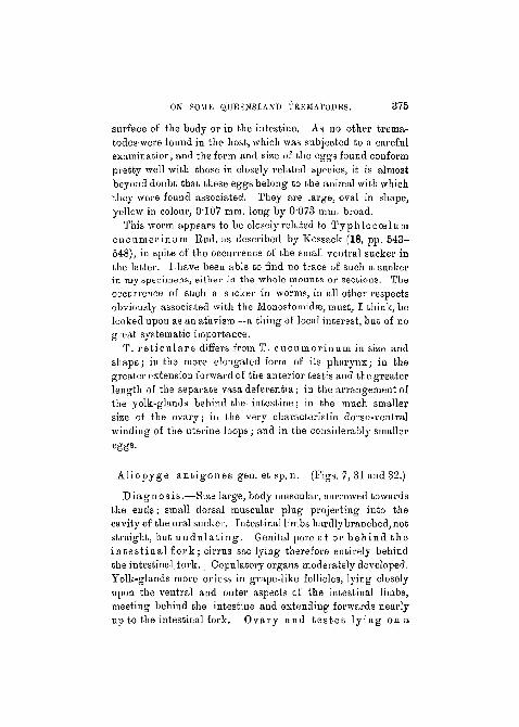
Allopyge ant igones gen. et sp.n. (Figs. 7, 31 and 32.)
Diagnosis.—Size large,body muscular, narrowed towardsthe euds; small dorsal muscular plug projecting into thecavity of the oral sucker. Intestinal limbs hardly branched, notstraight, but undu l a t i ng . Genital pore at or behind thein tes t ina l fork; cirrus sac lying therefore entirely behindthe intestinal fork. Copulatory organs moderately developed.Yolk-glands more or less in grape-like follicles, lying closelyupon the ventral and outer aspects of the intestinal limbs,meeting behind the intestine and extending forwards nearlyup to the intestinal fork. Ovary and tes tes ly ing on a

376 S. J. JOHNSTON.
s t r a i g h t line, inclined at an angle to the antero-posterior axisof the body; but the ovary not so closely associated with theposterior testis as in Hypt iasmus . Anterior testis separatedfrom the posterior and the ovary by a number of uterineloops. Uterine loops, iu the posterior half of the body, exten-ding out beyond the intestinal limbs ; but no loops ex ten-ding backwards beyond the posterior testes as in H y p t i -asmus. No Laurer's canal; a recep tacu lum semiuispresent or absent.
Type species : A. an t igones ; from the small intestine ofthe Australian crane, Ant igone a u s t r a l a s i a n a .
Type-specimen iu the Museum of the Australian Instituteof Tropical Medicine, No. T. 35.
Co-type iu the Australian Museum, No. W. 366.This genus differs from H y p t i a s m u s, 'to which it is closely
related, mainly, in the undulating course of the intestinallimbs, in the position of the genital pore, at, or just behind,the intestinal fork, and in the consequent situation of the cirrussac entirely behind it; in the absence of uterine loops passingbehind the posterior testis, and iu the presence (probablyinvariable) of a receptaculum seminis. With this genusprobably Hyptiasmus omiuosus Koss. and H. ado lph iStoss. should be associated. I have not been able to see theoriginal text; of Stossich's work (57), but know its contentsonly throughBraun'sabstracfcin the' ZoologischesCentralblatt'and by means of Kossack's criticisms of it, so that I do notknow whether H. adolphi is the worm of this group in whichStossich found the receptaculum seminis, but suspect that itis so. Three specimens of A. an t igones were obtained fromits host; one was sectioned, and the other two were made intowhole mounts. They are lnrge, flat, leaf-like worms, about20 mm. long and 4 mm. broad. While the ventral surface isflat the dorsal is somewhat convex. The integument is roughwith little tranveise corrugations.
The character of the oral sucker and the pharynx is exactlythe same as in T. r e t i cu la re . The structure which I havecalled the pharynx has been generally looked upon, in related
ON SOME QUEENSLAND TBUMATODES. 377
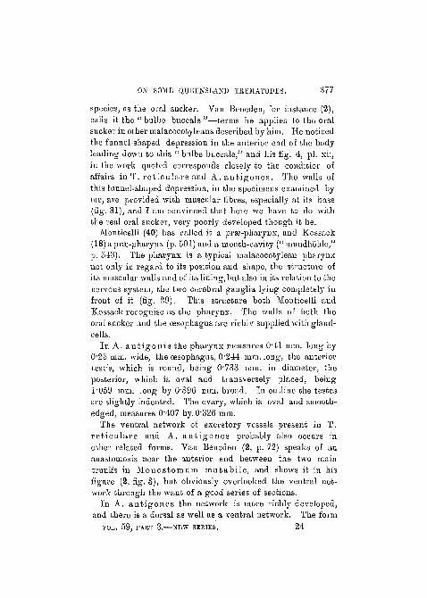
species, as the oral sucker. Van Benedeu, for instance (2),calls it the " bulbe buccale "—terms he applies to the oralsucker in other malacocotyleans described by him. He noticedthe funnel-shaped depression in the anterior end of the bodyleading down to this "bulbe buccale/' and his fig. 4, pi. xii,in the work quoted corresponds closely to the condition ofaffairs in T. re t icu lare and A. an t igones . The walls ofthis funnel-shaped depression, in the specimens examined byme, are provided with muscular fibres, especially at its base(fig. 31), and I am convinced that here we have to do withthe real oral sucker, very poorly developed though it be.
Monticelli (40) has called it a prfe-pharynx, and Kossack(18) a prse-pharynx (p. 501) and a mouth-cavity (" mundhohle,"p. 543). The pharynx is a typical malacocotylean pharynxnot only in regard to its position and shape, the structure ofits muscular walls and of its lining, but also in its relation to thenervous system, the two cerebral ganglia lying completely infront of it (fig. 29). This structure both Mouticelli andKossack recognise as the pharynx. The walls of botli theoral sucker and the oesophagus are richly supplied with gland-cells.
In A. an t igon i s the pharynx measures 041 mm. long byO25 mm. wide, the oesophagus, 0244 mm. long, the anteriortestis, which is round, being 0'733 mm. in diameter, theposterior, which is oval and transversely placed, beingT059 mm. long by 0896 mm. broad. In outline the testesare slightly indented. The ovary, which is oval and smooth-edged, measures 0"407 by. 0326 mm.
The ventral network of excretory vessels present in T.re t icu lare and A. an t igones probably also occurs inother related forms. Vau Beneden (2, p. 72) speaks of anauastomosis near the anterior end between the two maintrunk's in Monostomum mutabi le , and shows it in hisfigure (2, fig. 3), but obviously overlooked the ventral net-work through the want of a good series of sections.
In A. ant igones the network is more richly developed,and there is a dorsal as well as a ventral network. The form
VOL. 59, PART 3 . NEW SERIES. 24
378 S. J. JOHNSTON.
and position of the vesicle and its pore are much the same asin T. re t i cu la re . The nervous system of the former alsocorresponds pretty closely with the arrangement describedfor the latter.
In the uterus the loops lying between the anterior testisand the middle of the body extend outwards beyond theintestine almost to the edge of the body, while the moreanterior loops are confined to the space between the intestinallimbs. There are no backwardly directed loops extendingbehind the posterior testis as in Hypt iasmus . While thereis no Laurer's canal, a very distinct receptaculum is present(fig. 32).
The eggs are very numerous, smaller and narrower than inT. r e t i cu l a r e , O094 mm. long by 0-055 mm. broad, yellowto light brown in colour.
FEOM REPTILES.
Fain. Angiod ic ty idas.
Polyangium l ingua tu la Lss.
Ten specimens of this species were obtained from theintestine of Chelone mid as.
Octaugium sag i t t a Lss.About one hundred specimens were obtained from Chelone-
mi das, living along with the Polyangium.
Fam. Pronocephalidfe.
D ia sch i s to rch i s pandus Braun, gen. nov. (Figs. 9, 10,34-37.)
In addition to a single specimen obtained from the intestineof a hawksbill turtle, Chelone i m b r i c a t a , which I caughtin Port Jackson, I have received from the Institute sixspecimens of a worm obtained from the intestine of Chelonem id as, caught off the Queensland coast. These worms appear

ON SOME QUEENSLAND TREMATODES. 379
to me to be identical with Braun's Monostomum pandutn(9, p. 48) on the following grounds :
Professor Braun had at his disposal only a single specimen,which was mounted whole. As the worm is fairly thick anddense, it is, of course, impossible to view its anatomy com-pletely m a single specimen.
As far as Braim's description goes my whole mountsagree with it very well, the differences being of such minorimportance as to make it out of the question, in my opinion,to propose a separate species for my specimens. A study ofserial sections, however, not only makes clear important pointsin the animal's structure that are quite impossible to see inthe whole mounts, but also shows some of the appearancesin the latter to be somewhat misleading. While the worm isundoubtedly one of the Pronocephal idse Lss. (33), it doesnot fit into any of the genera at present established, so that Iam obliged to propose a new genus for it, addiug yet another tothe list of genera containing only a single species in this family.
Four of the worms available to me for study I cut intosections, one transverse, one sagittal, and two horizontallongitudinal. With the help of these series I shall supple-ment Braun's description (9, pp. 48-50), afterwards giving adiagnosis of the genus Diasch is to rch is , and a discussionof its relationships.
Ex te rna l Characters.—My specimens were all a littlesmaller than Braun's, varying in length from 8 to 9 mm.,while being 2'5 mm. broad at the level of the cirrus sac and3 mm. broad at the widest part, near the posterior end. Theform of body is skiff-like, rounded at both ends, rathernarrower in front, and gradually increasing in breadth tonear the posterior end. The collar is less conspicuous thanin the other members of the family, taking the form of a lowkidney-shaped elevation of the surface of the body round thesucker on the dorsal side, but not produced into lobes orprocesses of any kind at its ventro-lateral ends. The dorsalsurface of the worm is arched, both from side to side and, toa certain extent, antero-posteriorly; the ventral surface is

380 S. J. JOHNSTON.
concave, and sometimes the posterior edge is turned forwardsa little. The genital pore is found on the ventral surface,near the left side, some distance from the anterior end; theexcretory pore is on the dorsal surface, a little in front ofthe posterior extremity. The sucker measures 0'57 mm. by0"63 mm. in its longitudinal and transverse axes respectivelyin the smallest specimen, and 0717 mm. by 0-782 mm. in thelai'gest. Its opening is subterminal.
Al imenta ry Canal . — A thick - walled oesophagus,0'326 mm. long, leads back from the sucker, and joins theintestine almost at right angles, the limbs running at firsttransversely and in the same straight line. There is nopharynx. The intestinal limbs extend backwards to thelevel of the excretory pore, and are provided with numerousside branches, both on their outer and mesial surfaces.Some of these lateral ceeca become divided into two or threeshort branches (fig. 34). The intestinal limbs do not becomearched in towards the median plane in the region of thetestes, as so commonly happens in members of this family,but pui'sue a course fairly parallel to the sides of the body,converging a little at their posterior ends.
Exc re to ry System.—The excretory vesicle is providedwith the characteristic funnel-shaped end, bearing processeson its walls—the "rippen" of Looss (33)—as in E p i b a t h r aand other related genera. The capacious vesicle extendsforwards as far as the ovary, and is produced on each sideinto three or four wide diverticula. From one of these pairs—the second from the front—the main vessels are given offand extend outwards till they reach the outer sides of thetestes. Here each vessel divides into two branches, onerunning posteriorly, the other anteriorly (fig. 10). Eachanterior branch pursues an undulating course, in seven oreight large waves, ending blindly near the sucker. Branchesare given off both from the outer and inner aspects; theformer end blindly, but the latter anastomose with oneanother in the space between the intestinal limbs, ventral tothe uterus.
OX SOME QUEENSLAND TKiSJIATODES. 381
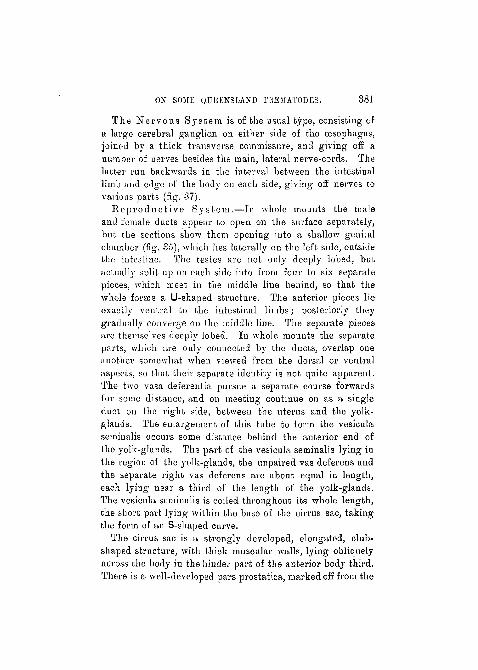
The Nervous System is of the usual type, consisting ofa large cerebral ganglion on either side of the oesophagus,joined by a thick transverse commissure, and giving off anumber of nerves besides the main, lateral nerve-cords. Thelatter run backwards in the interval between the intestinallimb and edge of the body on each side, giving off nerves tovarious parts (fig. 37).
Reproduc t ive System.—In whole mounts the maleand female ducts appear to open on the surface separately,but the sections show them opening into a shallow genitalchamber (fig. 35), which lies laterally on the left side, outsidethe intestine. The testes are not only deeply lobed, butactually split up on each side into from four to six separatepieces, which meet in the middle line behind, so that thewhole forms a U-shaped structure. The anterior pieces lieexactly ventral to the intestinal limbs; posteriorly theygradually converge ou the middle line. The separate piecesare themselves deeply lobed. In whole mounts the separateparts, which are only connected by the ducts, overlap oneunotlier somewhat when viewed from the dorsal or ventralaspects, so that their separate identity is not quite apparent.The two vasa deferentia pursue a separate course forwardsfor some distance, and on meeting continue on as a singleduct on the right side, between the uterus and the yolk-glands. The enlargement of this tube to form the vesiculaseminalis occurs some distance behind the anterior end ofthe yolk-glands. The part of the vesicula seminalis lying inthe region of the yolk-glands, the unparred vas deferens andthe separate right vas deferens are about equal in length,each lying near a third of the length of the yolk-glands.The vesicula seminalis is coiled throughout its whole length,the short part lying within the base of the cirrus sac, takingthe form of an S-shaped curve.
The cirrus sac is a strongly developed, elongated, club-shaped structure, with thick muscular walls, lying obliquelyacross the body in the hinder part of the anterior body third.There is a well-developed pars prostatica, marked off from the
382 S. J. JOHNSTON.
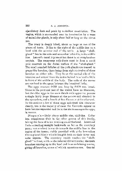
ejaculatory duct and penis by a shallow constriction. Thevagina, which is surrounded near its termination by a massof unicellular glands, is only about half as long as the cirrussac.
The ovary is deeply lobed, about as large as one of thepieces of testis. It lies to the right of the middle line on alevel with the anterior end of the testis. A. large " shell-gland" lies to the side and somewhat behind it, in the middleline. Laurer's canal is present but there is no receptaculumseminis. The transverse yolk-ducts meet to form a smallyolk reservoir on the dorsal surface of the "shell-gland."The small rounded follicles of the yolk-glands are massed ingrape-like bunches, there being from eight to twelve of thesebunches on either side. They lie on the ventral side of theintestine and extend from the testes behind to a level a littlein front of the middle of the body. The coils of the uterusare confined to the space between the intestinal limbs.
The eggs measure O033 mm. long by 0"019 mm. broad.Those in the proximal part of the uterus have no filaments,but the older eggs in the more distals coils appear to possessa single fairly large filament at the narrow end attached tothe operculum, and a bunch of fine filaments at the other end.In the sections a few of these eggs exhibited this characterclearly, but in the majority of cases the filaments appear tohave become separated and lie in the uterus amongst the eggs,in masses.
Diagnosis.—Body above middle size, skiff-like. Collarless conspicuous than in the other genera of this family,taking the form of a low swelling round the head. Intestinallimbs extending straight backwards as far as the excretorypore; not a rched inwards towards the middle line in theregion of the testes; richly provided with side branchesth roughou t the i r whole l eng th both on their inner andouter aspects. The excretory vesicle reaches the "shell-gland " in front, with wide l a t e r a l d iver t i cu la ; the mainbranches running up to the head end in an undulating course,givingoff branches, some of which anas tomose. Genital
ON SOME QUEENSLAND TBEMATODES. 383
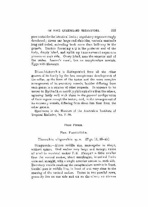
pore outside the intestinal limbs; copulatory organs stronglydeveloped ; cirrus sac large and club-like, vesicula seminalislong and coiled, extending back more than half-way to thegonads. Testes forming a U in the posterior end of thebody, deeply lobed, and spl i t Lip into several s epa ra t epieces on each side. Ovary lobed, near the anterior end ofthe testes. Laurer's caual, but no receptaculum seminis.Eggs with filaments.
Diaschis torchis is distinguished from all the othergenera of its family by the less conspicuous development ofthe collar, by the form of the testes and the more complexarrangement of its excretory vessels, besides differing fromeach genus in a number of other respects. In appears to benearer to E p i b a t h r a and Glyphicephalus than tlie others,agreeing fairly well with these in the general configurationof their organs except the testes; and, in the ai'rangenient ofits excretory vessels, differing from these less than from theother genera.
Specimens in the Museum of the Australian Institute ofTropical Medicine, No. T. 36.
• FEOM PISHES.
Fam. Fasciolidae.
Pleorchis ol igorchis sp. n. (Figs. 11, 3S-40.)
Diagnosis.—Above middle size, rectangular in shape,without spines. Oral sucker very large and strong; ra t ioof oral to vent ra l sucker 7:3. Pharynx a little smallerthan the ventral sucker, short oesophagus, intestinal limbswide and straight, with a single anterior cEecum on each sideExcretory vesicle reaching the receptaculum seminis in front.Genital pore in middle line, in front of and very close to theopening of the ventral sucker. Testes in two parallel rows,generally five on one side and six on the other; no c i r rus

381- S. S. JOHNSTON.
sac . O\rary s p h e r i c a l ; Laurer's canal and receptacnlnmpresent; uterus small; yolk-glands very richly developed.
Parasitic in the intestine of T e t r a o d o n h i s p i d u s Linn.Type specimen in the Museum of the Australian Instituteof Tropical Medicine, Townsville, No. T. 37.
Co-type in the Australian Museum, No. W. 367.The largest specimen, fixed under slight pressure, measured
12 mm. long by 5 mm. broad; the smallest from the blacktoad-fish ( T e t r a o d o n h i s p i d u s ?) 8 mm. long by 3'75 mm.broad. Eight specimens were obtained from this host; buta large number of specimens, which differed from these onlyin being smaller in size, 5-G mm. long by 2 mm. broad,were obtained from the iutestiue of the spotted toad-fish( T e t r a o d o n h i s p i d u s ) . The worms are flat and almostrectangular in shape in the preserved specimens, the sidesbeing fairly parallel aud the ends almost truncated. Theintegument is thick and shows a number of corrugations onthe surface. There are no spines.
The oral sucker is very large and strong, in many casesretaining within its grasp a piece of the mucous membrane ofthe host's intestine. I t measures l-666 mm. in diameter.The opening is terminal and comparatively small, with acondensation of the tissues round its edge so that the latteris specially tough. The ventral sucker is much smaller aud•weaker, and measures 0"714 mm. in diameter. The ratio ofthe oral to the ventral sucker is 7 : 3. The pharynx is a littlesmaller than the ventral sucker, and in most cases was com-pressed in the longitudinal direction. There is a distinctpras-pharynx, and a short oesophagus 0'12 mm. long (fig. 38)leading into the intestinal limbs, which run at first horizon-tally, where they bend round to become longitudinal; aCfficmn is given off which extends forwards as far as themiddle of the anterior sucker. The main posterior limbs,which are wide, extend straight back, parallel with the sidesof the body, into the extreme posterior end. They have nobranches, but in contracted specimens the walls are throwninto terasverse folds.
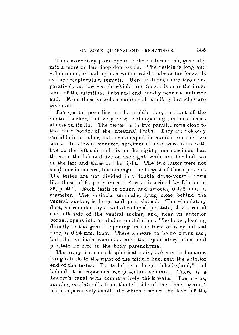
ON SOME QUEENSLAND TKKMATOPES. 385
The e x c r e t o r y p o r e opens at the posterior end, generallyinto a move or less deep depression. The vesicle is long andvoluminous, extending as a wide straight tube as far forwardsas the receptaculum seminis. Here it divides into two com-paratively narrow vessels which runs forwards near the innersides of the intestinal limbs and end blindly near the anteriorend. From these vessels a number of capillary branches aregiven off.
The genital pore lies in the middle line, in front of theventral sucker, and very close to its opening;, in most casesalmost on its lip. The testes lie in two parallel rows close tothe inner border oi the intestinal limbs. They are not onlyvariable in number, but also unequal in number on the twosides. In eleven mounted specimens there were nine withfive on the left side and six on the r i gh t ; one specimen hadthree on the left and five on the right, while another had twoon the left and three on the right. The two latter were notsmall nor immature, but amongst the largest of those present.The testes are not divided into double dorso-ventral rowslike those of P . p o l y o r c h i s Stoss., described by Linton in26, p . 460. Each testis is round and smooth, 0476 mm. indiameter. The vesicula seminalis, lying close behind theventral sucker, is large and pear-shaped. The ejaculatoryduct, surrounded by a well-developed prostate, skirts roundthe left side of the ventral sucker, and, near its anteriorborder, opens into a tubular genital sinus. The latter, leadingdirectly to the genital opening, in the form of a cylindricaltube, is 0'24 mm. long. There appears to be no cirrus sac ;but the vesicula seminalis and the ejaculatory duct andprostate lie free in the body parenchyma.
The ovary is a smooth spherical body, 0'57 mm. in diameter,lying a little to the right of the middle line, near the anteriorend of the testes. To its left is a large " shell-ghmd/' andbehind it a capacious receptaculum seminis. There is aLaurer 's canal with comparatively thick walls. The uterus,running out laterally from the left side of the "shel l-gland,"is a comparatively small tube which reaches the level of the
oob s. j . JOHNSTON.
anterior edge of the ventral sucker in three or four S-shapedbends, and opens into the genital sinus alongside the maleopening. The eggs are never numerous. The yolk-glands,consisting of small rounded follicles 0'048 mm. in diameter,are very richly developed. Iu front they reach the level ofthe anterior border of the ventral sucker and are in thisregion confined to the lateral parts of the body. Passingbackwards they gradually extend inwards to the middle liue,so that in the posterior half of the animal they fill up thewhole field, extending right across the body and back to theextreme posterior end. The eggs are thin-shelled, light incolour, 0-073-0-076 mm. long by 0-046 mm. wide.
This species differs from P . polyorohis Stossich in itslarger size, in the absence of spines, in its much larger oralsucker, in the smaller number of testes, and in its sphericalovary.
Fain. Ster ingophoridas .
S te r iugo t rema pulchrum sp. n. (Figs. 12,41 and 42).
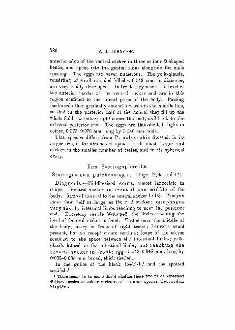
Diagnosis.—Middle-sized worms, almost lanceolate inshape. Ventral sucker in front of the middle of thebody. Ratio of the oral to the ventral sucker 1 : 1"8. Pharynxmore thau half as large as the oral sucker; oesophagusvery shor t ; intestinal limbs reaching to near the posteriorend. Excretory vesicle V-shaped, the limbs reaching thelevel of the oral sucker in front. Testes near the middle ofthe body; ovary in front of right testis; Laurer's canalpresent, but no receptaculum seminis; loops of the uterusconfined to the space between the intestinal limbs; yolk-glaiids lateral to the intestinal limbs, not r each ing theven t ra l sucker in f ron t ; eggs 0-043-0'048 mm. long by0-032-0'035 mm. broad, thick shelled.
In the gullet of the black toadfish,1 and the spottedtoadfish.1
1 There seems to be some doubt whether these Wo fishes representdistinct species or colour varieties of the same species, Tetraodonhispidus.
ON SOME QUEENSLAND TRBMATODES. 387
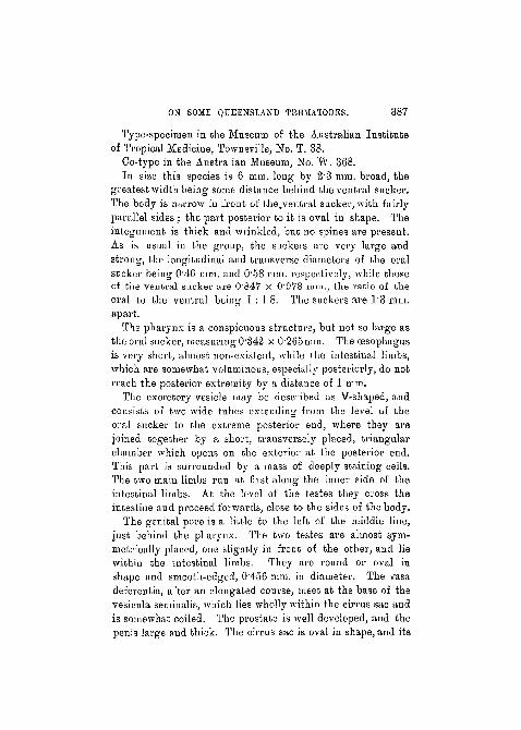
Type-specimen in the Museum of the Australian Instituteof Tropical Medicine, Townsville, No. T. 38.
Co-type in the Australian Museum, No. W. 368.In size this species is 6 mm. long by 23 mm. broad, the
greatest width being some distance behind the ventral sucker.The body is narrow in front of the.ventral sucker, with fairlyparallel sides; the part posterior to it is oval in shape. Theintegument is thick and wrinkled, but no spines are present.As is usual in the group, the suckers are very large andstrong, the longitudinal and transverse diameters of the oralsucker being 0'46 mm. and 0'58 mm. respectively, while thoseof the ventral sucker are 0'847 x O978 mm., the ratio of theoral to the ventral being 1 : 18. The suckers are 1'3 mm.apart.
The pharynx is a conspicuous structure, but not so large asthe oral sucker, measuring 0'342 x 0"265mm. The oesophagusis very short, almost non-existent, while the intestinal limbs,which are somewhat voluminous, especially posteriorly, do notreach the posterior extremity by a distance of 1 mm.
The excretory vesicle may be described as V-shaped, andconsists of two wide tubes extending from the level of theoral sucker to the extreme posterior end, where they arejoined together by a short, transversely placed, triangularchamber which opens on the exterior at the posterior end.This part is surrounded by a mass of deeply staining cells.The two main limbs run at first along the inner side of theintestinal limbs. At the level of the testes they cross theintestine and proceed forwards, close to the sides of the body.
The genital pore is a little to the left of the middle line,just behind the pharynx. The two testes are almost sym-metrically placed, one slightly in front of the other, and liewithin the intestinal limbs. They are round or oval inshape and smooth-edged, 0-456 mm. in diameter. The vasadeferentia, after an elongated course, meet at the base of thevesicula seminalis, which lies wholly within the cirrus sac andis somewhat coiled. The prostate is well developed, and thepenis large and thick. The cirrus sac is oval in shape, and its

388 S. J. JOHNSTON.
walls are thick and muscular. The ovary is three-lobed, thelobes being somewhat indistinctly marked off by shallowgrooves. It lies close in front of the right testis, and issmaller than that body, being 0"277 mm. in diameter. A smallshell-gland lies on its left side iu the middle line. There is aLaurer's canal, but no receptaculum seininis.
The coils of the uterus, in closely placed transverse loopson the left side, run back to the posterior end of the body—in the worm from which h'g. 12 was drawn they do not extendso far back as usual—while the ascending loops keep mainlyto the right of the middle line up to the level of the testes.From.this point the uterus has a pretty straight course to thegenital opening; the vagina is not conspicuously developed.The coils of the uterus are entirely confined to the spacebetween the intestinal limbs. The yolk-glands consist offrom six to eig'ht tree-like groups of small follicles on eachside. The follicles are about 0'017 mm. in diameter, andthere are 500 or 600 of them in each group. The number ofgroups on each side of the body is sometimes different. Eachgroup opens by a separate duct into the longitudinal duct.The yolk-glands lie iu the lateral field of the body outside theintestinal limbs, and extend forwards as far as the anteriorborder of the testes, whilst behind they extend a little beyondthe middle point between the testes and the posterior endof the body.
The oval eggs are thick-shelled (fig. 12, a aud b) with anoperculum at the narrow end, while the broad end is .oftenprovided with a blunt spike. The eggs measure 0-043-0'048mm. long by 0"032-0'035 mm. broad, aud the shells are0'0027 mm. thick.
A single immature specimen was found amongst the full-grown ones. It was 2"58 mm. long by 1*17 mm. broad. Thesuckers and pharynx bore the same relations in size as themature worms. The testes measured 0'179 mm. in diameter;the ovary 0'098 mm. There were no eggs, nor could anytrace of an uterus be made out.
In addition to the specimens from the gullet of the black

ON SOilE QUEENSLAND TREMATODES. 389
toadfish, a large number of specimens were obtained from thegullet of the spotted toadfish, and they appear to me not todiffer from this species except in their smaller size. Theymeasured up to 4'25 mm. long by 1*78 mm. broad.
While I place this species in Odhner's genus Steringo-trema, it appears to be more closely related to Distomumvibex Lintou (27, p. 291, figs. 48-51) than to any of thethree species enumerated by Odhner (51). Distomumvibex Linton evidently belongs to the same genus. S'.pulchrum differs from Linton's species mainly in the dis-position of the yolk-glauds, in the shorter intestinal limbs, inthe more confined distribution of the uterine loops, aud in thesize and shape of the eggs. It differs from S. cluthensisNicoll, S. pagell i van Ben. and S. divergens Rud.in size, in the relative sizes of the suckers, in the very shortoesophagus, in having the post-acetabular considerably longerthan the pre-acetabular region, and iu the very differentdisposition of the yolk-glands, as well as differing from eachof the three species named iu a number of other points.
Fam. Govgoderidee.
Sub-fam. Anaporrhutinas Lss.
Petalodistonium1 gen. nov.
Diagnosis.—Posterior part of the body very broad, almostcircular and plate-like. Muscular pharynx present;short oesophagus. Genital pore at or behind the intestinalfork. Cirrus sac very weak ; testes deeply lobed and dividedinto several distinct pieces, or broken up into a large numberof rounded follicles, lying wholly outside the intestinallimbs. Large receptaculum seminis present but no Laurer'scanal. Yolk-glands lying wholly within the intestinallimbs.
Type P. polycladum. Parasitic in the sting-ray, Dasy-batis kuhli i .
1 •KiTakov, a plate.

390 S. J. JOHNSTON.
The genus is closely related to Probo l i t r ema Lss.,differing from it principally in the fact that its yolk-glandsare close together, lying within the space bounded by theintestinal limbs, while in the latter the yolk-glands are farapart and lie in the lateral part of the body quite outside theintestinal limbs (Looss, 33, pp. 860 and 863, and Monticelli,42, tav v, fig. 52). It differs from AnaporrhutuniOfenheim in the testes being wholly outside the intestinallimbs.
Pe ta lod is tomum poly clad ura,1 sp. n. (Fig. 13.)
Diagnosis.—Under middle size, petal-like, with theposterior part of the body almost circular. Ratio of oralto vent ra l sucker 1 : 1"6. Branched intestinal limbs.Genital pore at the level of the intestinal fork. Testesvery large and more or less compact but divided intotwo or three pieces. Vesicula seminalis long, tubu la r andcoiled. Ovary t r i - lobed. Yolk-glands in two sets ofsmall rounded follicles, close together, not extendingoutwards beyond the intestinal limbs.
Found in the body-cavity of the sting-ray, Dasyba t i skuhlii .
Type-specimen in the Museum of the Australian Instituteof Tropical Medicine, Townsville, No. T. 39.
Co-type in the Australian Museum, Sydney, No. W. 369.Four specimens of this species, two of which were sectioned,
were obtained from the body-cavity of the sting-ray. The pos-terior part of the body is almost circular, with a short bluntanterior part. It maybe compared to the petal of a flower inwhich the lamina is circular and the claw short and blunt. Thelength of the animal varies from 3"3 to 3"76 mm., thebreadth from 3 to 3'5 mm. The integument is smooth,without spines of any kind. The mouth-opening is terminal,the oral sucker bowl-shaped and deep, but the ventral sucker
1 wokve and xXaloq (branch), referring to the branches of the intestinallimbs.
ON SOME QUEENSLAND TREMATODES. 391
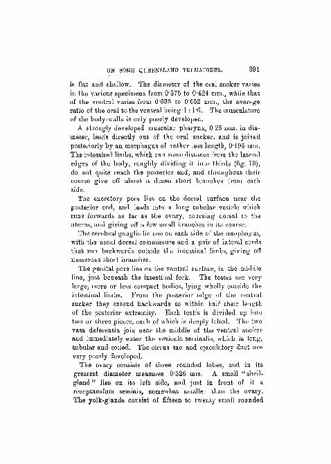
is flat and shallow. The diameter of the oral sucker variesin the various specimens from 0375 to 0'424 mm., while thatof the veutral varies from 0 6 3 6 to 0'652 mm., the averageratio of the oral to the ventral being 1 : 1"6. The musculatureof the body-walls is only poorly developed.
A strongly developed muscular pharynx, 0 2 5 mm. in dia-meter, leads directly out of the oral sucker, and is joinedposteriorly by an oesophagus of rather less length, 0"195 mm.The intestinal limbs, which run some distance from the lateraledges of the body, roughly dividing it into thirds (fig. 13),do not quite reach the posterior end, and throughout theircourse give off about a dozen short branches from eachside.
The excretory pore lies on the dorsal surface near theposterior end, and leads into a long tubular vesicle whichruns forwards as far as the ovary, coursing doi-sal to theuterus, and giving off a few small branches in its course.
The cerebral ganglia lie one on each side of the oesophagus,with the usual dorsal commissure and a pair of lateral cordsthat run backwards outside the intestinal limbs, giving offnumerous short branches.
The genital pore lies on the ventral surface, in the middleline, just beneath the intestinal fork. The testes are verylarge, more or less compact bodies, lying wholly outside theintestinal limbs. From the posterior edge of the ventralsucker they extend backwards to within half their lengthof the posterior extremity. Each testis is divided up iutotwo or three pieces, each of which is deeply lobed. The twovasa deferentia join near the middle of the ventral suckerand immediately enter the vesicula seminalis, which is long,tubular and coiled. The cirrus sac and ejaculatory duct arevery poorly developed.
The ovary consists of three rounded lobes, and in itsgreatest diameter measures 0"326 mm. A small "shel l -g l a n d " lies on its left side, and just in front of it areceptaculum seminis, somewhat smaller than the ovary.The yolk-glands consist of fifteen to twenty small rounded
392 S. J. JOHNSTON.
follicles (0'06-i mm. in diameter) grouped in two fairly com-pact masses. They do not extend outwards beyond the inneredge of the intestinal limbs, but lie fairly close together,one on either side of the ovary. This does not quite agreewith Looss' definition of the sub-family, "Dotterstocke auseinauder geriickt" (33, p. 863), aud perhaps that definition isa little too narrow. The short, directly transverse yolk-ductsmeet iu a small yolk-reservoir lying ventral to the "shell-gland" and opening into the ootype by a comparatively longduct.
The coils of the uterus, never far from the middle longi-tudinal Hue, are confined laterally to the space within theintestinal limbs, but they extend a little further back, runningout between the two ends of the intestine.
Tlie eggs are rather long and narrow, 0'052-0/063 mm. inlength by 0023 mm. broad.
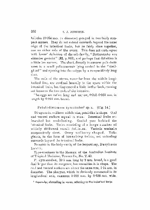
Pe talodistomum cymatodes1 sp. n. (Fig. 14.)
Diagnosis.—Above middle size, petal-like in shape. Oraland ventral suckers equal in size. Intestinal limbs un-branched but undu la t i ng . Genital pore behind theintestinal limbs. Testes consisting of a la rge number ofwidely diffused small fol l icles. Vesicula seminaliscomparatively short. Ovary mulber ry shaped. Yolk-glands, in the form of b ranch ing tubes , not extendingoutwards beyond the intestinal limbs.
Parasitic in the body-cavity of the leopard ray, Dasyba t i sk u h 1 i i.
Type-specimen in the Museum of the Australian Iustituteof Tropical Medicine, Towusville, No. T.40.
P. cymatodes , 10'5 mm. long by 8 mm. broad, is a gooddeal larger than its congener, but resembles it in shape. Theoral and ventral suckers are about the same size, 1*14 mm. indiameter. The pharynx, which is obviously contracted in itslongitudinal axis, measures 0-293 mm. by 0'538 mm. wide.
1 Kv^aruSiie, abounding in waves, referring to the intestinal limbs.

ON SOME QUEENSLAND TREMATODES. 393
The intestinal limbs are unbranched, but are thrown into anumber of snake-like undulations.
The genital pore is in the middle line, midway betweenthe intestinal fork and the anterior edge of the ventralsucker. The vesicula seminalis and cirrus sac are short, andsmaller than in P. polycladum.
The testes consist of about fifty small rounded follicles oneach side, O"1O7-O"129 mm. in diameter, and lying more orless dispersed from one another, in the region between theintestinal limbs and the lateral edges of the body. Theducts from the separate follicles join up in groups of five orsix and enter a main longitudinal vas deferens on each side;these two vessels join one another at the base of the vesiculaseminalis.
The ovary (0'375by 0244 mm.) is comparatively small, andis mulberry shaped. The receptaculum seminis is muchlarger, O73 x 057 mm.
The yolk-glands, on each side, consist of a much-branchedtube rather than of follicles, and, while lying further apartthan in P. polycladum, do not stretch outwards beyondthe inner limit of the intestinal limbs. The extension of theuterus also coincides with that of P. polycladum. Theeggs are larger, and especially broader than in the last-namedspecies, being 006—0064 in length by 0'03 mm. broad, andmany of them are seen to be provided with a short spike atone end.
Only a single specimen of this worm was obtained fromits host, the leopard ray, Dasybat i s kuhl i i , where itoccurred in the body-cavity.
Prom the Australian Institute of Tropical Medicine andthe Biological Department of the University of Sydney.
LITERATURE.
1. Arnsdorff, A.—"Monostomnm viciirium n. sp." ' Centralb.Bakt.,' vol. xlvii, 190S, p. 362.
2. Beneden, P. J. van.—" Memoir aur les Vers intestinaux," ' O.B.Acad. Sc. Paris,' 1858, Supplement.
VOL. 59, PART 3.—NEW SERIES. 25
394 S. J. JOHNSTON.
3. Befctendorf, H.—" Ueber Musculatur und Sinneszellen der Trema-toden," ' Zool. Jalirb. Anat.,' x, 1897, pp. 317-358.
4. Brandes, G.—" Revision der Monostomiden," ' Centailb. Bakt.,' xii,1892, p. 504.
5. Braun, M.—" Vernies," in ' Bronn. Klaas. Ordn. Tierveich. Leipsig,'1892 and 1893.
6. " Bericht uber thierische Parasiten," ' Csntralb. Bakt.,' xiii,1893, pp. 176-190.
7. " Cber Distomtuu cucuinerinum Rud.," ' Zool. Anz.,' xxii,1899, p. 465.
8. " Znr Kenntnis der Trematoden der Siiuge-thiere," ' Zool.Jahrb.,' xiv, 1901, p. 346.
9. " Trematoden der Chelonier," ' Mitteilungen aus dem Zool.Mus. Berlin,' 1901.
10. Cobn, L.—" Mitteilungen iiber Trematoden," ' Zool. Anz.,' xxv, 1902,p. 712.
11. " Helminthologische Mitteilungen," 'Arch. f. Naturg.,'lxix,1902, p. 53.
12. "Helminthologische Mitteilungen," 'Arch..f. Naturg.,' lxx,1904, p. 229.
13. Dietz, E.—"Die Echinostomiden der Vogel," ' Zool. Jahr.,' Suppl.xii, 1910, pp. 265-512.
14. Fischer, P. M.—" Uber den bau von Opi s tho t rema cochlearenov. gen. nov. spec, Ein Beitrag zur Kenntnis der Trematoden,"' Zeit. wiss. Zool.,' xl, 1884, pp. 1-41.
15. Fuhrmann, O.—" Neue Trematoden," ' Centralb. Bakt.,' xxxvii,1904, p. 58.
16. Gilbert, N. C. —" Occurrence of Echinostomum spinulo-suni R.," ' Amer. Natural.,' xxxix, 1905, p. 925.
17. Jfigerskiold, L. A.—" Uber den Bau von Ogmogaster plicatusCreplin," ' Svensk. Vetensk. Acad. Handl. Stockholm,' vol. xxiv,1891, pp. 1-32.
18. Kossack, W.—" Cber Monostomiden," ' Zool. Jahrb. Syst.,' xxxi,1911, pp. 419-590.
19. Lebour, Marie.—" Fish Trematodes of the Northumberland Coast,"in the ' Northumberland Sea Fisheries Report for 1907.'
20. " Trematodes of the Northumberland CoastNo. II," ' Trans.Nat. Hist. Soc. Northumberland, etc.,' new series, vol. ii, pt. i.
21. Leiper, R. T.—"An Account of some Helminths contained in Dr.C. M. Wenyon's Collection from the Sudan," in the ' Third Reportof the Wellcome Research Laboratories, Khartoum.'
ON SOME QUEENSLAND TREHATODES. 395
22. Linstow, 0. von.—" Einige neue Distomen nnd Bemerkungen iiberdie weiblichen sexualorgane der Trematoden," 'Arch. Naturg./xxxix, 1873, p. 95.
23. " Neniatoden, Treniatoden und Acantliocephalen gesammeltvon Prof. Fedtschenko in Turkestan," ' Arch. Naturg.,' xlix, 1883.
24. "Neue Helminthen," ' Centralb. Bakt.,' xxxvii, 1904, p. 078.25. Linton, E.—"Notes on Trematode Parasites of Pishes," ' Proc.
U.S. National Mus.,' xx, 1897.26. "Parasites of Fishes of the Woods Hole Region," 'Bull.
U.S. Fish Commission,' xix, 1899, pp. 405-492.27. "Fish Parasites collected at Woods Hole in 1898," 'Bull.
U.S. Fish Commission,' xix, 1899, pp. 267-304.28. " Notes on Parasites of Bermuda Fishes," ' Proc. U.S.
National Museum,' xxxiii, 1907, pp. 85-126.*29. Loose, A.—" Recherches sur la Faune Parasitaire de l'Egypte 1st
pt.," ' Mem. Inst. Egypt in Cairo,' 1896, Referat by M. Braun in' Centralb. Bakt.,' xx, 1896, pp. 107-116.
30. " Weitere Beitriige zur Kenntniss der Trematoden—Fauna^Igyptens," u.s.w., ' Zool. Jahrb. Syst.,' xii, 1899.
31. " Notizen zur Helminthologie Bgyptens IV. Ueber Trema-toden aus Seeschildkroten der egyptischen Kusten," ' Centralb.Bakt.,' xxx, 1901, p. 566.
32. " Na-tura doceri," ' Centralb. Bakt.,' xxix, 1902, p. 192.33. " Trematoden aus Seeschildkroten, etc.," ' Zool. Jahrb. Syst.,'
xvi, 1902.
34. "Einige Zum Teil neue Distomen der Europaischen Fauna,"' Centralb. Bakt.,' xliii, 1907, p. 604.
35. Liihe, M.—"Beitrage zur Helminthenfauna der Berberei," 'S . B.Akad. Wiss. Berlin,' 1898, p. 619.
36. "Uber Monostomum orbiculare," 'Centralb. Bakt.,'xxix, 1901, p. 49.
37. Anhang to " Zwei neue Distomen aus indischen Anuren,"1 Centralb. Bakt.,' xxx, 1901, p. 174.
38. MacCalluni, W. G.—" Echinos tonium garzetta) n. sp.," ' Zool.Jahrb. Syst.,' xx, 1904, pp. 541-548.
39. Magalhaes, P. S.—"Notes d'hehninthologie brcsilienne, 5," 'Arch.Parasit.,' vol. i, 1898, p. 361.
* The works marked by an asterisk (*) have not been accessible tome, so that I have had to depend on abstracts of them.

396 S. J. JOHNSTON.
40. Monticelli, F. S.—" Studii sui Trematodi endoparas&iti. Monosto-mum cymbium Dies, Contribuzione allo studio dei Monostomidi,"' Mem. B. Accad. Sc. Torino,' serie ii, torn. xlii.
*41. " Studii sui Trematodi endoparassiti. S\il genere Noto-cotyle Diesing," ' Boll. Soc. Natural Napoli,' 1, vol. vi, 1892,p. 26.
42. " Studii sui Trematodi endoparassiti. Primo contributo diosservazione sui distomidi," ' Zool. Jahrb. Suppl.,' 3, 1893.
43. Miihling P.—" Die Helminthenfauna der Wirbelthiere Ost-preussens," 'Arch. Naturg.,' lxiv, 1898, pp. 1-118.
44. Muller, A.—" Helminthologische BeobLichtungen an bekannten undunbekannten Bntozoen," ' Arch. Naturg.,' lx, 1894 p. 113.
45. Nicoll, W.—" Some new and little known Trematodes," ' Ann. Mag.Nat. Hist.,' 7, vol. xvii,p. 513.
46. " Studies on the Structure and Classification of the DigeneticTrematodes," ' Quart. Journ. Micr. Sci.,' vol. 53, 1909, pp.391-487.
47. Odhner, T.—"MitteilungenzurKenntnissderDistomen,"'Centralb.Bakt.,' xxxi, 1902, p. 55.
48. '•' Die Trematoden des arktischen Gebietes," ' Romer u.Schaudin, Fauna ai-tica,' vol. iv, p. 292. Jena, 1905.
49. "Der wahre Bau des Synaptobothr ium copulans v.Linstow," ' Zool. Anz.,' xxx, 1906.
50. " Nordostafrikanische Trematoden," ' Results of the SwedishExpedition to Egypt and the White Nile.' Uppsala, 1911.
51. " Znm naturlichen System der digenen Trematoden, III,"' Zool. Anz.,' xxxviii, 1911, pp. 97-117.
52. Ofenheirn, E. Von.—" Uber erne neue Distomidengattimg,"' Zeitschr. f. Naturwiss.,' lxxiii, 1900, p. 145.
*53. Parona.—" CatalogO di Elminti . . . Isola d'Elba," ' Boll.Mus. Zool. Anat. comp. Univ. Genoa,' No 77, 1899.
54. Stafford.—" Trematodes from Canadian Fishes," ' Zool. Anz.,'xxvii, 3904, p. 481.
55. Stiles and Hassall.—" An Inventory of the Genera and Subgeneraof the Trematode family Fasciolidai," 'Arch, parasit.,' 1, 1898,p. 97.
56. " Index Catalogue . . . Trematoda and TrematodeDiseases," 'Bull. No. 37 of the Hygienic Lab., Washington,' 1908.
*57. Stossich, M.—"II Monostomum mutabile Zeder e le sue formeaffini," 'Boll. Soc. Adriat.,' xxi, 1902. Summary by Braun in' Zool. Central!).,' ix, pp. 406, 407.

ON SOME QUEENSLAND TEJEMATODBS. 397
EXPLANATION OF PLATES 22—27,
illustrating Mi*. S. J. Johnston's paper " On some QueenslandTrematodes, with Anatomical Observations and Descrip-tions of Now Species and Genera."
[The drawings, winch were done by Mr. F. W. Atkins, of the TechnicalCollege.. Sydney, were all made with the help of the camera lucida.The micro-photographs are from " untouched-lip " negatives of sectionsand whole nwrants.]
REFEBENCE LETTERS.
c. g. Cerebral ganglion, c. s. Cirrus sac. Ej. d. Ejaculatory duct.E. Excretory vessel. Ex. j>. Excretory pore. Ex. v. Excretory vesicle.F. c. Flame-cell. G. p. Genital pore. G. s. Genital sinus, int. Intes-tinal limbs. L. c. Laurer's canal. L. t. Lateral nerve-trunk. M. o.Mouth-opening. N. Nervous system. (Es. (Esophagus. . 0. s. Oralsucker. 0. Ovary. 0. d. Oviduct. P. Penis. Fh. Pharynx. Pr.Prostate. B. s. Receptaculum seminis. 8. g. '' Shell-gland." Te. Testis.Ut. Uterus. Vug. Vagina. V. d. Vas deferens. V. s. Vesicula seiui-nalis. F. 8k. Ventral sucker. Y. d. Yolk-duct. Y. g. Yolk-glaud.Y. r. Yolk-reservoir.
Rhabdiopceus t ay lo r i , figs. 1-4.
Fig. 1.—Drawn from a cleared and transparent object mounted withthe dorsal side uppermost, x 8.
Fig. 2.—Posterior end, showing the multiple proboscis retracted intoits complex sheath, x 21.
Fig. 2A.—Egg from the proximal part of the uterus. X 550.
Fig. 2B.—Egg from the distal part of the uterus, x 550.
Fig. 3.—Transverse section through one of the proboscides about itsmiddle. x 110. C. c. Central cavity, c. in. Circular muscle-fibres.G. c. G-land-cell. L. m. Longitudinal muscle-fibres. R. Bundles ofrhabdites.
Fig. 4.—Transverse section through proboscis near its tip. X 230.11. = single rhabdites.

398 8. ,T. JOHNSTON.
Typhloccolum re t icu lare , figs. 5-7.
Fig. 5.—Whole mount, x 30.
Fig. 6.—Excretory system, x 21. Compiled from a series of hori-zontal longitudinal sections, by the camera lucida. F. c. Flame-cells.cap. Capillary vessel, net. Network of vessels.
Fig. V.—Nervous system, x 21. Compiled in the same manner andfrom the same series as used in fig. 6. 5̂. c. Posterior commissure.
Fig. 8.—Allopyge ant igones . x 7.Fig. 9.—Diaschistorcliis pandus. x 10. Unmounted specimen
viewed by direct light.
Fig. 10.—D. pandus . x 20. G. c. Gland-cells surrounding thetermination of the vagina.
Fig. 11.—Pleorchis ol igorchis . x 14. Viewed as a transparentobject from the dorsal aspect.
Fig. 12.—Steringotrema pulchrum. X 24.Fig. 12A. Egg with operculum.Fig. 12B. Egg from which the operculum has been removed. X S50.Fig. 13.—Petalodistomum polycladum. x 21.Fig. 14.—Petalodistomum cymatodes. x 11. Ex. v. Excretory
vessel.
MICRO-PHOTOGRAPHS.
Rhabdiopceus t ay lo r i , figs. 15-26.
Fig. 15.—Part of a longitudinal sagittal section showing spines, thickventral cuticle and the arrangement of the fibres in the muscular system,x 50. C. TO. Circular muscle. D. v.vi. Dorso-ventral muscle. L. in. Longi-tudinal muscle. N. c. Nerve-cord. O. m. Oblique muscle. Sp. Spine.
Fig. 1C.—Part of a transverse section showing the arrangement of themuscle-fibres in the muscular system of the body and in the cirrus sacand vagina. The section also shows the very thick cuticle and the spinesin transverse section, x 89.
Fig. 17.—Transverse section showing the bifid nature of the ventralspines, x 54.
Fig. 18.—Horizontal longitudinal section showing the relations ofthe nervous system, the alimentary canal and the excretory vessels.X 20.
Fig. 19,—Transverse section showing the brain, x 51.Fig. 20.—Transverse section through the proboscis chamber and
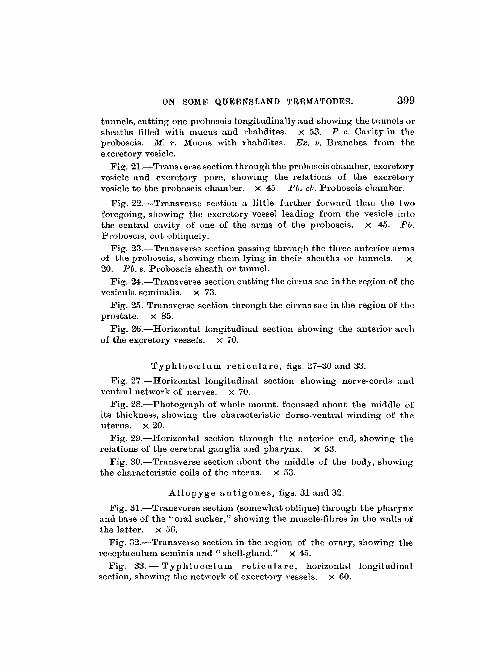
ON SOME QUEENSLAND TftEMATODES. 399
tunnels, cutting one proboscis longitudinally and showing the tunnels orsheaths filled with mucus and rhabdites. X 53. P. c. Cavity in theproboscis. M. r. Mucus with rhabdites. Ex. v. Branches from theexcretory vesicle.
Fig. 21.—Transverse section through the proboscis chamber, excretoryvesicle and excretory pore, showing the relations of the excretoryvesicle to the proboscis chamber, x 45. Ph. ch. Proboscis chamber.
Fig. 22.—Transverse section a little further forward than the twoforegoing, showing the excretory vessel leading from the vesicle intothe central cavity of one of the arms of the proboscis, x 45. Fb.Proboscis, cut obliquely.
Fig. 23.—Transverss section passing through the three anterior armsof the proboscis, showing them lying in their sheaths or tunnels. x20. Fb. s. Proboscis sheath or tunnel.
Fig. 24.—Transverse section cutting the cirrus sac in the region of thevesicula seminalis. x 73.
Fig. 25. Transverse section through the cirrus sac iu the region of theprostate, x 85.
Fig. 26.—Horizontal longitudinal section showing the anterior archof the excretory vessels, x 70.
Typhlocoeluni re t iculare , figs. 27-30 and 33.
Fig. 27.—Horizontal longitudinal section showing nerve-cords andventral network of nerves, x 70.
Fig. 28.—Photograph of whole mount, focussed about the middle ofits thickness, showing the characteristic dorso-ventral winding of theuterus, x 20.
Fig. 29.—Horizontal section through the anterior end, showing therelations of the cerebral ganglia and pharynx, x 53.
Fig. 30.—Transverse section about the middle of the body, showingthe characteristic coils of the uterus, x 53.
Allopyge ant igones , figs. 31 and 32.
Fig. 31.—Transverse section (somewhat oblique) through the pharynxand base of the "oral sucker," showing the muscle-fibres in the walls ofthe latter, x 56.
Fig. 32.—Transverse section in the region of the ovary, showing thereceptaculum seminis and " shell-gland." X 45.
Fig. 33.— Typhloccelum re t icu la re , horizontal longitudinalsection, showing the network of excretory vessels, x 60.
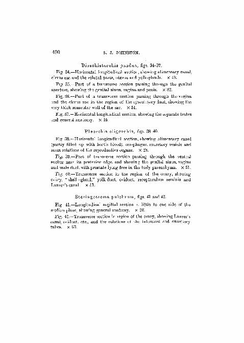
400 S. J. JOHNSTON.
Diaschisfcorchis pandus , figs. 34-37.Fig. 34.—Horizontal longitudinal section, showing alimentary canal,
cirrus sac and the related parts, uterus and yolk-glands. X 48.Fig. 35.—Part of a transverse section passing through the genital
aperture, showing the genital sinus, vagina and penis, x 82.Fig. 36.—Part of a transverse section passing through the vagina
and the cirrus sac in the region of the ejaculatory duct, showing thevery thick muscular wall of the sac. X 51.
Fig. 37.—Horizontal longitudinal section, showing the separate testesand general anatomy, x 15.
P leorch is o l igorchis , figs. 38-40.
Fig. 38.—Horizontal longitudinal section, showing alimentary canal(partly filled up with host's blood), cesophagns, excretory vesicle andmain relations of the reproductive organs, x 24.
Fig. 39.—Part of transverse section passing through the ventralsucker near its posterior edge, and showing the genital sinus, vaginaand male duct, with prostate lying free in the body parenchyma, x 51.
Fig. 40.—Transverse section in the region of the ovary, showingovary, " shell - gland," yolk-duct, oviduct, receptaculum seminis andLaurer's canal. X 51.
S te r ingot rema pulchrum, figs. 41 and 42.
Fig. 41.—Longitudinal sagittal section a little to one side of themedian plane, showing general anatomy, x 2G.
Fig. 42.—Transverse section in region of the ovary, showing Laurer'scanal, oviduct, etc., and the relations of the intestinal and excretorytubes, x 53.
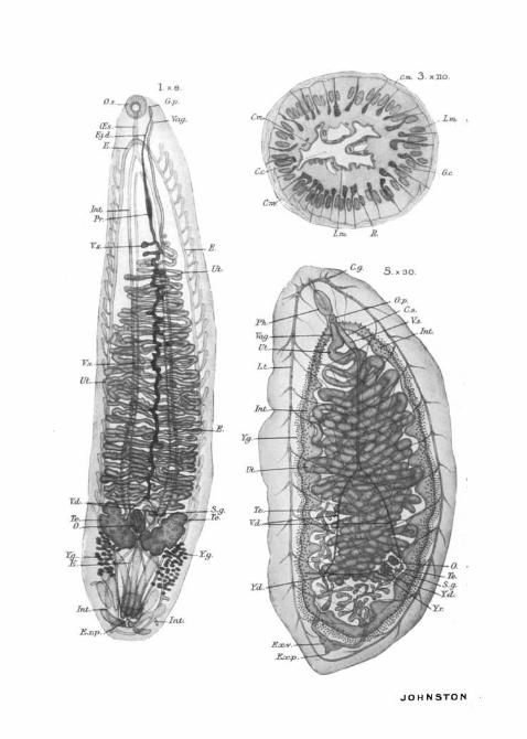
Cm. 3 . xHO.
O.s.
JOHNSTON -

TREMATO DES.
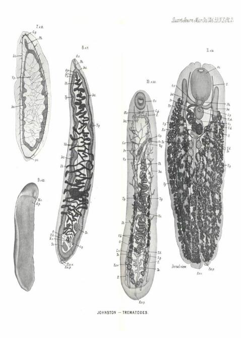
ScofVbC. 59JT.S.M. 23.7. xai.
8 . X 7.
O.s.PK.
Int.
Int.
10.x 20.
H. xl*.
Moo.p.
JOHNSTON —TREMATODES.
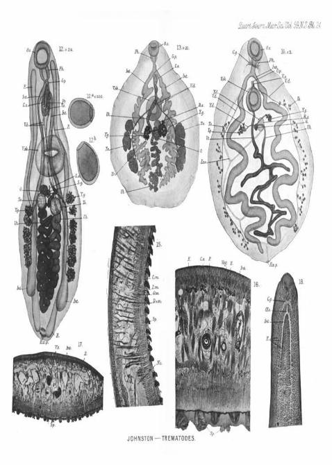
. 24
O.S. O.s.14. xJL.
JOHNSTON TREMATODES.sP.-

Sr. (ES.
JOHNSTON — TREMATODES.
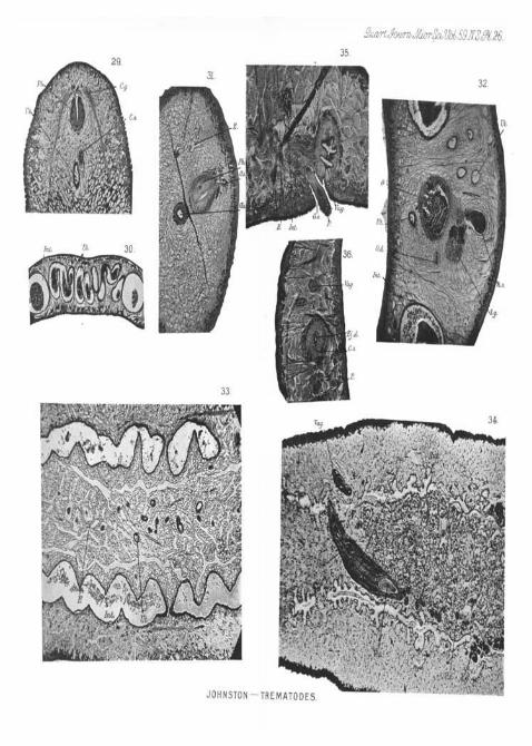
m
JOHNSTON —TREMATODES.
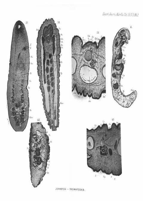
\
Im*.
JOHNSTON —TREMATODES.