on G rap h e n e Ox i de for D op ami n e D e te r mi n ...
22
Page 1/22 A Simple Fluorescent Aptasensing Platform Based on Graphene Oxide for Dopamine Determination Ahlem Teniou Ecole Nationale Superieure de Biotechnologie de Constantine amina rhouati ( [email protected] ) ECOLE NATIONALE SUPERIEURE DE BIOTECHNOLOGIE https://orcid.org/0000-0001-9782-9728 Gaëlle Catanante Universite de Perpignan Research Article Keywords: Dopamine, Aptamer, Fluorescence resonance energy transfer, Graphene oxide, Fluorescence quenching Posted Date: September 24th, 2021 DOI: https://doi.org/10.21203/rs.3.rs-922691/v1 License: This work is licensed under a Creative Commons Attribution 4.0 International License. Read Full License Version of Record: A version of this preprint was published at Applied Biochemistry and Biotechnology on January 8th, 2022. See the published version at https://doi.org/10.1007/s12010-022-03802-1.
Transcript of on G rap h e n e Ox i de for D op ami n e D e te r mi n ...

Page 1/22
A Simple Fluorescent Aptasensing Platform Basedon Graphene Oxide for Dopamine DeterminationAhlem Teniou
Ecole Nationale Superieure de Biotechnologie de Constantineamina rhouati ( [email protected] )
ECOLE NATIONALE SUPERIEURE DE BIOTECHNOLOGIE https://orcid.org/0000-0001-9782-9728Gaëlle Catanante
Universite de Perpignan
Research Article
Keywords: Dopamine, Aptamer, Fluorescence resonance energy transfer, Graphene oxide, Fluorescencequenching
Posted Date: September 24th, 2021
DOI: https://doi.org/10.21203/rs.3.rs-922691/v1
License: This work is licensed under a Creative Commons Attribution 4.0 International License. Read Full License
Version of Record: A version of this preprint was published at Applied Biochemistry and Biotechnology onJanuary 8th, 2022. See the published version at https://doi.org/10.1007/s12010-022-03802-1.

Page 2/22
AbstractDopamine (DA) is a catecholamine neurotransmitter playing an important role in different biologicalfunctions including central nervous, renal, cardiovascular, and hormonal systems. The sensitive andselective detection of this neurotransmitter plays a key role in the early diagnosis of various diseasesrelated to abnormal levels of dopamine. Therefore, it is of great importance to explore rapid, simple, andaccurate methods for detection of dopamine with high sensitivity and speci�city. We propose in this work,a �uorescent aptasensor based on graphene oxide (GO) as a quencher, for the rapid determination ofdopamine. The principle of this aptasensor is based on �uorescence resonance energy transfer (FRET),where GO was used as energy donor, and a carboxy�uorescein (FAM)-labeled aptamer as acceptor. In theabsence of DA, FAM-aptamer was adsorbed on the surface of GO through π-π stacking interactionsbetween nucleotide bases and the carbon network, leading to a weak FRET and a quenching of the FAM�uorescence. However, by adding the target, the aptamer undergoes a conformational change to bind toDA with high a�nity, resulting in a �uorescence recovery. Under the optimal experimental conditions, the�uorescence recovery was linearly proportional to the concentration of DA in the range of 3-1680 nM, witha limit of detection of 0.031 nM. Moreover, the developed assay exhibited minor response in the presenceof various interferents and it revealed a satisfactory applicability in human serum samples.
IntroductionDopamine (DA), is an important neurotransmitter that has a potential vital role in regulating functionalactivities such as the central nervous, renal, hormonal, and cardiovascular systems [1, 2]. In addition, DAhas an important effect on stress, behavior, attention, and other cognitive functions. It has been reportedthat high dopamine levels induce cardiotoxicity leading to rapid heart rates, hypertension, heart failure,and drug addiction [3, 4]. In parallel, a low dopamine level may be considered as a major cause ofpsychiatric disorders such as Parkinson [5], schizophrenia [6], depression [7]…etc [8, 9]. Therefore, theaccurate and rapid measurement of DA is crucially required for the diagnosis of these diseases [10, 11].Dopamine analysis is usually based on enzyme assays, liquid chromatography, mass spectroscopy, andcapillary electrophoresis [12–15]. However, most of these techniques are time-consuming and involvesophisticated pre-treatment process, expensive instruments and expertise for operation thus limiting theirapplications in routine detection of DA [16–18]. Great efforts are devoted for developing simple, accurate,and inexpensive methods for detecting DA in biological samples with high sensitivity and selectivity.
Among the different technologies, �uorescence resonance energy transfer (FRET) constitutes a promisingtechnique promoting diagnostics. It is a non-radiative energy transfer process that occurs through dipole-dipole interaction between an emitter (donor) and an absorber molecule (acceptor) often called the “FRETpair” [19]. FRET can only occur when the intermolecular distance between donors and acceptors issmaller than 10 nm, which allow the detection of interaction between molecules, thus providing accurateinformation at the nanoscale [20]. This phenomenon is based on the excitation of a donor �uorophoreaccompanied with an emission spectrum that overlaps the excitation spectrum of an acceptor, in veryclose proximity [20, 21]. In this context, �uorescently-labeled aptamers can be used as bioreceptors to
Page 3/22
develop several aptasensors using FRET as a detection method. Based on this principle, the targetamount is measured by monitoring the �uorescence change induced by the modi�cation of the aptamerconformation [22, 23].
Aptamers are short sequences of DNA or RNA, characterized with high a�nity and speci�city for theirtargets [24]. They are selected in vitro by the exponential enrichment process named SELEX (systematicevolution of the ligand by the exponential enrichment process) [25]. As compared to antibodies, aptamersare more stable and economical because they are chemically synthesized [26–28]. In addition, aptamerscan be functionalized with different probes including chemical groups [29], enzymes [30] and�uorophores [31]. Aptamer functionalization with a �uorescent probe provides a fundamental advantagein FRET to control the distance between acceptor and donor molecules [32]. Theoretically, any knownaptamer can be engineered into a molecular aptamer beacon (MBA) that shows a FRET in response to aspeci�c biomarker [33–35]. Traditional MBA are composed of a �uorophore and a quencher, eachattached at the end of hairpin-structured oligonucleotides. The �uorescence is quenched as a result of theproximity between the �uorophore and its quencher. Then, it will be recovered once the optimal distancebetween the FRET couple is broken [21, 36]. However, traditional MBs suffer from some limitations, suchas false-positive signals, high-cost synthesis, and di�cult selection of dye-quencher pair in certain cases[37]. Therefore, new types of MBs based on nanomaterials, such as gold nanoparticles (AuNPs), quantumdots (QDs), graphene oxide (GO), and carbon nanotubes (CNTs), have been developed as promisingcandidates for biosensor design [38, 39].
Due to its unique properties, GO has emerged as one of the most extensively studied nanomaterial. It is atwo-dimensional (2-D)-layered material synthesized by the oxidation of graphene [40, 41]. GO hasexcellent electronic, thermal, mechanical and photophysical properties. It has a great potential ofapplications ranging from molecular electronics to ultrasensitive biosensing [42, 43]. It has been shownthat GO has the ability to quench �uorescence of the adsorbed dyes. This is mainly due to the largeconjugated structure of GO which makes it an excellent electron acceptor during energy transferprocesses [44–46]. These characteristics offer very interesting opportunities to construct a variety ofbiosensors, in particular, aptasensors [45, 47]. Different GO-based aptasensors has been developed forthe detection of different targets such as glucose [48] and pathogens [49]. However, most of the�uorescent methods developed for dopamine aptasensing are mainly based on quantum dots [50, 51],nanoclusters[52–54], or nanoparticles [55–57]. The synthesis of such materials involves relativelycomplicated and time-consuming processes. In this work, we develop a simple �uorescent aptasensorbased on graphene oxide as a quencher for DA detection. The principle of this aptasensor is based onFRET by using the FAM-labeled aptamer as a donor and GO as acceptor. First, DA-aptamer was adsorbedonto the surface of GO where the �uorescence was e�ciently quenched. After the target binding, theaptamer changes its conformation in the way that it is released from GO surface, thus recovering the�uorescence that is theoretically proportional to the analyte concentration [58]. This homogeneous�uorescent assay exhibited a high speci�city towards DA with a detection limit of 0.031 nM. Besides, theapplication of this sensing system was demonstrated by detecting the DA levels in human serum.Compared with conventional methods, this sensing platform is simpler, faster, and shows

Page 4/22
comprehensible quantitative results. Moreover, it avoids expensive dual labeling of aptamer required inconventional molecular beacon-based platforms.
Material And MethodsReagents
The �uorescein amidite (FAM)-labeled dopamine aptamer with a sequence 5'-FAM-GTC TCT GTG TGCGCC AGA GAA CAC TGG GGC AGA TAT GGG CCA GCA CAG AAT GAG GCC C-3') was synthesized byMicrosynth company (Balgach, Switzerland, www.microsynth.ch). Graphene oxide (GO) solution,magnesium chloride (MgCl2), sodium chloride (NaCl), hydrochloric acid (HCl), Tris-hydroxymethyl-methane (Tris), glycine, glucose, lactic acid and Dopamine (DA) were purchased from Sigma Aldrich(https://www.sigmaaldrich.com). Cysteine was purchased from Biochem(https://www.biochemopharma.fr/). Human serum was obtained from a local biological analysislaboratory. The stock solution of GO (4mg/ml) was used to prepare a homogeneous GO aqueoussolutions of different concentration (0,2,3,4,5 µg/µl) using Tris-Hcl buffer (5 mM MgCl2, 0.5 M NaCl and50 mM Tris HCl, pH 7.4), and they were stored at room temperature prior to use. The stock aptamersolution (2µM) was prepared with Tris-HCl buffer (5 mM MgCl2, 0.5 M NaCl and 50 mM Tris HCl, pH 7.4),and stored at -20°C before use. The stock solution of DA (100µM) was also prepared using the same Tris-HCl buffer and stored at -4°C. Puri�ed water was used to prepare all the other solutions. All reagents areof analytical grade.
Apparatus
All �uorescence measurements were recorded with a microplate reader, employing a 96 well blackmicroplate. Aptamer pre-heating was occurred using a thermocycler (Bio-Rad).
Fluorescent detection of dopamine
The FAM-labeled aptamer (400 nM) was dissolved in Tris-Hcl buffer (5 mM MgCl2, 0.5 M NaCl and 50mM Tris HCl, pH 7.4), and then heated to 90°C for 5 min. After cooling at room temperature, 25 µl of FAM-labeled aptamer was incubated with 25 µl of different concentrations of DA for 25 min under vortexmixing to make sure that dopamine and DA-aptamer interacted with each other su�ciently. Then 25 µl ofGO (3 µg/ml) was added to the above solution, and the volume was completed to 200 µl using the Tris-Hcl buffer. The mixed solution was allowed to settle under a quit stirring for 30 min at room temperature.Finally, the �uorescence intensity was measured under the excitation and emission wave- lengths of 485and 538 nm, respectively. All experiments were repeated three times and carried out under the optimizedsensing conditions.
Interference studies

Page 5/22
Aiming to con�rm the speci�city of the proposed technique for dopamine detection, some interferingcompounds (Glycine, cycteine, Glucose, Lactic acid) were tested. The selectivity assay was conductedunder the same procedure as for DA detection. In brief, 25 µl of FAM-labeled aptamer (400 nM) dissolvedin Tris-HCl buffer were added to 25 µl of each interferents. Then, the mixture solution was incubated for25 min at room temperature, after that 25 µl of GO (3 µg/ml) was introduced to the solution. Finally, the�uorescence was measured after 30 min of quit stirring at room temperature.
Detection of DA in real samples
In order to con�rm the response of the sensor in real samples, human serum samples were diluted 10times with Tris-Hcl buffer. Then, the samples were spiked with different known concentrations of DA (3,7,280, 1120 nM). The �uorescence detection of DA in human serum sample was performed under the sameprocedure mentioned above.
Results And DiscussionMechanism of sensing detection
Figure 1 illustrates the sensing strategy for the DA detection based on the �uorescence resonance energytransfer (FRET) between aptamer and GO. In the absence of target molecule (DA), FAM-modi�ed aptameris adsorbed onto the surface of GO via hydrophobic and π-π stacking interaction between the nucleotidebases and the sp2 honeycomb network of carbon [59–61]. Therefore, these interactions induce theformation of a stable complex that will lead to quench the �uorescence of FAM easily through energytransfer from the FAM to GO [62]. However, in the presence of target molecule (DA), The conformation ofDA-aptamer is altered (target-induced allosteric effect), and switched from a random coil to rigid stem–loop structure-DA that have a weak a�nity to GO and keep the dyes away from graphene surface [63].Consequently, the FRET process will be hindered, and the FAM �uorescence is recovered and measured asa function of DA. In the experimental mixture, the �uorescence recovery increases by increasing DA-concentrations and the �uorescence will be proportional to the concentration of dopamine [64, 65]. On theother hand, the addition of interferents couldn’t change the conformation of DA-aptamer, so as it can’t bereleased from GO surface, resulting in non-emission of �uorescence due to the quenching effect of GO.
Based on ssDNA-GO interactions, Ye and collaborators [65] demonstrated the generation of a versatilemolecular beacon-like probe as a multiplex sensing platform. This probe has been effectively applied asan example of a �uorescent biosensor-based FRET method to detect a speci�c target, for instance:speci�c sequence of DNA, as well as thrombin, metal ions such as Ag+ and Hg2+, and amino acids suchas cysteine, with detection limits of 5 nM, 20 nM, 5.7 nM, and 60 nM, respectively.
Optimization of assay conditions
Aptamer and quencher concentrations, and incubation time are experimental variables that play animportant role in the fabrication of sensitive and selective aptasensing platforms based on FRET

Page 6/22
principle. In this context, different experiments have been realized to optimize aptamer and quencherconcentrations, as well as incubation time.
GO concentration
The choice of GO concentration is a critical factor because it may affect the performance of the sensingsystem. Optimizations were carried out by varying GO concentration between 0 and 5 µg/ml where theaptamer concentration was �xed at 400 nM. The quenching e�ciency was de�ned by 100% × (F–F0)/F,where F0 and F are the �uorescence intensities of the aptamer solution with and without GO, respectively.As shown in Fig. 2, the quenching of �uorescence increased dramatically with the increasing amounts ofGO, indicating that aptamers are still free in the solution. Then, the �uorescence quenching e�ciency (~ 75 %) stabilized when the GO concentration was higher than 3 µg/ml, indicating that the FAM-labeledaptamer was almost completely adsorbed onto the surface of GO. Since higher concentrations of GOhaven’t shown an enhanced quenching e�ciency, a concentration of 3 µg/ml was selected as the GOamount generating the optimum quenching. Thus, it has been used in the next experiments.
GO quenching time
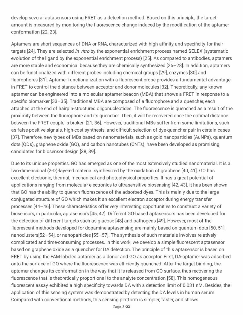
The effect of reaction time between the aptamer and GO on the �uorescence quenching of FAM probewas also investigated. In this context, different incubation times have been tested ranging from 0 to 30min. As we can see from Fig. 3, the quenching e�ciency increased by increasing the reaction timeindicating that the FAM-aptamer was adsorbed onto the surface of GO. After 10 min of incubation, thesignal reached the maximum of 75% of quenching and stabilized. Therefore, 10 min was chosen to bethe optimal incubation time for GO and FAM-aptamer.
Aptamer concentration
To obtain the maximum sensitivity of biosensing, four different concentrations of aptamer (200, 400, 600and 800 nM) were tested. According to the results presented in Fig. 4, the change of �uorescenceintensity kept increasing gradually with increasing aptamer concentrations. The optimal concentration isdetermined according to the sensitivity of the proposed aptasensor. It should be noted that a highconcentration of FAM-aptamer provides a better �uorescence signal. However, a high concentration ofFAM-aptamer may in�uence the assay sensitivity leading to an erroneous results [66]. Therefore, theaptamer concentration of 400 nM was adopted to perform the next experiments.
Detection of DA
The sensitivity of any detection method is considered as a key factor to determine its applicability. It wasinvestigated by monitoring the �uorescence intensity of increasing concentrations of DA at the emissionwavelength of 538 nm. The �uorescence intensity was quanti�ed by calculating the percentage of�uorescence recovery [(F-F0)/F0 × 100], where F is the �uorescence intensity in different dopamineconcentrations and F0 is the �uorescence intensity in the absence of dopamine. The calibration curve

Page 7/22
presenting the �uorescent recovery percentages as function of DA concentrations ranging from 3 nM to1680 nM was illustrated in Fig. 5. It was revealed that with increasing the concentration of DA, the�uorescence recovery increased gradually with a good linear relationship (Regression coe�cient R2 = 0.997), indicating the growth of the number of FAM-aptamer attached to the DA target even at lowconcentrations (3 nM). The detection limit was estimated to be 0.031 nM based on 3δ/S calculation (δ isthe standard deviation for the blank solution, and S is the slope of the calibration curve).
The performance of our technique was compared to other electrochemical and optical aptasensors,previously reported in the literature for dopamine detection. It has been noted that the LOD achieved inthis new approach is much lower than those of these techniques (Table 1). The high sensitivity of ouraptasensor could be attributed to the high a�nity of the aptamer toward DA, and the ultra-high�uorescence quenching ability of GO. The change of the conformation of the aptamer upon the additionof target molecules enhanced the distance between FAM-labeled signal probe and the surface of GO,inducing a higher �uorescence restoration. Moreover, since this assay was performed in a homogenoussolution, it avoided the need for time-consuming immobilization, coating and washing steps usuallyrequired in common heterogeneous assays [67–69]. Indeed, our biosensor provides a simple and rapiddetection of DA since it is based on monitoring the �uorescence change due to the target binding. Thesefeatures, as well as its other merits, such as low cost and �uorescence background, make it a promisingtool for a rapid label free detection of DA.

Page 8/22
Table 1Comparison between this method and other reported techniques for detection of dopamine
Detectionmethod
Detectionrange
nM
LOD
nM
Principle Refs
Electrochemical 5×10³-75×103
3.36 ×10³
DNA aptamer/AuNP/rGO/ modi�ed glassycarbon electrode.
[70]
5–300 2.1 Au-electrode/Au-NPs/PEI/CNTs. [67]
5–150 1 MB/MCH/DNA/Au electrode. [71]
1–1000 0.75 Aptamer/GCSC-GO/GCE. [72]
5 ×10³-50×103
1000 Split aptamer1/split aptamer 2 / Au-electrode.
[73]
5–5×10² 1.8 aptamer/AuNPs/GCE. [74]
Colorimetric 540–5400 360 Aptamer/ unmodi�ed AuNPs. [75]
200–1100 70 AHMT/ AuNPs. [76]
Fluorescence 26 − 2.90 × 10³
2 DNA/ rhodamine B/ AuNPs. [68]
0.1–10.0 0.08 tDNA/cDNA/aptamer/dopamine/hDNA/ ExoIII.
[69]
50 − 2.5×10⁵ 10 CQDs/AuNPs/aptamer. [77]
5× 10⁴ − 106 104 CB/ Fe2+/ DNA [29]
30–210 19 Ru/ QDs/ DNA [78]
3–1680 0.031 GO/DNA Thiswork
Selectivity of the sensing system
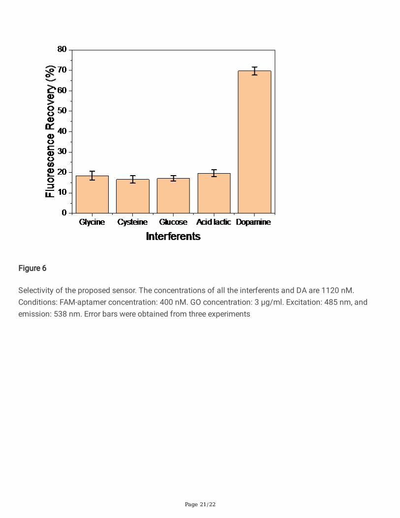
Selectivity is another important feature of a good sensing system as though the presence of manyinterferents in the real sample would affect the accuracy of the detection mechanism. In this regard, theselectivity of the aptamer-GO sensing platform was studied by monitoring the �uorescence recoverypercentage for different interferential substances including glycine, cysteine, glucose, and lactic acid. Forthat, we set the concentration of DA and the other interferents at 1120 nM, and then each one wasincubated with 25 µl of FAM-aptamer. After 25 min, 25 µl of GO was added to the mixture, and the�uorescence recovery was measured after 30 min of incubation. As shown in Fig. 6, under the sameexperimental conditions, the �uorescence recoveries of the sensing system in the presence of DA are

Page 9/22
highly signi�cant, whereas a minor �uorescence restoration was observed in the presence of the testedinterferents. Furthermore, it can be seen that the �uorescence enhancement in the presence of the targetwas more than 4-fold as compared to the other interferents. These results indicates that glycine, cysteine,glucose, and acid lactic are not recognized by DA-aptamer. Consequently, the aptamer remains adsorbedon the GO surface inducing the �uorescence quenching. In particular, the aptamer was strongly bound todopamine, thus inducing its release from the surface of GO and the �uorescence recovery. Therefore, thiscon�rms that the label-free �uorescent aptasensor based on GO exhibited an excellent speci�city for theDA detection, thus indicating its potential application in complex matrices.
Determination of DA in human serum
Table 2
Recoveries of DA from human serum samples (n = 3)Sample Standard value of DA (nM) Found (nM) Recovery (%) RSD (%, n = 3)
Human serum 3 3.02 100.53 0.76
7 7.01 100.21 2.09
280 288.8 103.16 0.61
1200 1999.83 99.99 1.89
In order to investigate the applicability of the method, our biosensor was tested in human serum samples.For that, the samples free of DA were collected from a local biomedical analysis laboratory. Then, theywere diluted 10 times and spiked with 3, 7, 280, and 1200 nM of DA. After incubation with the aptamer, 25µl of GO was added and the �uorescence recovery was measured. As it is shown in Fig. 7, the �uorescentrecovery percentages of human serum and tris-buffer were almost the same. This latter conclusion iscon�rmed by the analytical results for the samples spiked with DA presented in Table 2. As illustrated inthis table, the developed aptasensor exhibited good recoveries ranging from 89–103%, with RSD between0.61 and 2.09 %. These experimental results con�rm the good reliability and applicability of the proposedmethod for DA detection in complex biological samples.
ConclusionThis work describes the �rst label-free �uorescent aptasensor for DA detection using GO as a quencher.The developed aptasensor showed a linear relationship between the �uorescence recovery and DAconcentration in the range of 3-1680 nM. In addition, the present sensing platform exhibited an excellentselectivity and sensitivity. Moreover, the applicability of this sensing platform was con�rmed by detectingDA in complex biological matrices, with a prominent accuracy. By comparing the present technique tothat previously reported in the literature, our results have shown a high accuracy and reliability for rapidanalysis of dopamine. Furthermore, the developed device does not require any surface functionalization,thus simplifying the fabrication process as well as the analysis. Therefore, the ssDNA–GO platform could

Page 10/22
be an excellent alternative to universal molecular beacons in constructing sensing systems. It could bepotentially applied in biomedical diagnostics for dopamine monitoring as well as other biomarkers.
DeclarationsAcknowledgements This work was �nancially supported by Bioengineering laboratory, Higher nationalschool of biotechnology, Constantine-Algeria
Fundings No funding
Availability of data and material (data transparency) Not applicable
Code availability Not applicable
Con�ict of interests The authors declare no competing interests.
References1. Shang, N. G., Papakonstantinou, P., McMullan, M., Chu, M., Stamboulis, A., Potenza, A. … Marchetto,
H. J. A. (2008). f. m. Catalyst-free e�cient growth, orientation and biosensing properties of multilayergraphene nano�ake �lms with sharp edge planes. 18,3506–3514.
2. De Witte, P. J. A., & PRESS-, A. L. P. (1996). The role of neurotransmitters in alcohol dependence:animal research. 31,13–16.
3. Kirchon, A., Feng, L., Drake, H. F., Joseph, E. A., & Zhou, H. C. J. C. S. R. (2018) From fundamentals toapplications: a toolbox for robust and multifunctionalMOF materials.47,8611–8638.
4. Bucolo, C., Leggio, G. M., Drago, F., & Salomone, S. J. P. therapeutics(2019). Dopamine outside thebrain: The eye, cardiovascular system and endocrine pancreas. 203, 107392.
5. Napier, T. C., Kirby, A., Persons, A. L., & Psychiatry, B. J. P. i. N.-P., (2020). The role of dopaminepharmacotherapy and addiction-like behaviors in Parkinson’s disease. 102, 109942.
�. Xu, Y., Qiu, Z., Zhu, J., Liu, J., Wu, J., Tao, J., & Chen, L. J. B. n(2019). The modulation effect of non-invasive brain stimulation on cognitive function in patients with mild cognitive impairment: asystematic review and meta-analysis of randomized controlled trials. 20,1–11.
7. Whitton, A. E., Reinen, J. M., Slifstein, M., Ang, Y. S., McGrath, P. J., Iosifescu, D. V. … Schneier, F. R. J.B. (2020). Baseline reward processing and ventrostriatal dopamine function are associated withpramipexole response in depression. 143,701–710.
�. Chen, P. Y., Vittal, R., Nien, P. C., & Ho, K. C. J. B. Bioelectronics(2009). Enhancing dopamine detectionusing a glassy carbon electrode modi�ed with MWCNTs, quercetin, and Na�on®. 24,3504–3509.
9. Naranjo, C. A., Tremblay, L. K., Busto, U. E., & Psychiatry, B. J. P. i. N.-P., (2001). The role of the brainreward system in depression. 25, 781–823.

Page 11/22
10. Zhang, A., Neumeyer, J. L., & Baldessarini, R. J. (2007). Recent Progress in Development of DopamineReceptor Subtype-Selective Agents: Potential Therapeutics for Neurological and PsychiatricDisorders. Chemical Reviews, 107, 274–302
11. Liu, A., Wei, M. D., Honma, I., & Zhou, H. (2006). Biosensing Properties of TitanateNanotube Films:Selective Detection of Dopamine in the Presence of Ascorbate and Uric Acid. 16,371–376.
12. Nichkova, M., Wynveen, P. M., Marc, D. T., Huisman, H., & Kellermann, G. H. (2013). Validation of anELISA for urinary dopamine: applications in monitoring treatment of dopamine-related disorders.125,724–735.
13. Hows, M. E. P., Lacroix, L., Heidbreder, C., Organ, A. J., & Shah, A. J. (2004). High-performance liquidchromatography/tandem mass spectrometric assay for the simultaneous measurement ofdopamine, norepinephrine, 5-hydroxytryptamine and cocaine in biological samples. Journal ofNeuroscience Methods, 138, 123–132
14. Qian, T., Yu, C., Zhou, X., Wu, S., & Shen, J. (2014). Au nanoparticles decorated polypyrrole/reducedgraphene oxide hybrid sheets for ultrasensitive dopamine detection. Sensors and Actuators B:Chemical, 193, 759–763
15. Lakard, S., Pavel, I. A., & Lakard, B. (2021). Electrochemical Biosensing of DopamineNeurotransmitter. A Review, 11, 179
1�. Pérez-Fernández, V., Harman, D. G., Morley, J. W., & Cameron, M. A. (2017). Optimized method toquantify dopamine turnover in the mammalian retina. J. A. c, 89, 12276–12283
17. Matuszewski, B., Constanzer, M., & Chavez-Eng, C. J. A. c(2003). Strategies for the assessment ofmatrix effect in quantitative bioanalytical methods based on HPLC – MS/MS. 75, 3019–3030.
1�. Wei, N., Zhao, X. E., Zhu, S., He, Y., Zheng, L., Chen, G. … Liu, Z. J. T. (2016). Determination ofdopamine, serotonin, biosynthesis precursors and metabolites in rat brain microdialysates byultrasonic-assisted in situ derivatization–dispersive liquid–liquid microextraction coupled withUHPLC-MS/MS. 161,253–264.
19. Sahoo, H. J. J., o., P., & Reviews, P. C. P. (2011). Förster resonance energy transfer–A spectroscopicnanoruler: Principle and applications. 12, 20–30.
20. Medintz, I. L., & Hildebrandt, N. (2013). FRET-Förster resonance energy transfer: from theory toapplications. John Wiley & Sons
21. Pehlivan, Z. S., Torabfam, M., Kurt, H., Ow-Yang, C., Hildebrandt, N., & Yüce, M. J. M. A. (2019).Aptamer and nanomaterial based FRET biosensors: a review on recent advances (2014–2019).186,1–22.
22. Didenko, V. V. J. B. (2001). DNA probes using �uorescence resonance energy transfer (FRET): designsand applications. 31, 1106–1121.
23. Shi, J., Tian, F., Lyu, J., & Yang, M. J. J. o. m. c. B. (2015) Nanoparticle based �uorescence resonanceenergy transfer (FRET) for biosensing applications. 3, 6989–7005.
24. Jayasena, S. D. (1999). Aptamers: An Emerging Class of Molecules That Rival Antibodies inDiagnostics. Clinical Chemistry, 45, 1628–1650

Page 12/22
25. Ma, X., Wang, W., Chen, X., Xia, Y., Wu, S., Duan, N., & Wang, Z. J. E. F. R. Technology(2014). Selection,identi�cation, and application of A�atoxin B1 aptamer. 238,919–925.
2�. Shim, W. B., Kim, M. J., Mun, H., & Kim, M. G. J. B. Bioelectronics(2014). An aptamer-based dipstickassay for the rapid and simple detection of a�atoxin B1. 62,288–294.
27. Seok, Y., Byun, J. Y., Shim, W. B., & Kim, M. G. J. A. C. A. (2015). A structure-switchable aptasensor fora�atoxin B1 detection based on assembly of an aptamer/split DNAzyme. 886,182–187.
2�. Toh, S. Y., Citartan, M., Gopinath, S. C., & Tang, T. H. J. B. bioelectronics(2015). Aptamers as areplacement for antibodies in enzyme-linked immunosorbent assay. 64, 392–403.
29. Seto, D., Maki, T., Soh, N., Nakano, K., Ishimatsu, R., & Imato, T. (2012). A simple and selective�uorometric assay for dopamine using a calcein blue–Fe2 + complex �uorophore. Talanta, 94, 36–43
30. Salimi, A., Khezrian, S., Hallaj, R., & Vaziry, A. (2014). Highly sensitive electrochemical aptasensor forimmunoglobulin E detection based on sandwich assay using enzyme-linked aptamer. AnalyticalBiochemistry, 466, 89–97
31. He, Y., Lin, Y., Tang, H., & Pang, D. J. N. (2012). A graphene oxide-based �uorescent aptasensor for theturn-on detection of epithelial tumor marker mucin 1. 4,2054–2059.
32. Yüce, M., Ullah, N., & Budak, H. J. A. (2015). Trends in aptamer selection methods and applications.140, 5379–5399.
33. Yamamoto, R., & Kumar, P. K. J. G. t. c(2000). Molecular beacon aptamer �uoresces in the presenceof Tat protein of HIV-1. 5, 389–396.
34. Fang, X., Li, J. J., Perlette, J., Tan, W., & Wang, K. (2000). Peer reviewed: molecular beacons: novel�uorescent probes. ACS Publications
35. Vo-Dinh, T. (2003). Novel Fluorescent Molecular Beacon DNA Probes for Biomolecular Recognition(pp. 1533–1556). CRC Press
3�. Alivisatos, P. J. N. (2004). The use of nanocrystals in biological detection. 22,47–52.
37. Song, S., Liang, Z., Zhang, J., Wang, L., Li, G., & Fan, C. J. A. C. (2009). Gold-nanoparticle‐basedmulticolor nanobeacons for sequence‐speci�c. DNA analysis, 121, 8826–8830
3�. Wang, H., Li, J., Liu, H., Liu, Q., Mei, Q., Wang, Y. … Lu, Z. J. N. A. R(2002). Label-free hybridizationdetection of a single nucleotide mismatch by immobilization of molecular beacons on an agarose�lm. 30,e61-e61.
39. Mairal, T., Cengiz Özalp, V., Lozano Sánchez, P., Mir, M., Katakis, I., & O’Sullivan, C. K. (2008).Aptamers: molecular tools for analytical applications. Analytical and Bioanalytical Chemistry, 390,989–1007
40. Hunt, A., Dikin, D. A., Kurmaev, E. Z., Boyko, T. D., Bazylewski, P., Chang, G. S., & Moewes, A. (2012).Epoxide Speciation and Functional Group Distribution in Graphene Oxide. Paper-Like Materials, 22,3950–3957

Page 13/22
41. Gao, W., Alemany, L. B., Ci, L., & Ajayan, P. M. (2009). New insights into the structure and reduction ofgraphite oxide. Nature Chemistry, 1, 403–408
42. Lee, J., Kim, J., Kim, S., & Min, D. H. (2016). J. A. d. d. r. Biosensors based on graphene oxide and itsbiomedical application, 105, 275–287
43. Wang, C. F., Sun, X. Y., Su, M., Wang, Y. P., & Lv, Y. K. J. A. (2020). Electrochemical biosensors basedon antibody, nucleic acid and enzyme functionalized graphene for the detection of disease-relatedbiomolecules. 145,1550–1562.
44. Dong, H., Gao, W., Yan, F., Ji, H., & Ju, H. J. A. c(2010). Fluorescence resonance energy transferbetween quantum dots and graphene oxide for sensing biomolecules. 82, 5511–5517.
45. Chung, C., Kim, Y. K., Shin, D., Ryoo, S. R., Hong, B. H., & Min, D. H.J. A. o. c. r. (2013) Biomedicalapplications of graphene and graphene oxide.46,2211–2224.
4�. Yuan, H., Qi, J., Xing, C., An, H., Niu, R., Zhan, Y. … Wang, B. J., A. F. M.. (2015). Graphene-Oxide‐Conjugated Polymer Hybrid Materials for Calmodulin Sensing by Using. FRET Strategy, 25, 4412–4418
47. Hermann, T., & Patel, D. J. J. S. (2000). Adaptive recognition by nucleic acid aptamers. 287,820–825.
4�. Liu, Y., Yu, D., Zeng, C., Miao, Z., & Dai, L. J. L. (2010). Biocompatible graphene oxide-based glucosebiosensors. 26,6158–6160.
49. Jung, J. H., Cheon, D. S., Liu, F., Lee, K. B., & Seo, T. S. (2010). A graphene oxide based immuno-biosensor for pathogen detection. J. A. C, 122, 5844–5847
50. Zhao, J., Zhao, L., Lan, C., Zhao, S. J. S., & Chemical, A. B. (2016). Graphene quantum dots aseffective probes for label-free �uorescence detection of dopamine. 223,246–251.
51. Ma, Y., Chen, A., Xie, X., Wang, X., Wang, D., Wang, P. … Li, Y. J. T. (2019). Doping effect and�uorescence quenching mechanism of N-doped graphene quantum dots in the detection ofdopamine. 196,563–571.
52. He, W., Gui, R., Jin, H., Wang, B., Bu, X., & Fu, Y. J. T. (2018). Ratiometric �uorescence and visualimaging detection of dopamine based on carbon dots/copper nanoclusters dual-emittingnanohybrids. 178,109–115.
53. Govindaraju, S., Ankireddy, S. R., Viswanath, B., Kim, J., & Yun, K. J. S. r(2017). Fluorescent goldnanoclusters for selective detection of dopamine in cerebrospinal �uid. 7, 1–12.
54. Ling, Y., Wang, L., Zhang, X. Y., Wang, X. H., Zhou, J., Sun, Z. … Chemical, A. B. (2020). Ratiometric�uorescence detection of dopamine based on effect of ligand on the emission of Ag nanoclustersand aggregation-induced emission enhancement. 310,127858.
55. Sivakumar, P., Priyatharshni, S., & Kumar, K. J. M. C. Physics(2020). Fluorescent silver nanoparticlesfor sensitive and selective detection of dopamine. 240,122167.
5�. Qi, W., Zhao, M., Fu, Y., He, H., Tian, X., Wu, D. … Hu, P. P. J. D. Pigments(2020). Fluorescent detectionof uric acid through photoinduced electron transfer using luminol-terbium (III) nanoparticlessynthesized via aggregation-induced �uorescence strategy. 172,107797.

Page 14/22
57. Lin, J., Huang, B., Dai, Y., Wei, J., & Chen, Y. J. M. S. C, E(2018). Chiral ZnO nanoparticles for detectionof dopamine. 93,739–745.
5�. Zhu, Y., Cai, Y., Xu, L., Zheng, L., Wang, L., Qi, B., & Xu, C. J. A. a. m., interfaces(2015). Building anaptamer/graphene oxide FRET biosensor for one-step detection of bisphenol A. 7, 7492–7496.
59. Zheng, M., Jagota, A., Semke, E. D., Diner, B. A., McLean, R. S., Lustig, S. R., & Richardson, R. E., Tassi,N. G. J. N. m.. (2003). DNA-assisted dispersion and separation of carbon nanotubes, 2, 338–342
�0. Mao, B., Calatayud, D. G., Mirabello, V., Kuganathan, N., Ge, H., Jacobs, R. M. … Hodges, B. J. J. C.(2017). Fluorescence-Lifetime Imaging and Super‐Resolution Microscopies Shed Light on theDirected‐and Self‐Assembly of Functional Porphyrins onto Carbon Nanotubes and Flat Surfaces.23,9772.
�1. Chang, H., Tang, L., Wang, Y., Jiang, J., & Li (2010). Graphene �uorescence resonance energy transferaptasensor for the thrombin detection. J. J. A. c, 82, 2341–2346
�2. Duan, Y. F., Ning, Y., Song, Y., & Deng, L. J. M. A. (2014). Fluorescent aptasensor for the determinationof Salmonella typhimurium based on a graphene oxide platform. 181,647–653.
�3. Walsh, R., & DeRosa, M. C. J. B. communications, b. r(2009). Retention of function in the DNAhomolog of the RNA dopamine aptamer. 388, 732–735.
�4. Yang, M., Javadi, A., Li, H., & Gong, S. J. B. Bioelectronics(2010). Ultrasensitive immunosensor for thedetection of cancer biomarker based on graphene sheet. 26,560–565.
�5. Zhang, M., Yin, B. C., Tan, W., & Ye, B. C. J. B., Bioelectronics. (2011). A versatile graphene-based�uorescence “on/off”. switch for multiplex detection of various targets, 26, 3260–3265
��. Goud, K. Y., Sharma, A., Hayat, A., Catanante, G., Gobi, K. V., Gurban, A. M., & Marty, J. L. (2016).Tetramethyl-6-carboxyrhodamine quenching-based aptasensing platform for a�atoxin B1: Analyticalperformance comparison of two aptamers. J. A. b, 508, 19–24
�7. Azadbakht, A., Roushani, M., Abbasi, A. R., Menati, S., & Derikvand, Z. (2016). A label-free aptasensorbased on polyethyleneimine wrapped carbon nanotubes in situ formed gold nanoparticles as signalprobe for highly sensitive detection of dopamine. Materials Science and Engineering: C, 68, 585–593
��. Xu, J., Li, Y., Wang, L., Huang, Y., Liu, D., Sun, R. … Sun, C. (2015). A facile aptamer-based sensingstrategy for dopamine through the �uorescence resonance energy transfer between rhodamine B andgold nanoparticles. Dyes and Pigments, 123, 55–63
�9. Wang, Y., Kang, K., Wang, S., Kang, W., Cheng, C., Niu, L. M., & Guo, Z. (2020). A novel label-free�uorescence aptasensor for dopamine detection based on an Exonuclease III- and SYBR Green I-aided ampli�cation strategy. Sensors and Actuators B: Chemical, 305, 127348
70. Jarczewska, M., Sheelam, S. R., Ziółkowski, R., & Górski, Å. (2015). A Label-Free Electrochemical DNAAptasensor for the Detection of Dopamine. Journal of The Electrochemical Society, 163, B26–B31
71. Wang, Y., Li, Y., Tang, L., Lu, J., & Li, J. (2009). Application of graphene-modi�ed electrode forselective detection of dopamine. Electrochemistry Communications, 11, 889–892

Page 15/22
72. Wei, B., Zhong, H., Wang, L., Liu, Y., Xu, Y., Zhang, J. … Wang, H. (2019). Facile preparation of acollagen-graphene oxide composite: A sensitive and robust electrochemical aptasensor fordetermining dopamine in biological samples. International Journal of Biological Macromolecules,135, 400–406
73. Liang, Y., Guo, T., Zhou, L., Offenhäusser, A., & Mayer, D. (2020). Label-Free Split Aptamer Sensor forFemtomolar Detection of Dopamine by Means of Flexible. Organic Electrochemical Transistors, 13,2577
74. Liu, L., Xia, N., Meng, J. J., Zhou, B. B., & Li, S. J. (2016). An electrochemical aptasensor for sensitiveand selective detection of dopamine based on signal ampli�cation of electrochemical-chemicalredox cycling. Journal of Electroanalytical Chemistry, 775, 58–63
75. Zheng, Y., Wang, Y., & Yang, X. (2011). Aptamer-based colorimetric biosensing of dopamine usingunmodi�ed gold nanoparticles. Sensors and Actuators B: Chemical, 156, 95–99
7�. Feng, J. J., Guo, H., Li, Y. F., Wang, Y. H., Chen, W. Y., & Wang, A. J. (2013). Single MolecularFunctionalized Gold Nanoparticles for Hydrogen-Bonding Recognition and Colorimetric Detection ofDopamine with High Sensitivity and Selectivity. ACS Applied Materials & Interfaces, 5, 1226–1231
77. Ren, L., Hang, X., Qin, Z., Zhang, P., Wang, W., Zhang, Y., & Jiang, L. (2020). Determination ofdopamine by a label-free �uorescent aptasensor based on AuNPs and carbon quantum dots. Optik,208, 163560
7�. Huang, H., Shi, S., Gao, X., Gao, R., Zhu, Y., Wu, X. … Yao, T. (2016). A universal label-free �uorescentaptasensor based on Ru complex and quantum dots for adenosine, dopamine and 17β-estradioldetection. Biosensors and Bioelectronics, 79, 198–204
Figures

Page 16/22
Figure 1
Schematic illustration for the developed GO-based �uorescent aptasensors for DA detection

Page 17/22
Figure 2
Effect of GO concentration (0-5 µg/ml) on the quenching e�ciency toward FAM modi�ed aptamer at theemission wavelength of 538 nm. The aptamer concentration was �xed at 400 nM. Error bars wereobtained from three experiments
Page 18/22
Figure 3
FAM-labeled aptamer quenching kinetic behaviors with respect to the reaction time in the presence of 400nM FAM-aptamer, and 3 µg/ml GO in Tris-HCl buffer (5 mM MgCl2, 0.5 M NaCl and 50 mM Tris HCl, pH7.4) at the emission wavelength of 538 nm. Error bars were obtained from three experiments

Page 19/22
Figure 4
Effect of aptamer concentrations (200-800 nM) on the Fluorescence intensity at the emission wavelengthof 538 nm. Error bars were obtained from three experiments

Page 20/22
Figure 5
Calibration curve of �uorescence recovery as a function of DA concentrations: 3, 7, 14, 140, 280, 560,1200, 1680 nM. Conditions: FAM-aptamer concentration: 400 nM. GO concentration: 3 µg/ml. Excitation:485 nm, and emission: 538 nm. Error bars were obtained from three experiments
Page 21/22
Figure 6
Selectivity of the proposed sensor. The concentrations of all the interferents and DA are 1120 nM.Conditions: FAM-aptamer concentration: 400 nM. GO concentration: 3 µg/ml. Excitation: 485 nm, andemission: 538 nm. Error bars were obtained from three experiments

Page 22/22
Figure 7
Comparison of �uorescence recoveries e�ciency between buffer (green bars) and human serum samples(orange bars). Conditions: FAM-aptamer concentration: 400 nM. GO concentration: 3 µg/ml. Excitation:485 nm, and emission: 538 nm. Error bars were obtained from three experiments