Oesophageal Disorders - JU Medicine€¦ · Patho-Physiology Percent of time oesophageal Ph

V7 JAN 2014 1
Oesophageal squamous cell cancer: induction chemotherapy and
oesophagectomy versus induction chemotherapy and chemoradiotherapy - a
feasibility study
A feasibility trial of chemoradiation or surgery for oesophageal cancer
Protocol version 7: Jan 2014
NIHR RfPB number: PB-PG-0807-14131
EUDRACT number: 2009-013877-16
Chief investigator Jane M Blazeby
1,2
Principal investigators:
S Mike Griffin3
Tom Crosby4
Sara T Brookes2
1University Hospitals Bristol NHS Foundation Trust, Bristol BS2 8HW, Tel: 0117 9287231.
2Department of Social Medicine, Canynge Hall, 39 Whatley Road, Clifton, Bristol, BS8 2PS
3Northern Oesophago-gastric Unit, Royal Victoria Infirmary, Newcastle, NE1 4LP Tel: 0191 2820234, 4Velindre Cancer Centre, Velindre NHS Trust, Cardiff, CF14 2TL Tel: 029 2031 6202,

V7 JAN 2014 2
Chief investigator
Professor Jane M Blazeby, University Hospitals Bristol NHS Foundation Trust, Bristol BS2 8HW. Tel: 0117 9287231 email: [email protected]. Emergency contact: 07811 963031
Surgical lead
Professor S Mike Griffin, Northern Oesophago-gastric Unit, Royal Victoria Infirmary, Newcastle NE1 4LP. Tel: 0191 2820234, email: [email protected]
Radiotherapy lead
Dr Tom Crosby, Velindre Cancer Centre, Velindre NHS Trust , Whitchurch, Cardiff, CF14 2TL Tel: 029 2031 6202, Fax: 02920316267 email: [email protected]
Methodology lead
Dr Sara T Brookes, Department of Social Medicine, University of Bristol Canynge Hall, 39 Whatley Road, Bristol, Tel: 0117 9287218, email: [email protected]
Director of Bristol Randomised Trials Collaboration
Dr Alan Montgomery, Community Based Medicine, University of Bristol, 25/27 Belgrave Road, Clifton, Bristol, Tel: 0117 3313840, email: [email protected]
Study design and conduct
Dr Athene Lane, Department of Social Medicine, Canynge Hall, 39 Whatley Road, Bristol, Tel 0117 928: email: [email protected]
Qualitative Research
Professor Jenny Donovan, Department of Social Medicine, Canynge Hall, 39 Whatley Road, University of Bristol. Bristol Tel: 0117 9287203, email: [email protected]
Quality assurance surgery
Mr C Paul Barham, Consultant Surgeon, University Hospitals Bristol NHS Foundation Trust, Bristol. Tel: 0117 9283054, email: [email protected]
Mr Wyn Lewis, Consultant Surgeon, Cardiff & Vale NHS, Trust, Cardiff, [email protected]
Quality assurance oncology
Dr Stephen J Falk, Consultant oncologist, University Hospitals Bristol NHS Foundation Trust, Bristol. Tel: 0117 9287214, email: [email protected]
Dr Phillip Atherton, Consultant Clinical Oncologist, Northern Centre for cancer Care, Freeman Hospital, Newcastle upon Tyne, NE7 7DN Tel: 0191 2820234, email: [email protected]
Quality assurance pathology
Dr M Moorghen, Consultant Histopathologist, University Hospitals Bristol, NHS Foundation, Trust, Bristol
Prof Geraint Williams, Professor of Pathology and Consultant Histopathologist, Wales College of Medicine, Cardiff University, Heath Park, Cardiff, CF14 4XN
Professor Alastair D Burt, Professor of Pathology, University of Newcastle upon Tyne, Peacock Hall, Royal Victoria Infirmary, Newcastle upon Tyne, NE14LP. Email: [email protected]

V7 JAN 2014 3
Health economics Dr William Hollingworth, Department of Social Medicine, Canynge Hall, 39 Whatley Road, University of Bristol, Bristol, BS8 2PR Tel:0117 928 7355, email:[email protected] Patient representative: To be arranged in main trial Translational research: None arranged at present Sponsor: University Hospitals Bristol NHS Foundation Trust, Research and Development Department, Level 3, Bristol Education Centre, Upper Maudlin Street, Bristol BS2 8AE. Tel: 01173420233 Trial Office for trial management and coordination, contact for day-to-day queries:
Dr Joanna Nicklin, Research Team, Surgical Research Team, Room 106A, , Chapter House, Bristol Dental Hospital, BS1 2LY Tel: 0117 342 3882 [email protected]: 0117 342 4834
Address for completed Case Report Forms:
Dr Joanna Nicklin, Surgical Research Team, Room 106A, Chapter House, Bristol Dental Hospital, BS1 2LY Tel: 0117 342 3882 [email protected]: 0117 342 4834

V7 JAN 2014 4
Table of Contents Page
Summary
Lay summary ................................................................................... 7
Scientific summary ................................................................ 7
Background
Introduction ...................................................................................... 8
Rationale for feasibility study ........................................................... 8
Expected benefits to the NHS ......................................................... 9
Aims & objectives .............................................................................................. 9
Summary of trial design .................................................................................... 9
Overall trial schema 10
Selection of participants
Multi-disciplinary team meetings.................................................... 11
Meeting with consultant and nurse ……………………………………11
Screening for entry into the trial ..................................................... 11
Inclusion criteria............................................................................. 11
Exclusion criteria ........................................................................... 11
Second appointment with consultant and nurse ........................... 12
Consent to participate in randomised study ................................... 12
Randomisation............................................................................... 12
Alternative to randomisation .......................................................... 13
Withdrawal from allocated trial treatments ..................................... 13
Withdrawal from the trial follow up ................................................. 13
Assessments before randomisation
Tumour staging.............................................................................. 13
Assessment of fitness .................................................................... 13
Clinical data collection
Before randomization at clinic appointments ................................. 14
After study entry ............................................................................ 14
During treatment ............................................................................ 14
At end of induction chemotherapy ................................................ 15
During follow up ............................................................................. 15
Assessment of health-related quality of life ................................................. 15
Health economic data collection .................................................................... 16
Treatment protocols
Chemotherapy and surgery protocol ............................................. 16
Induction chemotherapy before surgery ........................................ 16
Surgical protocol 16
Chemoradiotherapy protocol ......................................................... 19
Induction chemotherapy before chemoradiotherapy 19
Definitive chemoradiotherapy protocol .......................................... 19
Salvage treatment ......................................................................... 21

V7 JAN 2014 5
Medications permitted ................................................................... 21
Medications permitted with caution ................................................ 21
Non-permitted medications .......................................................... 21
Clinical considerations ................................................................... 22
Pregnancy ..................................................................................... 22
Nutritional status ............................................................................ 22
DPD deficiency .............................................................................. 22
Drug supply, dosing, side effects and dose modifications ........... 22
Drug supply 22
Dosing and precautions 22
Cisplatin 22
Capecetibine 23
5 fluourouracil 24
Possible side effects of Cisplatin, Capecetibine / 5 fluourouracil 24
Dose modifications of chemotherapy 25
Adverse events 28
Adverse events related to surgery 28
Operative complications ............ 28
In-hospital complications 28
General complicaitons 29
In-hospital mortality ………. 29
Frequency of complications 29
Reporting adverse events related to chemotherapy ...................... 29
Definitions 29
Clarifications and expectations 30
Trial- specific exceptions ………………………………………………30
SAE excluded from expedited notification …………………………..30
Institution/Investigator Responsibilities ……………………………. 30
Investigator Assessment ……………………………………………. 31
Notification procedure ……………………………………………….. 32
Bristol Randomised Trials Collaboration responsibilities………… 32
Adverse events related to radiotherapy ......................................... 33
Statistical analyses 33
Analyses for intergrated qualitative research 33
Audio-recordings of consultations ……………………………………34
Semi-structured patient interviews ………………………………….. 34
Survey to agree clinical important treatment effects 34
Development of core information for patients 34
Ethical & governance issues
Regulatory status ........................................................................... 35
Study medication 35
Indemnity ....................................................................................... 35
Data protection .............................................................................. 35

V7 JAN 2014 6
Storage of records ......................................................................... 35
Publication policy ........................................................................... 35
Investigator responsibilities .......................................................... 36
Co-enrolment guidelines ................................................................ 35
Finance .......................................................................................... 35
Monitoring and audit ...................................................................... 35
Research government statement................................................... 36
Trial steering committee 37
Indepedant data monitoring committee 37
Participating centre selection ........................................................................ 38
Quality assurance of surgery, chemotherapy and radiotherapy ................. 39
References ................................................................................................ 37
Appendix A
Common toxicity criteria based on CTCAE version 3.0.......………………… 41
Appendix B
RTOG acute radiation morbidity scoring criteria ………..……………………. 45
Appendix C Cockroft and Gault formula ........................................................ 48

V7 JAN 2014 7
Summary
Lay summary Cancer of the gullet (oesophagus) is a serious disease that is difficult to treat. There are two types of oesophageal cancer and for the squamous cell type there are two treatments that are widely used to try and cure the disease. One involves chemotherapy (drug treatment that kills cancer cells) and then surgery (to excise the cancer within the oesophagus). The other treatment involves chemotherapy and then chemotherapy and radiotherapy together (radiotherapy burns and kills cancer cells). At present there is a lack of high quality evidence that compares the two treatments and selecting a treatment depends upon doctors' or patients’ preferences and not on evidence about which treatment produces the best outcomes. The aim of this study is to determine the feasibility of a randomised trial of chemotherapy and oesophagectomy versus chemotherapy and chemoradiotherapy for patients with oesophageal squamous cell cancer. If the full multi-centre trial is feasible it will provide high quality evidence to use in clinical practice and inform treatment choice. The feasibility study will also establish methods for recruiting participants from multi-disciplinary cancer team meetings into randomised trials. It will use qualitative research to work with doctors and nurses to improve communication with patients regarding the two types of treatments and to improve trial recruitment. Additionally surveys with health professionals and patients will be undertaken to identify the core information to be communicated about each treatment. Scientific summary The optimal treatment for localised oesophageal squamous cell cancer is controversial. Surgery alone or in combination with neoadjuvant treatment can achieve cure but is highly invasive, associated with significant risks and high rates of disease recurrence. Definitive chemoradiotherapy may lead to long-term survival although there may be local treatment failure. There are no adequate randomised controlled trials comparing the effectiveness and cost-effectiveness of surgical and non-surgical treatments. Previous attempts have failed because of difficulties in identifying patients and poor acceptance of randomisation. Treatment decision making, therefore, is not evidence-based and depends on clinician or patient preference. The overall aim of this study is to determine whether a full multi-centre randomised trial of the two standard treatments is feasible. This will be achieved by undertaking a randomised pilot trial of induction chemotherapy and oesophagectomy versus induction chemotherapy and definitive chemoradiotherapy with intergrated qualitative research to establish recruitment rates, to understand barriers to recruitment and to pilot procedures and trial outcome measures. In addition, research will be undertaken to establish methods to identify patients eligible for randomisation using records from multi-disciplinary team meetings. This will determine and maximize recruitment and allow estimation of potential numbers for a full trial. Methods will also be used to reach consensus between health professionals and participants regarding clinically important treatment effects. These treatment effects will then be tested in the full trial. The final part of the feasibility study is to develop an intervention (information checklist) for surgeons, oncologists and nurses to use during consultations to describe the two standard treatments. This will be informed by information obtained from systematic reviews, the consensus methods, qualitative analysis of audio-recordings of consultations and semi-structured interviews with participants, in which their understanding of information provided will be elicited.

V7 JAN 2014 8
Background
Introduction
Oesophageal squamous cell cancer is a serious health problem. In the UK there are 2,500 incident cases per year and only 30% of these patients may be offered potentially curative treatment. Potentially curative treatment may include oesophagectomy or definitive chemoradiotherapy (CRT), but both approaches are associated with risks of death, major complications and a detrimental impact on health-related quality of life. Reports of survival after surgical based treatments vary, with possible one year survival between 30% and 60% and five year survival between 15% and 35%1. In-hospital mortality rates after surgery have reduced to between 2% and 5% but complications are common2;3. Several longitudinal studies have shown that surgery has a major negative effect on all aspects of health related quality of life with residual deficits in long term survivors4;5. In the presence of such poor (and variable) survival rates and the significant risk of treatment-related death, morbidity and reduced health related quality of life, attention has turned to the outcomes of non-surgical management of oesophageal squamous cell cancer.
The non-surgical management of oesophageal squamous cell cancer has included radiotherapy, alone or in combination with chemotherapy. A systematic review comparing these modalities in patients unsuitable for surgery concluded that CRT was superior to radiotherapy alone6. Despite the seemingly poor prognostic features of the patients in the trials, definitive CRT was associated with long-term survival and disease control, reaching 52% and 27% survival at one-year and five years respectively6. Recently, a cohort study from the UK demonstrated a five year survival of 32% with CRT7. Toxicity and side effects also occur with CRT but there is some non-randomised evidence that health related quality of life with CRT may be less severe than surgery8. This evidence suggests that a trial of CRT compared to treatment including surgery is an imperative next development in this evolving area of importance and uncertainty.
There have been three attempts to undertake randomised controlled trials of surgical versus non-surgical treatments for oesophageal cancer, but each have suffered problems9-
11. Two trials were equivalence studies powered to determine whether the two treatments could be considered equivalent in terms of survival at two-years9;10. Equivalence was defined as a difference of less than 10% and 15% respectively. It is questionable for a cancer with such a low survival rate, that such differences would not be deemed clinically important. The third study was powered to detect superiority of one treatment over another but failed to report what magnitude of difference was considered superior11. None were able to rule out much larger beneficial and detrimental effects of treatment including surgery compared to CRT, all producing wide confidence intervals. In addition, none provided a robust assessment of health related quality of life.
Rationale for a feasibility study There is a need for a well designed trial to compare the effectiveness and cost-effectiveness of the current standard treatments for oesophageal squamous cell cancer. This trial was proposed to the National Cancer Research Institute (NCRI) Upper Gastrointestinal Clinical Study Group in November 2006. The group supported the need for such a trial, but felt that recruitment would be difficult because eligible patients may not arise incidentally in sufficient numbers to provide adequate power for a trial and because in the absence of randomised evidence in this area, clinicians and patients often have strong preferences for particular treatments making randomisation difficult. Indeed these problems were experienced by the US Intergroup and the MRC clinical trials unit who have

V7 JAN 2014 9
both made previously unsuccessful attempts to mount a trial of a surgical versus non-surgical therapy in this area 12. It was therefore proposed that a detailed feasibility study be undertaken to address issues raised by the NCRI group and resolve the design problems surrounding this complex trial. Expected benefits to the NHS The study will show whether a full randomised controlled trial (RCT) is feasible. A full trial would provide high quality evidence to help doctors and patients make better decisions. If a full trial is not feasible the proposed study will still have an important impact for cancer services in the UK. It will clarify a method for recruiting participants into randomised controlled trials using multi-disciplinary team records. This may have a major influence on recruitment into clinical trials in oncology. The study will also refine methods for developing information checklists that can be used to communicate clearly the benefits or risks of treatment for gastrointestinal cancer.
Aims and objectives
The overall aim of the research is to determine the feasibility of an RCT comparing two standard treatments, induction chemotherapy and definitive oesophagectomy versus induction chemotherapy and definitive chemoradiotherapy for oesophageal squamous cell cancer.
The study will address previous difficulties in mounting a trial by achievement of the following objectives:
1. to determine the number of patients eligible to enter a trial by identifying ‘cases’ using multi-disciplinary team records from the three feasibility centres
2. to undertake a pilot study for the main trial in three feasibility centres to
establish recruitment rates, to use qualitative research to explore barriers to recruitment and to pilot cost and outcome measures and procedures for the trial
3. to ascertain consensus from professionals and patients of clinically important
treatment effects to be tested in a trial and communicated to patients
4. to develop an information checklist to improve the communication between clinicians and patients about the two diverse treatments, this, in turn, will improve recruitment into a full trial
Summary of trial design This is an open randomised controlled pilot trial designed to determine the feasibility of a full multi-centre trial comparing induction chemotherapy and oesophagectomy versus induction chemotherapy and definitive chemoradiotherapy for localised oesophageal squamous cell cancer. Associated research, to underpin the pilot RCT, will produce methods to recruit participants from multi-disciplinary team meetings, develop an information checklist with key clinical and health related quality of life outcomes associated with both treatments for use in consultations and establish clinically important treatment effects to be tested in a full multi-centre trial. Figure 1 shows the design, selection and recruitment process for the RCT.

V7 JAN 2014 10
Figure 1. Trial schema - feasibility randomised trial with intergrated qualitative research
Maximising trial recruitment from multi-disciplinary team meetings
Details of consecutive participants with localised oesophageal squamous cell cancer will be reviewed and reasons for trial ineligibility documented.
Patients with adenocarcinoma selected for surgery or chemoradiotherapy will also be identified
Induction chemotherapy +
Definitive oesophagectomy
Induction chemotherapy +
Definitive chemoradiotherapy
Consent obtained To be randomised into the trial and/or
be followed up for 12 months
12 month follow up as per clinical and health related quality of life protocol until the end of feasibility study
Clinic appointment with surgeon/oncologist & or nurse
Participants screened for trial eligibility and the two standard treatments will be discussed.
Patients with adenocarcinoma will have one treatment discussed. The consultation will be audio-recorded. Second stage of eligibility assessment
Potentially eligible participants will be invited to clinic & sent an information sheet (1a) describing the qualitative study
Clinic appointment with surgeon/oncologist & or nurse
Further discussion of the two standard treatments.
The consultation will be audio-recorded.
Semi-structured home interview, audio-recorded before start of treatment
Randomisation Non randomised patients will be
followed up
Eligible patients will be given a further information sheet (1b) explaining the randomised trial and inviting them to participate in randomisation. They will have time to think and make a decision in the intervening time between
appointments
Further time to consider if they wish
to join the study
V7 JAN 2014 11
Selection of participants Multi-disciplinary team meetings Multi-disciplinary team meeting records from the participating centres will be collated by the local research nurse and/or an multi-disciplinary team core member in each centre and anonymised (to maintain patient confidentiality). Potentially eligible trial participants, and patients with adenocarcinoma that are selected for surgery or definitive chemoradiotherapy will be sent an appointment to meet the clinical team. This invitation will include information about the qualitative research (Patient information sheet 1a). The number of multi-disciplinary team decisions regarding patients with oesophageal sqaumous cell cancer and those potentially eligible for the trial will be recorded. Initial trial eligibility criteria: Histological evidence of squamous cell cancer No evidence of metastases Fit for radical treatment Meeting with consultant and nurse (clinical nurse specialist / research nurse) During this meeting potentially eligible trial participants will be informed about the nature of the two standard treatments (N.B participants will be aware of the diagnosis of cancer for between one and four weeks before this appointment). Patients with adenocarcinoma will be informed about their recommended treatment (surgery or chemoradiotherapy) The information will be given verbally from the specialist upper gastro-intestinal consultant surgeon or specialist gastro-intestinal consultant oncologist and a clinical nurse specialist, trained research nurse or information radiographer. This information appointment will be audio-recorded and patient consent for the audio-recording requested (Consent form No. 1) A member of the research team will explain the randomisation process and give the patient the information sheet explaining the randomisation process (patient information sheet 1b) to potentially eligible trial patients Screening for entry into the trial During this consultation the participant will be screened for study eligibility. Reasons for ineligibility will be recorded and care will continue as usual. Inclusion criteria This study will include participants:
1. Aged 18 years of age or older on the date of first clinic appointment.
2. With histologically confirmed oesophageal squamous cell cancer.
3. With tumours staged as T2N0/1M0, T3N0/1M0, T4N0/1M0, where the T4 tumour involves the diaphragmatic crura or mediastinal pleura only (TNM classification)13 .
4. With a total primary tumour and nodes less than 10cm length.
5. Considered sufficiently fit for both treatments in the trial by a surgeon and an oncologist, both of whom are members of the core multi-disciplinary team*.
6. Willing to use contraception, if applicable.
7. Able to give informed written consent to participate in the randomised trial.
*If the participant is of uncertain fitness for both treatments, then respiratory and cardiac function tests should be performed according to local practice within 4 weeks of
V7 JAN 2014 12
randomisation. Suggested levels: FEV1 >1.5 litres; cardiac ejection fraction >50% of normal echocardiography.
Exclusion criteria Participants will be excluded from entry into the trial if they have:
1. Concommitant or past malignancies within five years prior to randomisation, except basal or squamous cell carcinoma of the skin or in situ carcinoma of the cervix.
2. Prior treatment for oesophageal cancer (not including photodynamic therapy or laser therapy for high grade dysplasia or carcinoma in situ).
3. Type I or II tumours of the oesophago-gastric junction with more than 2cm gastric wall involvement (measured on EUS).
4. Previous treatment that compromises the ability to deliver definitive mediastinal chemoradiotherapy or to undergo oesophagectomy.
Any queries about whether a patient is eligible to enter the trial should be addressed to the local principle investigator before study entry. Concerns will then be discussed with the Chief Investigator. Second appointment with consultant and nurse Following this meeting, a second appointment with an oncologist or surgeon, clinical nurse
specialist, research nurse or information radiographer will be arranged and any further staging investigations or tests of fitness can be completed. If the first appointment happens to be with an oncologist (which may occur for pragmatic reasons), the second appointment will be with a surgeon to ensure that the participant has the opportunity to be given detailed information and ask surgical, chemotherapeutic and radiotherapy questions (and vice versa). At this second hospital appointment, the issues surrounding treatment for localised oesophageal squamous cell cancer will be discussed again. The advantages and disadvantages of both treatments will be explained using the checklist and the purposes of randomisation will be further described. This appointment will be audio-recorded where written consent is obtained (Consent form 1.0). Consent to participate in randomised study The decision to participate in the trial will be voluntary and participants will be competent to understand what is involved. It will be made clear to all participants they will be free to decline without prejudicing future care. Participants consenting to randomization will complete another consent form (Consent form No. 2). After the second appointment, participants will be given a further 60-hour period to reflect on their potential involvement in the study. During this time the participant is provided with the contact details of the research
nurse, specialist nurse, information radiographer or core multi-disciplinary team member to ask further questions about the study. If they then agree to participate they will be sent another appointment for completion of the consent forms.
Written consent for audio-recordings of all consultations will be gained at each separate consultation. Both investigators and participants will retain copies of each signed consent form.

V7 JAN 2014 13
Randomisation If the participant agrees to take part in the trial the research nurse will use the Bristol Randomised Trials Collaboration automated randomisation web based system to register the patient and allocate a unique participant number and treatment will be allocated. Treatment allocation will be determined using computer-generated random numbers, and will be stratified by participating centre. The unique participant number will be the primary way in which the participant will be identified and will be used in all correspondence. Alternative to randomisation If randomisation is unacceptable to the participant, a participant-led treatment decision will be reached. These participants will be asked to provide consent to be followed up (in the same way as the trial participants) for the duration of the feasibility study (Consent form no. 3). For each study participant a letter will be sent to the participant’s GP and referring clinical team (if appropriate) indicating participation in the study and treatment allocation (GP letter no. 1 or 2).
Withdrawal from allocated trial treatments Participants should be given every encouragement to receive per protocol treatment unless clinical issues occur or participant choose to withdraw from the trial treatment. The reasons for this should be recorded wherever possible. Clinical issues that may influence per protocol treatment implementation
i. tumour progression during treatment.
ii. unacceptable toxicity or intercurrent illness preventing treatment.
iii. any change in the participant’s condition which, in the clinician’s opinion, justifies the discontinuation of treatment.
Withdrawal from the trial follow up
Although a trial treatment may be withdrawn for the above reasons, participants should continue to be followed up in the trial. Participants who explicitly withdraw consent to have any data recorded or any follow up must be respected and this will be recorded. Participants can change their minds about withdrawal at any time and re-consent to participate in the trial. Follow up data can only be recorded from the point when consent was reinstated.
ASSESSMENTS BEFORE RANDOMISATION Tumour staging Each of the tumour staging investigations outlined below should be aimed at being performed within 4 weeks prior to randomisation. However if this is not possible, the last staging investigation, which may include CT, EUS, PET/CT and laparoscopy, should normally be performed within 4 weeks prior to randomisation. If the last staging investigation is > 4 weeks, please contact the Chief investigator.
1. physical examination of neck and abdomen. 2. spiral/multislice computerised tomography (CT) scan of thorax and abdomen (neck
and pelvis optional).

V7 JAN 2014 14
3. endoscopic ultrasound (EUS) of oesophagus and stomach. 4. bronchoscopy if the tumour is close to or invading either bronchii or trachea. 5. positron emission (PET)/CT, bone scans and laparoscopy are optional and should
be performed according to local practice. The final pre-treatment TNM stage will be based upon information collated from all tests. These are recommendations and should not replace local guidelines. Assessment of fitness These following investigations should be undertaken within four weeks prior to randomisation.
1. Clinical assessment of cardiovascular history function 2. Clinical assessment of respiratory history & function 3. Smoking and alcohol history 4. Detailed drug history 5. Body mass index 6. WHO Performance status 7. Full blood count 8. Serum renal, liver and bone profile (including serum magnesium). 9. Glomerular filtration rate 10. Electrocardiogram 11. Pregnancy test in females of child bearing age
If concerns are raised further investigations may be performed according to local guidelines. CLINICAL DATA COLLECTION Before randomisation at clinic appointments All potential participants who are discussed at the multi-disciplinary team meetings, will be logged on a database at the department of Social Medicine, University of Bristol. Data regarding whether these individuals were then approached to participate in the study, randomised and agreed to be followed up, will also be recorded in a screening and enrolment log by the local research nurses. For each patient anonymised clinical data will be collected regarding the reasons for trial ineligibility, the treatment recommendation of the MDT and the final treatment recieved .When consent to participate in the randomised trial/follow up is received the names and addresses of participants will be entered on the project database along with the date on which consent was given. Eligible men or women who decline participation in the study will have no further information recorded, however. Details of the reasons for non-participation in the study will be recorded at all stages of this process. After study entry The following information will be recorded after agreement to participate in the study:
1. Which part of the study they participant has consented to take part in (and randomisation and/or follow up)
2. Date of completion of the consent form (s) 3. Full clinical details of staging investigations and co-morbidity 4. Full details of dates of clinic appointments 5. Health related quality of life and health economics assessments

V7 JAN 2014 15
This information will be entered onto the study database at the earliest opportunity. It is possible that there will be several appointments and data collection must occur after each appointment. If the participant is excluded for any reason e.g. for health grounds, then this must be fully documented.
During treatment The research nurse in each centre will keep prospective records of the following details during treatment:
1. date of start and finish of treatment and dates and reasons for treatment delays; 2. full clinical details of hospital admissions or problems during treatment, including
toxicity, surgical morbidity and in-hospital mortality; 3. date of treatment finally received and reasons for changes; 4. full histopathological results from surgery. 5. Health related quality of life and health economics assessments
At end of induction chemotherapy After completion of induction chemotherapy in both study arms the following assessments will be undertaken:
1. a clinical examination and history, if new concerns regarding fitness to continue
with definitive treatment arise, tests of cardiac and/or respiratory function may be undertaken according to local policy;
2. spiral/multislice computerised tomography (CT) scan of thorax and abdomen (neck and pelvis optional);
3. optional re-discussion of participant and review of CT co-morbidity tests at multi-disciplinary team meeting.
During follow up
After completion of both treatments participants will be followed up according to local policies. In addition participants will be followed up by phone calls from the research nurse at 16 and 24 weeks post randomisation and after 12 months throughout the duration of the feasibility study. Where patients have passed the 12 month follow up stage, data already collected will be retained. Phone calls will record details of:
1. Hospital admissions, requirement for interventions/investigations
ASSESSMENT OF HEALTH RELATED QUALITY OF LIFE The purpose for measuring health related quality of life in the feasibility study is to trial best methods for data collection, establish whether the instruments are suitable in this setting and to use the results to inform the sample size calculation of the full trial.
Generic domains of health related quality of life will be assessed with the EORTC core questionnaire - the EORTC QLQ-C3014. Disease and treatment specific symptoms and side effects will be assessed with the oesophago-gastric cancer specific module - the EORTC QLQ-OG2515. The EQ-5D will be used to value quality of life and combine it with survival to calculate quality adjusted life years (QALYs) for the economic analysis (see below).
Baseline questionnaires will be completed by participants in clinic after written consent has been obtained and prior to randomisation (Consent form No.4). Follow-up questionnaires will be administered by post following a phone call reminder to the participant made
V7 JAN 2014 16
centrally from the research associate based in Bristol. Participants will be asked to fill out the questionnaires themselves as completely and accurately as possible and return within a stamp-addressed envelope, which will be provided by the research nurse. If this is not possible research staff may administer them and this will be recorded. Reasons for questionnaire non-completion will be documented. Timing of health related quality of life assessment
Baseline questionnaire.
During week 7 post randomisation (end of induction chemotherapy)
During week 16 post randomisation (after completion of treatment).
Post randomisation 24 weeks and 12 months.
HEALTH ECONOMIC DATA COLLECTION
Details of resources used during induction chemotherapy will be recorded by the research nurse from the medical record using a standardised data collection form. This will include drug regimen used and number of cycles. In the surgical group theatre time and duration of time on intensive care or surgical wards and any readmissions will be recorded. In the definitive chemoradiotherapy group documentation of the drug regimen, number of cycles, number of fractions of outpatient or inpatient therapy and hospital admissions will occur. In a brief addendum to the health related quality of life postal questionnaire at each follow up timepoint, we will ask patients about any cancer/ gastro-intestinal related use of health services in the interim. This will include medications taken, GP surgery/home visits, nurse visits, outpatient appointments, accident and emergency visits, inpatient admissions and utilisation of nursing home or hospice care. The pilot study will help refine the resource use data collection forms for a subsequent definitive trial. Unit costs of care will be identified from national sources (e.g. NHS reference costs) and local hospital finance departments. The costs will be combined with resource use to estimate the comparative costs of surgery and chemoradiotherapy. The pilot data will enable us to calculate the expected value of sample information from a definitive trial.
TREATMENT PROTOCOLS Following entry into the study, participants will receive one of the two standard treatments that will commence within one week of randomisation. Chemotherapy and surgery protocol Induction chemotherapy before surgery Standard induction chemotherapy will consist of two cycles of 21 days i.e. a total of 42 days therapy. There are two schedules that may be used according to local practice.
i) Cisplatin 80mg/m2 to be given by intravenous infusion on day 1, with pre and post
hydration ( a recommended hydration schedule is given on page 23, table 4).
5-Fluorouracil 1g/m2/day to be given by continuous intravenous infusion for 4 days starting
on day 1 (i.e. total cycle dose = 4g/m2).

V7 JAN 2014 17
ii) Cisplatin 80mg/m2 to be given by intravenous infusion on day 1, with pre and post
hydration ( a recommended hydration schedule is given on page 23, table 4).
Capecitabine 625mg/m2 to be given orally twice daily within 30 mins of food, starting in the
evening of day 1 and finishing on the morning of day 43.
Pre-meds and anti-emetics may be administered according to local practice.
Surgical protocol
Centres recruiting to this study will be expected to deliver surgical management that conforms to the standards set out in the NICE Improving Outcomes Guidance16. Surgery should be performed within four to six weeks after completion of induction chemotherapy. The operation may consist of a two or three-phase oesophagectomy and it may be performed with an open, laparoscopic, open or combined approach. The phase of
operation carried out first is dependant upon the planned operation and its approach. The stomach is the preferred organ for reconstruction, where this is unavailable, colonic or small bowel transposition should be performed. Abdominal phase The intra-abdominal contents should be carefully inspected, paying particular attention to the omentum and peritoneal surfaces in the supracolic compartment for metastases and the para-aortic region in patients with otherwise resectable nodal disease. Lesions suspicious of metastatic disease should be biopsied and sent for frozen section. Gastric mobilisation can proceed but the operation should be terminated if there is unequivocal evidence on frozen section that there is disease outside the proposed surgical field. Complete gastric mobilisation based on the right gastroepiploic and right gastric arteries should be achieved. The left gastric vein should be divided as low as possible, where it disappears behind the hepatic artery at the upper border of the pancreas. The left gastric artery should be divided at its origin. Lymphadenectomies along the hepatic artery and splenic artery will be performed, either en bloc or separately at the surgeon’s discretion. The hepatic artery dissection should remove all nodal tissue overlying the hepatic artery proper and the common hepatic artery. (This ensures removal of all group 8 nodes). Lymphadenectomy on the splenic artery extends from the origin of this vessel as far lateral as the point of ligation of the uppermost posterior short gastric vessel. The abdominal phase should result in a dissection that has removed lymph nodes from stations 1, 2, 3, 7, 8 and 11. The exact extent of the dissection at the diaphragm will be influenced by the results of pre-operative staging, as well as intra-operative assessment. For tumours at the level of the diaphragm, it may be necessary to remove crural fibres and a cuff of diaphragm. Mobilisation of the left lateral segments of the liver, division of the inferior phrenic vein well to the right and to the left of the oesophagus, may be used to facilitate extension into the mediastium. Both pleural cavities may be opened, although this can be deferred until the thoracic phase. Removal of the pericardial fat pad anteriorly and strips of parietal pleura should usually be achieved at this stage, to again minimise the risk of a positive radial margin, although these steps can be undertaken during the thoracic phase of the operation.
V7 JAN 2014 18
Preparation of the transection site on the lesser curve, without compromise to the extent of lymphadenectomy, can be undertaken during the abdominal or thoracic phase of the operation. Thoracic phase This may be either via the right or left thorax. The intra-thoracic contents should be carefully inspected, paying particular attention to the pleural surfaces. Lesions suspicious of metastatic disease should be biopsied and sent for frozen section. Oesophageal mobilisation can proceed but the operation should be terminated if there is unequivocal evidence on frozen section that there is disease outside the proposed surgical field. The oesophagus is fully mobilised with mediastinal pleura overlying the oesophagus excised in continuity. If necessary during a right thoractomy, the azygos vein is divided where it crosses the oesophagus. The posterior limit of the dissection should be the antero-lateral wall of the aorta, so that the thoracic duct is mobilised with the oesophagus and peri-oesophageal tissues. The thoracic duct is ligated and divided at the level of the diaphragm and at the upper level of transection. The mediastinal pleura overlying the left side of the oesophagus should also be excised to the level of the tumour. Para-oesophageal and diaphragmatic nodes (groups 108, 110, 111) are removed in continuity with the oesophagus. Lymph nodes at the tracheal bifurcation and along the right and left main bronchi to the pulmonary hilus, can be removed en bloc or separately (nodal groups 107, 109). The extent of lymphadenectomy for the upper thoracic para-oesophageal nodes (group 105), will be determined by the site of oesophageal transection. This must be above the aortic arch and preferably within 5 cm of the thoracic inlet. Enlarged nodes in the para-tracheal group (group 106), should be removed for sampling purposes. Complete dissection of the left sided recurrent laryngeal nerve nodal chain is not mandatory. The stomach is delivered into the chest and formed into an oesophageal conduit using a stapled or hand sewn technique. An anastomosis between the conduit and oesophagus is formed according using a stapled or hand sewn technique. An nasogastric tube is placed via the nasopharnyx into the conduit. Cervical phase The cervical incision is at the discretion of the surgeon (left neck, vertical or skin crease). The oesophagus is identified, preserving and minimising traction on the left recurrent laryngeal nerve. The oesophagus is divided maintaining the maximum proximal length to enable the anastomosis to be at the level of the skin (two stay sutures may be placed in the proximal oesophagus). The divided distal oesophagus is sutured closed with an attached NG tube. Using the abdominal incision the oesophagus (and attached NG tube) are delivered into the abdominal cavity and the tumour and oesophagus are removed by division of the proximal stomach (ensuring an adequate distal resection margin). Leaving the NG tube passing from the neck incision via the thorax into the abdominal wound. The free NG tube, is then fixed to the neo-oesophagus/conduit and this is delivered into the neck using gentle traction. An anastomosis between the conduit/neooesophagus and proximal oesophagus in the neck is performed using staples or sutures at the discretion of the surgeon. An NG tube (nasopharyngeal to the conduit) is placed under direct vision.

V7 JAN 2014 19
Optional procedures The placement of a feeding jejunostomy, intra-abdominal, intra-thoracic drains and cervical drains are optional. Pyloroplasty, pyloromyotomy or no drainage may also be performed at the surgeon’s preference. The techniques for closure of the laparotomy, thoracotomy or port sites is at the surgeon’s discretion. Lymph node numbers referred to are those used in the Japanese Classification for oesophageal cancer . Chemoradiotherapy protocol
Induction chemotherapy before definitive chemoradiotherapy
Standard induction chemotherapy before chemoradiotherapy will consist of two cycles of 21 days i.e. a total of 42 days therapy. There are two schedules that may be used according to local practice.
i) Cisplatin 80mg/m2 to be given by intravenous infusion on day 1, with pre and post
hydration ( a recommended hydration schedule is given on page23, table 4).
5-Fluorouracil 1g/m2/day to be given by continuous intravenous infusion for 4 days starting
on day 1 (i.e. total cycle dose = 4g/m2).
ii) Cisplatin 80mg/m2 to be given by intravenous infusion on day 1, with pre and post
hydration ( a recommended hydration schedule is given on page 23, table 4).
Capecitabine 625mg/m2 to be given orally twice daily within 30 mins of food, starting in the
evening of day 1 and finishing on the morning of day 43.
Pre-meds and anti-emetics may be administered according to local practice
Definitive chemoradiotherapy protocol Definitive chemoradiotherapy should start as soon as induction chemotherapy has finished (i.e. day 43). The chemotherapy component is identical to the induction chemotherapy and will continue for a further 2 cycles (i.e. from day 43 to day 84). The radiotherapy protocol also starts on day 43. Patients should be planned and treated in the supine position with their arms above their heads. The planning CT scan should be performed within two weeks of starting the induction chemotherapy and within six weeks of the staging spiral CT scan. The planning CT scan should be performed according to local practice but should be at 3-5mm slices incorporating the whole thorax to below the level of kidneys. It is helpful to use intravenous contrast to help distinguish the gross tumour volume (GTV) from surrounding tissues but in most cases is not helped by oral contrast which may interfere with planning calculations. In most cases the GTV (extent of primary and nodal disease) is defined by the EUS taking into account information from the diagnostic spiral CT scan, barium studies and 18-FDG PET scan if performed. Radiotherapy will be delivered in a single phase: 50Gy in 25 fractions. Gross tumour volume The target volumes are localised on axial slices of the planning CT scan. The EUS GTV is used to define the longitudinal margins with the aid of an EUS derived reference point (i.e. arch of aorta or tracheal carina which is easily seen on CT axial images e.g. if the carina is

V7 JAN 2014 20
26cm ab oral on EUS and the proximal tumour extent is at 28cm, this would be 4 x 5mm CT slices below the carina as seen on the CT. However, one should encompass ‘tumour’ seen on the CT plan even if outside the EUS defined disease extent i.e. the GTV should be the most proximal and distal extension of disease as seen on EUS or CT scan). The lateral and anterior-posterior GTV margins are derived from the planning CT scan. The GTV is marked on axial CT images and clinical target volume (CTV) and planning (PTV) increased with appropriate lateral margins. The CTV for the superior and inferior margins should be drawn manually to follow the axis of oesophagus, i.e. not generated automatically in the sup-inf direction by the planning system. Table 1: Clinical target volume
Superiorly & inferiorly The EUS defined extent of tumour (primary or nodal) with a 2cm margin along the axis of the oesophagus (see above)
Laterally & anteriorly 1cm margin around the tumour defined by the planning CT scan
Posteriorly 0.5-1cm cm margin around tumour defined by the planning CT scan
Table 2:Planning target volume
Superiorly & inferiorly CTV + an increase of 1cm by planning system i.e. geometrically superior-inferiorly
Laterally & anteriorly/posteriorly
CTV + 0.5cm increase by planning system
The maximum treatment field length is 17cm, i.e. maximum EUS disease length (primary tumour & lymph nodes 10cm assuming 1cm extension from PTV to field length).
Lower third tumours If the tumour involves the gastro-oesophageal junction, i.e. Siewert Type 1, the inferior margins to define the PTV should be 3 cm below the EUS defined GTV. However the inferior volume should be increased as described above to cover the lymph node stations along the lesser curve to include the para-cardial, and left gastric lymph nodes along the lesser curve of the stomach as seen on the CT scan. All treatment will be delivered in a single 3D (conformal) CT planned phase i.e. treatment cannot start following conventional simulation. Given the target volume described above and the normal tissue dose constraints below, it is up to individual participating centres to decide the field arrangements. It is recommended however that a 3 or 4 field technique is usually satisfactory, with anterior-posterior parallel opposed and two posterior oblique or lateral fields. The treatment should be delivered with a single phase technique. The plan should be verified in the simulator (or equivalent) prior to starting treatment and at least one portal image taken in the first week of treatment on the linear accelerator and weekly throughout treatment thereafter. The total dose is 50Gray delivered in 25 fractions, treating each field daily Mon-Fri and prescribed to the ICRU (International Commission on Radiation Units and Measurements) 50 reference point, usually the point of intersection of the central axes. The PTV min should be no less than 93% and the PTV max should be no more than 107% of the dose

V7 JAN 2014 21
prescribed to the ICRU 50 reference point. No point outside the PTV should receive >105%. Table 3:Normal tissue tolerance The following normal tissue tolerance doses should be observed and recorded for each patient plan;
Spinal cord Less than 5% of the spinal cord within the target volume should receive more than 40Gy and none should receive more than 45Gy i.e. D 40<5%, D45<0.
Heart Less than 30% of the total volume of heart should receive more than 40Gy i.e. V40 < 30%
Lung Less than 25% of the total lung volume should receive more than 20Gy i.e. V20 < 25%.
Unscheduled breaks in treatment should not lead to prolonged overall treatment time where possible e.g. by delivering two fractions per day (more that six hours apart). Salvage treatment Following either induction chemotherapy and definitive oesophagectomy or chemoradiotherapy participants may develop systemic and/or local recurrence. Where there is clinical, CT or PET evidence of systemic disease participants will be offered palliative treatment according to the local policy. Participants with evidence of localised disease within the oesophagus or at the level of the anastomosis may be offered salvage treatment with surgery or chemoradiotherapy according to local policy following discussion at the multi-disciplinary team meeting. Medications permitted Pre-medication with steroids such as dexamethosone, analgesic (e.g. paracetamol), anti-emetic (e.g. granisetron, ondansetron) and H2 blockers (e.g. ranitidine) are recommended prior to trial treatment.
Medications permitted with caution The following drugs have been shown to potentially have some interaction with cisplatin and may require dose modification:
Ototoxic drugs like aminoglycoside antibiotics or loop diuretics (e.g. furosemide), may increase ototoxic potential of cisplatin.
Cisplatin may reduce the serum level of phenytoin so levels should be monitored and the dose adjusted accordingly.
Cisplatin may reduce renal excretion of bleomycin and methotrexate which may increase toxicity of these agents.
Anti-gout agents (like allopurinol, colchicine, probenecid or sulfinpyrazone). The following drugs have been shown to potentially have some interaction with Pyrimidine analogues like Capecitabine or 5FU and may require dose modification:
Coumarin derivative anticoagulants (like warfarin) require more frequent monitoring due to altered coagulation parameters, and effects may occur up to several months after initiating capecitabine therapy.
There can also be interactions with cytochrome P-450 (isozymes 1A2, 2C9 and 3A4).

V7 JAN 2014 22
Allopurinol (reduce the efficacy of 5FU).
Phenytoin plasma levels may be increased, levels should be regularly monitored. Non permitted medications The following drugs have been shown to interact with cisplatin and should be avoided if possible:
Cumulative nephrotoxicity may be potentiated by aminoglycoside antibiotics e.g. gentamicin. These should not be administered, if possible, simultaneously or 1-2 weeks after treatment with cisplatin.
Cisplatin can also react with aluminium and all aluminium containing IV sets, needles, catheters and syringes should be avoided.
The following drugs have been shown to have some interaction with pyrimidine analogues like capecitabine or 5FU and should be avoided:
Sorivudine and analogues (would require a 4-week wash out prior to the trial)
Aluminium hydroxide or magnesium hydroxide containing antacids.
Methotrexate is not recommended with any form of chemotherapy. Clinical considerations
Pregnancy Patients should be advised that contraception should be used where applicable to avoid the possibility of pregnancy occurring to themselves, or their partners, due to the potentially harmful effects of chemo-radiation. In the very unlikely event that a pregnancy occurs patients should be advised to report this to their treatment centre immediately. The PI is responsible for reporting this information to the Chief investigator and the Trial Office immediately who will then inform the Research and Development Department at UH Bristol (sponsor). The pregnancy should be followed up by the PI, and follow-up reports provided to the Trial Office, who will provide the onward reports to the Research and Development Department at UH Bristol (sponsor) for pharmacovigilance purposes. Nutritional status
Patients with oesophageal cancer may have a poor nutritional status and centres should be aware that good clinical practice would normally include a basic dietician’s review. Patients within the trial would be expected to receive the same nutritional support as those not included, and where there is a clinical need for invasive enteral nutrition, such as insertion of a jejunostomy or a PEG, this is permitted, and data will be collected to confirm the rate of enteral nutritional required. DPD deficiency Occasionally (approximately 1-3%) a patient may have a markedly exaggerated toxicity due to reduced 5FU catabolism. If this occurs, await full recovery of toxicities. Further treatment should be carried out at a much reduced capecitabine or 5 FU dose (e.g. 50%). This should be discussed with the Chief Investigator or one of the clinical co-investigators as required.
DRUG SUPPLY, DOSING, SIDE EFFECTS AND MODIFICATIONS Chemotherapy should only be administered under the direction of oncologists in specialist units under conditions permitting adequate monitoring and surveillance. Supportive equipment should be available to control anaphylactic reactions

V7 JAN 2014 23
Drug Supply Cisplatin, capecitabine and 5FU (when used in place of capecitabine) shall be available through routine medical supplies. Patients will be asked to bring any remaining tablets to their next visit so that they can be counted for drug accountability and compliance. Patient diaries will also be used to check compliance.
Dosing and precautions Body Surface Area (BSA) should be calculated according to local policy. If required the formula and calculator are available at http://www.halls.md/body-surface-area/refs.htm. Cisplatin Cisplatin will be administered as an intravenous infusion with adequate pre- and post hydration and electrolyte correction (see recommended hydration schedule below). On days of concurrent chemo-radiation, cisplatin should be completed before radiotherapy treatment. Patients may undergo their radiotherapy during the post-hydration following cisplatin, which may be interrupted provided it is completed afterwards. Cisplatin should only be administered under the direction of oncologists in specialist units under conditions permitting adequate monitoring and surveillance. Supportive equipment should be available to control anaphylactic reactions. Table 4:Recommended fluid hydration for cisplatin
Infusion fluid & additives Volume Infusion time
Sodium Chloride 0.9% 1000ml 1 hour
Mannitol 20% 200ml 30 minutes
Ensure urine output >100ml/hour prior to giving Cisplatin. Give a single dose of furosemide 20mg iv if necessary.
Cisplatin 500ml 1 hour
Sodium Chloride 0.9% + 2g MgSO4 + 20mmol KCl 1000ml 2 hours
Capecitabine Capecitabine treatment is usually administered on an outpatient basis. Oral capecitabine 625mg/m2 twice a day (to the nearest achievable dose using 500mg and 150mg tablets). Patients will be asked to take the medication every 12 hours for example 8:00am / 9:00 am and 8:00 pm / 9:00 pm each day, within 30 minutes after eating. If a patient vomits after taking the capecitabine tablets they should not take another dose. The next dose of capecitabine should be taken at the planned usual time.
Table 5:Dosing
Calculated capecitabine dose at dose level 625 mg/m2.
Dose level 625 mg/m2 (twice daily)
Number of tablets administered in the
morning
Number of tablets administered in the
evening
Body surface area (m2)
Dose per administration (mg)
150 mg 500 mg 150 mg 500 mg
≤1.38 800 2 1 2 1

V7 JAN 2014 24
1.39 – 1.52 950 3 1 3 1
1.53 – 1.78 1000 - 2 - 2
1.79 – 1.92 1150 1 2 1 2
1.93 – 2.18 1300 2 2 2 2
≥ 2.19 1450 3 2 3 2
Table 6:Calculated capecitabine dose at dose level 75%.
75% Dose level
(twice daily)
Number of tablets
administered in the
morning
Number of tablets
administered in the
evening
Body surface
area (m2)
Dose per
administration (mg)
150 mg 500 mg 150 mg 500 mg
≤1.38 650 1 1 1 1
1.39 – 1.52 650 1 1 1 1
1.53 – 1.78 750 5 - 5 -
1.79 – 1.92 800 2 1 2 1
1.93 – 2.18 1000 - 2 - 2
≥ 2.19 1150 1 2 1 2
Table 7:Calculated capecitabine dose at dose level 50%.
50% Dose level (twice daily)
Number of tablets administered in the
morning
Number of tablets administered in the
evening
Body surface area (m2)
Dose per administration (mg)
150 mg 500 mg 150 mg 500 mg
≤1.38 450 3 - 3 -
1.39 – 1.52 500 - 1 - 1
1.53 – 1.78 500 - 1 - 1
1.79 – 1.92 600 4 - 4 -
1.93 – 2.18 650 1 1 1 1
≥ 2.19 650 1 1 1 1
Patients with difficulty swallowing capecitabine tablets may dissolve them in lukewarm water. The capecitabine tablets should be placed in approximately 200 mls of lukewarm water. By stirring for about 15 minutes the tablets should dissolve. There is no stability data for any form of capecitabine suspension, so this should be done immediately prior to use and the solution swallowed immediately, rinsing to ensure all of the contents are ingested. As the solution will have a bitter taste it could be flavoured with a fruit juice or squash but grapefruit juice should not be used. The solution may also be administered through a naso-gastric or other enteral feeding tube.
5-Fluorouracil 5-Fluorouracil will be administered as continuous intravenous infusion over 4-days according to local practice.
Possible side effects of Cisplatin and Capecitabine (or 5FU)

V7 JAN 2014 25
Haematological toxicity: Myelosuppression is uncommonly observed with cisplatin and capecitabine (or 5FU). Neutropaenia and thrombocytopaenia should be monitored according to the recommended protocol and appropriate dose modifications made. Anaemia may occur cumulatively with cisplatin and should be corrected during radiotherapy to maintain the haemoglobin > 12g/dL. Nephrotoxicity: Renal toxicity has been noted in about one third of patients given a single dose of cisplatin when prior hydration has not been employed. Renal toxicity becomes more prolonged and severe with repeated courses of the drug. Serum renal function should be used to estimate renal function as a baseline prior to and during treatment using the Cockroft & Gault formula (Appendix II). An EDTA clearance should be performed where indicated in the protocol and an appropriate dose adjustment made to the chemotherapeutic agents. Gastrointestinal toxicity: Nausea and vomiting is common following cisplatin, usually starting within 1 hour of treatment and lasting up to 24 hours. Anorexia, nausea and occasional vomiting may persist for up to one week. Nausea occurs less commonly with capecitabine (or 5FU). Diarrhoea occurs with capecitabine (or 5FU) and patients should receive advice regarding discontinuation of therapy and use of loperamide or codeine phosphate. Clinicians should be aware of infective causes of diarrhea (e.g. Clostridium difficile), and patients should be tested in cases of concern. Antibiotic treatment is not recommended routinely but may be required in such circumstances. Stomatitis occurs with capecitabine and patients should receive advice regarding good oral care, and the use of mouthwash (e.g. Corsodyl™). Hand-foot syndrome (Palmar-plantar erythrodysaesthesia/PPE): Occurs with capecitabine (or 5FU) and is reversible, where necessary with dose delay and reduction (see below). Other side effects include dermatitis, pigmentation and nail changes. Cardiac: An infrequent association with capecitabine (or 5FU) is acute chest pain that appears to be related to coronary artery spasm and occurs more frequently in patients with known ischaemic heart disease. Fluoropyrimidines are also associated with tachyarrhythmias and ECG changes. Neurotoxicity: Cisplatin neurotoxicity is characterised by peripheral neuropathies and paraesthesia in both upper and lower extremities and while reversible, may take a year or more to recover. Losses of taste and seizures have also been reported. This is quite different to 5FU associated neurotoxicity characterised by a transient reversible cerebellar syndrome. Ototoxicity: Ototoxicity is cumulative with cisplatin and is not reversible. Unilateral or bilateral tinnitus, which is usually reversible, and/or hearing loss in the high frequency range, may occur. These abnormalities usually appear within 4 days after drug administration. Patients with significant neuro- and ototoxicity are not eligible and where necessary patients should be changed to Carboplatin. Anaphylaxis: Reactions to cisplatin therapy have been occasionally reported in patients who were previously exposed to cisplatin. Patients who are particularly at risk are those with a prior history or family history of atopy. Serious reactions may be controlled by IV adrenaline, corticosteroids or antihistamines.
V7 JAN 2014 26
Serum electrolyte disturbances: Hypomagnesaemia, hypocalcaemia, hyponatraemia, hypokalaemia and hypophosphataemia have been reported to occur in patients treated with cisplatin and should be monitored according to the protocol. Other toxicities: Hair loss is not expected with this combination but may rarely occur with most chemotherapeutic agents. Vascular toxicities coincident with the use of cisplatin in combination with other antineoplastic agents have been reported rarely. These events may include myocardial infarction, cerebrovascular accident, thrombotic microangiopathy (haemolytic uraemic syndrome) or cerebral arteritis. There have been reports of optic neuritis, papilloedema and cerebral blindness following treatment with cisplatin. Dose modifications chemotherapy Protocol chemotherapy may have to be modified if toxicity arises. Full blood count, serum creatinine, magnesium, calcium and LFTs should be measured prior to each cycle of chemotherapy.
Haematological toxicity
If the neutrophil count is <1.0x10⁹/l and/or the platelet count <100x109/l, the next course of chemotherapy should be delayed for 1 week and blood counts repeated. If blood counts
have recovered (i.e. the neutrophil count is 1.0x10⁹/l and the platelet count is
100x109/l), treat with a 25% reduction of both cisplatin and 5FU/capecitabine. If, on repeating the blood count after the week’s delay, the blood counts have not recovered, delay the next course by one further week, and give it with a 50% dose reduction of both cisplatin and 5FU/ capecitabine once the blood counts have recovered.
Nephrotoxicity Cisplatin produces cumulative nephrotoxicity. If a baseline estimate of renal function using the Cockroft & Gault formula predicts the GFR to be ≥ 60mL/min full dose Cisplatin should be used. If the estimate is <60mls/min an EDTA Creatinine clearance should be performed and the appropriate cisplatin dose used (see table 8, below). In the case of a 25% deterioration in estimated renal function (using the Cockroft & Gault formula) on pre-treatment blood samples an EDTA Creatinine clearance should be performed and pending this an appropriate dose reduction in cisplatin should be made. The EDTA Creatinine clearance result, when available, takes precedent over estimated GFR for subsequent cisplatin dose calculations. Table 8
GFR (mls/min) baseline and prior to Day 1
Action
≥ 60ml/min
Continue full dose
50-59 ml/min
Cisplatin 50% dose
40-49ml/min
Cisplatin 50% dose Capecitabine 75% dose
30-39 ml/min Stop cisplatin, use carboplatin AUC 5 Capecitabine 50% dose
< 30ml/min Stop cisplatin, use carboplatin AUC 5 Stop capecitabine
Consider dose reduction of 5FU in cases of severe renal impairment (i.e. GFR ≤

V7 JAN 2014 27
10ml/min.
Neurotoxicity / ototoxicity
Patients with CTC grade 2 or greater neurotoxicity or new functional deterioration in hearing, new tinnitus or new significant high frequency hearing loss on audiogram should have cisplatin discontinued. In this situation, carboplatin at a dose of AUC5 may be substituted at the discretion of the investigator.
Capecitabine (or 5-FU) dose non-haematological modifications Toxicity due to capecitabine administration may be managed by symptomatic treatment and/or modification of the dose (treatment interruption or dose reduction). Once the dose has been reduced, it should not be increased at a later time. Patients taking capecitabine should be informed of the need to interrupt treatment immediately if moderate or worse toxicity occurs. Doses of capecitabine omitted for toxicity are not replaced or restored, instead patients should resume the planned treatment cycle. The following are the recommended dose modifications for toxicity. In addition patients should receive loperamide in case of diarrhoea, pyridoxine for PPE and mouthwashes and anti-emetics according to local policy. Toxicities should be graded according to CTCAE v3.0. An abridged version is included in Appendix A for use when completing CRFs. In particular, diarrhoea, nausea, vomiting, stomatitis and skin reactions are to be noted. Please use alternative specific toxicity for Hand-foot syndrome (PPE), the frequency of which in patients receiving capecitabine has led to altered toxicity ratings. Table 9
Grade of hand-foot syndrome
1 Numbness, dysaesthesia/paraesthesia, tingling, painless swelling or erythema of the hands and/or feet and/or discomfort which does not disrupt the patient's normal activities.
2 Painful erythema and swelling of the hands and/or feet and/or discomfort affecting the patient's activities of daily living.
3 Moist desquamation, ulceration, blistering and severe pain of the hands and/or feet and/or severe discomfort that causes the patient to be unable to work or perform activities of daily living.
If grade 2 or 3 hand-foot syndrome occurs, administration of capecitabine/5FU should be interrupted until the event resolves or decreases in intensity to grade 1. Following grade 3 hand-foot syndrome, subsequent doses of capecitabine/5FU should be decreased. Table 10:Capecitabine (or 5FU) dose reduction schedule for non-heamatological toxicities.
Toxicity
During a course of therapy Dose adjustment for next cycle (% of starting dose)
Grade 1
Maintain dose level Maintain dose level
Grade 2

V7 JAN 2014 28
1st appearance Interrupt until resolved to grade 0-1 100%
2nd appearance Interrupt until resolved to grade 0-1 75%
3rd appearance Interrupt until resolved to grade 0-1 50%
4th appearance Discontinue treatment permanently
Grade 3
1st appearance Interrupt until resolved to grade 0-1 75%
2nd appearance Interrupt until resolved to grade 0-1 50%
3rd appearance Discontinue treatment permanently
Grade 4
1st appearance Discontinue permanently or
If physician deems it to be in the patient’s best interest to continue, interrupt until resolved to grade 0-1
50%
Chest pain If unexplained chest pain occurs on treatment, capecitabine (or 5FU) should be stopped, an ECG performed and cardiac enzymes measured. In the case of angina or myocardial infarction being confirmed this should be managed according to usual local practice. Patients should not recommence capecitabine (or 5FU) therapy and further therapy should be discussed with the Chief Investigator. Such cardiac toxicity should be reported through a SAE form.
ADVERSE EVENTS
Complications and adverse events related to both treatment arms of this protocol (chemotherapy and oesophagectomy (surgery)) or chemotherapy and chemoradiotherapy are the same as those experienced by participants undergoing the treatments outside of this protocol. Adverse events will be recorded and reported in accordance with UH Bristol’s Research Related Adverse Event Reporting Policy.They are listed here.
Adverse events related to surgery
Oesophageal surgery is associated with complications. These may occur during the operation itself, whilst recovering from surgery in hospital and there are complications that may occur after discharge and during the months following surgery.
Operative complications During the operation it may be necessary to perform additional surgery because of damage to organs close to the oesophagus or stomach or because the blood supply to other organs is damaged during the dissection. Splenectomy may be necessary and although this does not usually lead to long-term problems after recovery from surgery it is necessary to remain on long term antibiotics.
Damage to the pancreas may occur during mobilisation of the stomach. This may lead to leakage of pancreatic enzymes and formation of an intrabdominal abscess or a fistula to the skin. Pancreatic collections/fistula may be small and lead to a delay in discharge, occasionally they are large and problematic requiring percutaneous drainage, prolonged courses of antibiotics and rarely re-operation.

V7 JAN 2014 29
Damage to the lung or airway may occur during mobilisation of the oesophagus in the chest. This will require immediate repair and it may lead to air leakage. Occasionally this requires prolonged ventilation and rarely re-operation. In hospital complications
Failure of an anastomosis or staple line to heal may occur following oesophagectomy. Leakage of the oesophago-gastric anastomosi or feeding jejenojejunostomy may occur. Leakage may also occur from the gastric staple line. Leakage may lead to problems with sepsis requiring intravenous antibiotics, or it may cause an intra-abdominal or intra-thoracic collection and abscess requiring percutaneous or open drainage. An anastomotic leak may also require reoperation or endoscopic stenting to adequately drain or repair the problem. If there is an anastomotic leak after surgery, it may result in multi-system organ failure and admission to an intensive care unit. Damage to the thoracic duct may occur during mobilisation of the thoracic oesophagus. This may lead to leakage of chyle in the early post-operative time. If the leakage is small this may be treated with a change in nutrition, however, a large leak will require a second operation to repair the duct.
General complications
Folllowing surgery, patients are at risk of general complications that may occur after any abdominal and thoracic procedure. These include chest infection (pneumonia), pulmonary embolic problems (deep vein thrombosis and pulmonary emboli), wound infections, cardiac dysfunction (arrthymia or ischaemia) or renal failure or urinary problem related to catheterisation. In-hospital mortality
Following any of the above complication there is a risk of death whilst in hospital. The estimated risk of in-hospital mortality after oeophagectomy is about 1 in 20 in large cancer centres.
Frequency of complications
The overall frequency of any complications occurring after oesophagectomy is between 40 and 60% in all published series. Risks of serious complications are less than 20% and there is a spectrum of all general cardio-respiratory, infective and thrombo-embolic complications. In addition in the rate of inoperability is approximately 5%
Reporting adverse events related to chemotherapy
Both the investigators and sponsors will follow specific procedures when reporting adverse events/reactions in clinical trials. These procedures are described in this section of the protocol and they are in line with current ICH GCP. Side effects of the individual chemotherapy drugs are described above.
Definitions
The definitions of the EU Directive 2001/20/EC Article 2 based on ICH GCP apply in this trial protocol. These definitions are given in Table 11, below.
Table 11:Table of definitions
Name (abbreviation) Definition
Adverse Event (AE) Any untoward medical occurrence in a patient or clinical trial subject to whom a medicinal product has been administered

V7 JAN 2014 30
including occurrences which are not necessarily caused by or related to that product.
Adverse Reaction (AR) Any untoward and unintended response to an investigational medicinal product related to any dose administered.
Unexpected Adverse Reaction (UAR)
An adverse reaction, the nature or severity of which is not consistent with the information about the medicinal product in question set out in the summary of product characteristics (or Investigator brochure) for that product.
Serious Adverse Event (SAE) or Serious Adverse Reaction (SAR) or Suspected Unexpected Serious Adverse Reaction (SUSAR)
Respectively any adverse event, adverse reaction or unexpected adverse reaction that: - results in death - is life-threatening* - requires hospitalisation or prolongation of existing hospitalisation** - results in persistent or significant disability or incapacity - consists of a congenital anomaly or birth defect
Clarifications and exceptions
*The term ‘life-threatening’ in the definition of ‘serious’ refers to an event in which the patient was at risk of death at the time of the event; it does not refer to an event which hypothetically might have caused death if it were more severe. **Hospitalisation is defined as an inpatient admission, regardless of length of stay, even if the hospitalisation is a precautionary measure for continued observation. Hospitalisations for a pre-existing condition (including elective procedures that have not worsened) do not constitute an SAE (serious adverse event). Medical judgement should be exercised in deciding whether an adverse event (AE) or adverse reaction (AR) is serious in other situations. Important AE/ARs that are not immediately life-threatening or do not result in death or hospitalisation but may jeopardise the subject or may require intervention to prevent one of the other outcomes listed in the definition above, should also be considered serious.
Trial-specific exceptions
Disease progression or death as a result of disease progression are not considered to be SAEs and should be reported on the relevant form
SAE excluded from expedited notification
The following situations that fulfil the definition of an SAE are excluded from expedited notification on an SAE form. Nausea, vomiting, diarrhoea and neutropenia should be reported on the Chemotherapy Form. i. Elective hospitalisation and surgery for treatment of oesophageal cancer or its complications. ii. Elective hospitalisation to simplify treatment or procedures. iii. Elective hospitalisation for pre-existing conditions that have not been exacerbated by trial treatment iv. Nausea and vomiting v. Central line complications vi. Diarrhoea caused by Capecitbine or 5FU vii. Neutropenia
Institution/Investigator responsibilities
All non-serious AEs/ARs, whether expected or not, should be recorded in the toxicity

V7 JAN 2014 31
(symptoms) section of the Chemotherapy forms and AEs/ARs section of the CRF and sent to the Trial Office within one month of the form being due. SAEs/SARs should be notified to the Trials Office as described below who will notify the Chief Investigator and the Research and Development Department at UH Bristol in accordance with UH Bristol’s Research Related Adverse Event Reporting policy. The severity (i.e. intensity) of all AEs/ARs (serious and non-serious) in this trial should be should be graded using Common Terminology Criteria for Adverse Events (CTCAE) v3.0 (http://ctep.cancer.gov/reporting/index.html), Appendix A. Any questions concerning this process should be directed to the local principal investigator and then to the Chief Investigator of the study in the first instance.
Investigator Assessment (a) Seriousness
When an AE/AR occurs the investigator responsible for the care of the patient must first assess whether the event is serious using the definitions given in Table 11. If the event is serious and not exempt from expedited reporting, then an SAE form must be completed and faxed to the Trial Office who will notify the Chief Investigator and the Research and Development Department at UH Bristol in accordance with UH Bristol’s Research Related Adverse Event Reporting policy. (b) Causality
The Investigator must assess the causality of all serious events/reactions in relation to the trial therapy using the definitions in Table 11. There are 5 categories: unrelated, unlikely, possible, probable and definitely related. If the causality assessment is unrelated or unlikely to be related the event is classified as a SAE. If the causality is assessed as either possible, probable or definitely related then the event is classified as a SAR.
Table 12. Definitions of causality
Relationship Description Event type
Unrelated There is no evidence of any causal relationship SAE
Unlikely There is little evidence to suggest there is a causal relationship (e.g. the event did not occur within a reasonable time after administration of the trial medication). There is another reasonable explanation for the event (e.g. the patient’s clinical condition, other concomitant treatment).
SAE
Possible There is some evidence to suggest a causal relationship (e.g. because the event occurs within a reasonable time after administration of the trial medication). However, the influence of other factors may have contributed to the event (e.g. the patient’s clinical condition, other concomitant treatments).
SAR
Probable There is evidence to suggest a causal relationship and the influence of other factors is unlikely.
SAR
Definitely There is clear evidence to suggest a causal relationship and other possible contributing factors
SAR
Trial office fax for the attention Joanna Nicklin 0117 342 4834

V7 JAN 2014 32
can be ruled out.
(c) Expectedness
If the event is a SAR the Investigator must assess the expectedness of the event. Please see page 24 in the protocol for the list of expected toxicities associated with the drugs being used in this trial. If a SAR is assessed as being unexpected it becomes a SUSAR.
(d) Notification
Investigators must notify the Trial Office of all SAEs occurring from the time of randomization until 30 days after the last protocol treatment administration. The Trial Office will notify the Chief Investigator and the Research and Development Department at UH Bristol in accordance with UH Bristol’s Research Related Adverse Event Reporting policy. SARs and SUSARs must be notified to the Trial Office indefinitely (i.e. no matter when they occur after randomisation) who will notify the Chief Investigator and the Research and Development Department at UH Bristol in accordance with UH Bristol’s Research Related Adverse Event Reporting policy . Notification Procedure: 1. The SAE form must be completed by the Investigator (consultant named on the signature list and delegation of responsibilities log who is responsible for the patient’s care), with due care being paid to the grading, causality and expectedness of the event as outlined above. In the absence of the responsible investigator the form should be completed and signed by a member of the site trial team. The responsible investigator should subsequently check the SAE form, make changes as appropriate, sign and then re-fax to the Trial Office as soon as possible. The Trial Office will notify the the Chief Investigator and Research and Development Department at UH Bristol in accordance with UH Bristol’s Research Related Adverse Event Reporting policy. The initial report shall be followed by detailed, written reports as appropriate. 2. Send the SAE form by fax to the Trial Office within 24 hours who will notify the Chief Investigator and the Research and Development Department at UH Bristol in accordance with UH Bristol’s Research Related Adverse Event Reporting policy within 24 hours. 3. Follow-up: Patients must be followed-up until clinical recovery is complete and laboratory results have returned to normal or baseline, or until the event has stabilised. Follow-up should continue after completion of protocol treatment if necessary. Follow-up information should be noted on a further SAE form by ticking the box marked ‘follow-up’ and faxing to the Trial Office 0117 928 7305 as information becomes available. Extra, annotated information and/or copies of test results may be provided separately. The patient must be identified by trial number, date of birth and initials only. The patient’s name should not be used on any correspondence. 4. The Research and Development Department at UH Bristol will notify the MHRA and Research Ethics Committee of any SAEs/SUSARs requiring expedited reporting in accordance with UH Bristol’s Research Related Adverse Event Reporting policy.
Trial office fax for the attention of Joanna Nicklin 0117 342 4834

V7 JAN 2014 33
Bristol Randomised Trials Collaboration responsibilities The responsibility of the BRTC is to provide expertise in the design and analysis of the study. The BRTC will also provide the Web-based randomisation service. Any interim analysis warranted by adverse events will be carried out by the study statistician.
The Chief Investigator (or a medically qualified delegate) may carry out a secondary clinical review of SAE reports received. The causality assessment given by the local Investigator at the hospital cannot be overruled and in the case of disagreement, both opinions will be provided in any subsequent reports.
The Trial Office and Chief Investigator will also keep all investigators informed of any safety issues that arise during the course of the trial.
Adverse events related to radiotherapy
Radiotherapy for oesophageal cancer is associated with complications. These may occur over the month in which it is administered and there are complications that may occur during the months following radiotherapy. Appendix B provides a commonly recognised grading system for recording events related to radiotherapy.
STATISTICAL ANALYSES
1. The primary outcome for the feasibility study is the proportion (and number) of eligible patients randomised. Secondary outcomes will include health related quality of life, survival at one and two years post-randomisation, treatment related toxicity and morbidity.
The proportion of eligible patients randomised will be calculated from data obtained sytematically from multi-disciplinary team meetings and screening and enrollment logs. This will be studied within each centre. Data on toxicity will also be recorded and graded according to acute and late NCI Common Toxicity Criteria (version 3). Health related quality of life will be assessed with validated self report questionnaires as described above.
2. Other important endpoints of the feasibility study are the development of an information checklist to use during consultations in the main trial to communicate clinical equipoise surrounding the two treatment arms and ensuring that procedures and outcomes are developed and ready to use in the main trial and acceptability of the proposed multi-centre trial design to staff and participants.
Descriptive statistics will be used to describe the characteristics of the patients randomised to the two groups, and to compare them with those eligible but not randomised. The magnitude of between group differences for survival at one- and two-years post randomisation will be estimated using Cox Proportional Hazards regression models (after distributional checks). Attention will be focused on 95% confidence intervals in order to estimate potential effect sizes to be tested for in a main trial. P values will not be considered in the feasibility study since there will be insufficient power for these to be informative.

V7 JAN 2014 34
Analyses for intergrated qualitative research
The purpose of the qualitative research intergrated into the feasibility study is to understand how information about the trial and two treatments is communicated to patients and to identify optimal ways for doing this to feedback to participating clinicians. It will involve:
1. Audio-recordings of the consultations
2. Semi-structured interviews with patients
3. Semi-structured interviews with healthcare professionals
Audio-recordings of consultations
Consultations will be recorded and transcribed in full. The text will be coded and codes scrutinised for emerging themes. A constant comparison approach will be undertaken until it is certain that no new themes are emerging.
Semi-structured patient interviews
Patients consenting to participate in the qualitative study will undergo audio-recorded semi-structured interviews (at their homes or in the hospital, according to their choice) wtihin a week of the out patient clinic appointment. The interviews will be based on a topic guide which will be refined as the study progresses. The guide will include, patients’ understanding and interpretation of the information given during the consultation; acceptance of clinical equipoise; ideas about how information giving could be improved; evaluations of different treatment outcomes in terms of survival and health related quality of life and perspectives on what are important differences in outcomes between treatments.
Transcribed audio-recordings of the interviews will be read and systematically coded and common themes determined. Data relating to each code will be retrieved and the relationships between codes explored.
Information from the audio-recorded consultations and patient interviews will identify best methods for communication of trial details and the equipoise surrounding both treatments. This will be iteratively fedback to participating consultants and research nurses to explore information provision and communication of equipoise. If required clinical meetings with key staff to discuss equipoise will also be arranged.
Semi-structured interviews with healthacre professionals Semi structured interviews will be undertaken, with consenting healthcare professionals, who are responsible for recruiting to the study. The interviews will be conducted with the aid of a topic guide and will aim to explore understanding of the study and the role of teamwork in recruitment. Transcribed audio-recordings of the interviews will be read and systematically coded and common themes determined. Data relating to each code will be retrieved and the relationships between codes explored.
Information gained from the analysis of these interviews will be used to further understand the role of teamwork in trial recruitment. Survey to agree clinical important treatment effects to be tested in the main trial The objective of this part of the study is to identify what constitutes an important clinical difference in outcome between surgical and definitive chemoradiotherapy treatment and what constitutes equivalence to be tested in the main trial. The main trial will then be powered to detect the minimally important difference with sufficient power. This will directly
V7 JAN 2014 35
inform the sample size calculation for the main trial and ensure an adequately powered study. Development of core information
The objective of this part of the research is to develop core information that is required by patients to provide them with sufficient knowledge to understand the two trial treatments. The core information will include data about survival, morbidity and health related quality of life and the uncertainty around these outcomes for each of the two treatments. The outcome information will be put together from systematic literature reviews of clinical and patient reported outcomes from trials in oesophageal cancer. This evidence will be synthesised to produce a summary of key outcomes and how commonly they occur with each treatment. Experts will be consulted using Delphi methodology. This will ask selected individuals to rate the importance of each outcome for communication to patients before randomisation and during information provision for informed consent. As is expected that approximately 100 health professionals will complete the survey and then the number of outcomes will be reduced and another round completed. Up to three rounds will reduce the list to core outcomes for information provision for patients. Once the core information is established this will be used by participating clinicians and nurses in consultations.
Health professionals will complete the survey using web based technology and patients will complete a separate survey using a paper questionnaire. Healthcare professionals will be provided with written infomation about the delphi process (Infomation sheet 4) and be consented (Consent 6, v2). Patients will be selected from the databases of participating NHS trusts.Participants attending a national upper gastrointestinal cancer patient support meeting will also be invited to participate in a Delphi study of core information. Participants will have been informed about the research presentation prior to attending the meeting. Information about the study will be given verbally to participants during the meeting. Written information will also be provided (patient information sheet 3, V2). These participants will consent (Consent form 7) participation in the Delphi survey by means of a postal questionnaire. Following implementation of the core information the qualitative interviews and observations of consultations will continue to established best methods for communicating the relevant details. ETHICAL AND GOVERNANCE ISSUES The study performed will be subject to Research Ethics Committee approval, including any provisions of Site Specific Assessment (SSA) and local Research and Development (R & D) approval. Regulatory status MHRA approval has been granted Study medication Study medication will be stored and dispensed by the trial site’s pharmacy department in accordance with Good Clinical Practice and Good Manufacturing Practice. Indemnity This is an NHS sponsored research study. For NHS sponsored research HSG (96) 48 reference no. 2 is relevant. If there is negligent harm during the clinical trial when the NHS body owes a duty of care to the person harmed, NHS indemnity covers NHS staff,

V7 JAN 2014 36
medical academic staff with honorary contracts and those conducting the trial. NHS indemnity does not offer no-fault compensation and is unable to agree in advance to pay compensation for non-negligent harm. Ex-gratia payments may be considered in the case of a claim. Data protection The University of Bristol department of Social Medicine and the Research and Development Department at UH Bristol will act to preserve patient confidentiality and will not disclose or reproduce any information by which patients could be identified, (except where patients are registered with the Office for National Statistics or traced via the NHS Central Register, which requires separate consent). Data will be collected and retained in accordance with the Data Protection Act 1998. Storage of records Participant data will be stored on a server located within the Department of Social Medicine, University of Bristol. Study documents (paper and electronic) will be retained in a secure location during and after the trial has finished. All stored documents will be retained for a period of 15-years following the end of the study. Where trial related information is documented in the medical records – those records will be identified by a ‘Do no destroy before dd/mm/yyy label’ where date is 15 years after the last patient’s visit. Publication policy Data from all centres will be analysed together and published as soon as possible. Individual participants may not publish data concerning their patients that are directly relevant to questions posed by the trial until the Trial Management Group (TMG) has published its report. The TMG will form the basis of the Writing Committee and advise on the nature of publications. All publications shall include a list of contributors, and if there are named authors, these should include the Chief Investigator, Co-Investigators, Trial Manager, and statistician(s) involved in the trial. If there are no named authors then a writing committee will be identified. Investigator responsibilities The principal clinical investigator at each centre will be responsible for the clinical conduct of the study staff. The clinical investigators will maintain a Trial Site File including a list and CVs of appropriately qualified persons to whom they have delegated significant trial-related duties. The investigator will ensure that all such identified persons will be thoroughly familiar with the protocol and study procedures, as well as being aware of the principles of Good Clinical Practice (GCP). A research nurse/research associate shall be appointed by the investigator at each centre and shall have responsibility for the efficient operation of the study to GCP guidelines.
Co-enrolment guidelines
It may be possible for patients to be enrolled in other clinical trials, but this will need to be discussed with the Chief Investigator or Co-Investigator. It is preferable that the Trial Office should be notified in writing, with details of the trial name, sponsor, randomisation arms, study endpoints and a declaration that follow up in this trial will not be impeded before the patients is co-enrolled.
Finance

V7 JAN 2014 37
This study is supported by a NIHR Research for Patient Benefit Grant PB-PG-0807-14131.
Monitoring and audit
The study will be monitored and audited in accordance with University Hospitals Bristol NHS Trust policy. All trial related documents will be made available on request for monitoring and audit by UH Bristol, the relevant Research Ethics Committee and for inspection by the Medicines and Healthcare products Regulatory Authority or other licensing bodies.
Research governance statement
This study will be conducted in accordance with
The Medicine for Human Use (Clinical Trial) Regulations 2004 as amended.
International Conference for Harmonisation of Good Clinical Practice (ICH GCP) guidelines
Research Governance Framework for Health and Social Care. Trial steering committee Consultant Oncologist Dr Ian Geh University Hospitals Birmingham NHS Foundation Trust Queen Elizabeth Hospital, Queen Elizabeth Medical Centre, Birmingham, B15 2TH 0121 697 8438 [email protected] Consultant surgeon Mr Richard Berrisford, Plymouth NHS Trust and Royal Devon & Exeter NHS Foundation trust: Oesophago-gastric centre Royal Devon and Exeter Hospital Barrack Road Exeter EX2 5DW 01392 402689 (secretary)
Statistician Gareth Griffiths Scientific Director

V7 JAN 2014 38
Wales Cancer Trials Unit School of Medicine Cardiff University 6th Floor, Neuadd Meirionnydd Heath Park Cardiff CF14 4YS 029 206 87456 Fax: 029 20687 501 [email protected] Data monitoring committee A Data Monitoring Committee (DMC) will be established. The DMC will meet annually and be responsible for reviewing recruitment, interim monitoring of safety and effectiveness, trial conduct and external data. The DMC will provide a recommendation to the Trial Steering Committee concerning study continuation as appropriate. PARTICIPATING CENTRE SELECTION The feasibility study will be carried out in three centres within the UK. Pre-requisites for centre participation include:
i. A weekly meeting of a specialist upper gastro-intestinal multi-disciplinary team ii. All members of the multi-disciplinary team consent to participate in qualitative
research processes including audio-recording of consultations iii. Experience with radical oesophagectomy and 2-phase lymphadenectomy iv. Experience with definitive chemoradiotherapy (participating in SCOPE I18) v. Access to contrast enhanced sprial or multi-slice CT and EUS vi. Appropriate pathologists who are experienced in the reporting of oesophageal
cancer, with agreement to peer review specimens and to participate in a laboratory based translational project.
The following accreditation documentation must be received by the Trial Office in order for a centre to be opened to recruitment into the trial:
Completed signed Site Specific Information form for each site
CV lead oncologist, surgeon and research nurse
Confirmation of Trust R&D approval.
Signed Clinical Trial agreement between each Trust R&D and UH Bristol NHS Trust.
A copy of the most recent version of the Patient Information Sheet and Consent Forms on local headed paper.
Completed delegation log (signature list and delegation of responsibilities).
Full contact details for all site personnel.
Set of lab normal ranges from laboratory being used for analyses.
Successful pass of quality assurance test for radiotherapy planning and treatment (per SCOPE 1 protocol).
Evidence of pathology outcomes for undertaking radical oesophagectomy (review of PI of last 10 procedures)
All this documentation must be stored in the Trial Site File at the centre. The Trial Office (and if applicable the Research and Development Department at UH Bristol) will be

V7 JAN 2014 39
notified of any changes to the trial personnel and their responsibilities during the running of the trial and the respective trial master files must contain this up to date information.
Once all the above documents have been received, confirmation of centre approval will be sent to the Principal Investigatior. Site initiation will be by attendance at regional set-up meetings and occasionally by teleconference or site visits. Recruitment may then commence.
Quality assurance of surgery, chemotherapy and radiotherapy
Oesophagectomy will be performed as described in the protocol. The quality assurance procedures in place to ensure protocol compliance are a very important aspect of this study and site visits will be performed to observe surgery.
The steering group will ensure that participating surgeons have a high level of expertise in performing the procedure. The results of their last 20 cases using oesophagectomy will be audited prior to involving them in the study. The Data monitoring and safety committee will be provided with anonymised surgeons’ data. If any surgeons are outside acceptable limits, the chair of the trial steering committee will be informed and will discuss the implications and necessary action with the principle investigators.
Radiotherapists with a special interest in gastro-intestininal oncology will be responsible for this treatment, and their results will be audited before and throughout the trial. Dr Crosby is on the study TSC, and will critically appraise progress of treatments administered within the trial. All participating centres must have previous experience in Upper gastro intestinal chemoradiotherapy. A CD ROM will be sent to centres wishing to participate in this study which will include a Practical Guide to Oesophageal Planning using the technique described, three example cases and a test case to be planned by each Investigator supervising patients in this trial.
Reference List (1) Malthaner R, Fenlon D. Preoperative chemotherapy for resectable thoracic
esophageal cancer. Cochrane Database of Systematic Reviews 2003; 4.
(2) Hulscher JB, van Sandick JW, de Boer AG, Wijnhoven BP, Tijssen JG, Fockens P et al. Extended transthoracic resection compared with limited transhiatal resection for adenocarcinoma of the esophagus. N Engl J Med 2002; 347(21):1662-1669.
(3) Griffin SM, Shaw IH, Dresner SM. Early complications after Ivor Lewis subtotal esophagectomy with two-field lymphadenectomy: Risk factors and management. Journal of the American College of Surgeons 2002; 194(3):285-297.
(4) Djarv T, Lagergren J, Blazeby JM, Lagergren P. Long-term health-related quality of life following surgery for oesophageal cancer. Br J Surg 2008; 95(9):1121-1126.
(5) Lagergren P, Avery KN, Hughes R, Barham CP, Alderson D, Falk SJ et al. Health-related quality of life among patients cured by surgery for esophageal cancer. Cancer 2007; 110(3):686-693.

V7 JAN 2014 40
(6) Wong R, Malthaner R. Combined chemotherapy and radiotherapy (without surgery) compared with radiotherapy alone in localized carcinoma of the esophagus (Review). 1-40. 2005. Wiley.
(7) Crosby TD, Brewster AE, Borley A, Perschky L, Kehagioglou P, Court J et al.
Definitive chemoradiation in patients with inoperable oesophageal carcinoma. Br J Cancer 2004; 90(1):70-75.
(8) Avery KN, Metcalfe C, Barham CP, Alderson D, Falk SJ, Blazeby JM. Quality of life during potentially curative treatment for locally advanced oesophageal cancer. Br J Surg 2007; 94(11):1369-1376.
(9) Stahl M, Stuschke M, Lehmann N, Meyer HJ, Walz MK, Seeber S et al. Chemoradiation with and without surgery in patients with locally advanced squamous cell carcinoma of the esophagus. J Clin Oncol 2005; 23(10):2310-2317.
(10) Bedenne L, Michel P, Bouche O, Milan C, Mariette C, Conroy T et al. Chemoradiation followed by surgery compared with chemoradiation alone in squamous cancer of the esophagus: FFCD 9102. J Clin Oncol 2007; 25(10):1160-1168.
(11) Chiu PW, Chan AC, Leung SF, Leong HT, Kwong KH, Li MK et al. Multicenter prospective randomized trial comparing standard esophagectomy with chemoradiotherapy for treatment of squamous esophageal cancer: early results from the Chinese University Research Group for Esophageal Cancer (CURE). J Gastrointest Surg 2005; 9(6):794-802.
(12) Urba S. Esophageal cancer: preoperative or definitive chemoradiation. Ann Oncol 2004; 15 Suppl 4:iv93-iv96.
(13) Sobin LH, Wittekind C. Oesophagus (ICD-O C15). In: Sobin LH, Wittkind C, editors. TNM Classification of Malignant Tumours. Sixth Edition ed. New York: A John Wiley & Sons, Inc; 2002. 60-69.
(14) Aaronson NK, Ahmedzai S, Bergman B, Bullinger M, Cull A, Duez NJ et al. The European Organization for Research and Treatment of Cancer QLQ-C30: a quality of life instrument for use in international clinical trials in oncology. J Natl Cancer Inst 1993; 85:365-376.
(15) Lagergren, P, Fayers, P, Conroy, T, Stein, HJ, Sezer, O, Hardwick, R, Hammerlid, E, Bottomley, A, Van Cutsem, E, Blazeby, JM & on behalf of the European Organisation for Research Treatment of Cancer Gastrointestinal and Quality of Life Groups. 'Clinical and psychometric validation of a questionnaire module, the EORTC QLQ-OG25, to assess health-related quality of life in patients with cancer of the oesophagus, the oesophago-gastric junction and the stomach', European Journal of Cancer, 43 (14), (pp. 2066-2073), 2007
(16) Executive NHS. Improving outcomes in upper gastro-intestinal cancers. The Manual 2001
(17) Japanese Society for Esophageal Diseases. Guidelines for clinical and pathologic
studies on carcinoma of the esophagus. Jpn J Surg 1976; 6:70-86.

V7 JAN 2014 41
(18) Crosby T, Griffiths G. SCOPE 1: A Randomised phase II/III multi-centre clinical trial of definitive chemo-radiation, with or without Cetuximab, in carcinoma of the oeseophagus. 1-108. 9-11-0006. Velindre NHS Trust.

V7 JAN 2014 42
APPENDIX A
COMMON TOXICITY CRITERIA BASED ON CTCAE VERSION 3.-0
Short Name 1 2 3 4
Allergic reaction
Transient flushing or rash; drug fever >38
oC
(<100.4oF)
Rash; flushing; urticaria; dyspnea; drug fever ≥30
oC
(≥100oF)
Symptomatic broncospasm, with or without urticaria; parenteral medication(s) indicated; allergy-related oedema / angioedema;
hypotension
Anaphylaxis
Hearing (without monitoring program)
-
Hearing loss not requiring hearing aid or intervention (i.e. not interfering with activities of daily living (ADL)
Hearing loss requiring hearing aid or intervention (i.e. interfering with ADL)
Profound bilateral hearing
loss (>90 dB)
Haemoglobin <LLN-10.0g/dL <10.0 - 8.0g/dL <6.5 - 8.0g/dL <6.5g/dL
Leukocytes (total wbc)
<LLN - 3.0x10⁹/L
3.0x10⁹/L -
2.0x10⁹/L
<2.0x10⁹/L - 1.0x10
9/L <1.0x10
9/L
Neutrophils < LLN-1.5 x 10⁹/L < 1.5 – 1.0 x 10
9/L <1.0 – 0.5 x 10
9/L <0.5 x 10
9/L
Platelets < LLN-75.0 x
109/L
< 75.0 – 50.0 x 10
9/L
<50.0 – 25.0 x 10⁹/L <25.0 x 10
9/L
ALT > ULN - 2.5 x
ULN 2.5 - 5 x ULN 5 - 20 x ULN > 20 x ULN
AST > ULN - 2.5 x
ULN 2.5 - 5 x ULN 5 - 20 x ULN > 20 x ULN
Bilirubin > ULN - 1.5 x
ULN 1.5 - 3.0 x ULN 3 - 10 x ULN > 10 x ULN
Cardiac Ischemia/infarction
Asymptomatic arterial narrowing
without ischaemia
Asymptomatic and testing suggesting ischaemia; stable
angina
Symptomatic and testing consistent with ischemia;
unstable angina; intervention indicated
Acute myocardial infarction
Hypertension
Asymptomatic, transient (<24hrs)
increase by > 20mmHg
(diastolic) or to > 150/100 if
previously WLNY
; intervention not indicated
Recurrent or persistent (>24hrs)
or symptomatic increase by >
20mmHg (diastolic) or to >150/100 if previously WLN;
monotherapy may be indicated
Requiring more than one drug or more intensive therapy than previously
Hypertensive crisis
Left ventricular systolic dysfunction
Asymptomatic, resting ejection
fraction (EF) <60-50%; shortening
fraction (SF) <30-24%
Asymptomatic, resting EF <50-
40% SF <24-15%
Symptomatic congestive heart failure (CHF)
responsive to intervention. EF <40—20% SF <15%
Refractory CHF or poorly
controlled; EF <20%;
intervention such as ventricular assist device,
ventricular reduction
surgery, or heart transplant indicated
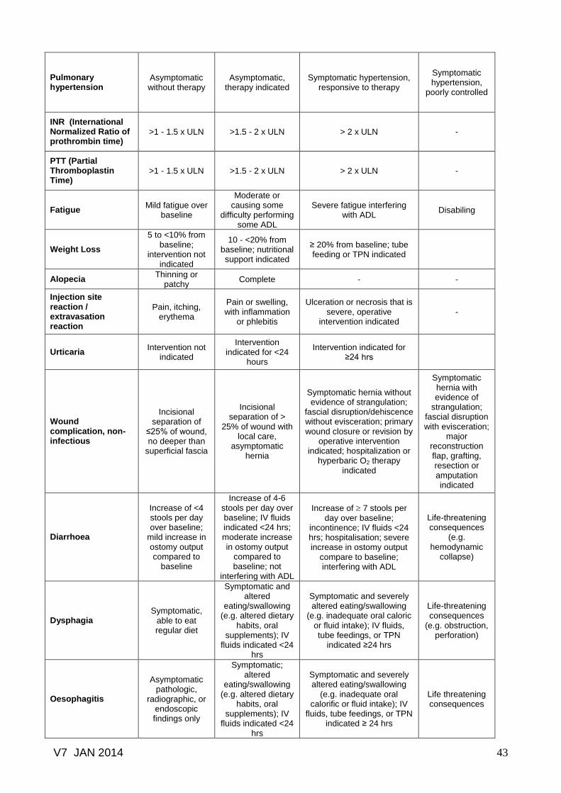
V7 JAN 2014 43
Pulmonary hypertension
Asymptomatic without therapy
Asymptomatic, therapy indicated
Symptomatic hypertension, responsive to therapy
Symptomatic hypertension,
poorly controlled
INR (International Normalized Ratio of prothrombin time)
>1 - 1.5 x ULN >1.5 - 2 x ULN > 2 x ULN -
PTT (Partial Thromboplastin Time)
>1 - 1.5 x ULN >1.5 - 2 x ULN > 2 x ULN -
Fatigue Mild fatigue over
baseline
Moderate or causing some
difficulty performing some ADL
Severe fatigue interfering with ADL
Disabiling
Weight Loss
5 to <10% from baseline;
intervention not indicated
10 - <20% from baseline; nutritional support indicated
≥ 20% from baseline; tube feeding or TPN indicated
Alopecia Thinning or
patchy Complete - -
Injection site reaction / extravasation reaction
Pain, itching, erythema
Pain or swelling, with inflammation
or phlebitis
Ulceration or necrosis that is severe, operative
intervention indicated -
Urticaria Intervention not
indicated
Intervention indicated for <24
hours
Intervention indicated for ≥24 hrs
Wound complication, non-infectious
Incisional separation of
≤25% of wound, no deeper than
superficial fascia
Incisional separation of >
25% of wound with local care,
asymptomatic hernia
Symptomatic hernia without evidence of strangulation;
fascial disruption/dehiscence without evisceration; primary wound closure or revision by
operative intervention indicated; hospitalization or
hyperbaric O2 therapy indicated
Symptomatic hernia with evidence of
strangulation; fascial disruption with evisceration;
major reconstruction flap, grafting, resection or amputation indicated
Diarrhoea
Increase of <4 stools per day over baseline;
mild increase in ostomy output compared to
baseline
Increase of 4-6 stools per day over baseline; IV fluids indicated <24 hrs; moderate increase in ostomy output
compared to baseline; not
interfering with ADL
Increase of 7 stools per day over baseline;
incontinence; IV fluids <24 hrs; hospitalisation; severe increase in ostomy output
compare to baseline; interfering with ADL
Life-threatening consequences
(e.g. hemodynamic
collapse)
Dysphagia Symptomatic,
able to eat regular diet
Symptomatic and altered
eating/swallowing (e.g. altered dietary
habits, oral supplements); IV
fluids indicated <24 hrs
Symptomatic and severely altered eating/swallowing
(e.g. inadequate oral caloric or fluid intake); IV fluids, tube feedings, or TPN
indicated ≥24 hrs
Life-threatening consequences
(e.g. obstruction, perforation)
Oesophagitis
Asymptomatic pathologic,
radiographic, or endoscopic findings only
Symptomatic; altered
eating/swallowing (e.g. altered dietary
habits, oral supplements); IV
fluids indicated <24 hrs
Symptomatic and severely altered eating/swallowing
(e.g. inadequate oral calorific or fluid intake); IV
fluids, tube feedings, or TPN indicated ≥ 24 hrs
Life threatening consequences
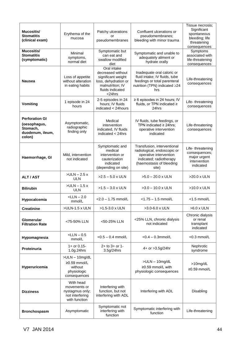
V7 JAN 2014 44
Mucositis/ Stomatitis (clinical exam)
Erythema of the mucosa
Patchy ulcerations or
pseudomembranes
Confluent ulcerations or pseudomembranes;
bleeding with minor trauma
Tissue necrosis; Significant
spontaneous bleeding; life threatening
consequences
Mucositis/ Stomatitis (symptomatic)
Minimal symptoms, normal diet
Symptomatic but can eat and
swallow modified diet
Symptomatic and unable to adequately aliment or
hydrate orally
Symptoms associated with life-threatening consequences
Nausea Loss of appetite without alteration in eating habits
Oral intake decreased without significant weight
loss, dehydration or malnutrition; IV fluids indicated
<24hrs
Inadequate oral caloric or fluid intake; IV fluids, tube feedings or total parenteral
nutrition (TPN) indicated 24 hrs
Life-threatening consequences
Vomiting 1 episode in 24
hours
2-5 episodes in 24 hours; IV fluids
indicated < 24hours
≥ 6 episodes in 24 hours; IV fluids, or TPN indicated ≥
24hrs
Life- threatening consequences
Perforation GI
(oesophagus, Stomach, duodenum, ileum, colon)
Asymptomatic, radiographic finding only
Medical intervention
indicated, IV fluids indicated < 24hrs
IV fluids, tube feedings, or TPN indicated ≥ 24hrs; operative intervention
indicated
Life-threatening consequences
Haemorrhage, GI Mild, intervention
not indicated
Symptomatic and medical
intervention or cauterization
indicated (depending on site)
Transfusion, interventional radiological, endoscopic or
operative intervention indicated; radiotherapy
(haemostasis of bleeding site)
Life- threatening consequences; major urgent intervention
indicated
ALT / AST >ULN – 2.5 x
ULN >2.5 – 5.0 x ULN >5.0 – 20.0 x ULN >20.0 x ULN
Bilirubin >ULN – 1.5 x
ULN >1.5 – 3.0 x ULN >3.0 – 10.0 x ULN >10.0 x ULN
Hypocalcemia <LLN – 2.0
mmol/L <2.0 – 1.75 mmol/L <1.75 – 1.5 mmol/L <1.5 mmol/L
Creatinine >ULN-1.5 x ULN >1.5-3.0 x ULN >3.0-6.0 x ULN >6.0 x ULN
Glomerular Filtration Rate
<75-50% LLN <50-25% LLN <25% LLN, chronic dialysis
not indicated
Chronic dialysis or renal
transplant indicated
Hypomagnesia <LLN – 0.5
mmol/L <0.5 – 0.4 mmol/L <0.4 – 0.3mmol/L <0.3 mmol/L
Proteinuria 1+ or 0.15-1.0g.24hrs
2+ to 3+ or 1-3.5g/24hrs
4+ or >3.5g/24hr Nephrotic syndrome
Hyperuricemia
>ULN – 10mg/dL
≥0.59 mmol/L without
physiologic consequences
>ULN – 10mg/dL
≥0.59 mmol/L with physiologic consequences
>10mg/dL
≥0.59 mmol/L
Dizziness
With head movements or
nystagmus only; not interfering with function
Interfering with function, but not
interfering with ADL Interfering with ADL Disabling
Bronchospasm Asymptomatic Symptomatic not interfering with
function
Symptomatic interfering with function
Life-threatening
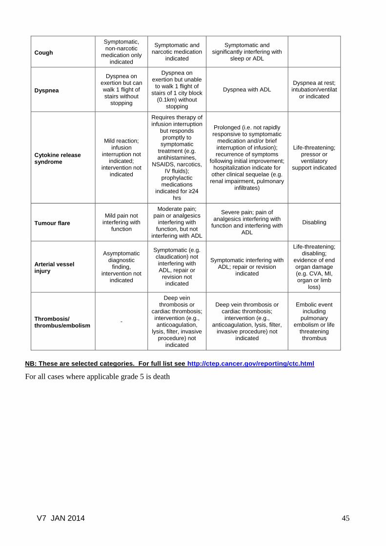
V7 JAN 2014 45
Cough
Symptomatic, non-narcotic
medication only indicated
Symptomatic and narcotic medication
indicated
Symptomatic and significantly interfering with
sleep or ADL
Dyspnea
Dyspnea on exertion but can walk 1 flight of stairs without
stopping
Dyspnea on exertion but unable to walk 1 flight of
stairs of 1 city block (0.1km) without
stopping
Dyspnea with ADL Dyspnea at rest; intubation/ventilat
or indicated
Cytokine release syndrome
Mild reaction; infusion
interruption not indicated;
intervention not indicated
Requires therapy of infusion interruption
but responds promptly to
symptomatic treatment (e.g. antihistamines,
NSAIDS, narcotics, IV fluids);
prophylactic medications
indicated for ≥24 hrs
Prolonged (i.e. not rapidly responsive to symptomatic
medication and/or brief interruption of infusion); recurrence of symptoms
following initial improvement; hospitalization indicate for
other clinical sequelae (e.g. renal impairment, pulmonary
infiltrates)
Life-threatening; pressor or ventilatory
support indicated
Tumour flare
Mild pain not interfering with
function
Moderate pain; pain or analgesics
interfering with function, but not
interfering with ADL
Severe pain; pain of analgesics interfering with
function and interfering with ADL
Disabling
Arterial vessel injury
Asymptomatic diagnostic
finding, intervention not
indicated
Symptomatic (e.g. claudication) not interfering with ADL, repair or
revision not indicated
Symptomatic interfering with ADL; repair or revision
indicated
Life-threatening; disabling;
evidence of end organ damage (e.g. CVA, MI, organ or limb
loss)
Thrombosis/ thrombus/embolism
-
Deep vein thrombosis or
cardiac thrombosis; intervention (e.g., anticoagulation,
lysis, filter, invasive procedure) not
indicated
Deep vein thrombosis or cardiac thrombosis; intervention (e.g.,
anticoagulation, lysis, filter, invasive procedure) not
indicated
Embolic event including
pulmonary embolism or life
threatening thrombus
NB: These are selected categories. For full list see http://ctep.cancer.gov/reporting/ctc.html
For all cases where applicable grade 5 is death
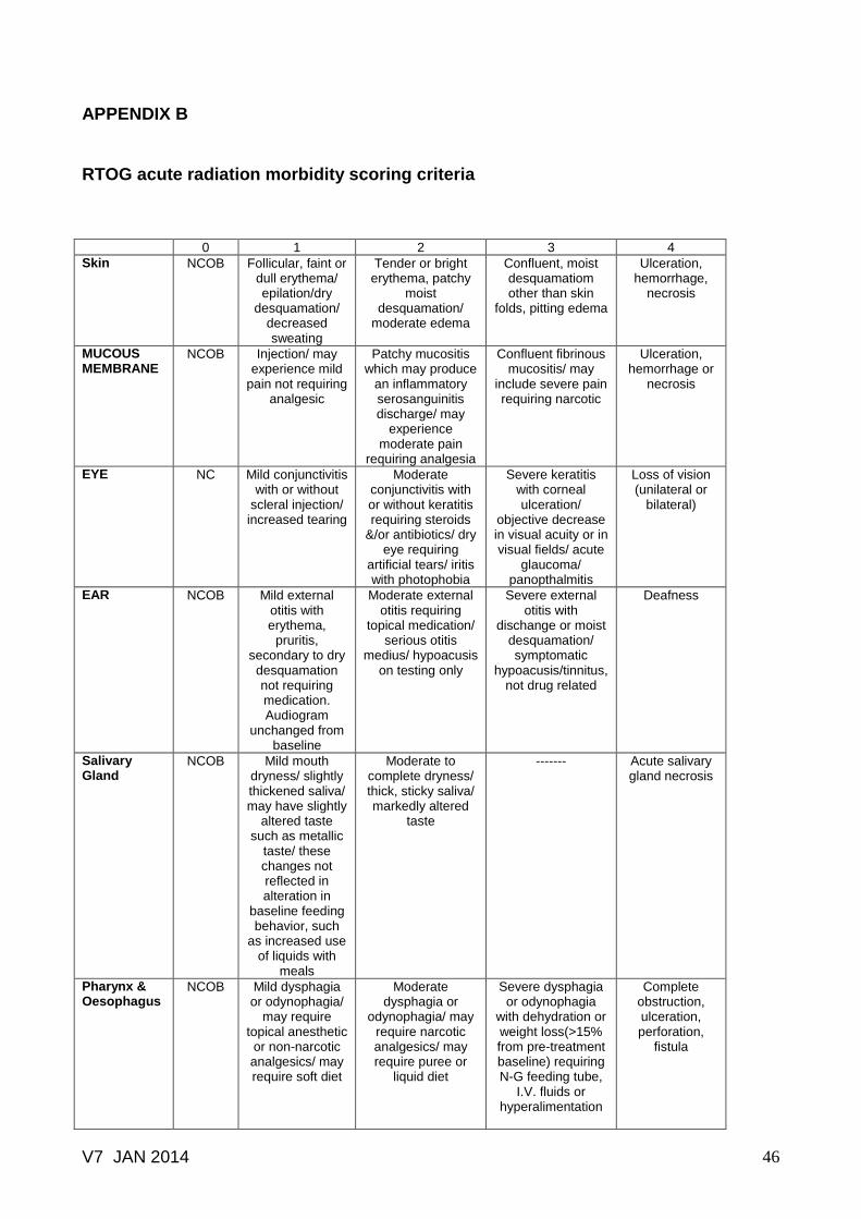
V7 JAN 2014 46
APPENDIX B
RTOG acute radiation morbidity scoring criteria
0 1 2 3 4
Skin NCOB Follicular, faint or dull erythema/ epilation/dry
desquamation/ decreased sweating
Tender or bright erythema, patchy
moist desquamation/
moderate edema
Confluent, moist desquamatiom other than skin
folds, pitting edema
Ulceration, hemorrhage,
necrosis
MUCOUS MEMBRANE
NCOB Injection/ may experience mild
pain not requiring analgesic
Patchy mucositis which may produce
an inflammatory serosanguinitis discharge/ may
experience moderate pain
requiring analgesia
Confluent fibrinous mucositis/ may
include severe pain requiring narcotic
Ulceration, hemorrhage or
necrosis
EYE NC Mild conjunctivitis with or without
scleral injection/ increased tearing
Moderate conjunctivitis with or without keratitis requiring steroids
&/or antibiotics/ dry eye requiring
artificial tears/ iritis with photophobia
Severe keratitis with corneal ulceration/
objective decrease in visual acuity or in visual fields/ acute
glaucoma/ panopthalmitis
Loss of vision (unilateral or
bilateral)
EAR NCOB Mild external otitis with erythema,
pruritis, secondary to dry desquamation not requiring medication. Audiogram
unchanged from baseline
Moderate external otitis requiring
topical medication/ serious otitis
medius/ hypoacusis on testing only
Severe external otitis with
dischange or moist desquamation/ symptomatic
hypoacusis/tinnitus, not drug related
Deafness
Salivary Gland
NCOB Mild mouth dryness/ slightly thickened saliva/ may have slightly
altered taste such as metallic
taste/ these changes not reflected in alteration in
baseline feeding behavior, such
as increased use of liquids with
meals
Moderate to complete dryness/ thick, sticky saliva/ markedly altered
taste
------- Acute salivary gland necrosis
Pharynx & Oesophagus
NCOB Mild dysphagia or odynophagia/
may require topical anesthetic or non-narcotic analgesics/ may require soft diet
Moderate dysphagia or
odynophagia/ may require narcotic analgesics/ may require puree or
liquid diet
Severe dysphagia or odynophagia
with dehydration or weight loss(>15% from pre-treatment baseline) requiring N-G feeding tube,
I.V. fluids or hyperalimentation
Complete obstruction, ulceration, perforation,
fistula

V7 JAN 2014 47
Larynx
NCOB Mild or intermittent
hoarseness/cough not requiring
antitussive/ erythema of
mucosa
Persistent hoarseness but able to vocalize/ referred ear pain,
sore throat, patchy fibrinous exudate or
mild arytenoid edema not
requiring narcotic/ cough requiring
antitussive
Whispered speech, throat pain or
referred ear pain requiring narcotic/ confluent fibrinous exudate, marked arytenoid edema
Marked dyspnea, stridor or hemoptysis
with tracheostomy or
intubation necessary
UPPER G.I. NC Anorexia with <=5% weight
loss from pretreatment
baseline/ nausea not requiring anti-
emetics/ abdominal
discomfort not requiring
parasympatholytic drugs or analgesics
Anorexia with <=15% weight loss from pretreatment
baseline/nausea &/ or vomiting
requiring anti-emetics/ abdominal
pain requiring analgesics
Anorexia with >15% weight loss from pretreatment
baseline or requiring N-G tube
or parenteral support. Nausea
&/or vomiting requiring tube or
parenteral support/abdominal
pain, severe despite
medication/hematemesis or melena/
abdominal distention (flat plate
radiograph demonstrates
distended bowel loops
Ileus, subacute or acute
obstruction, performation, GI bleeding
requiring transfusion/abd
ominal pain requiring tube
decompression or bowel diversion
LOWER G.I. INCLUDING PELVIS
NC Increased frequency or
change in quality of bowel habits not requiring medication/
rectal discomfort not requiring analgesics
Diarrhea requiring parasympatholytic
drugs (e.g., Lomotil)/ mucous
discharge not necessitating sanitary pads/
rectal or abdominal pain requiring
analgesics
Diarrhea requiring parenteral support/ severe mucous or blood discharge
necessitating sanitary
pags/abdominal distention (flat plate
radiograph demonstrates
distended bowel loops)
Acute or subacute
obstruction, fistula or
perforation; GI bleeding requiring
transfusion; abdominal pain
or tenesmus requiring tube
decompression or bowel diversion
LUNG NC Mild symptoms of dry cough or dyspnea on
exertion
Persistent cough requiring narcotic, antitussive agents/
dyspnea with minimal effort but
not at rest
Severe cough unresponsive to
narcotic antitussive agent or dyspnea at rest/ clinical or
radiologic evidence of acute
pneumonitis/ intermittent oxygen or steroids may be
required
Severe respiratory
insufficiency/ continuous oxygen or assisted
ventilation
GENITOURINARY
NC Frequency of urination or
nocturia twice pretreatment
habit/ dysuria, urgency not
requiring medication
Frequency of urination or
nocturia which is less frequent than
every hour. Dysuria, urgency,
bladder spasm requiring local
anesthetic (e.g., Pyridium)
Frequency with urgency and
nocturia hourly or more frequently/
dysuria, pelvis pain or bladder spasm requiring regular,
frequent narcotic/gross
hematuria with/ without clot
passage
Hematuria requiring
transfusion/ acute bladder obstruction not secondary to clot passage, ulceration or
necrosis

V7 JAN 2014 48
HEART NCOB Asymptomatic but objective
evidence of EKG changes or pericardial
abnormalities without evidence
of other heart disease
Symptomatic with EKG changes and radiologic findings of congestive heart failure or pericardial disease/ no specific treatment required
Congestive heart failure, angina
pectoris, pericardial disease responding
to therapy
Congestive heart failure,
angina pectoris, pericardial disease,
arrhythmias not responsive to non-surgical measures
CNS NCOB Fully functional status (i.e., able
to work) with minor neurologic
findings, no medication
needed
Neurologic findings present sufficient to require home case/ nursing assistance may be required/
medications including
steroids/anti-seizure agents may
be required
Neurologic findings requiring
hospitalization for initial management
Serious neurologic impairment
which includes paralysis, coma or seizures>3
per week despite
medication/hospitalization required
Haematologic WBC (X1000)
≥4.0 3.0 -<4.0 2.0 - <3.0 1.0 - < 2.0 <1.0
Platelets (X1000)
≥100 75 - <100 50 - <75 25 - <50 <25 or spontaneous
bleeding
Neutrophils ≥1.9 1.5 - <1.9 1.0 - <1.5 0.5 - <1.0 <0.5 or sepsis
Haemoglobin (GM%)
>11 11 – 9.5 <9.5 – 7.5 <7.5 – 5.0 -----
Haematocrit (%)
≥32 28 - <32 <38 Packed cell transfusion
required
-----
NC/OB: No change / over baseline GUIDELINES: The acute morbidity criteria are used to score/grade toxicity from radiation therapy. The criteria are relevant from day 1, the commencement of therapy, through day 90. Thereafter, the EORTC/RTOG Criteria of Late Effects are to be utilized. The evaluator must attempt to discriminate between disease- and treatment-related signs and symptoms. An accurate baseline evaluation prior to commencement of therapy is necessary. All toxicities Grade 3, 4 or 5
* must be verified by the Principal Investigator.
*ANY TOXICITY WHICH CAUSED DEATH IS GRADED 5.
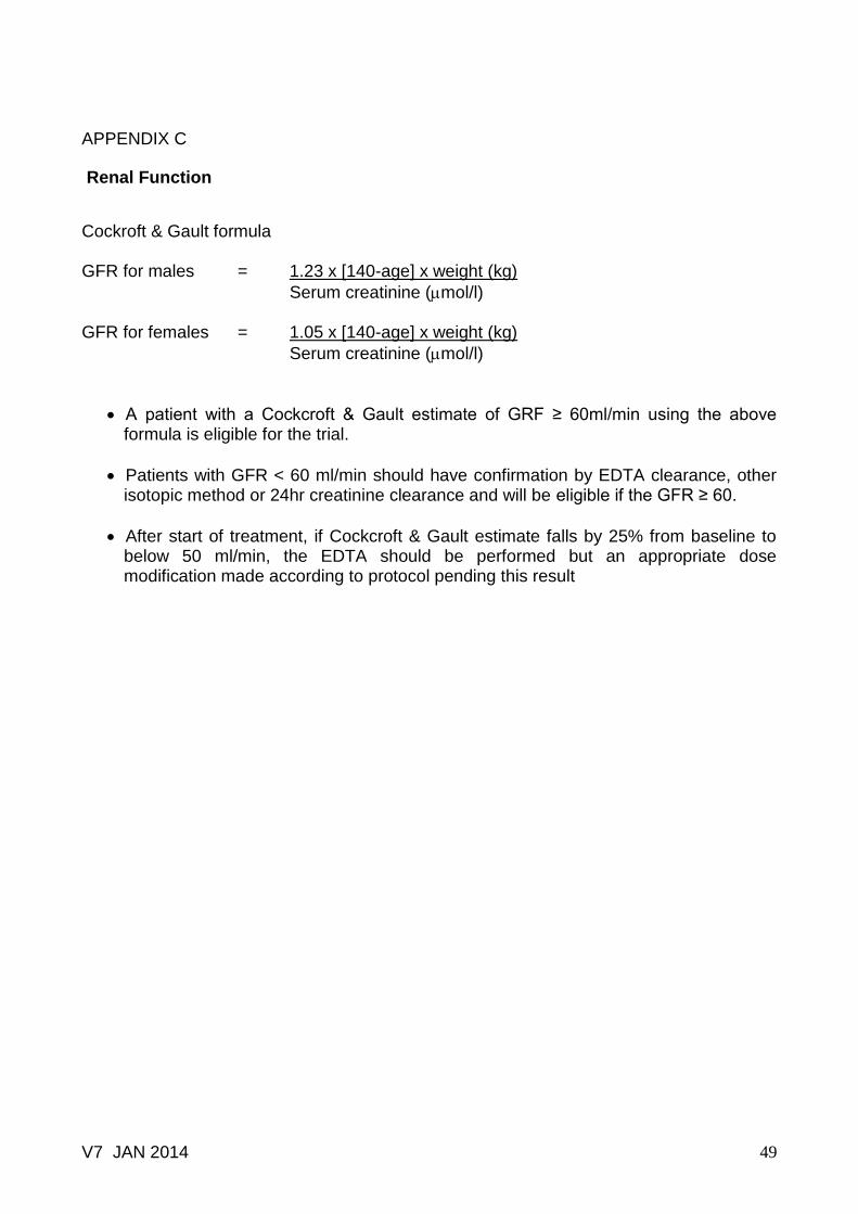
V7 JAN 2014 49
APPENDIX C
Renal Function
Cockroft & Gault formula GFR for males = 1.23 x [140-age] x weight (kg)
Serum creatinine (mol/l) GFR for females = 1.05 x [140-age] x weight (kg)
Serum creatinine (mol/l)
A patient with a Cockcroft & Gault estimate of GRF ≥ 60ml/min using the above formula is eligible for the trial.
Patients with GFR < 60 ml/min should have confirmation by EDTA clearance, other isotopic method or 24hr creatinine clearance and will be eligible if the GFR ≥ 60.
After start of treatment, if Cockcroft & Gault estimate falls by 25% from baseline to below 50 ml/min, the EDTA should be performed but an appropriate dose modification made according to protocol pending this result